Note: Descriptions are shown in the official language in which they were submitted.
CA 02678572 2011-10-26
MITOCHONDRIAL TARGETING AND IMPORT OF A VIRUS TO DELIVER A
NUCLEIC ACID
FIELD OF THE INVENTION
[0001] The invention is related to mitochondrial delivery systems and
expression of
mitochondrial genes.
BACKGROUND
[0002] In only 16,600 base pairs, human mitochondrial DNA (mtDNA) encodes 37
genes
(2 for ribosomal RNAs, 22 for tRNAs and 13 for proteins) that are essential
for cell viability.
These genes are buffered against the effect of mutations, because somatic
cells typically
contain around 1000 copies of mtDNA. While most cells are apparently uniform
with respect
to mtDNA composition, mammalian cells can tolerate a significant fraction of
aberrant
mtDNA, a condition called heteroplasmy, and retain respiratory function.
Nevertheless,
mitochondria lack nucleotide excision and recombination repair systems, so
that, with age,
deletions and point mutations accumulate in mtDNA. This accretion of defects
is thought to
contribute to the infirmities of advanced age.
[0003] Significant obstacles remain to correcting any mtDNA in living animals
since
there is no delivery system for DNA into mitochondria. Therefore, a need
exists in the art for
the targeting and delivery of nucleic acids and other therapeutic molecules
into the
mitochondria.
SUMMARY
[0004] Compositions for introducing nucleic acid molecules into mitochondria
are
provided. These compositions are used, inter alia, for correcting point
mutations in
mitochondrial protein-coding genes. Nucleic acid molecules such as cDNA coded
in the
mitochondrial genetic code are directly introduced into the mitochondria.
Methods for
introducing nucleic acid molecules are described.
[0005] In one preferred embodiment, a composition comprises a nucleic acid
backbone or
vector comprising a mitochondrial targeting sequence; a mitochondrial gene
operably linked
to a mitochondrial promoter. Any of these components can be in single copies;
multiple
copies; combinations of single copies of one component and multiple copies of
another; and
components can be tandem repeats or otherwise.
[0006] The recombinant vectors can be DNA plasmids or viral vectors.
Mitochondrial
gene expressing viral vectors can be constructed based on, but not limited to,
adeno-
CA 02678572 2012-02-15
associated virus, retrovirus, adenovirus, or alphavirus. The recombinant
vectors capable of
expressing the mitochondrial molecules can be delivered as described herein,
and persist in
the mitochondria. Alternatively, viral vectors can be used that provide for
transient
expression of mitochondrial molecules. Such vectors can be repeatedly
administered as
necessary. Once expressed, the mitochondrial molecules compensate for the gene
function or
expression of the mutated mitochondrial gene. Delivery of mitochondrial gene
expressing
vectors can be systemic, such as by intravenous or intramuscular
administration, by
administration to target cells ex-planted from a subject followed by
reintroduction into the
subject, or by any other means that would allow for introduction into the
desired target cell.
[0007] In a preferred embodiment, a composition comprises a nucleic acid
encoding a
vector; mitochondrial promoters; a mitochondrial gene and a mitochondrial
targeting
sequence. The vector can be viral, non-viral, naked DNA and the like.
[0007a] In another aspect, the present invention provides a composition
comprising at least
one virion comprising: i) a recombinant viral vector comprising a human
mitochondrial gene
operably linked to a mitochondrial promoter, and ii) a viral capsid comprising
a
mitochondrial targeting sequence.
[0007b] In another aspect, the present invention provides a composition
comprising (a) at
least one virion comprising: i) a recombinant viral vector comprising a human
mitochondrial
gene operably linked to a mitochondrial promoter, and ii) a viral capsid
comprising a
mitochondrial targeting sequence, and (b) a carrier or an excipient.
[0007c] In another aspect, the present invention provides an in vitro or ex
vivo method of
introducing nucleic acid molecules into mitochondria comprising:
providing a composition comprising at least one virion comprising: 1) a
recombinant
viral vector comprising a human mitochondrial gene operably linked to a
mitochondrial
promoter and ii) a viral capsid comprising a mitochondrial targeting sequence;
and
introducing the composition into a cell having mitochondria, wherein the at
least one
virion enters a mitochondrion and the human mitochondrial gene is expressed in
the
mitochondrion.
[0007d] In another aspect, the present invention provides the use of the above-
mentioned
composition in the manufacture of a medicament for the treatment of a
mitochondria-related
disease.
[0007e] In another aspect, the present invention provides the use of the above-
mentioned
composition for the treatment of a mitochondria-related disease.
-2-
CA 02678572 2011-10-26
[00081 In one embodiment, the viral vector is an Adeno Associated virus (AAV)
vector
and the AAV vector comprises Rep, Cap, and inverted terminal repeat (ITR)
sequences.
[00091 In one aspect, the mitochondrial targeting sequence is inserted into
the AAV
capsid open reading frame (cap ORF). However, the mitochondrial targeting
sequences can
be inserted into any region of a vector; insert single or multiple copies of
the same or
different targeting sequences.
[0010] In another preferred embodiment, the AAV is selected from the group
consisting
of AAV-1 to AAV-9 serotypes. The AAV genome can comprise Rep and Cap genes
from
other AAV serotypes and/or the AAV can be pseudotyped.
[00111 In another preferred embodiment, the mitochondrial gene is a normal
mitochondrial gene and expression thereof compensates for a mutated
mitochondrial gene.
Preferably, the vector comprises at least one mitochondrial gene and
mitochondrial targeting
sequences. In other aspects, the vector comprises at least one mitochondrial
gene and two or
more mitochondrial targeting sequences and combinations thereof. Other
examples, include a
vector comprising multiple copies of the mitochondrial gene and/or
mitochondrial targeting
sequences. The vector can comprises one or more types of mitochondrial genes
and/or
mitochondrial targeting sequences; fragments and variants thereof.
100121 In another preferred embodiment, the nucleic acids of the mitochondrial
gene
comprise mitochondrial codons from the mitochondrial genome.
[00131 In another preferred embodiment, an Adenovirus Associated Virion
comprising a
mitochondria) targeting sequence; a mitochondrial gene operably linked to a
mitochondrial
-2a-
CA 02678572 2009-08-12
WO 2008/101233 PCT/US2008/054216
promoter. Preferably, the mitochondrial gene is operably linked to the
mitochondrial
promoter and inserted into the AAV vector backbone.
[0014] In another preferred embodiment, the AAV is selected from the group
consisting
of AAV-1 to AAV-9 serotypes. The AAV can also be pseudotyped, comprise Rep and
Cap
genes from other AAV serotypes; and/or mutated Cap genes.
[0015] In another preferred embodiment, the AAV comprises a normal
mitochondrial
gene and expression thereof compensates for a mutated mitochondrial gene and a
mitochondrial targeting sequence; fragments and variants thereof.
[0016] In one preferred embodiment, the AAV comprises at least one
mitochondrial
gene and mitochondrial targeting sequences. The numbers and types of
mitochondrial genes
and sequences can vary. For example, in some embodiments, the AAV comprises at
least
one mitochondrial gene and two or more mitochondrial targeting sequences and
combinations
thereof. In other embodiments, the AAV n the vector comprises multiple copies
of the
mitochondrial gene and/or mitochondrial targeting sequences. In another
embodiment, the
AAV comprises one or more types of mitochondrial genes and/or mitochondrial
targeting
sequences.
[0017] In another preferred embodiment, a method of treating mitochondrial
related
diseases comprises administering to a patient a composition comprising an AAV
vector
encoding at least one mitochondrial gene wherein said gene is operably linked
to a
mitochondrial promoter, at least one mitochondrial targeting sequence; binding
and entering
of the AAV into mitochondria; expressing the mitochondrial gene in
mitochondria wherein
said gene is a normal gene and compensates for a mutation; and, treating
mitochondrial
related diseases.
[0018] In another preferred embodiment, the mitochondrial related diseases
comprise:
Alpers Disease; Barth syndrome; (3-oxidation defects; carnitine-acyl-carnitine
deficiency;
carnitine deficiency; co-enzyme Q10 deficiency; Complex I deficiency; Complex
II
deficiency; Complex III deficiency; Complex IV deficiency; Complex V
deficiency;
cytochrome c oxidase (COX) deficiency, LHON--Leber Hereditary Optic
Neuropathy; MM--
Mitochondrial Myopathy; LIMM--Lethal Infantile Mitochondrial Myopathy; MMC--
Maternal Myopathy and Cardiomyopathy; NARP--Neurogenic muscle weakness,
Ataxia, and
Retinitis Pigmentosa; Leigh Disease; FICP--Fatal Infantile Cardiomyopathy
Plus, a MELAS-
associated cardiomyopathy; MELAS--Mitochondrial Encephalomyopathy with Lactic
Acidosis and Strokelike episodes; LDYT--Leber's hereditary optic neuropathy
and Dystonia;
MERRF--Myoclonic Epilepsy and Ragged Red Muscle Fibers; MHCM--Maternally
inherited
-3-
CA 02678572 2009-08-12
WO 2008/101233 PCT/US2008/054216
Hypertrophic CardioMyopathy; CPEO--Chronic Progressive External
Ophthalmoplegia;
KSS--Kearns Sayre Syndrome; DM--Diabetes Mellitus; DMDF Diabetes
Mellitus+DeaFness;
CIPO--Chronic Intestinal Pseudoobstruction with myopathy and Ophthalmoplegia;
DEAF--
Maternally inherited DEAFness or aminoglycoside-induced DEAFness; PEM--
Progressive
encephalopathy; SNHL--SensoriNeural Hearing Loss; Encephalomyopathy;
Mitochondrial
cytopathy; Dilated Cardiomyopathy; GER-- Gastrointestinal Reflux; DEMCHO--
Dementia
and Chorea; AMDF--Ataxia, Myoclonus; Exercise Intolerance; ESOC Epilepsy,
Strokes,
Optic atrophy, & Cognitive decline; FBSN Familial Bilateral Striatal Necrosis;
FSGS Focal
Segmental Glomerulosclerosis; LIMM Lethal Infantile Mitochondrial Myopathy;
MDM
Myopathy and Diabetes Mellitus; MEPR Myoclonic Epilepsy and Psychomotor
Regression;
MERME MERRF/MELAS overlap disease; MHCM Maternally Inherited Hypertrophic
CardioMyopathy; MICM Maternally Inherited Cardiomyopathy; MILS Maternally
Inherited
Leigh Syndrome; Mitochondrial Encephalocardiomyopathy; Multisystem
Mitochondrial
Disorder (myopathy, encephalopathy, blindness, hearing loss, peripheral
neuropathy);
NAION Nonarteritic Anterior Ischemic Optic Neuropathy;NIDDM Non-Insulin
Dependent
Diabetes Mellitus; PEM Progressive Encephalopathy; PME Progressive Myoclonus
Epilepsy;
RTT Rett Syndrome; SIDS Sudden Infant Death Syndrome; MIDD Maternally
Inherited
Diabetes and Deafness; and MODY Maturity-Onset Diabetes of the Young, and
MNGIE.
[0019] In another preferred embodiment, the mitochondrial targeting sequences
comprise nucleic acid molecules encoding: cytochrome c oxidase (COX); ATP
synthase;
subunit c of human ATP synthase (ATPc); ATP synthase; hexokinase I, amine
oxidase
(flavin-containing) A, hexokinase IV, pancreatic beta cell form, peripheral
benzodiazepine
receptor-related protein, metaxin 2, putative mitochondrial outer membrane
protein import
receptor (hTOM), glutathione transferase; voltage-dependent anion channel 2
(outer
mitochondrial membrane protein porin), hexokinase IV, cytochrome b5,
peripheral
benzodiazepine receptor, germ cell kinase anchor S-AKAP84, A kinase anchor
protein,
carnitine O-palmitoyltransferase I precursor, hexokinase II, amine oxidase
(flavin-containing)
B, long-chain-fatty-acid--CoA ligase 2, long-chain-fatty-acid--CoA ligase 1
(palmitoyl-CoA
ligase), voltage-dependent anion channel 1, metaxin 1, Human putative outer
mitochondrial
membrane 34 kDa translocase hTOM34, voltage-dependent anion channel 4 (outer
mitochondrial membrane protein porin), cytochrome-b5 reductase, voltage-
dependent anion
channel 3 (outer mitochondrial membrane protein porin), Mitochondrial import
receptor
subunit TOM20 homolog (Mitochondria) 20 kd outer membrane protein) (Outer
-4-
CA 02678572 2009-08-12
WO 2008/101233 PCT/US2008/054216
mitochondrial membrane receptor TOM20), and tumorous imaginal discs homolog
precursor
(HTID-1); fragments and variants thereof.
[0020] In another preferred embodiment, a method for compensating for a mtDNA
mutation in a host comprising: introducing a host's mitochondria a composition
of AAV
encoding mitochondrial promoters; a mitochondrial gene and a mitochondrial
targeting
sequence; fragments and variants thereof. Preferably the viral vector is an
Adeno Associated
virus (AAV) vector and the AAV vector comprises Rep, Cap, and inverted
terminal repeat
(ITR) sequences.
[0021] In a preferred embodiment, a method of introducing nucleic acid
molecules into
mitochondria (intra-mitochondrially) comprises administering to a patient a
composition
comprising a vector encoding at least one mitochondrial gene wherein said gene
is operably
linked to a mitochondrial promoter, and at least one mitochondrial targeting
sequence;
binding and entering of the vector into mitochondria; and, introducing nucleic
acid molecules
into mitochondria.
[0022] In another preferred embodiment, the mitochondrial gene of interest is
introduced
into a patient by obtaining cells from a patient; culturing the cells ex vivo
with the
composition encoding the mitochondrial gene and mitochondrial targeting
sequences;
wherein the composition is targeted to the mitochondria and the desired gene
is expressed in
the mitochondria and re-administering the cells to the patient. The cells can
be obtained
either from the patient or from a donor. The cells can be obtained from
organs, skin, tissues,
muscles and bone marrow. The cells are preferably autologous, however they can
be HLA-
compatible or partially HLA matched.
[0023] In one embodiment, the cells are stem cells and are obtained from bone
marrow.
[0024] In another preferred embodiment, a method of introducing a nucleic acid
molecule into a mitochondrion in vitro, comprises culturing cells with a
composition
comprising a vector encoding at least one mitochondrial gene wherein said gene
is operably
linked to a mitochondrial promoter, at least one mitochondrial targeting
sequence; binding
and entering of the vector into mitochondria; expressing the mitochondrial
gene in
mitochondria ; and, introducing the nucleic acid molecule into a
mitochondrion.
[0025] In another preferred embodiment, a mitochondrial gene can be silenced,
down-
regulated or up-regulated. For example, the delivery of a nucleic acid in such
cases is in the
form of siRNA's which are directed to a gene to be silenced, a promoter,
enhancer etc which
can modulate gene expression.
-5-
CA 02678572 2009-08-12
WO 2008/101233 PCT/US2008/054216
[0026] Inhibition of gene expression may be quantified by measuring either the
endogenous target RNA or the protein produced by translation of the target
RNA.
Techniques for quantifying RNA and proteins are well known to one of ordinary
skill in the
art. In certain preferred embodiments, gene expression is inhibited by at
least 10%,
preferably by at least 33%, more preferably by at least 50%, and yet more
preferably by at
least 80%. In particularly preferred embodiments, of the invention gene
expression is
inhibited by at least 90%, more preferably by at least 95%, or by at least 99%
up to 100%
within cells in the organism. Note that although in certain embodiments of the
invention
inhibition occurs in substantially all cells of the subject, in other
preferred embodiments
inhibition occurs in only a subset of cells expressing the heterologous gene.
[0027] Other aspects of the invention are described infra.
BRIEF DESCRIPTION OF THE DRAWINGS
[0028] The invention is pointed out with particularity in the appended claims.
The
above and further advantages of this invention may be better understood by
referring to the
following description taken in conjunction with the accompanying drawings, in
which:
[0029] Figure 1 is an electron microscope scan showing that A20 immunogold
recognizes fully assembled AAV virions (arrow) in the mitochondria.
[0030] Figure 2 is an electron microscope scan showing that GFP and A20
immunogold
co-localization indicating fully assembled virus is imported into the
mitochondria.
[0031] Figure 3 is a scan of an agarose gel showing PCR product of mtDNA
isolated
from cells infected with mitochondrially targeted AAV containing the H strand
promoter
driving expression of the ND4 subunit gene in the mitochondrial genetic code
in Lanes 2-6
(arrow). Amplification of plasmid DNA is shown in lanes 7 to 11.
[0032] Figure 4 is a scan of an SfaNI digest showing 915 bp band (arrow)
indicating the
presence of wild-type mitochondrial ND4 DNA in mito-AAV transfected LHON
cybrids
homoplasmic for GI 1778A mtDNA. The GI 1778A mutation eliminates an SfaNI site
at the
ND4 locus shown above the digest.
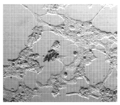
[0033] Figure 5 are micrographs of live cell cultures showing healthy
appearing LHON
cybrids infected with the mito-targeted AAV expressing wild-type ND4 even
after 5 days of
growth in galactose (left). Homoplasmic GI 1778A cells infected with the AAV
lacking the
MTS are dead or dying (right)
[0034] Figures 6A and 6B are graphs showing AAV mitoND4 rescue. After 48 hours
of
growth in galactose we found a 19% increase in cell survival (p <0.05)
relative to controls,
-6-
CA 02678572 2009-08-12
WO 2008/101233 PCT/US2008/054216
the cybrid cells infected with the same ND4 gene but in a virus without the
MTS attached to
the GFP expressing VP2. Figure 6B is a histogram showing a 48% increase in the
rate of
ATP synthesis in LHON cybrids infected with the mito-targeted AAV (8gfp)
expressing
wild-type ND4 relative to LHON cells infected with the same AAV vector lacking
the MTS
(cont), p <0.05.
[0035] Figure 7 is a schematic illustration showing COX8 MTS- AAV expressing
GFP
plasmid.
DETAILED DESCRIPTION
[0036] Vectors comprising mitochondrial genes and targeting sequences are
directed into
mitochondria. The mitochondrial genes are expressed in the mitochondria.
Methods of
transducing the mitochondria are described.
Definitions
[0037] Prior to setting forth the invention, the following definitions are
provided:
[0038] As used herein, the singular forms "a", "an" and "the" include plural
referents
unless the context clearly dictates otherwise.
[0039] As used herein, a "pharmaceutically acceptable" component is one that
is suitable
for use with humans and/or animals without undue adverse side effects (such as
toxicity,
irritation, and allergic response) commensurate with a reasonable benefit/risk
ratio.
[0040] The term "DNA construct" and "vector" are used herein to mean a
purified or
isolated polynucleotide that has been artificially designed and which
comprises at least two
nucleotide sequences that are not found as contiguous nucleotide sequences in
their natural
environment.
[0041] The term "plasmid" as used herein refers to any nucleic acid encoding
an
expressible gene and includes linear or circular nucleic acids and double or
single stranded
nucleic acids. The nucleic acid can be DNA or RNA and may comprise modified
nucleotides
or ribonucleotides, and may be chemically modified by such means as
methylation or the
inclusion of protecting groups or cap- or tail structures. Single or double
stranded DNA or
RNA and linear or circular. Single stranded DNA can be used for expression and
circular
RNA can also be used for expression.
[0042] As used interchangeably herein, the terms "oligo-nucleotides",
"polynucleotides",
and "nucleic acids" include RNA, DNA, or RNA/DNA hybrid sequences of more than
one
nucleotide in either single chain or duplex form. The term "nucleotide" as
used herein as an
-7-
CA 02678572 2009-08-12
WO 2008/101233 PCT/US2008/054216
adjective to describe molecules comprising RNA, DNA, or RNA/DNA hybrid
sequences of
any length in single-stranded or duplex form. The term "nucleotide" is also
used herein as a
noun to refer to individual nucleotides or varieties of nucleotides, meaning a
molecule, or
individual unit in a larger nucleic acid molecule, comprising a purine or
pyrimidine, a ribose
or deoxyribose sugar moiety, and a phosphate group, or phosphodiester linkage
in the case of
nucleotides within an oligonucleotide or polynucleotide. Although the term
"nucleotide" is
also used herein to encompass "modified nucleotides" which comprise at least
one
modifications (a) an alternative linking group, (b) an analogous form of
purine, (c) an
analogous form of pyrimidine, or (d) an analogous sugar, all as described
herein.
[0043] The phrase "having a length of N bases" or "having a length of N
nucleotides" is
used herein to describe lengths along a single nucleotide strand, of a nucleic
acid molecule,
consisting of N individual nucleotides.
[0044] As used herein, the term "bind", refers to an interaction between the
bases of an
oligonucleotide which is mediated through base-base hydrogen bonding. One type
of binding
is "Watson-Crick-type" binding interactions in which adenine-thymine (or
adenine-uracil)
and guanine-cytosine base-pairs are formed through hydrogen bonding between
the bases.
An example of this type of binding is the binding traditionally associated
with the DNA
double helix.
[0045] As used herein, the term "oligonucleotide" refers to a polynucleotide
formed from
naturally occurring bases and pentofuranosyl groups joined by native
phosphodiester bonds.
This term effectively refers to naturally occurring species or synthetic
species formed from
naturally occurring subunits or their close homologs. The term
"oligonucleotide" may also
refer to moieties which function similarly to naturally occurring
oligonucleotides but which
have non-naturally occurring portions. Thus, oligonucleotides may have altered
sugar
moieties or intersugar linkages. Exemplary among these are the
phosphorothioate and other
sulfur-containing species which are known for use in the art. In accordance
with some
preferred embodiments, at least some of the phosphodiester bonds of the
oligonucleotide
have been substituted with a structure which functions to enhance the ability
of the
compositions to penetrate into the region of cells where the RNA or DNA whose
activity to
be modulated is located. It is preferred that such substitutions comprise
phosphorothioate
bonds, methyl phosphonate bonds, or short chain alkyl or cycloalkyl
structures. In
accordance with other preferred embodiments, the phosphodiester bonds are
substituted with
other structures which are, at once, substantially non-ionic and non-chiral,
or with structures
-8-
CA 02678572 2009-08-12
WO 2008/101233 PCT/US2008/054216
which are chiral and enantiomerically specific. Persons of ordinary skill in
the art will be
able to select other linkages for use in practice of the invention.
[0046] Oligonucleotides may also include species which include at least some
modified
base forms. Thus, purines and pyrimidines other than those normally found in
nature may be
so employed. Similarly, modifications on the pentofuranosyl portion of the
nucleotide
subunits may also be effected, as long as the essential tenets of this
invention are adhered to.
Examples of such modifications are 2'-O-alkyl- and 2'-halogen-substituted
nucleotides. Some
specific examples of modifications at the 2' position of sugar moieties which
are useful in the
present invention are OH, SH, SCH3, F, OCH3, OCN, O(CH2) õNH2 or O(CH2) õCH3
where n
is from 1 to about 10, and other substituents having similar properties.
[0047] As used herein, the term "administering a molecule to a cell" (e.g., an
expression
vector, nucleic acid, and the like) refers to transducing, transfecting,
microinjecting,
electroporating, or shooting, the cell with the molecule. In some aspects,
molecules are
introduced into a target cell by contacting the target cell with a delivery
cell (e.g., by cell
fusion or by lysing the delivery cell when it is in proximity to the target
cell).
[0048] A "vector" (sometimes referred to as gene delivery or gene transfer
"vehicle")
refers to a macromolecule or complex of molecules comprising a polynucleotide
to be
delivered to a host cell, either in vitro or in vivo. The "vector" can be any
nucleic acid-
and/or viral-based technique used to deliver a desired nucleic acid. The
polynucleotide to be
delivered may comprise a coding sequence of interest in gene therapy. Vectors
include, for
example, viral vectors (such as adenoviruses ("Ad"), adeno-associated viruses
(AAV), and
retroviruses), liposomes and other lipid-containing complexes, and other
macromolecular
complexes capable of mediating delivery of a polynucleotide to a host cell.
Vectors can also
comprise other components or functionalities that further modulate gene
delivery and/or gene
expression, or that otherwise provide beneficial properties to the targeted
cells. As described
and illustrated in more detail below, such other components include, for
example,
components that influence binding or targeting to cells (including components
that mediate
cell-type or tissue-specific binding); components that influence uptake of the
vector nucleic
acid by the cell; components that influence localization of the polynucleotide
within the cell
after uptake (such as agents mediating nuclear localization); and components
that influence
expression of the polynucleotide. Such components also might include markers,
such as
detectable and/or selectable markers that can be used to detect or select for
cells that have
taken up and are expressing the nucleic acid delivered by the vector. Such
components can
be provided as a natural feature of the vector (such as the use of certain
viral vectors which
-9-
CA 02678572 2009-08-12
WO 2008/101233 PCT/US2008/054216
have components or functionalities mediating binding and uptake), or vectors
can be
modified to provide such functionalities. Other vectors include those
described by Chen et
al; BioTechniques, 34: 167-171 (2003). A large variety of such vectors are
known in the art
and are generally available.
[0049] A "recombinant viral vector" refers to a viral vector comprising one or
more
heterologous gene products or sequences. Since many viral vectors exhibit size-
constraints
associated with packaging, the heterologous gene products or sequences are
typically
introduced by replacing one or more portions of the viral genome. Such viruses
may become
replication-defective, requiring the deleted function(s) to be provided in
trans during viral
replication and encapsidation (by using, e.g., a helper virus or a packaging
cell line carrying
gene products necessary for replication and/or encapsidation). Modified viral
vectors in
which a polynucleotide to be delivered is carried on the outside of the viral
particle have also
been described (see, e.g., Curiel, D T, et al. PNAS 88: 8850-8854, 1991).
[0050] By "nucleic acid" is meant both RNA and DNA including: cDNA, genomic
DNA, plasmid DNA or condensed nucleic acid, nucleic acid formulated with
cationic lipids,
nucleic acid formulated with peptides, cationic polymers, RNA or mRNA. In a
preferred
embodiment, the nucleic acid administered is a plasmid DNA which constitutes a
"vector."
The nucleic acid can be, but is not limited to, a plasmid DNA vector with a
eukaryotic
promoter which expresses a protein with potential therapeutic action.
[0051] As used herein, the term a "plasmid" refers to a construct made up of
genetic
material (i.e., nucleic acids). It includes genetic elements arranged such
that an inserted
coding sequence can be transcribed in eukaryotic cells. In this case, a
preferred embodiment
comprises a mitochondrial targeting sequence, a mitochondrial gene operably
linked to a
mitochondrial promoter. Also, while the plasmid may include a sequence from a
viral
nucleic acid, such viral sequence preferably does not cause the incorporation
of the plasmid
into a viral particle, and the plasmid is therefore a non-viral vector.
Preferably, a plasmid is a
closed circular DNA molecule. The enhancer/promoter region of an expression
plasmid will
determine the levels of expression. Most of the gene expression systems
designed for high
levels of expression contain the intact human cytomegalovirus (CMV) immediate
early
enhancer/promoter sequence. However, down-regulation of the CMV promoter over
time
has been reported in tissues. The hypermethylation of the CMV promoter, as
observed when
incorporated into retroviral vectors, has not been observed for episomal
plasmids in vivo.
Nevertheless, the CMV promoter silencing could be linked to its sensitivity to
reduced levels
of the transcription factor NF-KB. The activity of the CMV promoter has also
been shown to
-10-
CA 02678572 2009-08-12
WO 2008/101233 PCT/US2008/054216
be attenuated by various cytokines including interferons (a and (3), and tumor
necrosis factor
(TNF-a). In order to prolong expression in vivo and ensure specificity of
expression in
desired tissues, tissue-specific enhancer/promoters have been incorporated in
expression
plasmids. The chicken skeletal alpha actin promoter has been shown to provide
high levels
of expression (equivalent to the ones achieved with a CMV-driven construct)
for several
weeks in non-avian striated muscles.
[0052] Additional genetic sequences in the expression plasmids can be added to
influence the stability of the messenger RNA (mRNA) and the efficiency of
translation. The
5' untranslated region (5' UTR) is known to effect translation and it is
located between the
cap site and the initiation codon. The 5' UTR should ideally be relatively
short, devoid of
strong secondary structure and upstream initiation codons, and should have an
initiation
codon AUG within an optimal local context. The 5' UTR can also influence RNA
stability,
RNA processing and transcription. In order to maximize gene expression by
ensuring
effective and accurate RNA splicing, one or more introns can be included in
the expression
plasmids at specific locations. The possibility of inefficient and/or
inaccurate splicing can be
minimized by using synthetic introns that have idealized splice junction and
branch point
sequences that match the consensus sequence. Another important sequence within
a gene
expression system is the 3' untranslated region (3' UTR), a sequence in the
mRNA that
extends from the stop codon to the poly(A) addition site. The 3' UTR can
influence mRNA
stability, translation and intracellular localization. The skeletal muscle
.alpha.-actin 3' UTR
has been shown to stabilize MRNA in muscle tissues thus leading to higher
levels of
expression as compared to other 3' UTR. This 3' UTR appears to induce a
different
intracellular compartmentalization of the produced proteins, preventing the
effective
trafficking of the proteins to the secretory pathway and favoring their
perinuclear
localization. One of the attractive features of plasmid expression systems is
the possibility to
express multiple genes from a single construct.
[0053] Viral "packaging" as used herein refers to a series of intracellular
events that
results in the synthesis and assembly of a viral vector. Packaging typically
involves the
replication of the "pro-viral genome", or a recombinant pro-vector typically
referred to as a
"vector plasmid" (which is a recombinant polynucleotide than can be packaged
in an manner
analogous to a viral genome, typically as a result of being flanked by
appropriate viral
"packaging sequences"), followed by encapsidation or other coating of the
nucleic acid.
Thus, when a suitable vector plasmid is introduced into a packaging cell line
under
appropriate conditions, it can be replicated and assembled into a viral
particle. Viral "rep"
- 11 -
CA 02678572 2009-08-12
WO 2008/101233 PCT/US2008/054216
and "cap" gene products, found in many viral genomes, are gene products
encoding
replication and encapsidation proteins, respectively. A "replication-
defective" or
"replication-incompetent" viral vector refers to a viral vector in which one
or more functions
necessary for replication and/or packaging are missing or altered, rendering
the viral vector
incapable of initiating viral replication following uptake by a host cell. To
produce stocks of
such replication-defective viral vectors, the virus or pro-viral nucleic acid
can be introduced
into a "packaging cell line" that has been modified to contain gene products
encoding the
missing functions which can be supplied in trans). For example, such packaging
gene
products can be stably integrated into a replicon of the packaging cell line
or they can be
introduced by transfection with a "packaging plasmid" or helper virus carrying
gene products
encoding the missing functions.
[0054] A "detectable marker gene" is a gene that allows cells carrying the
gene to be
specifically detected (e.g., distinguished from cells which do not carry the
marker gene). A
large variety of such marker gene products are known in the art. Preferred
examples thereof
include detectable marker gene products which encode proteins appearing on
cellular
surfaces, thereby facilitating simplified and rapid detection and/or cellular
sorting. By way of
illustration, the lacZ gene encoding beta-galactosidase can be used as a
detectable marker,
allowing cells transduced with a vector carrying the lacZ gene to be detected
by staining.
[0055] A "selectable marker gene" is a gene that allows cells carrying the
gene to be
specifically selected for or against, in the presence of a corresponding
selective agent. By
way of illustration, an antibiotic resistance gene can be used as a positive
selectable marker
gene that allows a host cell to be positively selected for in the presence of
the corresponding
antibiotic. Selectable markers can be positive, negative or bifunctional.
Positive selectable
markers allow selection for cells carrying the marker, whereas negative
selectable markers
allow cells carrying the marker to be selectively eliminated. A variety of
such marker gene
products have been described, including bifunctional (i.e. positive/negative)
markers (see,
e.g., WO 92/08796, published May 29, 1992, and WO 94/28143, published Dec. 8,
1994).
Such marker gene products can provide an added measure of control that can be
advantageous in gene therapy contexts.
[0056] As used herein, the term "safe and effective amount" refers to the
quantity of a
component which is sufficient to yield a desired therapeutic response without
undue adverse
side effects (such as toxicity, irritation, or allergic response) commensurate
with a reasonable
benefit/risk ratio when used in the manner of this invention. By
"therapeutically effective
amount" is meant an amount of a compound of the present invention effective to
yield the
-12-
CA 02678572 2009-08-12
WO 2008/101233 PCT/US2008/054216
desired therapeutic response. The specific safe and effective amount or
therapeutically
effective amount will vary with such factors as the particular condition being
treated, the
physical condition of the patient, the type of mammal or animal being treated,
the duration of
the treatment, the nature of concurrent therapy (if any), and the specific
formulations
employed and the structure of the compounds or its derivatives.
[0057] As used herein, a "pharmaceutical salt" include, but are not limited
to, mineral or
organic acid salts of basic residues such as amines; alkali or organic salts
of acidic residues
such as carboxylic acids. Preferably the salts are made using an organic or
inorganic acid.
These preferred acid salts are chlorides, bromides, sulfates, nitrates,
phosphates, sulfonates,
formates, tartrates, maleates, malates, citrates, benzoates, salicylates,
ascorbates, and the like.
The most preferred salt is the hydrochloride salt.
[0058] "Diagnostic" or "diagnosed" means identifying the presence or nature of
a
pathologic condition. Diagnostic methods differ in their sensitivity and
specificity. The
"sensitivity" of a diagnostic assay is the percentage of diseased individuals
who test positive
(percent of "true positives"). Diseased individuals not detected by the assay
are "false
negatives." Subjects who are not diseased and who test negative in the assay,
are termed
"true negatives." The "specificity" of a diagnostic assay is 1 minus the false
positive rate,
where the "false positive" rate is defined as the proportion of those without
the disease who
test positive. While a particular diagnostic method may not provide a
definitive diagnosis of
a condition, it suffices if the method provides a positive indication that
aids in diagnosis.
[0059] The terms "patient" or "individual" are used interchangeably herein,
and refers to
a mammalian subject to be treated, with human patients being preferred. In
some cases, the
methods of the invention find use in experimental animals, in veterinary
application, and in
the development of animal models for disease, including, but not limited to,
rodents including
mice, rats, and hamsters; and primates.
[0060] "Sample" is used herein in its broadest sense. A sample comprising
polynucleotides, polypeptides, peptides, antibodies and the like may comprise
a bodily fluid;
a soluble fraction of a cell preparation, or media in which cells were grown;
a chromosome,
an organelle, or membrane isolated or extracted from a cell; genomic DNA, RNA,
or cDNA,
polypeptides, or peptides in solution or bound to a substrate; a cell; a
tissue; a tissue print; a
fingerprint, skin or hair; and the like.
[0061] "Treatment" is an intervention performed with the intention of
preventing the
development or altering the pathology or symptoms of a disorder. Accordingly,
"treatment"
refers to both therapeutic treatment and prophylactic or preventative
measures. Those in need
-13-
CA 02678572 2009-08-12
WO 2008/101233 PCT/US2008/054216
of treatment include those already with the disorder as well as those in which
the disorder is
to be prevented.
[0062] As used herein, "ameliorated" or "treatment" refers to a symptom which
is
approaches a normalized value (for example a value obtained in a healthy
patient or
individual), e.g., is less than 50% different from a normalized value,
preferably is less than
about 25% different from a normalized value, more preferably, is less than 10%
different
from a normalized value, and still more preferably, is not significantly
different from a
normalized value as determined using routine statistical tests.
[0063] As used herein, "allogeneic" is used to refer to immune cells derived
from non-
self major histocompatibility complex donors. HLA haplotypes/allotypes vary
from
individual to individual and it is often helpful to determine the individual's
HLA type. The
HLA type may be determined via standard typing procedures.
[0064] As will be recognized by those in the art, the term "host compatible"
or
"autologous" cells means cells that are of the same or similar haplotype as
that of the subject
or "host" to which the cells are administered, such that no significant immune
response
against these cells occurs when they are transplanted into a host.
[0065] As used herein, "partially-mismatched HLA", refers to HLA types that
are
between about 20% to about 90% compatible to the host's HLA type.
[0066] Bone marrow derived progenitor cell" (BMDC) or "bone marrow derived
stem
cell" refers to a primitive stem cell with the machinery for self-renewal
constitutively active.
Included in this definition are stem cells that are totipotent, pluripotent
and precursors. A
"precursor cell" can be any cell in a cell differentiation pathway that is
capable of
differentiating into a more mature cell. As such, the term "precursor cell
population" refers to
a group of cells capable of developing into a more mature cell. A precursor
cell population
can comprise cells that are totipotent, cells that are pluripotent and cells
that are stem cell
lineage restricted (i.e. cells capable of developing into less than all
hematopoietic lineages, or
into, for example, only cells of erythroid lineage). As used herein, the term
"totipotent cell"
refers to a cell capable of developing into all lineages of cells. Similarly,
the term "totipotent
population of cells" refers to a composition of cells capable of developing
into all lineages of
cells. Also as used herein, the term "pluripotent cell" refers to a cell
capable of developing
into a variety (albeit not all) lineages and are at least able to develop into
all hematopoietic
lineages (e.g., lymphoid, erythroid, and thrombocytic lineages). Bone marrow
derived stem
cells contain two well-characterized types of stem cells. Mesenchymal stem
cells (MSC)
normally form chondrocytes and osteoblasts. Hematopoietic stem cells (HSC) are
of
-14-
CA 02678572 2009-08-12
WO 2008/101233 PCT/US2008/054216
mesodermal origin that normally give rise to cells of the blood and immune
system (e.g.,
erythroid, granulocyte/macrophage, magakaryocite and lymphoid lineages). In
addition,
hematopoietic stem cells also have been shown to have the potential to
differentiate into the
cells of the liver (including hepatocytes, bile duct cells), lung, kidney
(e.g., renal tubular
epithelial cells and renal parenchyma), gastrointestinal tract, skeletal
muscle fibers, astrocytes
of the CNS, Purkinje neurons, cardiac muscle (e.g., cardiomyocytes),
endothelium and skin.
[0067] As used herein, "mitochondrial related disorders" related to disorders
which are
due to abnormal mitochondria such as for example, a mitochondrial genetic
mutation,
enzyme pathways etc. Examples of disorders include and are not limited to:
loss of motor
control, muscle weakness and pain, gastro-intestinal disorders and swallowing
difficulties,
poor growth, cardiac disease, liver disease, diabetes, respiratory
complications, seizures,
visual/hearing problems, lactic acidosis, developmental delays and
susceptibility to infection.
The mitochondrial abnormalities give rise to "mitochondrial diseases" which
include, but not
limited to : AD: Alzheimer's Disease ; ADPD: Alzheimer's Disease and
Parkinsons's Disease;
AMDF: Ataxia, Myoclonus and Deafness CIPO: Chronic Intestinal
Pseudoobstruction with
myopathy and Ophthalmoplegia ; CPEO: Chronic Progressive External
Ophthalmoplegia ;
DEAF: Maternally inherited DEAFness or aminoglycoside-induced DEAFness ;
DEMCHO:
Dementia and Chorea; DMDF: Diabetes Mellitus & DeaFness; Exercise Intolerance;
ESOC:
Epilepsy, Strokes, Optic atrophy, & Cognitive decline; FBSN: Familial
Bilateral Striatal
Necrosis; FICP:: Fatal Infantile Cardiomyopathy Plus, a MELAS-associated
cardiomyopathy;
GER: Gastrointestinal Reflux; KSS Kearns Sayre Syndrome LDYT: Leber's
hereditary optic
neuropathy and DYsTonia; LHON: Leber Hereditary Optic Neuropathy; LIMM: Lethal
Infantile Mitochondrial Myopathy; MDM: Myopathy and Diabetes Mellitus; MELAS::
Mitochondrial Encephalomyopathy, Lactic Acidosis, and Stroke-like episodes;
MEPR:
Myoclonic Epilepsy and Psychomotor Regression; MERME: MERRF/MELAS overlap
disease; MERRF: Myoclonic Epilepsy and Ragged Red Muscle Fibers; MHCM:
Maternally
Inherited Hypertrophic CardioMyopathy; MICM: Maternally Inherited
Cardiomyopathy;
MILS: Maternally Inherited Leigh Syndrome; Mitochondrial
Encephalocardiomyopathy;
Mitochondrial Encephalomyopathy; MM: Mitochondrial Myopathy; MMC: Maternal
Myopathy and Cardiomyopathy; Multisystem Mitochondrial Disorder (myopathy,
encephalopathy, blindness, hearing loss, peripheral neuropathy); NARP::
Neurogenic muscle
weakness, Ataxia, and Retinitis Pigmentosa; alternate phenotype at this locus
is reported as
Leigh Disease; NIDDM:: Non-Insulin Dependent Diabetes Mellitus; PEM:
Progressive
-15-
CA 02678572 2009-08-12
WO 2008/101233 PCT/US2008/054216
Encephalopathy; PME: Progressive Myoclonus Epilepsy; RTT: Rett Syndrome ;
SIDS:
Sudden Infant Death Syndrome.
Adeno-associated viruses.
[0068] In a preferred embodiment, a composition comprises a nucleic acid
encoding a
viral vector; mitochondrial promoters; a mitochondrial gene and a
mitochondrial targeting
sequence; fragments and variants thereof.
[0069] In a preferred embodiment, the vector is an adeno-associated virus.
[0070] In one aspect, the mitochondrial targeting sequence is inserted into
the AAV
capsid open reading frame (cap ORF). However, the mitochondrial targeting
sequences can
be inserted into any region of a vector; insert single or multiple copies of
the same or
different targeting sequences.
[0071] In another preferred embodiment, the mitochondrial gene is operably
linked to
the mitochondrial promoter and inserted into the AAV vector backbone.
[0072] In another preferred embodiment, the AAV is selected from the group
consisting
of AAV-1 to AAV-9 serotypes. The AAV genome can comprise Rep and Cap genes
from
other AAV serotypes and/or the AAV can be pseudotyped.
[0073] The adeno-associated viruses (AAV) are DNA viruses of relatively small
size
which can integrate, in a stable and site-specific manner, into the genome of
the cells which
they infect. They are able to infect a wide spectrum of cells without inducing
any effects on
cellular growth, morphology or differentiation, and they do not appear to be
involved in
human pathologies. The AAV genome has been cloned, sequenced and
characterized. It
encompasses approximately 4700 bases and contains an inverted terminal repeat
(ITR) region
of approximately 145 bases at each end, which serves as an origin of
replication for the virus.
The remainder of the genome is divided into two essential regions which carry
the
encapsidation functions: the left-hand part of the genome, which contains the
rep gene
involved in viral replication and expression of the viral genes; and the right-
hand part of the
genome, which contains the cap gene encoding the capsid proteins of the virus.
[0074] In some embodiments, an AAV vector is a self-complementary AAV (scAAV)
vector containing a double-stranded vector genome. Such a vector is typically
generated by
deleting the terminal resolution site from one rAAV ITR in an rAAV vector
construct,
thereby preventing initiation of replication at the mutated end. When
propagated in a host
cell, such a construct generates single-stranded, inverted-repeat genomes
having a wild-type
ITR at each end and a mutated ITR in the middle. scAAV vectors may be
particularly useful
-16-
CA 02678572 2009-08-12
WO 2008/101233 PCT/US2008/054216
for gene transfer to the mitochondria, because the incubation time needed to
attain gene
expression from a single-stranded rAAV vector is typically 7 weeks, a period
of time that is
too long for some clinical applications. scAAV vectors enter cells as double-
stranded DNA,
eliminating the step of second-strand synthesis, the rate-limiting step for
gene expression of
single-stranded rAAV vectors.
[0075] The use of vectors derived from the AAVs for transferring genes in
vitro and in
vivo has been described (see WO 91/18088; WO 93/09239; U.S. Pat. No.
4,797,368, U.S. Pat.
No. 5,139,941, EP 488 528). These publications describe various AAV-derived
constructs in
which the rep and/or cap genes are deleted and replaced by a gene of interest,
and the use of
these constructs for transferring the said gene of interest in vitro (into
cultured cells) or in
vivo, (directly into an organism). The replication defective recombinant AAVs
according to
the invention can be prepared by cotransfecting a plasmid containing the
nucleic acid
sequence of interest flanked by two AAV inverted terminal repeat (ITR)
regions, and a
plasmid carrying the AAV encapsidation genes (rep and cap genes), into a cell
line which is
infected with a human helper virus (for example an adenovirus). The AAV
recombinants
which are produced are then purified by standard techniques. The invention
also relates,
therefore, to an AAV-derived recombinant virus whose genome encompasses a
sequence
encoding a nucleic acid encoding a mitochondrial gene and a mitochondrial
targeting
sequence, flanked by the AAV ITRs. The invention also relates to a plasmid
encompassing a
sequence encoding a nucleic acid encoding a desired gene flanked by two ITRs
from an
AAV. Such a plasmid can be used as it is for transferring the nucleic acid
sequence, with the
plasmid, where appropriate, being incorporated into a liposomal vector (pseudo-
virus).
[0076] Examples of mitochondrial related diseases which can be treated using
the
compositions of the invention comprise: Alpers Disease; Barth syndrome; (3-
oxidation
defects; carnitine-acyl-carnitine deficiency; carnitine deficiency; co-enzyme
Q10 deficiency;
Complex I deficiency; Complex II deficiency; Complex III deficiency; Complex
IV
deficiency; Complex V deficiency; cytochrome c oxidase (COX) deficiency, LHON--
Leber
Hereditary Optic Neuropathy; MM--Mitochondrial Myopathy; LIMM--Lethal
Infantile
Mitochondrial Myopathy; MMC--Matemal Myopathy and Cardiomyopathy; NARP--
Neurogenic muscle weakness, Ataxia, and Retinitis Pigmentosa; Leigh Disease;
FICP--Fatal
Infantile Cardiomyopathy Plus, a MELAS-associated cardiomyopathy; MELAS--
Mitochondrial Encephalomyopathy with Lactic Acidosis and Strokelike episodes;
LDYT--
Leber's hereditary optic neuropathy and Dystonia; MERRF--Myoclonic Epilepsy
and Ragged
Red Muscle Fibers; MHCM--Maternally inherited Hypertrophic CardioMyopathy;
CPEO--
-17-
CA 02678572 2009-08-12
WO 2008/101233 PCT/US2008/054216
Chronic Progressive External Ophthalmoplegia; KSS--Kearns Sayre Syndrome; DM--
Diabetes Mellitus; DMDF Diabetes Mellitus+DeaFness; CIPO--Chronic Intestinal
Pseudoobstruction with myopathy and Ophthalmoplegia; DEAF--Maternally
inherited
DEAFness or aminoglycoside-induced DEAFness; PEM--Progressive encephalopathy;
SNHL--SensoriNeural Hearing Loss; Encephalomyopathy; Mitochondrial cytopathy;
Dilated
Cardiomyopathy; GER--Gastrointestinal Reflux; DEMCHO--Dementia and Chorea;
AMDF--
Ataxia, Myoclonus; Exercise Intolerance; ESOC Epilepsy, Strokes, Optic
atrophy, &
Cognitive decline; FBSN Familial Bilateral Striatal Necrosis; FSGS Focal
Segmental
Glomerulosclerosis; LIMM Lethal Infantile Mitochondrial Myopathy; MDM Myopathy
and
Diabetes Mellitus; MEPR Myoclonic Epilepsy and Psychomotor Regression; MERME
MERRF/MELAS overlap disease; MHCM Maternally Inherited Hypertrophic
CardioMyopathy; MICM Maternally Inherited Cardiomyopathy; MILS Maternally
Inherited
Leigh Syndrome; Mitochondrial Encephalocardiomyopathy; Multisystem
Mitochondrial
Disorder (myopathy, encephalopathy, blindness, hearing loss, peripheral
neuropathy);
NAION Nonarteritic Anterior Ischemic Optic Neuropathy;NIDDM Non-Insulin
Dependent
Diabetes Mellitus; PEM Progressive Encephalopathy; PME Progressive Myoclonus
Epilepsy;
RTT Rett Syndrome; SIDS Sudden Infant Death Syndrome; MIDD Maternally
Inherited
Diabetes and Deafness; and MODY Maturity-Onset Diabetes of the Young, and
MNGIE.
[0077] In another preferred embodiment, the mitochondrial targeting sequences
comprise nucleic acid molecules encoding: cytochrome c oxidase (COX); ATP
synthase;
subunit c of human ATP synthase (ATPc); ATP synthase; hexokinase I, amine
oxidase
(flavin-containing) A, hexokinase IV, pancreatic beta cell form, peripheral
benzodiazepine
receptor-related protein, metaxin 2, putative mitochondrial outer membrane
protein import
receptor (hTOM), glutathione transferase; voltage-dependent anion channel 2
(outer
mitochondrial membrane protein porin), hexokinase IV, cytochrome b5,
peripheral
benzodiazepine receptor, germ cell kinase anchor S-AKAP84, A kinase anchor
protein,
carnitine O-palmitoyltransferase I precursor, hexokinase II, amine oxidase
(flavin-containing)
B, long-chain-fatty-acid--CoA ligase 2, long-chain-fatty-acid--CoA ligase 1
(palmitoyl-CoA
ligase), voltage-dependent anion channel 1, metaxin 1, Human putative outer
mitochondrial
membrane 34 kDa translocase hTOM34, voltage-dependent anion channel 4 (outer
mitochondrial membrane protein porin), cytochrome-b5 reductase, voltage-
dependent anion
channel 3 (outer mitochondrial membrane protein porin), Mitochondrial import
receptor
subunit TOM20 homolog (Mitochondria) 20 kd outer membrane protein) (Outer
-18-
CA 02678572 2009-08-12
WO 2008/101233 PCT/US2008/054216
mitochondrial membrane receptor TOM20), and tumorous imaginal discs homolog
precursor
(HTID-1); fragments and variants thereof.
Other Vectors
[0078] In other preferred embodiments, vectors delivering gene payloads for
the
treatment of mitochondrial disorders comprise viral and non-viral vectors are
used to
transduce the mitochondria.
[0079] Retrovirus vectors: In another preferred embodiment the mitochondrial
genes can
be introduced in a retroviral vector, e.g., as described in Anderson et al.,
U.S. Pat. No.
5,399,346; Mann et al., 1983, Cell 33:153; Temin et al., U.S. Pat. No.
4,650,764; Temin et
al., U.S. Pat. No. 4,980,289; Markowitz et al., 1988, J. Virol. 62:1120; Temin
et al., U.S. Pat.
No. 5,124,263; EP 453242, EP178220; Bernstein et al. Genet. Eng. 7 (1985) 235;
McCormick, BioTechnology 3 (1985) 689; International Patent Publication No. WO
95/07358, published Mar. 16, 1995, by Webster, K.A., Kubasiak, L.A., Prentice,
H. and
Bishopric, N.H.: Stable germline transmission of a hypoxia-activated molecular
gene switch.
From the double helix to molecular medicine, (ed.W.J. Whelan et al.), Oxford
University
Press, (2003); and Kuo et al., 1993, Blood 82:845. The retroviruses are
integrating viruses
which infect dividing cells. The retrovirus genome includes two LTRs, an
encapsidation
sequence and three coding regions (gag, pol and env). In recombinant
retroviral vectors, the
gag, pol and env genes are generally deleted, in whole or in part, and
replaced with a
heterologous nucleic acid sequence of interest. These vectors can be
constructed from
different types of retrovirus, such as, HIV, MoMuLV ("murine Moloney leukaemia
virus"
MSV ("murine Moloney sarcoma virus"), HaSV ("Harvey sarcoma virus"); SNV
("spleen
necrosis virus"); RSV ("Rous sarcoma virus") and Friend virus. Defective
retroviral vectors
are disclosed in W095/02697.
[0080] In general, in order to construct recombinant retroviruses containing a
nucleic
acid sequence, a plasmid is constructed which contains the LTRs, the
encapsidation sequence
and the coding sequence. This construct is used to transfect a packaging cell
line, which cell
line is able to supply in trans the retroviral functions which are deficient
in the plasmid. In
general, the packaging cell lines are thus able to express the gag, pol and
env genes. Such
packaging cell lines have been described in the prior art, in particular the
cell line PA317
(U.S. Pat. No. 4,861,719); the PsiCRIP cell line (W090/02806) and the GP+envAm-
12 cell
line (W089/07150). In addition, the recombinant retroviral vectors can contain
modifications within the LTRs for suppressing transcriptional activity as well
as extensive
-19-
CA 02678572 2009-08-12
WO 2008/101233 PCT/US2008/054216
encapsidation sequences which may include a part of the gag gene (Bender et
al., J. Virol. 61
(1987) 1639). Recombinant retroviral vectors are purified by standard
techniques known to
those having ordinary skill in the art.
[0081] Retroviral vectors can be constructed to function as infectious
particles or to
undergo a single round of transfection. In the former case, the virus is
modified to retain all
of its genes except for those responsible for oncogenic transformation
properties, and to
express the heterologous gene. Non-infectious viral vectors are prepared to
destroy the viral
packaging signal, but retain the structural genes required to package the co-
introduced virus
engineered to contain the heterologous gene and the packaging signals. Thus,
the viral
particles that are produced are not capable of producing additional virus.
Targeted gene
delivery is described in International Patent Publication WO 95/28494,
published October
1995.
[0082] Lentiviral Vectors: lentiviruses include members of the bovine
lentivirus group,
equine lentivirus group, feline lentivirus group, ovinecaprine lentivirus
group and primate
lentivirus group. The development of lentiviral vectors for gene therapy has
been reviewed
in Klimatcheva et al., 1999, Frontiers in Bioscience 4: 481-496. The design
and use of
lentiviral vectors suitable for gene therapy is described, for example, in
U.S. Pat. No.
6,207,455, issued Mar. 27, 2001, and U.S. Pat. No. 6,165,782, issued Dec. 26,
2000.
Examples of lentiviruses include, but are not limited to, HIV-1, HIV-2, HIV-
1/HIV-2
pseudotype, HIV-1/SIV, FIV, caprine arthritis encephalitis virus (CAEV),
equine infectious
anemia virus and bovine immunodeficiency virus. HIV-1 is preferred.
[0083] Autonomous parvoviruses are small DNA viruses that replicate
autonomously in
rapidly dividing cells. The genomes of autonomous parvoviruses do not
integrate, at least not
at a detectable level. Autonomous parvovirus genomes are single-stranded DNA
molecules
about 5 kilobases (kb) in size. The genomes are organized such that the NS
gene encoding
the nonstructural polypeptides NS 1 and NS2 is located on the left side of the
genome and the
VP gene encoding the structural polypeptides required for capsid formation are
on the right
side of the genome. Expression of the nonstructural polypeptides is controlled
by a
transcription control sequence called P4 in most parvoviruses, which is
located at about map
unit position 4 of the genome (assuming the entire genome is 100 map units and
numbering is
from left to right). Expression of the structural polypeptides is controlled
by a transcription
control sequence called P38, P39 or P40 in most parvoviruses, which is located
at about map
unit position 38 to about 40, depending on the autonomous parvovirus. NS 1
serves as a
trans-activator of the latter transcription control sequence. NS1 is also
essential for virus
-20-
CA 02678572 2009-08-12
WO 2008/101233 PCT/US2008/054216
replication and appears to be the primary mediator of parvovirus cytotoxicity,
particularly
against tumor cells. Autonomous parvovirus genomes also have inverted repeat
sequences
(i.e., palindromes) at each end which contain essential signals for
replication and
encapsidation of the virus. There have been several studies on the
mechanistics of
autonomous parvovirus replication, gene expression, encapsidation, and
cytotoxicity. See,
for example, Sinkovics, pp. 1281-1290, 1989, Anticancer Res., Vol 9.
[0084] Suitable autonomous parvovirus nucleic acid sequences include, but are
not
limited to, LuIII parvovirus (LuIll), minute virus of mice (MVM; e.g., MVMi
and MVMp),
hamster parvovirus (e.g., H1), feline panleukopenia virus, canine parvovirus,
porcine
parvovirus, latent rat virus, mink enteritis virus, human parvovirus (e.g.,
B19), bovine
parvovirus, and Aleutian mink disease parvovirus nucleic acid sequences. LuIII
parvovirus is
a parvovirus of unknown origin that was isolated as a contaminant of a
substrain of human
permanent cell line Lu106. The LuIII parvovirus exhibits high infectivity.
[0085] Non-viral Vectors: alternatively, the vector can be introduced in vivo
as nucleic
acid free of transfecting excipients, or with transfection facilitating
agents, e.g., lipofection.
For the past decade, there has been increasing use of liposomes for
encapsulation and
transfection of nucleic acids in vitro. Synthetic cationic lipids designed to
limit the
difficulties and dangers encountered with liposome mediated transfection can
be used to
prepare liposomes for in vivo transfection of a gene encoding a marker
[Feigner, et. al., Proc.
Natl. Acad. Sci. U.S.A. 84:7413-7417 (1987); see Mackey, et al., Proc. Natl
Acad. Sci.
U.S.A. 85:8027-8031 (1988); Ulmer et al., Science 259:1745-1748 (1993)]. The
use of
cationic lipids may promote encapsulation of negatively charged nucleic acids,
and also
promote fusion with negatively charged cell membranes [Feigner and Ringold,
Science
337:387-388 (1989)]. Particularly useful lipid compounds and compositions for
transfer of
nucleic acids are described in International Patent Publications W095/18863
and
W096/17823, and in U.S. Pat. No. 5,459,127. Other molecules are also useful
for facilitating
transfection of a nucleic acid in vivo, such as a cationic oligopeptide (e.g.,
International
Patent Publication W095/2193 1), peptides derived from DNA binding proteins
(e.g.,
International Patent Publication W096/25508), or a cationic polymer (e.g.,
International
Patent Publication W095/21931).
[0086] Naked DNA vectors can be introduced into the desired host cells by
methods
known in the art, e.g., transfection, electroporation, microinjection,
transduction, cell fusion,
DEAE dextran, calcium phosphate precipitation, use of a gene gun, or use of a
DNA vector
transporter [see, e.g., Wu et al., J. Biol. Chem. 267:963-967 (1992); Wu and
Wu, J. Biol.
-21-
CA 02678572 2011-10-26
WO 2008/101233 PCT/US2008/054216
Chem. 263:14621-14624 (1988); Williams et al., Proc. Nail. Acad. Sci. USA
88:2726-2730
(1991)]. Receptor-mediated DNA delivery approaches can also be used [Curie] et
al., Hum.
Gene Ther. 3:147-154(1992); Wu and Wu, J Biol. Chem. 262:4429-4432 (1987)].
Methods
for formulating and administering naked DNA to mammalian muscle tissue are
disclosed in
U.S. Pat. No. 5,580,859 and 5,589,466.
Mitochondrial Targeting
[0087] Mitochondria contain the molecular machinery for the conversion of
energy from
the breakdown of glucose into adenosine triphosphate (ATP). The energy stored
in the high
energy phosphate bonds of ATP is then available to power cellular functions.
Mitochondria
are mostly protein, but some lipid, DNA and RNA are present. These generally
spherical
organelles have an outer membrane surrounding an inner membrane that folds
(cristae) into a
scaffolding for oxidative phosphorylation and electron transport enzymes. Most
mitochondria have flat shelf-like cristae, but those in steroid secreting
cells may have tubular
cristae. The mitochondrial matrix contains the enzymes of the citric acid
cycle, fatty acid
oxidation and mitochondrial nucleic acids.
[0088] Mitochondrial DNA is double stranded and circular. Mitochondrial RNA
comes
in the three standard varieties; ribosomal, messenger and transfer, but each
is specific to the
mitochondria. Some protein synthesis occurs in the mitochondria on
mitochondrial
ribosomes that are different than cytoplasmic ribosomes. Other mitochondrial
proteins are
made on cytoplasmic ribosomes with a signal peptide that directs them to the
mitochondria.
The metabolic activity of the cell is related to the number of cristae and the
number of
mitochondria within a cell. Cells with high metabolic activity, such as heart
muscle, have
many well developed mitochondria. New mitochondria are formed from preexisting
mitochondria when they grow and divide. The inner membranes of mitochondria
contain a
family of proteins of related sequence and structure that transport various
metabolites across
the membrane. Their amino acid sequences have a tripartite structure, made up
of three
related sequences about 100 amino acids in length. The repeats of one carrier
are related to
those present in the others and several characteristic sequence features are
conserved
throughout the family.
[0089] Targeting of specific polynucleotides to organelles can be accomplished
by
modifying the disclosed compositions to express specific organelle targeting
signals. These
sequences target specific organelles, but in some embodiments the interaction
of the targeting
-22-
CA 02678572 2009-08-12
WO 2008/101233 PCT/US2008/054216
signal with the organelle does not occur through a traditional receptor:ligand
interaction. The
eukaryotic cell comprises a number of discrete membrane bound compartments, or
organelles. The structure and function of each organelle is largely determined
by its unique
complement of constituent polypeptides. However, the vast majority of these
polypeptides
begin their synthesis in the cytoplasm. Thus organelle biogenesis and upkeep
require that
newly synthesized proteins can be accurately targeted to their appropriate
compartment. This
is often accomplished by amino-terminal signaling sequences, as well as post-
translational
modifications and secondary structure.
[0090] In one embodiment, the nucleic acid molecules expressing a
mitochondrial
targeting signal can encode amino acids comprising at least two, preferably 5-
15, most
preferably about 11 charged groups. In another embodiment, the targeting
signal can contain
a series of charged groups that cause the targeting signal to be transported
into an organelle
either against or down an electromagnetic potential gradient. Suitable charged
groups are
groups that are charged under intracellular conditions such as amino acids
with charged
functional groups, amino groups, nucleic acids, and the like. Mitochondrial
localization/targeting signals generally consist of a leader sequence of
highly positively
charged amino acids. This allows the protein to be targeted to the highly
negatively charged
mitochondria. Unlike receptor:ligand approaches that rely upon stochastic
Brownian motion
for the ligand to approach the receptor, the mitochondrial localization signal
of some
embodiments is drawn to mitochondria because of charge.
[0091] In order to enter the mitochondria, a protein generally must interact
with the
mitochondrial import machinery, consisting of the Tim and Tom complexes
(Translocase of
the Inner/Outer Mitochondrial Membrane). With regard to the mitochondrial
targeting
signal, the positive charge draws the linked protein to the complexes and
continues to draw
the protein into the mitochondria. The Tim and Tom complexes allow the
proteins to cross
the membranes. Accordingly, one embodiment of the present disclosure delivers
compositions of the present disclosure to the inner mitochondrial space
utilizing a positively
charged targeting signal and the mitochondrial import machinery. In another
embodiment,
PTD-linked polypeptides containing a mitochondrial localization signal do not
seem to utilize
the TOM/TIM complex for entry into the mitochondrial matrix, see Del Gaizo et
al. (2003)
Mol GenetMetab. 80(1-2):170-80.
[0092] Mitochondrial mutations are the cause of maternally inherited diseases
affecting
tissues that primarily rely on oxidative energy metabolism, usually the
central and peripheral
nervous system, the heart and the skeletal muscles. The multi-systemic nature
of these
-23-
CA 02678572 2009-08-12
WO 2008/101233 PCT/US2008/054216
diseases is reflected in the acronyms by which they are sometimes named: for
example,
MERRF, for myoclonic epilepsy with ragged red fibers; MELAS, for mitochondrial
encephalopathy-lactic acidosis and stroke-like episodes; NARP, for neuropathy,
ataxia and
retinitis pigmentosa. Large deletions or rearrangements of DNA are the cause
of some
mitochondrial diseases, such as Kearns-Sayre syndrome that leads to
ophthalmoplegia,
pigmentary degeneration of the retina and cardiomyopathy. Almost all of these
cases are
sporadic and they are believed to be caused by a heavy burden of deleted mtDNA
in affected
tissues of a heteroplasmic individual during fetal development. In addition to
loss or
alteration of protein coding genes, MERRF and MELAS mutations alter tRNA
genes, though
MELAS mutations in tRNALeu(UUR) may also affect RNA processing. Patients are
frequently heteroplasmic for the mutation, explaining the fact that symptoms
may be delayed
until adolescence or even adulthood.
[0093] Given the importance of mitochondria in human disease, cell
proliferation, cell
death, and aging, embodiments of the present disclosure also encompasses the
manipulation
of the mitochondrial genome to supply the means by which known mitochondrial
diseases
(LHON, MELAS, etc.) and putative mitochondrial diseases (aging, Alzheimer's,
Parkinson's,
Diabetes, Heart Disease) can be treated.
[0094] Table 1: Specific disorders with mtDNA mutations
mtDNA Point mutations Multiple deletions Several types of mtDNA defect
Cardiomyopathy Aging Deafness
Leber's optic neuropathy Myositis Diabetes
Leigh's syndrome Inclusion body External ophthalmoplegia (PEO)
MELAS COX- muscle fibers Sporadic
MERRF MNGIE Maternal
NARP/MILS PEO Dominant
Wolfram Recessive
Leigh's
Single deletion or duplication Depletion of mtDNA Myopathy
Ataxia, Leukodystrophy Infantile myopathy Rhabdomyolysis
Diabetes: Maternal inheritance Fatal Sensory neuropathy
Kearns-Sayre "Later-onset" Systemic disorders
Pearson's AZT treatment
PEO: Sporadic
[0095] The examples of disorders with mtDNA mutations are shown in Table 1.
This
list is not meant to be exhaustive but provides some examples of diseases
which can be
treated using the compositions of the invention.
[0096] In one embodiment, a normal gene is introduced into mitochondria to
compensate
for the mutated mitochondrial gene giving rise to a disorder. For example, a
normal ND4
-24-
CA 02678572 2009-08-12
WO 2008/101233 PCT/US2008/054216
gene. The following table, Table 2, is illustrative of mitochondrial DNA
mutations and the
mitochondrial disorders which result.
[0097] In one embodiment, the mutated genes for which a normal gene is
introduced and
expressed in the mitochondria comprise mtDNA insertions, deletions,
substitutions,
inversions, point mutations and the like. Examples of mutations and loci are
shown in
Table 2.
Locus Disease Allele RNA
MT-TF *Mitochondrial Myopathy T582C tRNA Phe
MT-TF *MELAS / MM & EXIT G583A tRNA Phe
MT-TF *Myoglobinuria A606G tRNA Phe
MT-TF *Tubulointerstitial nephritis A608G tRNA Phe
MT-TF *MERRF G61 1A tRNA Phe
MT-TF *MM T618C tRNA Phe
MT-TF *EXIT & Deafness G622A tRNA Phe
MT- *DEAF A827G 12S rRNA
RNR 1
MT- *DEAF T961C 12S rRNA
RNR1
MT- *DEAF T961delT+C(n)ins 12S rRNA
RNR 1
MT- *DEAF T961insC 12S rRNA
RNR 1
MT- *DEAF T1005C 12S rRNA
RNR 1
MT- *SNHL T1095C 12S rRNA
RNR 1
MT- *DEAF A1116G 12S rRNA
RNR 1
MT- *DEAF C1494T 12S rRNA
RNR 1
MT- *DEAF A1555G 12S rRNA
RNR 1
MT-TV *AMDF G1606A tRNA Val
MT-TV *Leigh Syndrome C1624T tRNA Val
MT-TV *MELAS G1642A tRNA Val
MT-TV *Adult Leigh Syndrome G1644T tRNA Val
MT-TV *Movement Disorder T1659C tRNA Val
-25-
CA 02678572 2009-08-12
WO 2008/101233 PCT/US2008/054216
MT- *Rett Syndrome C2835T 16S rRNA
RNR2
MT- *MELAS C3093G 16S rRNA
RNR2
MT- *ADPD G3196A 16S rRNA
RNR2
MT-TL1 *MM G3242A tRNA Leu (UUR)
MT-TL1 *MELAS A3243G tRNA Leu (UUR)
MT-TL1 *DM / DMDF A3243G tRNA Leu (UUR)
MT-TL1 *CPEO / MM A3243G tRNA Leu (UUR)
MT-TL1 *MM A3243T tRNA Leu (UUR)
MT-TL1 *MELAS G3244A tRNA Leu (UUR)
MT-TL1 *KSS G3249A tRNA Leu(UUR)
MT-TL1 *MM / CPEO T3250C tRNA Leu (UUR)
MT-TL1 *MM A3251G tRNA Leu (UUR)
MT-TL1 *MELAS A3252G tRNA Leu (UUR)
MT-TL1 *MM C3254G tRNA Leu (UUR)
MT-TL1 *CPEO C3254T tRNA Leu (UUR)
MT-TL1 *MERRF/KSS overlap G3255A tRNA Leu (UUR)
MT-TL1 *MELAS C3256T tRNA Leu (UUR)
MT-TL1 *MELAS/Myopathy T3258C tRNA Leu (UUR)
MT-TL1 *MMC A3260G tRNA Leu (UUR)
MT-TL1 *DM T3264C tRNA Leu (UUR)
MT-TL1 *MELAS T3271C tRNA Leu (UUR)
MT-TL1 *DM T3271C tRNA Leu (UUR)
MT-TL1 *PEM T3271delT tRNA Leu (UUR)
MT-TL1 *Ocular myopathy T3273C tRNA Leu (UUR)
MT-TL1 *LHON C3275A tRNA Leu (UUR)
MT-TL1 *Myopathy A3280G tRNA Leu (UUR)
MT-TL1 *Myopathy A3288G tRNA Leu (UUR)
MT-TL1 *MELAS T3291C tRNA Leu (UUR)
MT-TL1 *MM A3302G tRNA Leu (UUR)
MT-TL1 *MMC C3303T tRNA Leu (UUR)
MT-TI *MM A4267G tRNA Ile
MT-TI *FICP A4269G tRNA Ile
-26-
CA 02678572 2009-08-12
WO 2008/101233 PCT/US2008/054216
MT-TI *CPEO / Motor Neuron Disease T4274C tRNA Ile
MT-TI *Varied familial presentation G4284A tRNA Ile
MT-TI *CPEO T4285C tRNA Ile
MT-TI *Progressive Encephalopathy T4290C tRNA Ile
MT-TI *Hypomagnesemic Metabolic Syndrome T4291C tRNA Ile
MT-TI *MHCM A4295G tRNA Ile
MT-TI *CPEO / MS G4298A tRNA Ile
MT-TI *MICM A4300G tRNA Ile
MT-TI *CPEO G4309A tRNA Ile
MT-TI *FICP A4317G tRNA Ile
MT-TI *Mitochondrial C4320T tRNA Ile
Encephalocardiomyop athy
MT-TQ *Encephalopathy / MELAS G4332A tRNA Gln
MT-TQ *ADPD/Hearing Loss &Migraine T4336C tRNA Gln
MT-TQ *Myopathy T4370AT tRNA Gln
MT-TQ *LHON A4381G tRNA Gln
MT-TM *MM T4409C tRNA Met
MT-TM *LHON modulator A4435G tRNA Met
MT-TM *Myopathy G4450A tRNA Met
MT-TW *MM G5521A tRNA Trp
MT-TW *Gastrointestinal Syndrome G5532A tRNA Trp
MT-TW *Leigh Syndrome A5537insT tRNA Trp
MT-TW *Encephalomyopathy G5540A tRNA Trp
MT-TW *DEMCHO G5549A tRNA Trp
MT-TA *Myopathy G5591A tRNA Ala
MT-TA *CPEO T5628C tRNA Ala
MT-TN *CPEO / MM T5692C tRNA Asn
MT-TN *CPEO / MM G5698A tRNA Asn
MT-TN *CPEO / MM G5703A tRNA Asn
MT-TC *Mitochondrial Encephalopathy T5814C tRNA Cys
MT-TY *FSGS / Mitochondrial Cytopathy A5843G tRNA Tyr
MT-TY *EXIT T5874G tRNA Tyr
MT-TS1 *DEAF A7445C tRNA Ser (UCN)
precursor precursor
MT-TS1 *SNHL A7445G tRNA Ser (UCN)
-27-
CA 02678572 2009-08-12
WO 2008/101233 PCT/US2008/054216
precursor precursor
MT-TS1 *PEM / AMDF C7472insC tRNA Ser (UCN)
MT-TS1 *MM T7480G tRNA Ser (UCN)
MT-TS1 *MM / EXIT G7497A tRNA Ser (UCN)
MT-TS1 *SNHL T7510C tRNA Ser (UCN)
MT-TS1 *SNHL T7511C tRNA Ser (UCN)
MT-TS1 *PEM / MERME T7512C tRNA Ser (UCN)
MT-TD *MEPR A7543G tRNA Asp
MT-TK *DMDF / MERRF/ HCM A8296G tRNA Lys
MT-TK *Encephalopathy C8302T tRNA Lys
MT-TK *MNGIE G8313A tRNA Lys
MT-TK *MELAS T8316C tRNA Lys
MT-TK *Mitochondrial cytopathy A8326G tRNA Lys
MT-TK *Mitochondrial Encephalopathy G8328A tRNA Lys
MT-TK *PEO and Myoclonus G8342A tRNA Lys
MT-TK *MERRF A8344G tRNA Lys
MT-TK *Cardiomyopathy A8348G tRNA Lys
MT-TK *Myopathy T8355C tRNA Lys
MT-TK *MERRF T8356C tRNA Lys
MT-TK *MERRF G8361A tRNA Lys
MT-TK *Myopathy T8362G tRNA Lys
MT-TK *MICM + DEAF / MERRF / Autism G8363A tRNA Lys
MT-TG *MHCM T9997C tRNA Gly
MT-TG *CIPO / Encephalopathy A10006G tRNA Gly
MT-TG *PEM T10010C tRNA Gly
MT-TG *Myopathy G10014A tRNA Gly
MT-TG *GER / SIDS A10044G tRNA Gly
MT-TH *MERRF-MELAS / Cerebral edema G12147A tRNA His
MT-TH *RP + DEAF G12183A tRNA His
MT-TH *MICM G12192A tRNA His
MT-TS2 *CIPO C12246A tRNA Ser (AGY)
MT-TS2 *DMDF C12258A tRNA Ser (AGY)
MT-TL2 *CPEO G12294A tRNA Leu (CUN)
MT-TL2 *Dilated Cardiomyopathy T12297C tRNA Leu (CUN)
-28-
CA 02678572 2009-08-12
WO 2008/101233 PCT/US2008/054216
MT-TL2 *CPEO / Stroke / CM / Renal & Prostate A12308G tRNA Leu (CUN)
Cancer Risk
MT-TL2 *CPEO T12311C tRNA Leu (CUN)
MT-TL2 *CPEO G12315A tRNA Leu (CUN)
MT-TL2 *MM A12320G tRNA Leu (CUN)
MT-TE *MM+DM T14709C tRNA Glu
MT-TT *Encephalomyopathy G15915A [0098] tRNA Thr
MT-TT *LIMM A15923G tRNA Thr
MT-TT *LIMM A15924G tRNA Thr
MT-TT *Multiple Sclerosis G15927A tRNA Thr
MT-TT *Multiple Sclerosis G15928A tRNA Thr
MT-TT *MM T15940delT tRNA Thr
MT-TT *LHON modulator A15951G tRNA Thr
MT-TP *MM C15990T tRNA Pro
MT-TP *Mitochondrial cytopathy G15995A tRNA Pro
Notes: LHON: Leber Hereditary Optic Neuropathy; MM: Mitochondrial Myopathy;
AD: Alzeimer's Disease;
LIMM: Lethal Infantile Mitochondrial Myopathy; ADPD: Alzheimer's Disease and
Parkinson's Disease; MMC:
Maternal Myopathy and Cardiomyopathy; NARP: Neurogenic muscle weakness,
Ataxia, and Retinitis
Pigmentosa; alternate phenotype at this locus is reported as Leigh Disease;
FICP: Fatal Infantile
Cardiomyopathy Plus, a MELAS-associated cardiomyopathy; MELAS: Mitochondrial
Encephalomyopathy,
Lactic Acidosis, and Stroke-like episodes; LDYT: Leber's hereditary optic
neuropathy and DYsTonia; MERRF:
Myoclonic Epilepsy and Ragged Red Muscle Fibers; MHCM: Maternally inherited
Hypertrophic
CardioMyopathy; CPEO: Chronic Progressive External Ophthalmoplegia; KSS:
Kearns Sayre Syndrome; DM:
Diabetes Mellitus; DMDF: Diabetes Mellitus + DeaFness; CIPO: Chronic
Intestinal Pseudoobstruction with
myopathy and Ophthalmoplegia; DEAF: Maternally inherited DEAFness or
aminoglycoside-induced
DEAFness: PEM: Progressive encephalopathy; SNHL: SensoriNeural Hearing Loss
[0099] Leber Hereditary Optic Neuropathy (LHON) was the first disease for
which a
mtDNA point mutation was identified. LHON usually presents as a bilateral loss
of central
vision that typically progresses over weeks without pain, until bilateral
scotomas remain.
The mean age of onset is in the mid-20's, although the range is extremely
broad. Initially, the
optic disc may be swollen and the peripapillary retinal nerve fiber layer
edematous, then the
optic disc atrophies. A common feature during the acute phase of LHON is
peripapillary
microangiopathy, which was first described by Leber in 1871. Histopathology of
end stage
nerves shows degeneration and secondary demyelination that likely limits
spontaneous
recovery of vision in 90% of patients with the G1 1778A point mutation. The
pattern visual
evoked potential (VEP) is affected in the early stages of LHON and becomes
extinguished at
the atrophic stage, indicating the loss of function of retinal ganglion cells.
Nevertheless,
electroretinograms (ERG) remain normal, suggesting the maintenance of
photoreceptor cells,
-29-
CA 02678572 2009-08-12
WO 2008/101233 PCT/US2008/054216
bipolar cells and the retinal pigment epithelium. Though LHON is typically
monosymptomatic and does not limit life-span, in early onset cases (2-4
years), other organ
systems are involved, and are characterized by muscle weakness, general
dystonic rigidity,
impaired speech and intelligence and short stature.
[0100] Most LHON cases are associated with mutations in one of three
mitochondrial
genes for subunits of NADH ubiquinone oxidoreductase which is complex I of the
mitochondrial respiratory chain. This enzyme contains 7 subunits encoded by
mtDNA that
are intimately associated with the inner mitochondrial membrane and 35
subunits that are
encoded by nuclear DNA and imported into the organelle. The connection between
LHON
and mtDNA was firmly established in 1988, when Wallace and colleagues reported
a
homoplasmic nucleotide transition from guanosine to adenosine at position
11778, which
results in an arginine to histidine substitution in ND4, a subunit of complex
I. Since then,
several other mutations in genes for NADH dehydrogenase, cytochrome b,
cytochrome
oxidase or ATP synthase subunits have been identified that also cause familial
LHON.
Approximately 50% of LHON patients have the GI 1778A mutation, 20% have the
G3460A
mutation, which affects the ND1 gene, and 10% have T14484C in the ND6 gene.
These
three mutations are considered the primary causes of LHON, and each presents a
significant
risk of blindness. Nevertheless, LHON shows incomplete penetrance and only
about 50% of
males and 10% of females in LHON families lose vision. In a minority of cases,
lack of
penetrance can be attributed to heteroplasmy: loss of vision is rare unless
more than 70% of
the mtDNA population carries the mutation. Heteroplasmy cannot explain the
gender bias.
Therefore modifier genes leading to physiological or behavioral differences
have been
offered as possible explanations for the gender bias.
[0101] Table 3 shows examples of symptoms and disorders associated with
mitochondrial diseases.
-30-
CA 02678572 2009-08-12
WO 2008/101233 PCT/US2008/054216
Table 3: Problems Associated with Mitochondrial Cytopathies
Organ Possible Problems
System
Brain Developmental delays, mental retardation, dementia, seizures, neuro-
psychiatric disturbances, atypical cerebral palsy, migraines, strokes.
...............................................................................
...............................................................................
........................................................
Weakness (which may be intermittent), neuropathic pain, absent reflexes,
Nerves gastrointestinal problem (gastroesophogeal reflux, delayed gastric
emptying,
constipation, pseudo-obstruction), fainting, absent or excessive sweating
resulting in temperature regulation problems.
Muscles Weakness, hypotonia, cramping, muscle pain.
Kidneys Proximal renal tubular wasting resulting in loss of protein,
magnesium,
phosphorous, calcium and other electrolytes.
Heart Cardiac conduction defects (heart blocks), cardiomyopathy.
Liver Hypoglycemia (low blood sugar), liver failure.
..............................
...............................................................................
...............................................................................
............................ Eyes Visual loss and blindness.
Ears Hearing loss and deafness.
Pancreas Diabetes and exocrine pancreatic failure (inability to make digestive
enzymes).
Systemic Failure to gain weight, short statue, fatigue, respiratory problems
including
intermittent air hunger.
Methods of Isolation of Cells
[0102] In an embodiment, the compositions described herein are administered in
a
pharmaceutical composition to a patient and the mitochondrial genes are
expressed in the
mitochondria of the patient.
[0103] In other preferred embodiment, a mitochondrial gene of interest is
introduced into
cells first prior to administering to a patient. One method includes culturing
cells with the
compositions of the invention to allow the gene of interest to be delivered
into the
mitochondria. In some embodiments, these cells can be a patients cells who is
being treated
for a mitochondrial related disorder. In such a case, cells from a patient are
obtained and
cultured ex vivo with the compositions to allow the mitochondrial gene to be
expressed in the
mitochondria. These cells can then be re-administered to the patient. The
cells can be from
any source including, blood, tissues, organs, muscles, skin, bone marrow and
the like. The
cells can be from a haplotype matched donor or a patients autologous cells.
One of skill in
the art would be familiar with obtaining such cells.
-31-
CA 02678572 2009-08-12
WO 2008/101233 PCT/US2008/054216
[0104] Sources of Stem Cells: Except where otherwise required, the invention
can be
practiced using stem cells of any vertebrate species. Included are stem cells
from humans; as
well as non-human primates, domestic animals, livestock, and other non-human
mammals in.
[0105] Embryonic Stem Cells: Embryonic stem cells can be isolated from
blastocysts of
members of the primate species (Thomson et al., Proc. Natl. Acad. Sci. USA
92:7844, 1995).
Human embryonic stem (hES) cells can be prepared from human blastocyst cells
using the
techniques described by Thomson et al. (U.S. Pat. No. 5,843,780; Science
282:1145, 1998;
Curr. Top. Dev. Biol. 38:133 ff., 1998) and Reubinoff et all Nature Biotech.
18:399 (2000)).
[0106] Briefly, human blastocysts are obtained from human in vivo
preimplantation
embryos. Alternatively, in vitro fertilized (IVF) embryos can be used, or one-
cell human
embryos can be expanded to the blastocyst stage (Bongso et al., Hum Reprod 4:
706, 1989).
Embryos are cultured to the blastocyst stage in G1.2 and G2.2 medium (Gardner
et al., Fertil.
Steril. 69:84, 1998). The zona pellucida is removed from developed blastocysts
by brief
exposure to pronase (Sigma). The inner cell masses are isolated by
immunosurgery, in which
blastocysts are exposed to a 1:50 dilution of rabbit anti-human spleen cell
antiserum for 30
min, then washed for 5 min three times in DMEM, and exposed to a 1:5 dilution
of Guinea
pig complement (Gibco) for 3 min (Solter et al., Proc. Natl. Acad. Sci. USA
72:5099, 1975).
After two further washes in DMEM, lysed trophectoderm cells are removed from
the intact
inner cell mass (ICM) by gentle pipetting, and the ICM plated on mEF feeder
layers.
[0107] After 9 to 15 days, inner cell mass-derived outgrowths are dissociated
into
clumps, either by exposure to calcium and magnesium-free phosphate-buffered
saline (PBS)
with 1 mM EDTA, by exposure to dispase or trypsin, or by mechanical
dissociation with a
micropipette; and then replated on mEF in fresh medium. Growing colonies
having
undifferentiated morphology are individually selected by micropipette,
mechanically
dissociated into clumps, and replated. ES-like morphology is characterized as
compact
colonies with apparently high nucleus to cytoplasm ratio and prominent
nucleoli. Resulting
ES cells are then routinely split every 1-2 weeks by brief trypsinization,
exposure to
Dulbecco's PBS (containing 2 mM EDTA), exposure to type IV collagenase
(.about.200
U/mL; Gibco) or by selection of individual colonies by micropipette. Clump
sizes of about
50 to 100 cells are optimal.
[0108] Antibodies are particularity useful for the preparation of
substantially pure stem
cells. By "substantially pure" herein is meant that at least about 50% of the
cells present after
sorting are stem cells, with at least about 70% preferred and at least about
90% preferred.
-32-
CA 02678572 2009-08-12
WO 2008/101233 PCT/US2008/054216
[0109] Appropriate markers or antigens for detecting bone marrow derived cells
(BMDC
) are polypeptides or nucleic acids not normally found in tissues outside of
the bone marrow.
Examples of such markers include, but are not limited to, Flk-1 (Swissprot:
locus
VGR2_HUMAN, accession P35968), Sca-1 (Swissprot: locus ICE3_HUMAN, accession
P42574), Thy-1 (Swissprot: locus THY1_HUMAN, accession P04216), Patched
(Accession
NP--000255.1 GI:4506247), CXCR (NP--003458.1 GI:4503175), survivin (Swissprot:
locus
BIR5_HUMAN, accession 015392), and the human homolog of mouse nucleostatin (NP-
-
705775.1 GI:23956324) polypeptides and nucleic acids encoding all or a portion
of these
proteins. These polypeptides and nucleic acids can be readily obtained using
methods well-
known to those skilled in the art. Other BMDC markers can also be identified,
for example,
using transcriptional profiling techniques well-known to those skilled in the
art, which can be
used to determine the expression of specific subsets of genes in BMDC's and
not in non-
BMDC tissues. Immunological based diagnostic and prognostic assays such as
those
described herein, utilize an antibody that is specific for a BMDC polypeptide
(i.e., an antigen
normally found only in BMDC's) which can be a polyclonal antibody or a
monoclonal
antibody and in a preferred embodiment is a labeled antibody.
[0110] In one preferred embodiment, the population of stem cells is purified.
A purified
population of stem cells contains a significantly higher proportion of stem
cells than the crude
population of cells from which the stem cells are isolated. For example, the
purification
procedure should lead at least to a five fold increase, preferably at least a
ten fold increase,
more preferably at least a fifteen fold increase, most preferably at least a
twenty fold increase,
and optimally at least a twenty-five fold increase in stem cells with respect
to the total
population. The purified population of stem cells should include at least 15%,
preferably at
least 20%, more preferably at least 25%, most preferably at least 35%, and
optimally at least
50% of stem cells.
[0111] The purified population of stem cells may be isolated by contacting a
crude
mixture of cells containing a population of stem cells that express an antigen
characteristic of
stem cells with a molecule that binds specifically to the extracellular
portion of the antigen.
Such a technique is known as positive selection.
[0112] The amount of cells administered to the patient will also vary
depending on the
condition of the patient and should be determined via consideration of all
appropriate factors
by the practitioner. Preferably, however, about 1x10⁶ to about 1x10¹², more
preferably about
1x10⁸ to about 1x10¹¹, more preferably, about 1x10⁹ to about 1x10¹⁰ cells are
utilized for
adult humans. These amounts will vary depending on the age, weight, size,
condition, sex of
-33-
CA 02678572 2012-02-15
WO 2008/101233 PCT/US2008/054216
the patient, the type of tumor to be treated, the route of administration,
whether the treatment
is regional or systemic, and other factors. Those skilled in the art should be
readily able to
derive appropriate dosages and schedules of administration to suit the
specific circumstance
and needs of the patient.
[0113] Methods of re-introducing cellular components are known in the art and
include
procedures such as those exemplified in U.S. Pat. No. 4,844,893 to Honsik, et
a]. and U.S.
Pat. No. 4,690,915 to Rosenberg. For example, administration of activated CD8+
cells via
intravenous infusion is appropriate.
[0115]
By their citation of various
references in this document, Applicants do not admit any particular reference
is "prior art" to
their invention.
EXAMPLES
Example 1: DNA Delivery to Mitochondria For Gene Therapy Of Diseases
[0116] Methods: To direct the targeting of the AAV vector into the
mitochondria we
inserted GFP cDNA under the direction of a mitochondrial targeting sequence
(MTS) into a
modified AAV capsid. We modified this vector by adding the COX8 MTS. It was
linked to
GFP and inserted into VP2 capsid of AAV at unique EAGI sites. We then linked a
normal
(wild-type) mitochondrial-encoded ND4 subunit gene with an appended FLAG
epitope to a
mitochondrial promoter, the H strand promoter in an AAV backbone containing
the inverted
terminal repeats. This plasmid, pTR-UF1 lmitoND4, was enveloped by the
mitochondrial
targeted COX8 MTS-GFP AAV was delivered to human 293 cells and LHON cybrids
homoplasmic for the GI 1778A mutation in mitochondrial DNA. Mitochondria were
labeled
by MitoTracker Red. To prove the entire AAV virion was directed to
mitochondria we co-
stained with a conformation antibody (A20) that recognizes only the fully
assembled virus.
ND4FLAG expression was detected in a one step incubation with anti-FLAG-Cy3
conjugated
-34-
CA 02678572 2009-08-12
WO 2008/101233 PCT/US2008/054216
antibody. We also isolated mtDNA from the transfected homoplasmic LHON cybrids
using
PCR primers flanking the H strand promoter and the 3' mitoND4. With these
primers the
endogenous mitochondrial DNA would produce a gene product > 10 Kb, while our H
strand
ND4 AAV should produce a 1.3 Kb product. We selected a PCR extension time of
two
minutes, too short for amplification of the endogenous mitochondrial DNA.
[0117] Results:- We found the translocation of viral capsid particles
expressing GFP to
mitochondria, by co-localization with the mitochondrial specific dye
MitoTracker. In
addition, the A20 specific antibody co-localized with COX8 MTS-GFP suggested
import of
the fully assembled virion. ND4FLAG expression was detected in the infected
cells.
Agarose gel electrophoresis of the isolated mtDNA of transfected LHON cybrids
revealed the
1.3 Kb band, indicating that our modified AAV vector did indeed deliver the
normal ND4
subunit gene to the mitochondria of homoplasmic LHON cybrid cells, thus
creating a
heteroplasmic allele expressing both wild-type and mutated mtDNA.
[0118] Conclusions:- Our findings suggest that a modified AAV vector may be
used to
deliver a gene directly to the mitochondria, thus offering hope for the
treatment of patients
with diseases caused by mutated mitochondrial DNA.
Example 2: Design and test the ability of a modified AAV vector for import
into
mitochondria.
[0119] Materials and Methods
[0120] Construction of ND4 and AA V Vectors - AAV capsid VP2 are modified to
accommodate a mitochondrial targeting sequence (COX8, ATPc) and GFP reporter.
The
AAV backbone pTR-UF11 is used to accept the wild-type ND4 in the mitochondrial
genetic
code linked to the mitochondrial H strand promoter. The rAAVs are packaged as
described
by Warrington et al (J Virol 2004; 78:6595-6609).
[0121] Cell Culture and Transfection - The study of the pathophysiology of
mtDNA
mutations has taken advantage of the use of trans-mitochondrial cell lines
known as cybrids
(King MP, Attardi G. Mitochondria-mediated transformation of human rho cells.
In Attardi
GM, Chomyn A (eds). Mitochondrial Biogenesis and Genetics. San Diego: Academic
Press;
1996:313-334). Cybrids are created by fusion of enucleated cells from patients
with mutated
mtDNA, in this case the GI 1778A mutation, with cells that have permanently
lost their
mtDNA after chronic exposure to ethidium bromide (p0). This procedure results
in the
production of a cell line with the mutated mtDNA of the patient and the
"neutral" nuclear
DNA of the host cell line. Unlike cybrids that contain mutated mtDNA, here the
hybrids
-35-
CA 02678572 2009-08-12
WO 2008/101233 PCT/US2008/054216
contain both the normal host cell mutant mtDNA and the AAV delivered wild-type
ND4
gene, by using osteosarcoma (143B.TK-) derived human cells containing the wild-
type
mtDNA. For AAV infections, the appropriate cell line at approximately 50-80%
confluency
will be transfected with 1 g of DNA with TranslT Transfection Reagent (Mires)
or 3.0 X
1010 rAAV viral particles in complete high-glucose medium. Selection in
galactose
containing media is performed in separate wells, with the cells treated with
the selective
media for three to five days. Cells infected with the AAV delivered ND4 gene
should be
somewhat resistant to galactose induced cell death while those mock-infected
should be more
sensitive and die out. The infected cells and mock-infected controls are
trypsinized then
quantitated using an automated Coulter Z-100 particle counter present in the
PI's lab.
[0122] Immunological Techniques & Microscopy - For immunohistochemistry, the
transfected cells are trypsinized and grown on glass slides. After the cells
reach confluence
they are incubated for 20 min with 250 nM of the mitochondrial-specific
fluorescent dye
MitoTracker Red or Green (Molecular Probes). Immunostaining of these cells or
ocular
tissues with mouse monoclonal anti-FLAG M2 antibodies (Sigma Immunochemicals),
A20
antibodies or anti-GFP antibodies (Clontech), the latter to increase detection
of cells with low
levels of GFP expression, is performed. Secondary anti-mouse or anti-rabbit
Cy2 (Jackson
Immunochemicals) is used for immunodetection. Immunofluorescence is visualized
on a
fluorescent microscope or in a BioRad Confocal Microscope. The collected
digital images
are pseudocolored red or green for MitoTracker, red for FLAG or green for GFP
depending
on the experiment then merged in RGB format for evaluation of co-localization.
Cells and
ocular tissues are further processed for immunogold detection of A20 and
ND4FLAG using a
Hitachi H7600 TEM. For Western blot analysis, sonicated proteins from
mitochondrial
lysates obtained from the transfected and restrictive media selected cells and
infected ocular
tissues are electrophoresed through a 10% polyacrylamide gel and electro-
transferred to a
polyvinylidene fluoride membrane (Bio-Rad). The membrane is immunostained with
mouse
monoclonal anti-FLAG M2 or rabbit anti-ND4 antibodies and then with the
appropriate HRP-
conjugated secondary antibodies and then detected using the ECL system
(Amersham).
[0123] Oxidative Phosphorylation Assays - Assays of complex I activity is
performed on
ND4 and mock-infected cells by the reduction of cytochrome c with NADH and
additionally
in the presence of the complex I inhibitor rotenone. Values obtained after
rotenone inhibition
are subtracted from those obtained without rotenone to give complex I
activity. ATP
synthesis is measured by a modified luciferin-luciferase assay in digitonin
permeabilized
cells using the complex I substrates malate and pyruvate. ATP synthesis with
malate and
-36-
CA 02678572 2009-08-12
WO 2008/101233 PCT/US2008/054216
pyruvate are also measured after the addition of 10 ng/ml oligomycin to
completely inhibit
mitochondrial ATP production, thus giving us the ATP levels obtained by
glycolysis outside
the mitochondria. This value is subtracted from the uninhibited ATP levels
will give us what
we are seeking here, i.e. the value for ATP that is synthesized inside the
mitochondrion.
[0124] Ribonuclease Protection Assay (RPA) - To quantify imported ND4 we will
measure mRNA levels and endogenous ND4, mitochondrial isolates will be briefly
exposed
to RNAase to digest any extra-mitochondrial RNA of mock-infected or
experimental cells
and ocular tissues, at each time point after infection, are prepared by using
a RNeasy Mini Kit
(Qiagen, Valencia, CA) according to the manufacturer's specifications. Each
experimental
reaction is conducted using an equal amount of RNA. For ND4 analysis, a 297-bp
PstI/HindIII fragment of the mouse or a 915-bp SfaNI/SfaNI human ND4 cDNA is
sub-
cloned into the pT7T3-19 vector and linearized for use as a template in the
RPA. The 297-nt
or 915-bp antisense probe that is complementary to the target RNA is
transcribed with T7
RNA polymerase, (Ambion, Austin, TX) in the presence of a-32P-UTP (ICN, Costa
Mesa,
CA). RPA is carried out using the RPAIIITm kit (Ambion, Austin, TX). Gel-
purified
riboprobes are hybridized with 30 g mt-RNA at 42 C for 18 h followed by RNase
A/T1
digestion at 37 C for 30 min. Protected fragments are heat denatured and
separated on 6%
denaturing polyacrylamide gels. A 304 nucleotide mouse P-actin or human
antisense probe is
used as an internal control. Radioactive signals are recorded and quantitated
by using a
Phosphorlmager. ND4 signal from AAV mediated transfer of the human ND4 gene
and the
mouse ND4 signal are normalized to the 3-actin signal from the same sample,
and the
normalized values are expressed as a percentage of the signal in the control.
[0125] Detection of ROS and Apoptosis - To detect intracellular ROS
generation, two
probes are used (Molecular Probes, Eugene, OR). The probe 2'-7'
dichlorofluorescein
diacetate (DCFDA) is used to detect cytosolic hydrogen peroxide (H2O2). DCFDA
has no
fluorescence until it passively diffuses into cells where intracellular
esterase cleaves the
acetates, and the oxidation of DCFDA by H202 produces a green-fluorescent
signal. The
mitochondria-specific dye dihydroethidium is used to detect intracellular
superoxide.
Superoxide oxidizes dihydroethidium to a red fluorescent signal. Cells
infected with the
AAV expressing mutant ND4 and mock-infected controls are incubated for 15 min
at 37 C
with 10 M of DCFDA & 1 M of dihydroethidium. We assess apoptotic cell death
with a
terminal deoxynucleotidyltransferase-mediated dUTP nick end labeling (TUNEL)
reaction
kit, (Roche, Indianapolis, IN)(appendix 3). All labeled cells are washed then
observed under
a fluorescence microscope and quantitated using a fluorospectrometer.
-37-
CA 02678572 2009-08-12
WO 2008/101233 PCT/US2008/054216
[0126] Morphometric Analysis - Images of toluidine blue stained sections of
the retina
and optic nerve are captured with a video camera mounted on a light
microscope, and then
the data is entered into computer memory. The area of the optic nerve head,
intensity of
toluidine blue staining and the number of retinal ganglion cells are
quantified using the NIH
IMAGE software. For statistical analysis, ND4 inoculated eyes are compared
with the
control eyes that received rAAV-gfp. Statistical analysis is performed by
Student's t-test for
unpaired data.
[0127] Construction of ND4 in the Mitochondrial Genetic Code - Relative to the
lengthy
construction of the allotopic allele our next step is to generate an ND4 in
the mitochondrial
genetic code. The gene is expressed in cells with normal mitochondrial DNA.
Therefore, we
isolated mitochondrial DNA from human cells with wild-type mtDNA, then used
PCR
primers designed to amplify the entire ND4 subunit of complex I using a high
fidelity
polymerase (Pfu). Mutations in this subunit gene are responsible for most
cases of LHON.
The ND4 derived PCR amplificant will be cloned into the TOPO vector system and
colonies
screened by direct sequencing.
[0128] Construction of a Promoter to Drive Mitochondrial Gene Expression - To
drive
mitochondrial gene expression we will first try the heavy (H) strand promoter.
Since the
mitochondrial H strand promoter is relatively short we annealed the single-
stranded
oligonucleotide "ggtaccgctgctaaccccataccccgaaccaacc-
aaaccccaaagacaccccccatctagaa" (SEQ
ID NO: 1) to its complementary
oligonucleotide"ttctagatggggggtgtctttggggtttggtt-
ggttcggggtatggggttagcagcggtacc" (SEQ ID NO: 2) to comprise a double-stranded H
strand
gene promoter that we then put into a TOPO vector (Invitrogen) for further
cloning. We may
incorporate regions of or the entire D loop region to drive ND4 gene
expression.
[0129] Insertion of the Promoter Gene Construct into pTR-UF Plasmid -The
mitochondrial encoded ND4 subunit gene is linked to the parent AAV pTR-UFI1
plasmid
from which the hybrid CMV and CBA promoters are removed, and in their place
the
mitochondrial heavy strand promoter substituted. For the studies of gene
expression the ND4
gene is constructed to which the FLAG epitope is appended at the C terminus
and the gene
for the red fluorescent protein in the mitochondrial genetic code. By using
site directed
mutagenesis sequences are corrected in the nuclear encoded gene of RFP that
are
incompatible with the mitochondrial genetic code. This involves changing only
three
adjacent bases in RFP, thus one mutagenesis. RFP provides a direct way to
visualize
expression of a gene delivered to mitochondria in living cell culture.
-38-
CA 02678572 2009-08-12
WO 2008/101233 PCT/US2008/054216
[0130] Infection of Cultured Cells with Mito-Targeted VP2 AAV Delivering ND4 -
The
plasmid pTR-HstrandMitoND4 enveloped by the mitochondrial targeted AAV capsid
is
delivered to 293 cells, LHON cybrids homoplasmic for the GI 1778A mutation in
mitochondrial DNA (this mtDNA mutation is responsible for most LHON cases) and
p0
cells. Infected LHON cybrids also expressed GFP that co-localized with
MitoTracker Red
and silver enhanced A20 immungold. Thus, the AAV virus was targeted to the
mitochondria
of cells with mutated mitochondrial DNA.
[0131] To eliminate the background and further support that wild-type ND4 gene
is
delivered to the mitochondria p0 cells that have absolutely no mitochondrial
DNA are
infected. We had used this cell line for our previously demonstrated allotopic
GFP protein
import into mitochondria (Owen R, IV, et al. Recombinant adeno-associated
virus vector-
based gene transfer for defects in oxidative metabolism. Hum Gene Ther 2000;
11:2067-
2078). Since these p0 cells contain no mitochondrial DNA, any DNA detected in
mitochondria isolated from these infected cells would have had to have been
delivered by the
modified VP2 AAV virus. Controls consist of the VP2 capsid linked to GFP that
is not
targeted to mitochondria, thus this vector should not deliver the payload ND4
DNA to the
mitochondria. Additional controls will consist of using the mito-targeted VP2
to deliver a
pTR-UF construct with the CBA promoter driving the allotopic ND4FLAG gene in
the
universal genetic code.
[0132] Anesthesia and Experimental Procedures - For the intraocular injection
of rAAV,
8 week-old DBA/1J mice are sedated by an intramuscular injection of a mixture
of ketamine
and xylazine. A local anesthetic (proparacaine HCl) is applied topically to
the cornea, then a
32-gauge needle attached to a Hamilton syringe is inserted through the pars
plana. The
needle tip is visualized in the vitreous with the use of the indirect
ophthalmoscope, and it is
positioned directly over the optic nerve head. Two microliters of rAAV-
expressing ND4 or
RFP are injected into the vitreous of the right eyes of the mice. As controls,
the left eyes will
receive rAAV with the gfp gene. At the appropriate time after viral
inoculations, the mice are
sacrificed and the ocular tissues processed for histologic, immunologic, and
genes assays.
[0133] Magnetic resonance imaging will be performed using a high field 4.7.0
Tesla
superconducting magnet (Oxford Instruments Limited) with a SUN computer-based
acquisition and processing system (Spectroscopy Imaging Systems) using a 6-cm
field of
view, a 256 x 192 matrix with four repetitions and a section thickness of
approximately 1.25
mm. A specially designed surface coil is placed over the head for an improved
signal-to-
noise ratio. Suppression of orbital fat is accomplished using a frequency
selective saturation
-39-
CA 02678572 2009-08-12
WO 2008/101233 PCT/US2008/054216
pulse method (1) with a Ti weighting (TIw) = 600 msec and a TE = 20 msec.
Images are
acquired in 3D with the animal lying prone, and sedated as for rAAV
injections.
[0134] For fMRI non-magnetic light emitting diodes are mounted on the birdcage
headcoil directly over the eyes. A flickering at 1 cycle per second is used
for visual
stimulation protocols. In each experiment, baseline images (no visual
stimulation) are
acquired for subtraction of the visual signal. Data processing is done by
conversion of
sequentially sampled data and two-dimensional Fourier transformation. Head
movement
artifacts are assessed by a center-of-mass analysis. Images are discarded from
analysis if the
center-of-mass deviation is greater than 25% of a pixel size. For each
experiment, the mean
image of the prestimulation baseline images is subtracted from the stimulation
images on a
pixel-by-pixel basis to get a functional map. Usually, 2-4 functional maps are
averaged to
give a single functional map. Each linearly interpolated map is then overlaid
onto the
corresponding anatomical image to reveal the location of visual activity in
the brain. Since
the decussation of the optic nerve at the chiasm is near complete in rodents
both eyes are
stimulated simultaneously. Therefore, the signal of the experimental right eye
is represented
in the left brain, and that of the control eye represented in the right brain.
Differences in the
right and left over regions of interest (ROI) are obtained by subtracting the
baseline pre-
stimulation values from each of those following stimulation at corresponding
time points.
[0135] Mean signal intensities over ROIs are used to measure the intensity of
Ti
weighted and fMRI signals. Regions of interest from the globe to the optic
chiasm (TIw
MRI) are evaluated. Differences for the right (transduced) and left (control)
ROIs of the
optic nerves are obtained by subtracting baseline values (obtained prior to
intravitreal
injections) to the follow-up values obtained after AAV gene inoculations at
the corresponding
time points. Multivariate analyses of covariance will be used to compare these
differences
for treatment effect at each follow-up time point. For each analysis, the
vector of right and
left optic nerve differences is the outcome of interest, baseline values are
adjusted for and the
effect of treatment is assessed using the Wilks' lambda test. During MRI, all
animals are
monitored for respiratory rate, pulse and blood oxygen saturation, and body
temperature.
Respiratory parameters are monitored using a MR compatible respiratory gating
circuit
consisting of an air-filled balloon lightly attached to the subject's abdomen.
Pulse oxymetry
will be performed using a footpad IR sensor positioned on the left hindlimb.
Body
temperature will be monitored using a rectal flouroptic probe.
[0136] Euthanasia. For evaluations at the end of the in vivo experiments,
animals will be
sedated as described above and euthanized with an overdose of pentobarbital,
I.M. This
-40-
CA 02678572 2009-08-12
WO 2008/101233 PCT/US2008/054216
method is consistent with the recommendations of the Panel on Euthanasia of
the American
Veterinary Medical Association.
[0137] Results and Discussion:
[0138] The vector - Adeno-associated Virus (AAV) possesses a unique profile of
biological properties that may make it useful for gene therapy. AAV can
provide long-lasting
gene expression in almost all ganglion cells of the retina following
intravitreal delivery. In
contrast, Ad-based vectors can also infect a wide variety of cell types, but
their clinical
usefulness is limited by their tendency to elicit significant inflammatory and
immune
responses.
[0139] A notable characteristic of AAV is its latency phase, during which it
establishes a
persistent infection with very little host response. AAV is a single-stranded
DNA parvovirus
with a 4.7kb genome and a particle diameter of approximately 20 nm. The AAV
genome
consists of two genes, rep and cap, which encode the non-structural Rep
proteins (Rep78,
Rep68, Rep52, and Rep40) and the capsid proteins (VP1, VP2, and VP3),
respectively.
Flanking these two genes are the inverted terminal repeat (ITR) sequences that
provide all the
cis-acting sequence required for replication, packaging and integration.
[0140] Allotopic Expression - Using the AAV vector we adapted the approach of
allotopic complementation in which a nuclear-encoded version of a gene
normally encoded
in mitochondrial DNA (ND4 in this case) specifies a protein expressed in the
cytoplasm that
is then imported into the mitochondria. To accomplish this goal we first
constructed a
synthetic full-length version of nuclear-encoded ND4 by PCR of overlapping
80mer
oligonucleotides that converted codons read in a non-canonical fashion by the
mitochondrial
genetic system to the universal genetic code. We appended to this re-coded ND4
gene
sequences specifying either the N-terminal region of 1) the P1 isoform of
subunit c of human
ATP synthase (ATPc) containing the entire 61-amino-acid mitochondrial
transport signal
(MTS) plus the first 5 amino acids of the mature P1 polypeptide or 2) the
aldehyde
dehydrogenase (Aldh) containing the first 19 amino-acid MTS. For detection of
import we
added the short FLAG epitope tag to the C-terminus of the P1ND4 gene or the
larger green
fluorescent protein (GFP) to the A1dhND4 gene.
[0141] Even though GFP was successfully imported into mitochondria by an MTS
fused
to the N-terminus thus making successful transfection easily detectable in
living cell culture,
when GFP or HA 55 was fused at the C-terminus to a re-coded mitochondrial gene
(ND4 or
ATP6) import of the fusion protein was unsuccessful. When fused to the FLAG
epitope
import was successful. To achieve stable and efficient expression of the
fusion gene in cells,
-41-
CA 02678572 2009-08-12
WO 2008/101233 PCT/US2008/054216
P1ND4FLAG was inserted into the AAV vectors, pTR-UF11 and pTR-UF12. The
chicken
3-actin promoter and CMV enhancer drive transgene expression in both vectors.
In addition,
pTR-UF12 also contains an internal ribosome entry site (IRES) linked to GFP
for
identification of transfected cells in living cell cultures. Thus, GFP
(lacking a MTS) is
expressed only in the cytoplasm, while the P1ND4FLAG fusion protein is
expressed in the
mitochondria of the same cell. Unlike plasmid transfection that requires the
addition of
chemical reagents to facilitate DNA entry into cells and produces only
transient expression of
the introduced gene, viral-mediated gene transfer permits efficient delivery
of genes into cells
and tissues for assays of transgene function.
[0142] Transduction of homoplasmic human cybrid cells containing the
mitochondria of
patients with the GI 1778A mutation in mtDNA with rAAV containing the
P1ND4FLAG
fusion gene showed that the ATPc mitochondrial targeting sequence directed the
allotopically-expressed ND4FLAG polypeptide into mitochondria. Cells
transfected with
P1ND4FLAG in AAV vector pTR-UF11 showed mitochondrially targeted FLAG that co-
localized with MitoTracker Red. Cells transfected with a COX8 MTS directing
GFP import
into mitochondria do show co-localization of GFP and MitoTracker Red. Lastly,
when ND4
was linked to GFP rather than FLAG the ND4GFP fusion did have a punctate
staining pattern
suggestive of import into mitochondria, but the poor co-localization of GFP
with
MitoTracker Red indicates that this fusion did not efficiently import or
assemble in the
mitochondrial matrix. The 52 Kd size of the imported ND4FLAG on SDS
polyacrylamide
gels was consistent with that of the imported fusion polypeptide.
[0143] AAV as a vehicle for import of DNA into mitochondria - Next, we asked
if this
technique could be modified to direct import of AAV to deliver a DNA payload,
in this case
the ND4 subunit gene, directly into the mitochondria. This technique differs
from allotopic
expression in which a mitochondrial gene was recoded in the nuclear genetic
code and the
translated protein then directed from the cytoplasm to mitochondria by a
targeting sequence.
Rather, here we would attempt to target the AAV to deliver its DNA payload
directly to the
mitochondrion for expression in the organelle. The VP2 capsid protein of AAV-
2, the
serotype best known for infection of retinal ganglion cells, can tolerate
large peptide
insertions at the N terminus. Remarkably, this insertion did not substantially
reduce
infectivity of the recombinant AAV. Direct insertion of amino acid sequences
into the AAV-
2 capsid open reading frame (cap ORF) is one strategy to redirect the
targeting of the
prototypical gene therapy vector. Using this system nearly wild-type levels of
recombinant
-42-
CA 02678572 2009-08-12
WO 2008/101233 PCT/US2008/054216
AAV-like particles were obtained even with ligands as large as 30-kDa inserted
into VP2.
Insertions at residue 138 in VP2 had a minimal effect on viral infectivity.
[0144] To direct the targeting of the AAV vector into the mitochondria we
inserted GFP
under the direction of a mitochondrial targeting sequence (MTS) into the
modified AAV
capsid provided to us by the Muzyczka laboratory. We modified this vector by
adding the
COX8 MTS that we had used above for allotopic expression of GFP into
mitochondria. Here
it was linked to GFP (purple) and inserted into the VP2 capsid at unique EAGI
sites at
residue 138.
[0145] It was our expectation that this vector should generate an AAV virion
whose
trafficking within the cell and import into the mitochondria can be monitored
in cultured cells
by fluorescence microscopy, as we had previously demonstrated for the
allotopic expression
of GFP. Unlike allotopic complementation that delivers the gene to the nucleus
(with the
expressed protein imported from the cytoplasm into the mitochondria) here we
are attempting
to deliver the DNA encoding the ND4 gene directly into the mitochondria.
[0146] To test our hypothesis we infected human 293 cells with the COX8MTS-GFP
AAV particles. Indeed, we found the translocation of viral capsid particles
expressing GFP
to mitochondria, by co-localization with the mitochondrial specific dye
MitoTracker Red.
[0147] To show that the entire AAV virion was directed to mitochondria we co-
stained
with a conformation antibody (A20) that recognizes only the fully assembled
viruses. We
found the silver enhanced immunogold stained conformational A20 specific
antibody co-
localized with COX8MTS-GFP. Thus, this finding indicates that the viruses are
indeed
targeted to the mitochondria.
[0148] Testing the ability of the modified AA V virion to deliver the payload
DNA to the
target mitochondrion.
[0149] To achieve this next step we isolated mitochondrial DNA from human
cells with
wild-type mtDNA, then used PCR primers designed to amplify the entire ND4
subunit of
complex I. Mutations in this subunit gene are responsible for most cases of
LHON. The
ND4 gene that we had designed for allotopic expression in our previous work
was in the
nuclear genetic code, thus not suitable for expression in mitochondria.
However, it could
provide an important control experiment, i.e. when delivered by our modified
AAV it should
not direct synthesis within mitochondria but only in the cytoplasm. If it did
then this finding
would suggest that the modified AAV delivered the ND4 payload to the nucleus
rather than
to the mitochondria.
-43-
CA 02678572 2009-08-12
WO 2008/101233 PCT/US2008/054216
[0150] Errors in the amplified ND4 gene in the mitochondrial genetic code were
corrected by site directed mutagenesis. We then linked this mitochondrial
encoded ND4
subunit gene to a mitochondrial promoter, the heavy H strand promoter in an
AVV backbone
(pTR-UF) containing the inverted terminal repeats (iTRs). This plasmid pTR-UF1
lmitoND4
enveloped by the mitochondrial targeted AAV capsid was delivered to cells
homoplasmic for
the GI 1778A mutation in mitochondrial DNA. This mtDNA mutation is responsible
for
most LHON cases. As was the case for the human 293 cells these LHON cybrids
also
expressed COX8GFP that co-localized with MitoTracker Red and silver enhanced
A20
immunogold. Thus, the AAV virion was targeted to the mitochondria of cells
with mutated
mitochondrial DNA.
[0151] Next, we selected infected cells expressing the wild-type ND4 gene
product using
glucose-free galactose media, as we had done for the allotopic expression of
ND4 and ATP
with the FLAG epitope. After 5 days of selection in glucose-free galactose
media, the cells
were amplified for several weeks in normal media then the mitochondria were
isolated. The
mtDNA was then isolated from this mito pellet. To test, whether the mito-
pellet contained
the wild-type ND4 gene delivered by our modified mito-targeted AAV vectors, we
designed
PCR primers with the forward one flanking the H strand promoter and the
reverse one
flanking the mitoND4. Using these primers the endogenous mitochondrial DNA
would
produce a gene product > 10 Kb while our H strand ND4 AAV should produce a 1.3
Kb
product. To detect our AAV gene product we selected a PCR extension time of
two minutes,
too short for amplification of the endogenous mitochondrial DNA.
[0152] We found the presence of the 1.3 KB band, indicating that our modified
AAV
vector did indeed deliver its payload DNA (the mitochondrially encoded ND4
subunit gene)
to the mitochondria. To further test this hypothesis we performed an SfaNI
digest. There are
two SfaNI sites in the wild-type ND4 gene. The G11778A mutation in mtDNA
results in loss
of one of these, thus only one is present in the LHON mutated mitochondrial
ND4 subunit
gene. With two sites the largest digestion product should be 915 base pairs,
but 1.2 Kb with
only the one site present in mutated LHON mtDNA. The SfaNI digest revealed a
915 bp
band indicating the presence of two SfaNI sites, thus characteristic of the
wild-type
mitochondrial ND4 DNA inserted by our modified AAV vector. This finding was
confirmed
by loss of the Mae III restriction site induced by the G to A transition at
nucleotide 11778 in
the mutated ND4 LHON DNA of the cybrids.
[0153] Next, we measured the degree of heteroplasmy introduced by the mito-
targeted
AAV ND4 subunit DNA into LHON cybrid cells harboring 100% mutated mtDNA with
the
-44-
CA 02678572 2009-08-12
WO 2008/101233 PCT/US2008/054216
A transition at nucleotide 11778. For this experiment we used PCR primers
designed to
amplify only the ND4 gene encompassing the 11778 nucleotide region, between
the forward
and reverse primers. On the sequencing chromatograph the G peak at nucleotide
position
11778 measured 94 and the A peak measured 503. Therefore, the degree of
heteroplasmy
introduced by the mito-targeted AAV was 19%, small but large enough to get a
measurable
effect. Growth of the LHON cybrid cells in the glucose-free galactose media
after five days
suggests that the LHON cells received, expressed and were rescued by the wild-
type ND4
subunit unit gene that we delivered with the mitochondrially targeted AAV.
These same cells
infected with the standard AAV-2 lacking the mitochondrial targeting sequence
in the VP2
capsid all died after five days in the glucose-free galactose media.
[0154] Test the ability of the mitochondrion to express the AAV delivered DNA:
To
demonstrate expression of the mitochondrial gene product we will use the
modified
COX8MTSGFP AAV VP2 capsid to deliver to the mitochondria the pTR-UF plasmid
containing the mitochondrial heavy (H) strand promoter driving expression of a
red
fluorescent protein (RFP) in the mitochondrial genetic code that we are
already constructing
by using site directed mutagenesis of the nuclear version of RFP. Bennett and
co-workers
used a similar version of RFP in the mitochondrial genetic code, driven by the
H strand
promoter to demonstrate mitochondrial gene expression (Khan SM, et al. J
Bioenerg
Biomembr 2004; 36:387-393). In addition, we are adding to the ND4 subunit gene
described
above the FLAG epitope appended to the C terminus, all in the mitochondrial
genetic code.
We will then detect RFP and the ND4FLAG fusion proteins in mitochondria by
fluorescence
co-localization with MitoTracker green, and the ND4FLAG fusion protein by
immunogold
and transmission electron microscopy. We have successfully demonstrated
allotopic
expression of the FLAG epitope fused in frame to the nuclear encoded version
of the ND4
and ATP6 subunit genes. We have also successfully demonstrated allotopic
expression of
GFP. Since the publication of those results we have used electron microscopy
to demonstrate
the expression of the ND4FLAG fusion protein in mitochondria both in cell
culture and in the
mouse optic nerve. In transmission electron micrographs we show immunogold
labeled
FLAG is present in mitochondria of human 293 cells.
[0155] The ability of the mitochondrially targeted AAV containing a normal ND4
gene
to rescue the defective respiration of LHON cells with mutated mtDNA: To
determine if
import led to functional complementation with the wild-type ND4, homoplasmic
cybrid cells
harboring mutant mtDNA (i.e. 100% GI 1778A mtDNA derived from a patient with
LHON
inserted into a neutral nuclear background) were infected with our modified
AAV with the
-45-
CA 02678572 2009-08-12
WO 2008/101233 PCT/US2008/054216
mitochondrial targeted VP2 capsid enveloping the pTR-UF plasmid containing the
wild-type
ND4 subunit gene, then tested for rescue by measuring cell growth in glucose-
deficient
galactose media. Cells harboring complex I mutations have a severe growth
defect compared
to wild-type cells in such medium. After 48 hours of growth in galactose we
found a 19%
increase in cell survival (p <0.05) relative to controls, the cybrid cells
infected with the same
ND4 gene but in a virus without the MTS attached to the GFP expressing VP2. We
have
found that increasing the DNA concentration 3 fold results in some colonies of
cybrid cells
infected with the mitochondrial targeted VP2 surviving after 5 days in
galactose, while the
controls infected with the VP2 capsid that did not contain the MTS all died.
One plausible
explanation is that the galactose medium selected for infected cybrids
expressed wild-type
ND4, suggesting these cells likely had improved respiration.
[0156] Since assays of complex I activity revealed no significant differences
between
G11778A mutant cybrids and the wild-type cell line, we were unable to measure
a difference
in NADH-dehydrogenase activity as a result of transduction with allotopically
expressed
ND4FLAG. Therefore, we here focused on changes in ATP synthesis using the
complex I
substrates malate and pyruvate as substrates for oxidative phosphorylation.
Relative to the
wild-type cell line with normal mtDNA, untreated cybrid cells with the G11778A
mtDNA
have a 60% reduction in the level of ATP synthesis. Allotopic expression of a
normal ND4
reversed this deficiency in ATP synthesis. Relative to control GI 1778A
cybrids transfected
with a AAV-GFP, GI 1778A cybrids infected with P1ND4F showed a 3-fold increase
in the
rate of ATP synthesis, thus leading to ATP production that was statistically
indistinguishable
from the corresponding wild-type cell-line. Therefore, we expect that if our
new approach to
append the COX8 mitochondrial targeting sequence driving GFP expression in the
AAV VP2
capsid results in the translocation of AAV containing the pTR-UF plasmid
expressing the
wild-type ND4 gene into the mitochondria of respiratory deficient LHON cells
then ATP
synthesis should improve. We found a 48% increase in the rate of ATP synthesis
in LHON
cybrids infected with the mito-targeted AAV expressing wild-type ND4 relative
to control
LHON cells infected with the AAV vector lacking the MTS. This difference was
statistically
significant (p <0.05) (n=7). Thus, we improved mitochondrial respiration by
the ND4 gene
transfer.
[0157] Expression of AA V delivered DNA by Mitochondria: Mitochondrially
targeted
VP2 capsid AAV is used to deliver the mitochondrial H strand promoter to drive
expression
of a mitochondrial encoded red fluorescent protein (RFP) or ND4 subunit gene
to which the
FLAG epitope is appended to the C terminus and inserted into the pTR-UF
backbone.
-46-
CA 02678572 2009-08-12
WO 2008/101233 PCT/US2008/054216
Expression of RFP and the ND4FLAG fusion is detected in mitochondria by
fluorescence co-
localization with MitoTracker and confirmation of FLAG immunogold within
mitochondria
by transmission electron microscopy.
[0158] Detection of Expression of AAV Transferred ND4 & RFP - Human 293 cells
containing normal mtDNA, LHON cybrids with 100% mutated mitochondrial DNA
(GI 177A) and p0 cells without mitochondrial DNA are infected with the
mitochondrially
targeted AAV VP2 containing the MTS, but without the GFP reporter and
containing the
payload ND4FLAG fusion gene or RFP in the mitochondrial genetic code. They are
then be
tested for expression of the fusion polypeptide. To determine if the
mitochondrially targeted
VP2 capsid AAV directed the expression of ND4FLAG or RFP in mitochondria,
immunocytochemistry is used to detect the FLAG epitope in the infected cells
and direct
visualization of red fluorescent protein by fluorescence microscopy.
Mitochondrial
expression of RFP and immunostaining with anti-FLAG antibody linked to cy2
(red) should
reveal a typical punctate mitochondrial pattern each co-localizing with the
mitochondrion-
specific dye (in this case MitoTracker Green). That these reporters were
expressed in
mitochondria fluorescence microscopy, is confirmed by the detection of FLAG
tagged
immunogold within mitochondria by using transmission electron microscopy.
Additionally,
detection of FLAG in mitochondrial isolates from these infected cells by
immunoblotting is
taken as evidence that the fusion protein containing the wild-type ND4 linked
to FLAG was
expressed.
[0159] Mitochondrially targeted AAV containing a normal ND4 gene rescue of the
defective respiration of LHON cells with mutated mtDNA: Cells homoplasmic for
the
G11778A mutation in mtDNA in a neutral nuclear background will be infected and
then
tested for rescue by measuring cell growth (in glucose-deficient galactose
media), apoptosis,
cellular respiration and ROS production.
[0160] While the study of the pathophysiology of mtDNA mutations has taken
advantage of the use of transmitochondrial hybrid cell lines known as cybrids,
we are now in
a position to study the effects of a hetero-allelic hybrid cell line
expressing both mutant ND4
and the wild-type ND4 delivered by AAV. Using our hybrids we will gauge the
effects of the
normal ND4 allele on cellular function relative to the homoplasmic cybrids
with 100%
mutated ND4 mtDNA by assays of cell growth, apoptosis, oxidative
phosphorylation and
ROS production.
[0161] Cell Survival Following ND4 Gene Transfer - To determine protective
effect,
cells homoplasmic for the G11778A LHON mutation in mitochondrial DNA are
infected. To
-47-
CA 02678572 2009-08-12
WO 2008/101233 PCT/US2008/054216
determine the impact of the transferred ND4 DNA, the growth of transfected
cells are
evaluated in selective media that forces the cells to rely on the respiratory
chain to produce
ATP. Immediately following the infection with AAV-expressing ND4, cells with
100%
mutated LHON mtDNA are grown in the standard glucose-rich medium for 3 days.
They are
placed in glucose-free medium containing galactose as the main carbon source
for glycolysis.
This medium forces the cells to rely predominantly on oxidative
phosphorylation to produce
ATP. Cells harboring complex I mutations have a severe growth defect compared
to wild-
type cells in such media. The number of cells relative to mock-infected cells
placed in this
selective media are counted. This latter approach should select for cells
expressing the
highest levels of wild-type ND4, for testing of its impact on oxidative
phosphorylation and
ROS production later. The colonies of ND4 transfected LHON cells will not die
out, thus
cell counts should be greater in these culture plates relative to mock
infected cultures. This
finding will be confirmed by immunostaining of the surviving cells for the
FLAG epitope.
[0162] Oxidative Phosphorylation Rescue by ND4 - Assays of complex I activity
by
spectrophotometric measurements of the NADH reduction of cytochrome c in cells
expressing the AAV delivered ND4FLAG relative to mock-infected cells are
performed. To
obtain the mitochondrial complex I activity, the value obtained after the
addition of the
complex I inhibitor rotenone are subtracted from the initial total value and
expressed relative
to the protein content of the sample.
[0163] Complexes Ito V of the respiratory chain result in the generation of
ATP. As a
measure of oxidative phosphorylation, we will focus on increases in ATP
synthesis induced
by the AAV mediated gene transfer by using malate and pyruvate as complex I
substrates.
We have already shown that relative to the wild-type cell line with normal
mtDNA, cybrid
cells containing the LHON GI 1778A mutation in mtDNA had a 60% reduction in
the rate of
ATP synthesis. Such substantial reductions in ATP synthesis likely contribute
to the
development of optic neuropathy in LHON patients with the GI 1778A mtDNA
mutation.
[0164] We found that allotopic expression of a normal ND4 reversed this
deficiency in
ATP synthesis. Relative to control GI 1778A cybrids infected with a AAV-GFP,
GI 1778A
cybrids infected with the allotopic P1ND4FLAG showed a 3-fold increase in the
rate of ATP
synthesis, thus leading to ATP production that was statistically
indistinguishable from the
corresponding wild-type cell-line. We expect that if appending the COX8
mitochondrial
targeting in the AAV VP2 capsid results in the translocation of AAV containing
the pTR-UF
plasmid expressing the wild type ND4 gene into the mitochondria of respiratory
deficient
LHON, then ATP synthesis should improve. LHON cybrids are infected with the
MTS
-48-
CA 02678572 2009-08-12
WO 2008/101233 PCT/US2008/054216
modified VP2 AAV containing the ND4 gene expressed in the mitochondrial
genetic code,
then measure ATP synthesis.
[0165] To measure the kinetics of ATP synthesis in cells the chemiluminescence
generated by endogenous cellular ATP in the presence of a luciferin-luciferase
cocktail is
used. This assay was modified by permeablizing the cells with digitonin as the
last step
before measuring chemiluminescence. This modification prevents loss of
cellular ATP prior
to measurements, thus resulting in more accurate measures of cellular ATP that
were higher
than had been previously reported with the two-minute digitonin treatment and
centrifugation, which resulted in loss of cellular ATP after washing out the
digitonin and
resuspending the cells in buffer.
[0166] Since cell lines infected with the wild-type ND4 will still harbor high
levels of
mutant ND4 mtDNA, the levels of increases in ATP synthesis are not predicted
to be as
significant as seen in the parental osteosarcoma cells with 100% wild-type
ND4. However,
we do expect the levels of increases in ATP synthesis in our hybrids to
reflect the ratio of the
allotopically expressed wild-type ND4 and the mutant R340H ND4 indigenous to
the cell
line.
[0167] We have shown the rate of ATP synthesis increased by 48% in LHON
cybrids
infected with the mito-targeted AAV expressing wild-type ND4 relative to
control LHON
cells infected with the AAV vector lacking the MTS. This difference was
statistically
significant (p <0.05) (n=7). Thus, mitochondrial respiration was improved by
the ND4 gene
transfer using the COX8 mitochondrial targeting sequence-GFP appended to the
VP2 capsid
of AAV. We expect that higher levels of ATP synthesis may be achieved by
delivering
higher levels of wild-type ND4 DNA than shown in our preliminary results. This
may be
achieved by using alternative (ATPc) or tandem targeting sequence appended to
VP2 and by
deleting the GFP reporter.
[0168] In support of oxidative phosphorylation deficiency as a mechanism for
optic
neuropathy, we have demonstrated that attacking expression of a critical
complex I subunit
gene (NDUFAI) reduces complex I activity by 80% and induces degeneration of
the optic
nerve in the mouse, with features similar to human LHON also caused by
defective complex I
subunit genes. Still reactive oxygen species also play a role, as we were able
to rescue the
optic neuropathy induced by complex I deficiency using an anti-ROS gene (SOD2)
coding
for the mitochondrial superoxide dismutase.
[0169] Reactive Oxygen Species & Apoptosis Reduced by ND4 - It appears that an
inverse relationship exists between the activity of complex I and ROS. In
fact, the
-49-
CA 02678572 2009-08-12
WO 2008/101233 PCT/US2008/054216
cytotoxicity induced by loss of complex I activity may not be entirely from
reductions in
oxidative phosphorylation, but may be due in part to increased production of
ROS. Within
the mitochondria, production of ROS occurs at complex I and complex III
(ubiquinone-
cytochrome c oxidoreductase), with complex I being the major source. In LHON,
electrons
that normally pass along the electron transport chain from complex Ito
ubiquinone react with
molecular oxygen, generating superoxide, thus creating oxidative stress that
may in turn
damage proteins, lipids, and DNA. This ultimately culminates in cell death and
optic
neuropathy.
[0170] In support of oxygen toxicity as a mechanism for optic neuropathy we
have
demonstrated that increasing ROS levels directly, by attacking synthesis of an
anti-ROS gene
(SOD2) that dismutes superoxide, induces optic nerve injury with features
similar to LHON
caused by mutated complex I subunit genes. Since complex I activity is only
mildly reduced
in the most common mutation associated with LHON (GI 1778A), many years may
pass
before elevated levels of ROS induce an injury to the optic nerve severe
enough to result in
loss of vision. This may contribute to the delay in the onset of optic
neuropathy of LHON
patients. However, in cells we are able to obtain the answer more quickly.
[0171] To test whether the wild-type ND4 gene transfer to LHON cells infected
with
rAAV will decrease the generation of reactive oxygen species that induce
apoptosis, levels of
superoxide and hydrogen peroxide suppression generated by infection with the
ND4 gene
will be measured, using commercially available kits from Molecular Probes.
Relative to
mock-infected cells, it is likely that we will find that expression of the
wild-type ND4 gene in
LHON cybrids decreases the production of ROS. We would expect that gene
transfer of the
wild-type ND4 will suppress ROS production and apoptosis. Apoptosis is
determined using a
terminal deoxynucleotidyltransferase-mediated dUTP nick end labeling (TUNEL)
reaction
kit, (Roche). After the TUNEL reaction, the samples are observed under a
fluorescence
microscope.
-50-