Note: Descriptions are shown in the official language in which they were submitted.
CA 02683819 2014-12-04
[0001] SELF-RETAINING SYSTEMS FOR SURGICAL PROCEDURES
FIELD OF THE INVENTION
[0002] The present invention relates generally to self-retaining systems
for
surgical procedures, methods of manufacturing self-retaining systems for
surgical
procedures, and their uses.
BACKGROUND
[0003] Wound closure devices such as sutures and staples have been widely
used in superficial and deep surgical procedures in humans and animals for
closing
wounds, repairing traumatic injuries or defects, joining tissues together
[bringing
severed tissues into approximation, closing an anatomical space, affixing
single or
multiple tissue layers together, creating anastomoses between two hollow
(luminal)
structures, adjoining tissues, attaching or reattaching tissues to their
proper anatomical
location], attaching foreign elements to tissues (affixing medical implants,
devices,
prostheses and other functional or supportive devices), and for repositioning
tissues to
new anatomical locations (repairs, tissue elevations, tissue
1
CA 02683819 2009-10-13
WO 2008/128113 PCT/US2008/060127
grafting and related procedures) to name but a few examples. Sutures typically
consist of a
filamentous suture thread attached to a needle with a sharp .point (attachment
of sutures and
surgical needles is described in 'U.S. Patent Nos. 3,981,307, 5,084,00,
5,102,41.8, 5,123,911,
5,500,991, 5,722,991, 6,012,216, and 0,163,948, and U.S. Patent Application
Publication No.
U.S, 2004/0088003). Classically, the needle is advanced through the desired
tissue on one,
side of the wound and then through the adjacent side of the wound to form a
"loop" which is
then completed by tying a knot in the suture.
10004j Sutures materials are broadly ela.ssified as being
bioabsorbable (i..e,, they break
down completely in the body over time), such as those composed of catgut,
glycolic acid
polymers and copolymers, lactic acid polymers and copolymers; or as being non-
absorbable
( permanent; n ondegrad ab e),. such as those made of polyami de,
polytetrafluoroethyleneõ
polyether-ester, polyurethane, metal alloys, metal (e.g., stainless steel
wire), polypropylene,
polyethelene, silk, and cotton. _Absorbable sutures have been found to be
particularly -useful
in situations where suture removal might jeopardize the repair or .where the
natural healing
process renders the support provided by the suture material unnecessary after
wound healing
has been completed; as in, for example, completim2 an uncomplicated skin
closure.
Nondegradable (non-absorbable) sutures are used in wounds where healing may be
expected
to be protracted or where the suture material is needed to provide physicai
support to the
wound for long periods of time; as in, for example, deep tissue repairs, high
tension wounds,
many orthopedic repairs and some types of surgical anastomoses. The present
invention
provides for polymeric formulations, surface properties, configurations and
diameters
designed to increase the holding power, durability and strength of self-
retaining closure
systems composed of both bioabsorbable and non-absorbable polymers.
100051 A. new type of suture ha.s been designed with barbs, or with
.frusto-conical.
retainers, for engaging tissue when the suture is pulled in a direction other
than that in which
it was originally deployed in the tissue. Ýnod ess tissue-approximating
devices having barbs
have been previously described in, for example, U.S. Pat. No. 5,374,268,
disclosing, armed
anchors having barb-lik_e projections, while .suture assemblies having barbed
lateral members
have been described in U.S. Pat. Nos, 5,584,.859 and 6,264,675. One of the
earlier patents
describing a barbed suture is U.S. Pat. No. 3,716,058, which discloses a.
suture having one or
more relatively rigid barbs at its opposite ends; the presence of the barbs
just at the ends of the
suture would litnit the barbs' effectiveness: Sutures having a plurality of
barbs positioned
along a greater portion attic suture are described in U.S. Pat No, 5,931,855,
which discloses
2
CA 02683819 2009-10-13
WO 2008/128113 PCT/US2008/060127
a unidirectional barbed suture, and U.S. Pat.. No, 6,241,747, which discloses
a bidirectional
barbed suture. Methods and apparatus for forming barbs on sutures have been
described in,
for example, U.S. Pat. Nos, 6,848,152, while methods of .manufac.turing
sutures with frusto-
conical retainers have also been described in European :Patent 1 075 843.
100061 :Despite their advantages over conventional sutures, current designs
of barbed
sutures can break, slip through tissue, incompletely deploy, not fully anchor
and/or rotate in
silk leading to suboptimal clinical results and limiting their utility. In the
present invention,
novel tissue retainer configurations, secondary retainer structures and
expanded segment
suture configurations are described that increase the ability of the self-
retaining sutures to
anchor into their surrounding tissue, strengthen their hold, increase the
amount of tension they
can withstand (without breakage or slippage) and increase their clinical
performance.
SUMMARY
100071 Sutures may be configured to more effectively distribute or
resist tensions
upon them when deployed in tissue.
100081 in one aspect, a suture may include an expanded section
disposed avvay from
either end of the suture.
100091frt another aspect, a suture may include one or More tissue retainers
having an
uneven or roughened surface.
100101 in another aspect, a suture may include a continuous helical tissue
retainer tha:t.
ìs uni di recti onal
100111 :In another aspect., a suture may include a continuous helical
tissue retainer that
is bidirectional: In the bidirectional configuration., the helix is oriented
in one direction
projecting "away" t7rom the needle until the midpoint (or transition point) of
the suture is
reached; at this point the configuration of the helix reverses itself 180
along the remaining
length of the suture thread before attaching to a second needle at the
opposite end.
100121 In another aspect, a method of manufacturing a. helical self-
retaining, suture
may include cutting a continuous helical tissue retainer in one chiral
direction angled away
from the suture deployment. end, and a second continuous helical tissue
retainer cut in .the
opposite chiral direction also angled away from the deployment end.
100131 In another aspect, a. method of manufacturing a bidirectional
helical self-
retaining suture may include cutting: a. continuous helical tissue retainer in
one chiral direction
angled away from a first suture deployment end and a second Continuous helical
tissue
CA 02683819 2009-10-13
WO 2008/128113 PCT/US2008/060127
retainer cut in the opposite chiral direction also angled away from the first
sunge deployment
end, both at a first portion of the suture that is proximal to the first
suture deployment end.
The method may further include cutting a continuous helical tissue retainer
.in one chiral
direction angled away from a second suture deployment end and a. second
continuous helical
tissue retainer cut in the opposite chiral direction also angled away from the
.second suture
deployment end, both at a second portion of the suture that. is proximal to
the second suture
deployment end and that is disposed away from the first portion
100141 In yet another aspect, a suture may include tissue retainer
"scales" that increase
the percentage of the surface area covered by retaining elements as compared
to intermittent
"barb" configurations, Such tissue retainer scales may include pointed or
munded tissue
penetrating edges.
100151 In another aspect, a method of manufacturing a "scaled" self-
retaining suture
may include cutting a continuous helical tissue retainer in one chiral
direction angled away
from the suture deployment end, while a second continuous helical tissue
retainer is cut in the
opposite chiral direction and (also angled away from the suture deployment
end),
100161 in yet another aspect, a suture may include one or more
primary tissue
retainers, with at least one such primaiN tissue retainer that further
includes one or more
secondaty tissue retainers. Such secondary tissue retainers may include
flanges, barbsõ and
filaments.
1001.71 In yet another a.spect, the surface of the portion of the suture
material that is
not composed of primary tissue retainers (i.e., the "unbarbed" areas of the
thread), are
modified such that they further include one or more secondary tissue
retainers. Such
secondary tissue retainers may include flanges, barbs, and. filaments,
1001.81 In yet another aspect, the surface of the portion of the
.suture material that. is
not composed of primary tissue retainers (i.e., the "non-barbed" areas of the
thread) and the
pfimary tissue retainers themselves are both ntodified to further include one
or more
secondary tissue retainers. Such secondary tissue retainers may include
flanges, barbs, and
filaments.
100191 In a further aspect., a method of making a self-retaining
suture includes the step
of cutting intersecting helical escarpments into the circumferential periphery
of a suture body.
Such helical escarpments may intersect due to differing, pitches or opposing
chi rality of the
hell ces.
[00201 In another aspect, a method of making a self retaining suture
includes an
4
CA 02683819 2009-10-13
WO 2008/128113 PCT/US2008/060127
expanded section of the thread such that the diameter of the expanded portion
of the thread is
greater than the diameter the end of the suture, or, if the suture is adapted
for deployment with
a needle, than the diameter of the needle.
10021.1 In yet another aspect, a method of making a bidirectional self
retaining suture
includes an expanded section of the tread such that the diameter of the thread
at a specified
distance from either needle attachment site (this distance will vary depending
upon the
clinical indication) is greater than the diameter of the needles that are
attached to it; fronï the
point where the barbed suture thread diameter is greatest, the diameter of the
thread then
tapers down (the rate and length of the tapering segment will vary depending
upon the clinical
indication) as it approaches the needle attachment sites until the thread
diameter is equal to, or
smaller than, the diameter of the needles it is attached to..
100221 The details of one or more embodiments are set forth in the
description below.
Other features, objects and advantages will be apparent. from the description,
the drawings,
and the claims, :in addition, the disclosures of all patents and patent
applications referenced
herein are incorporated by reference in their entirety.
BRIEF DESC.RIPTION OF THE DRAWINGS
[00231 FIGS, 1 a, lb, and 1.c are perspective views of an embodiment
according to the
present invention of a self-retaining .suture having uneven-surfaced
retainers,
100241 FIG, Id is a perspective view of a further embodiment according to
the present
invention of a self-retaining suture having uneven-surfaced retainers..
100251 FIG. 2a and 2b are perspective views of an embodiment
according to the
present invention of a self-retaining .suture having an expanded transition
.segment.
100261 FIGS. 3a, 3b, 3c, and 3d are perspective views of a use of an
enibodiment
according to the present invention of a self-retaining suture having an
expanded transition
segment.
10027] FIG. 3e is a. perspective view of a use of an embodiment
according, -to the
present invention of a self-retaining suture having an expanded transition
segment.
100281 FIGS. 4a and 4b are perspective views of embodiments according
.to the
present invention of single helix self-retaining sutures.
10029.1 FIG. 4c is perspective view of an embodiment according to the
present
invention of a double helix self-retaining suture in an unexpanded position.
[00301 FIG. 4d is perspective view of an embodiment according to the
present
5
CA 02683819 2009-10-13
WO 2008/128113 PCT/US2008/060127
invention of a double helix self-retaining suture in an expanded position.
100311 FIG. 5 is a perspective view of an embodiment according to the
present
invention of a bidirectional double helix self-retaining suture in an expanded
position.
10032.1 FIG. 6 is a perspective view of a further embodiment according
to the present
invention of a scaled self-retaining suture having rounded retainers.
[00331 F:IG, 7 is a perspective view of a further embodiment
according to the present
invention of a bidirectional scaled self-retaining suture having rounded
retainers in an
expanded position.
10034.1 :FIGS, 8a and 8b are perspective views of an embodiment
according to the
present invention of a bidirectional double helix self-retaining suture having
an expanded
transition segment.
100351 FIGS. 9a and 9b are perspective views of a further embodiment
according to
the present invention of a bidirectional scaled self-retaining suture having
rounded retainers
and an expanded transition segment.
100361 FIG, 10 is a perspective view of an embodiment according, to the
present
invention of a self-retaining suture having a secondary retainer on a primary
retainer.
100371 FIG. 11 is a perspective view of a further embodiment
according to the present
invention of a self-retaining suture having a plurality of secondary retainers
on a primary
retai n e r.
100381 FIG, 12 is a perspective view of a further embodiment according to
the present
invention of a self-retaining suture having a plurality of secondary retainers
on a primary
retainer and secondary retainers on the body of the suture.
10039j FIG. '1.3 is a perspective view of a further embodiment
according to the present
invention of a self-retaining suture having primary retainers and filamentary
secondary
retainers,
f0040-1 FIG. .14a is a partial cut away view of a cutting device for
cutting a helical
retainer in a suture body according to an embodiment of the present invention.
[00411 FIGS. 4b-14f are plan views of cutting fixtures for providing
relative helical
motion between a cutting device and a suture body according to embodiments of
the present
invention.
1004.2.1 FIGS, 15a-15b are side and end views of an alternative cutting
device
according to embodiments of the present invention,
[00431 FiG. I5c is a side view of an alternative cutting device
according to an
6
CA 02683819 2009-10-13
WO 2008/128113 PCT/US2008/060127
embodiment of the present invention.
:DETAILED DESCRIPTION OF THE INVENTION
10044.1 :Prior to setting forth the invention, it may be helpful to an
understanding
thereof to first set forth definitions of certain tems that are used
hereinafter.
10045i "Self-retaining system" refers to a self-retaining suture
together with means for
deploying the suture into tissue. Such deployment means include, WithOlat
limitation, suture
needles and other deployment devices as well as .sufficiently rigid and sharp
ends On the
suture itself to penetrate tissue,
00461 "Self-retaining suture" refers to a suture that does not require a
.knot or a suture
anchor at its end in order to maintain its -position into which it is deployed
during a. surgical
procedure. These may be monotilament sutures or braided sutures, and are
positioned in
tissue in two stages, namely deployment and affixation, and inc=lude at least
one tissue
retainer,
100471 "Tissue retainer- (or simply "retainer-) or "barb- refers -to a.
suture ele.ment
having a retainer body projecting from the suture body and a retainer end
adapted to penetrate
tissue. Each retainer is adapted to resist movement of the suture in a
direction other than the
direction in which the suture is deployed into the tissue by the surgeon, by
being oriented to
substantially face the deployment direction (i.e they lie flat when pulled in
the deployment
direction; and open or "fan out" when pulled in a direction contrary to the
deployment
direction). As the tissue-penetrating end of each retainer faces or points
away from the
deployment. direction when moving through tissue during deployment, the tiss-
ue retainers
should not catch or grab tissue during this phase. Once the self-retaining
suture has been
deployed, a force exerted in another directi On (often s-ubstantially opposite
-to the. deployment
direction) causes the retainers to be displaced from their deployment
positions (i.e. resting
substantially along the suture body), forces the retainer ends to open (or
"fan out") from the
suture body in a manner that catches and penetrates into the surrounding
tissue,. and results in
tissue being caught between the retainer and the suture body; thereby
"anchoring" or affixing
the self retaining suture in place.
1.00481 "Retainer configurations" refers to configurations of tissue
retainers and can
- =
include features such as size, shape, surface characteristics, and so forth.
These are
sometimes also referred to as "barb configurations":
[00491 "Bi di recti anal suture" refers to a self-retaining suture
having retainers oriented
7
CA 02683819 2009-10-13
WO 2008/128113 PCT/US2008/060127
in one direction at. one end and retainers oriented in the other direction at
the other end. A
bidirectional suture is typically armed with a needle at each end of the
suture thread. any
bidirectional sutures have a transitional segment. located between the two
barb orientations.
1005(1 "Transition segment" refers to a retainer-free (barb-free)
portion of a
bidirectional suture located between a first set of retainers (barbs) oriented
in one direction
and a second set of retainers (barbs) oriented in another direction.
[00511 "Suture thread" refers to the filamentary body component of
the suture, and,
for sutures requiring needle deployment, does not include the suture needle.
The suture thread
may be rnonofilanìentary, or, m ulti fi I am e mat.), .
[00521 "Monotilament suture" refers to a suture comprising a
monofilamentary suture
thread.
100531 "Braided suture" refers to a suture comprising a
multifilamentary suture thread.
The filaments in such suture threads are typically braided, twisted, or woven
together
[00541 "Degradable (also referred to as "biodegradable" or
"bioabsorbable") suture"
refers to a suture which, after intmduction into a tissue is broken down and
absorbed by the
body. Typically, the degradation process is at least partially mediated by, or
performed in, a
biological system. "Degradation" refers to a chain scission process by which a
polymer chain
is cleaved into oligomers and monomers. Chain scission may occur through
various
mechanisms, including, for example, by chemical reaction (e.g., hydrolysis,
oxidationlreduction, enzymatic mechanisms or a. combination or these) or 'by a
thermal or
photolytic process. Polymer degradation may be characterized, for example,
using gel
permeation chromatography (GPC), which monitors the polymer molecular mass
changes
during erosion flrtd breakdown. Degradable suture material may include
polymers such as
polyglycolic acid, copolymers of glycoli de and lactide, copolymers of
trimethylene carbonate
and glycolide with diethylene glycol (e.g., MA,XON'Em, Tyco Healthcare Group),
terpolymer
composed of g,lycolide, trimethylene carbonate, and dioxanone (e.g., BIOSYNTM
[glycolide
(60%), trimethylene carbonate (26%), and dioxanone (14%)], Tyco Ilealthcare
Group),
copolymers of gl y col i de, c a prof a ctorte, tri m ethy 1 ene carbonate,
and la cti de (e.g.,
C APROSYNTm, Tyco Healthcare Group). These sutures can be in either a braided
multifilament form or a monofilament form. The polymers used in the present
invention can
be linear polymers, branched polymers or multi-axial poly.mers. Examples of
multi-axial
polymers used in sutures are described in U.S. Patent Application Publication
Nos.
20020161168, 200002l9, and 20040116620. Sutures made from degradable suture
8
CA 02683819 2009-10-13
WO 2008/128113 PCT/US2008/060127
material lose tensile strength as the material degrades.
1.00551 "Non-degradable (also referred to as "non-absorbable") suture"
refers to a.
suture comprising material that is not. degraded by chain scission such as
chemical reaction
processes (e.g., hydrolysis, oxidationireduction, enzymatic mechanisms or a
combination or
these) or by a thermal or photolytic process. Non-degradable suture material
includes
poliarnide (also known as nylon, such as iìylon 6 and nylOTI 6.6), polyester
(e.g., poi ethylene
terephthlate), polytetrafluoroethylene(e.g., expanded
polytetrafluoroethylene), polyether-ester
such as polyb-utester (block copolymer of butylene terephthalate and polytetra
methylene ether
glycol), polyurethane, metal alloys, metal (e.g., stainless steel wire),
polypropylene,
polyethelene, silk, and cotton. Suture.s made of non-degradable suture
material are suitable
for applications in which the suture is meant to remain permanently or is
meant to be
physically removed from the body..
[00561 "Suture diameter' refers to the diameter of the body of the
suture. It is to be
understood that a variety of suture lengths may be used µvith the sutures
described herein and
that -while the term "diameter is often associated with a. circular periphery,
it is to be
understood herein to indicate a cross-sectional dimension associated with a
periphery of any
shape. Suture sizing is based upon diameter. United States Pharmacopeia
('US)?")
designation of suture size runs from 0 to 7 in the larger range and 1-0 to 11-
0 in the smaller
range,. in the smaller range, the -higher the value preceding the hyphenated
zero, the smaller
the suture diameter. The actual diameter of a. suture will depend on the
suture material, so
that, by way of example a suture of size 5-0 and made of collagen will have a
diameter of
0,15 MM., while sutures having, the same U.SP size designati On but made of a
synthetic
absorbable .material or a non-absorbable .material Will each have a diameter
of 0.1 m.m, The
selection of suture size for a. particular purpose depends upon factors such
as the nature of -the
tissue to be sutured and the importance of cosmetic concerns; while smaller
sutures may be
more easily manipulated through tight surgical sites and are associated with
less scarring, the
tensile strength of a suture manufactured from a given material tends to
decrease with
decreasing size. It is to be understood that the sutures and methods of
manufacturing sutures
disclosed herein are suited -to a variety of diameters, including without
limitation 7, 6, 5, 4, 3,
2,1, 0, 1-0, 2-0, 3-0, 4-0, 5-0, 6-0, 7-0, 8-0, 9-0, 10-0 and 11-0.
100571 "Suture deployment end" refers to an end of the suture to be
deployed into
tissue; one or both ends of -the suture may be suture deployment ends. The
suture deployment
end may be attached to deployment means such as a suture needle, or may be
.sufficiently
9
CA 02683819 2009-10-13
WO 2008/128113 PCT/US2008/060127
sharp and rigid to penetrate tissue on its own..
1.00581 "Armed suture" refers to a suture having a suture needle On at
least one suture
deployment end.
10059.1 "Needle attachment" refers to the attachment of a needle to a.
suture requiring
same for deployment into tissue, and can include methods such as crimping,
swaging, using
adhe.sives, and so forth. The point of attachment of the suture to the needle
is known as the
swage.
100601 "Suture needle" refers to needles used to deploy õsutures into
tissue, which
come in many different shapes, forms and compositions. There are two main
types of
needles, traumatic needles and atraumatic needles. Traumatic needles have
channels or
drilled ends (that is, holes or eyes) and are supplied separate from the
suture thread and are
threaded on site.. Atraumatic needles are eyeless and are attached to the
suture at the factory
by swaging whereby the suture material is inserted into a channel at the blunt
end of the
needle which is then deformed to a final shape to hold the suture and needle
together. As
such, atraumatic needles do not require extra nine on site for threading and
the suture end at
the needle attachment site is smaller than the needle body. In the traumatic
needle the thread
conies out of the needle's hole on both sides and often the suture rips the
tissues to a certain
extent as it passes through. IMost .modern sutures are swaged atraumatic
needles. Atraumatic
needles may be permanently swaged to the suture or may be designed to come off
the suture
with a sharp straight tug, These "pop-offs" are commonly used for interrupted
sutures, where
each suture is only passed once and then tied. For barbed sutures that. are
uninterrupted, these
atra um ati c needles would be i deal,
100611 Suture needles may also be classified according to their point
geometry. For
example, needles may be (i) "tapered" whereby the needle body is round and
tapers smoothly
to a point; (it) "cutting" whereby the needle body is triangular and has
sharpened cutting, edge
on the inside, (iii) "reverse cutting" whereby' the cutting edge is on the
outside; (iv) "trocar
point" or "tapercut" whereby the needle body is round and tapered, but ends in
a small
triangular cutting point; (v) "blunt" points for sewing friable tissues; (vi)
"side cutting" or
"spatula points" kv hereby the needle is flat on t.{.1) and bottom with a
cutting edge along the
front to one side (these are typically used for eye surgery).
1006.21 Suture needles may also be of several shapes including, (i)
straight, (ii) half
curved. or ski, (iii) IA circle, (iv) 3/8 circle, (v) 1/2 circle, (Vi) 5/8
circle, (v) and compound
curve,
CA 02683819 2009-10-13
WO 2008/128113 PCT/US2008/060127
[00631 Suturing needles are described, for example, in US Patent Nos.
6,322,581 and
6,2.14,030 (Mani, inc., japan), and 5,464,422 (W.L. Gore, Newark, L)E), and
5,941,899;
5,425,746; 5,306,288 and 5,156,615 (US Surgical Corp., Norwalk, CT); and
5,312,422
(Linvatee Corp., Largo, FL); and 7,063,716 (Tyco :Healthcare, North Haven,
CT): Other
suturing needles are described, for example, in US Patent Nos. 6,129,741,
5,897,572;
5,676,675; and 5,03,072, The sutures described herein may be deployed with a
variety of
needle types (including without limitation curved, straight, long, short,
micro, and so forth),
needle cutting surfaces (including without limitationõ cutting, tapered, and
so forth), and
needle attachment techniques (including without limitation, drilled end,
crimped, and so
forth). Moreover, the sutures described herein may theMselves include
sufficiently rigid and
sharp ends so as to dispense with the requirement for deployment needles
altogether.
100641 "Needle diameter" refers to the diameter of a suture
deployment needle at the
widest point of that needle. While the term "diameter" is often associated
with a circular
periphery, it is to be understood herein to indicate a cross-sectional
dimension associated with
a periphery of any shape.
100651 "Wound closure" refers to a surgical procedure for closing of
a wound. An
injury, especially one in which the skin or another external or internal
surface is cut, torn,
-pierced, or otherwise broken is known as a wound. A wound commonly occurs
when the
integrity of any tissue is compromised (e.g., skin breaks or burns, muscle
tears, or bone
fractures). A wound may be caused by an act, such as a gunshot, fall, or
surgical procedure;
by an infectious disease; or by an underlying medical condition. Surgical
wound Closure
facilitates the biological event of healing, by joining, or closely
approximating, the edges of
those wounds where the tissue has been torn, cut, or otherwise separated.
Surgical wound
closure directly apposes or approximates the tissue layers, which serves to
minimize the
volume new tissue formation required to bridge the gap between the two edges
of the wound.
Closure can serve both functional and aesthetic purposes. These purposes
include elimination
of dead space by approximating the subcutaneous tissues, minimization of scar
formation by
careful epidermal alignment, and avoidance of a depressed scar by precise
eversion or skin
edges.
100661 "Tissue elevation procedure" refers to a. surgical procedure for
repositioning
tissue from a lower elevation to a higher elevation (i.e. MOVing the tissue in
a direction
opposite to the direction of gravity). The retaining ligaments of the face
support facial soft
tissue in the normal anatomic position. However, with age, gravitational
effects achieve a
11
CA 02683819 2009-10-13
WO 2008/128113 PCT/US2008/060127
downward puli on this tissu.e and the underlying ligaments, and fat descends
into the plane
between the superficial and deep facial fascia, thus allowing facial tissue to
sag. Face-lift
procedures are designed to lift these sagging tissues, and are one example of
a more general
class of medical procedure known as a tissue elevation procedure: More
generally; a tissue
elevation procedure reverses the appearance change that results front
gravitation effects over
time, and other temporal. effects -that cause tissue to sag, s-uch as genetic
effects. it should be
noted that tissue can also be repositioned lvithout elevation in some
procedures tissues are
repositioned laterally (away from the midline), medially. (towards the
midline) or inferiorly
(lowered) in order to restore symmetry (i.e. repositioned such that the left
and right sides of
the body "match").
100671 "Medical device" or "implant" refers to any object placed in
the body for the
purpose a restoring physiological function, reducing/alleviating symptoms
associated with
disease, and/or repairing/replacing damaged or diseased organs and tissues.
While normally.
composed of biologically compatible synthetic materials (e.g., medical-grade
stainless steel,
titanium and other metals.: polymers such as polyurethane, silicon, PLA. PLGA
and other
materials) that are exogenous, some medical devices and implants include
materials derived
front animals (e.g., "xenografts" such as whole animal organs; animal tissues
such as heart
valves; naturally occurring or chemically-modified molecules such as collagen,
hyaluronic
acid, proteins, carbohydrates and others), human donors (e.g., "allografts"
such as whole
organs; tissues such as bone grafts, skin grafts and others), or from the
patients themselves
(e.g., "autogra.fts" such as saphenous vein grafts, skin grafts,
tendon/ligament/muscle
-transplants). Medical devices that can be used in procedures in conjunction
with the present
invention include, but are not restricted to, orthopaedic implants (artificial
joints, ligaments
and tendons; screws, plates, and other implantable hardware), dental implants,
intravascular
implants (arterial and venous vascular bypass grafts, hemodialysis access
grafts; both
autologous and synthetic), skin grafts (autologous, synthetic), -tubes,
drains, implantable tissue
bulking agents, pumps,. shunts, sealants, surgical meshes (e.g., hernia repair
meshes, tissue
scaffolds), ti stula. treatments, spinal implants (eg., artificial
intervertebral discs, spinal fusion
devices, etc.) and the like.
100681 As discussed above, the present invention provides compositions,
configurations, methods of manufacturing and methods of using self-retaining
systems in
surgical procedures which greatly increase their ability to anchor into the
surrounding tissue
to provide superior holding strength and improve clinical performance,
12
CA 02683819 2009-10-13
WO 2008/128113 PCT/US2008/060127
A. Self-Retaining Sutures
100691 Self-retaining sutures (including barbed sutures) differ from
conventional
sutures in that they possess numerous tiny tissue retainers (such a.s barbs)
which anchor into
the. tissue following deployment and resist movement of the suture in a
direction opposite to
that in which the retainers face, thereby eliminating the need to tie knots to
affix adjacent
tissues together (a "knotless" closure). By eliminating knot tying, associated
complications
are eiiniiria.ted, including, but not limited to (i) spitting (a condition
where the suture, usually
a knot) pushes through the skin after a subcutaneous closure), (ii) infection
(bacteria are often
able to attach and grow in the spaces created by a knot), (iii) bulk/mass (a
significant amount
of suture material left in a wound is the portion that comprises the knot),
(iv) slippage (knots
can slip or come untied), and (v) irritation (knots serve as a bulk "foreign
body" in a wound).
Suture loops associated with knot tying may lead to ischemia. (they create
tension points that
cart strangulate tissue and limit blood flow to the region) and increased risk
of dehiscence or
rupture at the surgical wound. Knot tying is also labor intensive and can
comprise a
si gni fi can t percentage of the ti me spent closing a surgical wound, A dd i
ti mai operative
procedure time is not only bad for the patient (complication rates rise with
time spent under
anesthesia), but it also adds to the overall cost of the operation (many
surgical procedures are
estimated to cost between $15 and $30 per minute of operating time). Thus,
knotless sutures
not only allow patents to experience an improved clinical outcome, but they
also save time
and costs associated with extended surgeries and follow-up treatments.
100701 Self-retaining systems for wound clos-ure also result. in
better approximation of
the AVOUlld edges, evenly distribute the, tension along, the length of the
wound (reducing areas
of tension that can break or lead to ischemia), decrease the bulk of suture
material re.maining
in the wound (by eliminating knots) and reduce spitting (the extrusion of
suture material -
typically knots - through the surface of the skin. All of these features are
thought to reduce
scarring, improve cosmesis, and increase wound strength relative to litround
closures effected
N.vith plain .sutures or staples,.
100711 The ability of self-retaining sutures .to anchor and hold
tissues in place. even -in
the absence of tension applied to the suture is a feature that also provides
superiority over
plain sutures. When closing a wound that is under tension, this advantage
manifests itself in
several ways: (i) a multiplicity of retainers can dissipate tension along the
entire length of the
suture (providing hundreds of "anchor" points as opposed to knotted
interrupted sutures
13
CA 02683819 2009-10-13
WO 2008/128113 PCT/US2008/060127
which concentrate the tension at discrete points; this produces a .superior
cosmetic result and
lessens the chance that the suture will "slip" or pull through); (ii)
complicated wound
geometries can be closed (circles, arcs, j a gged edges) in a uniform manner
with more
precis-ion and accuracy than can be achieved with interrupted sutures; (iii)
they eliminate the
need for a "third hand"' which is often required for maintaining tension
across the wound
during traditional suturing and knot tying (to prevent "slippage" when tension
is momentarily
released during tying); (iv) they are superior in procedures where knot tying
is technically
difficult, such as in deep wounds or laparoscopie procedures; and (v) they can
be used to
approximate and hold the wound prior to definitive closure. As a result, self
retaining sutures
provide easier handling in anatomically tight or deep places (such as the
pelvis, abdomen and
thorax) and make it easier to approximate tissues in laparoscopic and
minimally invasive
procedures; all without having to .secure the closure via a knot. Greater
accuracy allows self-
retaining .sutures to be used fir more complex closures (such as those with
dia.meter
mismatches, larger defects or -purse .string suturing) than can be
accomplished with plain
sutures.
100721 Self retaining sutures also lend =themselves to a variety of
specialized
indications; for example, they are suitable for tissue elevation procedures
where tissue is
moved from .its previous location and repositioned into a new anatomical
location (this is
typically performed in cosmetic procedures where "drooping" tissue is elevated
and fixed in a
more "youthful" position; or where "out-of-position" tissue is moved back to
its correct.
anatomical locafion). Such procedures include facelifts, brow lifts, breast
lifts, buttocks lifts,
and so forth.
10073j A self-retaining .suture may be unidirectional, having one or
more retainers
oriented in one direction along the length of the suture thread; or
bidirectional, typically
having one or more retainers oriented in one direction along a portion of the
thread, followed
by one or more retainers oriented in another (often opposite) direction OVer
the remainder of
the thread (as described with barbed retainers in U.S, Pat, Nos. 5,931,855
and, 6,241,747),
[00741 Although any nutnber of sequential or intermittent
configurations of retainers
are possible, a common form involves a needle at one end, Wowed by ba.rbs
projecting
"away" from the needle until the transition point (often the midpoint) of the
suture is reached;
at the transition point the configuration of barbs reverses itself about 180'
(such that the barbs
are now facim2 in the opposite direction) along the remainim2 length of the
suture thread
before attaching to a .second needle at the opposite end (with the result that
the barbs on this
14
CA 02683819 2009-10-13
WO 2008/128113 PCT/US2008/060127
portion of the suture also face away from the nearest needle), .Put another
way, the barbs on
both "halves" of a bidirectional self-retaining suture point towards the
middle, with a.
transition. segment (lacking retainers) interspersed between thou, and lvith a
needle attached
to either end.
100751 'Despite the multitude of advantages of unidirectional and
bidirectional .self
retaining sutures, there remains a need to improve upon the design of the
suture such that a
variety of common limitation.s can be eliminated. Specifically, several
problems common to
existing self retaining sutures can be addressed by the embodiments of this
invention,
including, but not limited to: (i) retainers or barbs that are fragile and
break (or bend back)
when deployed in tissue; (ii) inadequate "hold" provided by the retainers for
some surgical.
procedures; resulting in retainers or barbs do not sufficiently anchor in the
surrounding tissue
and "pull through," (iii) insufficient contact between the retainers and the
surrounding tissue
(often occurring when the thread diameter is too small relative to the
diameter of the hole
created by a larger needle; this limits the ability of the retainers to
contact and "grip" the
surrounding tissue); (iv) breakage of the self retaining suture during
tensioning and wound
apposition; and (y) rotation and .slippage of the retainers after deployment.
The following self
retaining .sutures solve many of the aforementioned. problems.
B. Tissue Engagement Surface Configurations
100761 The affixation of self-retaining sutures after deployment entails
the penetration
of retainer ends into the surrounding tissue resulting in tissue being caught
between the
retainer and the suture body. The inner surface of the retainer that is
actually in contact with
the tissue that is caught between the retainer and the .suture body, herein
referred to as the
"tissue engagenion surface" or "inner retainer surface," can be adapted to
better engage the
tissue. With reference to FIG. 1, suture =l00 includes retainer 104 projecting
from suture body
1.02, where retainer 104 includes retainer body 106, tissue-penetrating end
108, and tissue
engagement surface 1.10. A.s shown in MG. 1, the tissue engagement surface 110
of retainer
104 can be provided with an uneven configuration thereby increasing the
surface area in
contact. with tissue and enhancing the resistance of the suture 100 to
moyonent in a direction
other than the deployment direction.. It is to be understood that the term
"uneven" as used
herein indicates any surface configuration that is not fiat and therefore
comprises a greater
surface area than would a comparably-sized flat surface. As such, the term may
encompass,
without limitation, .surfsaces that are rippled, corrugated, rough, dimpled,
serrated, knobby,
CA 02683819 2009-10-13
WO 2008/128113 PCT/US2008/060127
ridged, filamented, concave, convex, and so forth. The increased surface area
not only
increases the interaction between the suture material and the tissue, it also
provides a
supportive matrix for cellular attachment and ingrowth that can facilitate
healing. This can be
expected not only to increase the holding power of the self retaining suture
acutely (i.e.
shortly after deployment) due to the increased area of contact between the
suture and the
tissue, but as healing progresses, the holding strewth will be further
increased due to the
attachment and growth of healing tissue onto the tissue engagement surface.
100771 The tissue engagement surface can be provided with an uneven
configuration
either during or after the manufacture of the self-retaining suture. ln the
former case, a
method of .forrning retainers on a suture can include: providing a suture
having a longitudinal
axis and a circumferential periphery, a cutter, a displacer for pivoting the
cutter, the suture, or
both about the longitudinal axis; engaging the cutter with the suture; and,
cutting an uneven-
surfaced escarpment into the periphery of the suture.. To achieve a rough
surface, the cutter
may be a grinding wheel, a burr grinder, have an abrasive surface, etc., while
other uneven
surface configurations may be achieved with cutters such as, without
limitation, arcuate or
corrugated blades.
10(781 As shown in FIG. Id, providing the surface 162 of the body 152
of suture I50
that faces the inner surface 160 of retainer 154 with an uneven configuration
can further
enhance .surface area, increase the interaction between the suture material
and the tissue,
increase tissue retention and increase resistance to movement in a direction
other than the
deployment direction. As with self-retaining sutures having an uneven inner
retainer surface,
uneven tissue engagement surfaces on both retainer and suture body in sutures
such as the one
in FIG, Id .may similarly be formed during or after the manufacture of the
self-retaining
suture. To achieve a rough surfaces on both aspects of the suture., the cutter
may be a
grinding wheel (roughened on both sides), a burr grinder, have an double-sided
abrasive
surt7ace, etc., while other uneven surface configurations may be achieved -
with cutters such as,
-without limitation, arcuate or corrugated blades.. Further, cutters creating
uneven tissue
engagement surfaces on both suture body and retainer may be employed during
suture
manufacture and, if desired,. selected tissue engagement surfaces having the
resultant uneven
configurations may subsequently be scraped or polished to provide a smoother
surface. This
embodiment can be expected to .further increase the immediate holding power of
the self
retaining suture acutely due to the increased area of contact between the
suture and the tissue
and provide greater holding strength as healing tissue attaches and grows onto
both surfaces.
16
CA 02683819 2009-10-13
WO 2008/128113 PCT/US2008/060127
[00791 Alternatively, an uneven tissue engagement .surthce andlor
suture body surface
configuration can be obtained after the manufacture of the self-retaining
suture. A .inethod of
forming retainers on a suture can include: providing a suture having a
longitudinal axis and a
circumferential periphery, a cutter, and a di splacer for pivotimi; the
cutter, the suture, or both
about the longitudinal axis; engaging the cutter with the suture; cutting an
escarpment into the
periphery of the suture; and rendering -uneven at least a portion of at least
one (or both) cut
surface(s) of the escarpment. The last step may include, without limitation,
treating that
portion with an abrasive agent, a polymerising agentõ an acid etcham, or a
base etchant.
C. Diameter Expansion
100801 Both -unidirectional and bidirectional self retaining sutures
can be provided
with an expanded thread section between the two ends. For example, a. suture
may include an
expanded section of the thread such that the diameter of the expanded portion
of the thread is
greater than the diameter of the end of the suture, or, if the suture is
adapted for deployment
with a needle that has a greater diameter than the end of the suture, than the
diameter of the
needle, For sutures adapted for deployment with one or inore needles, the
diameter of the
thread at a specified distance from the needle attachment site (this distance
will vaiy
depending upon the clinical indication) is greater than the diameter of the
needle that is
attached to it. From the point where the barbed su.ture thread diameter is
greatest, the
diameter of the thread then tapers down as it approaches the needle attachment
site until the
thread diameter is equal to, or smaller than, the diameter of the needle to be
used to deploy the
suture (at the needle's widest point). The rate, distance, degree and length
of the tapering of
the thread can be adju.sted depending upon the clinical indication.
10081.1 In another aspect, a bidirectional barbed suture may include
an. expanded
segment of the thread which is located at the transition point where the barb
configurations
change orientation (typically, but n.ot always, located at or near the middle
of the. thread).
The maximum diameter of the suture thread occurs. somewhere along the
transition segment
and is reater than the diameter a either end of the suture, Or, in the case of
armed sutures,
the deployment. needle attached .to the suture end. In this aspect., the
diameter of the thread is
greatest at the transition point (typically in the middle) and then tapers
down from this
segment in both directions towards the needle attachment point until the
thread diameter is
equal to, or smaller than, the diameter of the ends of the suture or, in the
casc of armed
sutures, the diameter of the needle (at the needle's widest point) attached to
it. The rate,
17
CA 02683819 2009-10-13
WO 2008/128113 PCT/US2008/060127
distance, degree and length of the tapering of the thread can be adjusted
depending upon the
clinical indication. In the case of bidirectional self-retaining sutures, one
end of the suture is
deployed into the tissue at a substantially central first point a the suture
path and then the
other end is deployed ftom a second point near the first point but in the
opposite direction, in
order to avoid engaging the retainers in tissuc prior to completing the full
deployment of the
suture along. the desired path. Thus, in the case of bidirectional sutures, a
transitional segment
of the suture, that .segment between a first retainer or plurality of
retainers facing away from a.
first end of the suture and a second retainer or plurality of retainers facing
away from a second
end of the suture, may be the portion of the suture upon which the greatest
tension is exerted
and therefore most likely to fail (break and/or pull through), both dining
deployment and/or
after affixation, as the suture is elTectiyely pulled from substantially
opposing directions
during both d.eployment and affixation. The portion or the suture upon which
the magnitud.e
of the longitudinal .forces is greatest can be enhanced .to resist those
forces by increasing the
diameter of the suture thread at that portion; thus reducing the likelihood
that it will break. In
addition, the larger diameter suture thread will be forced into a smaller
diameter "hole"
created by the needle (or the smaller diameter suture end); this has the
effect of "sinking" the
expanded suture thread into the needle track, increasing the likelihood that
the retainers will
contact and embed in the surrounding tissue, and reducing the .probability
that the .self
retaining suture will pull through the tissue when tension is applied to it
(something that is
more prevalent when the needle track is larger than the thread dia.meter, as
is the case with
previously described barbed sutures). This embodiment is not only useful for
wound closure
applications,. but is particularly helpful for tissue retention applications,
an example of which
is described below in relation to FIG, 3e.
100821 The expanded thread segment can be created during manufacture
of the suture
thread (by, for example, extruding a. larger amount of suture material for a
particular length of
a suture thread during an extrusion manufacturing process, cutting or stamping
a suture thread
-with an expanded diameter portion, and so forth.), or after the manufacture
of the suture thread
(by, for example, cutting material away from the ends of a suture thread,
adding material to
the desired portion of the thread, and so forth).
1.00831 Referring now to FIGS. 2a and 2b, bidirectional. suture 200
includes first
plurality of retainers .210 facing substantially toward and pointing
substantially away from
first suture end 204 and .second plurality of retainers 212 facing
substantially toward and
pointing substantially away from .second suture end 206 (which may or may not
correspond to
18.
CA 02683819 2009-10-13
WO 2008/128113 PCT/US2008/060127
the actual mid-point of the suture, depending on the arrangement of
retainers). As shown in
FIG. 2a, to enhance the ability of transition segment 208 to withstand tension
and increase
tissue hold during clinical deployment, transition segment 208 can be expanded
such that the
diameter at transition segment 208 of suture 200 is greater than the diameter
of suture 200 at
either end 204 or 206. Further, as shown in FIG, 2b, such expansion can be
incremental, with
the diameter increasing from each end 204 and 206 of suture 200 and reaching;
a maximum at
transition segment 208; the incremental expansion mav commence at a point
outside the
transitional segment, such that some part of the retainer-bearing portions of
the suture thread
may also have a greater diameter than that of a suture end. The actual
proportion of the
diaMeter increase will depend on several factors, including without limitation
the initial
diameter at the ends of the suture, the nature of the tissue being sutured,
the strength and
flexibility of the suture material, the degree of tissue "anchorage" reqUi
red, the amount of
tension across the wound, etc. In FIG, 2b, for instance,, the ratio of the
suture diameter at ends
204 and 206 to increasirmly central suture diameter at positions x and x'; y
and y', and z and
z', vdlere z and z' define the boundaries of transition segment 208, rise
respectively from 1:1,
1:1.5, and I :2, The precise values and ratios suitable for any particular
self-retaining suture
having an expanded diameter at least some portion of the transition segment
will vary
depending on the purpose of the suture, the tissues for which it is intended,
the suture
material, and so fbrth. lt should also be noted that for many indications the
transition segment
ma.y be quite short; it is exaggerated in these figures for illustrative
purposes.
NOM A use of a bidirectional self-retaininF suture 300 having an
expanded transition
segment 368 is depicted in FIG-. 3e. With reference to FIG. 3a, suture 300
attached to needle
314 at suture end 304 is deployed in a subcuticular stitch through wound edges
at about the
central portion of the wound, First plurality of retainers 310, disposed proxi
In al to and Pacing
suture end 304, are pulled through tissue at the wound edges in the deployment
direction. As
illustrated in FIG. 3b, upon subcuticulady connecting wound edges with two
stitches 318
running from the center -to the end of the wound, suture :300 is pulled in the
deployment
direction to approximate the wound edges together. Wrth each pair or
subcuticular stitches
3.18, Sliture 300 is pulled in the deployment direction to progressively
approximate wound
edges until the last stitch in that deployment direction is at an end of the
wound and the
portion of the wound stitched together is closed, as shown in FIG, 3c, with
the retainer-free
segment (the part of the thread where one barb orientation transitions into
the opposite barb
orientation) remaining at the central portion of the wound. Then, as
illustrated in FIG., 3d, the
19
CA 02683819 2009-10-13
WO 2008/128113 PCT/US2008/060127
process is repeated for the rest of the wound with a second set of
subcuticular stitches 319
deployed with .needle 316 at suture end 306 and second plurality of retainers
312, resulting in
a closed wound, When, on the second half of the vk'ound closure surgery,
suture 300 is drawn
-through the tissue to approximate the wound edges on the open remainder of
the wound, the
act of pulling the suture 300 in the second deployment direction (that is,
towards the second
end of the wound) comprises the necessary affixation -force for the first
plurality of retainers
310, thus ca.u.sing first plurality of retainers 310 to engage the tissue.
Conversely, once suture
300 is pulled sufficiently tightly to close the second half of the wound, the
eng.agement force
of the tissue exerted against the first plurality of retainers 310 affixes the
second plurality of
retainers 312, The expansion of some or part of the transition segment (208 in
fie_ 2b, 3(8 in
Fig. 3e) renders the suture 300 more resistant to failure (breakap) caused by
the resultant
opposing longitudinal tissue engagement forces and the expanded segment also
anchors better
into the subeuticular tissue, lessening the chances that the retainers will
"pull through" or
disengage if the wound is under tension. Moreover, as the transition segment
is the last
portion of the suture 300 to be deployed into the tissue and as the diameter
of the tissue
perforation made by the needle (or sharpened end of the suture or other
deployment element,
whatever the case may he is smaller than the diameter of the expansion portion
of the suture,
the engagement of the expansion portion of the suture in the tissue entails
the exertion of an
outwardly radial force from the expansion portion of the .suture on the
surrounding tissue. As
the tissue is elastic and so in return exerts a force back upon the expansion
portion of the
suture, the positioning of the expansion portion of the suture in the tissue
is better secured and
thus the suture 300 can better resist movement in the tissue due to the
longitudinal forces
acting on the deployed .suture,
100851 Expanded diameter self-retaining sutures are also useful for
tissue
approximation procedures, that is, those used to bring wounds under high
tension closer
together to hold them in place while a definitive surface closure is
performed; this is
illustrated by way of example in FIG. 3e. fin a gaping wound (or a wound that
would be
difficult to bring together because of tension across it, a bidirectional self-
retaining suture
350 is deployed to bring the tissues into closer approximation. In this
procedure,. needle 364
at suture end 354 (and proximal to first plurality of retainers 360, which are
oriented or
pointing away .from needle 364) is inserted through the -wound edge, passed
radially outwards
from .wound, and withdrawn at a distance from the wound edge; the distance is
selected to .suit
the nature of the wound and .surrounding tissues, while bearing in mind that
the farther the
CA 02683819 2009-10-13
WO 2008/128113 PCT/US2008/060127
distance, the greater the holding strength). The procedure is then repeated on
the other side of
the wound vµith the opposite needle 366 at suture end 356 and the second
plurality of retainers
362 oriented or pointing in the opposite direction opposite to the first
plurality of retainers
360. For :large wounds, several self-retaining sutures may be required. The
tissue can then be
progressively "ratcheted" together over the retainers until it is as close
together as is required
(or as is prudent). I-bving an xpanded diameter at the transition segment 368
that tapers
down towards a needle (on either end of suture 350) not only provides
additional strength
where it is needed most (at. the center), but also increases the anchorage of
the retainers 360
and :362 into the tissue on either side of the wound; thereby increasing the
amount of tension
the suture can withstand without pulling through the tissue.
100861 While these examples illustrate the deployment of a sell-
retaining suture in
skin closure and in tissue approximation procedures, it is to be understood
that the benefits of
using a suture with an expanded diameter along part or all of its transition
.segment (or, in the
case of sutures that are not bidirectional, at .some portion away from an end
of the suture) can
be enjoyed in other suture applications., such as other -types of wound
closure, tissue
repositioning, and so forth. It should also be obvious to one of skill in the
art that an
expanded diameter .segment would be of utility in the creation of any self
retaining suture
including .retainer designs described in the prior art as well as in all the
novel retainer designs
disclosed in the present invention (in Sections B, D and E).
D. -Hel ca 1 Retai n er Con film rati on s
100871 'Uneven tension distribution on retainers may also cause
suture failure,
particularly where retainers are spaced too far apart. from one another, the
retainer
configuration allows "twisting" after deployment, or where there are not
enough retainers (or
anchorage points) present to provide sufficient holding strength. A self-
retaining suture can
include improved retainer configurations, SUCh as helical retainers (along,
the continuous
length of which tension is distributed substantially evenly) and scaled
retainers. (which
provide maximal tissue engagement surface area, thereby optimising the tension
distribution).
:For example,. \kith reference to FIG. 4a, a self-retaining suture 400 can
include a helical
retainer 404 disposed along at least part of the suture body 402, the helical
retainer 404
including a retainer body 408 and a tissue penetrating edge 410, the retainer
404 facing in a
direction and being adapted for resisting movement of the .suture, when in
tissue, in an
opposite direction from the direction in which it faces. Such helical self-
retaining suture may
21.
CA 02683819 2009-10-13
WO 2008/128113 PCT/US2008/060127
be unidirectional or bidirectional, wherein the latter can have the helical
retainer disposed at
least in part proximal to and with the penetrating edge 410 facing away from
one end of the
suture and can include a second helical retainer disposed at least in part
proximal to and with
the penetrating edge 410 facing away from the other end of the suture, the
suture including a
retainer-free m id-section.
100881II. 4b also discloses a helical self-retaining suture in the deployment
position
(that is, the unexpand.ed position), the suture 401 including a -helical
retainer 405 disposed
along at least part of rhe suture body 403, rhe helical retainer 405 including
a retainer body
40) and a tissue penetrating, edge 411. The retainer 405 also faces in a
direction and is
adapted for resisting movement of the suture, when in ti.s.sue, in an opposite
direction .from the
direction in -which it faces. fibwever, helical retainer 404 of suture 400 is
chirally opposite to
helical retainer 405 a suture 401.
[0089.1 The helical self-retaining, sutures of FIGS. 4a and 4b can be
produced by a
method including 0) providing a suture having a longitudinal axis and a
circumference; a
cutter; and, a displacer for longitudinally displacing and pivoting about the
longitudinal axis
at least one of the cutter and suture relative to one another; (ii) placing
the cutter and suture
into cutting engagenient at a transverse cut angle; and, (iii) cutting a first
helical
circumferential escarpment about the suture.
100901 The pivoting displacement of the cutter and/or suture relative
to each other can
be effected in one of several ways. For example, one of the cutter or suture
may be moved:
the cutter may revolve in a path about the circumference of the suture or the
suture may be
rotated on i.ts longitudinal axis. Alternatively, both suture and cutter may
be pivotally
displaced about the longitudinal axis a the suture, the suture being rotated
while the cutter
revolves. In the latter case, suture and cutter may be pivoted either in the
same direction or in
opposing directions about the longitudinal axis. If both cutter and suture are
pi-voted in the
same direction, the angular velocity at which each is pivoted about the
longitudinal axis must
differ in order to effect a circumferential escarpment on the suture. Where
only one of the
two is moved., or both are moved in opposing directions, the pivot velocity
does not affect the
C tti IV of the circumferential escarpment, although there ma.y be operational
limitations on
the pivot velocity.
100911 Similarly, thelongitudinal displacement of the suture and
cutter relative to one
another may be achieved in several ways. If it is desired to only move one of
the two, then
either the suture can be moved while the cutter remains longitudinally
stationary (for
CA 02683819 2014-12-04
example, without limitation, by pulling the suture past the location of the
cutter) or the cutter can
be moved along the length of the suture. Alternatively, both the cutter and
suture can be moved
longitudinally, either in opposing directions or in the same direction at
differing longitudinal
speeds. The net velocity of longitudinal displacement (that is, the velocity
at which the cutter
and suture move longitudinally relative to one another) affects the pitch of
the helical
circumferential escarpment, and so may be varied to achieve the desired pitch:
the greater the net
longitudinal displacement velocity, the longer the helical pitch. It is to be
understood that the
displacer of this method can be either a single component effecting both
longitudinal displacement
and pivoting displacement or a combination of components to effect both types
of displacement.
[0092] As the angle at which the cutter cuts into the suture (that is, the
transverse cut angle)
bears upon the direction in which the tissue retainer will face, the
transverse cut angle can be an
angle selected in the range between 90 and 180 relative to the longitudinal
axis of the suture
away from the suture deployment end that the retainer is to face; the
transverse cut angle can
typically be selected to be greater that 135 and less than 180 from the
suture end that the retainer
is to face, and can often be in the range of about 160 to about 170 from the
suture deployment
end. The greater the transverse cut angle, the deeper the cut creating the
circumferential
escarpment may be without eroding the integrity of the suture. Of course, as
the transverse cut
angle increases, the thickness of the resulting escarpment decreases, so the
selection of the
transverse cut angle may depend on the strength and/or resilience and/or
rigidity of the suture
material, the tissues for which the suture is intended, and so forth.
Similarly, the selection of the
depth of the circumferential cut may depend on factors including without
limitation the
aforementioned transverse cut angle, the diameter of the suture, and the
tissues for which the
suture is intended; exemplary parameters such as transverse cut angles, ratios
of cut depth to
suture diameter and cut distance to suture diameter and so forth are described
in U.S. Pat. No.
8,100,940. Where smaller retainer configurations are desired or larger suture
diameters are
present, a series of parallel helical cuts may be provided, as opposed to a
single cut.
100931 A step of cutting a second helical circumferential escarpment can be
added to this
method; such second helical escarpment may or may not intersect the first
helical escarpment. The
first and second helical escarpments may have the same or differing pitches
(which depend on the
rate of longitudinal displacement during the cutting of the escarpment) and
cut depths, and/or the
same or opposing chiralities. Depending upon the length of the
23
CA 02683819 2009-10-13
WO 2008/128113 PCT/US2008/060127
escarpments and the respective transverse cut angles, a difference in pitch or
chirality can
result in intersection of the escarpments to create an escarpment point. The
cutter can be,
without limitation, a laser, a blade, a grinding wheel, or a cutting disc; the
cutting surface can
be abraded or "roughened" on one or both sides (as described in Section B
above) to increase
the. surfa.ce area of one or both sides of the retainer. The cutter ma.y be
selected to cause the
cut surfaces to disengage from one another, in order to facilitate engagement
of the retainer
with the tissue during affixation of the suture. Where a grinding WheCI is
selected as the
cutter, the grinding action of the wheel removes some of the suture material
in forming the
escarpment and thereby increases the likelihood that the cut surfaces of the
escarpment would
disengage from one another during affixation. Further, where the cutter is a
blade or cutting
wheel, for example; it May be selected to have slightly thicker or wedge-
shaped configuration
to achieve this separation of cut surface& Cutting devices for cutting helical
retainers and
cutting fixtures for creating relative motion of cutting device and suture
body to cut an
escarpment in a helical pattern are descfibed in more detail in Section F
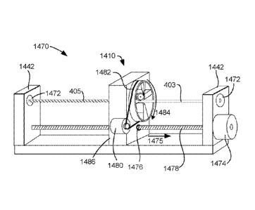
below.
100941 FIG, 4c discloses a scaled self-retaining suture 450 in an
unexpanded position,
produced by the foregoing method, vherein two intersecting helical escarpments
are cut
having substantially similar transverse cut angles, pitches, and cut depths,
but opposirm
chiralities (i.e. the effect of combining 4a and 4b together). The resultant
overlap of
escarpment points 452 of suture 450 produces a pattern of scale-like retainers
454 having
tissue-penetrating edges 456 and tissue-erit.oging surfaces 458; as the
retainers 454 created by
this method cover the entire circumferential suture surface on which they are
cut, the. tension
On the suture 450 is optimally distributed and the risk of suture failure
minimized. lAihen such
retainers 454 ot7 suture 450 engage tissue, retainers 454 flare away from the
suture body, as
shown in the expanded position in FIG. 4d.
100951 The foregoing steps may be carried out at the opposite end of the
suture to
create a bidirectional scaled self-retaining suture For such a suture, a
transition segment at
some point between the ends of the suture is left retainer-free the length of
the transitional
segment may be selected depending on the purpose of the suture, and the
transitional segment
may be located at or near the middle of the suture. Thus, to manufacture
bidirectional scaled
self-retaining sutures, the first pair of helical escarpments is cut along one
end of the suture to
a selected point some distance from the other suture end while the second pair
of helical
escarpments is cut to a selected point away from the first pair, so as to
avoid having the first
and second pair of escarpments from overlapping with one another and thereby
providing a
24
CA 02683819 2009-10-13
WO 2008/128113 PCT/US2008/060127
retainer-free transitional segment. For some bidirectional scaled self-
retaining sutures, the
orientation of one pair of helical escarpments at one end is about 18(r in
orientation from the
other pair of helical escarpments at the other end, thus creating an identical
"mitTored" pattern
of helical escarpments: Where smaller -retainer configurations are desired or
larger suture
diameters are present, a series of parallel helical cuts may be provided, as
opposed to a single
cut: Both ends of the resulting bidirectional scaled self-retaining suture can
function as suture
deployment ends, and can therefore be adapted for attachment to deployment
devices such as
suture needles or for direct deployment into tissue without a deploy:mem
device Referring
now to FIG. 5, bidirectional scaled self-retaining suture 500 includes a first
plurality of scale-
like retainers 504 having retainer bodies 508 and tissue-penetrating, edges
510 and a second
plurality 514 of retainers 516 having retainer bodies 518 and tissue-
penetrating edges 520.
First retainer plurality 504 is disposed proximally to -first suture
deployment end 502, thus
retainers 508 are oriented or pointed substantially a-way from end 502.
COnVerSely, second
retainer plurality 514 is disposed proximally to second suture deployment end
512, being
accordingly oriented or pointed substantially away from end 512. First and
second retainer
pluralities 504 and 514 are separated by transition segment 522, that portion
of a self-retaining
bidirectional suture that is retainer-free.
[00961 :If desired, a scaled self-retaining suture may be modified by
rounding off or
blunting the escarpment points. Referring now to FIG. 6, there is disclosed a
scaled self-
retainini,..? suture 600 including on the suture body scale-like retainers 604
having retainer
bodies 606 and tissue-penetrating edges 608. The tissue-penetrating edges 608
are rounded,
producing, a fish-scale effect, and are less likely bend or break. without
penetrating through
and engaging the tissue. Similarly, a bidirectional self-retaining suture may
be provided with
rounded "scales", as shown in FM. 7, Referring to that figure, bidirectional
scaled self-
retaining suture 700 (shown in the expanded position) includes a first
plurality of scale-like
retainers 704 having retainer bodies 708 and rounded tissue-penetrating edges
710 and a
second plurality of retainers 714 having retainer bodies 718 and rounded
tissue-penetrating
edges 720. First retainer plurality 704 is disposed proximally to first suture
deployment end
702, thus retainers 708 are oriented substantially away from end 702.
Conversely, second
retainer plurality 714 is disposed proximally to second suture deployment end
712, being
separated from first retainer plurality 704 by transition segment 722, with
retainers 718 being
accordingly oriented substantially away from end 712.
[00971 The bidirectional sutures described herein may be further
provided with an
CA 02683819 2009-10-13
WO 2008/128113 PCT/US2008/060127
expanded transition segment for to further resist movement in tissue.
Referring now to FIGS,
8a and 8b, bidirectional scaled self-retaining suture 800 (shown in the
expanded position)
includes a first plurality 804 of scale-like retainers 806 having retainer
bodies 808 and tissue-
penetrating edges 810 and a second plurality 814 of retainers 816 having
retainer bodies 818
and tissue-penetrating edges 821. First retainer plurality 804 is disposed
proximally to first
suture deployment end 802, thus retainers 808 are oriented or pointed
substantially away from
end 802. Conversely, second retainer plurality 814 is disposed proximally to
second suture
deployment end 812, being separated from first retainer plurality 804 by
transition segment
820õ with retainers 818 being accordingly oriented or pointed substantially
away from end
812. At transition segment 820, suture 800 has a greater diameter than at.
either end 802 or
812. As shown in this example, such expansion can be incremental, with the
diameter
increasing from each end 802 and 812 of suture 800 and reaching a. maximum at
transition
segment 820, the incremental expansion may commence at a point outside the
transitional
segment, such that some part of the retainer-bearing portions of the suture
thread may also
have a greater diameter than that of a suture end.
100981 in like fashion, the bidirectional scaled self-retaining
suture 900 in FIGS, 9a
and 9b (shown in the expanded position) includes a first plurality 904 of
scale-like retainers
906 having retainer bodies 908 and rounded tissue-penetrating edges 910 and a
second
plurality 914 of retainers 916 having retainer bodies 918 and rounded tissue-
penetrating edges
921: First retainer plurality 904 is disposed proximally to first suture
deployment, end 902,
and the retainers 906 are oriented or pointed substantially away from end 902.
Conversely,
second retainer plurality 914 is disposed proximally to second suture
deployment end 912,
being separated from first retainer plurality 904 by transition segment 920.
Retainers 918 are
oriented or pointed substantially a.way from end 91.2. Suture 900 has a
greater diameter at
transition segment 920 than at either end 902 or 912. The expansion is shown
in this exa.mple
to be incremental, with the diameter increasing from each end 902 and 912 of
suture 900 and
reaching a maximum at transition segment 920; the incre.mental expansion May
commence at
a point outside the transitional segment, such that some part of the retainer-
bearing portions of
the suture thread may also have a greater diameter than that. of a. suture
end.
100991 Bidirectional scaled self-retaining sutures may further be provided
with
expanded diameter transitional segments as described in Section C herein,
increased surface
area provided by the tissue engagement surface configurations described in
Section B, and
secondary retainer structure configurations as described in Section E.
26
CA 02683819 2009-10-13
WO 2008/128113 PCT/US2008/060127
E. Secondary Retainer Structure Configurations
[00100] To further reduce issues of uneven tension di.stribution or
insufficlent tissue
et:I._-);agement from arising and potentially leading to suture failure, self-
retaining sutures may
be provided with a secondary retainer structure, that is, secondary retainers
upon the. primaiy
retainers'. Secondary retainers further increase the surface area of the
suture and thereby
increase the amount of interaction between the suture and tissue. Moreover,
providing
secondary retainers on the primary retainers increases the total number of
retainers on the
suture (and thus the total number of points of tissue penetration) without a
reduction in suture
tensile strength that may occur by cutting a.dditional primary retainers into
the suture. body.
The increase in the number of retainers and the increase in the suture's -
tissue engage.ment
surface area results in better distribution of tension on the suture and
increased tissue hold.
FIGS. 6 through 8, inclusive, sho-w self-retaining sutures having secondary
retainers
projecting from the primary retainers; the sutures can be unidirectional or
bidirectional, In
this connection, a self-retaining suture can include at least one primary
retainer having a
primary retainer body projecting from the suture body and a tissue penetrating
end, and an
inner surface .substantially facing the suture body and an outer surface
facing sub.stantially
away from the suture body, the primary retainer facing and deployed in. a
direction and being
adapted for resisting move-merit of the suture when in tissue, in an opposite
direction from the
direction in which the primary retainer faces and is deployed. The primary
retainer can further
include at least one. secondary retainer projecting from the primary retainer
body. Such
secondary retainers can be oriented in a direction at least about 90" away
from the direction in
which the primal), retainer faces and is deployed, thus facliitating "double-
hooking" of tissue,
and may be located on any surface of the primary retainer. The primary
retainer can be a
barb, it can be frusto-conical, helical, and so forth. The secondary retainer
can also be a barb,
frusto-conical. helical, as well a.s filamentary.
100101 I :Referring. to FIG. 10, a self-retaining suture 1000 is shown,
the suture 1000
including a suture body 1002 and a plurality of primary retainers 1004 with
face 1005
extending from suture body. 1002 and having primary retainer bodies 1006 and
primary
retainer edges 1008 and primary retainer tissue engagement .surfaces 1016. The
primary
retainers 1004 further include a secondary retainer 1010 disposed on the
tissue engagement
surface 1016 at the primary retainer edge 1008 with face 1009 and with the
primary retainer
1004 facing the deployment direction and with the .second retainer 1010 facing
substantially
,T
I
CA 02683819 2009-10-13
WO 2008/128113 PCT/US2008/060127
away from the deployment direction. The secondary retainers 1010 include a
secondaiy
retainer body 1012 and a .secondary .retainer edge 1014. Both primal), and
.secondary retainers
1004 and 1010, respectively, are configured as barbs, such that the
combination of the two
have a "fishhook" appearance and function much the same way. That is, during
suture
deployment, primary retainers 1004 rest along suture body 1002 and secondary
retainers 1010
in turn rest between primary retainers 1004 and suture body 1002, During
anixation, primary
retainers 1004 are displaced away front suture body 1002 thereby exposing
secondary
retainers 1.010 and allowing them to further engage tissue being engaged
between primary
retainers. 1004 and suture body 1002, Such a "fish:hook" combination of
primary and
secondary retainers may be produced by forming a primary retainer on the
..suture thread by a
single transverse cut and then removing some suture. material from the tissue
engage.ment
surface of the retainer (such as by hand-cutting or laser-cutting) to t7orm
the secondary
retainer. Alternatively, this combination may be made by making a first
transverse cut on the
suture thread to form the primary retainer, followed by back-cutting a second
transverse cut
onto the primary retainer to form the secondary retainer: these cuts may be
made in a single
continuous motion or may be made in separate motions. Similarly, the
combination of
primary retainers with "breakback" secondary retainers disposed on the tissue
engagement
surfaces of the primary retainers may be provided with frusto-conical and
helical primary
retainers; the secondary retainers may be cut onto the tissue engagement
surfaces of such
primary retainers to provide a secondary retainer lip along at least part of
the length of the
tissue penetrating edge of the primary retainer,
1001021 FIG. 11 discloses a primary/secondary retainer combination as
well, but with a
plurality of secondary retainers 1110 with face 1111 on primary retainer 1104
with face 1105,
Thus, self-retaining suture .1100 includes suture body 1102 and primary
retainers 1.104 having
a plurality of secondary retainers 1110 projecting from primary retainer body
1106 and with
the secondary retainer 1110 located between the primary retainer 1104 and the
suture. body
11 02 facing away from the direction of deployment Both primary and secondary
retainers
1104 and 1110, respectively, are configured as barbs, such that the
combination of the two
resemble the microstructure of a porcupine's quill and l'unction in much the
.same way. That
is, during suture deployment, primary retainers 1.104 rest along suture body
1102 and
secondary retainers 1110 disposed on inner surface 1116 in turn rest between
primary
retainers 1104 and suture body 1102, As secondary retainers 1110 on the
surfaces of the
primary retainers 1104 other than inner surface 1116 are exposed during suture
deployment,
28
CA 02683819 2009-10-13
WO 2008/128113 PCT/US2008/060127
tissue resistance during deployment may increase .slightly. However, once the
primary barbs
1104 are affixed, the secondary retainers 1110 can enhance the hold or
positioning of the
primary retainers 1104. In addition, the uneven primary retainer surface
resulting from these
secondary retainers 1110 may increase local tissue disruption and increa.se
.surface area, both
of which can promote healing and fixation. It is to be understood that such
pluralities a
secondary retainers may be provided on t7rusto-conical and helical primary
retainers as well.
[00103] Seif-retaìnìng sutures having secondary retainers associated
with primary
retainers may further be provided with secondary retainers along the suture
body, as disclosed
in FIG-. 12. Suture 1200 in FIG. 12 includes suture body -1202 and primary -
retainers. -1204
having a plurality of secondary retainers 12.10 (that project from primary
retainer body 1206)
and with the secondary retainer 1210 located between the primal)! retainer
1204 and the
suture body 1202 facing away from the direction of deployment Suture body 1202
further
includes secondary retainers 1212, -which point away from the direction of
deployment and
face the directIN, of deployment; given their orientationõ secondary retainers
1212 rest along
body 1202 during deployment of suture 1.200 and so do not increase tissue
resistance as suture
1200 is deployed through it. As do primal), retainers 1204, secondary
retainers 1212 fan
away from suture body 1202 and engage tissue during affixation of the suture
1200.
Provision of secondary retainers 1212 along suture body 1202 further increases
surface area
and therefore the holding strength of the suture. While retainers 1212 are
configured as barbs,
it is to be understood that other retainer configurations may be used.
100104] Yet another secondary retainer structure is illustrated in
FIG. 13, which shows
a self-retaining suture 1300 including a. suture body 1302 and primary
retainers 1304 having a
plurality of secondary retainers 1310 extending t7rom primary retainer bodies
1306, which
secondary retainers 1310 are filamentary. These filamentary retainers 1310 may
be created
on the primary -retainers. 1304 by polymer grafting or growing techniques, by
which polymer
chains grown of-lathe surface of the primary retainers .1304 increase the
microscopic surface
area of the primar,,, retainers 1304 thereby improving tissue engagement.
F. Manufacture of Self-Retaining Sutures
100105] Suture threads described herein may be produced by any suitable
method,
including without limitation injection moulding, stamping, cutting, laser,
extrusion, and so
forth. With respect to cutting, polymeric thread or filaments may be
manufactured or
purchased for the .suture body, and the retainers can be subsequently cut onto
the suture body;
CA 02683819 2009-10-13
WO 2008/128113 PCT/US2008/060127
they may be hand-cut, laser-cut, or mechanically machine-cut using blades,
cutting wheeis,
grinding wheels, and so forth. The sutures may be .made of any suitable
bi.ocompatible
material, and may be fiather treated with any .suitable biocompatible
material, whether to
enhance the sutures' strength, resilience, longevity, or other qualities., or
to equip the sutures
to fuiltil additional functions besides joining tissues together,
repositioning tissues, or
attaching .foreign elements to ti.ssues,
[00106] As described above, helical or spirai self-retaining sutures
can be produced -by
a method including (i) providing a. suture having a longitudinal axis and a
circumference; a
cutter; and, a displacer for longitudinally displacing, and pivoting, about
the longitudinal axis
at least. one of the cutter and suture relative to one another; (ii) placing
the cutter and suture
into cutting engagement at a transverse cut angle; and, (iii) cutting a
helical circumferential
escarpment about the suture. FIG.. 14a shows an embodiment a a. cutting device
for .17(n-filing a
helical retainer 405 disposed along a suture. body 403 of a self-retaining.
suture 401, the helical
retainer 405 including a retainer body 409 and a tissue penetrating edge 41 1,
1001.07] As shown in FIG. 14a, a cutting device 1410 creates an escarpment
in the
surface of suture body 403. As shown in FIG. .14A the cutting device 1410
comprises, in one
embodiment, a rotary cutting head 1412 can be, if desired, positioned adjacent
suture body
403. Rotary cutting head is driven by motor -1418. Rotary cutting head 1412 is
.mounted such.
that the depth of tip 1414 of rotary cutting head 1412 is precisely controlled
so that the depth
of the escarpment is kept. constant. In one embodiment rotary cuttim.t; head
may be held in
fixed relationship with a roller 1416 which rides on .suture body 403 thereby
controlling the
depth of the escarpment relative to the surface of the suture body 403. This
allows for a
constant escarpment depth even vhere the .suture body varies in diameter. This
is particular
useful where the suture body has an expanded region of greater diameter as
described above.
1001.08] Roller -1416 and rotary cutting head 1412 are mounted to a
floating. armature
1.430 which is pushed into contact with suture body 403 by springs 1432, The
drive
mechanism 1434 connecting motor 1418 and rotary cutting head 1412 allows
move:meat of
rotary cutting head 141_2 without binding. Rotary cutting head 141_2 is driven
at a very high
rotation rate compared to the translation and rotation of suture. body 403. In
alternative
embodiments other cutting devices may be used including knives, abrasive
wheels., lasers and
the :like. Alternative mechanisms may also be utilized to control the depth of
cut including an
electronically controlled sensor/servo system or other mechanical "following"
systems,
Where the diameter of suture body 403 is .sufficiently uniform, the distance
between roller
CA 02683819 2009-10-13
WO 2008/128113 PCT/US2008/060127
1416 and cutter support 1420 may be fixed rather than varying with the
diameter of the suture
body,
[00109] An anvil or cutter support 1420 is provided on the opposite
side of suture body
403 from cutting device 14 10 to support the suture body 403 during the
cutting operation. The
cutter support 1420 is designed to allow suture body 403 to translate
longitudinally (arrow
1404) and rotate (arrow 1402) relative to cutting device 1410 but not to move
laterally or
vertically while passing through cutting device 1410. Cutter support 1420 thus
maintains
suture body 403 in the correct alignment with rotary cutting head 141.2 during
the cutting
action. Suture body 403 is held between roller 1416 and cutter support 1420.
Roller 1416 may
also !Unction to prevent lateral and vertical motion of suture body 403.
100.11.0] In order for cutting device 1410 to create a helical
escarpment in the surface of
suture body 403 one of (Laming device 1410 or suture body 403 is rotated
(arrow 1402)
relative to the longitudinal axis of suture body 403 and translated (arrow
1404) along the
longitudinal axis of suture body 403at the same time. Alternatively, both
suture body 403 and
cutting device 1410 may be rotated about the longitudinal axis of the suture
body 403, the
suture body 403 being rotated while the cutting device 1410 revolves. In the
latter case, suture
body 403 and cutting device 1410 may be rotated either in the same direction
or in opposing
directions about the longitudinal axis of suture body 403. If both cutting
device 1410 and
suture body 403 are rotated in the same direction, the angular velocity at
which each is rotated
about, the longitudinal axis must differ in order to effect a circumferential
escarpment on the
suture.
f0O1 111 The speed of translation compared to the rela.tive rate of
rotation of cutting
devic..e 1410 and suture body 403 controls the pitch of the helical retainer
405. The pitch of
the helical retainer 405 is the distance between turns. The pitch of helical
retainer 405 is
shown in FIG 14a a.s dimension 1406. The faster the translation for a given
rotation, the larger
the pitch. Conversely, the faster the relative rotation for a given
translational speed, the
smaller the pitch. In some embodiments, the translation velocity and the
rotational velocity
will have a fixed relationship along the length of the suture body 403.
However, in other
embodiments it may be desirable for the pi tc h of the helical escarpment to
be larger in some
regions of the suture 401 than in others. This may be achieved by altering
either or both of the
relative rate of rotation and speed of translation during the passage of the
suture body 403
through the cutting device 1410.
[00112] In order to form a helic.al retainer, a mechanism is used to
generate the desired
31
CA 02683819 2009-10-13
WO 2008/128113 PCT/US2008/060127
rate of rotation and speed of translation of the suture body 403 relative to
the cutting device
1410. FIG. 14b shows one embodiment of a cutting fixture 1440 for forming a
helical retainer
405 disposed along a suture body 403 of a self-retaining su.ture 401. Cutting
fixture 1440
includes cutting device 1410 along witb a mechanism for rotating suture body
403 and
translating cutting device 1410 along suture body 403. As shown in Fig 14b,
suture body 403
is tensioned bet.w.een two supports 1442. Suture body 403 is held to each
support 1442 by a
gear driven. chuck. 1444. A drive mechanism 1446, which may be an electric
motor, drives a
screw drive 1.448 which runs between the supports 1.442. Gears 1450 at each
end of screw
drive 1448 rotate chucks -1444 at the same speed and in the same direction as
shown by
arrows 1445. Screw drive 1.448 also passes through a -threaded passage in the
base 1452 of
cutting device 1410. As screw drive 1448 turns, screw drive 1448 drives
cutting device 1410
lineariy. along the length of suture body 403 as ShOWII by. arrow 1454. Thus
in cutting fixture
1440, suture body 403 is rotated relative to cutting device 1410 and cutting
device 1410 is
translated relative to suture body 403 thereby cutting a helical retainer 405
on suture body
403. N-ote that gears 1450 fix the ratio of translation speed of cutting
device 1410 compared to
the rate of rotation of suture body 403. Cutting fixture 1440 will, therefore,
produce a helical
retainer 405 with a constant pitch. Gears 1450 may be changed in order to
change the pitch.
Alternatively, if a variable pitch is desired, a separate drive .mechanism
1446 may be .provided
for the chucks 1444 thereby allowing the rate of rotation of suture body 403
to be varied
independent of the rate of translation of cutting device 1410. For example
chucks 1446 and
screw drive 1548 may be driven by separate computer-controlled electric drive
systems.
1001131 FIG. 14c shows an alternative cutting fixture 1460 .thr
.forming a helical
retainer 405 disposed along a suture body 403 of a self-retaining suture 401.
Cutting fixture
1460 includes cutting device 14.10 along with a mechanism for rotating suture
body 403 and
translating suture body 403 relative to cutting device 1410. As shown in Fig
14c, suture body
403 is tensioned between two supports 1442.. Suture body 403 is held to each
support 1442 by
a feed spool 1461 or uptake spool 1462. Spools 1461 and 1462 rotate about
their axes in the
same speed and direction such that as the suture body 403 is played out by
feed spool 1461 it
is taken -up uptake .spool 1.462 translating suture body 403 in the direction
of arrows 1463. A
mechanical tensioning mechanism (not shown) may also be provided to help
maintain a
constant tension in suture body 403 during the cutting process and to take up
any slack cause
by slight variation in the rate of suture play out and take up. A linear drive
mechanism such as
a .screw drive may be used to translate suture body 403 as an alternative to
drums or spools
32
CA 02683819 2009-10-13
WO 2008/128113 PCT/US2008/060127
1461, 1462. A drive mechanism 1464 drives shaft 1468 which runs between the
supports
1442. Gears 1466 at each end of shaft 1468 rotate gimbles 1469 which hold
spools 1461,
1462 to supports 1442. The gimbles 1469 rotate the spools 1461, 1462 about the
longitudinal
axis of suture body 403 as shown by arrows 1465. Thus, in cutting fixture
1460, suture body
403 is rotated a.nd translated relative to a fixed-position cutting device
1410 thereby cutting a
helical retainer 405 on suture body 403. lf a variable pitch is desired, a
separate drive
mechanism 1446 may be provided for the rotation of the spools 1461, 1462 than
the drive
mechanism 1464 for shaft. 1468 and gimbles 1469, thereby allowing the rate of
rotation of
suture body 403 to be varied independent of the rate of suture body 403.
f0011.41 EIG, 14d shows an alternative cutting fixture 1470 for fonning a
helical
retainer 405 disposed along a suture body 403 of a self-retaining suture 401.
Cutting fixture
1470 includes cutting device 1410 along with a mechanism for rotating cutting
device 1410
and translating cut fin device 1410 relative to suture body 403 As shown in
Fig -14d, suture
body 403 is tensioned between two supports 1442. Suture body 403 is held to
each support
1442 by a fixed chuck 1472. A tensioning mechanism (not shown) may also be
provided to
help maintain a constant tension in suture body 403 during the cutting
process. A drive
mechanism -1474 drives screw drive -1478 which runs between the supports 1442.
Screw drive
1478 passes through a threaded passage 1476 in the base 1486 of cutting device
1410 such
that, as screw drive 1478 turns, cutting device 1410 is translated along the
length of suture
body 403 as shown by arrow 1475: A separate drive mechanism 1480 drives a belt
1482
which rotates cutting device 1410 along a circular track within base 1486 as
shown by arrow
-1484. In cutting fixture 1470 the cutting device 1410 is thereby moved along
a helical path
relative to the fixed suture body 403. Thus, in cutting fixture 1470, suture
body 403 remains
in a. fixed position and cutting device 14-10 is rotated and translated
relative to suture body
403 thereby cutting a helical retainer 405 on suture body 403. The pitch of
helical retainer 405
may be controlled, in this embodiment, by adjusting the relative speeds of
drive mechanism
1480 and drive mechanism 1.474 which may be computer-controlled electric
motors. If a fixed
pitch is desired, a gear linkage may be used to drive the rotation of cutting
device 1410 within
base 1.452 from screw drive 1478.
100115] FIG, 14e shows an alternative cutting fixture 1490 for forming a
helical or
spiral retainer 405 disposed along a suture 'body 403 of a self-retaining
suture. Cutting fixture
1490 includes cutting device 14-10 along with a mechanism for rotating cutting
device -1410
relative to suture body 403 and translating suture body 403 relative to
cutting device -1410. As
33
CA 02683819 2009-10-13
WO 2008/128113 PCT/US2008/060127
shown in Fig 14e, suture body 403 is tensioned between two spools 1491, 1492.
Feed spool
1491 and uptake spool 1492 rotate about their axes in the same speed and
direction such that
as the suture body 403 is played out by spool 1491 it is taken up spool 1492
translating suture
body 403 in the direction of arrows 1493, A tensioning mechanism (not shown)
may also be
provided to help maintain a constant tension in suture body 403 during the
cutting process and
to take up any slack cause by slight variation in the rate of suture play out
and take up A
linear drive mechanism such as a screw drive may be used to translate suture
body 403 as an
alternative to spools 1491, 1492. As with cutting fixture 1470 of FIG, 14d, a
drive nìeciìanis.in
1480 drives a belt 1482 which rotates cutting device 1410 within base 1486 as
shown by
arrow 1484. However, in cutting -fixture 1490 base 1486 is stationary while
suture body 403
translates. Thus in cutting fixture 1490, suture body 403 translates relative
to cutting, device
1410 and cutting device 1410 rotates relative to suture body 403 thereby
cutting a helical
retainer 405 on suture body 403. The pitch of helical retainer 405 may be
controlled in this
entbodiment by adjusting the relative speeds of drive mechaniSM 1480 and
spools 1491., 1492.
If a fixed pitch is desired, a gear linkage may be used to drive spools 1491,
1492 from drive
mechanism 1480, _Alternatively separate drive mechanisms may be used and
electronically
controlled to maintain constant or varying rotational and translational speeds
to create the
constant of varying pitch desired,
100116] Still alternatively, the mechanisms of Fig. 14c and Fig. 14e
can be combined if
it is desired to have the cutter revolve or rotate about the suture, and
translate or be displa.ced
along the longitudinal length of the suture. in combination with having the
suture both
translate or be displaced along the longitudinal length, as well rotate or
revolve about the axis
of said suture. To accomplish this functionality the mechanism that
longitudinally displaces or
translates, and rotates the suture, which includes for example, the spools and
gimbles and
gears in Fig. 140 would replace the fixed chuck of Fig. 14d, Additionally, the
mechanisms of
Fig 14b and Fig. 14e can be combined, if it is desired, to have the cutter
revolve or rotate
about the suture and translate or be displaced along the longitudinal length
of the suture, in
combination with the suture being rotated about the axis of the suture. To
accomplish this
functionality, the driven chucks of Fig 14b would replace the fixed chucks of
Fig 14d
Alternatively to have the suture translate longitudinally and be in
combination with having the
cutter revolve or rotate and translate, the suture. translation mechanism of
Fig. 14e can be
added to the device of Fig, 14d. Thus, the fixed chucks of Fig. 14d that hold
the suture can be
replaced in Fig_ 14d with the spools of Fig. 140 so that the suture can
translate or be displaced
34
CA 02683819 2009-10-13
WO 2008/128113 PCT/US2008/060127
longitudinally .
1001171 As, shown in FIG. 14f, as an alternative embodiment the cutter
rotating or
revo1ving and cutter translating mechanism of :Fig. 14d can be replaced with a
spiral track or
cylinder having an interior helical track that the cutter can be moved on:
:FIG. 14f shows an
alternative cutting fixture 1471 for forming a helical retainer 405 disposed
along a suture
body 403 of a self-retaining suture 401, Cutting fixture 1471 includes cutting
device 1410
along with a mechanism for rotating cutting device 1410 and translating
cutting device 1410
relative to suture body 403. .As shown in Fig 14fõ suture body 403 is
tensioned between two
supports 1442. Suture body 403 is held to each support 1442 by a fixed chuck
1472. A
tensioning m ech an i sin (not shown) May al so be provided to hel p maintain
a constant ten si on
in suture body 403 during the cutting process. Cutting device 1410 which
includes a cutting
head 1412 and a cutter support 1420 travels along a helical track 1479 as
shown by arrow
1477. In cutting fixture 1471 the cutting device 1410 is thereby moved along
a. helical path
relative to the fixed suture body 403. Thus, in cutting fixture 1471, suture
body 403 remains
in a fixed position and cutting device 1410 is rotated and translated relative
to suture body
403 thereby cutting a helical retainer 405 on suture body 403. The pitch of
helical retainer 405
is fixed by the pitch of helical track 1479. As the cutting device 1.410 is
moved on the helical
or spiral track, a helical or spiral cut is made on the suture body 403. It is
to be noted that use
of a helical track or a cylinder with an interior helical or spiral track, can
be made with any of
the other embodiments herein, if it is desired, to have the cutter 'both
rotate or revolve about
the suture and have the cutter translate along the length of the suture. Still
further, if desired in
any of these embodiments, the suture can itself be pre-twisted prior to being
loaded into any
of the fixtures described.
(00.118] FIG. 15a shows an alternative cutting device 1510 that may be
used in the
cutting fixtures of FIGS. 14b-14f. As shown in FIG. 15a. the suture body 403
passes between
cutter support .1520 and support 1522. A rotary cutting blade 1512 rotates as
shown by arrow
1514. Rotary cutting blade 1512 is mounted on shaft 1516 which passes through
support
1522. The geometry of rotary cutting blade 1512 is thereby fixed relative to
suture body 403,
:It may, however, be desirable to provide set screws or the like in order -to
adjust the angle and
depth of the rotary cutting blade 1512 relative to the suture body 403. Rotary
cutting blade
may be a knife blade in which case the rotation of the blade may be either
passive or driven.
The knife blade made cif a suitable ceramic or metal material for cutting into
suture body 403.
If the rotation is passive, rotary cutting blade 1512 turns in response to the
rotation of suture
CA 02683819 2009-10-13
WO 2008/128113 PCT/US2008/060127
body 403 against rotary cutting blade 1112. If the rotation of rotary cutting
blade 1512 is
driven. rotaiN cutting blade 1512 is driven by Motor 1515, Rotary cutting
blade 1512 may be
an abrasive wheel or the like in which case rotary cutting blade 1512 is
preferably driven at
high speed or very high speed relative to the rotation of suture body 403. If
the rotation of
rotary cutting blade 1512 is driven, it may be driven in the same direction as
suture body 403
or in the opposite direction Rotary cutting blade 1512 is moved along, a
helical path relative
to suture body 403 utilizing a cutting fixture which provides for relative
translation and
rotation of cutting device 1510 and the suture body 403 as shown by arrows
150,2 and 1504
respectively. Suitable cutting fixtures are described with respect to FIGS.
14b-14f above, As
described above, either or both of cutting device 151.0 and suture body 403
may be moved in
order to generate the desired helical path of the cutting device 1510 about
the suture body
403,
[001191 FIG. 15b shows an end view of cutting device 1510 illustrating
the entrance
aperture 1506 into cutting device 1510. Entrance aperture 1506 into cutting
device 1510 is
defined by the space between supports 1520 and 1522. Supports 1520 and 1522
are each
provided Avith a polished radius or polished chamfer 1521 to guide suture body
403 (position
shown by dashed line) into aperture 1506 without abrading suture body 403.
R.otaiN cutting
blade 1512 can seen protruding into aperture 1506 adjacent support 1521. in
alternative
embodiments a plurality of cutting blades may be provide in order to cut a
plurality of helical
retainers at the satne time. As suture body 403 enters aperture 1506 (into the
pa.ge) suture
body 403 rotates in the direction of arrow 1508 relative to cutting device.
151(). Note that
either the suture body 403 or the cutting device 1510 may be rotated to
achieve the desired
relative rotation of cutting device 1510 and suture body 403,
(00120] FIG. 15c shows another alternative cutting device 1530 that
may be used in the
cutting fixtures of FIGS. :14b-14f. As shown in :FIG. 15a. the suture body 403
passes between
cutter support 1520 and support .1522. A -fixed cutting blade 1532is mounted
to support 1522
by machine screw 1534. Machine screw 1534 allows the position of cutting blade
1532 to be
adjusted relative to suture body 403, it may be desirable to provide further
set screws or the
like in order to better adjust the angle and depth of the rotary cutting blade
1512 relative to the
suture body 403. Cutting blade may be a knife blade made of a suitable ceramic
or metal
material for cutting into suture body 403. Cutting blade 1532 is moved along a
helical path
relative to suture body 403 utilizing a cutting fixture which provides for
relative translation
and rotation of cutting device 1510 and the suture body 403 as shown by arrows
1502 and
36
CA 02683819 2009-10-13
WO 2008/128113 PCT/US2008/060127
1504 respectively, Suitable cutting fixtures are described with respect to
.F1GS, 14b-14f
above. As described above, either or both of cutting device 1.510 and suture
body 403 may be
moved in order to generate the desired helical path of the cutting device
.1510 about the suture
body 403.
100121] Self-retaininF sutures described herein may also incorporate
materials that
further promote tiss-ue engagement. In addition to tis.sue engagement at the
retainers, use of
tissue engagement-promoting materials in at least part of the suture bodies
(whether or not
such materials also make up all or part of the retainers) can enhance the
ability of the sutures
to stay in place. One such class of tissue engagement-promoting materials are
porous
polymers that can be extruded -to forrn suture bodies, including both
microporous polymers
and
polymers that can be extruded with bubbles (whether b oab sorb able or
flonbioab.sorbable). .A suture synthesized with such materials can have a
three-dimen.sional
lattice structure that increases tissue engagement surface area and permits
tisslie infiltration
into the suture body itself, thus having a primary structure that promotes
successful suture
use. :Moreover, by optimizing pore size, fibroblast ingrowth can be
encouraged,. further
facilitating the suture to be anchored in the tissue.
1001221
One such microporous polymer is e.PTFE (expanded polytetra-fluoroethylene).
Self-retaining incorporating ePTFE (and related microporous materials) are
well-suited to
uses requiring a strong and permanent lift (such as breast lifts, face lifts,
and other tissue
repositioning procedures), as tissue infiltration of the suture results in imp-
roved fixation and
engraftment of the suture and the surrounding tissue thus providing superior
hold and greater
lOng,evity of the lift.
1001231
Additionally, self-retaining sutures described herein may be provided wit.h.
compositions to promote. healing and prevent undesirable effects such as sca.r
formation,.
infection, pain, and so forth. This can be accomplished in a variety of
manners, including for
example: (a) by directly affixing to the suture a formulation (e.g., by either
spraying the
suture with a polymer/drug film, or by dipping the suture. into a
poly.merldrug solution), (b) by
coating the suture with a substance such as a hydro4e1 which -will in turn
absorb the
cam posi ti on (c) by interweavi iïg form ulati on-coated thread (or the pol y
er itself formed i nto
3) a -thread) into the suture structure in the case of multi-filamentary
sutures, (d) by inserting the
suture into a sleeve or mesh which is comprised of, or coated with, a.
formulation, or (e)
constructing the suture itself with a composition. Such compositions may incl-
ude without
limitation anti -prol iterative agents, anti -a ngi ogen c agents, anti -
infective agents, fi b rosi s-
37
CA 02683819 2009-10-13
WO 2008/128113 PCT/US2008/060127
inducing agents, anti-scarring agents, lubricious agents, echogenic agents,
anti-inflammatory
agents, cell cycle inhibitors, analgesics, and anti-microtubule agents. For
example, a
composition can be applied to the su.ture before the retainers are formed, so
that when the
retainers engage, the engaging surface is substantially free of the coating.
fln this way, tissue
beinF sutured contacts a coated surface of the suture as the suture is
introduced, but when the
retainer engages, a non-coated surface of the retainer contacts the tissue..
Alternatively, the
suture .may be coated after or during formation of retainers on the suture if,
for example, a.
fully-coated rather than selectively-coated suture is desired.. In yet another
alternative., a
suture may be selectively coated either during or after formation of retainers
by exposing only
selected portions of the suture to the coating.. The particular purpose to
which the suture is to
be put or the composition may determine -whether alblly-coated or selectively-
coated suture
is appropriate; t7or example., with lubricious coatings, it may be desirable
to selectively coat
the suture, leaving, for instance, .the tissue-engaging surfaces of the
sutures uncoated in order
to prevent the tissue engagement function of those surfaces from being
impaired. On the other
hand, coatings such as those comprising such compounds as anti-infective
agents may
suitably be applied to the entire suture, while coatings such as those
comprising fibrosing
agents may .suitably be applied to all or part of the suture (such as the
tissu.e-engaging
surfaces.). The purpose of the suture may also determine the sort of coating
that is applied to
the sutum for example, .self-retaining sutures having anti-proliferative
coatings may be used
in closing tumour excision sites, while self-retaining sutures vi,,ith
fibrosing coatings may be
used in tissue repositioning procedures and those having anti-scarring
coatings may be used
for wound closure on the skin. As well, the structure of the suture may
influence the choice
and extent of coating for example, .sutures having an expanded segment may
include a
fibrosis-inducing composition on the expanded segment to further secure the
segment in
position in the tissue.. Coatings may also include a plurality of compositions
either together or
on different portions of the suture, where the multiple compositions can be
selected either for
different purposes (such as combinations of analgesics, anti-infective and
anti-scarring agents)
or for 'their synergi sti c effects.
G. Clinical Uses
1001.24] In addition to the general wound closure and soft tissue
repair applications
described in the preceding sections, self retaining sutures can be used in a
variety of other
inidcations.
38
CA 02683819 2009-10-13
WO 2008/128113 PCT/US2008/060127
[00125] Self-retaining sutures described herein may be used in various
dental
procedures, i.e., oral and maxillothcial surgical procedures and thus may be
referred to as
"self-retaining dental sutures." The above-mentioned procedures include, but
are not limited
to, oral .surgery (e.g., removal of impacted or broken teeth), surgery to
provide bone
augmentation, surgery to repair dentofacial deformities, repair following
trauma (e.g., facial
bone fractures and injuries), .surg,ical treatment of odontogenic and non-
odontogenic tumors,
reconstructive .surgeries, repair of cleft lip or cleft palate, congenital
craniofacial deformities,
and esthetic .facial surgery, Self-retaining dental sutures may be degradable
or non-
degradable, and may typically range in size from LISP 2-0 to USP 6-0.
f001.261 Self-retaining. sutures described herein may also be used in ti .s
sue repositioning
surgical procedures and thus may be referred to as "self-retaining, tissue
repositioning
sutures" ,Such .surgical procedures include, without limhation, face lifts,
neck lifts, brow lifts,
thigh !ills, and breast !ills. Self-retaining sutures used in tissue
_repositioning procedures ma.y
vary depending on the tissue being repositioned; for example, sutures with
larger and further
spaced-apart retainers may be suitably employed with relatively soft tissues
such a.s fatty
tissues.
[001271 Self-retaining sutures described herein may also be used in
microsurgical
procedures that are performed under a .surgical microscope (and thus may be
referred to as
"self-retaining micro.sutures"). Such surgical procedures include, but are not
limited to,
reattachment and repair of peripheral nerves, spinal microsurgery,
microsurgery of the hand,.
v ari ou s plastic microsurgi cal procedures (e.g., facial reconstruction), m
icrosurgery of the
male or female reproductive systems, and various types of reconstructive
microsurgery.
Microsurgical reconstruction is used for complex reconstructive surgery
problems when other
options such as primary closure, healing by secondary intention, skin
grafting, local .flap
transfer, and distant flap transfer are not adequate. Self-retaining
microsutures have a very
small caliber, often as small as USP 9-0 or LISP 10-0, and may have an
attached needle of
corresponding size. They may be degradable or non-degradable.
[00128] Self-retaining sutures as described herein may be used in
similarly .small
caliber ranges for ophthalmic surgical procedures and thus may be referred .to
as "ophthalmic
self-retaining suturee. Such procedures include but are not li.m.ited to
keratoplasty, cataract,
and vitreous retinal inicrosurgi cal procedures. Ophthalmic self-retaining
sutures may be
d.egrad.able or non-degradable, and have an attached needle of correspondingly-
small caliber,
[00129] Self retaining sutures can be used in a variety of veterinary
applications for a
39
CA 02683819 2014-12-04
wide number of surgical and traumatic purposes in animal health.
[00130]
Although the present invention has been shown and described in detail with
regard to only a few exemplary embodiments of the invention, it should be
understood by
those skilled in the art that it is not intended to limit. Various
modifications, omissions,
and additions may be made to the disclosed embodiments without materially
departing
from the novel teachings and advantages of the foregoing teachings.