Note: Descriptions are shown in the official language in which they were submitted.
CA 02685492 2009-10-28
WO 2007/143139 PCT/US2007/013022
- 1 -
ABCB5 POSITIVE MESENCHYMAL STEM CELLS AS IMMUNOMODULATORS
FIELD OF INVENTION
The present invention is directed at ABCB5 positive immunomodulatory
mesenchymal stem cells and to their use in the treatment of immune mediated
disease.
BACKGROUND OF INVENTION
Stem cell-based immunomodulatory strategies are a new therapeutic frontier in
clinical allotransplantation (Frank, et. al., Lancet 363:1411(2004)). It has
been found,
for example, that adult bone marrow-derived mesenchymal stem cells (BM-MSC)
can
inhibit T cell proliferation in response to mitogens and alloantigens in vitro
(Le Blanc, et
al., Scand. J. Immunol. 57:11(2003); Rasmusson, et al., Exp. Cell Res. 305:33
(2005)).
Because of its easy accessibility, skin is a particularly attractive potential
source of
therapeutically useful stem cells. However, identification of molecular
markers for the
isolation and expansion of pure dermal stem cells is a significant problem and
the
specific biological effects of these cells are largely unknown.
Recently, a novel human ATP-binding cassette (ABC) transporter, ABCB5 P-
glycoprotein, was cloned and it has been suggested that this protein may serve
as a
marker for the isolation of a subpopulation of stem cells (Frank, et. al., J
Biol. Chem.
278:47156 (2003); Frank, et al., Cancer Res. 65:4320 (2005); US 6,846,883).
SUMMARY OF INVENTION
The present invention is based in part upon the discovery that dermal
mesenchymal stem cells that express ABCB5 have immunomodulatory properties and
can be useful for the treatment of immune mediated diseases. The ABCB5 protein
is
.expressed on the surface of the stem cells and can be used both in their
identification,
e.g., using immunofluorescence, and purification, e.g., using antibodies
immobilized on
an inert substrate.
In an aspect, the invention is directed to a method of obtaining
immunomodulatory dermal mesenchymal stem cells (dermal MSC) from a sample of
human skin and then isolating ABCB5 positive cells from the sample. ABCB5
cells may
be identified, for instance, by immunofluorescence using antibodies against
the human
protein and by other methods as well. In this regard, it should be noted that
the full
CA 02685492 2009-10-28
WO 2007/143139 PCT/US2007/013022
- 2 -
amino acid and gene sequences for human ABCB5 are known in the art (see
GenBank
Accession No. NM 178559 and NP 848654) and monoclonal antibodies specific for
this
protein have been previously described (see e.g. Frank, et al., Cancer Res.
65:4320-4333
(2005); Frank, et al., J. Biol. Chem. 278:47156-47165 (2003)). A preferred
method for
the isolation of ABCB5 positive cells is through the use of antibodies that
specifically
recognize this protein and which have been immobilized, e.g., on a bead or
column
packing. Once obtained, the cells can be cloned by limiting dilution and
expanded using
methods well known in the art (see the Examples section for further
discussion).
Purified ABCB5 dermal MSC may be administered to a subject for the purpose of
modulating the activity of immunity-associated cells, e.g. for inhibiting the
activation of
T-lymphocytes. This can be accomplished, for instance, by intravenously
injecting or
infusing the subject with between 1x10⁷ ¨ 1x10¹⁰ cells.
In other aspects the invention relates to a method for modulating immune
molecule expression in a cell of a subject by administering to a subject ABCB5
positive
dermal mesenchymal stem cells in an effective amount to modulate immune
molecule
expression in cells of the subject. For instance, it is demonstrated herein
that in vivo
transplantation of A13CB5+-derived dermal mesenchymal stem cells can inhibit
an APC-
expressed positive costimulatory signal critically involved in T cell
activation. In some
embodiments the subject is administered 1x10⁷¨ lx101 ABCB5 positive dermal
mesenchymal stem cells by intravenous injection or infusion.
In other aspects of the invention a method for promoting allograft survival is
provided. The method is achieved by administering to a subject having an organ
transplant an effective amount of ABCB5 positive dermal mesenchymal stem cells
to
promote allograft survival. The ABCB5 positive dermal mesenchymal stem cells
may be
administered to the subject prior to, at the same time as or after the organ
transplant. In
some embodiments the allograft is a heart; a lung; a liver; or a kidney.
In some embodiments the ABCB5 positive dermal mesenchymal stem cells are
syngeneic. In other embodiments the ABCB5 positive dermal mesenchymal stem
cells
are allogeneic, for instance the ABCB5 positive dermal mesenchymal stem cells
may be
autologous to a person that donated the organ or derived from a third party.
Treatment may be given as far as seven days in advance of transplantation and
still be effective. Administration may be repeated at regular intervals, e.g.,
daily,
CA 02685492 2009-10-28
WO 2007/143139 PCT/US2007/013022
- 3 -
weekly, or monthly, to further suppress immune cell activation and prevent
rejection of
transplanted organs. The cells may be administrated intravenously by injection
or
infusion and it is expected that a single treatment will involve the
administration of
between 1x10⁷ and lx101 cells and more typically, between 1x10⁸ and 5x10⁹
cells. This
treatment may be either given alone or in conjunction with other treatments to
promote
graft acceptance, e.g., the administration of cyclosporine.
The ability to reduce the activity of immune cells will also prove useful in
the
treatment of other types of subjects as well. For example, the cells may be
given as a
treatment in autoimmune diseases such as: multiple sclerosis; rheumatoid
arthritis;
systemic lupus erythematosus, scleroderma psoriasis; myasthenia gravis;
Grave's
disease, Crohn's disease; and ulcerative colitis. In addition, the cells can
be given for the
treatment of graft-versus-host disease.
Thus, a method of treating autoimmune disease is provided according to another
aspects of the invention. The method involves administering to a subject
having
autoimmune disease ABCB5 positive dermal mesenchymal stem cells in an
effective
amount to treat the autoimmune disease.
In other aspects the invention is a method of treating liver disease by
administering to a subject having a liver disease ABCB5 positive dermal
mesenchymal
stem cells in an effective amount to treat the liver disease. In one
embodiment the liver
disease is hepatitis.
In yet other aspects the invention is a method of treating a neurodegenerative
disease by administering to a subject having a neurodegenerative disease ABCB5
positive dermal mesenchymal stem cells in an effective amount to treat the
neurodegenerative disease, wherein the neurodegenerative disease is associated
with an
immune response against host cells. In one embodiment the neurodegenerative
disease is
amyotrophic lateral sclerosis.
A method of treating cardiovascular disease is provided according to other
aspects of the invention. The method involves administering to a subject
having
cardiovascular disease ABCB5 positive dermal mesenchymal stem cells in an
effective
amount to treat the cardiovascular disease, wherein the cardiovascular disease
is
associated with tissue remodeling. In one embodiment the cardiovascular
disease is
CA 02685492 2009-10-28
WO 2007/143139 PCT/US2007/013022
- 4 -
atherosclerosis. In another embodiment the cardiovascular disease is
myocardial
infarction.
A method for promoting tissue regeneration by identifying dermal mesenchymal
stem cells as ABCB5 positive dermal mesenchymal stem cells and administering
to a
subject in need thereof the ABCB5 positive dermal mesenchymal stem cells in an
effective amount to promote tissue regeneration is provided according to
another aspect
of the invention. In one embodiment the tissue is cartilage or bone.
In some embodiments of the methods described herein the ABCB5 positive
dermal mesenchymal stem cells are syngeneic. In other embodiments the ABCB5
positive dermal mesenchymal stem cells are allogeneic. In yet other
embodiments the
ABCB5 positive dermal mesenchymal stem cells are non-autologous.
In some embodiments the ABCB5 positive dermal mesenchymal stem cells are
administered to the subject by intravenous injection or infusion.
In other aspects the invention is an isolated preparation of immunomodulatory
dermal mesenchymal stem cells characterized by the expression of ABCB5 on
their cell
surface. In some embodiments the stem cells are substantially pure. A
prefilled injection
vial, ampoule or infusion bag of in unit dose form, encompassing the isolated
dermal
mesenchymal stem cells is also provided. The injection vial, ampoule or
infusion bag
may comprises lx107¨ lx101 of the dermal mesenchymal stem cells. In other
embodiments the injection vial, ampoule or infusion bag comprises 1x10⁸ ¨
5x10⁹ of the
dermal mesenchymal stem cells.
A kit is provided according to other aspects of the invention. The kit
includes the
prefilled injection vial, ampoule or infusion bag or, the isolated preparation
of
immunomodulatory dermal mesenchymal stem cells characterized by the expression
of
ABCB5 together with instructions on the administration of the dermal
mesenchymal
stem cells to either a subject that has undergone or is about to undergo an
organ
transplant, a subject having an autoimmune disease, a liver disease, a
neurodegenerative
disease, or a cardiovascular disease. Other kits may include reagents for
isolating and
purifying such cells.
According to some embodiments the ABCB5 positive dermal mesenchymal stem
cells have an exogenous nucleic acid. The exogenous nucleic acid may be a
vector.
=
CA 02685492 2015-07-23
64371-1004
- 5 -
Several methods are disclosed herein of administering to a subject a
composition for treatment of a particular condition. It is to be understood
that in each such
aspect of the invention, the invention specifically includes, also, the
composition for use in the
treatment of that particular condition, as well as use of the composition for
the manufacture of
a medicament for the treatment of that particular condition.
In another aspect, the invention provides a method of obtaining an isolated
substantially pure preparation of ABCB5 positive immunomodulatory dermal
mesenchymal
stem cells comprising: a) providing a sample of skin from a human donor; b)
isolating
ABCB5 positive dermal cells from the skin sample using antibody that
specifically binds to
ABCB5 P-glycoprotein; c) expanding the ABCB5 positive dermal cells by limiting
dilution
cloning or expansion culturing the cells to produce the isolated substantially
pure preparation
of immunomodulatory dermal mesenchymal stem cells; and d) formulating the
ABCB5
positive dermal cells in a pharmaceutically acceptable carrier, wherein ABCB5
positive
dermal cells comprise at least 95% of the isolated substantially pure
preparation of ABCB5
positive dermal mesenchymal stem cells.
In another aspect, the invention provides a composition of ABCB5 positive
dermal mesenchymal stem cells for use in therapy.
In another aspect, the invention provides a composition of an isolated
substantially pure preparation of ABCB5 positive dermal mesenchymal stem cells
characterized by the expression of ABCB5 on their cell surface formulated in a
pharmaceutically acceptable carrier or excipient that is a sterile isotonic
aqueous buffer,
wherein ABCB5 positive dermal cells comprise at least 95% of the isolated
substantially pure
preparation of ABCB5 positive dermal mesenchymal stem cells.
In another aspect, the invention provides a composition of ABCB5 positive
dermal mesenchymal stem cells for treating a subject having an organ
transplant to promote
allograft survival, and a pharmaceutically acceptable carrier.
CA 02685492 2015-07-23
64371-1004
- 5a -
In another aspect, the invention provides a composition of ABCB5 positive
dermal mesenchymal stem cells for treating autoimmune disease, and a
pharmaceutically
acceptable carrier.
In another aspect, the invention provides a composition of ABCB5 positive
dermal mesenchymal stem cells for treating liver disease, and a
pharmaceutically acceptable
carrier.
In another aspect, the invention provides a composition of ABCB5 positive
dermal mesenchymal stem cells for treating a neurodegenerative disease,
wherein the
neurodegenerative disease is associated with an immune response against host
cells, and a
pharmaceutically acceptable carrier.
In another aspect, the invention provides a composition of ABCB5 positive
dermal mesenchymal stem cells for treating cardiovascular disease, wherein the
cardiovascular disease is associated with tissue remodeling, and a
pharmaceutically acceptable
carrier.
In another aspect, the invention provides a composition of ABCB5 positive
= dermal mesenchymal stem cells for treating tissue damage in a subject in
need thereof,
wherein the tissue is bone or cartilage, and a pharmaceutically acceptable
carrier.
In another aspect, the invention provides a composition of ABCB5 positive
dermal mesenchymal stem cells for treating kidney disease.
In another aspect, the invention provides an isolated preparation of
immunomodulatory dermal mesenchymal stem cells characterized by the expression
of
ABCB5 on their cell surface.
In another aspect, the invention provides a kit comprising a prefilled
injection
vial, ampoule or infusion bag in unit dose form comprising isolated
immunomodulatory
dermal mesenchymal stem cells which express ABCB5 on their cell surface,
optionally
CA 02685492 2015-07-23
64371-1004
- 5b -
wherein the injection vial, ampoule or infusion bag comprises 1 x 10⁸ ¨ 5 x
109 of the dermal
mesenchymal stem cells, together with instructions on the administration of
the dermal
mesenchymal stem cells to either a subject that has undergone or is about to
undergo an organ
transplant, a subject having an autoimmune disease, a liver disease, a
neurodegenerative
disease, or a cardiovascular disease.
In another aspect, the invention provides a composition of ABCB5 positive
dermal mesenchymal stem cells for use in inducing tissue generation, wherein
the ABCB5
positive dermal mesenchymal stem cells are prepared by isolating a preparation
of ABCB5
positive dermal mesenchymal stem cells, wherein at least 95% of the isolated
preparation is
ABCB5 positive dermal mesenchymal stem cells, growing undifferentiated ABCB5
positive
dermal mesenchymal stem cells through mitotic expansion and promoting
differentiation of
the ABCB5 positive dermal mesenchymal stem cells to form tissue, and a
pharmaceutically
acceptable carrier.
In another aspect, the invention provides a matrix seeded with a population of
dermal stem cells, wherein at least 95% of the population of dermal stem cells
are ABCB5+
dermal mesenchymal stem cells.
In another aspect, the invention provides a composition of ABCB5 positive
dermal mesenchymal stem cells for use in reconstructing a damaged cornea in a
subject
having a damaged cornea.
Other advantages and novel features of the present invention will become
apparent from the following detailed description of various non-limiting
embodiments of the
invention when considered in conjunction with the accompanying figures. In
cases where the
present specification and a document incorporated by reference include
conflicting and/or
inconsistent disclosure, the present specification shall control.
This invention is not limited in its application to the details of
construction and
the arrangement of components set forth in the following description or
illustrated in the
drawings. The invention is capable of other embodiments and of being practiced
or of being
CA 02685492 2015-07-23
64371-1004
- 5c -
carried out in various ways. Also, the phraseology and terminology used herein
is for the
purpose of description and should not be regarded as limiting. The use of
"including,"
"comprising," or "having," "containing," "involving," and variations thereof
herein, is meant
to encompass the items listed thereafter and equivalents thereof as well as
additional items.
BRIEF DESCRIPTION OF DRAWINGS
The accompanying drawings are not intended to be drawn to scale. In the
drawings, each identical or nearly identical component that is illustrated in
various figures is
represented by a like numeral. For purposes of clarity, not every component
may be labeled
in every drawing. In the drawings:
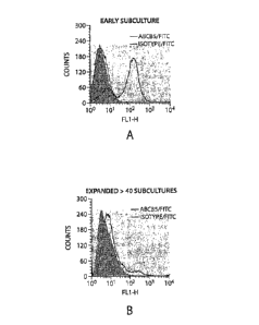
Figure 1. Graphs depicting single-color flow cytometry analyses of surface
ABCB5 expression of murine Balb/c skin-derived cultures in early subcultures
(A) and
after >40 subcultures (B), light gray line: ABCB5; shaded: isotype control;
dark black line:
unstained.
Figure 2. A bar graph depicting the expression pattern of MSC markers on
unseparated (solid bars) versus ABCB5" (dotted bars) or ABCB5 + (striped bars)
dermal MSC
determined by dual-color flow cytometry.
Figure 3. Kaplan-Meier graphs depicting graft survival in (A) donor-derived
MSC-treated B6 recipients of Balb/c donor hearts, and (B) with and without
concurrent
CA 02685492 2009-10-28
WO 2007/143139 PCT/US2007/013022
- 6 -
blockade of CD4OL, using the anti-CD4OL naAb, MR1. (C) Kaplan-Meier graphs
depicting graft survival in third-party derived MSC-treated B6 recipients of
C3H donor
hearts, and in (D) recipient-derived MSC-treated Balb/c recipients of B6 donor
hearts.
Statistical differences as indicated by P values for each strain combination
were assessed
using the log-rank test.
Figure 4. Dot plots depicting dual color flow cytometry analysis of murine
Balb/c skin-derived cultures for ABCB5 expression (FITC, FL1 fluorescence) and
for
the PD-1, PD-L1, and PD-L2 markers (PE, FL2 fluorescence). ABCB5+ cells
coexpressing the respective surface markers are found in the top right
quadrant of each
fluorescence plot.
Figure 5. Bar graphs depicting the expression of PD-1, PD-L1, and PD-L2 on
fully-mismatched recipient CD4+, CD8+, and CD11c+ cells in vivo 7 days after
allo-
transplantation of 3x10⁶ MSC, determined by flow cytometry. Untreated controls
(solid
bars), MSC-treatment (dotted bars), splenocyte treatment (striped bars); NS:
not
significant; p-value(s) indicate statistically significant changes
Figure 6. Bar graph depicting CD40 expression (% positivity, mean SEM)
determined by dual color flow cytometry on CD1 1c+ APC splenocyte subsets
derived
from spleens of either MSC-treated (7 days post i.v. injection of 3x10⁶ ABCB5+
dermal .
MSC) (gray bar, labeled MSC Treatm.) or untreated control animals (black bar,
labeled
No Treatm.). B and C. Graphs depicting 3H-thymidine uptake of T cells purified
from
ABCB5+ dermal MSC-treated (7 days post i.v. injection of 3x10⁶ ABCB5+ dermal
MSC)
or untreated C57BL6 mice, upon allostimulation for 120 hours in standard one-
way
mixed lymphocyte reactions (MLR) with irradiated (1750 rad) naïve Balb/c (B)
or
C3H/HeJ (C) splenocytes. Illustrated are mean values SEM plotted against
increasing
stimulator to responder ratios. D. Bar graph depicting 3H-thyrnidine uptake of
T cells
derived from spleens of either MSC-treated (7 days post i.v. injection of
3x10⁶ ABCB5+
dermal MSC) (gray bar, labeled MSC Treatm) or untreated (black bar, labeled No
Treatm.) C57BL6 mice upon mitogen (ConA)-stimulation for 72 hours. Illustrated
are
mean values SEM.
=
CA 02685492 2009-10-28
WO 2007/143139
PCT/US2007/013022
-.7-.
DETAILED DESCRIPTION
The present invention is based in part upon the development of methods for
isolating a subpopulation of dermal mesenchymal stem cells that are
characterized by the
expression of the P-glycoprotein ABCB5 on their cell surface and by the
expression of
the immune regulator PD-1. It has been found that ABCB5+ dermal mesenchymal
stem
cells markedly prolong heterotopic murine cardiac allograft survival in vivo,
and, with
concurrent CD4O-CD4OL positive costimulatory pathway blockade, induce long-
term
allograft survival. The exact mechanism responsible for reducing immune
rejection of
transplanted organs has not been determined with certainty but it appears that
the dermal
stem cells express PD-1, a factor believed to inhibit T lymphocyte activation.
The
results obtained suggest that ABCB54- dermal mesenchymal stem cells are more
effective
as in vivo modulators of allograft rejection than bone marrow ¨mesenchymal
stem cells
(BM-MSC), which to date have been shown to induce only modest prolongation of
allograft survival (Bartholomew, et al., Exp. Hematol 30:42 (2002)) However,
dermal
mesenchymal stem cells may be otherwise comparable to BM-MSC in that they
display a
similar differentiation capacity and show a.nearly identical profile with
respect to other
surface markers (Fernandes, et al., Nat. Cell Biol. 6:1082 (2004); Shih, et
al., Stem Cells
23:1012 (2005)). The potential promise of clonal dermal mesenchymal stem cells
for
use in cell-based immunomodulatory therapeutic strategies in
allotransplantation and the
other diseases described herein is underscored by the additional advantage of
easy
accessibility of skin as a tissue source for stem cell isolation. The dermal
mesenchymal
stem cells described herein are easily isolated and expanded. ABCB5+ dermal
mesenchymal stem cells can be purified, cloned, propagated and expanded among
clonally-derived differentiating cultures for greater than 50 passages.
Antigen-dependent T cell activation requires two distinct signals: on antigen
encounter, naïve T cells receive signal 1 through T cell receptor engagement
with the
MHC-plus antigenic peptide complex, and signal 2 through positive
costimulatory
pathways leading to full activation. Negative T cell costimulatory signals, on
the other
hand, function to down-regulate immune responses (Rothstein, et al., ImmunoL
Rev.
196:85 (2003)). PD-1, a constituent of the novel PD-1-(PD-Ll/PD-L2) negative
costimulatory pathway (Carter, et al, Eur. J ImmunoL 32:634 (2002); Freeman,
et al,
Exp. Med. 192:1027 (2000); Ito, et al., I ImmunoL /74:6648 (2005)), has
already been
CA 02685492 2009-10-28
WO 2007/143139
PCT/US2007/013022
- 8 -
shown to be expressed on BM-MSC and to inhibit lymphocyte activation in vitro
(Augello, et al., Eur. J. Immunol. 35:1482 (2005)). While not being held to
any
particular theory, it is believed that ABCB5 + dermal mesenchymal stem cells
may
similarly function to down-regulate in vivo alloimmune responses via PD1-(PD-
L1/PD-
L2)-mediated negative costimulatory signaling, and that allografted ABCB5 +
dermal
mesenchymal stem cells may therefore synergize with CD4O-CD4OL costimulatory
blockade to further suppress allograft rejection compared to either therapy
alone.
"ABCB5 positive dermal mesenchymal stem cells" as used herein refers to cells
of the skin having the capacity to self-renew and to differentiate into mature
cells of
multiple adult cell lineages such as bone, fat and cartilage. These cells are
characterized
by the expression of ABCB5 on the cell surface. In culture, mesenchymal stem
cells
may be guided to differentiate into bone, fat, cartilage, or muscle cells
using specific
media. (Hirschi ICK and, Goodell MA. Gene Ther. 2002; 9: 648-652. Pittenger
MF, et
al., Science. 1999; 284: 143-147. Schwartz RE, et al., J Clin Invest. 2002;
109: 1291-
1302. Hirschi K and Goodell M. Di(erentiation. 2001; 68: 186-192.)
Mesenchymal stem cells have been shown to exert immunoregulatory functions:
For example, adult BM-MSC can inhibit T cell proliferation to cognate antigen,
alloantigen and mitogen in vitro and attenuate graft-versus-host disease
(GVHD),
allograft rejection and cell-mediated autoimmunity in vivo. mesenchymal stem
cells
express MHC class I antigens and can be induced to express MHC class II
molecules by
exposure to interferon-y, which indicates an ability to provide signal 1 in a
proinflammatory environment. While mesenchymal stem cells do not express the
positive costimulatory pathway members CD80, CD86, CD40, or CD4OL to provide
signal 2, they can express PD-1, a constituent of the novel PD-1-(PD-L1/PD-L2)
negative costimulatory pathway, which, upon engagement to its ligands on
target
immune-competent cells, may be responsible for mesenchymal stem cells-mediated
lymphocyte activation in vitro. These findings raise the possibility that
allogeneic or
autologous mesenchymal stem cells might exert their immunoregulatory effects
at sites
of inflammation via provision of inhibitory costimulatory signals to antigen-
reactive T
cells, because such signals can be provided in cis or trans leading to T-cell
inactivation.
In addition, mesenchymal stem cells might exert immunoregulatory effects and
retain
imrnunoprivilege in the inflammatory environment via secretion of soluble
CA 02685492 2009-10-28
WO 2007/143139
PCT/US2007/013022
- 9 -
immunoregulatory mediators: Members of the TGF-13 superfamily, which are
produced
by mesenchymal stem cells, can suppress T cell-mediated antigen responses in
vitro, and
production of bone morpho genetic protein 2 (BMP-2) by mesenchymal stem cells
might
mediate immunosuppression via the generation of CD8+ TREGs. Therefore, several
distinct mechanisms by which mesenchymal stem cells modulate immune response
activation are likely operative, including induction of T and B cell anergy,
inhibition of
APC maturation as evidenced by CD40 down-regulation, and generation of TREGs.
The ABCB5 positive dermal mesenchymal stem cells can be obtained from skin.
The skin may be derived from any subject having skin, but in some embodiments
is
preferably human skin. The skin may be derived from a subject of any age but
in some
embodiments is preferably adult skin, rather than adolescent or infant skin.
The ABCB5 positive dermal mesenchymal stem cells may be derived from a
subject by isolating a sample of skin and then purifying the ABCB5 positive
dermal
mesenchymal stem cells. It will be apparent to those of ordinary skill in the
art that skin
can be enriched for cells having ABCB5 in a number of ways. For example, the
cells
= can be selected for, via binding of the ABCB5 on the cell surface
molecules with
antibodies or other binding molecules. Examples of methods are set forth in
the
examples below. Skin samples can be obtained directly from a donor or
retrieved from
cryopreservative storage. The dermal mesenchymal stem cells may, for instance,
be
isolated using antibodies against ABCB5 and maintained in culture using
standard
methodology or frozen, e.g., in liquid nitrogen, for later use.
To study the immunomodulatory properties of murine ABCB5 + dermal
mesenchymal stem cells, a protocol may be used for isolating, cloning,
propagating, and
expanding this stem cell population in vitro under defined medium conditions
such as
those in the examples below. Briefly, murine skin was harvested from adult
Balb/c or
C57BL/6 strain mice, dissected into small pieces and dissociated with
collagenase,
followed by isolation of ABCB5 + cells using anti-ABCB5 mAb, goat anti-mouse
Ig-
coated magnetic microbeads and MiniMACS separation columns, and subsequent
cell
cloning by limiting dilution. Surface expression of murine ABCB5 was
determined in
clonally-derived successive cell passages using immunofluorescence staining
with anti-
ABCB5 mAb and flow cytometry.
CA 02685492 2009-10-28
WO 2007/143139 PCT/US2007/013022
- 10 -
The present invention contemplates any suitable method of employing
monoclonal antibodies to separate mesenchymal stem cells from other cells.
Accordingly, included in the present invention is a method of producing a
population of
mesenchymal stem cells comprising the steps of providing a cell suspension of
skin
containing mesenchymal stem cells; contacting the cell suspension with one or
a
combination of monoclonal antibodies which recognize an epitope, including
ABCB5,
on the mesenchymal stem cells; and separating and recovering from the cell
suspension
the cells bound by the monoclonal antibodies. The monoclonal antibodies may be
linked
to a solid-phase and utilized to capture mesenchymal stem cells fromskin
samples. The
bound cells may then be separated from the solid phase by known methods
depending on
the nature of the antibody and solid phase.
Monoclonal based systems appropriate for preparing the desired cell population
include magnetic bead/paramagnetic particle column utilizing antibodies for
either
positive or negative selection; separation based on biotin or streptavidin
affinity; and
high speed flow cytometric sorting of immunofluorescent-stained mesenchymal
stem
cells mixed in a suspension of other cells. Thus, the method of the present
invention
includes the isolation of a population of mesenchymal stem cells and
enhancement using
monoclonal antibodies raised against surface antigen ABCB5.
The ABCB5 positive dermal mesenchymal stem cells are preferably isolated. An
"isolated ABCB5 positive dermal mesenchymal stem cell" as used here in refers
to a
preparation of cells that are placed into conditions other than their natural
environment.
The term "isolated" does not preclude the later use of these cells thereafter
in
combinations or mixtures with other cells or in an in vivo environment.
The ABCB5 positive dermal mesenchymal stem cells may be prepared as
substantially pure preparations. The term "substantially pure" means that a
preparation
is substantially free of skin cells other than ABCB5 positive stem cells. For
example, the
ABCB5 cells should constitute at least 70 percent of the total cells present
with greater
percentages, e.g., at least 85, 90, 95 or 99 percent, being preferred. The
cells may be
packaged in a finished pharmaceutical container such as an injection vial,
ampoule, or
infusion bag along with any other components that may be desired, e.g., agents
for
preserving cells, or reducing bacterial growth. The composition should be in
unit dosage
form.
CA 02685492 2009-10-28
WO 2007/143139
PCT/US2007/013022
-11 -
The ABCB5 positive dermal mesenchymal stem cells are useful for treating
immune mediated diseases. Immune mediated diseases are diseases associated
with a
detrimental immune response, i.e., one that damages tissue. These diseases
include but
are not limited to transplantation, autoimm.une disease, cardiovascular
disease, liver
disease, kidney disease and neurodegenerative disease.
It has been discovered that mesenchymal stem cells can be used in
transplantation
to ameliorate a response by the immune system such that an immune response to
an
antigen(s) will be reduced or eliminated. Transplantation is the act or
process of
transplanting a tissue or an organ from one body or body part to another. The
mesenchymal stem cells may be autologous to the host (obtained from the same
host) or
non-autologous such as cells that are allogeneic or syngeneic to the host. Non-
autologous cells are derived from someone other than the patient or the donor
of the
organ. Alternatively the mesenchymal stem cells can be obtained from a source
that is
xenogeneic to the host.
Allogeneic refers to cells that are genetically different although belonging
to or
obtained from the same species as the host or donor. Thus, an allogeneic human
mesenchymal stem cell is a mesenchymal stem cell obtained from a human other
than
the intended recipient of the mesenchymal stem cells or the organ donor.
Syngeneic
refers to cells that are genetically identical or closely related and
immunologically
compatible to the host or donor, i.e.., from individuals or tissues that have
identical
genotypes. Xenogeneic refers to cells derived or obtained from an organism of
a
different species than the host or donor.
Thus, the mesenchymal stem cells are used to suppress or ameliorate an immune
response to a transplant (tissue, organ, cells, etc.) by administering to the
transplant
recipient mesenchymal stem cells in an amount effective to suppress or
ameliorate an
immune response against the transplant.
Accordingly, the methods may be achieved by contacting the recipient of donor
tissue with mesenchymal stem cells. The mesenchymal stem cells can be
administered
to the recipient before or at the same time as the transplant or subsequent to
the =
transplant. When administering the stem cells prior to the transplant,
typically stem cells
should be administered up to 14 days and preferably up to 7 days prior to
surgery.
Administration may be repeated on a regular basis thereafter (e.g., once a
week).
CA 02685492 2009-10-28
WO 2007/143139 PCT/US2007/013022
- 12 -
The mesenchymal stem cells can also be administered to the recipient as part
of
the transplant. For instance, the mesenchymal stem cells may be perfused into
the organ
or tissue before transplantation. Alternatively the tissue may be transplanted
and then
treated during the surgery.
Treatment of a patient who has received a transplant, in order to reduce the
severity of or eliminate a rejection episode against the transplant may also
be achieved
by administering to the recipient of donor tissue mesenchymal stem cells after
the donor
tissue has been transplanted into the recipient.
Reducing an immune response by donor tissue, organ or cells against a
recipient,
i.e. graft versus host response may be accomplished by treating the donor
tissue, organ or
cells with mesenchymal stem cells ex vivo prior to transplantation of the
tissue, organ or
cells into the recipient. The mesenchymal stem cells reduce the responsiveness
of T cells '
in the transplant that may be subsequently activated against recipient antigen
presenting
cells such that the transplant may be introduced into the recipient's (host's)
body without
the occurrence of, or with a reduction in, an adverse response of the
transplant to the
host. Thus, what is known as "graft versus host" disease may be averted.
The mesenchymal stem cells can be obtained from the recipient or donor, for
example, prior to the transplant. The mesenchymal stem cells can be isolated
and stored
frozen until needed. The mesenchymal stem cells may also be culture-expanded
to
desired amounts and stored until needed. Alternatively they may be obtained
immediately before use.
The mesenchymal stem cells are administered to the recipient in an amount
effective to reduce or eliminate an ongoing adverse immune response caused by
the
donor transplant against the host. The presentation of the mesenchymal stem
cells to the
host undergoing an adverse immune response caused by a transplant inhibits the
ongoing
response and prevents restimulation of the T cells thereby reducing or
eliminating an
adverse response by activated T cells to host tissue.
As part of a transplantation procedure the mesenchymal stem cells may also be
modified to express a molecule to enhance the protective effect, such as a
molecule that
induces cell death. As described in more detail below, the dermal mesenchymal
stem
cells can be engineered to produce proteins using exogenously added nucleic
acids. For
instance, the mesenchymal stem cells can be used to deliver to the immune
system a
CA 02685492 2009-10-28
WO 2007/143139
PCT/US2007/013022
- 13 -
molecule that induces apoptosis of activated T cells carrying a receptor for
the molecule.
This results in the deletion of activated T lymphocytes and in the suppression
of an
unwanted immune response to a transplant. Thus, dermal mesenchymal stem cells
may
be modified to express a cell death molecule. In preferred embodiments of the
methods
described herein, the mesenchymal stem cells express the cell death molecule
Fos ligand
or TRAIL ligand.
In all cases an effective dose of cells (i.e., a number sufficient to prolong
allograft
survival should be given to a patient). The number of cells administered
should
generally be in the range of 1 x 10⁷ ¨ lx 1010 and, in most cases should be
between 1 x
108 and 5 x 10⁹. Actual dosages and dosing schedules will be determined on a
case by
case basis by the attending physician using methods that are standard in the
art of clinical
medicine and taking into account factors such as the patient's age, weight,
and physical
condition. In cases where a patient is exhibiting signs of transplant
rejection, dosages
and/or frequency of administration may be increased. The cells will usually be
administered by intravenous injection or infusion although methods of
implanting cells,
e.g. near the site of organ implantation, may be used as well.
The mesenchymal stem cells may be administered to a transplant patient either
as the sole immunomodulator or as part of a treatment plan that includes other
immunomodulators as well. For example, patients may also be given: monoclonal
antibodies or other compounds that block the interaction between CD40 and
CD4OL;
inhibitors of lymphocyte activation and subsequent proliferation such as
cyclosporine,
tacrolimus and rapamycin; or with immunosuppressors that act by other
mechanisms
such as methotrexate, azathioprine, cyclophosphamide, or anti-inflammatory
compounds
(e.g., adrenocortical steroids such as dexamethasone and prednisolone).
The dermal mesenchymal stem cells of the invention are also useful for
treating
and preventing autoimmune disease. Autoimmune disease is a class of diseases
in which
an subject's own antibodies react with host tissue or in which immune effector
T cells
are autoreactive to endogenous self peptides and cause destruction of tissue.
Thus an
immune response is mounted against a subject's own antigens, referred to as
self
antigens. Autoimmune diseases include but are not limited to rheumatoid
arthritis,
Crohn's disease, multiple sclerosis, systemic lupus erythematosus (SLE),
autoimmune
encephalomyelitis, myasthenia gravis (MG), Hashimoto's thyroiditis,
Goodpasture's
CA 02685492 2009-10-28
WO 2007/143139 PCT/US2007/013022
- 14 -
syndrome, pemphigus (e.g., pemphigus vulgaris), Grave's disease, autoimmune
hemolytic anemia, autoimmune thrombocytopenic purpura, scleroderma with anti-
collagen antibodies, mixed connective tissue disease, polymyositis, pernicious
anemia,
idiopathic Addison's disease, autoimmune-associated infertility,
glomerulonephritis
(e.g., crescentic glomerulonephritis, proliferative glomerulonephritis),
bullous
pemphigoid, Sjogren's syndrome, insulin resistance, and autoimmune diabetes
mellitus.
A "self-antigen" as used herein refers to an antigen of a normal host tissue.
Normal host
tissue does not include cancer cells.
An example of autoimmune disease is anti-glomerular basement membrane
(GBM) disease. GBM disease results from an autoimmune response directed
against the
noncollagenous domain 1 of the 3 chain of type IV collagen (3(IV)NC1) and
causes a
rapidly progressive glomerulonephritis (GN) and ultimately renal failure in
afflicted
patients. As described in the examples below the effectiveness of dermal
mesenchymal
stem cells in a model of GBM has been demonstrated. Autoreactive antibodies
recognizing 3(IV)NC1 are considered hallmark of the disease. In addition,
3(IV)NC1-
autoreactive T helper (Th)l-mediated cellular immunity has been implicated in
its
pathogenesis. Anti-GBM disease can be induced experimentally in susceptible
mouse
strains by immunization with antigen preparations containing recombinant
3(IV)NCI
(r3(IV)NC1), providing for a valuable disease model system to study responses
to
therapeutic immunomodulation. Antigen-dependent T cell activation and
resultant
production of interleukin 2 (IL-2) requires two distinct signals: On antigen
encounter,
naive T cells receive signal 1 through the T cell receptor engagement with the
Major
Histocompatibility Complex (MHC)-plus antigenic peptide complex on antigen
presenting cells (APCs), and signal 2 through positive costimulatory pathways
leading to
full activation. The critical role of one such positive costimulatory pathway,
the
interaction of APC-expressed CD40 with its Th ligand CD4OL, for disease
development
in experimental anti-GBM autoimmune GN has recently been demonstrated, and
CD40-
CD4OL pathway blockade has been found to prevent the development of autoimmune
autoimmune GN. Negative T cell costimulatory signals, on the other hand,
function to
down-regulate immune responses. Regulatory T cells (TREGs) and soluble
cytokine
mediators, such as interleukin 10 and members of the transforming growth
factor 13
(TGF- 13) family, can also attenuate T cell activation and immune effector
responses.
CA 02685492 2009-10-28
WO 2007/143139 PCT/US2007/013022
- 15 -
Another autoimmune disease is Crohn's disease. Clinical trials for the
treatment
of Crohn's disease using mesenchymal stem cells have been conducted. Crohn's
disease
is a chronic condition associated with inflammation of the bowels and
gastrointestinal
tract. Based on the conducted trials the use of mesenchymal stem cells for the
treatment
of Crohn's disease appears promising.
When used in the treatment of an autoimmune disease, the ABCB5 positive
dermal mesenchymal stem cells will preferably be administered by intravenous
injection
and an effective dose will be the amount needed to slow disease progression or
alleviate
one or more symptoms associated with the disease. For example, in the case of
relapsing
multiple sclerosis, an effective dose should be at least the amount needed to
reduce the
frequency or severity of attacks. In the case of rheumatoid arthritis, an
effective amount
would be at least the number of cells needed to reduce the pain and
inflammation
experienced by patients. A single unit dose of cells should typically be
between 1 x 10⁷
and 1 x 101 cells and dosing should be repeated at regular intervals (e.g.,
weekly,
monthly etc.) as determined to be appropriate by the attending physician.
The ABCB5 positive dermal mesenchymal stem cells are also useful in the
treatment of liver disease. Liver disease includes disease such as hepatitis
which result
in damage to liver tissue. More generally, the ABCB5 positive dermal
mesenchymal
stem cells of the present invention can be used for the treatment of hepatic
diseases,
disorders or conditions including but not limited to: alcoholic liver disease,
hepatitis (A,
B, C, D, etc.), focal liver lesions, primary hepatocellular carcinoma, large
cystic lesions
of the liver, focal nodular hyperplasia granulomatous liver disease, hepatic
granulomas,
hemochromatosis such as hereditary hemochromatosis, iron overload syndromes,
acute
fatty liver, hyperemesis gravidarum, intercurrent liver disease during
pregnancy,
intrahepatic cholestasis, liver failure, fulminant hepatic failure, jaundice
or asymptomatic
hyperbilirubinemia, injury to hepatocytes, Crigler-Najjar syndrome, Wilson's
disease,
alpha-l-antitrypsin deficiency, Gilbert's syndrome, hyperbilirubinemia,
nonalcoholic
steatohepatitis, porphyrias, noncirrhotic portal hypertension, noncirrhotic
portal
hypertension, portal fibrosis, schistosomiasis, primary biliary cirrhosis,
Budd-Chiari
syndrom, hepatic veno-occlusive disease following bone marrow transplantation,
etc.
Stress on the body can trigger adult stem cells to change into specialized
cells
that migrate to the damaged area and help repair the injury. For example, a
damaged
CA 02685492 2009-10-28
WO 2007/143139
PCT/US2007/013022
- 16 -
liver can send signals to stern cells which respond by creating liver cells
for the damaged
liver. (Journal of Clinical Investigation 2003 July 15;112 (2):160-169)
In some embodiments, the invention is directed to treating a neurodegenerative
disease, with dermal mesenchymal stem cells. In some cases, the invention
contemplates
the treatment of subjects having neurodegenerative disease, or an injury to
nerve cells
which may lead to neuro-degeneration. Neuronal cells are predominantly
categorized
based on their local/regional synaptic connections (e.g., local circuit
intemeurons vs.
longrange projection neurons) and receptor sets, and associated second
messenger
systems. Neuronal cells include both central nervous system (CNS) neurons and
peripheral nervous system (PNS) neurons. There are many different neuronal
cell types.
Examples include, but are not limited to, sensory and sympathetic neurons,
cholinergic
neurons, dorsal root ganglion neurons, proprioceptive neurons (in the
trigeminal
mesencephalic nucleus), ciliary ganglion neurons (in the parasympathetic
nervous
system), etc. A person of ordinary skill in the art will be able to easily
identify neuronal
cells and distinguish them from non-neuronal cells such as glial cells,
typically utilizing
cell-morphological characteristics, expression of cell-specific markers,
secretion of
certain molecules, etc.
"Neurodegenerative disorder" or "neurodegenerative disease" is defined herein
as
a disorder in which progressive loss of neurons occurs either in the
peripheral nervous
system or in the central nervous system. Non-limiting examples of
neurodegenerative
disorders include: (i) chronic neurodegenerative diseases such as familial and
sporadic
amyotrophic lateral sclerosis (FALS and ALS, respectively), familial and
sporadic
Parkinson's disease, Huntington's disease, familial and sporadic. Alzheimer's
disease,
multiple sclerosis, olivopontocerebellar atrophy, multiple system atrophy,
progressive
supranuclear palsy, diffuse Lewy body disease, corticodentatonigral
degeneration,
progressive familial myoclonic epilepsy, strionigral degeneration, torsion
dystonia,
familial tremor, Down's Syndrome, Gilles de la Tourette syndrome, Hallervorden-
Spatz
disease, diabetic peripheral neuropathy, dementia pugilistica, AIDS Dementia,
age
related dementia, age associated memory impairment, and amyloidosis-related
neurodegenerative diseases such as those caused by the prion protein (PrP)
which is
associated with transmissible spongiform encephalopathy (Creutzfeldt-Jakob
disease,
CA 02685492 2009-10-28
WO 2007/143139
PCT/US2007/013022
- 17 -
Gerstmann-Straussler-Scheinker syndrome, scrapie, and kuru), and those caused
by
excess cystatin C accumulation (hereditary cystatin C angiopathy); and (ii)
acute
neurodegenerative disorders such as traumatic brain injury (e.g., surgery-
related brain
injury), cerebral edema, peripheral nerve damage, spinal cord injury, Leigh's
disease,
Guillain-Barre syndrome, lysosomal storage disorders such as lipofuscinosis,
Alper's
disease, vertigo as result of CNS degeneration; pathologies arising with
chronic alcohol
or drug abuse including, for example, the degeneration of neurons in locus
coeruleus and
cerebellum; pathologies arising with aging including degeneration of
cerebellar neurons
and cortical neurons leading to cognitive and motor impairments; and
pathologies arising
with chronic amphetamine abuse.including degeneration of basal ganglia neurons
leading to motor impairments; pathological changes resulting from focal trauma
such as
stroke, focal ischemia, vascular insufficiency, hypoxic-ischemic
encephalopathy,
hyperglycemia, hypoglycemia or direct trauma; pathologies arising as a
negative side-
effect of therapeutic drugs and treatments (e.g.,.degeneration of cingulate
and entorhinal
cortex neurons in response to anticonvulsant doses of antagonists of the NMDA
class of
glutamate receptor), and Wernicke-Korsakoff s related dementia.
Neurodegenerative
diseases affecting sensory neurons include Friedreich's ataxia, diabetes,
peripheral
neuropathy, and retinal neuronal degeneration. Neurodegenerative diseases of
limbic
and cortical systems include cerebral amyloidosis, Pick's atrophy, and Retts
syndrome.
The foregoing examples are not meant to be comprehensive but serve merely as
an
illustration of the term "neurodegenerative disorder or "neurodegenerative
disease".
Most of the chronic neurodegenerative diseases are typified by onset during
the
middle adult years and lead to rapid degeneration of specific subsets of
neurons within
the neural system, ultimately resulting in premature death. Compositions
comprising
dermal mesenchymal stem cells may be administered to a subject to treat
neurodegenerative disease alone or in combination with the administration of
other
therapeutic compounds for the treatment or prevention of these disorders or
diseases.
Many of these drugs are known in the art. For example, antiparkinsonian agents
include
but are not limited to Benztropine Mesylate; Biperiden; Biperiden
Hydrochloride;
Biperiden Lactate; Cannantadine; Ciladopa Hydrochloride; Dopamantine;
Ethopropazine
Hydrochloride; Lazabemide; Levodopa; Lometraline Hydrochloride; Mofegiline
Hydrochloride; Naxagolide Hydrochloride; Pareptide Sulfate; Procyclidine
CA 02685492 2009-10-28
WO 2007/143139
PCT/US2007/013022
- 18 -
Hydrochloride; Quinelorane Hydrochloride; Ropinirole Hydrochloride; Selegiline
Hydrochloride; Tolcapone; Trihexyphenidyl Hydrochloride. Drugs for the
treatment of
amyotrophic lateral sclerosis include but are not limited to Riluzole. Drugs
for the
treatment of Paget's disease include but are not limited to Tiludronate
Disoditun.
The utility of adult stem cells in the treatment of neurodegenerative disease
has
been described. It has been demonstrated that mesenchymal stem cells can
change into
neuron-like cells in mice that have experienced strokes. Journal of Cell
Transplantation
Vol. 12, pp. 201-213, 2003. Additionally, stem cells derived from bone marrow
developed into neural cells that hold promise to treat patients with
Parkinson's disease,
amyotrophic lateral sclerosis (ALS), and spinal cord injuries.
The methods of the invnetion are also useful in the treatment of disorders
associated with kedney disease. Mesenchymal stem cells previously injected
into
kidneys have been demonstrated to result in an almost immediate improvement in
kidney
function and cell renewal. Resnick, Mayer, Stem Cells Brings Fast Direct
Improvement,
Without Differentiation, in Acute Renal Failure, EurekAlert!, August 15, 2005.
Thus,
the dermal mesenchymal stem cells of the invention may be administered to a
subject
having kidney disease alone or in combination with other therapeutics or
procedures,
such as dialysis, to improve kidney function and cell renewal.
Other diseases which may be treated according to the methods of the invention
include diseases of the cornea and lung. Therapeis based on the adminstration
of
mesenchymal stem cells in these tissues have demonstrated positive results.
For
instance, human mesenchymal stem cells have been used to reconstruct damaged
corneas. Ma Y et al, Stem Cells, August 18, 2005. Additionally stem cells
derived from
bone marrow were found to be important for lung repair and protection against
lung
injury. Rojas, Mauricio, et al., American Journal of Respiratory Cell and
Molecular
Biology, Vol. 33, pp. 145-152, May 12, 2005. Thus the dermal mesenchymal stem
cells
of the invention may also be used in the repair of corneal tissue or lung
tissue.
Mesenchymal stem cells from sources such as bone marrow have also been used
in therapies for the treatment of cardiovascular disease. Bone marrow stem
cells can
help repair damaged heart muscle by helping the heart develop new, functional
tissue.
Goodell MA, Jackson KA, Majka SM, Mi T, Wang H, Pocius J, Hartley CJ, Majesky
MW, Entman ML, Michael LH, Hirschi KK. Stem cell plasticity in muscle and bone
CA 02685492 2009-10-28
WO 2007/143139 PCT/US2007/013022
- 19 -
marrow. Ann N Y Acad Sci. 2001 hm;938:208-18. Bone marrow stem cells placed in
damaged heartsafter myocaridal infarction improved the hearts' pumping ability
by 80%.
Nature Medicine Journal September 2003 vol. 9 no. 9: 1195-1201.
Cardiovascular disease refers to a class of diseases that involve the heart
and/or
blood vessels. While the term technically refers to diseases that affects the
the heart
and/or blood vessels, other organs such as, for example, the lungs, and joints
might be
affected or involved in the disease. Examples of cardiovasular diseases
include, but are
not limited to athersclerosis, arteriosclerosis, aneurysms, angina, chronic
stable angina
pectoris, unstable angina pectoris, myocardial ischemia (MI), acute coronary
syndrome,
coronary artery disease, stroke, coronary re-stenosis, coronary stent re-
stenosis, coronary
stent re-thrombosis, revascularization, post myocardial infarction (MI)
remodeling (e.g.,
post MI remodeling of the left ventricle), post MI left ventricular
hypertrophy,
angioplasty, transient ischemic attack, pulmonary embolism, vascular
occlusion, venous
thrombosis, arrhythmias, cardiomyopathies, congestive heart failure,
congenital heart
disease, myocarditis, valve disease, dialated cardiomyopathy, diastolic
dysfunction,
endocarditis, rheumatic fever, hypertension (high blood pressure),
hypertrophic
cardiomyopathy, anneurysms, and mitral valve prolapse.
Atherosclerosis is a disease of large and medium-sized muscular arteries and
is
characterized by endothelial dysfunction, vascular inflammation, and the
buildup of
lipids, cholesterol, calcium, and/or cellular debris within the intimal layer
of the blood
vessel wall. This buildup results in plaque (atheromatous plaque) formation,
vascular
remodeling, acute and chronic lumina' obstruction, abnormalities of blood
flow, and
diminished oxygen supply to target organs.
Atherosclerosis may cause two main problems First, the atheromatous plaques
may lead to plaque ruptures and stenosis (narrowing) of the artery and,
therefore, an
insufficient blood supply to the organ it feeds. Alternatively, an aneurysm
results. These
complications are chronic, slowly progressing and cumulative. Most commonly,
plaque(s) suddenly ruptures ("vulnerable plaque") causing the formation of a
thrombus
that will rapidly slow or stop blood flow (e.g., for a few minutes) leading to
death of the
tissues fed by the artery. This event is called an infarction. One of the most
common
recognized scenarios is called coronary thrombosis of a coronary artery
causing
myocardial infarction (MI) (commonly known as a heart attack). Another common
CA 02685492 2009-10-28
WO 2007/143139 PCT/US2007/013022
- 20 -
scenario in very advanced disease is claudication from insufficient blood
supply to the
legs, typically due to a combination of both stenosis and aneurysmal segments
narrowed
with clots. Since atherosclerosis is a body wide process, similar events also
occur in the
arteries to the brain, intestines, kidneys, legs, etc.
Atherosclerosis may begin in adolescence, and is usually found in most major
arteries, yet is asymptomatic and not detected by most diagnostic methods
during life. It
most commonly becomes seriously symptomatic when interfering with the coronary
=
circulation supplying the heart or cerebral circulation supplying the brain,
and is
considered the most important underlying cause of strokes, heart attacks,
various heart
diseases including congestive heart failure and most cardiovascular diseases
in general.
Though any artery in the body can be involved, usually only severe narrowing
or
obstruction of some arteries, those that supply more critically-important
organs are
recognized. Obstruction of arteries supplying the heart muscle result in a
heart attack.
Obstruction of arteries supplying the brain result in a stroke. Atheromatous
palque(s) in
the arm or leg arteries producing decreased blood flow cause peripheral artery
occlusive
disease (PAOD)
Cardiac stress testing is one of the most commonly performed non-invasive
testing method for blood flow limitation. It generally detects lumen narrowing
of--75%
or greater. Areas of severe steno sis detectable by angiography, and to a
lesser extent
"stress testing" have long been the focus of human diagnostic techniques for
cardiovascular disease, in general. Most severe events occur in locations with
heavy
plaque. Plaque rupture can lead to artery lumen occlusion within seconds to
minutes,
and potential permanent tissue damage and sometimes sudden death.
Various anatomic, physiological and behavioral risk factors for
atherosclerosis
are known. These risk factors include advanced age, male gender, diabetes,
dyslipidemia
(elevated serum cholesterol or triglyceride levels), high serum concentration
of low
density lipoprotein (LDL, "bad cholesterol"), Lipoprotein(a) (a variant of
LDL), and / or
very low density lipoprotein (VLDL) particles, low serum concentration of
functioning
high density lipoprotein (HDL, "good cholesterol") particles, tobacco smoking,
hypertension, obesity (e.g., central obesity, also referred to as abdominal or
male-type
obesity) , family history of cardiovascular diease (eg. coronary heart disease
or stroke),
elevated levels of inflammatory markers (e.g., C-reactive protein (CRP or hs-
CRP),
CA 02685492 2009-10-28
WO 2007/143139
PCT/US2007/013022
- 21 -
sCD4OL, sICA.M, etc.), elevated serum levels of homocysteine, elevated serum
levels of
uric acid, and elevated serum fibrinogen concentrations.
The term myocardial infarction (MI) is derived from myocardium (the heart
muscle) and infarction (tissue death due to oxygen starvation). MI is a
disease state that
occurs when the blood supply to a part of the heart is interrupted. Acute MI
(A.MI) is a
type of acute coronary syndrome, which is most frequently (but not always) a
manifestation of coronary artery disease. The most common triggering event is
the
disruption of an atherosclerotic plaque in an epicardial coronary artery,
which leads to a
clotting cascade, sometimes resulting in total occlusion of the artery. The
resulting
ischemia or oxygen shortage causes damage and potential death of heart tissue.
Important risk factors for MI or AlVII include a previous history of vascular
disease such as atherosclerotic coronary heart disease and/or angina, a
previous heart
attack or stroke, any previous episodes of abnormal heart rhythms or syncope,
older ag
(e.g., men over 40 and women over 50), tobacco smoking, excessive alcohol
consumption, high triglyceride levels, high LDL ("Low-density lipoprotein")
and low
HDL ("High density lipoprotein"), diabetes, hypertension, obesity, and stress.
Symptoms of of MI or AMI include chest pain, shortness of breath, nausea,
vomiting, palpitations, sweating, and anxiety or a feeling of impending doom.
Subjects
frequently feel suddenly ill. Approximately one third of all myocardial
infarctions are
silent, without chest pain or other symptoms.
A subject suspected of having a MI receives a number of diagnostic tests, such
as
an electrocardiogram (ECG, EKG), a chest X-ray and blood tests to detect
elevated
creatine kinase (CK) or troponin levels (markers released by damaged tissues,
especially
the myocardium). A coronary angiogram allows to visualize narrowings or
obstructions
on the heart vessels.
Myocardial infarction causes irreversible loss of heart muscle cells leading
to a
thin fibrotic scar that cannot contribute to heart function. Stem cell therapy
provides a
possible approach to the treatment of heart failure after myocardial
infarction as well as
atherosclerosis associated with remodeling. The basic concept of stem cell
therapy is to
increase the number of functional heart muscle cells by injecting immature
heart muscle
cells directly into the wall of the damaged heart. Myocardial infarction leads
to the loss
of cardiomyocytes, followed by pathological remodeling and progression to
heart failure.
CA 02685492 2009-10-28
WO 2007/143139 PCT/US2007/013022
-22 -
One goal of stem cell therapy is to replace caxdiomyocytes lost after
ischemia, induce
revascularization of the injured region. Another goal is to prevent
deleterious
pathological remodeling after myocardial infarction and associated with
atheroschlerosis.
Autologous or allogeneic mesenchymal stem cells are considered to be one of
the
potential cell sources for stem cell therapy. Thus, the dermal mesenchymal
stem cells of
the invention may be used in the treatment of cardiovascular diseases.
Another use for the dermal mesenchymal stem cells of the invention is in
tissue
regeneration. In this aspect of the invention, the ABCB5 positive cells are
used to
generate tissue by induction of differentiation. Isolated and purified
mesenchymal stem
lo cells can be grown in an undifferentiated state through mitotic
expansion in a specific
medium. These cells can then be harvested and activated to differentiate into
bone,
cartilage, and various other types of connective tissue by a number of
factors, including
mechanical, cellular, and biochemical stimuli. Human mesenchymal stem cells
possess
the potential to differentiate into cells such as osteoblasts and
chondrocytes, which
produce a wide variety of mesenchymal tissue cells, as well as tendon,
ligament and
dermis, and this potential is retained after isolation and for several
population expansions
in culture. Thus, by being able to isolate, purify, greatly multiply, and then
activate
mesenchymal stem cells to differentiate into the specific types of mesenchymal
cells
desired, such as skeletal and connective tissues such as bone, cartilage,
tendon, ligament,
muscle, and adipose, a process exists for treating skeletal and other
connective tissue
disorders. The term connective tissue is used herein to include the tissues of
the body =
that support the specialized elements, and includes bone, cartilage, ligament,
tendon,
stroma, muscle and adipose tissue.
The methods and devices of the invention utilize isolated dermal mesenchymal
progenitor cells which, under certain conditions, can be induced to
differentiate into and
produce different types of desired connective tissue, such as into bone or
cartilage
forming cells.
In another aspect, the present invention relates to a method for repairing
connective tissue damage. The method comprises the steps of applying the
dermal
mesenchymal stem to an area of connective tissue damage under conditions
suitable for
differentiating the cells into the type of connective tissue necessary for
repair.
CA 02685492 2009-10-28
WO 2007/143139 PCT/US2007/013022
- 23 -
The term "connective tissue defects" refers to defects that include any damage
or
irregularity compared to normal connective tissue which may occur due to
trauma,
disease, age, birth defect, surgical intervention, etc. Connective tissue
defects also refers
to non-damaged areas in which bone formation is solely desired, for example,
for
cosmetic augmentation.
The dermal mesenchymal stem cells may be administered directly to a subject by
any known mode of administration or may be seeded onto a matrix or implant.
Matrices
or implants include polymeric matrices such as fibrous or hydrogel based
devices. Two
types of matrices are commonly used to support the mesenchymal stem cells as
they
differentiate into cartilage or bone. One form of matrix is a polymeric mesh
or sponge;
the other is a polymeric hydrogel. The matrix may be biodegradeable or
nonbiodegradeable. The term biodegradable, as used herein, means a polymer
that
dissolves or degrades within a period that is acceptable in the desired
application, less
than about six months and most preferably less than about twelve weeks, once
exposed
to a physiological solution of pH 6-8 having a temperature of between about 25
C and
38 C. A matrix may be biodegradable over a time period, for instance, of less
than a
year, more preferably less than six months, most preferably over two to ten
weeks.
Fibrous matrices can be manufactured or constructed using commercially
available materials. The matrices are typically formed of a natural or a
synthetic
polymer. Biodegradable polymers are preferred, so that the newly formed
cartilage can
maintain itself and function normally under the load-bearing present at
synovial joints.
Polymers that degrade within one to twenty-four weeks are preferable.
Synthetic
polymers are preferred because their degradation rate can be more accurately
determined
and they have more lot to lot consistency and less immunogenicity than natural
polymers. Natural polymers that can be used include proteins such as collagen,
albumin,
and fibrin; and polysaccharides such as alginate and polymers of hyaluronic
acid.
Synthetic polymers include both biodegradable and non-biodegradable polymers.
Examples of biodegradable polymers include polymers of hydroxy acids such as
polylactic acid (PLA), polyglycolic. acid (PGA), and polylactic acid-glycolic
acid
(PLGA), polyorthoesters, polyanhydrides, polyphosphazenes, and combinations
thereof.
õ Non-biodegradable polymers include polyacrylates, polymethacrylates,
ethylene vinyl
=
CA 02685492 2009-10-28
WO 2007/143139 PCT/US2007/013022
-24 -
acetate, and polyvinyl alcohols. These should be avoided since their presence
in the
cartilage will inevitably lead to mechanical damage and breakdown of the
cartilage.
In the preferred embodiment, the polymers form fibers which are intertwined,
woven, or meshed to form a matrix having an interstitial spacing of between
100 and 300
microns. Meshes of polyglycolic acid that can be used can be obtained from
surgical
supply companies such as Ethicon, N.J. Sponges can also be used. As used
herein, the
term "fibrous" refers to either a intertwined, woven or meshed matrix or a
sponge matrix.
The matrix is preferably shaped to fill the defect. In most cases this can be
achieved by trimming the polymer fibers with scissors or a knife;
alternatively, the
matrix can be cast from a polymer solution formed by heating or dissolution in
a volatile
solvent.
The mesenchymal stem cells are seeded onto the matrix by application of a cell
suspension to the matrix. This can be accomplished by soaking the matrix in a
cell
culture container, or injection or other direct application of the cells to
the matrix.
The matrix seeded with cells is implanted at the site of the defect using
standard
surgical techniques. The matrix can be seeded and cultured in vitro prior to
implantation,
seeded and immediately implanted, or implanted and then seeded with cells. In
the
preferred embodiment, cells are seeded onto and into the matrix and cultured
in vitro for
between approximately sixteen hours and two weeks. It is only critical that
the cells be
attached to the matrix. Two weeks is a preferred time for culture of the
cells, although it
can be longer. Cell density at the time of seeding or implantation should be
approximately 25,000 cells/mm3.
Polymers that can form ionic or covalently crosslinked hydrogels which are
malleable are used to encapsulate cells. For example, a hydrogel is produced
by cross-
linking the anionic salt of polymer such as alginic acid, a carbohydrate
polymer isolated
from seaweed, with calcium cations, whose strength increases with either
increasing
concentrations of calcium ions or alginate. The alginate solution is mixed
with the cells
to be implanted to form an alginate suspension. Then the suspension is
injected directly
into a patient prior to hardening of the suspension. The suspension then
hardens over a
short period of time due to the presence in vivo of physiological
concentrations of
calcium ions.
CA 02685492 2009-10-28
WO 2007/143139 PCT/US2007/013022
- 25 -
The polymerie material which is mixed with cells for implantation into the
body
should form a hydrogel. A hydrogel is defined as a substance formed when an
organic
polymer (natural or synthetic) is cross-linked via covalent, ionic, or
hydrogen bonds to
create a three-dimensional open-lattice structure which entraps water
molecules to form a
gel. Examples of materials which can be used to form a hydrogel include
polysaccharides such as alginate, polyphosphazines, and polyacrylates, which
are
crosslinked ionically, or block copolymers such as Pluronics.TM. or
Tetronics.TM.,
polyethylene oxide-polypropylene glycol block copolymers which are crosslinked
by
temperature or pH, respectively. Other materials include proteins such as
fibrin,
polymers such as polyvinylpyrrolidone, hyaluronic acid and collagen.
In general, these polymers are at least partially soluble in aqueous
solutions, such
as water, buffered salt solutions, or aqueous alcohol solutions, that have
charged side
-groups, or a monovalent ionic salt thereof. Examples of polymers with acidic
side groups
that can be reacted with cations are poly(phosphazenes), poly(acrylic acids),
poly(methacrylic acids), copolymers of acrylic acid and methacrylic acid,
poly(vinyl
acetate), and sulfonated polymers, such as sulfonated polystyrene. Copolymers
having
acidic side groups formed by reaction of acrylic or methacrylic acid and vinyl
ether
monomers or polymers can also be used. Examples of acidic groups are
carboxylic acid
groups, sulfonic acid groups, halogenated (preferably fluorinated) alcohol
groups,
phenolic OH groups, and acidic OH groups.
Examples of polymers with basic side groups that can be reacted with anions
are
poly(vinyl amines), poly(vinyl pyridine), poly(vinyl imidazole), and some
imino
substituted polyphosphazenes. The ammonium or quaternary salt of the polymers
can
also be formed from the backbone nitrogens or pendant imino groups. Examples
of basic
side groups are amino and imino groups.
Alginate can be ionically cross-linked with divalent cations, in water, at
room
temperature, to form a hydrogel matrix. Due to these mild conditions, alginate
has been
the most commonly used polymer for hybridoma cell encapsulation, as described,
for
example, in U.S. Pat. No. 4,352,883 to Lim. In the Lim process, an aqueous
solution
containing the biological materials to be encapsulated is suspended in a
solution of a
water soluble polymer, the suspension is formed into droplets which are
configured into
discrete microcapsules by contact with multivalent cations, then the surface
of the
CA 02685492 2009-10-28
WO 2007/143139 PCT/US2007/013022
- 26 -
microcapsules is crosslinked with polyamino acids to form a semipermeable
membrane
around the encapsulated materials.
Polyphosphazenes are polymers with backbones consisting of nitrogen and
phosphorous separated by alternating single and double bonds. The
polyphosphazenes
suitable for cross-linking have a majority of side chain groups which are
acidic and
capable of forming salt bridges with di- or trivalent cations. Examples of
preferred acidic
side groups are carboxylic acid groups and sulfonic acid groups. Polymers can
be
synthesized that degrade by hydrolysis by incorporating monomers having
imidazole,
amino acid ester, or glycerol side groups. For example, a polyanionic
poly[bis(carboxylatophenoxy)Jphosphazene (PCPP) can be synthesized, which is
cross-
linked with dissolved multivalent cations in aqueous media at room temperature
or
below to form hydrogel matrices.
The water soluble polymer with charged side groups is ionically crosslinked by
reacting the polymer with an aqueous solution containing multivalent ions of
the
opposite charge, either multivalent cations if the polymer has acidic side
groups or
multivalent anions if the polymer has basic side groups. The preferred cations
for cross-
linking of the polymers with acidic side groups to form a hydrogel are
divalent and
trivalent cations such as copper, calcium, aluminum, magnesium, strontium,
barium,
zinc, and tin, although di-, tri- or tetra-functional organic cations such as
alkylammonium
salts. Aqueous solutions of the salts of these cations are added to the
polymers to form
soft, highly swollen hydrogels and membranes. The higher the concentration of
cation, or
the higher the valence, the greater the degree of cross-linking of the
polymer.
Concentrations from as low as 0.005 M have been demonstrated to cross-link the
polymer. Higher concentrations are limited by the solubility of the salt.
Preferably the polymer is dissolved in an aqueous solution, preferably a 0.1 M
potassium phosphate solution, at physiological pH, to a concentration forming
a
polymeric hydrogel, for example, for alginate, of between 0.5 to 2% by weight,
preferably 1%, alginate. The isolated cells are suspended in the polymer
solution to a
concentration of between 1 and 10 million cells/ml, most preferably between 10
and 20
million cells/ml.
In an embodiment, the cells are mixed with the hydrogel solution and injected
directly into a site where it is desired to implant the cells, prior to
hardening of the
CA 02685492 2009-10-28
WO 2007/143139 PCT/US2007/013022
-27 -
hydrogel. However, the matrix may also be molded and implanted in one or more
different areas of the body to suit a particular application. This application
is particularly
relevant where a specific structural design is desired or where the area into
which the
cells are to be implanted lacks specific structure or support to facilitate
growth and
proliferation of the cells.
The site, or sites, where cells are to be implanted is determined based on
individual need, as is the requisite number of cells. One could also apply an
external
mold to shape the injected solution. Additionally, by controlling the rate of
polymerization, it is possible to mold the cell-hydrogel injected implant
Alternatively, the mixture can be injected into a mold, the hydrogel allowed
to
harden, then the material implanted.
The suspension can be injected via a syringe and needle directly into a
specific
area wherever a bulking agent is desired, especially soft tissue defects. The
suspension
can also be injected as a bulking agent for hard tissue defects, such as bone
or cartilage
defects, either congenital or acquired disease states, or secondary to trauma,
burns, or the
like. An example of this would be an injection into the area surrounding the
skull where
a bony deformity exists secondary to trauma. The injection in these instances
can be
=
made directly into the needed area with the use of a needle and syringe under
local or
general anesthesia.
The dermal mesenchymal stem cells may be modified to express proteins which
are also useful in the therapeutic indications, as described in more detail
below. For
example, the cells may include a nucleic acid that produces at least one
bioactive factor
which further induces or accelerates the differentiation of the mesenchymal
stem cells
into a differentiated lineage. In the instance that bone is being formed, the
bioactive
factor may be a member of the TGF-beta superfamily comprising various tissue
growth
factors, particularly bone morphogenic proteins, such as at least one selected
from the
group consisting of BMP-2, BMP-3, BMP-4, BMP-6 and BMP-7.
The cells of the invention may be useful in a method for inducing T cell
anergy,
in vitro. Induction of T cell anergy involves culturing the dermal mesenchymal
stem
cells in the presence of antigen under conditions sufficient to induce the
formation of T
cells and/or T cell progenitors and to inhibit activation of the formed T
cells and/or T cell
progenitors. Anergy is defined as an unresponsive state of T cells (that is
they fail to
CA 02685492 2009-10-28
WO 2007/143139 PCT/US2007/013022
-28 -
produce IL-2 on restimulation, or proliferate when restimulated)(Zamoyska R,
Curr
Opin Immunol, 1998, 10(1):82-87; Van Parijs L, et al., Science, 1998,
280(5360:243.-
248; Schwartz RH, Curr Opin Immunol, 1997, 9(3):351-357; Immunol Rev, 1993,
133:151-76). Anergy can be measured by taking the treated T cells and
restimulating
them with antigen in the presence of APCs. If the cells are anergic they will
not respond
to antigen at an appropriate concentration in the context of APCs.
As used herein, a subject is a human, non-human primate, cow, horse, pig,
sheep,
goat, dog, cat or rodent. Human dermal mesenchymal stem cells and human
subjects are
particularly important embodiments.
In a still further aspect of the invention described herein, mesenchymal stem
cells
may be genetically engineered (or transduced or transfected) with a gene of
interest. The
transduced cells can be administered to a patient in need thereof,.for example
to treat
genetic disorders or diseases.
The ABCB5 positive dermal mesenchymal stem cells, and progeny thereof, can
be genetically altered. Genetic alteration of a ABCB5 positive dermal
mesenchymal stem
cell includes all transient and stable changes of the cellular genetic
material which are
created by the addition of exogenous genetic material. Examples of genetic
alterations
include any gene therapy procedure, such as introduction of a functional gene
to replace
a mutated or nonexpressed gene, introduction of a vector that encodes a
dominant
negative gene product, introduction ofa vector engineered to express a
ribozyme and
introduction of a gene that encodes a therapeutic gene product. Natural
genetic changes
such as the spontaneous rearrangement of a T cell receptor gene without the
introduction
of any agents are not included in this concept. Exogenous genetic material
includes
nucleic acids or oligonucleotides, either natural or synthetic, that are
introduced into the
dermal mesenchymal stem cells. The exogenous genetic material may be a copy of
that
which is naturally present in the cells, or it may not be naturally found in
the cells. It
typically is at least a portion of a naturally occurring gene which has been
placed under
operable control of a promoter in a vector construct.
Various techniques may be employed for introducing nucleic acids into cells.
Such techniques include transfection of nucleic acid-CaPO4 precipitates,
transfection of
nucleic acids associated with DEAE, transfection with a retrovirus including
the nucleic
acid of interest, liposome mediated transfection, and the like. For certain
uses, it is
CA 02685492 2009-10-28
WO 2007/143139 PCT/US2007/013022
- 29 -
preferred to target the nucleic acid to particular cells. In such instances, a
vehicle used
for delivering a nucleic acid according to the invention into a cell (e.g., a
retrovirus, or
other virus; a liposome) can have a targeting molecule attached thereto. For
example, a
molecule such as an antibody specific for a surface membrane protein on the
target cell
or a ligand for a receptor on the target cell can be bound to or incorporated
within the
nucleic acid delivery vehicle. For example, where liposomes are employed to
deliver the
nucleic acids of the invention, proteins which bind to a surface membrane
protein
associated with endocytosis may be incorporated into the liposome formulation
for
targeting and/or to facilitate uptake. Such proteins include proteins or
fragments thereof
tropic for a particular cell type, antibodies for proteins which undergo
internalization in
cycling, proteins that target intracellular localization and enhance
intracellular half life,
and the like. Polymeric delivery systems also have been used successfully to
deliver
nucleic acids into cells, as is known by those skilled in the art. Such
systems even permit
oral delivery of nucleic acids.
One method of introducing exogenous genetic material into the dermal
mesenchymal stem cells is by transducing the cells using replication-
deficient
retroviruses. Replication-deficient retroviruses are capable of directing
synthesis of all
virion proteins, but are incapable of making infectious particles.
Accordingly, these
genetically altered retroviral vectors have general utility for high-
efficiency transduction
of genes in cultured cells. Retroviruses have been used extensively for
transferring
genetic material into cells. Standard protocols for producing replication-
deficient
retroviruses (including the steps of incorporation of exogenous genetic
material into a
plasmid, transfection of a packaging cell line with plasmid, production of
recombinant
retroviruses by the packaging cell line, collection of viral particles from
tissue culture
media, and infection of the target cells with the viral particles) are
provided in the art.
The major advantage of using retroviruses is that the viruses insert
efficiently a
single copy of the gene encoding the therapeutic agent into the host cell
genome, thereby
permitting the exogenous genetic material to be passed on to the progeny of
the cell
when it divides. In addition, gene promoter sequences in the LTR region have
been
reported to enhance expression of an inserted coding sequence in a variety of
cell types.
The major disadvantages of using a retrovirus expression vector are (1)
insertional
mutagenesis, i.e., the insertion of the therapeutic gene into an undesirable
position in the
CA 02685492 2009-10-28
WO 2007/143139 PCT/US2007/013022
- 30 -
target cell genome which, for example, leads to unregulated cell growth and
(2) the need
for target cell proliferation in order for the therapeutic gene carried by the
vector to be
integrated into the target genome. Despite these apparent limitations,
delivery of a
therapeutically effective amount of a therapeutic agent via a retrovirus can
be efficacious
if the efficiency of transduction is high and/or the number of target cells
available for
transduction is high.
Yet another viral candidate useful as an expression vector for transformation
of
dermal mesenchymal stem cells is the adenovirus, a double-stranded DNA virus.
Like
the retrovirus, the adenovirus genome is adaptable for use as an expression
vector for
gene transduction, i.e., by removing the genetic information that controls
production of
the virus itself. Because the adenovirus functions usually in an
extrachromosomal
fashion, the recombinant adenovirus does not have the theoretical problem of
insertional
mutagenesis. On the other hand, adenoviral transformation of a target dermal
mesenchymal stem cell may not result in stable transduction. However, more
recently it
has been reported that certain adenoviral sequences confer intrachromosomal
integration
specificity to carrier sequences, and thus result in a stable transduction of
the exogenous
genetic material.
Thus, as will be apparent to one of ordinary skill in the art, a variety of
suitable
vectors are available for transferring exogenous genetic material into dermal
mesenchymal stem cells. The selection of an appropriate vector to deliver a
therapeutic
agent for a particular condition amenable to gene replacement therapy and the
optimization of the conditions for insertion of the selected expression vector
into the cell,
are within the scope of one of ordinary skill in the art without the need for
undue
experimentation. The promoter characteristically has a specific nucleotide
sequence
necessary to initiate transcription. Optionally, the exogenous genetic
material further
includes additional sequences (i.e., 'enhancers) required to obtain the
desired gene
transcription activity. For the purpose of this discussion an "enhancer" is
simply any
nontranslated DNA sequence which works contiguous with the coding sequence (in
cis)
to change the basal transcription level dictated by the promoter. Preferably,
the
exogenous genetic material is introduced into the dermal mesenchymal stem cell
genome
immediately downstream from the promoter so that the promoter and coding
sequence
are operatively linked so as to permit transcription of the coding sequence. A
preferred
CA 02685492 2009-10-28
WO 2007/143139 PCT/US2007/013022
- 31 -
retroviral expression vector includes an exogenous promoter element to control
transcription of the inserted exogenous gene. Such exogenous promoters include
both
constitutive and inducible promoters.
Naturally-occurring constitutive promoters control the expression of essential
cell
functions. As a result, a gene under the control of a constitutive promoter is
expressed
under all conditions of cell growth. Exemplary constitutive promoters include
the
promoters for the following genes which encode certain constitutive or
"housekeeping"
functions: hypoxanthine phosphoribosyl transferase (HPRT), dihydrofolate
reductase =
(DHFR) (Scharfmann et al., Proc. Natl. Acad. Sci. USA 88:4626-4630 (1991)),
adenosine deaminase, phosphoglycerol kinase (PGK), pyruvate kinase,
phosphoglycerol
mutase, the actin promoter (Lai etal., Proc. Natl. Acad. Sci. USA 86: 10006-
10010
(1989)), and other constitutive promoters known to those of skill in the art.
In addition,
many viral promoters function constitutively in eukaryotic cells. These
include: the early
and late promoters of SV40; the long terminal repeats (LTRS) of Moloney
Leukemia
Virus and other retroviruses; and the thymidine kinase promoter of Herpes
Simplex
Virus, among many others. Accordingly, any of the above-referenced
constitutive
promoters can be used to control transcription of a heterologous gene insert.
Genes that are under the control of inducible promoters are expressed only or
to a
greater degree, in the presence of an inducing agent, (e.g., transcription
under control of
the metallothionein promoter is greatly increased in presence of certain metal
ions).
Inducible promoters include responsive elements (REs) which stimulate
transcription
when their inducing factors are bound. For example, there are REs for serum
factors,
steroid hormones, retinoic acid and cyclic AMP. Promoters containing a
particular RE
can be chosen in order to obtain an inducible response and in some cases, the
RE itself
may be attached to a different promoter, thereby conferring inducibility to
the
recombinant gene. Thus, by selecting the appropriate promoter (constitutive
versus
inducible; strong versus weak), it is possible to control both the existence
and level of
expression of a therapeutic agent in the genetically modified dermal
mesenchymal stem
cell. Selection and optimization of these factors for delivery of a
therapeutically
effective dose of a particular therapeutic agent is deemed to be within the
scope of one of
ordinary skill in the art without undue experimentation, taking into account
the above-
disclosed factors and the clinical profile of the subject.
CA 02685492 2009-10-28
WO 2007/143139 PCT/US2007/013022
- 32 -
In addition to at least one promoter and at least one heterologous nucleic
acid
encoding the therapeutic agent, the expression vector preferably includes a
selection
gene, for example, a neomycin resistance gene, for facilitating selection of
dermal
mesenchymal stem cells that have been transfected or transduced with the
expression
. vector. Alternatively, the dermal mesenchymal stem cells are transfected
with two or
= more expression vectors, at least one vector containing the gene(s)
encoding the
therapeutic agent(s), the other vector containing a selection gene. The
selection of a
suitable promoter, enhancer, selection gene and/or signal sequence is deemed
to be
within the scope of one of ordinary skill in the art without undue
experimentation.
The selection and optimization of a particular expression vector for
expressing a
= . specific gene product in an isolated dermal mesenchymal stem cell is
accomplished by
obtaining the gene, preferably with one or more appropriate control regions
(e.g.,
promoter, insertion sequence); preparing a vector construct comprising the
vector into
which is inserted the gene; transfecting or transducing cultured dermal
mesenchymal
stem cells in vitro with the vector construct; and determining whether the
gene product is
present in the cultured cells.
Thus, the present invention makes it possible to genetically engineer dermal
mesenchymal stem cells in such a manner that they produce polypeptides,
hormones and
proteins not normally produced in human stem cells in biologically significant
amounts
or produced in small amounts but in situations in which overproduction would
lead to a
therapeutic benefit. These products would then be secreted into the
bloodstream or other
areas of the body, such as the central nervous system. The human stem cells
formed in
this way can serve as a continuous drug delivery systems to replace present
regimens,
which require periodic administration (by ingestion, injection, depot infusion
etc.) of the
needed substance. This invention has applicability in providing hormones,
enzymes and
drugs to humans, in need of such substances. It is particularly valuable in
providing such
substances, such as hormones (e.g., parathyroid hormone, insulin), which are
needed in
sustained doses for extended periods of time.
For example, it can be used to provide continuous delivery of insulin, and, as
a
result, there would be no need for daily injections of insulin. Genetically
engineered
human mesenchymal stem cells can also be used for the production of clotting
factors
CA 02685492 2009-10-28
WO 2007/143139 PCT/US2007/013022
- 33 -
such as Factor VIII, or for continuous delivery of dystrophin to muscle cells
for muscular
dystrophy.
Incorporation of genetic material of interest into dermal mesenchymal stem
cells
is particularly valuable in the treatment of inherited and acquired disease.
In the case of
inherited diseases, this approach is used to provide genetically modified
human
mesenchymal stem cells and other cells which can be used as a metabolic sink.
That is,
such dermal mesenchymal stem cells would serve to degrade a potentially toxic
substance. For example, this could be used in treating disorders of amino acid
catabolism including the hyperphenylalaninemias, due to a defect in
phenylalanine
hydroxylase; the homocysteinemias, due to a defect in cystathionine beta-
synthase.
The dermal mesenchymal stem cells may further be modified to express a cell
death molecule to enhance the elimination of activated T cells, in the
treatment of organ
or tissue transplantation.. For example, the cell death molecule may be
expressed by the
mesenchymal stem cells which have been engineered to express the exogenous
cell death
molecule. As used herein, a "cell death molecule" is a molecule that interacts
or binds
with its cognate receptor on a stimulated T cell inducing T cell death or
apoptosis. Fas
mediates apoptosis of recently activated T cells which are again exposed to
stimulation
(van Parijs et al., Immunity 4: 321-328 (1996)). Fas is a type I membrane
receptor that
when crosslinked by its cognate ligand induces apoptosis in a wide variety of
cells. The
interaction between the Fas molecule (CD95) on target T cells and its ligand
Fas L on
mesenchymal stem cells results in receptor aggregation, which transduces
signals leading
to apoptosis of the target cell. The Fas system has been shown to be involved
in a
number of cell functions in vivo including negative selection of thymocytes,
maintaining
immune privilege sites within the body, and cytotoxic T-lymphocyte (CTL)-
mediated
cytotoxicity (Green and Ware, Proc Nat! Acad Sci, 94(12):5986-90 (1997)).
Other members of the tumor necrosis factor receptor (TNFR) family have roles
in
programmed cell death. TRAIL ligand, which interacts with its receptor DR4 can
induce
apoptosis in a variety of transformed cell lines (G. Pan etal. Science,
277:815-818
(1997)); and the expression of CD27 and its ligand CD70 (Prasad et al., Proc
Natl Acad
Sci, 94:6346-6351 (1997)) also induces apoptosis. Fas expression is restricted
to
stimulated T cells and sites of immune privilege. TRAIL is detected in many
normal
tissues.
CA 02685492 2009-10-28
WO 2007/143139 PCT/US2007/013022
-34 -
Both Trail-ligand and CD27, but not Fas-ligand, are expressed on unmanipulated
human mesenchymal stem cells. Activated, but not resting, T cells express the
Trail
receptor and CD70. Most of the T cells found in the body are in the resting
state; T cells
are activated when they encounter cells both in the context of MHC and the
appropriate
co-stimulatory molecule such as B7-1 or 1B7-2.
Thus, the engagement of cell death receptors on activated T cells with their
ligands expressed on the mesenchymal stem cells results in T cell death via
apoptosis.
Ligands and their receptors other than those specifically mentioned above,
either present
within the mesenchymal stem cell or introduced into the mesenchymal stem cell
can
perform this function. Therefore, mesenchymal stem cells administered to an
individual
delete activated T cells, reducing the severity or incidence of transplant
rejection disease.
The dose of the dermal mesenchymal stem cells varies within wide limits and
will, of course be fitted to the individual requirements in each particular
case. The
number of cells used will depend on the weight and condition of the recipient
and other
variables known to those of skill in the art.
The cells can be administered by a route which is suitable for the particular
tissue
or organ to be treated. Modes of administration of the mesenchymal stem cell
preparation include but are not limited to systemic intravenous injection and
injection
directly to the intended site of activity. The preparation can be administered
by any
convenient route, for example by infusion or bolus injection and can be
administered
together with other biologically active agents. Administration is preferably
systemic, i.e.,
parenterally, by intravenous injection. In some cases, the dermal mesenchymal
stem
cells are delivered to the site of desired treatment or therapy and can be
targeted to a
particular tissue or organ.
In general, in the case of parenteral administration, it is customary to
administer
from about 0.01 to about 5 million cells per kilogram of recipient body
weight. The
number of cells used will depend on the weight and condition of the recipient,
the
number of or frequency of administrations, and other variables known to those
of skill in
the art. The mesenchymal stem cells can be administered by a route which is
suitable for
the tissue, organ or cells to be transplanted. They can be administered
systemically, i.e.,
parenterally, by intravenous injection or can be targeted to a particular
tissue or organ,
such as bone marrow. The human mesenchymal stem cells can be administered via
a
CA 02685492 2009-10-28
WO 2007/143139
PCT/US2007/013022
- 35 -
subcutaneous implantation of cells or by injection of stem cell into
connective tissue, for
example muscle.
The cells can be suspended in an appropriate diluent, at a concentration of
from
about 0.01 to about 5x10⁶ cells/ml. Suitable excipients for injection
solutions are those
that are biologically and physiologically compatible with the cells and with
the recipient,
such as buffered saline solution or other suitable excipients. The composition
for
administration must be formulated, produced and stored according to standard
methods
complying with proper sterility and stability. Other excipients include water,
isotonic
common salt solutions, alcohols, polyols, glycerine and vegetable oils. The
composition
for administration must be formulated, produced and stored according to
standard
methods complying with proper sterility and stability.
The mesenchymal stem cell can be administered alone, however in a preferred
embodiment, the mesenchymal stem cells are utilized in the form of
pharmaceutical
compositions. Such compositions comprise a therapeutically effective amount of
the
dermal mesenchymal stem cells, and a pharmaceutically acceptable carrier or
excipient.
Such a carrier includes but is not limited to saline, buffered saline,
dextrose, water, and
combinations thereof.
In a preferred embodiment, the mesenchymal stem cell preparation or
composition is formulated in accordance with routine procedures as a
pharmaceutical
composition adapted for intravenous administration to human beings. Typically,
compositions for intravenous administration are solutions in sterile isotonic
aqueous
buffer. Where necessary, the composition may also include a local anesthetic
to
ameliorate any pain at the site of the injection. Generally, the ingredients
are supplied
either separately or mixed together in unit dosage form, for example, as a
cryopreserved
concentrate in a hermetically sealed container such as an ampoule indicating
the quantity
of active agent. Where the composition is to be administered by infusion, it
can be
dispensed with an infusion bottle containing sterile pharmaceutical grade
water or saline.
Where the composition is administered by injection, an ampoule of sterile
water for
injection or saline can be provided so that the ingredients may be mixed prior
to
administration.
The invention also includes prefilled pharmaceutical containers (e.g.,
injection
vials, ampoules, infusion bags etc.) containing a unit dose of cells (i.e.,
the number of
CA 02685492 2009-10-28
WO 2007/143139
PCT/US2007/013022
- 36 -
cells to be given at a single time to a patient). The cells may be suspended
in a solution
(e.g., sterile isotonic saline) together with agents that help in their
preservation (e.g.,
glycerol). Other agents such as antibiotics, pharmaceutically acceptable
salts, buffers
and excipients may also be included. The prefilled pharmaceutical containers
in unit
dose form may be kept refrigerated or stored frozen. They may also be supplied
as part
of a kit that has instructions for the administration of the cells to
transplant patients or
patients with an autoimmune disease.
The present invention also provides any of the above-mentioned compositions in
kits, optionally including instructions for use of the composition for the
treatment of a
condition described herein. That is, the kit can include a description of use
of the
composition for participation in any biological or chemical mechanism
disclosed herein.
The kits can further include a description of activity of the condition in
treating the
pathology, as opposed to the symptoms of the condition. That is, the kit can
include a
description of use of the compositions as discussed herein. The kit also can
include
instructions for use of a combination of two or more compositions of
theinvention, or
instruction for use of a combination of a composition of the invention and one
or more
other compounds indicated for treatment of the diseases. Instructions also may
be
provided for administering the composition by any suitable technique as
previously
described, for example, orally, intravenously, pump or implantable delivery
device, or
via another known route of drug delivery. The kits may also be one or more
reagents
associated with the isolation and purification fo the dermal mesenchymal stem
cells, i.e.
ABCB5 antibodies, and instructions for isolating and/or purifying the cells.
The kits described herein may also contain one or more containers, which may
contain the composition and other ingredients as previously described. The
kits also may
contain instructions for mixing, diluting, and/or administrating the
compositions of the
invention in some cases. The kits also can include other containers with one
or more
solvents, surfactants, preservative and/or diluents (e.g., normal saline (0.9%
NaC1), or
5% dextrose) as well as containers for mixing, diluting or administering the
components
in a sample or to a subject in need of such treatment.
The compositions of the kit may be provided as any suitable form, for example,
as liquid solutions or as dried powders. When the composition provided is a
dry powder,
the composition may be reconstituted by the addition of a suitable solvent,
which may
CA 02685492 2009-10-28
WO 2007/143139 PCT/US2007/013022
- 37 -
also be provided. In embodiments where liquid forms of the composition are
used, the
liquid form may be concentrated or ready to use. The solvent will depend on
the
composition and the mode of use or administration. Suitable solvents for drug
compositions are well known, for example as previously described, and are
available in
the literature. The solvent will depend on the composition and the mode of use
or
administration.
Examples
Example 1:
A. ABCB5 marks murine dermal MSC
Human ABCB5 P-glycoprotein marks MSC phenotype-expressing stem cell
subpopulations in physiological skin and malignant melanomas (Frank, et. at.,
J. Biol.
Chem. 278:47156 (2003); Frank, et. al., Cancer Res. 65:4320 (2005)). Murine
ABCB5,
which was found in transfection experiments to be recognized by the anti-ABCB5
mAb
3C2-1D12 directed against a species-conserved extracellular epitope of the
molecule
(Frank, et. al., J. Biol. Chem. 278:47156 (2003)), marks identical dermal MSC
subpopulations, as determined by HRP-immunoenzymatic staining for ABCB5, when
frozen tissue sections across the anastomosis of human to SCID mouse skin
xenografis
were compared.
B. Cloning and Characterization of ABCB5 Dermal Mesenchymal Stem Cells
A protocol was developed to study the immunomodulatory properties of murine
ABCB5 + dermal MSC. The protocol included isolating, cloning, propagating, and
expanding this stem cell population in vitro under defined, previously
described medium
conditions (Frank, et. at., J. Biol. Chem. 278:47156 (2003)). Briefly, murine
skin was
harvested from adult (6-10 weeks old) 13a1b/c or C57BL/6 strain mice (C57BL/6
(H-2b)
and BALB/c (H-2d) wild-type mice were obtained from Taconic Farms, Germantown,
NY), dissected into small pieces and dissociated with collagenase, followed by
isolation
of ABCB5 + cells using anti-ABCB5 mAb, goat anti-mouse Ig-coated magnetic
microbeads and MiniMACS separation columns, and subsequent cell cloning by
limiting
dilution. Surface expression of murine ABCB5 was determined in clonally-
derived
successive cell passages using immunofluorescence staining with anti-ABCB5 mAb
and
flow cytometry (Fig. 1A, B).
CA 02685492 2009-10-28
WO 2007/143139
PCT/US2007/013022
- 38 -
While ABCB5 was expressed in early passages by the majority of cells (Fig.
1A),
we found ABCB5 expressed by 5-15% of cells in successive single cell-derived
cultures
(Fig. 1B), indicating that ABCB5 + stem cells give rise in culture to more
differentiated,
= ABCB5- progeny. ABCB5 + dermal cells nevertheless maintained constant
relative
abundance ratios vis-à-vis ABCB5- bulk populations during long-term culture
expansion,
demonstrating self-renewal capacity of this cell subset. Further phenotypic
characterization of murine Balb/c single cell-derived cultures revealed
significant
expression of the MSC-associated markers CD29 (98.1+/-0.1%; mean+/-SD), CD44
(95.6+/- 4.7%), CD49e (95.2+/-3.5%), CD166 (14.5+/-2.4%), and CD133 (4.0+/-
1.4%)
among all cells, even in later passages (>30, n=3), with preferential
expression of the
most primitive stem cell markers CD133 and CD166 noted on ABCB5 + compared to
ABCB5- subpopulations (76.4+/-18.5% vs. 0.7+/-0.9% and 70.7+/-17.9% vs. 10.8+1-
2.8%, respectively; mean+/-SD) (Fig. 2). Clonally-derived ABCB5 + dermal MSC
were
found to possess multipotent differentiation potential, further indicative of
their MSC
phenotype, under distinct culture conditions in vitro, with a capacity to
generate myosin
heavy chain-expressing multinucleated myocytes, osteocytes, and adipocytes,
when
stained with appropriate lineage markers (FITC-conjugated anti-myosin heavy
chain
mAb, Alizarin Red S staining, Oil Red staining, respectively).
Example 2:
Immunomodulatory Effect of ABCB5 Positive Dermal MSC
A.. The immunomodulatory function of clonal ABCB5-derived dermal MSC
was studied in vivo, using a murine heterotopic cardiac allotransplantation
model as
previously described (Yamada, et al., J. ImmU nol. 167:140 (2001)). In a fully
mismatched strain combination, treatment of C57BL/6 recipients of Balb/c
cardiac
allografts with donor-type dermal MSC (3x10⁶ cells i.v., day -7) resulted in
significant
prolongation of allograft survival compared to donor-type splenocyte-treated
or untreated
control recipients (median graft survival 29.5 days vs. 10 days (P=0.012) or
7.5 days
(P=0.006), respectively), (Fig.3A) demonstrating the in vivo efficacy of
dermal MSC to
delay graft rejection.
B. Long-term allograft survival >100 days was achieved in n= 4/4 animals when
dermal MSC treatment was combined with CD4OL-directed costimulatory blockade
using the anti-CD4OL mAb MR1 (250 mg/kg i.p. q.o.d. from day 0-10) as
previously
CA 02685492 2009-10-28
WO 2007/143139
PCT/US2007/013022
- 39 -
described (Ozkaynak, et al., .1 Immunol. 169:6546(2002); Kishimoto, et al., J.
Clin.
Invest. 106:63 (2000)) (Fig. 3B). Third-party, Balb/c strain dermal MSC
also
prolonged cardiac allograft survival in C57BL/6 recipients of C3H/HeJ hearts
compared
to untreated controls (median graft survival 28 days vs. 7.5 days, P=0.016)
(Fig. 3C).
Balb/c recipients of C57BL/6 heart allografts treated with recipient-strain
MSC,
however, rejected donor hearts with the same tempo as untreated control mice
(Fig. 3D).
The results suggest that administration of MSC for prolongation of allograft
survival is
most effective under these conditions in the presence of a stem cell-dependent
allogeneic
stimulus. However, this does not rule out activity under more stringent
conditions with
non-allogeneic cells.
Example 3:
ABCB5 dermal MSC Coexpress PD-1 and Upon Allotransplantation Activate
Expression of the PD-1 Ligand PD-L2 on recipient T cells In Vivo
The PD-1-(PD-L1/PD-L2) negative costimulatory pathway has recently been
implicated in BM-MSC-mediated immune regulation in vitro (Augello, et al.,
Eur.
Immunol. 35:1482 (2005)). The mechanism underlying dermal MSC-mediated
prolongation of cardiac allograft survival, was addressed by systematically
analyzing
expression of known costimulatory receptors and their ligands (Rothstein, et
al.,
Immunol. Rev. 196:85 (2003)) on Balb/c ABCB5+-derived dermal MSC.
Out of >20 costimulatory molecules examined, immunofluorescence double
staining and flow cytometry revealed specific coexpression of ABCB5 with the
negative
costimulatory molecule PD-1, (Fig. 4F) but not its ligands, PD-L1 (Fig. 40)
and PD-L2
(Fig. 4H), which are not expressed by dermal MSC, and compared to the controls
(Fig.
4A, E, B-D). No significant expression of positive costimulatory molecules was
detected. Examination of in vivo costimulatory molecule expression on
peripheral
immune cells of C57BL/6 recipients 7 days following i.v. injection of 3x10⁶
allogeneic
Balb/c dermal MSC, before cardiac allotransplantation, revealed that dermal
MSC
treatment had specifically and significantly (P<0.01) activated expression of
the PD-1
ligand PD-L2 on 12.5+1-3.8% of recipient CD4+ T cells (mean+/-SD) and 12.9+/-
2.4% of
recipient CD8+ T cells, but not on recipient CD1le APCs, when compared to
Balb/c
splenocyte-treated or naïve control animals (Fig. 5C). Moreover, recipient T
cell
expression of the PD-1 ligand PD-Li was preserved in dermal MSC-treated
animals,
CA 02685492 2009-10-28
WO 2007/143139
PCT/US2007/013022
-40 -
unlike in allogeneic Balb/c splenocyte-treated mice, where PD-Li expression
was
markedly down-regulated (Fig. 5B). No differential expression pattern of PD-1
on
recipient immune cells was detected (Fig. 5A).
These findings suggest that the immunosuppressive effect of ABCB5 dermal
MSC in allotransplantation is closely tied to specific coexpression of the
negative
costimulator PD-1. Furthermore, dermal MSC-mediated specific induction of PD-
L2
expression on recipient T cells suggest a functional role of these not
previously
recognized cell populations in the prolongation of cardiac allograft survival.
Example 4:
In vivo immunomodulatoty function of ABCB5+ murine dermal MSC.
When 3x10⁶ autologous, clonally-derived murine ABCB5 dermal MSC were
intravenously (i.v.) grafted to C57/BL6 mice, ABCB5+ dermal MSC administration
resulted in a 3.4-fold reduction in surface expression of the costimulatory
pathway
member CD40 on CD11 e APCs isolated 7 days post transplantation from spleens
of
MSC-treated animals compared to CD1le APCs derived from untreated controls
(48.71
11.43% vs. 14.34 4.53%, P < 0.05, mean SEM) (Fig. 6A), indicating that in
vivo
transplantation of ABCB5 -derived dermal MSC can inhibit an APC-expressed
positive
costimulatory signal critically involved in T cell activation. T cells derived
from
autologous MSC-treated animals exhibited significantly impaired proliferation
compared
to those derived from untreated controls, to either allogeneic stimulation in
standard one-
way mixed lymphocyte reactions (MLR) with irradiated naïve Balb/c or C3H/HeJ
splenocytes (inhibition 82% 9% for Balb/c stimulators and 84% 5% for
C3H/HeJ
stimulators at 1:1 stimulator to responder ratios, mean SD, P < 0.001,
respectively)
(Fig. 6B and 6C), or to mitogenic stimulation with ConA (Fig. 6D). These
findings show
that ABCB5+ murine dermal MSC can exert distinct modulatory effects on both
APC
maturation and T cell activation in vivo.
What is claimed is: