Note: Descriptions are shown in the official language in which they were submitted.
CA 02695697 2010-02-05
WO 2009/020744 PCT/US2008/070137
RTX5008
PROTEIN FORMULATIONS COMPRISING GDF-5-IN AQUEOUS ACIDIC SOLUTION
FIELD OF THE INVENTION
The invention relates to liquid compositions of bone morphogenetic proteins
for
improved stability during handling and prolonged storage at reduced
temperatures. More
specifically, the invention relates to biocompatible liquid compositions
comprising GDF-
5 in an acidic solution having a pH of from about 3.0 to about 3.6, having
improved
protein stability during handling and prolonged storage at reduced
temperatures.
BACKGROUND
GDF-5 is a member of the Bone Morphogenetic Proteins (BMP), which is a
subclass of the TGF-13 superfamily of proteins. GDF-5 includes several
variants and
mutants, including mGDF-5 first isolated from the mouse by Lee (US Pat No
5,801,014).
Other variants include MP52, which is the patented name (WO 95/04819) for the
human
form of GDF-5, which is also known as hGDF-5 and also as LAP-4 (Triantfilou,
et al.
Nature Immunology 2, 338-345 (2001)); also CDMP-1, an allelic protein variant
of
hGDF-5 (WO 96/14335); also rhGDF-5, the recombinant human form manufactured in
bacteria (EP 0955313); also rhGDF-5-A1a83, a monomeric variant of rhGDF-5;
also
BMP-14, a collective term for hGDF-5/CDMP-1 like proteins; also Radotermin,
the
international non-proprietary name designated by the World Health
Organization; also
HMW MP52's, high molecular weight protein variants of MP52; also C465A, a
monomeric version wherein the cysteine residue responsible for the
intermolecular cross-
link is substituted with alanine; also other active monomers and single amino
acid
substitution mutants including N445T, L441P, R438L, and R438K. For the
purposes of
this applciation the term "GDF-5" is meant to include all variants and mutants
of the
GDF-5 protein, wherein rhGDF-5 is the exemplary member having 119 amino acids.
1
CA 02695697 2010-02-05
WO 2009/020744 PCT/US2008/070137
RTX5008
All members of the BMP family share common structural features including a
carboxy terminal active domain and share a highly conserved pattern of
cysteine residues
that create three intramolecular disulfide bonds and one intermolecular
disulfide bond.
The active form can be either a disulfide-bonded homodimer of a single family
member
or a heterodimer of two different members (see Massague, et al. Annual Review
of Cell
Biology 6:957 (1990); Sampath, et al. Journal ofBiological Chemistry 265:13198
(1990);
Celeste et al. PNAS 87:9843-47 (1990); U.S. Pat. No. 5,011,691, and U.S. Pat.
No.
5,266,683). The proper folding of the GDF-5 protein and formation of these
disulfide
bonds are essential to biological functioning, and misfolding leads to
inactive aggregates
and cleaved fragments.
The production of BMP's from genetically modified bacteria, and of GDF-5 in
particular, utilizes plasmid vectors to transform E. coli to produce monomer
GDF-5
protein in high yield (see for example Hotten US 6,764,994 and Makishima US
7,235,527). The monomer is obtained from inclusion bodies, purified, and
refolded into
homodimers of GDF-5 protein to produce the biologically active dimer of the
GDF-5
protein. The steps leading to the dimer utilize various pharmaceutically
unacceptable
materials to modify the solubility in order to enable the separation and
purification of the
GDF-5 protein.
The degradation of proteins in general has been well described in the
literature,
but the storage and solubility of bone morphogenetic proteins, particularly
GDF-5 has not
been well described. BMP-2 is readily soluble at concentrations greater than 1
mg/ml
when the pH is below 6, and above pH 6 the solubility can be increased by the
addition of
1 M NaC1, 30% isopropanol, or 0.1 mM heparin (Ruppert, et al. Eur J Biochem
237, 295-
302 (1996). GDF-5 is nearly insoluble in physiological pH ranges and buffers
and is only
soluble in water at extreme pH (Honda, et al. Journal of Bioscience and
Bioengineering
89(6), 582-589 (2000)). GDF-5 is soluble at an alkaline pH of about 9.5 to
12.0, however
proteins degrade quickly under these conditions and thus acidic conditions are
used for
the preparation of GDF-5 protein.
2
CA 02695697 2010-02-05
WO 2009/020744 PCT/US2008/070137
RTX5008
Biocompatible compositions of the GDF-5 protein present great challenges to
obtain reasonable solubility and concurrent stability of the protein. The
current method of
storage for GDF-5 protein utilizes 10 mM HC1 at pH 2 and -80 C for long-term
storage,
but even these conditions provide for some degradation of the protein,
particularly with
repeated freeze-thaw cycles. We performed a trypsin digestion of late eluting
species of
the GDF-5 protein and found non-tryptic peptide fragments using MALDI-TOF
(matrix
assisted laser desorption ionization - time of flight mass spectrometry)
analysis,
indicating acid-catalyzed cleavage of the protein during storage and
subsequent
aggregation of the fragments. We also separately performed sequential freeze-
thaw
cycles and prolonged exposure to elevated temperatures of GDF-5 protein
solutions. Both
of these tests showed degradation of the protein in the current 10 mM HC1
storage
solvent. Thus there is a need for improved compositions for the handling and
storage of
GDF-5 protein solutions.
SUMMARY OF THE INVENTION
The present invention provides compositions for the handling and long-term
storage of GDF-5 solutions at reduced temperatures that provide for improved
stability of
the GDF-5 protein. By increasing the pH of the storage solution from 2 to
about 3, a
significant improvement in the stability of the GDF-5 protein is realized,
without
adversely affecting the solubility of the protein. Suitable solvent systems
include, but are
not limited to hydrochloric acid (HC1), acetic acid, trifluoroacetic acid
(TFA), and
phosphoric acid, in amounts to provide a pH of from about 3.0 to about 3.6.
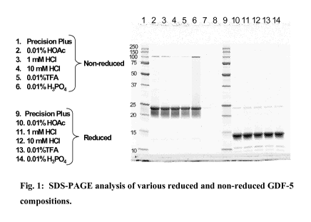
BRIEF DESCRIPTION OF THE DRAWINGS
Figure 1 shows SDS-PAGE analysis of various reduced and non-reduced GDF-5
compositions.
Figure 2 shows circular dichroism analysis of various GDF-5 compositions.
Figure 3 shows circular dichroism analysis of a stock 10 mM HC1 GDF-5 solution
further
diluted with water.
Figure 4a shows the DSC spectra of GDF-5 in 10 mM HC1.
Figure 4b shows the DSC spectra of GDF-5 in 1 mM HC1.
Figure 4c shows the DSC spectra of GDF-5 in 0.01% (v/v) TFA.
3
CA 02695697 2010-02-05
WO 2009/020744 PCT/US2008/070137
RTX5008
Figure 4d shows the DSC spectra of GDF-5 in 0.01% (v/v) phosphoric acid.
Figure 4e shows the DSC spectra of GDF-5 in 0.01% (v/v) acetic acid.
Figure 5a shows an rp-HPLC chromatogram of a GDF-5 reference standard in 10 mM
HCI.
Figure 5b shows an rp-HPLC chromatogram of GDF-5 in 10 mM HC1 after 5 freeze-
thaw
cycles.
Figure 5c shows an rp-HPLC chromatogram of GDF-5 in 50 mM acetic acid after 5
freeze-thaw cycles.
Figure 5d shows an rp-HPLC chromatogram of GDF-5 in 0.01% (v/v) TFA after 5
freeze-thaw cycles.
Figure 5e shows an rp-HPLC chromatogram of GDF-5 in 1 mM HC1 after 5 freeze-
thaw
cycles.
Figure 6 shows the percentage change in peak 1 of the various samples shown in
figures
5b-e.
Figure 7 shows an rp-HPLC chromatogram of GDF-5 in 12 mM HC1 after 6 days at
RT.
Figure 8 shows degradation trends of GDF-5 in different solvents at RT.
Figure 9 shows degradation trends of GDF-5 in different solvents at 2-8 C.
DETAILED DESCRIPTION OF THE INVENTION
We investigated the use of a number of different solvent systems in order to
improve the stability of GDF-5 protein solutions during handling and storage,
and herein
describe useful compositions for working with this protein. Since it's
discovery and the
subsequent development of recombinant human forms, GDF-5 has been stored in a
10
mM HC1 solvent system at -80 C to preserve the protein structure. Partly
because of its
lack of glycosylation, GDF-5 is less soluble than other BMP's, including BMP-
2, for
which the bulk of the scientific literature is directed to. There are few
reports, if any,
available on the solubility and stability of GDF-5. The preparation and
isolation of the
GDF-5 monomer from plasmid transformed bacteria and the subsequent refolding
into
dimer presents a different set of issues and problems than the handling and
storage of the
bioactive dimer. On the other hand, working with the mature dimer GDF-5
protein in
biocompatible compositions presents a different set of problems, and the
literature yields
4
CA 02695697 2010-02-05
WO 2009/020744 PCT/US2008/070137
RTX5008
very little physicochemical information regarding the solubility and stability
of the GDF-
protein.
It is an object of the present invention to provide a composition of GDF-5
protein
5 in a solvent system that provides for improved protein stability during
handling and
storage. It is another object of the present invention to provide a
biocompatible solution
of GDF-5 protein that is stable during prolonged storage at reduced
temperatures. It is
another object of the present invention to provide a biocompatible solution of
GDF-5
protein that is stable during handling at room temperature. It is another
object of the
present invention to provide a method of preserving a solution of GDF-5
protein by
providing a solvent system having a pH of from about 3.0 to about 3.6, wherein
the GDF-
5 protein is stabilized and has reduced susceptibility to acid catalyzed
cleavage while still
maintaining a useful solubility.
For the purposes of this application definitions of the following terms will
be
useful. The term "GDF-5" is meant to include all synonyms, variants and
mutations of
the GDF-5 protein molecule, including, but not limited to GDF-5, mGDF-5, hGDF-
5,
MP-52, LAP-4, radotermin, CDMP-1, C465A, and rhGDF-5, wherein rhGDF-5 is the
exemplary member of the group. The term "GDF-5" is also understood to include
monomeric GDF-5 proteins, which have also been shown to be biologically
active. The
term "room temperature", herein abbreviated as "RT" or "R.T.", is understood
to mean
the ambient temperature of an ordinary office or laboratory, being from about
18 to about
C. The term "bulk", as used herein when referring to "bulk protein" or "bulk
solution"
is understood to mean a solution of GDF-5 in 10 mM HC1 and stored at -80 C
after
25 isolation and purification from the production process, and is equivalent
with the terms
"stock", "stock protein", and "stock solution".
We undertook several studies of bulk GDF-5 solution to determine the extent of
protein degradation and the need for improved solvent systems and conditions
for the
handling and storage of the GDF-5 protein. We performed MALDI-TOF analysis
after
performing a trypsin digestion of the late eluting peak (aggregates) from
extracts of GDF-
5
CA 02695697 2010-02-05
WO 2009/020744 PCT/US2008/070137
RTX5008
protein isolated from HEALOS TM mineralized collagen sponges, which were
loaded
with the GDF-5 protein 10 mM HC1 solution and subsequently lyophilized. We
observed
non-tryptic fragments, indicative of acid-catalyzed cleavage of the GDF-5
protein.
5 In efforts to discover improved compositions for the handling and storage of
GDF-5 we examined the physicochemical properties of the protein in five
different
solvent environments: 10 mM HC1(the current solvent system for bulk protein),
1 mM
HC1, 0.01 %(v/v) acetic acid, 0.01 %(v/v) TFA, and 0.01 %(v/v) phosphoric
acid.
MALDI-TOF analysis of the GDF-5 protein was done at the Mass Spectrometry Core
Facility, Dana-Farber Cancer Institute in Boston, MA. Samples were mixed with
sinapinic acid, spotted and allowed to dry on a stainless steel plate, and
then analyzed on
a Voyager DE-STR mass spectrometer in linear mode (manufactured by Applied
Biosystems, Framingham, MA). The percentage aggregate estimated by peak height
analysis was found to be about 23.5% in 10 mM HC1 as opposed to 8-12% in the
remaining four solvents. In this estimation, we assumed any mass greater than
27 kDa to
be an aggregate. It should be noted that MALDI is not a quantitative
technique, so the
absolute percentage of aggregates in each solvent is only an approximation.
Nevertheless,
the data clearly indicated that there was a greater proportion of aggregates
in 10 mM HC1
than in the other four solvents.
We performed SDS-PAGE analysis of GDF-5 in the same set of solvent systems.
Figure 1 shows the SDS-PAGE analysis of reduced and non-reduced GDF-5 in the
five
different solvent environments (10 mM HC1, 1 mM HC1, 0.01 %(v/v) TFA, 0.01
%(v/v)
acetic acid, and 0.01 % (v/v) phosphoric acid). In the non-reduced gel, a
small amount of
aggregate was observed, while in the reduced gel there was clear indication of
the
presence of low molecular weight species, probably resulting from acid
cleavage. No
significant difference was noted between the migration profiles of GDF-5
reconstituted in
the five different solvent environments.
We also performed far UV circular dichroism (CD) of GDF-5 protein in the same
five solvent environments (10 mM HC1, 1 mM HC1, 0.01 % (v/v) TFA, 0.01 % (v/v)
acetic
6
CA 02695697 2010-02-05
WO 2009/020744 PCT/US2008/070137
RTX5008
acid, and 0.01% (v/v) phosphoric acid). The results are shown in figure 2 as
an overlay
plot, and demonstrate a unique CD spectrum for GDF-5 in 10 mM HC1, distinctly
different from the spectra in the other solvents. No significant difference in
the secondary
structural distribution of GDF-5 was noted when the remaining four solvent
environments were compared to each other. In another experiment, bulk GDF-5
solution
(3.8 mg/mL in 10 mM HC1) was diluted with water to achieve a desired protein
concentration of 0.2 mg/mL while increasing the pH (through dilution), and
then the CD
analysis was done using water as a blank. The spectrum is shown in Figure 3
and clearly
demonstrates a subtle pH-dependent structural change in GDF-5. At pH 3, the
GDF-5
protein becomes relatively more structured, with less random and more Beta
contribution,
than the spectrum at lower pH.
We performed Differential Scanning Calorimetry (DSC) on GDF-5 protein in the
same five solvent environments (10 mM HC1, 1 mM HC1, 0.01 %(v/v) acetic acid,
0.01%
(v/v) TFA, and 0.01% (v/v) phosphoric acid). Figures 4a through 4e show the
DSC
thermal data of the samples after instrument baseline and solvent subtraction
and
concentration normalization. Bulk GDF-5 in 10 mM HC1(figure 4a) shows a weak
thermal transition with Tm < 30 C and also a broad weak transition near 65 C.
The heat
transfer was significantly poor. In contrast, GDF-5 protein dialyzed against 1
mM HC1
(figure 4b), 0.01 %(v/v) TFA (figure 4c), and 0.01 %(v/v) phosphoric acid
(figure 4d),
showed a large transition near 40 C and a smaller endothermic transition near
85 C. In
0.01 %(v/v) acetic acid (figure 4e), the results showed a significant increase
in both
transitions: TMi- 60 C and TM2 - 94 C. The thermodynamic parameters, namely AH
and
AS values were also significantly higher in 0.01 %(v/v) acetic acid. This
result suggests
that the GDF-5 protein's thermal stability is much greater in an acetic acid
environment
or at a higher pH. In an earlier study, we noted that the C465A monomer, which
cannot
form an intermolecular disulfide bridge, did not exhibit the first endotherm
near 40 C,
suggesting that this transition represents disulfide interaction between the
two monomer
units.
7
CA 02695697 2010-02-05
WO 2009/020744 PCT/US2008/070137
RTX5008
In another set of experiments we have shown that even as few as two freeze-
thaw
cycles of GDF-5 in 10 mM HC1 can lead to a substantial increase in fragments
and
degradation products, as shown by rp-HPLC. Figure 5a shows an rp-HPLC
chromatogram of a reference standard of bulk GDF-5, showing good purity and
very little
additional peaks. Figure 5b shows an rp-HPLC chromatogram of bulk GDF-5 after
5
freeze-thaw cycles, showing an increase in the fragments appearing as
additional peaks
(peak 1& peak 2). Figure 5c shows an rp-HPLC chromatogram of GDF-5 in 50 mM
acetic acid after 5 freeze-thaw cycles, showing little, if any, increase in
the fragments
appearing as additional peaks (peak 1& peak 2). Figure 5d shows an rp-HPLC
chromatogram of GDF-5 in 0.01 %(v/v) TFA after 5 freeze-thaw cycles, showing
little, if
any, increase in the fragments appearing as additional peaks (peak 1& peak 2).
Figure 5e
shows an rp-HPLC chromatogram of GDF-5 in 1 mM HC1 after 5 freeze-thaw cycles,
showing little, if any, increase in the fragments appearing as additional
peaks (peak 1&
peak 2).
Figure 6 shows a plot directly comparing only the changes in peak 1, and shows
approximately a 30% increase in the peak 1 of the GDF-5 protein in 10 mM HC1
sample
after only 2 freeze-thaw cycles, whereas the other solvent systems show
minimal changes
to peak 1 after 5 freeze-thaw cycles. After 5 freeze-thaw cycles the percent
change in
peak 1 for the bulk 10 mM HC1 solution was approximately 75%, whereas the
other
solvent systems showed very little change in peak 1.
In another group of experiments we investigated the potential of various
solvent
systems to provide improved stability to liquid GDF-5 protein solutions at
temperatures
of 2-8 C and at room temperature (RT, approximately 25 C). In these
experiments the
stability of GDF-5 protein was evaluated by rp-HPLC in the following solvent
systems:
1.3 mM HC1, 5 mM HC1, 12 mM HC1, 0.01% (v/v) TFA, and 50 mM acetic acid.
Samples of the GDF-5 protein solutions were prepared by dialysis with the
selected
solvents at 2-8 C overnight and transferred as aliquots into small vials at
about 1 mL/vial
and placed accordingly at 2-8 C or at room temperature. At each designated
time point,
one vial from each set was removed and stored at -80 C until the analysis was
8
CA 02695697 2010-02-05
WO 2009/020744 PCT/US2008/070137
RTX5008
performed. The results show that GDF-5 was stable in both 50 mM acetic acid
(pH 3.3)
and 0.0 1%(v/v) TFA (pH 3.3) solutions at room temperature after three days
and in 1.3
mM HC1(pH 3.3) after 2 days, while it was not stable at room temperature in
either 5
mM HC1(pH 2.5) or 12 mM HC1(pH 2.1) after 2 days (see figures 7 and 8).
At 2-8C, the GDF-5 protein was stable for at least 30 days in 50 mM acetic
acid
or 0.01 %(v/v) TFA solution, and stable for at least 6 days in 1.3 mM HC1. In
contrast,
the GDF-5 protein was degraded in 5 mM HC1 and 12 mM HC1 solutions at 2-8 C,
and
formed degradation species after 2 days as evidenced by rp-HPLC (see figure
9).
The following examples are meant only to be illustrative in nature of the
present
invention, and not to be limiting in scope. One skilled in the art would
easily conceive of
other embodiments that would be considered within the scope of the present
invention.
Example 1
Four different solvent systems, 1 mM HC1, 0.01 %(v/v) acetic acid, 0.01 %(v/v)
TFA, and 0.01 % (v/v) phosphoric acid, were tested for their ability to
provide improved
GDF-5 protein stability over the standard 10 mM HC1 solvent system currently
used.
Approximately 1-2 ml of bulk GDF-5 protein (3.8 mg/ml) in 10 mM HC1 was taken
from
a freshly thawed sample and dialyzed for 24 hours at 2-8 C with 3 changes each
of 1 liter
of test solution to produce a GDF-5 protein solution in each of the four
different solvent
systems. The concentration of the dialysates was determined from the
absorbance value
at 280 nm using an extinction coefficient of 1.16 for a 1 mg/ml solution and a
pathlength
of 1 cm. The GDF-5 protein solutions were then analyzed by SDS-PAGE, Circular
Dichroism (CD), Differential Scanning Calorimetry (DSC), and MALDI-TOF.
SDS-PAGE
The GDF-5 protein samples were diluted in Bio-Rad 8-16% gradient gel
appropriate sample buffer (provided by the manufacturer) either with (reduced)
or
without (non-reduced) 50 mM dithiothreitol (DTT). The samples were denatured
by
heating at 90 C for 5 min and then centrifuged briefly at 5000 rpm.
Electrophoresis was
9
CA 02695697 2010-02-05
WO 2009/020744 PCT/US2008/070137
RTX5008
carried out at 200 volts constant for 1 hour on an 8-16% Bio-Rad criterion gel
with lx
tris-glycine-SDS running buffer. Gels were incubated in 100 mL 10% methanol,
7%
acetic acid (Ruby fix / destain solution) for 1 hour on an orbital shaker at
45 rpm. The fix
solution was decanted and 80 mL Sypro-Ruby (Bio-Rad) was added. Gels were
incubated
overnight in the dark on an orbital shaker at 45 rpm. The Sypro-Ruby was
decanted and
100 mL destain solution was added. Gels were incubated for 3 hours on an
orbital shaker
at 45 rpm. Finally, gels were imaged on a Bio-Rad Gel Doc imager.
In the non-reduced gels, a small amount of aggregate was observed, while in
the
reduced gels there was clear indication of the presence of low molecular
weight species,
probably resulting from acid cleavage. No significant difference was noted
between the
migration profiles of GDF-5 reconstituted in the five different solvent
environments.
Circular Dichroism
Circular Dichroism was carried out on an AVIV Mode160DS Circular Dichroism
Spectropolarimeter. For each sample, scans were taken between 190 and 250 nm.
For
each scan, data were collected at 1 nm intervals for 2 sec at each interval.
The scan
temperature was 23 C. The final protein concentration was 0.2 mg/mL. Data
represented
the average of three scans. A buffer blank was also recorded under identical
conditions
and the CD spectrum of the buffer blank was subtracted from that of the
sample. All runs
were made using 0.01 % TFA as a blank. Cuvettes had a path length of 1 mm. The
scans
were normalized using Mean residue weight (a value of 115) and inserting it
into the
equation:
[0] =[0.1 x ReS1d1e] / [conc. (mg/mol) x light path].
The value of [0] was calculated at each wavelength to give mean residue
ellipticities. Finally, an estimate of secondary structure was determined
using the
program PROSEC v.2.1 (copyright 1987 by AVIV Associates).
Differential Scanning CalorimetrX
Figures 4a through 4e show DSC thermal data for the GDF-5 protein in the five
different solvent environments, after instrument baseline and solvent
subtraction and
CA 02695697 2010-02-05
WO 2009/020744 PCT/US2008/070137
RTX5008
concentration normalization. The samples were stored at -80 C; thawed and
degassed
under vacuum with stirring for 8 minutes at room temperature prior to loading
in the DSC
cell and scanned in duplicate at 60 C/hr from 5-100 C on a MicroCal VP-DSC.
The
protein concentration was 0.51 mg/mL for all samples. Bulk GDF-5 in 10 mM HC1
shows a weak thermal transition with Tm < 30 C and a broad weak transition
near 65 C.
The heat transfer was significantly poor. In contrast, protein dialyzed
against 1 mM HC1,
0.01 % TFA and 0.01 % phosphoric acid showed a large transition near 40 C and
a
smaller endothermic transition near 85 C. In 0.01 % acetic acid, the results
showed a
significant increase in both transitions: TMi- 60 C and TM2 - 94 C. The
thermodynamic
parameters, namely AH and AS values were also significantly higher in the 0.01
% acetic
acid sample. This result suggests that the protein's thermal stability is much
greater in an
acetic acid environment or at a higher pH. We noted in an earlier study that
the C465A
monomer, which cannot form an intermolecular disulfide bridge, did not exhibit
the first
endotherm near 40 C, suggesting that this transition represents disulfide
interaction
between the two monomer units.
MALDI-TOF:
MALDI-TOF analysis of intact protein in five different solvent environments
was
done at the Mass Spectrometry Core Facility, Dana-Farber Cancer Institute in
Boston,
MA. Samples were mixed with sinapinic acid, spotted and allowed to dry on a
stainless
steel plate, and then analyzed on a Voyager DE-STR mass spectrometer in linear
mode
(manufactured by Applied Biosystems, Framingham, MA). No significant
difference was
noted in the weight average molecular weight of the major dimer as well as the
other
higher oligomer species in any of these solvents. All five spectra had their
27 kDa peak
normalized to 100% relative intensity. The percentage aggregate estimated by
peak
height analysis was found to be about 23.5% in 10 mM HC1 as opposed to 8-12%
in the
remaining four solvents. In this estimation, we assumed any mass > 27 kDa to
be an
aggregate. It should be noted that MALDI is not a quantitative technique, so
the absolute
percentage of aggregates in each solvent is only an approximation.
Nevertheless, the data
clearly indicate that there is a greater proportion of aggregates in 10 mM HC1
than in the
other four solvents.
11
CA 02695697 2010-02-05
WO 2009/020744 PCT/US2008/070137
RTX5008
Overall, the combined results showed that in each of the four different
solvent
systems tested GDF-5 protein had good linearity in serial dilution and
exhibited improved
stability over the 10 mM HC1 composition.
Example 2
An attempt was made to assess solubility of GDF-5 in 20 mM acetic acid. Stock
GDF-5 in 10 mM HC1(3.8 mg/mL) was dialyzed against 20 mM acetic acid with a
3,500
MW cut off membrane, then lyophilized, and finally, the dried mass was
reconstituted in
20 mM acetic acid. The OD at 280 nm was determined. It was noted that a clear
solution
was readily obtained with -6.5 mg/mL GDF-5 in 20 mM acetic acid. In a separate
attempt the GDF-5 protein in 20 mM acetic acid was lyophilized and then
reconstituted in
1 mM HC1. Again, the OD at 280 nm was determined and the results indicated
that a
clear solution could be readily obtained with a GDF-5 protein concentration of
-6.5
mg/mL.
Example 3
The stability of GDF-5 protein was evaluated through five freeze/thaw cycles
in
different storage solvents, including 1 mM HC1, 10 mM HC1, 0.01% (v/v) TFA,
and 50
mM acetic acid. Bulk GDF-5 in 10 mM HC1 was removed from -80 C and thawed at 2-
8 C. The GDF-5 protein solution was then dialyzed with the selected solvents
at 2-8 C
overnight (dialysis cassettes: Pierce, Cat # 66380, 10000 MWCO). The dialyzed
samples
were transferred into small vials at about 1 mL/vial and placed at -80 C. In
each
freeze/thaw cycle, the test samples were frozen at -80 C for at least 19 hours
and thawed
at room temperature for at least 5 hours. At the end of each cycle one vial of
each solvent
sample was removed and stored at -80 C prior to analysis so that all the
samples were
analyzed at same time for visual appearance, rp-HPLC, UV spectroscopy, and pH.
The test samples in glass vials were checked for clarity and particles. The
sample
vials were inspected using a vertical light against a black background. The
clarity of the
test samples was compared with a pure water sample. All samples appeared clear
and
12
CA 02695697 2010-02-05
WO 2009/020744 PCT/US2008/070137
RTX5008
transparent; the GDF-5 protein was still soluble at the concentration of 3.6
mg/mL after
the five-freeze/thaw cycles.
A non-reduced rp-HPLC method was used to monitor GDF-5 protein contents and
degradation species. Briefly, the test samples were diluted with 1 mM HC1 to
0.1 mg
GDF-5/mL and the diluted sample (50 1) was directly injected onto the HPLC
column
(Vydac 218TP52, Cl8 column) which was eluted with 0.15% (v/v) TFA in water and
0.15% (v/v) TFA in acetonitrile as the mobile phase. The eluted peaks were
monitored at
214 nm. The peak areas were compared to reference standard areas to determine
the
GDF-5 protein content. The percentage of each peak area was calculated to
monitor the
changes of the main peak and minor peaks (degradation peaks).
Representative chromatograms are shown in figures 5 a-e. The main peak of
GDF-5 and other degradation peaks are indicated in the figures. No significant
changes
in protein concentration were observed in the samples under all storage
conditions. The
GDF-5 protein was stable with 100% main peak recovery after five freeze/thaw
cycles in
1 mM HC1, 50 mM acetic acid, and 0.01% (v/v) TFA solution. However, GDF-5 was
less
stable in the 10 mM HC1 solution, as peak 1 increased dramatically after the
second
freeze/thaw cycle (see figure 6).
The protein content was also determined by UV spectroscopy. The test samples
were diluted with an appropriate solvent prior to analysis. The concentration
of GDF-5
was calculated using an extinction coefficient of 1.16 mL/mg*cm at 280 nm. UV
results
indicate that there was no significant change in protein concentration in all
samples
during the course of study. The protein concentrations as determined by UV
spectroscopy
and HPLC were similar. The pH of the samples was measured directly using a
calibrated
pH meter without dilution. The pH of all samples was stable and the storage
conditions
did not shift the pH.
The results show that GDF-5 was stable after 5 freeze/thaw cycles in 1 mM HC1,
50 mM acetic acid, and 0.01% (v/v) TFA solutions. In contrast, GDF-5 was less
stable in
13
CA 02695697 2010-02-05
WO 2009/020744 PCT/US2008/070137
RTX5008
mM HC1 solution and degradation species started forming after the second
freeze/thaw
cycle.
Example 4
5 In this example the stability of GDF-5 protein was evaluated in various
acidic
solvents including 1.3 mM HC1, 5 mM HC1, 12 mM HC1, 0.01% (v/v) TFA, and 50 mM
acetic acid for prolonged exposure to temperatures of 2-8 C and also at room
temperature
(approximately 25 C). Bulk GDF-5 in 10 mM HC1 was removed from -80 C and
thawed at 2-8 C. The GDF-5 protein solution was then dialyzed with the
selected
10 solvents at 2-8 C overnight (dialysis cassettes: Pierce, Cat # 66380, 10000
MWCO). The
dialyzed samples were transferred as aliquots into small vials at about 1
mL/vial and
placed accordingly at 2-8 C or room temperature. At each designated time
point, one vial
from each set was removed and stored at -80 C until the analysis was performed
using
rp-HPLC, UV spectroscopy, and pH meter.
The results show that GDF-5 was stable in both 50 mM acetic acid (pH 3.3) and
0.01 % (v/v) TFA (pH 3.3) solutions at room temperature for three days and in
1.3 mM
HC1(pH 3.3) for 2 days, while it was not stable at room temperature in either
5 mM HC1
(pH 2.5) or 12 mM HC1(pH 2.1). At 2-8C, GDF-5 protein was stable for at least
30
days in 50 mM acetic acid or 0.01% (v/v) TFA solution, and stable for at least
6 days in
1.3 mM HC1. In contrast, GDF-5 was rapidly degraded in 5 mM HC1 as well as in
12
mM HC1 solutions at 2-8 C, forming degradation species within 6 days as
evidenced on
HPLC (see figure 9). The studies using HC1 were terminated at 6 days.
Although this invention has been described with reference to specific
embodiments, variations and modifications of the methods and means for
increasing the
pH of a solution of GDF-5 protein will be readily apparent to those skilled in
the art.
Such variations and modifications are intended to fall within the scope of the
appended
claims.
14