Note: Descriptions are shown in the official language in which they were submitted.
CA 02696450 2010-01-15
WO 2009/015240 PCT/US2008/070938
LENS DELIVERY SYSTEM
BACKGROUND OF THE INVENTION
[0001] Intraocular implants such as an intraocular lens ("IOL") can be
delivered into the
eye through a small incision made in the cornea. Delivery devices have been
developed to
aid in the delivery and insertion of such implants into the eye.
[0002] A corneal or scleral incision allows access to the eye and the smaller
the incision
the less damage will be done and the less time will be needed for the incision
to heal. In
addition, the intraocular lens is preferably not damaged during delivery, or
at most,
minimally damaged such that it will not effect the functionality of the
intraocular lens.
[0003] Depending on the physical characteristics of the intraocular lens
(e.g., shape, size,
etc.), the shape and/or configuration of the intraocular lens may need to be
reduced in size
or altered during the delivery process to enable the intraocular lens to be
inserted through a
small incision. The reduction in size or adjustment of the configuration/shape
of the lens
allows for a smaller delivery profile.
[0004] A delivery device is therefore needed that will reduce the delivery
profile of the
intraocular lens such that it can be delivered into the eye through a small
incision.
Additionally, the delivery device minimizes and preferably eliminates damage
done to the
lens during the delivery process, including the loading of the intraocular
lens into the
delivery device.
SUMMARY OF THE INVENTION
[0005] One aspect of the invention is a method of hydraulically loading an
intraocular lens
into a delivery system. The method includes positioning an intraocular lens
within a
compression chamber and adjacent a delivery device, wherein the compression
chamber
and the delivery device are in fluid communication. The method includes
flowing a fluid
through the compression chamber and into the delivery device, wherein flowing
the fluid
through the compression chamber comprises loading the intraocular lens into
the delivery
device.
[0006] In some embodiments loading the intraocular lens into the delivery
device
comprises compressing the intraocular lens from an unstressed expanded
configuration to
a stressed delivery configuration. Compressing the intraocular lens can
increase the length
of the intraocular lens. The intraocular lens can comprise a fluid therein,
and wherein
1
CA 02696450 2010-01-15
WO 2009/015240
PCT/US2008/070938
compressing the intraocular lens comprises redistributing the fluid with the
intraocular
lens.
[0007] In some embodiments the intraocular lens comprises an optic portion, a
first haptic,
and a second haptic, and wherein positioning the intraocular lens within the
compression
chamber comprises positioning the first haptic distal to the optic portion.
[0008] One aspect of the invention is a hydraulic loading system for loading
an
ophthalmic device into a delivery device. The system includes a compression
chamber
with a tapered inner surface, wherein the compression chamber contains a fluid
therein.
The system includes a delivery device comprising an elongate loading element
wherein
the elongate loading element and the compression chamber are in fluid
communication.
The system includes an ophthalmic device disposed in a first configuration
within the
compression chamber. The system also includes a loading device adapted to
cause the
fluid to flow through the compression chamber and into the elongate loading
element,
thereby loading the ophthalmic device into the elongate loading element. In
some
embodiments the fluid contains a lubricant.
[0009] In some embodiments the ophthalmic device is an intraocular lens. In
some
embodiments the loading device comprises a plunger to direct the fluid through
the
compression chamber and into the elongate loading element.
[0010] One aspect of the invention is a method of loading an intraocular lens
into a
delivery device. The method comprises providing a delivery device comprising
an
everting tube comprising an inner tube portion and an outer tube portion,
wherein the
everting tube is coupled to a first actuation element. The method includes
loading the
intraocular lens into an end of the everting tube by actuating the first
actuation element,
wherein actuating the first actuation element everts a section of the outer
tube portion into
the inner tube portion about the end of the everting tube.
[0011] In some embodiments loading the intraocular lens into an end of the
everting tube
comprises compressing the intraocular lens within the inner tube portion. In
some
embodiments loading the intraocular lens into an end of the everting tube
comprises
loading a first haptic into the end of the everting tube before loading an
optic portion of
the intraocular lens. Loading the first haptic into the end of the everting
tube can include
forcing a volume of fluid from the first haptic into the optic portion.
[0012] In some embodiments loading the intraocular lens into an end of the
delivery tube
comprises engaging the intraocular lens and the inner tube portion, wherein
the inner tube
portion compresses the intraocular lens as the everting tube everts. In some
embodiments
2
CA 02696450 2010-01-15
WO 2009/015240
PCT/US2008/070938
actuating the first element moves the first actuation element in a proximal
direction or a
distal direction.
[0013] One aspect of the invention is a method of loading an intraocular lens
into a
delivery device. The method includes compressing an intraocular lens from a
first
configuration to a second configuration within a first portion of the delivery
device,
wherein compressing the intraocular lens comprises applying a compressive
force to the
intraocular lens in a direction generally orthogonal to a longitudinal axis of
the delivery
device. The method also includes actuating a second portion of the delivery
device to
move the second portion of the delivery device relative to the first portion
of the delivery
device in a direction generally parallel to the longitudinal axis of the
delivery device,
wherein actuating the second portion relative to the first portion loads the
intraocular lens
into the delivery device.
[0014] In some embodiments applying a compressive force to the intraocular
lens
comprises applying the compressive force indirectly to the first portion of
the intraocular
lens. In some embodiments applying a compressive force to the intraocular lens
comprises applying the compressive force directly to a third portion of the
intraocular lens,
wherein the method further comprises engaging the third portion and the first
portion.
[0015] In some embodiments the first portion and the second portion slidingly
engage one
another, and wherein actuating a second portion comprises sliding the second
portion over
the first portion. The delivery device can include a third portion engaging an
outer surface
of the first portion, and wherein sliding the second portion over the first
portion displaces
the third portion from the first portion.
[0016] In some embodiments compressing the intraocular lens within a first
portion of the
delivery device comprises moving a first half of the first portion closer to a
second half of
the first portion.
[0017] One aspect of the invention is a loading system for loading an
intraocular lens into
a delivery device. The system comprises an outer loading tube adapted to be
inserted
through an incision in the eye and an inner sleeve slidingly engaged with the
outer loading
tube and adapted to be disposed within the outer loading tube. The inner
sleeve is adapted
to engage an intraocular lens therein. The system includes a compressing
member
disposed adjacent an outer surface of the inner sleeve.
[0018] In some embodiments the inner sleeve comprises a first sleeve element
and a
second sleeve element, and wherein the first sleeve element and the second
sleeve element
3
CA 02696450 2015-04-07
= 52723-33
are disposed apart from one another in a first configuration and are moved
towards one
another in a delivery configuration, thereby compressing the intraocular lens.
[0019] In some embodiments the compressing member comprises a first
compressing element
and a second compressing element, and the first compressing element engages an
outer
surface of the first sleeve element and the second compressing element engages
an outer
surface of the second sleeve element. The first compressing element and the
second
compressing element can be disposed apart from one another in a first
configuration and are
moved towards one another in a second configuration. The outer loading tube
can be adapted
to be actuated to displace the compressing member.
[0020] In some embodiments the outer loading tube is coupled to a loading tube
actuator and
the inner sleeve is coupled to an inner sleeve actuator, and wherein actuation
of either the
loading tube actuator or the inner sleeve actuator moves the outer loading
tube relative to the
inner sleeve.
[0021] In some embodiments, there is provided a method of loading an
intraocular lens,
comprising: providing an intraocular lens comprising an optic portion and a
non-optic
peripheral portion comprising a distal haptic and a proximal haptic, the optic
portion in fluid
communication with the non-optic peripheral portion, and a fluid disposed
within the optic
portion and the non-optic peripheral portion; positioning the distal haptic
such that it extends
axially in a distal direction from the optic portion while the proximal haptic
is not extending
axially in a proximal direction from the optic portion; and loading the
intraocular lens into a
loading device through a compression chamber while changing the volume of the
fluid within
the optic portion and changing the volume of fluid within the non-optic
peripheral portion.
BRIEF DESCRIPTION OF THE DRAWINGS
[0022] The novel features of the invention are set forth with particularity in
the appended
claims. A better understanding of the features and advantages of the present
invention will be
obtained by reference to the following detailed description that sets forth
illustrative
embodiments, in which the principles of the invention are utilized, and the
accompanying
drawings of which:
4
CA 02696450 2015-04-07
= 52723-33
[0023] Figures 1A- 1C illustrate an exemplary fluid-driven accommodating
intraocular lens.
[0024] Figure 2-3 show an exemplary delivery device.
[0025] Figures 4-8 illustrate an exemplary embodiment of an everting tube with
a slit therein.
[0026] Figures 9-10 illustrate an exemplary delivery device incorporating an
everting tube.
[0027] Figures 11A-11D show an exemplary delivery device incorporating an
everting tube.
[0028] Figures 12A-12C illustrate the loading of an exemplary intraocular lens
in a delivery
device.
[0029] Figures 13A-13C illustrate the deploying of an exemplary intraocular
lens from a
delivery device.
[0030] Figure 14 illustrates an exemplary delivery device relative to an
exemplary intraocular
lens.
[0031] Figures 15-17 illustrate an alternative delivery device.
[0032] Figures 18A-18E illustrate an alternative delivery device.
[0033] Figure 19A shows an exemplary hydraulic loading system for loading an
intraocular
lens.
[0034] Figure 20B illustrates an alternative hydraulic loading system for
loading an
intraocular lens.
[0035] Figure 21 illustrates an exemplary peristaltic loading concept.
DETAILED DESCRIPTION OF THE INVENTION
[0036] The present invention relates generally to delivery devices for
delivering an intraocular
implant, such as an IOL, through an incision in an eye. The delivery devices
generally
compress and increase the length of the IOL (or at least portions of the IOL)
into a delivery
configuration such that it can be delivered through a small incision, relative
to the size of the
5
CA 02696450 2015-04-07
- * 52723-33
IOL, into the eye. In addition, the delivery devices minimizes shear and
tensile forces to the
JUL during the delivery process to minimize and preferably eliminate damage to
the JUL.
[0037] The IOLs described herein are accommodating IOLs implanted within a
lens capsule
after the native lens has been removed from the eye. In particular, the 10Ls
contain flowable
media such as a fluid that is, in response to ciliary muscle movement, moved
in the JUL to
change the power of the JUL. Such exemplary IOLs are described more fully in
U.S. Patent
Nos. 7,122,053; 7,261,737; 7,247,168; 7,217,288; 8,361,145; and 7,637,947. Is
it also
contemplated that the delivery devices described herein can, however, be used
to deliver other
types of accommodating IOLs (e.g., non fluid-driven accommodating IOLs), non-
accommodating IOLs, and even other types of intraocular implants. In addition,
it is
contemplated that the delivery devices can be used to deliver
5a
CA 02696450 2010-01-15
WO 2009/015240
PCT/US2008/070938
the IOL or other ophthalmic device to portions of the eye other than within
the lens
capsule, such as the anterior chamber or to the posterior chamber after a lens
capsule has
been removed.
[0038] The delivery devices reduce the delivery profile of the IOL by
compressing the
IOL, or portions of the IOL, from an expanded configuration to a delivery
configuration.
In some embodiments the IOL assumes a generally circular shape before being
loaded of
the delivery device, but is compressed into a lengthened generally cylindrical
shape by the
delivery device. One advantage of the delivery devices is that they minimize
the amount
and/or types of forces acting on the IOL during the delivery procedure
(including the
loading and deployment), which can help minimize the amount of damage to the
IOL
during delivery. This can be advantageous for delicate IOLs (comprised, for
example, of
polyermic materials) and/or IOLs which comprise a plurality of interconnected
components, the mating or bonded elements of which can be damaged by certain
types of
forces acting on the IOL during a loading and deployment procedure.
[0039] In preferred embodiments, the delivery devices minimize shear and
tensile forces
on the IOL during the delivery process, and instead reshape the IOL under
compression.
[0040] Figures 1A- 1C illustrate an exemplary fluid-driven accommodating IOL
210 that
can be delivered within the lens capsule with the delivery devices described
herein. IOL
210 includes a non-option peripheral portion which includes haptics 212 and
214. IOL 10
also includes an option portion which includes anterior element 216,
intermediate layer
218, and posterior element, or substrate, 222. Intermediate layer 218 includes
actuator
220. Haptics 212 and 214 define interior volumes 224 which are in fluid
communication
with active channel 226 defined by posterior element 222 and intermediate
layer 218. The
haptics engage the capsular bag such that zonule relaxation and tightening
causes
deformation of the haptics, which distributes a fluid disposed in the haptics
and active
channel between the haptics and the active channel. When fluid is directed
from the
haptics to the active channel, the pressure increase in the active channel
deflects actuator
220 in the anterior direction, causing the curvature of anterior element 216
to become
steeper. This increases the power of the IOL. This process is described in
more detail in
any of the exemplary patent applications and patents listed above.
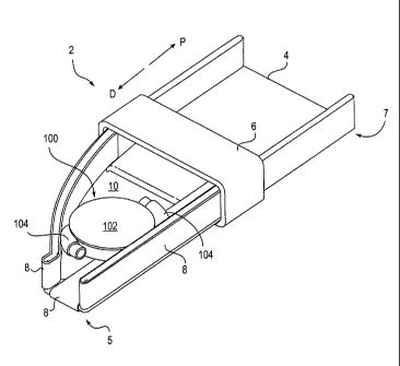
[0041] Figure 2 illustrates an exemplary embodiment of delivery device 2 and
IOL 100.
IOL 100 comprises optic portion 102 and haptics 104 (see Figure 3) positioned
within the
delivery device in an unstressed, or expanded, configuration. Delivery device
2 includes
inserter body 4, pull block 6, and belts 8. The pull block is connected to the
belts on the
6
CA 02696450 2010-01-15
WO 2009/015240 PCT/US2008/070938
belt portions that are on the exterior surfaces and bottom surface of the
inserter body
(bottom not shown), but is not connected to the belts on the inside of the
inserter body.
Movement of the pull block in either of the direction of arrows D and P moves
the portion
of the belts on the inside of the inserter body to move in the direction
generally opposite
the direction of movement of the pull block. For example, when pull block 6 is
moved in
the proximal, or P direction, the belt portions on the interior of the body
move generally in
the D direction. The belts act generally as conveyor belts to move the IOL the
pull block
is actuated.
[0042] In use, when the pull block is pulled in the proximal direction (the
direction of
arrow P in Figure 2), this causes the portion of the belts on the inside of
the inserter body
to move in the general distal direction (the direction of arrow D) along the
interior surfaces
of the inserter body. The belts on the outside and bottom of the inserter body
move in the
proximal direction as well. As the portion of the belts on the inside of the
inserter body
move distally, they eventually move around distal end 5 of the inserter body
to the outside
of the inserter body.
[0043] Similarly, when the pull block is pushed distally, or in direction D,
the portion of
the belts on the outside and bottom of the inserter body move distally and the
portion of
the belts on the inside of the inserter body move proximally. This causes the
IOL in the
inside of the body to move in the proximal direction.
[0044] The delivery device is configured so that only the belts and not the
inserter body
(or as little of the inserter body as possible) engage the IOL. Because the
IOL does not
make contact with the inserter body (or any other parts of the delivery device
that may be
added), the inserter body does not apply tensile force or shear forces/stress
on the IOL as
the IOL is moved by the belts. In addition, because the belts move with the
IOL, the
amount of shear and tensile forces applied to the IOL by the belts are
minimized. As
shown in Figure 2, there is an opening or space 10 formed in the bottom
surface of the
inserter body. The opening in the inserter body is created to avoid contact
between the
inserter body and the IOL to help minimize unwanted forces on the IOL.
[0045] To deliver the IOL into the eye, the IOL is positioned in the interior
of the inserter
body, making contact with substantially only the belts. The IOL is positioned
in an
expanded configuration so it is just barely making contact with the belts (as
shown in
Figure 2). The pull block is actuated in the proximal direction and the IOL is
moved in the
distal direction towards the distal end 5 of the device. Because of the
reduced width of the
distal end of the device compared to proximal end 7, the IOL is compressed as
it moves
7
CA 02696450 2010-01-15
WO 2009/015240 PCT/US2008/070938
distally and then passes out of distal end 5. It is delivered from the distal
end of the device
into the eye, where it expands after being released from the delivery device.
[0046] When compressing a closed-system fluid-filled IOL (as is shown in 1A-1C
and in
Figure 2) in the conveyor system, the portion of the IOL nearest to distal end
5 of inserter
body 4 will begin to compress before the rest of the IOL. As the distal end of
the IOL
begins to compress, fluid contained within the IOL will generally be squeezed
or forced
into more proximally positioned portions of the IOL. In addition, the first
portion of the
IOL to be deployed from the delivery device will begin to expand, and while
more
proximal portions of the IOL continue to be compressed, some fluid will begin
to be
squeezed distally into the now free and expanding distal portion of the IOL.
[0047] It may therefore be advantageous to orient the IOL in the inserter body
prior to
compression such that fluid will be distributed throughout the IOL in a
predictable manner
to enable compression and minimize damage to the IOL. For example, Figure 3
shows
distal end 5 of the inserter body in more detail. The IOL is positioned in the
inserter body
so that a leading (or distal) haptic 12 begins to be deployed first from the
inserter body.
When the leading haptic begins to be released from the inserter body, the
leading haptic
can receive fluid that is squeezed from the optic portion and/or trailing
haptic 14.
[0048] This embodiment may require high tensile forces on the belts, so a
pulling
mechanism would preferably utilize features designed to increase mechanical
advantage.
Levers, screws, and/or ratchets could be used to give a user the control as
well as the
required force.
[0049] The inserter body is generally a rigid structure with a general tapered
shape with
the width decreasing towards the distal end to compress the IOL as it is moved
in the distal
direction. In some embodiments the distal end of the inserter body is less
than about 50%
of the width of the proximal end. This is not intended to be a limitation and
may be less
than about 40%, about 30%, about 20%, about 10%, or less, than the width of
the proximal
section. While the embodiment shown only includes a bottom surface, the
inserter body
could also have a top surface (with a similar space as in the bottom surface
to avoid
sliding). If the inserter body did have a top surface, a fourth belt could
then also be
included in the device.
[0050] The pull block and belts can be made of a relatively rigid material
such as Mylar or
an elastomeric material such as a silicone.
[0051] While three belts are shown in this embodiment there may be more, such
as 4, or
fewer in the delivery device.
8
CA 02696450 2010-01-15
WO 2009/015240
PCT/US2008/070938
[0052] Figure 4 illustrates a second embodiment of a delivery device. In this
embodiment
the delivery device comprises an everting tube 30 that includes at least one
slit or cut 32
along at least a portion of the length of the tube. The term everting as used
herein
generally means that at least one section of the tube is adapted to roll back
or fold back
onto the tube, like a pair of socks or the cuff on a pair of pants. In some
embodiments,
however, the everting tube does not have a slit.
[0053] Everting as used herein can refer both to the step when the inner
surface of the tube
rolls outward and back and becomes an outer surface of the tube, or when an
outer surface
of the tube rolls inward and becomes an inner surface.
[0054] Figure 4 shows everting tube 30 in a non-everted state (no section of
tube is
everted, or rolled back). Slit 32 is shown running parallel to the
longitudinal axis LA of
the tube 30.
[0055] Figure 5 is a cross sectional view of the tube with a distal portion 34
everted,
however the portion of the tube including slit 32 has not yet been everted.
[0056] Figure 6 shows a perspective view of an exemplary everting tube 30 as
the portion
of the tube including the slit has begun to evert. The slit in the tube causes
the portion of
the tube circumferentially surrounding the slit to "blossom" as the distal end
of the slit
reaches the distal end of the tube and as the portion of the tube
circumferentially
surrounding the slit begins, and continues, to evert. Figure 7 shows the slit
continuing to
blossom. Figure 8 is a distal end view of the slit blossomed. Once the slit
portion of the
tube is fully everted, the remainder of the tube continues to evert in the
same manner as
did the portion of tube disposed proximally to the slit. It is in this manner
that the slit in
the tube allows for a greater expansion or opening of the tube as it is
everted.
[0057] In one embodiment of the everting tube concept as shown in Figure 9,
the everting
tube is coupled to a syringe-like device 40. Device 40 includes an outer body
42
comprising an inner bore or channel through which inner body 44 passes. Inner
body 44
includes handle 46 at its proximal end. The proximal end 50 of everting tube
30 is
coupled to distal portion 45 of inner body 44 and distal end 52 of the
everting tube 30 is
coupled to outer body 42. When inner body 44 is actuated in the distal
direction (e.g., by
pushing handle 46 distally), inner body 44 moves distally relative to outer
body 42.
Because the proximal end of the everting tube is coupled to distal portion 45
of the inner
tube, this movement also moves the proximal portion of the everting tube in
the distal
direction. Distal end 52 of the everting tube remains coupled to outer body 42
and thus
does not move. Similarly, when the inner body is moved or pulled proximally,
such as by
9
CA 02696450 2010-01-15
WO 2009/015240 PCT/US2008/070938
pulling on the handle in the proximal direction (or otherwise actuating inner
body 44),
inner body 44 moves proximally relative to outer body 42 and therefore so does
the
proximal end of the everting tube. It is noted that it is the relative
movement of the inner
and outer bodies that controls the movement (and thus the everting) of the
everting tube,
and the outer body can similarly be advanced in the distal direction or
retracted in the
proximal direction over the inner body to cause the relative movement.
[0058] In addition the inner and outer bodies may be disposed within an outer
sheath such
that the user of the delivery device would not see the inner and outer bodies.
The inner
and outer bodies could also be coupled to an actuator such as a control knob
which a user
could use to carefully control the advancement of the inner body relative to
the outer body
or the retraction of the outer body relative to the inner body. This could
give the user
precise control over the delivery of the IOL.
[0059] To deliver an IOL into the eye, an IOL is first loaded into the distal
end of the
delivery device shown in Figure 9 as follows. Handle 46 is advanced distally
(or a knob is
rotated, or other actuator to control the relative movement of the inner and
outer bodies) as
shown in Figure 9 such that a portion of the everting tube is disposed outside
and distal to
outer body 42. The slit in the everting tube is exposed, or outside of the
outer body, and
has "bloomed." The IOL is placed into the blooming opening and the handle is
then
actuated in the proximal direction, or the outer body is advanced in the
distal direction, or
both. As the inner layer of the everting tube moves in the proximal direction,
causing
more of the outer layer of the tube to roll inward and become part of the
inner layer of the
tube, the slit is retracted within the outer layer of the tube. The slit is
thereby forced
closed and the device is compressed in the tube via the hoop forces on the
closed, or intact,
portion of the tube.
[0060] Because the tube is everting inward and moving with the IOL (similar to
the belts
in the embodiment shown in Figures 2 and 3), the amount of shear and tensile
forces on
the IOL are minimized. Substantially all of the sliding (and accompanying
shear forces)
occurs between the two layers of the everting tube, so there is no (or very
little) sliding
between the everting tube and the IOL. In some embodiments a lubricant is
applied to the
everting tube to minimize shear and forces.
[0061] As the handle continues to be pulled in the proximal direction, the IOL
continues
to be loaded into the outer body as the IOL moves further proximally into the
channel. In
this embodiment, the compression is accomplished as the hoop forces force the
IOL to be
compressed as it is drawn into the everting tube.
CA 02696450 2010-01-15
WO 2009/015240 PCT/US2008/070938
[0062] Figure 10 shows a cross sectional view of an exemplary IOL 100 with a
portion of
the IOL loaded into the delivery device (and within the everting tube), as
described by the
loading process above. The exemplary IOL 100 is a soft, flexible,
accommodating IOL
which includes an optic portion 102 and a peripheral portion comprising
haptics 12 and 14
in fluid communication with the optic portion. The IOL comprises fluid which
is
transferred between the haptics and optic portion to accommodate the IOL in
response to
ciliary muscle movement.
[0063] When compressed into the delivery configuration, the length of IOL 100
increases
(as is shown in Figure 10) while the IOL narrows. When compressed, the fluid
within the
IOL is squeezed from the portion of the IOL loaded first. As shown in Figure
10,
proximal, or trailing, haptic 14 is loaded first, which squeezes the fluid
from the proximal
haptic into the optic portion (and likely into distal haptic 12 as well). As
the optic portion
is loaded into the delivery device (e.g., as the handle continues to be pulled
proximally),
the optic portion is compressed by the everting tube and the fluid in the
optic portion is
squeezed into the distal haptic 12.
[0064] Figure 10 shows the IOL in the loaded, or delivery, configuration.
Distal haptic 12
is external to the delivery device and contains a larger volume of fluid that
it contains
when the IOL is in an expanded configuration. Similarly, optic portion 102 and
trailing
haptic 14 contain less fluid than they do when in an expanded configuration.
In this
delivery configuration, the IOL has been partially compressed and elongated,
and much of
the fluid has been squeezed into the distal, or leading, haptic.
[0065] To deploy the IOL into the eye (e.g., into the lens capsule of which
the native lens
has been removed), the distal, or leading, haptic is pushed through the
corneal incision and
into the capsule. Then inner body 42 is pushed distally (or the outer body is
pulled
proximally, or both), which causes the everting tube and the loaded IOL to
move distally
together, deploying the IOL from the delivery device and into the eye by
squeezing out
through the blooming slit portion of the everting tube. As the optic portion
of the IOL
begins to be released from the outer body, the fluid moves from the distal
haptic to the
optic portion, causing the optic portion to expand in volume. Then, as the
proximal haptic
is released from the delivery device it begins to refill with fluid and
increases in volume.
Once the IOL has completely been deployed outside of the delivery device (and
into the
capsule), the IOL has generally returned to its pre-loaded, generally
expanded,
configuration (although the shape of the IOL may be slightly altered after
implantation due
11
CA 02696450 2010-01-15
WO 2009/015240 PCT/US2008/070938
to forces acting on the IOL by the lens capsule). The delivery device is then
removed
from the eye.
[0066] Figures 11A ¨ 11D show an alternative embodiment of delivery device 70
comprising outer body 72 and inner body 74 with knob 76. To load the IOL, knob
76 is
rotated which actuates inner body 74 in the proximal direction and/or actuates
the outer
body in the distal direction. To deploy the IOL, the knob 76 is rotated which
actuates the
inner body in the distal direction and/or actuates the outer body 72 in the
proximal
direction. Sheath 73 covers outer body 72 and provides the surgeon a stable
handle with
which to work. Figure 11D shows a close-up perspective view of distal end 77
of everting
tube 78.
[0067] In the embodiments shown in Figures 11A-11D, distal end 77 of the
everting tube
can be adapted such that it does not move relative to the eye during the
implantation
procedure. The tube will evert (the inner tube become outer tube, or the outer
tube
becomes inner tube), however the distal end remains substantially fixed in
space. This is
important because the user does not have to worry about distal end 77
contacting and
disrupting the eye during the procedure. The user also does not have to worry
about
moving the distal end of the delivery system relative to the eye during the
deployment
procedure.
[0068] Figures 12A-12C show the loading of IOL 80 into delivery device 70 as
described
above. IOL 80 comprises trailing haptic 82, optic portion 84, and leading
haptic 86.
Delivery of the IOL into an eye occurs in the reverse order of the steps shown
in Figures
12A-12C.
[0069] Figures 13A ¨ 13C show deployment of IOL from delivery device 70.
Figure 13A
shows a leading haptic extending from the distal end of the everting tube.
Figure 13B
shows the optic portion emerging, and Figure 13C shows the trailing haptic
almost
completely deployed. Figure 14 illustrates the size of delivery device 70 next
to IOL 80.
[0070] In some embodiments the everting tube is a thin, tough, generally
stretchy material
that is adapted to be everted. To evert a tube it is generally preferred to be
somewhat
stretchy and very thin relative to the inner diameter of the tube. A composite
material with
relatively different axial and circumferential stiffnesses may also be used.
For instance, a
tube can contain fibers running along the longitudinal axis of the tube that
serve to stiffen
the tube in the axial direction while maintaining the elastic properties in
the
circumferential direction. Alternatively, the everting tube can be formed by
being drawn
to provide extra stiffness along its length.
12
CA 02696450 2010-01-15
WO 2009/015240
PCT/US2008/070938
[0071] While the embodiments above show and describe one slit in the everting
tube, the
delivery device may have more than one slit, such as 2, 3, 4, 5, or more
slits. The slits
may be positioned around and along the length of the tube in any orientation
that helps
minimize the shear and tensile forces on the IOL during loading or deployment.
In some
embodiments the everting tube has no slits.
[0072] A variety of actuation mechanisms may be used to deliver the device.
For example
without limitation, a knob, a trigger, or a lever mounted on a grip may be
used as
alternatives to the syringe design.
[0073] Figure 15 illustrates an alternative delivery device 60 which comprises
body 62,
inserter 64, and advancement mechanism, or actuator, 66, which is coupled to
inserter 64.
Inserter 64 is a sheet that is rolled up along its length wherein one edge of
the inserter
overlaps the other, as shown in Figure 16. The proximal end (not shown) of
inserter 64 is
coupled to the distal end (not shown) of advancement mechanism 66. As
advancement
mechanism 66 is actuated in the proximal direction, inserter 64 is withdrawn
into body 62.
Body 62 generally compresses inserter 64 when inserter 64 is withdrawn in to
body 62.
This causes the diameter of inserter 64 to decrease and the sheet forms a
tighter roll or
curl.
[0074] Figure 16 is a distal end view of the inserter and Figure 17 shows a
perspective
view of a distal end of body 62 with inserter 64 withdrawn into body 62.
[0075] To load the IOL into the delivery device 60, the advancement mechanism
is
pushed distally to deploy inserter 64 from the distal end of body 62 (as shown
in Figure
15). The distal end of the inserter body will assume a more open (i.e., the
curl is not as
tight), or first, configuration, allowing the IOL to be positioned in the
distal end of the
inserter. After placement of the IOL in the distal opening of the inserter,
the advancement
mechanism is pulled proximally (or body 62 is pushed distally). This pulls the
inserter
into body 62 whereby the body 62 exerts a compressive force on the inserter,
causing it to
fold more tightly into itself. The inserter thus applies a compressive force
to the IOL. As
in the other embodiments above, because the IOL moves proximally with the
inserter, it is
compressed within the inserter. The inserter and IOL move together and
therefore shear
and tensile forces acting on the IOL are minimized.
[0076] Once loaded into the delivery device, the IOL can then be inserted
through the
wound as described above.
[0077] Once body 62 has been advanced into the wound advancement mechanism 66
is
advanced distally, which begins to deploy the folded inserter from the body.
The IOL
13
CA 02696450 2010-01-15
WO 2009/015240
PCT/US2008/070938
moves with the inserter as it is advanced out of body 62. As the inserter is
pushed from
body 62, it begins to unroll, or open, allowing the optic and trailing haptic
to begin to
expand and again fill with fluid that had been squeezed into the leading
haptic when the
IOL was in the loaded delivery configuration.
[0078] This embodiment may be used with an additional secondary advancement
mechanism to further advance the IOL from the rolled inserter. For example, a
plunger-
like device could be disposed within an internal bore or channel in the
advancement
mechanism. The plunger-like device could be pushed distally through the
advancement
mechanism to make contact with the IOL to completely deploy the IOL from the
folded
inserter. Because the IOL might be in a generally uncompressed state after the
inserter has
been pushed as far distally as possible, only a small amount of additional
force may be
needed to completely push the IOL from the folded inserter. Therefore the
plunger-like
device would not damage the IOL.
[0079] An alternative secondary advancement mechanism uses a hydraulic force
to fully
deploy the IOL from the folder inserter. A lumen within the advancement
mechanism can
be used to deliver fluid within the inserter thereby forcing the IOL out of
the inserter.
Fluid will also minimize the amount of shear or tensile forces acting on the
IOL. A
sealing mechanism such as a plug or other insert (such as a silicone material)
can also be
positioned into the rolled inserter to help create a seal between the IOL and
the inserter to
aid in the hydraulic ejection of the IOL.
[0080] In general the rolled inserter is a very thin material. In one
embodiment the rolled
inserter comprises mylar and is about .004" thick. The cross section of the
inserter may
assume a variety of cross-sectional shapes, such as round, oval, or
elliptical.
[0081] Figures 18A-18E illustrate an embodiment of loading and delivery system
300 for
loading and delivering intraocular lens 310. The system includes rigid outer
tube 302,
flexible inner sleeve 304 (split into two halves as shown), and compressor
clips 306.
Outer tube 302 is adapted to fit through about a 4mm incision in the eye.
Outer tube 302
is coupled to outer tube actuator 322 and inner sleeve 304 is coupled to inner
sleeve
actuator 324. The outer tube and inner sleeve can axially move with respect to
one
another by actuation of one or both of outer sleeve actuator 322 and inner
sleeve actuator
324. The compressor clips can be lightly bonded (e.g., using a weak bonding
material
such as Loctite 495) or unbonded to the inner sleeve.
[0082] To load lens 310 into outer tube 302, the intraocular lens is first
positioned in the
system as shown in Figure 18A (also shown in more detail in Figure 18B).
Haptics 312
14
CA 02696450 2010-01-15
WO 2009/015240
PCT/US2008/070938
are first positioned axially from optic portion 314 (one haptic leading and
the other haptic
trailing). This assists in the loading process. A compressive force in the
general direction
of arrows C is then applied to one or both of compressor clips 306. The
compressive force
can be applied by a vise or other similar device that brings two elements
together to cause
compressive force C to be applied to the compressor clips. As a result, a
compressive
force is applied to the lens and causes the lens to be compressed between the
two halves of
the inner sleeve. The inner sleeves, and not the compressor clips, engage the
lens. The
compressive force is applied until the two halves of the inner sleeve come
together such
that the lens is fully compressed within the two halves of the inner sleeve.
The
compressor clips can be compressed until they engage with each other or there
may be a
slight space between the edges of the compressor clips. During the compression
process
the lens is compressed and elongated.
[0083] After the compressor clips are compressed to the closed (or
substantially closed)
position shown in Figure 18C, outer tube actuator 322 is advanced distally in
the direction
of arrow D (shown in Figure 18D) and inner sleeve actuator 324 is held in
place. The
movement of outer tube actuator 322 causes the outer sleeve to be advanced
distally over
the inner sleeve (which is held in place). The inner sleeve could also be
retracted
proximally while the outer tube is held in place. Advancing the outer tube
displaces the
compressor clips in the distal direction, which also move relative to the
inner sleeve. The
outer tube is advanced until the inner sleeve (and therefore the lens) is
disposed within the
outer tube, as shown in Figure 18E. During this loading step sliding occurs
between the
outer tube and the inner sleeve, not between the lens and the inner sleeve.
This minimizes
shear and tensile forces acting on the lens.
[0084] The outer tube is then advanced through an incision made in the eye. To
deploy
the lens from the delivery system and into the lens capsule, inner sleeve
actuator 324 is
advanced distally in direction D. This causes inner sleeve to be advanced
distally relative
to the outer tube. As the inner sleeve emerges from the distal end of the
outer tube, the
inner sleeve will begin to split along the slit and the lens will begin to
expand. The lens
can therefore be delivered into the capsule.
[0085] The outer tube is generally rigid and in one embodiment is a stainless
steel tube.
The inner sleeve is generally a flexible material and in one embodiment is
PTFE. The
compressor clips can be any suitably rigid material.
[0086] Increasing the outer tube volume increases the volume into which the
lens can be
compressed. It is generally desirable for the outer tube to have the largest
cross sectional
CA 02696450 2010-01-15
WO 2009/015240 PCT/US2008/070938
area possible while still allowing the outer tube to be advanced into the
smallest incision
possible. It has been found than using an outer tube in which the cross
section is generally
elliptically-shaped allows the largest cross sectional area through the
smallest incision.
[0087] In an alternative embodiment the inner sleeve as shown in Figures 18A -
18E can
be replaced with a rolled sheet such as inserter 64 shown in Figures 15-17.
The system
would work similarly to the described above in references to Figure 18A-18E.
[0088] Figures 19-21 shows alternative embodiments of a hydraulic lens loading
system.
Using a hydraulic system to load the intraocular lens into the delivery device
(as well as a
hydraulic system to deploy the intraocular lens) minimizes shear and tensile
forces on the
lens. The lens is forced into a delivery device using a generally lubricous
liquid or fluid,
which minimizing shear and tensile forces acting on the lens as it is
compressed and
elongated. Figure 19A shows loading system 400 for loading intraocular lens
402 into
loading tube 408. The system includes syringe 404 including plunger 406.
Distal region
412 of syringe 404 includes a tapered inner surface 410 which has a smaller
cross
sectional diameter at the distal end than at the proximal end. The distal
region of the
syringe contains the lens as well as fluid 414. The fluid can be a liquid such
as saline and
can include or can be a known viscoelastic lubricant such as, for example
without
limitation, aqueous solutions of sodium hyaluronate, hydroxypropylmethyl
cellulose, and
chondroitin sulfate.
[0089] To advance the lens into loading tube 408, the plunger is actuated in
the distal D
direction which causes fluid 414 and lens 402 to be advanced distally towards
loading tube
108. The plunger continues to be advanced distally until the lens is forced
through
proximal end 416 of loading tube 108. By moving the lens with a lubricious
material,
shear and tensile forces on the lens are minimized.
[0090] Figure 20B shows an alternative hydraulic loading system 600 for
loading
intraocular lens 602 into loading tube 608. The system is similar to previous
embodiments
and includes syringe 604 with plunger 606. The syringe includes lens chamber
612 which
has a generally circular shape to retain the generally circular shape of lens
602. The
syringe also includes tapered section 610 which directs the lens into loading
tube 608.
Lens 602 is initially positioned in lens chamber 612 with distal haptic 605
extending
distally from optic portion 603 and into tapered section 610. This initial
positioning helps
direct the lens into a compressed configuration within loading tube 608 when
fluid 614 is
forced through lens chamber 612. The plunger is advanced distally to direct a
fluid
through lens chamber 612, which forces lens 602 into loading tube 608.
16
CA 02696450 2010-01-15
WO 2009/015240
PCT/US2008/070938
[0091] In an alternative design the intraocular lens can be loaded into the
loading tube
under vacuum pressure.
[0092] After the lens is loaded into the loading tube, the lens is
hydraulically delivered
into the eye. The loading tube is first detached from the loading apparatus.
The loading
tube is then inserted through an incision in the eye and a fluid (such as a
lubricious fluid)
is directed through the loading tube to eject the lens from the loading tube
and into the
eye. Hydraulic deployment also minimizes shear and tensile forces acting on
the lens. A
syringe can be used to direct the fluid through the loading tube.
Alternatively, a small
piston drives down the tube, pushing a short column of fluid distally to the
piston. The
piston is controlled with an actuator such as a knob, lever, ratchet, etc. The
piston can be
attached to either end of the loading tube. This means the lens can be ejected
from the
same end in which it is loaded, or it can be deployed from the other end of
the loading
tube.
[0093] Figure 21 illustrates an alternative loading system concept using
peristaltic
movement to load an intraocular lens (not shown). In this design, purely
compressive
loads on the lens are separated in time from shear loads on the lens. The lens
is "inched"
along into a fully compressed state. System 700 includes rigid large tube 702,
rigid small
tube 706, and flexible tube 704 with a generally conical or tapered shape.
Fluid 710 is
contained within the system to lubricate the system and also to help push the
lens through
the system. The lens is moved from the rigid large tube 702 through flexible
tube 704 and
into a fully compressed state within small rigid tube 706. Large tube 702 has
a larger
diameter than small tube 706. There is generally a pressure gradient between
P1 and P2
with P1 being higher. The difference in pressure between P1 and P2 (which is
the driving
pressure) is equal to P1 minus P2. The pressure P3 from a compressive force on
the
flexible tube is used to compress the lens in a direction that is
substantially orthogonal to
the axis A. P3 is pulsed out of phase from the driving pressure, which is also
pulsed. To
load the lens, P3 is initially increased to compress the lens radially. Then
P3 is decreased
while the driving pressure is increased, so the device is pushed in the
direction D a small
distance and reexpands radially. When P3 is decreased the flexible wall moves
radially
away from the lens and shear forces are reduced. P3 is then increased again,
compressing
the lens radially. P3 is then decreased as the driving pressure is increased,
which again
moved the lens in the direction D. The lens is therefore moved in small
increments in the
distal direction D, compressing it as it moves. This movement is repeated
until the lens is
17
CA 02696450 2010-01-15
WO 2009/015240 PCT/US2008/070938
fully compressed within small tube 706. The lens can then be deployed using
any of the
methods described herein.
[0094] In any or all of the embodiments described herein, the method of
delivery includes
creating a wound in the eye which generally comprises an incision in the eye.
In some
embodiments the incision is about 4mm and preferably about 3.7mm. The incision
can,
however, be slightly larger or smaller.
[0095] In any of the embodiments described herein, the position and/or
orientation of the
IOL may need to be adjusted for the loading step. For example, when loading an
IOL with
haptics, it may be necessary to align the haptics so they are oriented
generally along the
longitudinal axis of the delivery device before compressing the lens (see, for
example,
Figure 18B). Alternatively, only one haptic may be straightened while a second
haptic can
be positioned peripherally around the optic portion (see, for example, Figure
21A). These
orientations can provide for a better delivery profile and minimizes the
chance of damage
to the IOL during deployment.
[0096] To compress any of the fluid-filled accommodating IOL described herein,
it may
be necessary to apply a compressive side force of about .5 pounds. This can
vary,
however, depending on the size, composition, and volume of the IOL.
[0097] While only these embodiments have been described, they all attempt to
minimize
the amount of shear and tensile forces acting on the IOL during the loading
and/or delivery
process. One common method is minimizing the amount of sliding that occurs
between
the IOL and the delivery system components. Other embodiments are included in
this
invention which allow the IOL to be loaded into and deployed from the delivery
device
with (or in conjunction with) a delivery device component, in order to reduce
these
unwanted forces.
18