Note: Descriptions are shown in the official language in which they were submitted.
CA 02700523 2010-03-23
WO 2009/042130 PCT/US2008/011040
MRI-GUIDED MEDICAL INTERVENTIONAL
SYSTEMS AND METHODS
RELATED APPLICATIONS
[001] The present application claims the benefit of and priority to U.S.
Provisional Patent Application No. 60/974,821, filed September 24, 2007, the
disclosures
of which are incorporated herein by reference as if set forth in their
entireties.
FIELD OF THE INVENTION
[002] The present invention relates generally to medical systems and methods
and, more particularly, to in vivo medical systems and methods.
BACKGROUND OF THE INVENTION
[003] Deep Brain Stimulation (DBS) is becoming an acceptable therapeutic
modality in neurosurgical treatment of patients suffering from chronic pain,
Parkinson's
disease or seizure, and other medical conditions. Other electro-stimulation
therapies have
also been carried out or proposed using internal stimulation of the
sympathetic nerve
chain and/or spinal cord, etc.
[004] One example of a prior art DBS system is the Activa system from
Medtronic, Inc. The Activa system includes an implantable pulse generator
stimulator
that is positioned in the chest cavity of the patient and a lead with axially
spaced apart
electrodes that is implanted with the electrodes disposed in neural tissue.
The lead is
tunneled subsurface from the brain to the chest cavity connecting the
electrodes with the
pulse generator. These leads can have multiple exposed electrodes at the
distal end that
are connected to conductors which run along the length of the lead and connect
to the
pulse generator placed in the chest cavity.
1
CA 02700523 2010-03-23 ~=
WO 2009/042130 PCT/US2008/011040
[005] It is believed that the clinical outcome of certain medical procedures,
particularly those using DBS, may depend on the precise location of the
electrodes that
are in contact with the tissue of interest. For example, to treat Parkinson's
tremor,
presently the DBS probes are placed in neural tissue with the electrodes
transmitting a
signal to the thalamus region of the brain. DBS stimulation leads are
conventionally
implanted during a stereotactic surgery, based on pre-operative MRI and CT
images.
These procedures can be long in duration and may have reduced efficacy as it
has been
reported that, in about 30% of the patients implanted with these devices, the
clinical
efficacy of the device/procedure is less than optimum. Notwithstanding the
above, there
remains a need for alternative MRI-guided interventional tools for DBS, as
well as for
other interventional medical procedures.
SUMMARY OF THE INVENTION
[006] In view of the above, MRI-guided interventional systems and methods are
provided. Embodiments of the present invention provide methods, devices and
systems
for highly localized placement and/or delivery of diagnostic or therapeutic
devices or
substances.
[007] According to embodiments of the present invention, an MRI-guided
interventional system includes a frame with a cooperating targeting cannula.
The frame
is configured to be secured to the body of a patient, and is configured to
translate and
rotate such that the targeting cannula can be positioned to a desired
intrabody trajectory.
The frame may include one or more MRI-visible fiducial markers that allow
frame
location/orientation to be determined within an MRI image.
[008] Embodiments of the present invention may be particularly suitable for
placing neuro-modulation leads, such as Deep Brain Stimulation ("DBS") leads,
implantable parasympathetic or sympathetic nerve chain leads and/or CNS
stimulation
leads, as well as other devices within the brain.
[009] Embodiments of the present invention may be suitable for a number of
interventional procedures in many locations inside the body including, but not
limited to,
brain, cardiac, spinal, urethral, and the like. Embodiments of the present
invention may
be suitable for a number of MRI-guided drug delivery procedures, MRI-guided
ablation
procedures, etc.
[0010] A plurality of user-activatable actuators are operably connected to the
2
4 i CA 02700523 2010-03-23
WO 2009/042130 PCT/US2008/011040
frame and are configured to translate and rotate the frame relative to the
body of a patient
so as to position the targeting cannula to a desired intrabody trajectory. In
some
embodiments, the actuators are dials or thumbscrew-type devices that allow
manual
manipulation thereof. In other embodiments, the actuators are manipulated
remotely
using remote controls and cables.
100111 The targeting cannula includes an axially-extending guide bore
therethrough that is configured to guide placement of an interventional device
in vivo.
Various instrumentation and equipment (e.g., stimulation leads, ablation
probes or
catheters, injection or fluid delivery devices, biopsy needles, extraction
tools, etc.) can be
inserted through the targeting cannula to execute diagnostic and/or surgical
procedures.
[0012] According to some embodiments of the present invention, the frame
includes a base, a yoke movably mounted to the base and that is rotatable
about a roll
axis, and a platform movably mounted to the yoke and that is rotatable about a
pitch axis.
The platform includes an X-Y support table that is configured to move in an X-
direction
and Y-direction relative to the platform. The base has a patient access
aperture formed
therein, and is configured to be secured to the body of a patient such that
the aperture
overlies an opening in the body. A roll actuator is operably connected to the
yoke and is
configured to rotate the yoke about the roll axis. A pitch actuator is
operably connected
to the platform and is configured to rotate the platform about the pitch axis.
An X-
direction actuator is operably connected to the platform and is configured to
move the X-
Y support table in the X-direction. A Y-direction actuator is operably
connected to the
platform and is configured to move the X-Y support table in the Y-direction.
[0013) The base may include a plurality of locations for attaclunent to a body
of
a patient via fasteners. In some embodiments, one or more attachment locations
may
include multiple adjacent apertures configured to receive a fastener
therethrough. For
embodiments where the frame is configured to be attached to the skull of a
patient, the
base can be configured to be secured to the skull of a patient such that the
patient access
aperture overlies a burr hole formed in the patient skull.
[0014] According to some embodiments of the present invention, the yoke
includes a pair of spaced apart arcuate arms. The platform engages and moves
along the
yoke arcuate arms when rotated about the pitch axis. The base includes at
least one
arcuate arm. The yoke engages and moves along the base arcuate arm when
rotated about
the roll axis.
3
CA 02700523 2010-03-23
WO 2009/042130 PCT/US2008/011040
[0015] According to some embodiments of the present invention, at least one of
the yoke arcuate arms includes a thread pattern formed in a surface thereof.
The pitch
actuator includes a rotatable worm with teeth configured to engage the thread
pattern.
Rotation of the worm causes the platform to rotate about the pitch axis.
Similarly, at least
one of the base arcuate arms includes a thread pattern formed in a surface
thereof. The
roll actuator includes a rotatable worm with teeth configured to engage the
thread
pattern, and wherein rotation of the worm causes the yoke to rotate about the
roll axis.
[0016] In some embodiments, the actuators are color-coded such that each
different actuator has a respective different color. This allows a user to
quickly determine
which actuator is the correct one for a particular desired movement of the
frame.
[0017] According to some embodiments of the present invention, an ergonomic
remote control unit is provided that allows a user to remotely translate and
rotate the
frame such that the targeting cannula can be positioned to a desired intrabody
trajectory.
The remote control unit includes a plurality of position controls. Each
control is operably
connected to a respective frame actuator by a respective cable. One or more of
the
position controls can include both "gross" and "fine" adjustments.
[0018] Movement of a position control operates a respective actuator via a
respective control cable. For example, the remote control unit includes a roll
adjustment
control, a pitch adjustment control, an X-direction adjustment control, and a
Y-direction
adjustment control. A roll control cable is operably connected to the roll
adjustment
control and to the roll actuator. Movement of the roll adjustment control
operates the roll
actuator via the roll control cable. A pitch control cable is operably
connected to the
pitch adjustment control and to the pitch actuator. Movement of the pitch
adjustment
control operates the pitch actuator via the pitch control cable. An X-
direction control
cable is operably connected to the X-direction control and to the X-direction
actuator.
Movement of the X-direction adjustment control operates the X-direction
actuator via the
X-direction control cable. A Y-direction control cable is operably connected
to the Y-
direction control and to the Y-direction actuator. Movement of the Y-direction
adjustment control operates the Y-direction actuator via the Y-direction
control cable.
[0019] In some embodiments, the roll adjustment control, pitch adjustment
control, X-direction adjustment control, and Y-direction adjustment control
are
manually-operated thumbwheels, and rotation of each thumbwheel by a user
causes
corresponding axial rotation of a respective control cable and corresponding
axial
4
CA 02700523 2010-03-23
WO 2009/042130 PCT/US2008/011040
rotation of a respective actuator. In other embodiments, one or more of the
roll
adjustment control, pitch adjustment control, X-direction adjustment control,
and Y-
direction adjustment control are electric motor-assisted, rotatable controls.
[0020] In some embodiments, locking mechanisms are associated with the
remote unit position controls, and are configured to prevent user operation of
the controls
when in a locked position.
[0021] In some embodiments, each control cable has a geometrically shaped
rigid
end that is configured to removably engage a free end of a respective
actuator. Each
control cable rigid end may have a shape that is different from the other
control cable
rigid ends such that each control cable free end can only removably engage one
of the
respective actuator free ends. Each control cable includes a flexible
elastomeric collar
that is configured to surround a respective actuator free end and to maintain
engagement
of a cable end to a respective actuator free end. Each flexible collar can be
rolled or
folded back then released to cover and conformably compress against an
actuator free
end to hold the end of the cable in position; then the collar can be pushed
back to easily
release the cable from an actuator free end.
[0022] According to some embodiments, a safety lanyard may be used to connect
the remote control module to a rigid object, such as a patient support frame
or head coil
(or even the gantry or gantry housing) to prevent over extension of the cables
or
unwanted adjustments to the trajectory.
[0023] According to some embodiments, a drape is provided that is configured
to
be positioned near the body of a patient within a magnet of an MRI scanner.
The drape
includes a pocket that is configured to removably receive the remote control
unit therein.
The drape also includes one or more apertures through which the cables extend
from the
remote control unit to the frame.
[0024] According to some embodiments of the present invention, a camera
and/or video imaging device is removably secured to the frame via a bracket.
The
bracket includes a sleeve that is configured to slidably receive the imaging
device
therein.
[0025] An elongated tubular member extends through the platform and yoke and
is secured to the X-Y table of the frame. The targeting cannula is slidably
secured within
the tubular member and is movable between extended and retracted positions.
The
targeting cannula is configured to translate in response to translational
movement of the
CA 02700523 2010-03-23
WO 2009/042130 PCT/US2008/011040
X-Y support table and to rotate in response to rotational movement of the yoke
and
platform to define different axial trajectories extending through the patient
access
aperture of the base. The tubular member is configured to lock the targeting
cannula in
an extended position and in a retracted position.
(0026] A depth stop is removably secured within a proximal end of the tubular
member. The depth stop receives a sheath therein, and is configured to limit
the distance
that the sheath can extend into the body of a patient. The sheath is
configured to receive
an elongated interventional device (e.g., imaging probe, stimulation lead,
ablation
device, injection device, etc.). In some embodiments, the sheath is removable.
A locking
mechanism is removably secured to the depth stop and is configured to prevent
axial
movement of an elongated interventional device extending through the sheath.
[0027] According to some embodiments of the present invention, an MRI-guided
interventional system includes a frame with a cooperating targeting cannula
that has a
guide bore therethrough that is configured to guide placement of an
interventional device
in vivo. The frame is configured to rotate such that the targeting cannula can
be
positioned to a desired intrabody trajectory. The frame includes a base having
a patient
access aperture formed therein, wherein the base is configured to be secured
to the body
of a patient; a yoke movably mounted to the base and rotatable about a roll
axis; and a
platform movably mounted to the yoke and rotatable about a pitch axis. A
plurality of
user-activatable actuators are operably connected to the frame and are
configured to
rotate the frame relative to the body of the patient so as to position the
targeting cannula
to a desired intrabody trajectory. In some embodiments, the actuators are
color-coded
such that each actuator has a respective different color. In some embodiments,
the frame
includes a roll actuator operably connected to the yoke and configured to
rotate the yoke
about the roll axis; and a pitch actuator operably connected to the platform
and
configured to rotate the platform about the pitch axis.
[0028] In some embodiments, the system includes a remote control unit
comprising a plurality of elongate control devices. Each control device
includes first and
second elongate rods axially connected at respective first ends via a first
cable. The first
rod second end is operably connected to a respective actuator via a second
cable.
Rotational movement of the second end of the second rod operates the actuator
via the
second cable. Each second cable may have a geometrically shaped rigid end
configured
to removably engage a free end of a respective actuator.
6
CA 02700523 2010-03-23
WO 2009/042130 PCTIUS2008/011040
[00291 MRI-guided interventional methods, according to embodiments of the
present invention, include affixing a frame with a cooperating targeting
cannula to the
body of a patient, wherein the frame is configured to translate and rotate
such that the
targeting cannula can be positioned to a desired intrabody access path
trajectory. The
targeting cannula includes a guide bore therethrough that is configured to
guide
placement of an interventional device in vivo. The targeting cannula position
is adjusted
(e.g., rotated about a roll axis, rotated about a pitch axis, and/or
translated in X-Y
directions) so that the targeting cannula is aligned with the desired access
path trajectory
while the patient is positioned within a magnetic field associated with an MRI
scanner.
Once the targeting cannula is repositioned, an interventional device is
inserted through
the targeting cannula guide bore and into the body of the patient for
therapeutic and/or
diagnostic purposes. The targeting cannula is movable between retracted and
extended
positions, and is moved to the extended position and locked in the extended
position
prior to the adjusting the access path trajectory thereof.
[00301 The necessary rotational and translational adjustments required to
reposition the targeting cannula to the desired access path trajectory are
displayed to a
user via a graphical user interface. Both the actual access path trajectory
and desired
access path trajectory can be displayed, as well. In addition, the user can
view the actual
trajectory changing as he/she adjusts the position of the targeting cannula.
In some
embodiments, an indication of when the actual trajectory is aligned with a
desired
trajectory can be displayed to the user.
[0031] According to some embodiments, an MRI-guided interventional system
for use with a body of patient and an interventional device includes a base
and a
targeting cannula. The base is configured to be secured to the body of the
patient. The
targeting cannula has an elongate guide bore extending axially therethrough
and an inlet
and an outlet at opposed ends of the guide bore. The guide bore defines a
trajectory axis
extending through the inlet and the outlet and being configured to guide
placement of the
interventional device. The frame is operable to move the targeting cannula
relative to the
base to position the trajectory axis to a desired intrabody trajectory to
guide placement of
the interventional device in vivo. The inlet 'tapers from an outer diameter
distal from the
guide bore to an inner diameter proximate the guide bore to guide and
facilitate insertion
of the interventional device into the guide bore.
[0032] The system may include an elongate interventional device configured to
7
CA 02700523 2010-03-23 1 . .
WO 2009/042130 PCT/US2008/011040
be serially inserted through the inlet, the guide bore and the outlet and into
the body of
the patient in vivo.
[0033] In some embodiments, the trajectory guide frame further includes a
tubular cannula guide member defining a cannula guide member passage and
having an
inlet and outlet on opposed ends of the cannula guide member passage, and the
targeting
cannula is slidably mounted within the cannula guide member passage to move
between
extended and retracted positions. In some embodiments, the targeting cannula
has a
main body portion and an extension portion, the extension portion including
the inlet of
the targeting cannula, and the main body portion has a primary outer diameter
that is
greater than a diameter of the inlet of the cannula guide member and the
extension
portion has a reduced outer diameter that is less than the primary outer
diameter and is
sized to be received in the inlet of cannula guide member when the targeting
cannula is
in the retracted position.
[0034] The inlet of the cannula guide member can have a diameter that is at
least
as great as the outer diameter of the inlet of the targeting cannula.
[0035] According to embodiments of the present invention, an MRI-guided
interventional system for use with a body of patient and an interventional
device includes
a base, a targeting cannula, and a bracket. The base is configured to be
secured to the
body of the patient. The targeting cannula has an elongate guide bore
extending axially
therethrough, defining a trajectory axis, and being configured to guide
placement of the
interventional device. The frame is operable to move the targeting cannula
relative to the
base to position the trajectory axis to a desired intrabody trajectory to
guide placement of
the interventional device in vivo. The bracket is secured to the trajectory
guide frarne
such that the bracket is rotatable about the trajectory axis and axially fixed
with respect
to the trajectory axis. The bracket is configured to receive a light
transmission scope to
secure the light transmission scope to the trajectory guide frame.
[0036] In some embodiments, the trajectory guide frame includes one of a
groove
and a projection and the bracket includes the other of the groove and the
projection, and
the projection is cooperatively seated in the groove to permit rotation of the
bracket with
respect to the trajectory axis while preventing axial translation of the
bracket along the
trajectory axis. The groove may be configured to limit rotation of the bracket
with
respect to the trajectory axis to a prescribed range of rotation.
[0037J In some embodiments, the bracket and the trajectory guide frame are
8
CA 02700523 2010-03-23
WO 2009/042130 PCT/US2008/011040
configured to permit the bracket to be alternatively mounted on each of two
opposed
sides of the targeting cannula.
[0038] The bracket may be configured to removably snap fit onto the trajectory
guide frame.
[0039] The system can include a locking device to secure the light
transmission
scope to the bracket.
[0040] According to some embodiments, the system includes the light
transmission scope and the light transmission scope is a fiber scope.
[0041] According to embodiments of the present invention, a trajectory guide
frame for guiding an interventional device with respect to a body of a patient
in an MRI-
guided procedure includes a base, a yoke and a targeting cannula. The base has
a patient
access aperture therein. The base is configured to be secured to the body of
the patient.
The yoke is mountable on the base in a prescribe orientation with respect to
the base.
The targeting cannula is mounted on the yoke for movement therewith relative
to the
base. The targeting cannula includes a guide bore therethrough that is
configured to
guide placement of the interventional device in vivo. First and second spaced
apart pivot
holes are provided in one of the base and the yoke and first and second pivot
pins are
associated with the other of the base and the yoke. The yoke is configured to
be
mounted on the base in the prescribed orientation with the first pivot pin
received in the
first pivot hole and the second pivot pin received in the second pivot hole,
whereby the
yoke is pivotable relative to the base about the first and second pivot pins
about a roll
axis. The first and second holes are relatively configured to prevent
operative
engagement between the first pivot pin and the second pivot hole to inhibit
pivotal
mounting of the yoke on the base in an orientation other than the prescribed
orientation.
[0042] The first pivot pin may have a greater diameter than the second pivot
pin
and the second pivot hole.
[0043] In some embodiments, at least one of the first and second pivot pins is
adjustable to selectively change a length of said adjustable pivot pin
extending toward its
associated one of the first and second pivot holes.
[0044] In some embodiments, the first and second pivot holes are located in
the
base, and the base includes a pair of spaced apart mount arms each having a
guide slot
therein extending to a respective one of the first and second guide holes to
receive and
guide the first and second pivot pins to the first and second pivot holes.
According to
9
CA 02700523 2010-03-23
WO 2009/042130 PCT/US2008/011040
some embodiments, the first and second pivot pins are located on first and
second yoke
mount arms, respectively, and the base is configured to elastically deflect
the first and
second yoke mount arms apart as the first and second pivot pins are slid down
the guide
slots to the first and second pivot holes to mount the yoke on the base.
[0045] The first and second pivot pins can be releasably spring loaded into
engagement with the first and second pivot holes to permit the yoke to be
selectively
dismounted from the base.
[0046] According to embodiments of the present invention, a trajectory guide
frame for guiding an interventional device with respect to a body of a patient
in an MRI-
guided procedure includes a base, a platform, a targeting cannula, and a
stabilizer
mechanism. The base has a patient access aperture therein. The base is
configured to be
secured to the body of the patient. The platform is mounted on the base and
includes a
support table and a moving plate that is movable relative to the support table
and the
base. The targeting cannula is mounted on the moving plate for movement
therewith
relative to the support table and the base. The targeting cannula includes a
guide bore
therethrough that is configured to guide placement of the interventional
device in vivo.
The stabilizer mechanism is operable to selectively control movement between
the
support table and the moving plate to stabilize a position of the targeting
cannula with
respect to the base.
[0047] The trajectory guide frame may further include a yoke movably mounted
on the base and rotatable relative to the base about a pivot axis. The
platform is mounted
on the yoke for rotation therewith and the platform is configured to permit
translational
movement of the moving plate with respect to the yoke.
[0048] In some embodiments, the stabilizer mechanism includes an adjustment
device and a rub bar, and the rub bar and the support table cooperatively
define a slot
through which the moving plate slides in contact with the rub bar. The
adjustment
device may be operable to apply a load to the rub bar to compressively load
the moving
plate in the slot between the support table and the rub bar. The loading
device can
include at least one screw.
[0049] According to embodiments of the present invention, a trajectory guide
frame for guiding an interventional device with respect to a body of a patient
in an MRI-
guided procedure includes a base, a platform, a targeting cannula and a lock
clip. The
base has a patient access aperture therein. The base is configured to be
secured to the
CA 02700523 2010-03-23
WO 2009/042130 PCT/US2008/011040
body of the patient. The platform is mounted on the base and includes a
support table
and a moving plate that is movable relative to the support table and the base.
The
targeting cannula is mounted on the moving plate for movement therewith
relative to the
support table and the base. The targeting cannula includes a guide bore
therethrough that
is configured to guide placement of the interventional device in vivo. The
lock clip is
mounted on the platform and configured, when in a locking position, to prevent
relative
movement between the support table and the moving plate. The lock clip is
removable
to permit relative movement between the support table and the moving plate.
[00501 The trajectory guide frame may include a first lock hole in the support
table and a second lock hole in the moving plate. The lock clip extends
through the first
and second holes when in the locking position.
[00511 According to some embodiments, a trajectory guide frame for guiding an
interventional device with respect to a body of a patient in an MRI-guided
procedure
includes a base, a targeting cannula and an MRI-visible fiducial marker. The
base has a
patient access aperture therein. The base is configured to be secured to the
body of the
patient. The base defines a fiducial cavity and a tab aperture communicating
with a
fiducial cavity. The targeting cannula is mounted on the base for movement
relative
thereto. The targeting cannula includes a guide bore therethrough that is
configured to
guide placement of the interventional device in vivo. The MRI-visible fiducial
marker is
mounted on the base and includes a body portion containing an MRI-visible
material and
a fill tab extending from the body portion. At least a portion of the body
portion is
disposed in the fiducial cavity and the fill tab extends through the tab
aperture.
[0052] According to some embodiments, a trajectory guide frame for guiding an
interventional device with respect to a body of a patient in an MRI-guided
procedure
includes a base, a targeting cannula and an MRI-visible fiducial marker. The
base has a
patient access aperture therein. The base is configured to be secured to the
body of a
patient. The base includes a fiducial mount structure and a fiducial locator
feature
associated with the fiducial mount structure. The targeting cannula is mounted
on the
base for movement relative thereto. The targeting cannula includes a guide
bore
therethrough that is configured to guide placement of the interventional
device in vivo.
The MRI-visible fiducial marker is mounted on the fiducial mount structure.
The MRI-
visible fiducial marker is toroidal and defines a central opening. The
fiducial marker
extends into the central opening to positively locate the MRI-visible fiducial
marker with
11
.
CA 02700523 2010-03-23 1 =
WO 2009/042130 PCT/US2008/011040
respect to the base.
100531 According to some embodiments, a trajectory guide frame for guiding an
interventional device with respect to a body of a patient in an MRI-guided
procedure
includes a base, a targeting cannula and a plurality of MRI-visible fiducial
markers. The
base has a patient access aperture therein. The base is configured to be
secured to the
body of a patient. The targeting cannula is mounted on the base for movement
relative
thereto. The targeting cannula includes a guide bore therethrough that is
configured to
guide placement of the interventional device in vivo. The plurality of MRI-
visible
fiducial markers are mounted on the base. The MRI-visible fiducial markers are
relatively positioned in an asymmetric layout to facilitate positive
determination of an
orientation of the base in free space from an MR image.
[0054] In some embodiments, the plurality of MRI-visible fiducial markers
includes at least first, second and third MRI-visible fiducial markers each
located on a
circle, and a circumferential spacing between the first and second MRI-visible
fiducial
markers is less than a circumferential spacing between the first and third MRI-
visible
fiducial markers.
[0055] Further features, advantages and details of the present invention will
be
appreciated by those of ordinary skill in the art from a reading of the
figures and the
detailed description of the preferred embodiments that follow, such
description being
merely illustrative of the present invention.
BRIEF DESCRIPTION OF THE DRAWINGS
[0056] Fig. 1A is a block diagram of an MRI-guided interventional system,
according to some embodiments of the present invention.
[0057] Fig. 1B illustrates a user interface that displays, and that allows a
user to
adjust, the trajectory of a targeting cannula, according to some embodiments
of the
present invention.
[0058] Fig. 2A is a top perspective view of a burr hole formed in the skull of
a
patient, and a burr hole ring overlying the burr hole and secured to the
skull.
[0059] Fig. 2B is a top perspective view of a removable centering device
positioned on the burr hole ring of Fig. 1.
[0060] Fig. 3A is a perspective view of a trajectory frame utilized in the MRI-
guided interventional system, according to some embodiments of the present
invention.
12
CA 02700523 2010-03-23
WO 2009/042130 PCT/US2008/011040
[0061] Figs. 3B-3E are side view, schematic illustrations of the trajectory
frame
being secured to the skull of a patient.
[0062] Figs. 4-5 are partial perspective views of the frame of Fig. 3A
illustrating
the base of the frame being positioned on the skull of a patient with the
centering device
of Fig. 2B extending through the patient access aperture.
[0063] Fig. 6 illustrates the base secured to the skull of a patient.
[0064] Fig. 7 is an enlarged partial perspective view of the base illustrating
an
attachment location with a pair of adjacent apertures for receiving fasteners
therethrough,
according to some embodiments of the present invention.
[0065] Fig. 8A is a perspective view of the frame of Fig. 3A secured to the
body
(e.g., skull) of a patient, and with the targeting cannula in an extended
position.
[0066] Fig. 8B is a cut-away perspective view of the frame of Fig. 3A,
illustrating a targeting cannula according to some embodiments of the present
invention.
[0067] Figs. 9 and 10A-10C illustrate a remote control unit for remotely
controlling the positioning actuators of the frame of Fig. 3A, according to
some
embodiments of the present invention.
[00681 Fig. 11 is a perspective view of the base of the frame of Fig. 3A
illustrating fiducial markers associated therewith and illustrating an arcuate
arm with a
thread pattem formed in a surface thereof that is configured to be engaged by
a roll axis
actuator, according to some embodiments of the present invention.
[0069] Fig. 12 is a partial perspective view of the frame of Fig. 3A
illustrating a
yoke arcuate arm with a thread pattern formed in a surface thereof that is
configured to
be engaged by a pitch axis actuator, according to some embodiments of the
present
invention.
[0070] Figs. 13A-13B illustrate an optic fiber scope for a video imaging
camera
mounted to the frame of Fig. 3A so as to view a burr hole, according to some
embodiments of the present invention.
[0071] Fig. 14 is an enlarged, partial perspective view of the frame of Fig.
3A
illustrating the targeting cannula locked in an extended position, according
to some
embodiments of the present invention.
[0072] Fig. 15 is an enlarged, partial perspective view of the frame of Fig.
3A
illustrating control cables removably engaged with respective actuators, and
illustrating
flexible elastomeric collars configured to surround respective actuator free
ends and to
13
CA 02700523 2010-03-23
WO 2009/042130 PCT/US2008/011040
maintain engagement of the cable ends to a respective actuator free end,
according to
some embodiments of the present invention.
[0073] Fig. 16A is a partial perspective view of a frame of an MRI-guided
interventional system, according to other embodiments of the present
invention, and
illustrating actuators positioned on a side of the frame and illustrating
control cables
removably engaged with the respective actuators.
[0074] Fig. 16B is a partial perspective view of an exemplary prototype
actuator
illustrating a remote control cable end about to be inserted into a slot in
the actuator free
end, according to some embodiments of the present invention.
[0075] Fig. 16C is a partial perspective view of the actuator of Fig. 16B with
the
remote control cable end inserted into the actuator and with an elastomeric
collar
engaging the free end of the actuator to prevent the cable from being
inadvertently
removed from the actuator.
[0076] Figs. 16D-16E are partial perspective views of the actuator of Fig. 16C
illustrating removal of the elastomeric collar and cable (Fig. 16E) from the
free end of
the actuator.
[0077] Fig. 17 illustrates the frame of Fig. 3A secured to the skull of a
patient
and illustrates a desired trajectory for an interventional device, and also
illustrates the
actual trajectory of the interventional device as oriented by the frame.
[0078] Fig. 18 illustrates the frame of Fig. 17 after reorientation via
manipulation
of one or more frame actuators such that the actual trajectory is adjusted to
be in
alignment with the desired trajectory.
[0079] Fig. 19A is an enlarged, partial perspective view of the frame of Fig.
3A
illustrating the X-Y support table, according to some embodiments of the
present
invention.
(0080] Fig. 19B schematically illustrates X-Y translation of an X-Y support
table
and rotational movement of the yoke and platform, according to some
embodiments of
the present invention.
[0081] Fig. 19C is partial perspective view of an X-Y support table, according
to
some embodiments, with elements removed to reveal internal components of an X-
direction actuator and Y-direction actuator.
[0082] Fig. 20 illustrates a depth stop with a peel-away sheath inserted
therein,
according to some embodiments of the present invention.
14
CA 02700523 2010-03-23
WO 2009/042130 PCT/US2008/011040
[0083] Fig. 21 illustrates an imaging probe inserted within the peel-away
sheath
of Fig. 20 and with the depth stop advanced to a depth mark on the peel-away
sheath,
according to some embodiments of the present invention.
[0084] Fig. 22 illustrates the depth stop and probe being inserted into the
targeting cannula of the frame of Fig. 3A.
[0085] Fig. 23 illustrates the probe of Fig. 22 being removed from the peel-
away
sheath and depth stop.
[0086] Fig. 24 illustrates a lead lock secured to the depth stop of Fig. 23.
[0087] Fig. 25 illustrates a lead being inserted through the lead lock of Fig.
24
and through the targeting cannula.
[0088] Fig. 26A is a perspective view of the frame of Fig. 3A with the lead of
Fig. 25 inserted into the brain of a patient and with the peel-away sheath
being removed,
according to some embodiments of the present invention.
[0089] Fig. 26B is an enlarged view of the distal end of the peel-away sheath
with the distal end of the lead extending therethrough, prior to removal of
the sheath.
[0090] Fig. 27 illustrates a clamp inserted within and attached to the burr
hole
ring that is configured to prevent the lead from being retracted from the
brain as the
frame is removed from the skull of the patient.
[0091] Figs. 28A-28G are side view, schematic illustrations of the trajectory
frame illustrating exemplary operation of the device for the insertion of
interventional
devices within the body of a patient via the targeting cannula.
[0092J Fig. 29 illustrates a drape configured to be positioned adjacent to a
patient
and that has a pocket configured to removably receive the remote control unit
of Figs. 9
and 10A-10C.
[0093] Fig. 30 illustrates a safety lanyard according to some embodiments of
the
present invention, wherein the safety lanyard is attached to the remote
control unit of
Figs. 9 and 10A-10C and to a rigid object to prevent inadvertent detachment of
the
control cables.
[0094J Fig. 31 is a schematic illustration of a patient positioned within an
MRI
scanner and a user utilizing a remote control apparatus 400 and display
monitors to
position a targeting cannula, according to some embodiments of the present
invention.
[0095] Figs. 32A-32C illustrate a remote control unit for remotely controlling
the
positioning actuators of the frame of Fig. 3A, according to other embodiments
of the
CA 02700523 2010-03-23
WO 2009/042130 PCT/US2008/011040
present invention.
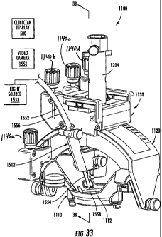
[0096] Figure 33 is a perspective view of a trajectory guide frame and a
camera
bracket according to further embodiments of the present invention.
[0097] Figure 34 is a top plan view of the trajectory guide frame bf Figure 33
with a yoke and platform thereof removed and the camera bracket shown mounted
thereon in alternative positions.
[0098] Figure 35 is a partial perspective view of the trajectory guide frame
and
camera bracket of Figure 33.
[0099] Figure 36 is an exploded, partial perspective view of the trajectory
guide
frame and camera bracket of Figure 33.
[00100] Figure 37 is a cross-sectional view of the trajectory guide frame
and camera bracket of Figure 33 taken along the line 37-37 of Figure 35. .
[00101] Figure 38 is a cross-sectional view of the trajectory guide frame
and camera bracket of Figure 33 taken along the line 38-38 of Figure 33,
wherein a
targeting cannula thereof is in a retracted position.
[00102] Figure 39 is a partial top plan view of the trajectory guide frame
of Figure 33.
[00103] Figure 40 is a partial cross-sectional view of the trajectory guide
frame of Figure 33, wherein the targeting cannula is in an extended position.
[00104] Figure 41 is a partial cross-sectional view of the trajectory guide
frame of Figure 33, wherein an alternative targeting cannula according to
further
embodiments of the present invention is in a retracted position.
[00105] Figures 42 and 43 are exploded perspective views of a base and a
yoke of the trajectory guide frame of Figure 33 illustrating a mounting system
according
to some embodiments of the present invention.
[00106] Figure 44 is a cross-sectional view of the base and yoke of Figure
42 taken along the line 44-44 of Figure 43.
[00107] Figure 45 is a partial perspective view of the trajectory guide
frame of Figure 33 illustrating a stabilizer system according to some
embodiments of the
present invention.
[00108] Figure 46 is an exploded, partial perspective view of the trajectory
guide frame of Figure 33.
[00109] Figure 47 is a further perspective view of the trajectory guide
16
CA 02700523 2010-03-23
WO 2009/042130 PCT/US2008/011040
frame of Figure 33.
[00110] Figure 48 is a perspective view of a lock clip for use with the
trajectory guide frame of Figure 33.
[00111] Figure 49A is a top plan view of the base of the trajectory guide
frame of Figure 33 illustrating a layout of MRI-visible fiducial markers of
the trajectory
guide frame.
[00112] Figure 49B is a schematic view of a display including an image
based on MRI image data including representations of a patient's head and a
base and
fiducial markers of the trajectory guide frame.
[00113] Figure 50 is an enlarged, fragmentary, perspective view of the
trajectory guide frame of Figure 33 illustrating a fiducial marker positioning
feature
according to some embodiments of the present invention.
[00114] Figure 51 is an enlarged, fragmentary, perspective view of the
trajectory guide frame of Figure 33 illustrating a fiducial marker tab relief
feature
according to some embodiments of the present invention.
DETAILED DESCRIPTION
[00115] The present invention now is described more fully hereinafter with
reference to the accompanying drawings, in which some embodiments of the
invention
are shown. This invention may, however, be embodied in many different forms
and
should not be construed as limited to the embodiments set forth herein;
rather, these
embodiments are provided so that this disclosure will be thorough and
complete, and will
fully convey the scope of the invention to those skilled in the art.
[00116] Like numbers refer to like elements throughout. In the figures,
the thickness of certain lines, layers, components, elements or features may
be
exaggerated for clarity.
[00117] The terminology used herein is for the purpose of describing
particular embodiments only and is not intended to be limiting of the
invention. As used
herein, the singular forms "a", "an" and "the" are intended to include the
plural forms as
well, unless the context clearly indicates otherwise. It will be further
understood that the
terms "comprises" and/or "comprising," when used in this specification,
specify the
presence of stated features, steps, operations, elements, and/or components,
but do not
preclude the presence or addition of one or more other features, steps,
operations,
17
CA 02700523 2010-03-23 =
WO 2009/042130 PCT/IJS2008/011040
elements, components, and/or groups thereof. As used herein, the term "and/or"
includes
any and all combinations of one or more of the associated listed items.
[001181 Unless otherwise defined, all terms (including technical and
scientific terms) used herein have the same meaning as commonly understood by
one of
ordinary skill in the art to which this invention belongs. It will be further
understood that
terms, such as those defined in commonly used dictionaries, should be
interpreted as
having a meaning that is consistent with their meaning in the context of the
specification
and relevant art and should not be interpreted in an idealized or overly
formal sense
unless expressly so defined herein. Well-known functions or constructions may
not be
described in detail for brevity and/or clarity.
[001191 It will be understood that when an element is referred to as being
"on", "attached" to, "connected" to, "coupled" with, "contacting", etc.,
another element,
it can be directly on, attached to, connected to, coupled with or contacting
the other
element or intervening elements may also be present. In contrast, when an
element is
referred to as being, for example, "directly on", "directly attached" to,
"directly
connected" to, "directly coupled" with or "directly contacting" another
element, there
are no intervening elements present. It will also be appreciated by those of
skill in the
art that references to a structure or feature that is disposed "adjacent"
another feature
may have portions that overlap or underlie the adjacent feature.
[001201 Spatially relative terms, such as "under", "below", "lower", "over",
"upper" and the like, may be used herein for ease of description to describe
one element
or feature's relationship to another element(s) or feature(s) as illustrated
in the figures. It
will be understood that the spatially relative terms are intended to encompass
different
orientations of the device in use or operation in addition to the orientation
depicted in the
figures. For example, if the device in the figures is inverted, elements
described as
"under" or "beneath" other elements or features would then be oriented "over"
the other
elements or features. Thus, the exemplary term "under" can encompass both an
orientation of "over" and "under". The device may be otherwise oriented
(rotated 90
degrees or at other orientations) and the spatially relative descriptors used
herein
interpreted accordingly. Similarly, the terms "upwardly", "downwardly",
"vertical",
"horizontal" and the like are used herein for the purpose of explanation only
unless
specifically indicated otherwise:
[00121] The term "MRI visible" means that a device is visible, directly or
18
CA 02700523 2010-03-23
WO 2009/042130 PCTIUS2008/011040
indirectly, in an MRI image. The visibility may be indicated by the increased
SNR of the
MRI signal proximate to the device (the device can act as an MRI receive
antenna to
collect signal from local tissue) and/or that the device actually generates
MRI signal
itself, such as via suitable hydro-based coatings and/or fluid (typically
aqueous solutions)
filled channels or lumens.
[00122] The term "MRI compatible" means that a device is safe for use in
an MRI environment and/or can operate as intended in an MRI environment, and,
as
such, if residing within the high-field strength region of the magnetic field,
is typically
made of a non-ferromagnetic MRI compatible material(s) suitable to reside
and/or
operate in a high magnetic field environment.
[00123] The term "high-magnetic field" refers to field strengths above
about 0.5 T, typically above 1.OT, and more typically between about 1.5T and
IOT.
[00124] The term "targeting cannula" refers to an elongate device,
typically having a substantially tubular body that can be oriented to provide
positional
data relevant to a target treatment site and/or define a desired access path
orientation or
trajectory. At least portions of a targeting cannula contemplated by
embodiments of the
invention can be configured to be visible in an MRI image, thereby allowing a
clinician
to visualize the location and orientation of the targeting cannula in vivo
relative to
fiducial and/or internal tissue landscape features. Thus, the term "cannula"
refers to an
elongate device that can be associated with a trajectory frame that attaches
to a patient,
but does not necessarily enter the body of a patient.
[00125] The term "imaging coils" refers to a device that is configured to
operate as an MRI receive antenna. The term "coil" with respect to imaging
coils is not
limited to a coil shape but is used generically to refer to MRI antenna
configurations,
loopless, looped, etc., as are known to those of skill in the art. The term
"fluid-filled"
means that the component includes an amount of the fluid but does not require
that the
fluid totally, or even substantially, fill the component or a space associated
with the
component. The fluid may be an aqueous solution, MR contrast agent, or any
material
that generates MRI signal.
[00126] The term "two degrees of freedom" means that the trajectory
frame described herein allows for at least translational (swivel or tilt) and
rotational
movement over a fixed site, which may be referred to as a Remote Center of
Motion
(RCM).
19
CA 02700523 2010-03-23
WO 2009/042130 PCT/US2008/011040
[00127] The term "programmatically" refers to operations directed and/or
primarily carried out electronically by computer program modules, code and
instructions.
1001281 The term "fiducial marker" refers to a marker that can be
identified using electronic image recognition, electronic interrogation of MRI
image
data, or three-dimensional electrical signals to define a position and/or find
the feature or
component in 3-D space.
[00129] Embodiments of the present invention can be configured to guide
and/or place diagnostic or interventional devices and/or therapies to any
desired internal
region of the body or object using MRI and/or in an MRI scanner or MRI
interventional
suite. The object can be any object, and may be particularly suitable for
animal and/or
human subjects. Some embodiments can be sized and configured to place
implantable
DBS leads for brain stimulation, typically deep brain stimulation. Some
embodiments
can be configured to deliver tools or therapies that stimulate a desired
region of the
sympathetic nerve chain. Other uses inside or outside the brain include stem
cell
placement, gene therapy or drug delivery for treating physiological
conditions. Some
embodiments can be used to treat tumors. Some embodiments can be used for RF
ablation, laser ablation, cryogenic ablation, etc. In some embodiments the
trajectory
frame and/or interventional tools can be configured to facilitate high
resolution imaging
via integral intrabody imaging coils (receive antennas), and/or the
interventional tools
can be configured to stimulate local tissue, which can facilitate confirmation
of proper
location by generating a physiologic feedback (observed physical reaction or
via flVIRI).
[001301 Some embodiments can be used to deliver bions, stem cells or
other target cells to site-specific regions in the body, such as neurological
target and the
like. In some embodiments, the systems deliver stem cells and/or other cardio-
rebuilding
cells or products into cardiac tissue, such as a heart wall via a minimally
invasive MRI
guided procedure, while the heart is beating (i.e., not requiring a non-
beating heart with
the patient on a heart-lung machine). Examples of known stimulation treatments
and/or
target body regions are described in U.S. Patent Nos. 6,708,064; 6,438,423;
6,356,786;
6,526,318; 6,405,079; 6,167,311; 6539,263; 6,609,030 and 6,050,992, the
contents of
which are hereby incorporated by reference as if recited in full herein.
[00131] Generally stated, some embodiments of the invention are directed
to MRI interventional procedures and provide interventional tools and/or
therapies that
may be used to locally place interventional tools or therapies in vivo to site-
specific
CA 02700523 2010-03-23
WO 2009/042130 PCT/US2008/011040
regions using an MRI system. The interventional tools can be used to define an
MRI-
guided trajectory or access path to an in vivo treatment site. Some
embodiments of the
invention provide interventional tools that can provide positional data
regarding location
and orientation of a tool in 3-D space with a visual confirmation on an MRI.
Embodiments of the invention may provide an integrated system that may allow
physicians to place interventional devices/leads and/or therapies accurately
and in shorter
duration procedures over conventional systems (typically under six hours for
DBS
implantation procedures, such as between about 1-5 hours).
1001321 In some embodiments, MRI can be used to visualize (and/or
locate) a therapeutic region of interest inside the brain or other body
locations and utilize
MRI to visualize (and/or locate) an interventional tool or tools that will be
used to deliver
therapy and/or to place a chronically implanted device that will deliver
therapy. Then,
using the three-dimensional data produced by the MRI system regarding the
location of
the therapeutic region of interest and the location of the interventional
tool, the system
and/or physician can make positional adjustments to the interventional tool so
as to align
the trajectory of the interventional tool, so that when inserted into the
body, the
interventional tool will intersect with the therapeutic region of interest.
With the
interventional tool now aligned with the therapeutic region of interest, an
interventional
probe can be advanced, such as through an open lumen inside of the
interventional tool,
so that the interventional probe follows the trajectory of the interventional
tool and
proceeds to the therapeutic region of interest. It should be noted that the
interventional
tool and the interventional probe may be part of the same component or
structure. A
sheath may optionally form the interventional tool or be used with an
interventional
probe or tool.
1001331 In particular embodiments, using the MRI in combination with
local or internal imaging coils and/or MRI contrast material that may be
contained at
least partially in and/or on the interventional probe or sheath, the location
of the
interventional probe within the therapeutic region of interest can be
visualized on a
display or image and allow the physician to either confirm that the probe is
properly
placed for delivery of the therapy (and/or placement of the implantable device
that will
deliver the therapy) or determine that the probe is in the incorrect or a non-
optimal
location. Assuming that the interventional probe is in the proper desired
location, the
therapy can be delivered and/or the interventional probe can be removed and
replaced
21
CA 02700523 2010-03-23
WO 2009/042130 PCT/US2008/011040
with a permanently implanted therapeutic device at the same location.
[00134] In some embodiments, in the event that the physician determines
from the MRI image produced by the MRI and the imaging coils, which may
optionally
be contained in or on the interventional probe, that the interventional probe
is not in the
proper location, a new therapeutic target region can be determined from the
MRI images,
and the system can be updated to note the coordinates of the new target
region. The
interventional probe is typically removed (e.g., from the brain) and the
interventional
tool can be repositioned so that it is aligned with the new target area. The
interventional
probe can be reinserted on a trajectory to intersect with the new target
region. Although
described and illustrated herein with respect to the brain and the insertion
of deep brain
stimulation leads, it is understood that embodiments of the present invention
may be
utilized at other portions of the body and for various other types of
procedures.
[00135] Exemplary embodiments are described below with reference to
block diagrams and/or flowchart illustrations of methods, apparatus (systems
and/or
devices) and/or computer program products. It is understood that a block of
the block
diagrams and/or flowchart illustrations, and combinations of blocks in the
block
diagrams and/or flowchart illustrations, can be implemented by computer
program
instructions. These computer program instructions may be provided to a
processor of a
general purpose computer, special purpose computer, and/or other programmable
data
processing apparatus to produce a machine, such that the instructions, which
execute via
the processor of the computer and/or other programmable data processing
apparatus,
create means (functionality) and/or structure for implementing the
functions/acts
specified in the block diagrams and/or flowchart block or blocks.
[00136] These computer program instructions may also be stored in a
computer-readable memory that can direct a computer or other programmable data
processing apparatus to function in a particular manner, such that the
instructions stored
in the computer-readable memory produce an article of manufacture including
instructions which implement the functions/acts specified in the block
diagrams and/or
flowchart block or blocks.
[00137] The computer program instructions may also be loaded onto a
computer or other programmable data processing apparatus to cause a series of
operational steps to be performed on the computer or other programmable
apparatus to
produce a computer-implemented process such that the instructions which
execute on the
22
CA 02700523 2010-03-23
WO 2009/042130 PCT/US2008/011040
computer or other programmable apparatus provide steps for implementing the
functions/acts specified in the block diagrams and/or flowchart block or
blocks.
[00138] Accordingly, exemplary embodiments may be implemented in
hardware and/or in software (including firmware, resident software, micro-
code, etc.).
Furthermore, exemplary embodiments may take the form of a computer program
product
on a computer-usable or computer-readable storage medium having computer-
usable or
computer-readable program code embodied in the medium for use by or in
connection
with an instruction execution system. In the context of this document, a
computer-usable
or computer-readable medium may be any medium that can contain, store,
communicate,
propagate, or transport the program for use by or in connection with the
instruction
execution system, apparatus, or device.
[00139] The computer-usable or computer-readable medium may be, for
example but not limited to, an electronic, magnetic, optical, electromagnetic,
infrared, or
semiconductor system, apparatus, device, or propagation medium. More specific
examples (a non-exhaustive list) of the computer-readable medium would include
the
following: an electrical connection having one or more wires, a portable
computer
diskette, a random access memory (RAM), a read-only memory (ROM), an erasable
programmable read-only memory (EPROM or Flash memory), an optical fiber, and a
portable compact disc read-only memory (CD-ROM). Note that the computer-usable
or
computer-readable medium could even be paper or another suitable medium upon
which
the program is printed, as the program can be electronically captured, via,
for instance,
optical scanning of the paper or other medium, then compiled, interpreted, or
otherwise
processed in a suitable manner, if necessary, and then stored in a computer
memory.
[00140] Computer program code for carrying out operations of data
processing systems discussed herein may be written in a high-level programming
language, such as Java, AJAX (Asynchronous JavaScript), C, and/or C++, for
development convenience. In addition, computer program code for carrying out
operations of exemplary embodiments may also be written in other programming
languages, such as, but not limited to, interpreted languages. Some modules or
routines
may be written in assembly language or even micro-code to enhance performance
and/or
memory usage. However, embodiments are not limited to a particular programming
language. It will be further appreciated that the functionality of any or all
of the program
modules may also be implemented using discrete hardware components, one or
more
23
CA 02700523 2010-03-23
WO 2009/042130 PCT/1JS2008/011040
application specific integrated circuits (ASICs), or a programmed digital
signal processor
or microcontroller.
[00141] Embodiments of the present invention will now be described in
detail below with reference to the figures. Fig. 1A is a block diagram of an
MRI-guided
interventional system 50, according to some embodiments of the present
invention. The
illustrated system 50 includes an MRI scanner 75, a trajectory frame 100
attached to the
body of a patient positioned within a magnetic field of the MRI scanner 75, a
remote
control unit 400, a trajectory guide software module 300, and a clinician
display 500.
The trajectory frame 100 supports a targeting cannula through which various
interventional devices may be inserted into the body of a patient. The frame
100 is
adjustable such that the targeting cannula is rotatable about a pitch axis,
about a roll axis,
and such that the targeting cannula can translate in X-Y directions. The frame
100 may
be attached to the body of a patient directly or indirectly and may be
configured to be
attached to various parts of the body.
[00142] In some embodiments, a remote control unit 400 is provided to
allow a user to remotely adjust the position of the targeting cannula. The
trajectory guide
software module 300 allows a user to define and visualize, via display 500, a
desired
trajectory (D, Figs. 17-18) into the body of a patient of an interventional
device
extending through the targeting cannula. The trajectory guide software module
300 also
allows the user to visualize and display, via display 500, an actual
trajectory (A, Fig. 17)
into the body of an interventional device extending through the targeting
cannula. The
trajectory guide software module 300 displays to the user the necessary
positional
adjustments (e.g., pitch axis rotation, roll axis rotation, X-Y translation)
needed to align
the actual trajectory of the targeting cannula with the desired trajectory
path (Fig. 1B). In
addition, the user can view, via display 500, the actual trajectory changing
as he/she
adjusts the position of the targeting cannula. The trajectory guide software
module 300 is
configured to indicate and display when an actual trajectory is aligned with a
desired
trajectory.
[00143] Fig. 2A illustrates a burr hole 10 formed in the skull S of a patient.
A burr hole ring 12 overlies the burr hole 10 and is secured to the skull S.
The illustrated
burr hole ring 12 has a pair of ears 14, each configured to receive a
respective fastener
(e.g., screw) therethrough for securing the burr hole ring 12 to the skull. In
the illustrated
embodiment, the burr hole ring 12 is secured to the skull S via screws 16.
Fig. 2B
24
CA 02700523 2010-03-23
WO 2009/042130 PCT/US2008/011040
illustrates a removable centering device 18 positioned on the burr hole ring
12. The
centering device 18 includes cut out portions 20 that fit over the ears 14 of
the burr hole
ring 12. The function of the centering device 18 is to facilitate centering a
trajectory
frame 100, described below, over the burr hole 10. After the frame 100 is
attached to the
skull of a patient, the centering device 18 is removed.
[00144] Referring to Fig. 3A, a trajectory frame 100 with a targeting
cannula 200 associated therewith is illustrated. The trajectory frame 100
allows for the
adjustability (typically at least two degrees of freedom, including rotational
and
translational) and calibration/fixation of the trajectory of the targeting
cannula 200 and/or
probe or tool inserted through the targeting cannula 200. The targeting
cannula 200
includes an axially-extending guide bore (not shown) therethrough that is
configured to
guide the desired therapeutic or diagnostic tool, e.g., intra-brain placement
of a
stimulation lead (or other type of device) in vivo, as will be described
below. Intra-brain
placement of devices may include chronically placed devices and acutely placed
devices.
The trajectory frame 100 may include fiducial markers 117 that can be detected
in an
MRI to facilitate registration of position in an image.
[001451 The illustrated trajectory frame 100 is configured to be mounted to
a patient's skull around a burr hole ring (12, Fig. 1) and over a burr hole
(10, Fig. 1), to
provide a stable platform for advancing surgical devices, leads, etc. in the
brain. The
frame 100 includes a base 110, a yoke, 120, a platform 130, and a plurality of
actuators
140a-140d. The base 110 has a patient access aperture 112 formed therein, as
illustrated.
The base 110 is configured to be secured (directly or indirectly) to the skull
of a patient
such that the patient access aperture 112 overlies the burr hole 10 in the
patient skull.
The patient access aperture 112 is centered over the burr hole 10 via the
removable
centering device 18. The yoke 120 is movably mounted to the base 110 and is
rotatable
about a roll axis RA. A roll actuator 140a is operably connected to the yoke
120 and is
configured to rotate the yoke 120 about the roll axis RA, as will be described
in detail
below. In some embodiments, the yoke 120 has a range of motion about the roll
axis RA
of about seventy degrees (70 ). However, other ranges, greater and lesser than
70 , are
possible, e.g., any suitable angle typically between about 10 - 90 , 30 - 90 ,
etc. The
illustrated platform 130 is movably mounted to the yoke 120 and is rotatable
about a
pitch axis PA. In some embodiments, the platform 130 has a range of motion
about the
pitch axis PA of about seventy degrees (70 ). However, other ranges, greater
and lesser
CA 02700523 2010-03-23
WO 2009/042130 PCT/US2008/011040
than 70 , are possible, e.g., any suitable angle typically between about 10 -
90 , 30 -
90 , etc.
[00146] Figs. 3B-3E are side view, schematic illustrations of the trajectory
frame being secured to the skull of a patient. Fig. 3B illustrates use of the
centering
device 18 to align the frame 100 relative to the burr hole 10. In Fig. 3C, the
frame 100 is
secured to the skull with fasteners and such that the patient access aperture
112 in the
base 110 is centered around the centering device 18. In Fig. 3D, the yoke 120
is rotated
out of the way such that the centering device 18 can be removed. In Fig. 3E,
the
targeting cannula 200 is moved to an extended position and locked in the
extended
position via prongs 208.
[00147] The platform 130 includes an X-Y support table 132 that is
movably mounted to the platform 130. The X-Y support table 132 is configured
to move
in an X-direction and Y-direction relative to the platform 130. An X-direction
actuator
140c is operably connected to the platform 130 and is configured to move the X-
Y
support table 132 in the X-direction. A Y-direction actuator 140d is operably
connected
to the platform 130 and is configured to move the X-Y support table 132 in the
Y-
direction. A pitch actuator 140b is operably connected to the platform 130 and
is
configured to rotate the platform 130 about the pitch axis PA, as will be
described in
detail below.
[00148] The actuators 140a-140d are configured to translate and/or rotate
the frame. The targeting cannula 200 is configured to translate in response to
translational movement of the X-Y support table 132 and to rotate in response
to
rotational movement of the yoke 120 and platform 130 to define different axial
intrabody
trajectories extending through the patient access aperture 112 in the frame
base 110.
[00149] The actuators 140a-140d may be manually-operated devices, such
as thumbscrews, in some embodiments. The thumbscrews can be mounted on the
frame
100 or may reside remotely from the frame 100. A user may tum the actuators
140a-
140d by hand to adjust the position of the frame 100 and, thereby, a
trajectory of the
targeting cannula 200. In other embodiments, the actuators 140a-140d are
operably
connected to a remote control unit 400 (Figs. 9-10) via a respective plurality
of non-
ferromagnetic, flexible drive shafts or control cables 150a-150d. The remote
control unit
400 includes a plurality of position controls 402a-402d, and each cable 150a-
150d is
operably connected to a respective position control 402a-402d and to a
respective
26
CA 02700523 2010-03-23
WO 2009/042130 PCT/US2008/011040
actuator 140a-140d. Movement of a position contro1402a-402d operates a
respective
actuator 140a-140d via a respective control cable 150a-150d, as will be
described below.
The cables 150a-150d may extend a suitable distance (e.g., between about 1-4
feet, etc.)
to allow a clinician to adjust the settings on the trajectory frame 100
without moving a
patient and from a position outside the bore of a magnet (where such magnet
type is
used) associated with an MRI scanner.
[001501 Referring to Figs. 6-7, the base 110 includes a plurality of
locations 112 for attaching the base 110 to a skull of a patient via
fasteners. Each
location may include two or more adjacent apertures 114. Each aperture 114 is
configured to receive a fastener (e.g., a screw, rod, pin, etc.) therethrough
that is
configured to secure the base 110 to the skull of a patient.
[00151] The base 110 also includes MRI-visible fiducial markers 117 that
allow the location/orientation of the frame 100 to be determined within an MRI
image
during an MRI-guided procedure. In the illustrated embodiment, the fiducial
markers 117
have a torus or "doughnut" shape and are spaced apart. However, fiducial
markers
having various shapes and positioned at various locations on the frame 100 may
be
utilized.
[00152] The base 110 also includes a pair of spaced apart arcuate anns
116, as illustrated in Fig. 11. The yoke 120 is pivotally attached to pivot
points 113 for
rotation about the roll axis RA. The yoke 120 engages and moves along the base
arcuate
arms 116 when rotated about the roll axis RA. In the illustrated embodiment,
one of the
base arcuate arms 116 includes a thread pattern 118 formed in (e.g., embossed
within,
machined within, etc.) a surface 116a thereof. However, in other embodiments,
both
arms 116 may include respective thread patterns. The roll actuator 140a
includes a
rotatable worm 142 with teeth that are configured to engage the thread pattern
118, as
illustrated in Fig. 5. As the worm 142 is rotated, the teeth travel along the
thread pattern
118 in the arcuate arm surface 116a. Because the base 110 is fixed to a
patient's skull,
rotation of the roll actuator worm 142 causes the yoke 120 to rotate about the
roll axis
RA relative to the fixed base 110. Rotation about roll axis RA is illustrated
in Figs. 4-5.
For example, in Fig. 5, the yoke 120 is rotated about the roll axis RA
sufficiently to
allow removal of the centering device 18.
1001531 Referring to Fig. 12, the yoke 120 includes a pair of spaced apart
upwardly extending, arcuate arms 122. The platform 130 engages and moves along
the
27
CA 02700523 2010-03-23
WO 2009/042130 PCT/US2008/011040
yoke arcuate arms 122 when rotated about.the pitch axis PA. In.the illustrated
embodiment, one of the yoke arcuate arms 122 includes a thread pattern 124
formed in
(e.g., embossed within, machined within, etc.) a surface 122a thereof.
However, in other
embodiments, both arms 122 may include respective thread patterns. The pitch
actuator
140b includes a rotatable worm 146 with teeth 148 that are configured to
engage the
thread pattern 124. As the worm 146 is rotated, the teeth 148 travel along the
thread
pattern 124 in the arcuate arm surface 122a. Because the base 110 is fixed to
a patient's
skull, rotation of the pitch actuator worm 146 causes the platform 130 to
rotate about the
pitch axis PA relative to the fixed base 110.
[00154] As illustrated in Fig. 19A, the X-Y support table 132 includes a
moving plate 134 that moves in both the X-direction and Y-direction. The X-
direction
actuator 140c, when rotated, causes translational movement of the moving plate
134
along the X-axis. For example, clockwise rotation of the X-direction actuator
140c
causes movement toward the "-X direction (i.e., to the left) in Fig. 19A; and
counterclockwise rotation of the X-direction actuator 140c causes movement
along the
+X direction (i.e., to the right) in Fig. 19A, etc. The Y-direction actuator
140d, when
rotated, causes translational movement of the moving plate 134 along the Y-
axis. For
example, clockwise rotation of the Y-direction actuator 140d causes movement
along the
-Y direction (i.e., out of the paper) in Fig. 19A; and clockwise rotation of
the Y-direction
actuator 140d causes movement along the +Y direction (i.e., into the paper) in
Fig. 19A.
In the illustrated embodiment, graduation scales 136, 137 are provided on the
platform
adjacent the moving plate 134. The moving plate 134 includes a pair of marks
or
indicators 138 that provide visual indication of X-Y movement of the moving
plate 134.
Fig. 19B illustrates X-Y translation of an X-Y support table 132, according to
some
embodiments of the present invention.
[00155] Various internal drive mechanisms may be utilized for causing
translational movement of the moving plate 134 in response to user rotation of
the X-
direction actuator 140c and the Y-direction actuator 140d. For example, drive
belts,
linkages, gears, or worm drives may be utilized, as would be understood by one
skilled
in the art of X-Y tables. Embodiments of the present invention are not limited
to any
particular mechanism for translating the X-Y table 132 along the X and Y
directions.
Fig. 19C is partial perspective view of an X-Y support table, according to
some
embodiments, with elements removed to reveal internal components of an X-
direction
28
CA 02700523 2010-03-23
WO 2009/042130 PCT/US2008/011040
actuator 140c and Y-direction actuator 140d.
[00156] As illustrated in Fig. 3A, the roll actuator 140a, pitch actuator
140b, X-direction actuator 140c, and Y-direction actuator 140d each extend
outwardly
from the frame 100 along the same direction (e.g., upwardly from the platform
130).
This configuration facilitates easy connection of the control cables 150a-150d
to the
actuators 140a-140d (where used) and also facilitates bundling of the cables
150a-150d
to reduce clutter or provide ease of handling and set-up. Embodiments of the
present
invention are not limited to the illustrated embodiment, however. The
actuators 140a-
140d may extend in various directions and these directions may be different
from each
other. In addition, the actuators 140a-140d may extend along the same
direction from the
frame, but in a different direction than that illustrated in Fig. 3A. For
example, Fig. 16
illustrates an embodiment where the actuators 140a-140d extend from a common
side of
the platform 130.
[00157] Referring to Figs. 9 and 10A-10C, the remote control unit 400 of
the illustrated system 50 includes a plurality of manually-operable position
controls
402a-402d. Specifically, the control unit 400 includes a roll adjustment
control 402a, a
pitch adjustment control 402b, an X-direction adjustment contro1402c, and a Y-
direction
adjustment control 402d. A roll control cable 150a is operably connected to
the roll
adjustment control 402a and to the roll actuator 140a such that movement of
the roll
adjustment control 402a operates the roll actuator 140a via the roll control
cable 150a. A
pitch control cable 150b is operably connected to the pitch adjustment control
402b and
to the pitch actuator 140b such that movement of the pitch adjustment control
402b
operates the pitch actuator 140b via the pitch control cable 150b. An X-
direction control
cable 150c is operably connected to the X-direction control 402c and to the X-
direction
actuator 140c such that movement of the X-direction adjustment control 402c
operates
the X-direction actuator 140c via the X-direction control cable 150c. A Y-
direction
control cable 150d is operably connected to the Y-direction control 402d and
to the Y-
direction actuator 140d such that movement of the Y-direction adjustment
control 402d
operates the Y-direction actuator 140d via the Y-direction control cable 150d.
[00158J In the illustrated embodiment, each of the position controls 402a-
402d is a thumbwheel control that can be rotated by a user's finger in
clockwise and
counterclockwise directions. Rotation of each thumbwheel 402a-402d by a user
causes
corresponding axial rotation of a respective control cable 150a-150d and
corresponding
29
CA 02700523 2010-03-23
WO 2009/042130 PCT/US2008/011040
axial rotation of a respective actuator 140a-140d.
[00159] Fig. lOB illustrates position controls,.according to additional
embodiments of the present invention, that utilize two thumbwheels. One
thumbwheel
402a' is for "fine" adjustments; the other thumbwheel 402a" is for "gross"
adjustments.
The amount of fine and gross adjustment is correlated to the diameter of each
thumbwheel, as would be understood by one skilled in the art. Fig. 10C
illustrates a
position control 402a"', according to additional embodiments of the present
invention,
that indicates incremental X-Y variable markings.
[00160] In the illustrated embodiment, locking mechanisms 404a-404c are
associated with the thumbwheels 402a-402d and prevent user rotation thereof
when in a
locked position. For example, a locking mechanism 404a is operably associated
with the
roll adjustment control 402a and is configured to prevent rotation thereof by
a user when
in a "locked" position. Locking mechanism 404b is operably associated with
pitch
adjustment control 402b and is configured to prevent rotation thereof by a
user when in a
"locked" position. Locking mechanism 404c is operably associated with X-
direction
control 402c and Y-direction control 402d and is configured to prevent
rotation of X-
direction control 402c and Y-direction control 402d by a user when in a
"locked"
position.
[00161] Each control cable 150a-150d can have a geometrically shaped
rigid end 151a-151d that is configured to removably engage a free end of a
respective
actuator 140a-140d. As illustrated in Fig. 16, the respective free ends 141a-
141d of the
actuators 140a-140d may have a slot 143 formed therein that is configured to
removably
receive a respective cable end. Exemplary cable end shapes include, but are
not limited
to, "L" shapes, "U" shapes, square shapes, rectangular shapes, ovaVcircular
shapes, and
other polygonal shapes. Each cable end has sufficient rigidity such that axial
rotation of
the cable causes the cable free end to impart rotational motion to a
respective actuator.
Fig. 15 illustrates the free end of cable 150b having a connector 151b with a
geometric
shape attached thereto that is configured to matingly engage a respective slot
143 in
actuator 140b, according to other embodiments of the present invention.
[00162] In some embodiments, the free end of an actuator 140a-140d may
be configured to receive only a specific one of the control cables 150a-150d.
For
example, in Fig. 15, the connector 151b may not fit within the slots 143 of
any of the
other actuators. As such, a control cable cannot be inadvertently connected to
the wrong
CA 02700523 2010-03-23
WO 2009/042130 PCT/US2008/011040
actuator. For example, the roll adjustment actuator free end 141a may be
configured to
only receive the free end 151a of the control cable 150a associated with the
roll control
402a. Similarly, the pitch adjustment actuator free end 141b may be configured
to only
receive the free end 151b of the control cable 150b associated with the pitch
control
402b.
[00163] Each control cable 150a-150d also has a flexible elastomeric (e.g.,
silicone, rubber, etc.) collar 154a-154d that is configured to surround a
respective
actuator 140a-140d and maintain engagement of the respective cable end 151a-
151d
within the respective actuator. Each elastomeric collar 154a-154d is
configured to
prevent removal of a cable by a user, for example, as a result of inadvertent
tugging on
the cable by a user, or by movement of the remote control unit 400. Each of
the
illustrated collars 154a-154d can be rolled or folded back then released to
cover and
conformably compress against an actuator to hold the end of a respective cable
in
position. Each collar 154a-154d can then be pushed back to easily release the
cable from
the actuator. In the illustrated embodiment, each actuator 140a-140d has a
circumferential groove 145 configured to receive a corresponding
circumferential ridge
156 of a collar 154a-154d in mating 'relation therewith.
[00164] Fig. 16B illustrates remote control cable end 151a about to be
inserted into slot 143 in the actuator free end 141a. The cable end 151a is
inserted into
the slot 143 and then the elastomeric collar 154a is fitted around the
actuator 140a such
that the circumferential ridge 156 engages the circumferential actuator groove
145, as
illustrated in Fig. 16C. Because of the elastomeric nature of the collar 154a,
the collar
snuggly fits the actuator 140a and retains the cable end 151a within slot 143.
To remove
the cable end 151a, the circumferential ridge 156 is pulled out of the groove
145 and the
collar 154a is rolled back on itself as illustrated in Figs. 16D-16E.
[00165] Embodiments of the present invention are not limited to the
illustrated elastomeric collars 154a-154d. Other ways of retaining the cable
ends 151a-
151d within respective actuators 140a-140d may be utilized without limitation.
1001661 In some embodiments, the actuators 140a-140d are color coded
such that each actuator has a different respective color for easy
identification by a user.
For example, the roll actuator 140a may be colored red and the pitch actuator
140b may
be colored yellow such that a user can easily identify the two respective
actuators when
positioning the frame 100. In some embodiments, the elastomeric collars 154a-
154d may
31
CA 02700523 2010-03-23
WO 2009/042130 PCT/US2008/011040
also be color coded so as to match the color of a respective actuator 140a-
140d. In some
embodiments, the cable ends 151a-151d may also be color coded so as to match
the
color of a respective actuator 140a-140d.
[00167] In some embodiments of the present invention, the control cables
150a-150d are formed of NITINOL or other MR-compatible materials. One or more
portions of the frame 100 and the targeting cannula 200 may also be formed of
NITINOL
or other MR-compatible (non-paramagnetic) materials.
[00168] Fig. 31 illustrates a patient positioned within an MRI scanner and
a user utilizing the remote control apparatus 400 and display monitors to
adjust the
trajectory a targeting cannula, according to some embodiments of the present
invention.
[00169] Figs. 32A-32C illustrate a remote control unit 800 for remotely
controlling the positioning actuators of the frame of Fig. 3A, according to
other
embodiments of the present invention. The illustrated remote control unit 800
includes a
separate, multi-rod control device 802a-802d for each respective actuator 140a-
140d.
Each of the control devices 802a-802d can be identical in structure and, as
such, only
one will be described in detail hereinafter. However, each control device 802a-
802d may
be color coded with a color different from the other control devices 802a-
802d, and each
control device 802a-802d may have different lengths, shapes or sizes for ease
of
assembly and/or operation. Moreover, each control device 802a-802d may be
color
coded so as to match the color of a respective actuator 140a-140d.
[00170] Each control device 802a-802d includes a pair of elongated rods
804, 806 (e.g., solid, MRI-compatible rods) joined at respective ends via a
flexible
member such as a cable 807, as illustrated. The rods 804, 806 may be formed of
wood,
polymeric material, or other suitable relatively lightweight MR]-compatible
material. In
addition, the rods 804, 806 are not required to be solid.
[00171] The cable 807 is relatively short in length relative to the length of
rods 804, 806 and serves as a universal joint to allow the two rods 804, 806
to rotate
even when oriented transverse to each other. For example, the cable 807 may be
between
about one quarter inch and about one inch (0.25" - 1") in length. However,
embodiments
of the present invention are not limited to this range for the length of cable
807; cable
807 may have other lengths. In some embodiments, the cable 807 is an MRI-
compatible
cable (e.g., NITINOL, etc.). In the illustrated embodiment, the distal end
804b of rod 804
is joined to the proximal end 806a of rod 806 via cable 807. The cable 807 may
be joined
32
CA 02700523 2010-03-23
WO 2009/042130 PCT/US2008/011040
to rods 804, 806 in any of various ways including, but not limited to, via
adhesives, via
fasteners, via threaded connections, etc.
[00172] The proximal end 804a of rod 804 includes an endcap 808 secured
thereto. The endcap 808 may be formed from tactile material to facilitate
rotation of rod
804 by a user. A knob or other device that facilitates rotation may be secured
to the
proximal end of rod 804 in lieu of endcap 808, in other embodiments. In
operation, a
user rotates the proximal end 804a of rod 804 in a clockwise or
counterclockwise
direction to correspondingly rotate actuator 140a.
[00173J The distal end 806b of rod 806 is joined to actuator 140a via a
flexible member such as a cable 810, as illustrated. The cable 810 is
relatively short in
length relative to the length of rod 806 and serves as a universal joint to
allow rod 806 to
rotate even when oriented transverse to actuator 140a. In some embodiments,
the cable
810 is an MRI-compatible cable. For example, the cable 810 may be between
about one
half inch and about one and one half inch (0.5" - 1.5") in length. However,
embodiments
of the present invention are not limited to this range for the length of cable
810; cable
810 may have other lengths.
[00174] Cable 810 may be joined to rod 806 in any of various ways
including, but not limited to, via adhesives, via fasteners, via threaded
connections, etc.
The free end of cable 810 may have a rigid, geometrical shape, as described
above with
respect to the embodiments of cables 150a-150d, and may be configured to
engage a slot
within the actuator 140a, as described above. An elastomeric collar, as
described above
with respect to Figs. 16A-16E, may or may not be necessary to retain the free
end of
cable 810 within actuator 140a. In the illustrated embodiment, an elastomeric
collar is
not utilized. When used, an elastomeric collar can also be color coded to the
actuator
and/or control device rods 804, 806.
[00175] In the illustrated embodiment, the control devices 802a-802d are
supported by a pair of spaced apart separator devices 812. Each separator
device 812
includes a plurality of substantially parallel, spaced apart bores passing
therethrough that
are configured to receive each of the rods 804 for the respective control
devices 802a-
802d. The separator devices 812 are configured to maintain the rods 804 in
substantially
parallel, spaced apart relationship, as illustrated. In the illustrated
embodiment, only rods
804 pass through the two separator devices 812. However, embodiments of the
present
invention are not limited to the illustrated use, configuration, location, or
number of
33
CA 02700523 2010-03-23
WO 2009/042130 PCT/US2008/011040
separation devices 812. In addition, although shown as two rods forming each
control
device 802a-802d, they may include three or more spaced apart rods (not
shown), or
only a single rod (not shown). Moreover, other types of devices may be
utilized without
limitation.
[00176] Referring to Figs. 14 and 19, an elongated tubular member 204
extends through the platform 130 and yoke 120 and is secured to the X-Y
support table
132. The targeting cannula 200 is slidably secured within the tubular member
204 and is
movable between extended and retracted positions. Figs. 3A-3D and 4-6
illustrate the
targeting cannula 200 in a retracted position above the burr hole 10 and Figs.
3E and 8A
illustrate the targeting cannula 200 in an extended position. The tubular
member 204 is
configured to lock the targeting cannula 200 in an extended position and in a
retracted
position, as illustrated in Fig. 14. The tubular member 204 has a pair of
radially opposed
elongated, axial-extending slots 206, as illustrated. The ends 206a, 206b of
each slot 206
include a transverse portion 207 that is configured to retain the targeting
cannula 200 in
respective extended and retracted positions. The targeting cannula 200
includes a
respective pair of radially extending prongs 208, as illustrated. Each prong
208
cooperates with a respective slot 206. When the targeting cannula 200 is moved
upwardly to the retracted position, the targeting cannula 200 is rotated
slightly (or the
tubular member 204 is rotated slightly) such that prongs 208 each engage a
respective
transverse portion 207. When so engaged, the targeting cannula 200 is retained
in the
retracted position. When the targeting cannula 200 is moved downwardly to the
extended
position, the targeting cannula 200 is rotated slightly (or the tubular member
204 is
rotated slightly) such that prongs 208 each engage a respective transverse
portion 207.
When so engaged, the targeting cannula 200 is retained in the extended
position.
[00177] Embodiments of the present invention are not limited to the
illustrated configuration or number of slots 206 in the tubular member 204 or
to the
number of prongs 208 extending from the targeting cannula 200. For example,
the
targeting cannula 200 may have a single prong 208 that is configured to
cooperate with a
respective single slot 206 in the tubular member 204. In addition, slot 206
may have a
different configuration than illustrated.
[00178] Referring to Figs. 20-26, a depth stop 210 with a removable sheath
212 inserted and secured therein is illustrated. The illustrated depth stop
210 has a
generally cylindrical configuration with opposite proximal and distal ends
210a, 210b
34
CA 02700523 2010-03-23
WO 2009/042130 PCT/US2008/011040
and is adapted to be removably secured within the proximal end 204a of the
tubular
member 204. The depth stop 210 is configured to limit a distance that the
sheath 212
extends into the body of a patient when the depth stop is inserted within the
tubular
member 204. The sheath 212 is configured to receive and guide an elongated
interventional device therethrough, as will be described below. The sheath 212
includes
opposing tabs 214a, 214b that, when pulled apart, cause the sheath 212 to peel
away for
removal from the targeting cannula 200.
[00179] Prior to insertion within the tubular member 204, the distal end
210b of the depth stop 210 is positioned adjacent to a mark 215 on the
removable sheath
212, as illustrated in Fig. 21. Locking screw 216 is then tightened to prevent
axial
movement of the sheath 212 relative to the depth stop 210. An elongate
location
verification probe 217, such as an imaging probe or rigid stylet, is then
inserted within
the sheath 212 as illustrated in Fig. 21.
[00180] Referring to Figs. 22-23, the opposing tabs 214a, 214b of the
sheath are pulled apart slightly, and the depth stop 210, sheath 212 and
imaging probe
217 are inserted into the-proximal end 204a of tubular member 204. When so
inserted, as
illustrated in Fig. 23, the sheath 212 and imaging probe 217 pass through the
axial bore
of the targeting cannula 200. The imaging probe 217 is then utilized to verify
that the
distal end 212b of the sheath 212 is positioned at the correct location within
the body of
a patient. Upon verifying that the sheath 212 is accurately positioned, the
imaging probe
217 is removed (Fig. 23).
[00181] The probe 217 extending through the targeting cannula guide bore
can include at least one electrode (which may include a coil winding) on a
distal tip
portion thereof. The electrode can be a recording and/or stimulating
electrode. The
electrode can be configured to deliver test voltages for physiologic
confirmation of
location/efficacy that can be done by fMRI or by feedback from a non-
anesthetized
patient. Thus, a patient can be stimulated with an interventional probe (the
stimulation
may be via a transducer on a distal tip portion of the probe), to help confirm
that the
interventional probe is in the correct location (i. e., confirm proper
location via
anatomical as well as provide physiologic information and feedback). During
(and
typically substantially immediately after) stimulation from the interventional
probe, the
physician can monitor for a physiologic response from the patient that can be
observed
either directly from the patient as a physical response or via an fMRI-visible
response.
CA 02700523 2010-03-23
WO 2009/042130 PCT/US2008/011040
[00182] The elongate probe 217 can be MRI-visible and may optionally be
configured to define an MRI antenna. The system 50 can be configured to allow
for real-
time tracking under MRI, with an SNR imaging improvement in a diameter of at
least 1-
mm surrounding the probe.
[00183] Next, a locking mechanism 220 is removably secured to the
proximal end 210a of the depth stop 210. The locking mechanism 202 includes
opposing
axially extending slots 221, as illustrated. The portions of the sheath 212
that have been
peeled away extend through these slots as illustrated. The locking mechanism
220 is
secured to the depth stop/tubular member via locking screw 222.
[00184] The targeting cannula 200 is now ready to receive an
interventional device therein. As illustrated in Fig. 25, an interventional
device 230, such
as a brain stimulation lead, is inserted into the locking mechanism and
through the
removable sheath 212. A locking screw 224 is tightened to secure the
interventional
device 230 and prevent axial movement thereof.
[00185] The targeting cannula 200 can be MRI-visible. In some particular
embodiments, the cannula 200 may optionally comprise a plurality of spaced
apart
microcoils configured to provide data used to provide 3-D dimensional data in
MRI 3-D
space, such as a trajectory, or 3-D spatial coordinates of position of the
cannula 200. The
microcoils can each provide data that can be correlated to a three-dimensional
(X,Y, Z)
position in 3-D space in the body. The microcoils can be in communication with
the MRI
scanner, and tracking sequences can be generated and data from one or more of
the MRI
scanner channels can be used to define positional 3-D positional data and a
trajectory
thereof.
[00186] In some particular embodiments, the progress of the cannula 200
and/or interventional probe 230 or other device may optionally be tracked in
substantially real-time as it advances to the target via the coils (similar
ones of which
may also or alternatively be on or in the probe or other device) and/or
antenna. However,
real-time tracking may not be desired in some embodiments.
[00187] In some embodiments, the cannula 200 can.include at least one
axially extending fluid-filled hollow lumen (Fig. 8B) or closed channel with
fluid that
can generate MRI signal that can be detected by an MRI scanner and/or by an
internal
MRI antenna incorporated on and/or into the cannula 200 that can increase the
SNR of
the fluid to increase its visibility in an MRI. The fluid may be an aqueous
solution (able
36
CA 02700523 2010-03-23
WO 2009/042130 PCT/US2008/011040
to resonate at the proton frequency). The cannula 200 can include an axially
extending,
relatively thin segment, which creates a high contrast MRI image (a segment
filled with
water or other suitable contrast solution filled section/lumen). The thickness
of the
segment may be between about 0.25-15 mm (and the segment can have a tubular
shape
with a diameter or may define another cross-sectional shape such as a square
section).
The cannula 200 may include MRI imaging coils to increase the signal from the
high
contrast fluid. See, e.g., co-pending U.S. Patent Application Serial No.
12/066,862,
which is incorporated here by reference in its entirety.
[00188] As illustrated in Figs. 26A and 26B, the interventional device 230
is positioned at the desired location within the body of a patient. The
removable sheath
212 is then completely removed by pulling apart the opposing tabs 214a, 214b.
A clamp
240 (Fig. 27) is inserted within the burr hole ring 12 that grips the
interventional device
230 to prevent inadvertent removal of the interventional device 230 from the
body of the
patient. The clamp 240 is secured to the burr hole ring 12. The frame 100 can
then be
removed from the body of the patient, leaving behind only the interventional
device 230
that has been inserted into the patient body.
[00189] Figs. 28A-28G are side view, schematic illustrations of the
trajectory frame 100 illustrating an exemplary series of operations for the
insertion of
interventional devices within the body of a patient via the targeting cannula
200. In Fig.
28A, the locking mechanism 220, depth stop 210, and removable sheath 212 are
positioned within the targeting cannula 200. In Fig. 28B, an interventional
device, e.g.,
lead 230, is inserted within the sheath 212 and into the brain of the patient.
The locking
mechanism 220 secures the lead against axial movement (Fig. 28C). The
targeting
cannula 200 is then retracted (Figs. 28D-28E). The locking mechanism 220 is
unlocked
(Fig. 28F) and the frame 100 is removed from the skull of the patient (Fig.
28G).
[00190] In some embodiments of the present invention, a video imaging
device (e.g., a fiber optic probe with visual access to the burr hole 10
and/or trajectory
frame 100 in communication with a camera and display in a clinician
workstation) 500
for viewing the burr hole 10 is removably secured to the frame 100 or to the
targeting
cannula tubular member 204 via a bracket 502. For example, as illustrated in
Fig. 13A, a
bracket 502 is secured to the targeting cannula tubular member 204. The
illustrated
bracket 502 is configured to adjustably slide axially along the tubular member
204 for
positioning. The illustrated bracket 502 includes a sleeve 504 that is
configured to
37
CA 02700523 2010-03-23
WO 2009/042130 PCT/US2008/011040
slidably receive an imaging device 500 therein. Threaded locking device 506 is
configured to secure the imaging device 500 in position within the sleeve 504
for
positioning of the imaging device 500 relative to the body of a patient. A
clinician views
images captured by the video imaging device 500 via a monitor, as illustrated
in Fig.
13B.
[00191] In the illustrated embodiment of Fig. 29, a sterile drape 600 is
provided for holding the remote control unit 400. The drape 600 is configured
to be
positioned near the body of a patient and includes a pocket 602 configured to
removably
receive the remote control unit 400 therein. The drape 600 also includes an
aperture 604
through which the cables 150a-150b extend from the remote control unit 400 to
the
frame 100. In the illustrated embodiment, the drape 600 is attached to the
magnet
housing M of an MRI scanner system.
[00192] In some embodiments, the control cables 150a-150d are
configured to have a limited length. Accordingly, as illustrated in Fig. 30, a
safety
lanyard 700 may be attached to the remote control unit 400 and to a rigid
object to
prevent inadvertent detachment of the control cables 150a-150d from the frame
actuators
140a-140d caused by moving the remote control unit 400 too far from the frame
100.
[00193] Operations associated with a typical surgical procedure using the
trajectory frame 100, according to some embodiments of the present invention,
will now
be described. These operations relate to deep brain stimulation procedures.
Embodiments
of the present invention are not limited to use with deep brain stimulation
procedures,
however.
[00194] Initially, a patient is placed within an MR scanner and MR images
are obtained of the patient's head that visualize the patient's skull, brain,
fiducial markers
and ROI (region of interest or target therapeutic site). The MR images can
include
volumetric high-resolution images of the brain. To identify the target ROI,
certain known
anatomical landmarks can be used, i.e., reference to the AC, PC and MCP points
(brain
atlases give the location of different anatomies in the brain with respect to
these point)
and other anatomical landmarks. The location of the burr hole 10 may
optionally be
determined manually by placing fiducial markers on the surface of the head or
programmatically by projecting the location in an image.
[00195] Images in the planned plane of trajectory are obtained to confirm
that the trajectory is viable, i.e., that no complications with anatomically
sensitive areas
38
, CA 02700523 2010-03-23
WO 2009/042130 PCT/US2008/011040
should occur. The patient's skull is optically or manually marked in one or
more desired
locations to drill the burr hole. The burr hole 10 is drilled and a burr hole
ring 12 is
affixed to the skull overlying the burr hole.
[00196] The trajectory frame 100 is then fixed to the skull of the patient
and the targeting cannula 200 is properly fitted thereto. A localization scan
is obtained to
determine/register the location of the targeting cannula 200, in direct
orientation of the
trajectory frame 100. The settings to which the trajectory frame 100 should be
adjusted
are electronically determined so that the targeting cannula 200 is in the
desired trajectory
plane. Frame adjustment calculations are provided to a clinician who can
manually or
electronically adjust the orientation of the frame 100. The desired trajectory
plane is
confirmed by imaging in one or more planes orthogonal to the desired
trajectory plane.
1001971 Once the targeting cannula 200 has the desired trajectory plane,
the probe 217 and delivery sheath 212 are advanced through the targeting
cannula 200.
The advancement of the probe 217 is monitored by imaging to verify that the
probe will
reach the target accurately. If the probe 217 and delivery sheath 212 are at
the desired
target, the sheath is left in place and the probe 217 is removed. The sheath
212 will now
act as the delivery cannula for the implantable lead 230.
[00198] If the probe 217 and delivery sheath 212 are not at the
desired/optimal location, a decision is made as to where the probe 217 and
delivery
sheath 212 need to be. The sheath 212 and the probe 217 are withdrawn from the
brain.
The trajectory frame 100 is adjusted accordingly via the actuators 140a-140d
and the
probe 217 and delivery sheath 212 are re-advanced into the brain. Once the
probe 217
and delivery sheath 212 are at the desired location, the probe 217 is removed
and the
delivery sheath is left in place. The lead 230 is then advanced to the target
location using
the sheath 212 as a guide. The location of the lead is confirmed by reviewing
an image,
acoustic recording and/or stimulation. The sheath 212 is then removed, leaving
the lead
in place.
[00199] It is contemplated that embodiments of the invention can provide
an integrated system 50 that may allow the physician to place the
interventional
device/leads accurately and in short duration of time. In some embodiments,
once the
burr hole is drilled, and the trajectory frame is fixed to the skull; the
trajectory frame is
oriented such that the interventional device advanced using the trajectory
frame follows
the desired trajectory and reaches the target as planned in preoperative setup
imaging
39
CA 02700523 2010-03-23
WO 2009/042130 PCT/US2008/011040
plans. As described herein, the system 50 can employ hardware and software
components to facilitate an automated or semiautomated operation to carry out
this
objective.
[00200] With reference to Figures 33-38, a trajectory guide frame 1100
according to further embodiments of the present invention is shown therein.
The frame
1100 can be generally constructed and used in the same or similar manner as
discussed
above with regard to the trajectory guide frame 100, except as discussed
herein. The
frame 1100 includes a base 1110, a yoke 1120, a platform 1130, a targeting
cannula 1200
(Figure 38), and a tubular trajectory guide member 1204 corresponding to the
components 110, 120, 130, 200 and 204, respectively, of the frame 100. The
targeting
cannula 1200 has a guide lumen tube 1258 (Figure 38) to receive an
interventional
device. The targeting cannula 1200 can slide up and down in the passage 1204C
(Figure
38) of the targeting cannula guide member 1204 as discussed above with the
targeting
cannula 200 and the guide member 204. The frame 1100 further includes a scope
bracket 1502. The bracket 1502 can be used to selectively and adjustably
secure an
imaging device, a light transmission scope or other suitable device.
[00201] The light transmission scope can include an optical fiber scope
1552 connected to a video camera 1551 (schematically illustrated in Figure
33), for
example, which is in turn connected to a display such as the display 500
(Figures 1 and
33). The fiber scope 1552 may include an inner tube 1554 containing a bundle
of optical
fibers, and an outer tubular jacket 1556 surrounding a portion of the inner
tube 1554. A
lens 1558 can be provided at the terminal end of the inner tube 1554. The
optical fibers
can transmit light to the camera 1551. Optical fibers may also be provided in
the inner
tube 1554 to transmit light from a light source 1553 to the terminal end of
the fiber scope
1552 to illuminate the local region (e.g., burr hole) observed via the lens
1558.
[00202] Turning to the bracket 1502 in more detail and with reference to
Figure 36, the bracket 1502 includes a frame mount collar or portion 1504, a
camera
probe mount portion 1506, and a connector leg 1508 joining the portions 1504,
1506. A
lock screw 1510A is provided in the frame mount portion 1504 and a lock screw
1510B
is provided in the camera probe mount portion 1506. The frame mount portion
1504
includes opposed clamp arms 1512. Opposed projections 1514A and 1514B extend
inwardly from the clamp arms 1512. A bore 1516 (Figure 38) is defined in the
bracket
1502 and has an upper section 1516A and a lower section 1516B, the lower
section
CA 02700523 2010-03-23
WO 2009/042130 PCT/US2008/011040
1516B having a reduced diameter as compared to that of the upper section
1516A.
[00203] Opposed arcuate, circumferential mount grooves 1204A and
1204B (Figures 36 and 37) are defined in the outer surface of the targeting
cannula
guide member 1204. The clamp arms 1512 are engaged about the guide member 1204
with the projections 1514A and 1514B seated in or mating with the grooves
1204A and
1204B, respectively. In this manner, the bracket 1552 is secured to the guide
member
1204 beneath the yoke 1120. The bracket 1502 may be further secured to the
guide
member 1204 by tightening the lock screw 1510A.
1002041 In use, the fiber scope 1552 is inserted through the bore 1516 such
that the outer jacket 1556 is received in the bore section 1516A, the inner
tube 1554 is
received in the bore section 1516B, and the lens 1558 is directed at the
patient access
aperture 1112 and the burr hole, when present. The fiber scope 1552 may be
secured in
the bracket 1502 by tightening the lock screw 1510B.
[00205] The bracket 1502 can be rotated in each of opposed rotation
directions Rl and R2 (Figures 35 and 37) about the trajectory axis TCA
(defined by the
guide bore or lumen 1250 (Figure 37) of the targeting cannula 1200 (Figure
38)) by
rotating the clamp arms 1512 circumferentially about the guide member 1204.
The
relative dimensions of the projections 1514A, 1514B and the grooves 1204A,
1204B
may permit the bracket 1502 to rotate within a prescribed range. The
prescribed range of
rotation can be selected to prevent or reduce the risk of interference between
the bracket
1502 and the yoke 1120. According to some embodiments, the range of rotation
is less
than 180 degrees on each side of the yoke 1120. According to some embodiments,
the
range of rotation is in the range of from about 10 to 80 degrees on each side
of the yoke
1120. The bracket 1502 can be locked in a selected rotational position by
tightening the
lock screw 1510A.
[00206] While the bracket 1502 can be rotated about the guide member
1204 and the trajectory axis TCA, the axial position of the bracket 1502 along
the
trajectory axis TCA is fixed. The engagement between the projections 1514A,
1514B
and the grooves 1204A, 1204B prevents the bracket 1502 from moving up or down
(i.e.,
axially along) the guide member 1204. In this manner, a desired distance
between the
lens 1558 and the patient access aperture 1112 can be reliably maintained.
[00207] The position of the bracket 1502 on the guide member 1204 can
also be reversed so that the bracket 1502 extends from the opposite side of
the yoke
41
CA 02700523 2010-03-23
WO 2009/042130 PCT/US2008/011040
1120. More particularly, the clamp arms 1512 can be mounted on the guide
member
1204 such that the projections 1514A and 1514B slidably seat in the grooves
1204B and
1204A, respectively. Figure 34 illustrates various positions of the bracket
1502 on
either side of the frame 1100 and across an exemplary range of rotational
positions on
each side.
[00208] In some applications, the bracket 1502 may be mounted on the
guide member 1204 as described above without tightening the screw 1510A. In
this
case, the bracket 1502 may be free to rotate about the guide member 1204 if
the bracket
1502 is struck by the yoke 1120, thereby preventing binding or interference
with the
operation of the yoke 1120.
[00209] Other types of light transmission scopes 1552 and associated
devices may be employed in accordance with embodiments of the present
invention. In
some embodiments, the light transmission scope 1552 is a laser transmission
scope
operatively connected to a laser generator to direct a laser beam at the
patient through the
patient access opening 1112. In some embodiments, the light transmission scope
1552 is
operatively connected to a light source without provision of a camera device.
[00210] According to some embodiments, the bracket 1502 is configured
such that, when a fiber scope 1552 of prescribed dimensions is inserted in the
bore 1516,
the outer jacket 1556 bottoms out in the bore 1516A and the lens 1558 projects
a
prescribed distance below the bracket 1502. In this manner, the lens 1558 is
disposed at
a prescribed distance from and angle with respect to the access opening 1112
and the
burr hole.
[00211] Referring to Figure 36, according to some embodiments, the
bracket 1502 can be readily snapped on and off of the guide member 1204. The
bracket
1502 may be formed of a resilient semi-rigid material permitting sufficient
deflection of
the clamp arms 1512 to release the projections 1514A, 1514B from the grooves
1204A,
1204B when desired.
[00212] With reference to Figures 33 and 38-40, the targeting cannula
1200 of the frame 1100 corresponds to the targeting cannula 200 of the frame
100 except
as follows. The frame 1100 and targeting cannula 1200 can be used in the same
manner
as the frame 100 and the targeting cannula 200 and are further modified or
configured to
facilitate insertion of an interventional device 1217 (e.g., a probe, sheath,
stylet, or the
like; shown in dashed lines in Figure 40) into and through the targeting
cannula 1200.
42
CA 02700523 2010-03-23
WO 2009/042130 PCT/US2008/011040
The targeting cannula 1200 can be slid up and down in the passage 1204C of the
guide
member 1204 as discussed above with regard to the targeting cannula 200 and
the guide
member 204 to place (and, in some embodiments, secure) the targeting cannula
1200. in a
raised or retracted position as shown in Figure 38 and in a lowered or
extended position
as shown in Figure 40.
[002131 Referring to Figure 38, the targeting cannula 1200 has a.guide
lumen tube 1258. The targeting cannula 1200 has an inlet 1252. and an outlet
1254 on
the trajectory axis TCA and communicating with the guide bore 1250 of the
guide lumen
tube 1258 on the upper and lower ends thereof. An annular tapered wall 1256
extends
inwardly from the inlet 1252 to an inner opening 1257.
[00214] A guide member support cap 1261 is mounted on the upper end of
the tubular guide member 1204. The cap 1261 has a through bore 1260 (Figure
40) and
an inlet 1262 (Figure 40) communicating with the bore 1260. An annular wall
1264
(Figures 39 and 40) tapers from the inlet 1262 to a narrowed (as compared to
the inlet
1262) passage 1266.
[00215] As shown in Figure 39, according to some embodiments, the inlet
1252 of the targeting cannula 1200 has a diameter D1 (Figure 39) that is at
least 20%
greater than the diameter D2 of the inner opening 1257 and the guide bore
1250.
According to some embodiments, the taper angle of the tapered wall 1256 with
respect to
the trajectory axis TCA is in the range of from about 20 to 70 degrees.
According to
some embodiments, the depth from the inlet 1252 to the inner opening 1257 is
in the
range of from about 0.01 to 0.1 inch.
[00216] According to some embodiments, the inlet 1262 of the support cap
1261 has a diameter D4 that is at least 5% greater than the diameter D5
(Figure 39) of
the passage 1266 (Figure 40). According to some embodiments, the taper angle
of the
tapered wall 1264 (Figure 40) with respect to the trajectory axis TCA is in
the range of
from about 20 to 70 degrees.
[002171 The enlarged, tapered inlet 1252 aids the operator (e.g., physician)
in inserting the elongate interventional device 1217 into the targeting
cannula 1200. The
tapered wall 1258 can also aid the operator in fully feeding the
interventional device
1217 through the guide bore 1250. This assistance may be particularly
advantageous
when the interventional device 1217 is to be inserted into the targeting
cannula 1200
while the targeting cannula 1200 is in its extended position (Figure 40)
and/or the
43
CA 02700523 2010-03-23
WO 2009/042130 PCT/US2008/011040
patient is in the bore of a magnet limiting a clinician's access to the
cannula. Insertion of
the interventional device 1217 may be further facilitated by the enlarged,
tapered inlet
1262 of the support cap 1261.
[00218] With reference to Figure 41, the frame 1100 is shown therein with
an alternative targeting cannula 1200' mounted in the guide member 1204 in
place of the
targeting cannula 1200 according to further embodiments of the present
invention.
[00219] The targeting cannula 1200' can be configured in the same manner
as the targeting cannula 1200 but further includes a tubular extension 1259'
having a
reduced outer diameter D6 as compared to the inner diameter of the guide
member
passage 1204C. The reduced outer diameter of the extension 1259' permits the
extension 1259' to be received into or through the passage 1266 of the support
cap 1261
when the targeting cannula 1200' is in the retracted position as shown in
Figure 41.
[002201 According to some embodiments, when the targeting cannula
1200' is in the retracted position, the tapered wall 1256' begins below the
tapered wall
1264 of the cap 1261 and extends up to a position adjacent or overlapping with
the cap
1261 to provide a continuous tapered wall from the opening 1262 to the inner
opening
1257'. According to some embodiments, the top of the extension 1259' is
substantially
flush with the top of the narrowed passage 1266 of the cap 1261 when the
targeting
cannula 1200' is in the retracted position.
[00221] The extension 1259' provides additional length to the targeting
cannula 1200' so that, when the targeting cannula 1200' is in its extended
position, the
distance between the inlet 1252' and the inlet 1262 is reduced, thereby making
it easier
for the operator to insert the interventional device 1217 through the guide
bore 1250'.
By configuring the extension 1259' to be flush with but not extend above the
upper end
of the passage 1266 of the cap 1261, interference between the extension 1259'
and other
components mounted on the cap 1261 (e.g., the depth stop 220) can be
prevented.
[002221 With reference to Figures 33 and 42-44, the frame 1100 can
include a mounting system 1101 configured to allow assembly of the yoke 1120
onto the
base 1110 by an operator or the like when desired. According to some
embodiments, the
mounting system 1101 also permits the yoke 1120 to be dismounted from the base
1110
when desired. As discussed in more detail below, the mounting system 1101 can
be
configured to prevent or reduce the risk of installing the yoke 1120 on the
base 1110
with an orientation other than a prescribed orientation.
44
CA 02700523 2010-03-23
WO 2009/042130 PCT/US2008/011040
[002231 A first part of the mounting system 1101 may be integrated into
the base 1110. The base 1110 includes a hub 1111 defining the patient access
opening
1112 and which can be secured to the patient by screws inserted through the
screw holes
1114. A pair of struts 1115 extend from the hub 1111 to support first and
second arcuate
rails or arms 1116A and 1116B. A first mount tab or extension 1160 depends
from the
first arm 1116A and a second mount tab or extension 1162 depends from the
second arm
1116B. A generally vertical first mount slot 1161 is provided in the first
extension 1160
(accessible via the outer wall). The first mount slot 1161 has a slot inlet
1161A on its
upper end and a pivot hole 1161B on its lower end. A generally vertical second
mount
slot 1163 is provided in the second extension 1162. The second mount slot 1163
has a
slot inlet 1163A on its upper end and a pivot hole 1163B on its lower end.
[00224] The yoke 1120 has first and second depending mount wings, tabs
or arms 1164 and 1.165. As shown in Figure 44, a mount hole 1164A is formed in
the
mount arm 1164 and has internal threads 1164B (Figure 44). A mount hole 1165A
is
formed in the mount arm 1165 and has internal threads 1165B (Figure 44).
[00225] As shown in Figures 42 and 44, the mounting system 1101 further
includes first and second pivot screws 1166 and 1167 each including a
respective knob
1166A, 1167A, an externally threaded shank 1166B, 1167B, and a smooth pivot
pin or
post 1166C, 1167C. The first pivot screw 1166 is screwed into the first mount
hole
1164A such that the pivot post 1166C projects inwardly beyond the hole 1164A.
The
second pivot screw 1167 is screwed into the second mount hole 1165A such that
the
pivot post 1167C projects inwardly beyond the hole 1165A.
[00226] Referring to Figure 42, to assemble the frame 1100, the yoke
1120 can be mounted on the base 1110 as follows. According to some
embodiments, the
yoke 1120 is mounted on the base 1110 before the base 1110 is mounted on the
patient
and, according to some embodiments, after the frame 1100 components are
shipped from
the manufacturer to the end user. According to some embodiments, the platform
1130,
the targeting cannula 1200, and the guide member 1204 are preassembled by the
manufacturer onto the yoke 1120.
1002271 With the pivot screws 1166, 1167 installed in the holes 1164A,
1165A and the opposed posts 1166C, 1167C projecting inwardly toward one
another, the
posts 1166C and 1167C are positioned above the slots 1161, 1163 and aligned
with the
slot inlets 1161A and 1163A, respectively. The yoke 1120 is lowered onto the
arcuate
CA 02700523 2010-03-23
WO 2009/042130 PCT/US2008/011040
arms 1116 of the base 1110 such that the posts 1166C and 1167C slide into the
slots
1161 and 1163, respectively, through the slot inlets 1161A and 1163A. The
assembler
continues to lower the yoke 1120 so that the posts 1166C, 1167C slide down the
slots
1161, 1163 until the posts 1166C and 1167C align with the pivot holes 1161B
and
1163B, respectively, whereupon the posts 1166C and 1167C seat within the pivot
holes
1161B and 1163B. The yoke 1120 is thereby mounted on the base 1110 to pivot
about
the roll axis RA (i.e., about the pivot posts 1166C, 1167C) as discussed above
with
regard to the frame 100, the yoke 120 and the base 110.
1002281 According to some embodiments, the spacing between the slots
1161, 1163 is greater than the spacing between the posts 1166C, 1167C and the
mount
extensions 1160, 1162 are resilient so that the extensions 1160, 1162 are
deflected
outwardly until the posts align with the pivot holes 1161B, 1163B, whereupon
the spring
bias of the mount extensions 1160, 1162 forces the posts 1166C, 1167C to seat
or snap
fit into the respective pivot holes 1161B, 1163B. In this manner, unintended
disengagement between the posts 1166C, 1167C and the pivot holes 1161B, 1163B
can
be prevented or inhibited by the persistent spring bias resisting removal of
the posts
1164C, 1165C from the pivot holes 1161B, 1163B. The amount of spring loading
between the posts 1166C, 1167C and the mount extensions 1160, 1162 can be
adjusted
by screwing the pivot screws 1166, 1167 into or out of the mount holes 1164A,
1165A.
[00229] According to some embodiments, the bottom walls of the opposed
slots 1161, 1163 taper outwardly with respect to one another in the direction
from the
slot inlets 1161A, 1163A to the pivot holes 1161B, 1163B. With this
configuration, it
may be easier for the assembler to initiate insertion of the posts 1166C,
1167C into the
slots 1161, 1163 with greater resistance being presented as the posts 1166C,
1167C
travel down the slots 1161, 1163 and outwardly deflect the mount arms 1164,
1165.
According to some embodiments, the slots 1161, 1163 taper outwardly at an
angle of
between about 1 and 15 degrees with respect to one another.
[00230] If desired, the yoke 1120 can be dismounted from the base 1110
by pulling the mount arms 1164, 1165 apart to release the posts 1166C, 1167C
from the
pivot holes 1161B, 1163B and sliding the posts 1166C, 1167C back up and out of
the
slots 1161, 1163. The pivot screws 1166, 1167 can be screwed outwardly so that
it is not
necessary to spread the arms.1164, 1165 to release the posts 1166C, 1167C. If
desired,
the screws 1166, 1167 can be fully removed from the yoke 1120. The knobs
1166A,
46
CA 02700523 2010-03-23
WO 2009/042130 PCT/US2008/011040
1167A can be grasped and pulled by the operator to pull the arms 1164, 1165
apart.
(00231] According to some embodiments, the first and second posts
1166C, 1167C have different diameters D7 (Figure 43) and D8 (Figure 42) from
one
another, the first and second slots 1161, 1163 have different widths Wl
(Figure 42) and
W2 (Figure 43) from one another, and the first and second pivot holes 1161B
and
1163B have different diameters Wl' and W2' (Figure 44) from one another. More
particularly, the diameter D7 is greater than the width W2 and the diameter
W2' and
somewhat less than the width WI and the diameter Wl'. The diameter D8 is
somewhat
less than the width W2 and the diameter W2'. As a result, the first post 1166C
can only
be inserted into the first slot 1161, thereby preventing the assembler from
mounting the
yoke 1120 on the base 1110 with the wrong relative orientation between the
base 1110
and the yoke 1120.
[00232] With reference to Figures 33, 38, 45 and 46, the frame 1100 may
further include a stabilizer system 1171 (Figure 45) to control relative
movement
between the moving plate 1134 and a support table 1132 thereof (corresponding
to the
moving plate 134 and the support table 132, respectively, of the frame 100).
The
stabilizer system 1171 can thereby control relative movement between the
targeting
cannula 1200 and the platform 1130.
[00233] The support table 1132 has sideward opening slots 1132A through
which ends of the moving plate 1134 slide when the moving plate 1134 is
translated in
the X-direction or the Y-direction with respect to the support table 1132. The
stabilizer
system 1171 includes a pair of opposed stabilizer mechanisms 1170 mounted in
respective ones of the slots 1132A. The stabilizer mechanisms 1170 can be
configured
and used in the same manner and only one of the stabilizer mechanisms 1170
will be
described in detail hereinafter, it being appreciated that this description
likewise applies
to the other stabilizer mechanism 1170.
1002341 Referring to Figures 45 and 46, the stabilizer mechanism 1170
includes a pressure member or bearing or rub bar 1172 and set screws 1174. The
rub bar
1172 is mounted in the slot 1132A between the top of the moving plate 1134 and
an
overlying crossbar 1132B of the support table 1132. The set screws 1174 are
threadedly
mounted in the threaded bores 1176 in the crossbar 1132B and extend into a
groove
1172B in the rub bar 1172 to retain the rub bar 1172 in the slot 1132A. The
rub bar 1172
may include indicia 1172C corresponding to the indicia 138 of the frame 100
and
47
CA 02700523 2010-03-23 . ' ,.
WO 2009/042130 PCT/US2008/011040
cooperating with a scale 1136 on the moving plate 1134.
[00235] The screws 1174 and bores 1176 can serve as an adjustment
mechanism by rotating the screws 1174 in or out with respect to the crossbar
1132C to
selectively adjust the height H of the slot 1132A. In this manner, the slack
or gap
between the moving plate 1134 and the support table 1132 in the slot 1132A can
be
reduced or effectively eliminated.
[00236] According to some embodiments, the screws 1174 and the
threaded bores 1176 serve as a loading mechanism to apply a load to the rub
bar 1172.
More particularly, the operator or assembler can rotate the screws to force
the rub bar
1172 to compressively load the moving plate 1134 via an engagement surface
1172A of
the rub bar 1172. The rub bar 1172 may present a frictional resistance to
sliding motion
of the moving plate 1134.
[00237] According to some embodiments, the operator or assembler sets
the compressive load on the moving plate 1134 using the stabilizer mechanisms
1170 at
a level that permits the moving plate 1134 to slide in the slots 1132A without
binding but
which does not permit significant movement between the moving plate 1134 and
the
support table 1132 other than in the X and Y directions. According to further
embodiments, the screws 1174 are adjusted so that the rub bar 1172 does not
compressively load the moving plate 1172.
[00238] The stabilizer system 1171 can be used to prevent or reduce tilting
of the moving plate 1134, and thus the targeting cannula 1200, relative to the
support
table 1132 and the base 1110. The stabilizer system 1171 can correct for
oversized
tolerances between the moving plate 1134 and the support table 1132 (e.g.,
from
manufacturing and/or usage of the frame 1100).
[00239] With reference to Figures 38, 47 and 48, the frame 1100 can
include a lock system 1179 to temporarily prevent or resist movement of the
upper (i.e.,
Y-direction movement) moving plate 1134 arid/or the lower (i.e., X-direction
movement)
moving plate 1135 (Figure 46) with respect to the support table 1132. Such
movement
control may be desirable to prevent damage to the frame 1110 during shipping
and/or to
prevent unintended X-Y displacement of the targeting cannula 1200 when setting
the
targeting trajectory, for example.
[00240] With reference to Figure 38, the lock system 1179 includes a lock
clip 1178, a through hole 1134L (in the upper moving plate 1134), a through
hole 1135L
48
CA 02700523 2010-03-23
WO 2009/042130 PCT/US2008/011040
(in the lower moving plate 1135), and a hole 1132L (in the support table). As
shown in
Figure 48, the lock clip 1178 may be generally U-shaped and integral with a
lock leg
1178A, a clip leg 1178B, a bend 1178C, and a latch feature 1178D. The lock
clip 1178
may be formed of any suitable MRI-compatible material, such as a substantially
rigid
polymeric material.
[00241] In use, the lock leg 1178A of the lock clip 1178 is inserted through
the holes 1134L, 1135L, 1132L and the latch feature 1178D is secured
underneath a
protruding end portion of the upper moving plate 1134 (see Figures 38 and 47).
According to some embodiments, the lock clip 1178 is installed at the assembly
factory.
The lock clip 1178 may be retained in the locking position until after the
frame 1110 is
mounted on the patient's head, for example. The lock clip 1178 can be removed
from
the platform 1130 by pulling the latch feature 1178D free of the end of the
moving plate
1134 and withdrawing the lock leg 1178A from the holes 1134L, 1135L, 1132L.
[002421 With reference to Figure 49A, according to some embodiments,
fiducial markers are relatively configured on the frame 1100, and in some
embodiments
on the base 1110, in a manner that facilitates or enables positive
determination of an
orientation of the frame 1100 from an MR image. According to some embodiments,
the
spacing between adjacent fiducial markers on the frame 1100 (e.g., on the base
1110) is
non-uniform. According to some embodiments, the fiducial markers are mounted
on the
frame 1100 in a rotationally asymmetric layout. For example, according to some
embodiments, the fiducial markers 1182, 1184, 1186 are mounted on the base
1110 in a
rotationally asymmetric layout or configuration. By reference to this
prescribed
asymmetry, an affirmative orientation of the base 1110 (and, thus, the frame
1100 and
the trajectory guide axis TCA) in free space can be determined from MRI image
data of
an MR image of the frame 1100.
[002431 According to some embodiments, the rotationally asymmetric
layout includes locating the fiducial markers such that a first fiducial
marker (e.g., the
fiducial marker 1184) is closer to an adjacent second fiducial marker (e.g.,
the fiducial
marker 1186) than it is to a third adjacent fiducial marker (e.g, the fiducial
marker
1182). According to some embodiments, the rotationally asymmetric layout
includes
locating the fiducial markers 1182, 1184, 1186 such that two of the fiducial
markers
1182, 1184, 1186 are closer to one another than either is to the third.
According to some
embodiments and as illustrated, the fiducial markers 1182, 1184, 1186 are
positioned on
49
CA 02700523 2010-03-23
WO 2009/042130 PCTIUS2008/011040
the base 1110 such that their center points (i.e., the center points of their
center openings
1187C) are each located on a circle C. The fiducial markers 1184 and 1186 are
circumferentially spaced apart from one another on the circle C a shorter
distance than
either fiducial marker 1184, 1186 is circumferentially spaced apart from the
fiducial
marker 1182 (i.e., the arc lengths of the circle segments between the fiducial
markers
1184, 1186 and the fiducial marker 1182 are greater than the arc length of the
circle
segment between the fiducial markers 1184, 1186).
[00244] In some embodiments, the center point CP of the circle C is
substantially coincident with the center point of the patient access opening
1112.
According to some embodiments, the center point CP lies on the guide axis TCA
(Figure 38) when the targeting cannula 1200 is positioned in a top dead center
position
with respect to the base 1110 and the frame 1100 is in a home position as
shown in
Figures 33, 34, 38 and 47. According to some embodiments, when the frame 1100
is in
the home position, the trajectory guide axis TCA is substantially orthogonal
with the
plane of the patient access opening 1112 or the burr hole. According to some
embodiments, when the lock clip 1178 is in the locking position (Figures 38
and 47) the
trajectory guide axis TCA intersects the center point CP irrespective of the
pitch or roll
setting of the frame 1100 (e.g., the center point CP may be coincident with
the remote
center of motion (RCM)).
[00245] According to some embodiments, the fiducial markers 1182, 1184,
1186 and the circle C all lie on a common fiducial marker plane F-F (Figures
38 and
44). According to some embodiments, the fiducial marker plane F-F is spaced
apart
above (and in some embodiments, parallel to) the plane of the patient access
opening
1112 and the burr hole (when mounted on the patient).
[00246] According to some embodiments, the frame 1100 is secured to the
patient's head and the patient is placed within the MRI scanner 75. The MRI
scanner
scans the patient and the frame 1100 and generates corresponding MR image
data. From
the MR image data, MR images are obtained of the patient's head that visualize
the
patient's skull and brain. Certain known anatomical landmarks can be included
in the
MR images. The MR images also visualize the MRI-visible fiducial markers 1182,
1184, 1186, which serve as MRI-visible landmarks associated with the frame
1100. The
MR images can include volumetric high-resolution images of the brain.
[00247] With reference to Figure 49B, the location and orientation of the
frame
CA 02700523 2010-03-23
WO 2009/042130 PCT/US2008/011040
1100 on the head is identified in the MR images. Certain known anatomical
landmarks
can be used. For example, reference may be made to physiological landmarks
such as
the AC, PC and MCP points (brain atlases give the location of different
anatomies in the
brain with respect to these points) and other anatomical landmarks of the
patient's head.
[00248] The location and orientation of the frame 1100 may be determined by
visually displaying the fiducial markers 1182, 1184, 1186 or representations
thereof in an
image 40 on the display 500 where they can be readily identified by the
operator (for
example, as shown in Figure 49B). The image 40 may further include the MR
image of
the patient 11. The operator can determine the orientation of the base 1110 on
the
patient's head and in free space by determining the relative positions of the
fiducial
markers 1182, 1184, 1186.
1002491 Altematively or additionally, an electronic controller 302 may
programmatically identify or recognize and analyze and/or report and/or
visualize the
fiducial markers 1182, 1184, 1186 in the image data.
[00250] According to some embodiments, the controller 302 processes the
acquired image data to programmatically recognize, orient and place the base
1110 in the
logical space (i.e., MR volume) frame of reference. According to some
embodiments,
the controller 302 uses an algorithm to programmatically determine the
position of the
base 1110 in the logical space. According to some embodiments, the controller
302 uses
a pre-stored reference image or images to programmatically determine the
position of the
base 1110 in the logical space.
[00251] Once the controller 302 has assessed the position (e.g., including
orientation) of the base 1110 in the logical space, the controller 302 can use
this data to
assess or track the frame 1100 or enable or assist identification of the frame
orientation
by the operator. For example, the controller 302 may enhance (e.g., add
increased image
contrast) or insert highlighted representations of the fiducial markers 1182,
1184, 1186
into the image 40 as provided on the display 500 in order to make the fiducial
markers
1182, 1184, 1186 visually stand out in the image 40. The controller 302 can
provide
various additional functionality once it has recognized the fiducial markers
1182, 1184,
1186 in the MR image. Further methods and operations of the controller are
disclosed in
U.S. Patent Application No. 12/236,854, filed September 24, 2008 [Attorney
Docket
No. 9450-34IP4], the disclosure of which is incorporated herein by reference.
[002521 According to some embodiments, the controller 302 generates, fits and
51
CA 02700523 2010-03-23
WO 2009/042130 PCT/US2008/011040
overlays or superimposes a graphical grid overlay 45 onto the MR image 40 as
shown in
Figure 49B, for example, to delineate the location and distribution of the
fiducial
markers 1182, 1184, 1186, the base 1110 and/or the access opening 1111 on the
head 11.
The positions and orientations of fiducial markers 1182, 1184, 1186 or the
base 1110
may be correlated to the image of the head 11 by the graphical grid overlay
45.
According to some embodiments, the controller 302 processes the MR image
without
displaying the fiducial markers 1182, 1184, 1186 and/or the grid overlay 45 to
the
operator.
[00253] With reference to Figures 44, 49A and 50, the frame 1100 may be
provided with features to reliably and accurately locate the fiducial markers
1182, 1184,
1186 on and with respect to the base 1110. The fiducial markers 1182, 1184,
1186 are
each toroidal in shape and have a central opening 1187C. As shown in Figures
42, 43
and 49A, the base 1110 can include cavities 1110A', 111OB', and 111OC',
defined in part
by platforms 1110A, 1110B, 1110C, configured to receive the fiducial markers
1182,
1184, and 1186, respectively. The base 1110 further includes a central locator
projection
1111 in each cavity 1110A, 1110B, 1110C extending up from the platform 1110A',
1110B', and 1110C' thereof. When the fiducial markers 1182, 1184,1186 are
mounted
in the cavities 1110A, 1110B, 1110C, each location projection 1111 is received
in the
respective central opening 1187C to positively locate the fiducial marker
1182, 1184,
1186 in the cavity 1110A, 1110B, 1110C. According to some embodiments, each
locator projection 1111 is located on the circle C (Figure 49A).
[00254] According to some embodiments, the outer diameter of each
locator projection 1111 is substantially the same as the inner diameter of the
receiving
central opening 1187C.
[002551 With reference to Figure 51, each fiducial marker 1182, 1184,
1186 may include a torus body 1187A (surrounding the central opening 1187C)
and a fill
nipple or tab 1187B extending radially from the body 1187A. The fill tab 1187B
may be
a manufacturing artifact from a tube used to fill the body 1187A with an MRI-
visible
material such as an MRI-visible liquid, for example. Suitable fiducial markers
may
include donut-shaped markers available from Beekley Corporation.
[00256] The seat cavity 1110A substantially fully (i.e., 360 degrees)
circumferentially surrounds the fiducial marker 1182 when the fiducial marker
1182 is
installed on the base 1110. However, in order to accommodate the tab 1187B of
the
52
CA 02700523 2010-03-23
WO 2009/042130 PCT/US2008/011040
fiducial marker 1182, a relief or cutout 1110D is formed in the base 1110 at
or
contiguous with the cavity 1110A. The tab 1187B of the fiducial marker 1182
extends
through the cutout 1110D.
[00257] The order steps may be different from that described herein and
not all steps may be used or some of the steps may be used with other steps.
In some
embodiments, the frame 1100 can be mounted and assembled in a different order
than
that set forth above. According to some embodiments, the frame 1100 is mounted
on the
patient and assembled as follows. The burr hole is formed. The base 1100 is
then
mounted on the head about the burr hole. A burr hole ring is then mounted on
the burr
hole through the access opening 1112. According to some embodiments, the
access
opening 1112 is sized and shaped to prevent contact between the burr hole ring
and the
base 1110 when both are mounted on the patient's head. The yoke 1120 is then
mounted
on the base 1110 (e.g., in the manner described above). According to some
embodiments, the yoke 1120 is removed from the base 1110 after the
interventional
procedure is executed using the frame 1100 in order to facilitate access to
the screws
securing the base 1110 to the head. The screws can then be removed to dismount
the
base 1110 from the head.
[00258] According to some embodiments, the lock clip 1178 is retained in
the locking position (Figures 38 and 47) at least until the yoke 1120 is
mounted on the
base 1110. According to some embodiments, the lock clip 1178 is retained in
the
locking position at least until the pitch and roll adjustments have been made
to place the
targeting cannula 1200 in the desired orientation. The lock clip 1178 may
thereafter be
removed in order to make an X-Y adjustment or adjustments to the targeting
cannula
1200.
[00259] As discussed above with reference to Figure 10A, the remote
control unit 400 may include locking mechanisms 401a, 404b, 404c to secure the
settings of the frame 1100. Additionally or alternatively, the frame 1100 may
be
constructed and configured to effectively self-lock the positions of the frame
1100 (i.e.,
the settings of the yoke 1120, the support table 1132, the X-direction moving
plate 1134
and the Y-direction moving plate 1135) up to at least a prescribed load, in
which case the
locking mechanisms 404a, 404b, 404c may be omitted or remain unused. More
particularly, according to some embodiments, the tolerances between the
relatively
movable components of the frame 1100 and/or the resistance of the onboard
adjustment
53
CA 02700523 2010-03-23
WO 2009/042130 PCT/US2008/011040
mechanisms (e.g., the gear drives) are selected such that at least a
relatively high
prescribed threshold force or torque must be applied to each respective
actuator 1140a,
1140b, 1140c, 1140d (Figure 33) in order to change the corresponding roll,
pitch, X or
Y adjustment (i. e. , to change the relative position of the corresponding
component).
With respect to the X and Y adjustments, this may be accomplished by adjusting
the
stabilizer systems 1171 so that the rub bars 1172 sufficiently bear upon the
moving plate
1134. The drive shafts or control cables 150a-d can be permitted to float
freely.
[00260] In some embodiments, the frames 100, 1100 are sized and shaped
so that they may be simultaneously mounted side-by-side on a patient's head to
conduct
a bilateral surgical procedure as described herein without contacting or
interfering with
one another.
[00261] The foregoing is illustrative of the present invention and is not to
be construed as limiting thereof. Although a few exemplary embodiments of this
invention have been described, those skilled in the art will readily
appreciate that many
modifications are possible in the exemplary embodiments without materially
departing
from the teachings and advantages of this invention. Accordingly, all such
modifications
are intended to be included within the scope of this invention as defined in
the claims.
The invention is defined by the following claims, with equivalents of the
claims to be
included therein.
54