Note: Descriptions are shown in the official language in which they were submitted.
CA 02701132 2010-03-29
WO 2009/047494 1 PCT/GB2008/003397
Apparatus For Stereotactic Neurosurgery
The present invention relates to apparatus for use in neurosurgery and to
methods of
neurosurgery. In particular, the present invention relates to apparatus and
methods for
use in stereotactically targeted treatment of abnormalities of brain function,
and for
accurately guiding instruments directly into the brain parenchyma.
There are many situations where there is a requirement to deliver therapeutic
agents
to specific targets within the brain parenchyma via implanted catheters.
Furthermore,
many of these therapeutic agents will cause unwanted side effects if delivered
to
healthy parts of the brain. Examples of treating abnormalities of brain
function
include the acute infusion of Gamma-amino-buturic-acid agonists into an
epileptic
focus or pathway to block transmission, and the chronic. delivery of opiates
or other
analgesics to the peri-aqueductal grey matter or to thalamic targets for the
treatment
of intractable pain. Also, cytotoxic agents can be delivered directly into a
brain
tumour. Intraparenchymal infusion can also be used to deliver therapeutic
agents to
brain targets that can not be delivered systemically because they will not
cross the
blood-brain barrier. For example, the treatment of patients with Parkinson's
disease,
Alzheimer's disease, head injury, stroke and multiple sclerosis may be carried
out by
the infusion of neurotrophic factors to protect and repair failing or damaged
nerve
cells. Neurotrophins may also be infused to support neural grafts transplanted
into
damaged or malfunctioning areas of the brain in order to restore function.
It is also known to insert instruments other than catheters, such as
electrodes, directly
in the brain parenchyma. For example, stimulating and lesioning electrodes are
used
in a variety of surgical procedures, including deep brain stimulation (DBS)
electrodes. A surgeon wishing to stimulate or lesion a particular area of
nervous
tissue can target the end of an electrode to the target site so that a desired
electrical
current can be delivered.
The above described methods. rely on targeting the required site as accurately
as
possible. Sub-optimal placement of the instrument. being inserted may lead to
CA 02701132 2010-03-29
WO 2009/047494 2 PCT/GB2008/003397
significant morbidity or treatment failure. For example, brain targets for
treating
functional disorders are usually deeply situated and have small volumes. A
desired
target for treating Parkinson's disease is situated in the sub-thalamic
nucleus and is 3-
4mm in diameter, or an ovoid of 3-4mm in diameter and 5-6mm in length. Other
targets such as the globus palladus or targets in the thalamus are usually no
more than
1-2mm larger. For such a small target sub-optimal placement of as little as
1mm will
not only reduce the effectiveness of the treatment, but may also induce
unwanted side
affects such as weakness, altered sensation, worsened speech and double
vision. It is
also desirable to minimise trauma in certain regions of the brain; for
example, the
mesencephalon (which includes the subthalamic nucleus, the substantia nigra
and the
pedunculor-pontine nucleus) is a critical region of the brain where is it is
important to
minimise trauma from the passage of an electrode or catheter.
A variety of stereotactic devices and methods have thus been developed
previously in
an attempt to allow instruments to be accurately guided towards a target
identified
by a surgeon (e.g. using x-rays or magnetic resonance imaging) with the
minimum of
trauma to other regions of the brain. Examples of prior systems are given in
EP1509153, US6609020 and US6328748.
US6609020 describes an elongate guide tube having a threaded head for
attachment
to a burr hole formed in a skull. EP 1509153 describes a stereoguide that is
fixable to
a stereotactic frame that includes a stereotactic base ring secured to a
subject's skull
by a plurality of screws. The stereoguide of EP1509153 comprises two guide
members that provide an axis of insertion through which instruments may be
passed.
Two clamps are also provided on the stereoguide to allow the instruments to be
clamped as required. Such an arrangement allows the insertion of catheters,
electrodes or guide tubes of the type described in US6609020 to identified
targets in
the brain. Although the arrangement of EP1509153 typically provides reliable
instrument positioning, moving the various clamps into and out of position can
sometimes be a somewhat involved and time consuming process for a surgeon.
It is also known, as an alternative to attaching a stereotactic frame to a
subject, to
CA 02701132 2010-03-29
WO 2009/047494 3 PCT/GB2008/003397
attach a lockable ball joint assembly to the outer surface of the skull of a
patient. For
example, US6,328,748 describes a guide that comprises a holder formed from a
lower ring and an upper ring that, when assembled together, capture a ball
held on a
stalk that has a channel through which medical instruments can be passed. The
lower
ring also comprises an external threaded surface that can be screwed into a
burr hole
formed in a patients skull. In use, the lower ring is attached to the skull
and the ball
inserted therein. The upper ring is then screwed onto the lower ring to
capture the
ball. An alignment tool is then inserted through the stalk and into the ball
and aligned
along a required axis of insertion with the aid of a stereotactic pointer.
Once the
required alignment has been set, the upper ring is screwed further into
engagement
with the lower ring thereby locking the ball in position and fixing the
orientation of
the channel provided through the ball. Instruments may then be inserted
through the
ball along the required axis of insertion to obtain biopsy material or the
like. Such
instruments are then withdrawn from the subject and the instrument guide is
unscrewed from the burr hole and removed from the subject. Although devices of
this
type are simpler for a surgeon to use than a stereotactic frame based system,
they can
not typically achieve the same levels of targeting accuracy that are possible
with
stereotactic frame based techniques.
According to a first aspect of the present invention, a skull mount is
provided that is
attachable to a hole formed in the skull of a subject, the skull mount
comprising an
alignment guide defining an alignment axis along which neurosurgical
instruments
can be passed, characterised in that the skull mount, when attached to a hole
in a
skull, does not substantially protrude from the outermost surface of the skull
and does
not extend into the brain parenchyma.
The present invention thus provides a skull mount that can be located within
or
substantially within an aperture or hole formed in the skull of a subject. The
skull
mount comprises an alignment guide or guide member, such as a channel or
passageway, that defines an alignment axis along which neurosurgical
instrument,
such as tubes or wires, can be passed. As outlined in more detail below, the
alignment axis of the alignment guide of the skull mount can be adjusted to
coincide
CA 02701132 2010-03-29
WO 2009/047494 4 PCT/GB2008/003397
with a required (e.g. predetermined) axis of neurosurgical instrument
insertion. The
skull mount does not substantially protrude from the outermost surface of the
skull;
e.g. the proximal end of the skull mount may be located mostly or
substantially
within or below the skull bone to which it is attached such that it does not
protrude
by a significant amount from the outer surface of the skull. Furthermore, the
skull
mount does not extend into the brain parenchyma. In other words, the distal
end of
the skull mount is arranged to protrude only a short distance, if at all, into
the skull
cavity such that there is no significant portion of the skull mount located
within the
brain parenchyma.
Advantageously, the skull mount is arranged such that, when inserted in a hole
formed in the skull of a subject, it is substantially flush to the outermost
surface of
the skull. The skull mount may not protrude at all from the skull or may even
be
located completely below the skull surface (e.g. it may be sub-flush to the
skull). In a
preferred embodiment, the skull mount protrudes from the outer skull surface
by no
more than 1 cm, more preferably by no more than 5mm and more preferably by no
more than 3mm.
The other dimensions of a skull mount of the present invention will depend on
the
thickness of the skull bone and may vary from subject to subject and for
different
species. To avoid contact with the brain parenchyma, it is preferred that the
skull
mount extends no more than approximately 5-10mm into a human skull cavity. The
skull bones of an average human range in thickness from around 6mm to 10mm;
although it is not uncommon for there to be variations of several millimetres
outside
of this range. It is thus preferred that the skull mount extends into the
skull from the
outer surface of the skull by no more than 20mm, more preferably by no more
than
15mm, more preferably by no more than 10mm, more preferably by no more than
8mm and more preferably by no more than 5mm. It can thus be seen that the
preferred length of the skull mount along the axis of insertion is no more
than 3cm,
more preferably no more than 2cm and more preferably no more than 1 cm.
CA 02701132 2010-03-29
WO 2009/047494 5 PCT/GB2008/003397
A skull mount of the present invention does not protrude a substantial amount
from
..the skull and can therefore, if required, remain implanted in a subject
after a surgical
procedure has been performed. For example, the present invention permits a
skull
mount to be provided that is suitable for long term, subcutaneous,
implantation
within a subject. This should be contrasted to devices of the type described
in
US6328748 that are designed for short term attachment to a subject. (e.g. to
collect
biopsy samples) and are detached from the subject after completion of the
required
surgical procedure and prior to removal of the subject from the sterile
environment of
the operating theatre. Skull mounts of the type described in US6328748 are
predominantly located outside of the skull and would be unsuitable for long
term
implantation as they could not be buried subcutaneously and would therefore
pose a
substantial risk of channelling infection into the brain if left attached
after surgery. It
should be noted that, as described below, a skull mount of the present
invention is
particularly suitable for use with a stereoguide and, in a preferred
embodiment, the
alignment axis of the alignment guide of the skull mount may be aligned with
an axis
of instrument insertion defined by the stereoguide. Instruments may then be
inserted
into the brain parenchyma with guiding providing by both the stereoguide and
the
skull mount. A skull mount of the present invention can thus be seen to also
improve
the targeting accuracy of stereoguide based neurosurgical apparatus.
As noted above, the skull mount is advantageously suitable for long term,
subcutaneous, implantation within a subject. Long term implantation may mean
the
skull mount remaining with the body for weeks, months or even years at a time;
i.e.
long after the initial surgical intervention. In such a case, the skull mount
is
conveniently formed from materials that are suitable for long term
implantation
within the body. For example, the skull mount may be formed from a plastic
material
such as Barex (Trademark), PEEK (Polyaryletheretherketone) or a thermoplastic
polyurethane elastomer (TPU) such as carbothane (Trademark). The skull mount
is
conveniently fabricated from a material that is opaque to x-rays or is
detectable using
MRI so that it can be readily identified after implantation. Conveniently, the
skull
mount comprises only non-magnetic material so that a patient with the mount
implanted therein can be safely subjected to an MRI scan. As outlined in more
detail
CA 02701132 2010-03-29
WO 2009/047494 6 PCT/GB2008/003397
below, the implanted skull mount maybe provided as part of a long term
implanted
drug delivery or deep brain stimulation system.
Preferably, the alignment guide of the skull mount comprises a member having a
channel formed therethrough defining the alignment axis. The orientation of
the skull
mount within a hole in the skull can then be adjusted during attachment of the
skull
mount to the skull to align the alignment axis with the required axis of
neurosurgical
instrument insertion. In other words, the skull mount may have a channel
having a
fixed location relative to the rest of the skull mount. The orientation of the
skull
mount within a hole formed in a skull may then be adjusted to provide the
required
alignment of the alignment axis. The aligned skull mount may then be fixed in
the
skull hole with an adhesive, such as Cyanoacrylate, Polymethyl methacrylate
(PMMA) or a UV curable adhesive. A layer of such adhesive may also, or
alternatively, provide the alignment guide itself, e.g. by curing the adhesive
so as to
form a channel co-axial with the alignment axis. The skull mount may also be
fixed
in place by a press-fit attachment.
Alternatively, the alignment guide of the skull mount may conveniently'
comprise a
member defining. the alignment guide and a socket attachable to a hole formed
in a
subject's skull. The member defining the alignment guide maybe moveable
relative
to, and optionally retained by, .the socket. In such an example, the socket
may be
provided as an integral part of the skull mount and may be locatable
substantially
within a hole formed in a subject's skull. The socket may have a lip or rim
that is
larger than the underlying socket portion in which the ball is located. The
rim may
then sit on, and be attached (e.g. screwed) to, the outer surface of the skull
whilst the
socket portion is substantially located within or below the hole formed in the
skull. In
a preferred embodiment, the moveable member providing the alignment guide may
comprise a ball or similar that has a channel formed therethrough to define
the
alignment axis. The ball may be retained within the socket.
Preferably, the moveable member (e.g. the ball) can be immobilised relative to
the
socket thereby allowing the alignment axis to be fixed or locked in place. For
CA 02701132 2010-03-29
WO 2009/047494 7 PCT/GB2008/003397
example, an adhesive may be used to lock the ball in position relative to the
socket
after alignment of the skull mount. Alternatively, a releasable locking
mechanism
(such as a locking screw) may be provided to immobilise the ball relative to
the
socket when required. An arrangement of this type allows the skull mount to be
implanted within the hole formed in the skull using, for example, an adhesive,
a
press-fit attachment or a screw-fit attachment. Once the socket is attached to
the
skull, an alignment process may be used to align the alignment axis defined by
the
moveable member (e.g. the ball) of the socket. The moveable member may then be
locked in place within the socket after alignment. Such a post-attachment
alignment
technique would simply not be possible using stereotactically inserted guide
tubes of
the type described in US6609020.
An alternative ball and socket arrangement may be provided in which the socket
is, at
least partially, formed by a suitably shaped hole formed in the skull of a
subject. For
example, a socket may be provided that includes a recess formed in the skull
that has
an upper part comprising a chamber in which the ball is located and a lower
part that
comprises a recess having a smaller cross section against which the ball is
seated. A
capping portion may also be provided that can be screwed in place on the
surface of
the skull to retain the ball within the chamber.
If the alignment guide is provided in the form of a channel as described
above, the
skull mount may also comprise a fluidic seal to prevent any fluid passing
through the
channel when no neurosurgical instruments are present in the channel and/or to
provide a seal against an inserted instrument: For example,-the channel may
include a
septum seal or similar to seal the channel when access to the brain is not
required. A
separate sealing cap may also be provided that is attachable to the skull
mount (e.g.
when no neurosurgical instruments are inserted through the skull mount) to
provide a
fluidic sealing function.
Advantageously, the skull mount comprises a recess or other suitable feature
that
allows releasable attachment of the skull mount to a neurosurgical alignment
instrument. A neurosurgical alignment instrument may thus hold the skull mount
CA 02701132 2010-03-29
WO 2009/047494 8 PCT/GB2008/003397
during the procedure of attaching the skull mount to a hole formed in a
subject's
skull. The surfaces of the skull mount defining the recess preferably carry a
screw
thread for releasable attachment to a complimentary protrusion provided on
that
associated neurosurgical alignment instrument. The recess may be co-axial with
the
alignment guide of the skull mount. In this manner, the skull mount may be
screwed
onto a neurosurgical alignment instrument, such as an instrument according to
the
second aspect of the invention as described below.
Conveniently, after stereotactic implantation, a surface of the skull mount
provides a
fixed reference position or datum marker. For example, the position of an
outermost
surface of the skull mount may be measured along the axis of insertion
relative to a
reference point on the stereotactic frame. The position of a brain target
along the axis
of insertion may also be known relative to the reference point on the
stereotactic
frame. It thus follows that the distance from the reference surface of the
skull mount
to the brain target can be readily determined and the depth of insertion of
neurosurgical instruments can subsequently be measured relative to the skull
mount
reference surface.
It should be remembered that it is only the skull mount that does not
substantially
protrude from the surface of the skull or enter the brain parenchyma. The
whole
purpose of the skull mount, when implanted, is to guide other neurosurgical
instruments (e.g. catheters, electrodes, guide tubes) to one or more desired
targets
within the brain. Furthermore,. the process of implanting the skull mount may
result
in some penetration of the brain parenchyma and/or may temporarily require a
structure to protrude outwardly from the skull. For example, as described
below, a
separate neurosurgical alignment instrument may be used to attach the skull
mount
using a stereotactic frame; this alignment instrument may also penetrate the
dura and
possibly forge a passageway through the cortex. It would also be possible to
provide
a detachable implantation member(s) that is attached to the skull mount during
implantation but subsequently detached therefrom. For example, the skull mount
may
be attached to and/or formed integrally with an implantation member (e.g. an
elongate tube that is co-axial with the alignment axis) that. is used during
the
CA 02701132 2010-03-29
WO 2009/047494 9 PCT/GB2008/003397
implantation process. The implantation member may be inserted into the brain,
or
protrude outwardly from the skull, during the skull mount implantation
process. The
implantation member .may then be detached from the skull mount (e.g. it may be
snapped or cut from the skull mount) after implantation and withdrawn from the
subject.
According to a second aspect of the present invention, a neurosurgical
alignment
instrument is provided for aligning a skull mount, the skull mount being
attachable to
a hole formed in the skull of a subject and including an alignment guide
defining an
alignment axis along which neurosurgical instruments can be passed, the
instrument
comprising; an elongate shaft and an element protruding from the distal end of
the
elongate shaft for engaging and aligning the alignment guide of an associated
skull
mount; characterised in that, when the instrument is engaged with a skull
mount
attached to a hole formed in the skull of a subject, the protruding element
passes
through the alignment guide of the skull mount and penetrates the cortex of
the
subject's brain.
A neurosurgical alignment instrument is thus provided for aligning the
alignment axis
of a skull mount, such as a skull mount according to the first aspect of the
present
invention. The alignment instrument comprises an elongate shaft having a
protruding
element at its distal end that can engage the alignment guide of an associated
skull
mount, such as a skull mount according to the first aspect of the invention.
In
addition to providing an alignment function, the distal end of the protruding
element
of the instrument is arranged to pass completely through the alignment guide
of the
skull mount. When the skull mount is attached or is being attached to a hole
formed
in the skull, the distal end of the protruding element passes through the
alignment
guide and into the brain cortex, optionally penetrating the dura. Unlike
alignment
devices of the type described in US6328748 (e.g. see pointer 19 shown in
figure 2 of
US6.328748), the alignment instrument of the present invention performs a dual
role
of aligning the alignment axis of the skull mount and also entering the brain
cavity to
form an pathway through the brain tissue (e.g. by forcing a path through the
dura
and/or cortex) .
CA 02701132 2010-03-29
WO 2009/047494 10 PCT/GB2008/003397
Advantageously, the elongate shaft of the alignment instrument is
appropriately
dimensioned such that it can be guided along a required axis of insertion by
an
associated stereoguide. The elongate shaft may, for example, be of
substantially
circular cross-section and have a constant radius along its length. The
elongate shaft
may be formed from a resilient material, such as stainless steel, that.
exhibits a
minimal amount of distortion during use. The associated stereoguide may hold
the
alignment instrument such that the central longitudinal axis of the elongate
shaft of
the instrument lies substantially along the axis of insertion that is defined
by the
stereoguide as it is moved towards the skull of the subject. In a preferred
embodiment, the stereoguide comprises two or more alignment guides for guiding
the
elongate shaft of the alignment instrument.
Conveniently, the protruding element is substantially co-axial with the
longitudinal
axis of the elongate shaft. In this manner, the protruding element may be
passed
through the alignment guide of the skull mount (thereby aligning the alignment
axis
of the mount with the axis of insertion defined by the stereoguide) and forced
into
contact with the brain of the subject from a direction that corresponds to the
axis of
insertion defined by the stereoguide. The protruding element advantageously
comprises a length of wire; for example, the protruding element may be formed
from
a length of wire having an outer diameter of 0.5mm to 1.5mm (e.g. 1 mm). The
distal
end of the protruding element may comprise a sharp tip for piercing the dura.
Preferably, the protruding element is arranged to penetrate between 10mm to
12mm
into the brain thereby not only piercing the dura but also forming a
passageway
through the cortex. As explained in more detail below, the brain tissue
underlying the
cortex is generally significantly softer than the cortex and dura. The
alignment
instrument of the present invention can thus be seen to forge a passage
through the
toughest, outermost, layers of the brain thereby easing any subsequent
introduction of
a guide wire and/or guide tube into the softer tissue underlying the cortex.
Advantageously, an attachment member is provided at the distal end of the
elongate
shaft, the attachment member being releasably engageable with an associated
skull
CA 02701132 2010-03-29
WO 2009/047494 11 PCT/GB2008/003397
mount. The attachment member may comprise, for example, a threaded protrusion
or
stump that is co-axial with the protruding member and elongate shaft. This
allows a
skull mount to be attached (e.g. screwed) to the end of the alignment
instrument and
then passed along the axis of insertion and into engagement with the hole
formed in
the skull. The skull mount may then be affixed to the skull hole using an
adhesive;
the alignment instrument ensuring that the alignment axis of the skull mount
is kept
in alignment with the insertion axis defined by the stereoguide whilst the
adhesive
cures. It should be noted that the attachment member is by no means essential.
For
example, the alignment instrument may be used to align a skull mount (e.g. a
ball and
socket type skull mount as described above) that has already been attached to
the
skull.
Preferably, a plurality of scale markings are provided on the elongate shaft.
Providing such markings allows the distance between the distal end of the
elongate
shaft and a point on the stereoguide to be measured. This distance information
can
then be used to calculate the distance from the skull mount to the desired
brain target
along the axis of insertion thereby enabling the length of any subsequently
inserted
neurosurgical instruments (e.g. guide wires, guide tubes, catheters etc) to be
precisely
calculated.
According to a third aspect of the invention, an applicator instrument for
inserting a
guide wire directly into the brain parenchyma of a subject is provided,
characterised
in that the instrument comprises an elongate shaft having a hollow channel for
retaining a guide wire, the hollow channel being substantially co-axial with
the
longitudinal axis of the elongate shaft, wherein, in use, a guide wire is
retained by the
hollow channel and arranged to protrude therefrom such that, when the
instrument is
moved along an axis of insertion towards a subject, the distal end of the
guide wire is
also moved along the required axis of insertion.
The present invention thus provides an applicator instrument for inserting a
guide
wire directly into the brain parenchyma of a subject. The applicator
instrument is
particularly suitable for inserting a guide wire through a skull mount
according to the
CA 02701132 2010-03-29
WO 2009/047494 12 PCT/GB2008/003397
first aspect of the invention that has had its alignment axis aligned with a
required
axis of insertion using a neurosurgical alignment instrument according to the
second
aspect of the invention. The applicator instrument comprises an elongate shaft
having
a centrally located hollow channel running along its length. Advantageously,
the
elongate shaft is rigid and is dimensioned such that it can be guided along a
required
axis of insertion by an associated stereoguide. The hollow channel is arranged
to
receive and retain a guide wire and, in use, to have a length of guide wire
protruding
therefrom. Conveniently, a clamp is provided to prevent longitudinal movement
of a
guide wire when retained by the instrument. The applicator instrument is
arranged
such that, in use, movement of the instrument by a stereoguide along the axis
of
insertion drives the protruding wire along the required axis of insertion and
in to the
brain parenchyma.
Preferably, the distal end of the elongate shaft comprises a feature or
features for
engaging a neurosurgical instrument. For example, the feature may comprise a
recess
or protrusion for engaging (e.g. by a frictional fit) 'a corresponding feature
of the
neurosurgical instrument, Conveniently, the feature may comprise a recess that
is
shaped for releasably engaging the hub of a guide tube. For example, the
elongate
shaft may be arranged to engage the hub of the guide tube described in
W003/07785
and shown in figures 8 and 9 thereof.
Advantageously, the hollow core of the applicator instrument has a
substantially
circular cross-section. A guide wire having a substantially circular cross-
section may
also be provided that is retained within the hollow core. The outer diameter
of the
guide wire and the internal diameter of the hollow channel are preferably
selected
such that the guide wire can be slideably retained within the channel without
any
substantial relative radial movement between the guide wire and the elongate
shaft.
In other words, the wire preferably fits snugly within the hollow channel. A
suitable
lubricant may also be provided to facilitate insertion of the wire into the
hollow
channel, if required.
CA 02701132 2010-03-29
WO 2009/047494 13 PCT/GB2008/003397
According to a fourth aspect of the invention, neurosurgical apparatus
comprises; a
stereoguide for guiding neurosurgical instruments along a defined axis of
insertion;a
skull mount comprising an alignment guide having an alignment axis; and a
skull
mount alignment instrument for aligning the alignment axis of the skull mount;
wherein, in use, the skull mount alignment instrument is carried by the
stereoguide
and aligns the alignment axis of the skull mount with the axis of insertion
defined by
the stereoguide.
The present invention thus provides neurosurgical apparatus comprising a skull
mount that can be attached to a hole formed in the skull of a subject. The
apparatus
also includes a skull mount alignment instrument for aligning the alignment
axis of
the skull mount relative to the skull to which it is attached and a
stereoguide for
carrying the neurosurgical instrument. In use, the skull mount alignment
instrument is
carried by the stereoguide and allows the alignment axis of the skull mount to
be
aligned with the axis of insertion that is defined by the stereoguide. In this
manner,
an additional or tertiary guiding element is provided near the surface of the
brain by
the skull mount thereby enabling neurosurgical instruments (e.g. guide wires,
guide
tube etc) to be moved along the required axis of insertion with guidance from
both
the stereoguide and from the skull mount. In this manner, neurosurgical
instruments
can be driven along the desired axis of insertion into the brain parenchyma
with a
higher level of accuracy than would be possible using a stereoguide or skull
mount
based system alone.
After insertion and alignment of the skull mount, a guide wire may be inserted
into
the brain parenchyma through the skull mount with guidance from the
stereoguide.
The apparatus thus conveniently comprises an applicator instrument for
retaining a
guide wire. In use, the applicator instrument may be carried by the
stereoguide to
allow a guide wire to be passed through the alignment guide of an implanted
skull
mount and into the brain parenchyma of a subject, the stereoguide and the
alignment
guide of the skull mount acting so as to guide the guide wire along the
defined axis of
insertion. In a preferred embodiment, the applicator instrument may
conveniently
comprise an instrument according to the third aspect of the invention.
CA 02701132 2010-03-29
WO 2009/047494 14 PCT/GB2008/003397
Advantageously, the applicator instrument is arranged to insert a guide wire
surrounded by a guide tube into the brain parenchyma.
Any skull mount having an alignment guide that can be adjusted so that its
alignment
axis corresponds to the required axis of insertion may be used. Preferably,
the
apparatus comprises a skull mount according to the first aspect of the present
invention that does not substantially protrude from the skull surface.
Similarly, any
type of appropriate skull mount alignment instrument may be used in
combination
with the stereoguide, although the skull mount alignment instrument is
preferably an
instrument according to the second aspect of the invention. The skull mount
alignment instrument may also be arranged to carry and insert the skull mount
into
the hole formed in the skull.
Advantageously, the stereoguide comprises two or more alignment guides for
guiding
neurosurgical instruments, such as the skull mount alignment instrument and/or
the
applicator instrument, along a defined axis of insertion. If appropriate, the
alignment
guides of the stereoguide may be fitted with different inserts for guiding
instruments
of different dimensions. The stereoguide may thus comprise at least a first
alignment
guide and a second alignment guide for guiding a neurosurgical instrument, the
first
and second alignment guides providing an axis of insertion for neurosurgical
instruments. Advantageously, stereotactic frame is provided that includes the
stereoguide and a base ring, the base ring being directly attachable to the
skull of a
subject. For example, the stereotactic frame of the type sold by Elekta may be
used.
A localiser box having a plurality of fiducial markers may also be separately
mountable to the base ring thereby allowing a required axis of insertion to be
established using an imaging technique (e.g. MRI) and then related to the
stereoguide
position.
The apparatus may further comprise at least one of a guide wire, a catheter, a
guide
tube, an electrode and a biopsy needle. The catheter, guide tube and/or
electrode may
be suitable for long term implantation within a subject and may thus form part
of an
implanted drug delivery or deep brain stimulation system.
CA 02701132 2010-03-29
WO 2009/047494 15 PCT/GB2008/003397
According to a fifth aspect of the invention, a method for aligning a skull
mount
relative to a hole formed in a subject's skull is provided, the skull mount
comprising
an alignment guide defining an alignment axis along which neurosurgical
instruments
can be passed, the method comprising the step of (i) using a stereoguide to
align said
alignment axis with a predetermined axis of insertion. Preferably, the skull
mount is a
skull mount according to the first aspect of the invention.
The method of the present invention thus provides a procedure for accurately
aligning the alignment axis of a skull guide using a stereoguide. Unlike
previous
skull mounts of the type described in US6328748, the use of a stereoguide to
provide
skull mount alignment enables higher accuracy alignment to be acheived.
Conveniently, step (i) comprises the step of using a stereoguide that forms
part of a
stereotactic frame that is mounted to the subject's skull. The stereotactic
frame may
also comprise a stereotactic base ring that can be securely affixed to the
subject's
skull using screws or the like. As explained above, the stereoguide may be
releasably
attached to the stereotactic base ring. In this manner, the stereoguide is
separately
mounted to the skull of the subject and is not supported or aligned in any way
by the
skull mount.
Advantageously, step (i) is preceded by a step of configuring the stereoguide
so as to
guide neurosurgical instruments along the predetermined axis of insertion. For
example, the stereoguide may have at least two alignment guides that define an
axis
.25 of insertion along which neurosurgical instruments may be passed. The step
of
configuring the stereoguide may then comprise setting the at least two
alignment
guides so that the stereoguide can guide neurosurgical instruments along the
required
axis of insertion.
Conveniently, step (i) is preceded by the step of determining the axis of
insertion
along which neurosurgical instruments are to be guided to a desired target in
the
brain parenchyma. The axis of insertion may be found, for example by a
surgeon,
CA 02701132 2010-03-29
WO 2009/047494 16 PCT/GB2008/003397
from diagnostic images acquired of the subject's brain. The step may thus be
performed of imaging the subject's head, for example using MRI or an X-ray
based
device, and determining the desired brain target and axis of instrument
insertion from
the acquired images. The imaging step may also include the step of attaching a
so-
called localiser box to a stereotactic base ring that is in turn attached to
the subject's
head as described above. The localiser box is advantageously repeatably
attachable to
the base ring and contains a plurality of fiducial markers thereby enabling
the co-
ordinates of targets identified from the image to be measured relative to the
base ring.
The stereoguide may also be affixed to the base ring in a known, repeatable,
location
after removal of the localiser box and may thus be positioned to provide the
axis of
instrument insertion as determined by a surgeon from the acquired images.
Advantageously, step-(i) comprises using the stereoguide to guide a
neurosurgical
alignment instrument along the predetermined axis of insertion, the
neurosurgical
alignment instrument comprising an elongate shaft and an element protruding
from
the distal end thereof. The neurosurgical alignment instrument used in this
step may
be an instrument according to the second aspect of the invention. Step (i) may
then
further comprise bringing the protruding element of the neurosurgical
alignment
instrument into engagement with the alignment guide of the skull mount,
thereby
aligning the alignment axis of the skull mount with the predetermined axis of
insertion. Furthermore, the distal end of the protruding element of the
neurosurgical
alignment instrument is preferably arranged to pass through the alignment
guide of
the skull mount, wherein step (i) may then comprise the step of forcing the
distal end
of the protruding element in to the subject's brain cortex, optionally
piercing the dura
in the process. The method of the present invention may thus employ the
neurosurgical alignment instrument to not only align the alignment guide but
to also
penetrate or pierce the dura of the subject and/or provide deeper penetration,
e.g. into
the brain cortex, if required.
The skull mount may be attached to the hole formed in the subject's skull and
then
aligned. Advantageously, the skull mount is both aligned and attached to the
hole in a
single action. Step (i) may thus comprise using the neurosurgical alignment
CA 02701132 2010-03-29
WO 2009/047494 17 PCT/GB2008/003397
instrument to carry a skull mount along the axis of insertion and into
engagement
with the hole formed in the subjects skull. The dura may be pierced before
step (i) or
as the skull mount is brought into engagement with the hole formed in the
skull.
After the skull mount has been inserted and aligned, the orientation of the
alignment
axis of the skull mount may be locked in position. A step (ii) of fixing the
orientation
of the alignment axis of the alignment guide of the skull mount may thus
follow the
alignment step (i).
Once the skull mount has been implanted and aligned, the method conveniently
comprises the step (iii) of using the stereoguide to pass a guide wire,
optionally
inserted into a guide tube, through the alignment guide of the skull mount and
along
the predetermined axis of insertion into the brain parenchyma. Step (iii) may
be
conveniently performed using an applicator instrument according to the third
aspect
of the invention. Passing such a wire through the aligned alignment guide of
the skull
mount improves the accuracy with which the wire follows the axis of insertion.
As noted above, step (iii) may include inserting a guide wire inserted through
a guide
tube in the brain parenchyma. In such a case, a step (iv) may be performed of
withdrawing the guide wire from the subject whilst leaving the guide tube in
situ.
The guide wire can thus be seen to provide rigidity to ensure the guide tube
follows
the required axis of insertion. Once the guide tube is properly aligned, the
guide wire
may be withdrawn back through the guide tube. Conveniently, the guide tube may
have a hub at its proximal end connectable to the skull mount. The step of
inserting
the guide wire and the guide tube may thus comprise attaching (e.g. screwing,
clipping or snap/press fitting) the guide tube to the skull mount. In this
manner, the
guide wire can be withdrawn without causing any displacement of the guide
tube.
Once the guide tube is implanted, neurosurgical instruments may be passed
along the
guide tube to the identified brain target. For example, a step (v) may be
performed of
inserting at least one of an intraparenchymal catheter and an intraparenchymal
electrode into the brain parenchyma through the guide tube.
CA 02701132 2010-03-29
WO 2009/047494 18 PCT/GB2008/003397
The hole formed in the subject's skull for receiving the skull mount may be
provided
by any technique. Advantageously, step (i) is preceded by the step of using a
drill bit
to drill a hole in the skull of the subject, wherein the stereoguide is used
to pass the
drill bit along the predetermined axis of insertion into contact with the
subject's skull.
In this manner, the hole may also be aligned with the axis of insertion.
It should be noted that although the description contained herein is
predominantly
directed to method and apparatus for inserting intracranial catheters for
delivering
therapeutic agents, the invention can also be used in other applications. For
example,
catheters may be implanted to drain fluid from the brain or electrodes may be
inserted for deep brain stimulation. A person skilled in the art would also
recognise
the various other uses of the apparatus and methods described herein.
The invention will now be described, by way of example only, with reference to
the
accompanying drawings in which;
Figure 1 shows a known stereoguide frame,
Figure 2 illustrates a skull mount insertion and alignment device,
Figures 3a-3c show a skull mount,
Figure 4 illustrates the skull mount insertion and alignment device of figure
2
carrying a skull mount of figure 3 and attached to a stereoguide frame of
figure 1,
Figure 5 shows the skull mount insertion and alignment device when fully
engaged
with the skull,
Figure 6 shows a skull mount after retraction of the skull mount insertion and
alignment device,
Figure 7 illustrates a guide tube applicator retaining a length of guide wire,
CA 02701132 2010-03-29
WO 2009/047494 19 PCT/GB2008/003397
Figure 8 illustrate a plastic guide tube having a slotted hub,
Figure 9 illustrates the guide tube applicator, guide wire and guide tube
prior to
insertion into the skull mount,
Figure 10 illustrates engagement of the guide tube hub and skull mount device,
Figure 11 illustrates the guide tube when attached to the skull mount,
Figure 12 illustrate a fine catheter inserted through the guide tube for
delivery of
therapeutic substances to a target region of the brain,
Figure 13 illustrates an alternative, pivotable, skull mount,
Figure 14 illustrates a further skull mount formed partially from skull bone,
Figure 15 illustrates a skull mount having an adhesive based alignment guide,
Figure 16 illustrates a further skull mount of the present invention, and
Figure 17 is an exploded view showing the components of the skull mount of
figure
16.
In order to perform neurosurgery, the surgeon, in the first instance,
identifies the
position of the desired target or targets within the brain. Stereotactic
localisation of a
brain target or targets can be accomplished by securely fixing a stereotactic
base ring
to the subject's skull and identifying the position of the target using
imaging
techniques, such as magnetic resonance imaging (MRI). The position of the
target
can be identified in three dimensional co-ordinates by making measurements
with
reference to radio-opaque fiducials that are attached, in known positions, to
the
stereotactic base ring. The radio-opaque fiducials may be contained in what is
termed
CA 02701132 2010-03-29
WO 2009/047494 20 PCT/GB2008/003397
a localiser box that is repeatably mountable to the stereotactic base ring.
After acquiring the necessary MRI data, the localiser box can be detached from
the
stereotactic base ring, which remains attached to the patient. A stereoguide
can then
be attached to the stereotactic base ring and used as a platform from which to
guide
neurosurgical instruments to the identified target(s). In is important to note
that in
such an arrangement the position of the radio-opaque fiducials of the
localiser box
and the position of the stereoguide are both known relative to the
stereotactic base
ring. This allows the stereoguide to guide instruments to the target co-
ordinates
identified from the MRI images. A stereotactic system of this type is
commercially
available from Elekta AB, Stockholm, Sweden.
Referring now to figure 1, a stereoguide 2 of the type described above is
illustrated
when attached to a stereotactic base ring 4 that is in turn securely attached
(e.g.
screwed) to the head 6 of a subject. The stereoguide 2 comprises an arced
portion 8
that is attached to the stereotactic base ring 4 by rotatable mounts 10. A
platform 12
is also provided that can be slid around the arced portion 8. The platform
carries a
first (upper) guide member 14 attached to the platform by a first slidable
mount 16
and a second (lower) guide member 18 attached to the platform by a second
slidable
mount 20. The first and second guide members 14 and 18 are arranged such that
they
are aligned to provide an axis of insertion 22. Furthermore, the first and
second
slidable mounts 16 and 20 allow the radial position of the first and second
guide
members 14 and 18 to be adjusted without altering the defined axis of
insertion. The
platform 12 also be moved around the arced portion 8, and the arced portion 8
can be
rotated relative to the base ring 4 using mounts 10, to alter the axis of
insertion 22 as
required.
It should be noted that the stereoguide also comprises scale markings (not
shown)
that provide an accurate measure of (a) the position of the first and second
guide
members 14 and 18 relative to the platform 12, (b) the angular position of the
platform 12 relative to the arced portion 8 and (c) the rotational position of
the arced
portion 8 relative to the stereotactic base ring 4 (i.e. the angular
orientation adopted
CA 02701132 2010-03-29
WO 2009/047494 21 PCT/GB2008/003397
by rotatable mounts 10). In this manner, it is possible to relate the
orientation of the
axis of insertion 22 and any positions measured relative to the guide members
14 and
18 to the stereotactic base ring 4 and hence to target(s), such as target 24,
that have
been identified by a surgeon from the acquired MRI images.
After a target has been identified, the surgeon selects a suitable axis of
insertion that
reaches that target and configures the stereoguide accordingly. It should be
noted that
selecting the axis of insertion is not typically an.arbitrary choice but is
chosen so as
- to minimise the impact of the procedure on the subject. For example, the
axis of
insertion may be selected so as to avoid major blood vessels in the brain
and/or any
critical brain regions as identified by the MRI imagery. The stereoguide 2 may
thus
be set to provide the required axis of insertion 22 to.the target 24.
- The first stage of the surgical procedure is to drill a hole in the skull of
the subject 6.
To drill such a hole, a cranial drill is inserted through the first and second
guide
members 14 and 18 of the stereoguide 2 and brought into contact with the skull
along
axis 22. A hole can then be drilled through the skull bone, the hole being
aligned with
the axis of insertion 22.
The next stage of the surgical procedure, which will be described in detail
with
reference to figures 2 to 6, is to implant a skull mount within the hole using
a skull
mount insertion and alignment device.
Referring to figure 2, a skull mount insertion and alignment device 30 is
illustrated.
The device 30 comprises an elongate shaft 32 having a substantially circular
cross-
.section. The distal end of the shaft 32 carries a protrusion 34 having a
circular cross-
section of smaller radius than the shaft 32. A screw thread is provided on the
outer
surface of the protrusion 34 for engaging the skull mount described below with
reference to figure 3. A stiff wire 36 having a diameter of around 0.8mm
passes
through the centre of the protrusion 34 and extends from the distal end of the
protrusion by about 10-12mm. The distal end of the wire 36 may, if required,
be
tapered to a point. The proximal end of the shaft 32 carries an end stop 38
having a
CA 02701132 2010-03-29
WO 2009/047494 22 PCT/GB2008/003397
marking 40 to identify the angular orientation of the alignment device 30. The
centres
of the shaft 32, protrusion 34, wire 36 and end stop 38 are all substantially
aligned
along a common central axis of rotation 42. A scale 33 is marked on the shaft
32 to
provide a measure of the distance (y) between the end (reference) surface 35
of the
shaft 32 and an associated mark formed on the stereoguide in which the device
is
mounted during use.
Referring to figures 3a to 3c, a skull mount 50 is illustrated: In particular,
figure 3a
shows a side view of the skull mount and figures 3b and 3c are cross-sectional
views
through the skull mount along the planes identified in figure 3a as I-I and II-
II
respectively. The skull mount 50 comprises an (upper) annular attachment
portion 52
comprising a ring portion 54 defining a cavity 64 and having an outer threaded
surface 56 and inner threaded surface 58. The skull mount 50 also comprises a
(lower) cylindrical tapered portion 60 having a central aperture 62 formed
therethrough. The cavity 64 and the inner threaded surface 58 are arranged to
compliment the protrusion 34 of the alignment device 30 described above with
reference to figure 2. Similarly, the aperture 62 is configured to allow the
stiff wire
36 of the above described alignment device 30 to pass therethrough. In this
manner,
the skull mount 50 can be screwed on to the distal end of the alignment device
30.
Referring to figure 4, a skull mount 50 attached to the end of a skull mount
insertion
and alignment device 30 is illustrated when being inserted into a stereoguide
2. As
illustrated, the distal end of the alignment device 30 which carries the skull
mount
can be passed though the first and second guide members 14 and 18 of the
stereoguide. The skull mount 50 can thus be passed along the axis of insertion
and
located within the hole 60 that has been previously formed in the subject's
skull.
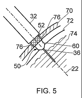
Figure 5 illustrates in more detail the skull mount 50 and the skull mount
insertion
and alignment device 30 after the skull mount 50 has been located within the
hole
formed in the subjects skull bone 70. In particular, it can be seen from
figure 5 how
the stiff wire 36 of the skull mount insertion and alignment device 30 passes
along
the axis of insertion 22 and performs the function of perforating the dura 72
and
CA 02701132 2010-03-29
WO 2009/047494 23 PCT/GB2008/003397
forming a passageway through the cortex 74 (which is typically 10-12mm thick).
The
device 30 can thus be thought of as a cortical obturator dural perforator
(CODP).
Although perforating the dura may be performed using the skull mount insertion
and
alignment device 30, it is also possible to pierce the dura prior to such a
procedure;
this prior piercing of the dura (e.g. manually by a surgeon using a scalpel or
the like)
can help to ensure no blood vessels are ruptured during the surgical
procedure. An
adhesive 76 is also provided to securely fix the skull mount 50 to the skull
70. The
adhesive 76 is allowed to cure whilst the skull mount insertion and alignment
device
30 remains attached to the skull mount 50.
Referring now to figure 6, it is shown how the skull mount insertion and
alignment
device 30 can (after the adhesive 76 has cured) be unscrewed from the skull
mount
50 and withdrawn back through the stereoguide 2. In this manner, it can be
seen that
the aperture provided through the skull mount 50 is then accurately aligned
with the
axis of insertion as defined by the stereoguide. The implanted skull mount 50
can
thus be considered a tertiary guide member that can aid the guiding of
instruments
along the axis of insertion. It can also be seen in figure 6 that the upper
surface of the
skull mount 50 is substantially flush to the surface of the skull after
implantation.
After implantation of the skull mount, a guide tube is implanted having a
distal end
that terminates just short of the required target area. A guide tube
applicator and
guide tube will now be described with reference to figures 7 to 11
Referring to figure 7, a guide tube applicator 80 is illustrated. The guide
tube
applicator 80 comprises an elongate shaft 82 having a central hollow channel
through
which a guide wire 84 can be passed. The outer diameter of the shaft 82 is
preferably
the same as the outer diameter of the shaft 32 of the skull mount insertion
and
alignment device 30. A clamp 86 is provided at the proximal end of the
applicator 80
to prevent unwanted axial movement of the guide wire 84 relative to the guide
tube
applicator 80. The distal end of the applicator 80 comprises a dome shaped
recess 88
having a central linear bar 90. An aperture through the bar 90 is provided for
the
CA 02701132 2010-03-29
WO 2009/047494 24 PCT/GB2008/003397
guide wire 84. The shape of the recess 88 and bar 90 are complimentary to the
shape
of the guide tube hub described in more detail with reference to figure 8.
Referring to figure 8, a guide tube 100 of known type is shown. The guide tube
100
comprises a length of tubing 102 having a hub 104 at its proximal end. The
sides of
the hub carry a screw thread 106 and the top surface 108 of the hub, which has
a lip
extending further radially than the screw thread 106, is dome shaped and has a
slot
110 formed therein. The slot 110 also provides the opening via which the lumen
of
tubing 102 can be accessed. As mentioned above, the top surface 108 of the
guide
tube hub 104 can be received in the recess 88 of the guide tube applicator 80.
The
slot 110 of the hub is also arranged to engage the bar 90 of the guide tube
applicator
80 thereby preventing relative rotation of the guide tube 100 and guide tube
applicator 80 when mated.
Figure 9 illustrates a guide tube 100 attached to the distal end of a guide
tube
applicator 80 prior to its insertion into the guide members of the stereoguide
2. The
required length of the guide tube 100 and the length of the guide wire 84 that
protrudes from the guide tube applicator 80 can be calculated relative to the
top
surface of the skull mount 50; this calculation can be performed using the
reading
taken from the scale 33 of the skull mount insertion and alignment device 30
during
the process of inserting the mount 50 into the hole.
Referring to figure 10, the guide tube applicator 80 is fed through the first
and second
guide members of the stereoguide (only the second guide member 18 being shown
in
figure 10) towards the subject. The guide tube 100, which is stiffened by the
guide
wire 84, passes through the skull mount 50 and into the brain of the subject.
The skull
mount 50 also acts as a guide member and may thus be considered a third or
tertiary
guide member. The guide wire 84 and guide tube 100 are thus driven together
through brain tissue along the axis of insertion with a high level of
accuracy. In
particular, the provision of the third guide member (which is also aligned
with the
axis of insertion as described above) provides accurate guiding in the
immediate
CA 02701132 2010-03-29
WO 2009/047494 25 PCT/GB2008/003397
proximity of the brain thereby minimising the possibility of suboptimal guide
tube
placement.
It should also be noted that using the skull mount insertion and alignment
device 30
that is described above also improves the accuracy of guide wire 84 and guide
tube
100 insertion. This is because, as also mentioned above, device 30 forms a
passageway through the cortex and may also pierce the dura. The dura is a
tough
membrane and the cortex is around 10-12mm of relatively tough brain tissue.
Inserting the guide wire 84 and guide tube 100 through the pre-formed
passageway in
the dura and cortex reduces any deflection away from the axis of insertion
that could
occur if the guide wire 84 alone was to be urged. into the brain.
Alternatively, the
guide wire 84 can have a smaller diameter (thereby having a lower stiffness)
than
would be necessary if it was required to penetrate the dura and cortex.
Insertion continues until the hub 104 of the guide tube 100 makes contact with
the
skull mount 50. As described above with reference to figure 3, the skull mount
includes a cavity 64 having a threaded wall 58. The hub 104 of the guide tube
100 is
configured so that it can be screwed into cavity ' 64 of the skull mount. This
is
achieved by rotating the guide tube applicator 80. Once the hub 104 is screwed
into
place, the guide tube applicator 80 (including the guide wire 84) can be
withdrawn
back through the guide members of the stereoguide. As shown in figure 11, the
skull
mount 50 and guide tube 100 are then retained in the subject's skull.
Referring to figure 12, use of the above described implanted guide tube 100
for
receiving a catheter 120 is illustrated. In particular, figure 12 shows a
skull mount 50
secured in a skull hole by an adhesive 76. The guide tube 100 is screwed into
the
skull mount 50 and comprises a length of tubing 102 located along the axis of
insertion and terminating just short of the required target 24. Figure 12 also
shows a
catheter 120 that has been passed through the guide tube and is arranged to be
of a
length such that its distal end reaches the required target 24. The proximal
end of the
catheter 120 may be secured to the skull by a clip 122. The catheter 120 may
also be
in fluid communication with a drug delivery pump (not shown) via a wider bore
CA 02701132 2010-03-29
WO 2009/047494 26 PCT/GB2008/003397
supply tube 124. In this manner, the required therapeutic agent may be
delivered to
the target site 24 via catheter 120. To minimise the risk of infection passing
the
blood-brain barrier, the catheter 120 and guide tube 100 may be subcutaneously
mounted and the supply tube 124 subcutaneously channelled to an implanted drug
delivery pump. It should be noted at this point that the catheter 120 may be
inserted
through the guide tube without the use of a stereoguide and can thus be
relatively
easily replaced if necessary.
Referring now to figures 13 and 14, alternative skull mounts suitable for use
in the
above described surgical procedure are illustrated.
Figure 13 shows a skull mount insertion and alignment device 30 having a
pivotable
skull mount 150 attached to its distal end. The pivotable skull mount 150
comprises a
truncated ball 152 having a cavity with an internal screw thread surface for
receiving
the protrusion 34 of the device 30 and a channel through which the stiff wire
36 of
the device 30 passes. The pivotable skull mount 150 also comprises a casing or
socket portion 154 for retaining the ball 152. The casing portion is suitable
for
insertion into a hole formed through the skull 156.
In use, the upper rim of casing portion 154 can be secured to the skull using
adhesive
or screws etc (not shown). The skull mount insertion and alignment device 30
may
then be moved along the axis of insertion using the stereoguide and engaged
with the
truncated ball 152. As shown in figure 13, the channel through the truncated
ball 152
becomes aligned with the axis of insertion as defined by the stiff wire 36 of
the
device 30. The ball 152 may then be locked in position relative to the casing
portion
154; such locking may be permanent (e.g. adhesive) or releasable (e.g. by
using
releasable locking screws). This pivotable arrangement has several advantages.
For
example, it allows an axis of insertion to be used that deviates significantly
from the
skull normal. It can also simplify the skull mount insertion process and, if a
releasable locking mechanism is used, allows subsequent angular adjustments to
the
axis of insertion.
CA 02701132 2010-03-29
WO 2009/047494 27 PCT/GB2008/003397
Figure 14 shows a skull mount 170 that is a variant to the skull mount 150 of
figure
13 and is also suitable for use with the above described skull mount insertion
and
alignment device 30. The skull mount 170 comprises a truncated ball 172
retained
within a cavity. The bottom and sides of the cavity are formed by a recess
drilled in
the skull bone 174. A plate 176 having a triangular cross-section aperture is
placed
over the recess and screwed to the skull thereby forming the top of the
cavity. In this
manner, a lower complexity skull mount may be provided, albeit with a
requirement
for the surgeon to provide a stepped recess in the skull 174. A threaded
recess may
also be provided on the internal surface of the channel formed through the
ball 172
for mating with the skull mount insertion and alignment device.
Referring to figure 15, a further skull mount 200 is illustrated. The skull
mount 200
comprises a layer of (uncured) UV curable adhesive 202 and is attached to a
hole
formed in the skull 204 (e.g. with adhesive or by a screw thread attachment).
After
skull mount attachment to the skull, an alignment instrument 206 comprising a
protruding member 208 is passed along the required axis of insertion 210 and
penetrates the layer of adhesive. An ultraviolet (UV) light source 212 is then
used to
cure the adhesive layer 202 with the alignment instrument in situ. The
protruding
member is formed from, or coated with, a material (e.g. a surfactant) that
does not
2 0 adhere to the cured adhesive. It is thus possible to retract the alignment
instrument
206 after the adhesive layer 202 has been cured thereby providing an alignment
guide
in the form of an alignment channel 214 in a layer of cured adhesive 216; the
alignment channel 214 being aligned with the axis of insertion 210.
25. Referring to figures 16 and 17, a further skull mount 300 of the present
invention is
illustrated.
The skull mount 300 comprises a skull insert 302 and a retaining ring 304. The
skull
insert 302 is dimensioned so as to fit in a hole formed in the skull and has a
30 protruding lip for engaging the outer surface of the skull around the
periphery of the
hole formed in the skull. The skull insert 302 is held in place by the ring
304 which
can in turn be secured to the skull by bone screws. An elastomeric septum seal
CA 02701132 2010-03-29
WO 2009/047494 28 PCT/GB2008/003397
guiding member 306 fits within a cavity defined by the skull insert 302 and
the
retaining ring 304. The septum seal guiding member 306 includes an aperture
that
defines an axis of insertion 312. The septum seal guiding member 306 also
provides a
fluidic seal with a catheter or other neurosurgical instrument passed through
its
aperture along the axis of insertion 312. A cap 310 and a cap sealing bung 308
are
also provided. The cap sealing bung 308 fits within, and forms a seal with,
the
septum seal guiding member 306 and is held in place by the cap 310 which is
attachable to the retaining ring 304 by a snap fit. The skull mount 300 thus
provides a
sealed passageway into the brain for a catheter or electrode etc. Furthermore,
appropriate alignment of the aperture of the septum seal guiding member 306
(e.g.
using a skull mount alignment device) allows that member to provide a guiding
function.
The above examples are directed to accurately inserting guide tubes through
which
catheters may then be passed for delivery of therapeutic substances (e.g.
drugs) to the
brain. The techniques and apparatus described above are, however, also
applicable
for inserting electrodes into the brain for deep brain stimulation. For
example, the
catheter 120 shown in figure 12 may be replaced with an electrode that is
connected
to a suitable power source. Alternatively, the guide wire 84 and guide tube
100
inserted into the brain by the guide tube applicator 80 as described with
reference to
figures 7-10 may be left in place for DBS purposes. It is even possible for
the guide
tube to be omitted altogether and the guide tube applicator 80 as described
with
reference to figure 7 may be used to insert only a guide wire (e.g. guide wire
84)
through the skull mount and into the brain. Furthermore, although the
insertion of
only one guide tube into a subject is described above, the technique may be
repeated
multiple time on a single subject to insert multiple guide tubes and/or
electrodes to
different target areas of the brain.