Note: Descriptions are shown in the official language in which they were submitted.
CA 02701453 2010-03-31
WO 2009/032307 PCT/US2008/010421
HEART BAND WITH FILLABLE CHAMBERS
TO MODIFY HEART VALVE FUNCTION
TECHNICAL FIELD
[0001] The present invention relates to devices and methods for treating
dilatation of heart
valves by applying localized pressure to surface areas of the heart.
BACKGROUND OF THE INVENTION
[0002] Dilatation of the base of the heart occurs with various diseases of
the heart and often is
a causative mechanism of heart failure. In some instances, depending on the
cause, the dilatation
may be localized to one portion of the base of the heart (e.g., mitral
insufficiency as a
consequence of a heart attack affecting the inferior and basal wall of the
left ventricle of the
heart), thereby affecting the valve in that region. In other cases, such as
cardiomyopathy, the
condition may be global affecting more of the heart and its base, causing
leakage of particularly
the mitral and tricuspid valves. Other conditions exist where the mitral valve
structure is
abnormal, predisposing to leakage and progressive dilatation of the valve
annulus (area of valve
attachment to the heart). This reduces the amount of blood being pumped out by
the ventricles of
the heart, thereby impairing cardiac function further.
[0003] In patients with heart failure and severe mitral insufficiency, good
results have been
achieved by aggressively repairing mitral and/or tricuspid valves directly,
which requires open-
heart surgery (Bolling, et al). The mitral valve annulus is reinforced
internally by a variety of
prosthetic rings (Duran Ring, Medtronic Inc) or bands (Cosgrove-Edwards
Annuloplasty Band,
Edwards Lifesciences Inc). The present paradigm of mitt-al valve
reconstruction is therefore repair
from inside the heart, with the annulus being buttressed or reinforced by the
implantation of a
prosthetic band or ring. Since this is major open-heart surgery with intra-
cavitary reconstruction,
there is the attendant risk of complications and death associated with mitral
valve surgery.
Another approach has been to replace the mitral valve, which while addressing
the problem, also
1
= ' CA 02701453 2010-03-31
=
WO 2009/032307 PCT/US2008/010421
requires open-heart surgery and involves implantation of a bulky artificial,
prosthetic valve with
all its consequences. Because every decision to perform major surgery requires
some risk vs.
benefit consideration, patients get referred for risky surgery only when they
are significantly
symptomatic or their mitral valve is leaking severely.
[0004] In contrast to the more invasive approaches discussed above, in
specific instances of
inferior left ventricular wall scarring causing mitral regurgitation, Lid-
Cohen and co-workers
have suggested localized pressure or support of the bulging scar of the
inferior wall of the heart
from the outside (Lid-Cohen. N. et al. (2000) "Design of a new surgical
approach for ventricular
remodeling to relieve ischemic mitral regurgitation: insights from 3-
dimentsional
echocardiography". Circulation 101 (23):2756-2763).
[0005] Another less invasive approach to preventing global heart dilation
is ventricular
containment with a custom made polyester mesh, or cardiac support device (U.S.
Pat. Nos.
6,077,218 and 6,123,662). These devices are designed to provide .a passive
constraint around both
ventricles of the heart, and constrain diastolic expansion of the heart. Other
devices include
ventricular assist devices that provide cardiac assistance during systole and
dynamic ventricular
reduction devices that actively reduce the size of the heart. However, this
technique does not
specifically address valve leakage using a device that reinforces the base of
the heart in all phases
of the cardiac cycle.
[0006] Percutaneous approaches (including "edge-to-edge", placating the
annulus and
coronary sinus approaches) of accessing the heart through the femoral artery
have been used.
Disadvantages of percutaneous approaches include fixture-made clots being sent
downstream, and
the dangers of potential patient allergy to contrast media. In addition,
percutaneous approaches
require complicated systems and are very dependent on the anatomy of
the'patient. As a result
these systems require the help of an experienced and trained interventional
cardiologist to assist
with the procedure.
2
CA 02701453 2010-03-31 '
I '
WO 2009/032307 PCT/US2008/010421
[00071 An example of a system that provides a less invasive approach to
base stabilization is
found in U.S. Patent 6,716,158 to Raman et. al. However, although the Raman
et. al. system
operates to stabilize the base of the heart, it does not provide a system to
modulate or modify heart
valve function by applying localized pressure to particular regions of the
heart, for example, to
tissues adjacent to heart valve. Such a system would advantageously apply
inward pressure to
tissue adjacent to the heart valves so as to modify the shape or reduce the
size of a heart valve
itself. Accordingly, there is a need to non-invasively repair or re-configure
the shape of a mitral
and/or tricuspid valve so as to treat valve dilation and resulting valve
insufficiency problems.
[0008] The present invention is directed to solving the above mentioned
problems and can
advantageously be applied to both patient populations requiring heart valve
modification by
applying localized pressure, and to patient populations simply requiring
external stabilization of
the base of the heart.
SUMMARY OF THE INVENTION
[0009] The present invention addresses the problems discussed above by
providing a device
for the treatment of certain heart disorders, in particular mitral and/or
tricuspid valve
insufficiency. The device aims to apply localized pressures to the heart
and/or reduce the size of
the base of the heart that contains these valvular structures. The device also
provides a system for
applying inward pressure to tissue adjacent to the heart valves so as to shape
the mitral and/or
tricuspid valve itself. In addition, the present invention can be used to
address progressive
dilatation of any localized area of the heart, such as the atrial or
ventricular myocardium, or the
cardiac base. It does so by optionally providing external re-enforcement or
remodeling of the
cardiac base while still providing support of the valve at annular and sub-
annular levels. As used
herein, the surgical procedure for implanting the device is referred to as
basal annuloplasty of the
cardia externally (BACEThl) and the device is referred to as the external
cardiac basal
annuloplasty system BACE S ystem. '
3
CA 02701453 2010-03-31
=
WO 2009/032307 PCT/US2008/010421
[0010] An advantage of the present system is that it overcomes the
disadvantages of
percutaneous approaches by overcoming the disadvantages of systems accessing
the heart through
the femoral artery.
[0011] Another advantage of the present invention is that it remodels the
heart while re-
shaping the valve(s). As such, the present invention operates to both prevent
heart disease and to
treat it as well. In addition, in one embodiment of the present invention
uniquely incorporates the
use of subcutaneous ports that allows adjustment and post operative re-shaping
of the valve(s)
without making incisions in the patient.
[0012] In one aspect, the present invention provides an external heart
device, comprising: a
band dimensioned to be received around a patient's heart, the band comprising
an inner layer and
an outer layer, wherein areas of the inner layer and outer layer are bound to
one another; and at
least one finable chamber in the band, the at least one finable chamber being
located in areas
where the inner layer and the outer layer are not bound to one= another.
[0013] In various embodiments, the at least one finable chamber may either
be formed or
inserted into the areas where the inner layer and the outer layer are not
bound to one another,
thereby providing a band structure with one or more integral finable chambers.
[0014] In various embodiments, the band may be transparent, and may
optionally be made of
silicone rubber, or other suitable bio compatible implantable material.
[0015] In various embodiments, the present invention may be formed with the
inner layer and
outer layer being bound to one another by adhesives, crosslinking, heat and/or
pressure, or even
by stitching.
[0016] In various embodiments, the interior surface of the inner layer may
optionally be
textured so as to remain in position around the heart, yet still permit the
device to be removed in
future without damaging the surface of the heart.
4
CA 02701453 2010-03-31 =
WO 2009/032307 PCT/US2008/010421
[0017] In various embodiments, the device has a plurality of finable
chambers, with two of
the tillable chambers being positioned spaced apart from one another, and with
the band forming
a bridge portion therebetween. Advantageously, the bridge portion in the band
may be
dimensioned to be positioned over vasculature on the exterior of the heart
when at least one of the
tillable chambers are filled.
[0018] Advantageously as well, the dimensions of the finable chambers and
their positioning
in the band may also provide a system to apply inward pressure to tissues
adjacent to a heart valve
so as to modify or change the shape of the valve to a more desired shape. In
one exemplary
application of the present invention, two of the tillable chambers are
positioned on opposite sides
of a mitral valve of the heart to shape the mitral valve to prevent mitral
valve dilation, and
resulting mitral regurgitation.
[0019] In various embodiments, each of the tillable chambers has a filling
tube in fluid
communication therewith. In different embodiments, the filling tubes may
optionally be fillable
through a blunt needle port, a sharp needle port, or through a subcutaneous
port, Luer port fitting,
or various combinations thereof. In one exemplary embodiment, all but one of
the filling tubes are
tillable through a subcutaneous port, and the plurality of subcutaneous ports
are disposed together
on a sheet. The sheet may optionally be made of silicone or polyester or other
suitable material
and may be used to position these subcutaneous ports at a convenient location
within the patient's
body.
[00201 The filling tubes may optionally be made of silicone or other
suitable bio-implantable
material. Depending upon the method of manufacturing the present device, the
individual filling
tubes may be integrally formed as the filling chambers of the device are
formed, or they may be
inserted after the fillable chambers have been formed.
[0021] In various embodiments and applications, the various finable
chambers may be filled
either by saline, a hardening polymer, a gel, a gas, or other suitable
material.
CA 02701453 2010-03-31
WO 2009/032307 PCT/US2008/010421
[0022] In optional embodiments, one or more sleeves may be positioned
around an exterior
surface of the outer layer of the device. Such sleeves may advantageously
operate to hold the band
at a preferred location on the patient's heart. Specifically, such sleeves are
designed to promote
tissue ingrowth to hold the device in place. These sleeves may be made of
polyester or other
suitable materials. In one exemplary embodiment, they are 5/8" wide, however,
the present
invention is not limited to any particular dimensions.
[0023] In one exemplary embodiment, the band may be between 2 and 5 cms wide
and may
be secured by clips, sutures, or other fasteners, with some on the posterior
side and some on the
anterior side of the heart. Specific care is taken to avoid injury to the
circumflex and right
coronary arteries and the coronary sinus. This procedure may be performed
either as a stand-alone
procedure or as an adjunct to other cardiac surgery. Additionally, it may be
performed with or
without the aid of cardio-pulmonary bypass.
[0024] Optional variations of the device include a complete stabilization
of the base of the
heart, or a partial stabilization around the expansile portions of the mitral
and tricuspid valves. It
is to be understood, however, that the present invention is not simply
directed to stabilizing the
base of the heart. Instead, the present invention is well suited to modifying
heart valve function
(and optional valve re-shaping) by therapeutically applying localized
pressures to various regions
of the heart.
[0025] Mother variation seeks to use ports along the device that will
facilitate delivery of
specialized drugs, gene therapeutic agents, growth factors, etc.
[0026] A specific variation incorporates the use of epicardial bi-
ventricular, and multi-site
pacing electrodes implanted along with the BACE-System, where multi-site
pacing might be
indicated. One iteration has multiple electrodes arranged as an array along
the left and right
ventricular walls of the heart, close to the base of the heart. The option
then exists to allow
6
CA 02701453 2012-05-16
selection of various sites along the heart to allow for optimal
resynchronisation or optimization
of contractility.
[0027] The present invention also provides a method of implantation,
which may be
through a conventional full median sternotomy with the strip being secured by
sutures, or a
minimally invasive thoracotomy approach whereby the device/strip may be
folded/rolled and
implanted by a specialized implantation system and secured using adhesives,
self-firing clips,
sutures, etc.
[0028] Another application of the device is the local application to
stabilize scars of the
heart to prevent their expansion (local ventricular stabilization).
In accordance with an aspect of the present invention, there is provided an
external heart device, comprising: (a) a band dimensioned to be received
around a patient's heart,
the band comprising an inner layer and an outer layer, wherein some but not
all areas of the inner
layer and outer layer are bound to one another; and (b) at least two fillable
chambers in the band,
the at least two tillable chambers being spaced apart from one another and
located in areas where
the inner layer and the outer layer are not bound to one another, the at least
two fillable chambers
being separated by a gap, wherein the gap and each of the at least two
fillable chambers have a
circumferential length, and wherein the circumferential length of the gap is
greater than the
circumferential length of each of the at least two fillable chambers; wherein
the at least two
fillable chambers are positioned on the heart spaced apart from one another
with the gap being
placed over heart vasculature so as to form a pressure-reducing bridge over
the heart vasculature
contacting and applying constant localized pressure to the heart in a
predetermined location to
modify a heart valve shape when the at least two fillable chambers are filled.
In accordance with an aspect of the present invention, there is provided a
method of
modifying heart valve function, comprising: placing a band around a patient's
heart, the band
comprising an inner layer and an outer layer, wherein areas of the inner layer
and outer layer are
bound to one another, and wherein at least two fillable chambers in the band
are spaced apart
from each other and are located in areas where the inner layer and the outer
layer are not bound
to one another, the at least two fillable chambers being separated by a gap,
wherein the gap and
7
CA 02701453 2015-06-16
each of the at least two fillable chambers have a circumferential length, the
circumferential
length of the gap being greater than the circumferential length of each of the
at least two
fillable chambers, the at least two fillable chambers positioned on the heart
spaced apart from
one another with the gap placed over heart vasculature to form a pressure-
reducing bridge
over the heart vasculature; and filling the at least two fillable chambers
with a fluid to cause
the at least two fillable chambers to contact and apply constant localized
pressure to the heart
in a predetermined location to modify heart valve shape.
In accordance with an aspect of the present invention, there is provided a use
of a
band around a patient's heart for modifying heart valve function, the band
comprising an
inner layer and an outer layer, wherein areas of the inner layer and outer
layer are bound to
one another, and wherein at least two fillable chambers in the band are spaced
apart from
each other and are located in areas where the inner layer and the outer layer
are not bound to
one another, the at least two tillable chambers being separated by a gap,
wherein the gap and
each of the at least two fillable chambers have a circumferential length, the
circumferential
length of the gap being greater than the circumferential length of each of the
at least two
fillable chambers, the at least two fillable chambers positioned on the heart
spaced apart from
one another with the gap placed over heart vasculature to form a pressure-
reducing bridge
over the heart vasculature; wherein in use, the at least two fillable chambers
are fillable with
a fluid for causing the at least two fillable chambers to contact and apply
constant localized
pressure to the heart in a predetermined location for modification of heart
valve shape.
In accordance with an aspect of the present invention, there is provided a use
of an
implantable device and an implantation system for implanting the implantable
device for
reducing or eliminating mitral valve regurgitation in a patient having mitral
valve
regurgitation, wherein the implantable device is configured for positioning
around a full
circumference of the patient's heart and comprises at least one fillable
chamber; wherein the
implantation system is useable to implant and position the device in the
patient by a
minimally invasive thoracotomy approach, the device being positionable on the
heart such
that the implantable device is for encompassing at least the full
circumference of the atrial-
ventricular junction of the heart, the device further being positionable such
that a particular
one fillable chamber of the at least one fillable chamber is positionable on a
selected
epicardial surface portion of the heart adjacent the heart's mitral valve, the
selected epicardial
surface portion comprising at least one of a posterior epicardial surface and
a lateral
7a
CA 02701453 2015-06-16
epicardial surface; and wherein the particular one fillable chamber is
fillable to expand the
particular one fillable chamber to a selected level, wherein after filling the
particular one
fillable chamber remains filled at the selected level throughout the cardiac
cycle and is for
applying a desired localized inward pressure on the selected epicardial
surface portion
adjacent the mitral valve for reshaping the mitral valve in a manner that
reduces or eliminates
mitral valve regurgitation.
In accordance with an aspect of the present invention, there is provided a
system for
reducing or eliminating mitral valve regurgitation, the system comprising an
implantable
device comprising: a support structure configured to encompass at least the
full
circumference of the atrial-ventricular junction of the heart, the support
structure having an
outer surface that faces outwardly from the heart when the device is inserted,
the outer
surface of the support structure serving as an outermost surface of the
device; and at least
one fillable chamber, each of the at least one fillable chamber being secured
to the support
structure such that an outwardly facing surface of each of the at least one
fillable chamber
bears against an inwardly facing surface of the support structure; and a
dedicated filling tube
for each of the at least one fillable chamber; wherein the device is
configured such that, when
the device is implanted and positioned on the heart such that the implantable
device
encompasses at least the full circumference of the atrial-ventricular junction
of the heart and
the device is further positioned such that a particular one of the at least
one fillable chamber
is positioned on a selected epicardial surface portion of the heart adjacent
the heart's mitral
valve, the selected epicardial surface portion comprising at least one of a
posterior epicardial
surface and a lateral epicardial surface, filling the particular one fillable
chamber to a selected
level causes the particular one fillable chamber to expand to a constant
volume and apply a
desired localized inward pressure on the selected epicardial surface adjacent
the mitral valve
to reshape the mitral valve in a manner that reduces or eliminates mitral
valve regurgitation.
In accordance with an aspect of the present invention, there is provided a use
of an
implantable device for reducing or eliminating mitral valve regurgitation in a
patient having
mitral valve regurgitation, wherein the implantable device is configured for
positioning
around a full circumference of the patient's heart and comprises at least one
fillable chamber;
wherein the implantable device is implantable in the patient so that the
device is positionable
on the heart such that the device is for encompassing at least the full
circumference of the
atrial-ventricular junction of the heart, the device further being
positionable such that a
7b
CA 02701453 2015-06-16
particular one of the at least one fillable chamber is positionable on a
selected epicardial
surface portion of the heart adjacent the heart's mitral valve, the selected
epicardial surface
portion comprising at least one of a posterior epicardial surface and a
lateral epicardial
surface; and wherein the particular one fillable chamber is fillable to expand
the particular
one fillable chamber to a selected level, wherein after filling the particular
one fillable
chamber remains filled at the selected level throughout the cardiac cycle and
is for applying
a desired localized inward pressure on the selected epicardial surface portion
adjacent the
mitral valve for reshaping the mitral valve in a manner that reduces or
eliminates mitral valve
regurgitation.
In accordance with an aspect of the present invention, there is provided an
external
heart device, comprising: a band dimensioned to be received around the base of
a patient's
heart; at least one fillable chamber comprised in the band, wherein the at
least one fillable
chamber is positioned spaced apart from another at least one fillable chamber,
if present in
the band; and a gap in the band that does not comprise a fillable chamber.
BRIEF DESCRIPTION OF THE FIGURES
[0029] Fig. 1 depicts a cross-section of the heart, showing the
approximate location of
a representative embodiment of the device of the present invention by dashed
lines, and the
distance between the top and the bottom of the heart represented by "X".
[0030] Fig 2 depicts a cross-section of the base of the heart between the
dashed lines
depicted in Fig. 1.
[0031] Fig. 3 is a perspective view of a representative embodiment of the
device of
the present invention.
[0032] Fig. 4 is a side elevation view of the embodiment of the device of
Fig. 3.
[0033] Fig. 5 is a proximal end view of the embodiment of the device of
Fig. 3.
[0034] Fig. 6A is a perspective view of the device of Fig. 3, shown
received around a
patient's heart, prior to filling of the fillable chambers.
7c
CA 02701453 2010-03-31
=
WO 2009/032307 PCT/US2008/010421
[0035] Fig. 6B is a perspective view of the device of Fig. 3, shown
received around a patient's
heart, after filling of the finable chambers.
[0036] As depicted in Figs. 7A to 7D, PV=pulmonary valve, MV=mitral valve,
AV=aortic
valve and TV=tricuspid valve.
[0037] Fig. 7A depicts a cross-sectional schematic diagram of the base of
the heart showing
the present invention prior to re-shaping the mitral valve by filling chamber
30E.
[0038] Fig. 7B depicts a cross-sectional schematic diagram of the base of
the heart showing
the present invention after re-shaping the mitral valve by filling chamber 30E
(i.e.: showing the
band forming a bridge portion between two of the tillable chambers 30A and
30B, and showing
the modification of the shape of a patient's mitral valve to treat mitral
dilation.)
[0039] Fig. 7C depicts a cross-sectional schematic diagram of the base of
the heart showing
the present invention prior to re-shaping the mitral valve by filling chamber
30D.
[0040] Fig. 7D depicts a cross-sectional schematic diagram of the base of
the heart showing
the present invention after re-shaping the mitral valve by filling chamber 30D
(i.e., showing the
band forming a bridge portion between two of the finable chambers 30A and 30B,
and showing
the modification of the shape of a patient's mitral valve to treat mitral
dilation.)
[0041] Fig. 8 is a perspective view of a second representative embodiment
of the device of the
present invention three sleeves received around the band for attachment to the
exterior of the
heart.
[0042] Fig. 9 is an illustration of a first system for manufacturing the
present invention using
three separate layers of material.
8
CA 02701453 2010-03-31 =
WO 2009/032307 PCT/US2008/010421
[0043] Fig. 10 is an illustration of a second system for manufacturing the
present invention
using one layer of material folded on top of itself with a second layer of
material inserted
therebetween.
[0044] Fig. 11 is an illustration of a third system for manufacturing the
present invention
using one layer of material having regions that are pinched onto itself to be
bound together,
showing the insertion of filling tubes into the separate fillable chambers.
[0045] Fig. 12 is an illustration of an alternate embodiment of the
invention having pockets in
the device with tillable chambers inserted therein.
[0046] Fig. 13 is an illustration of the blunt needle port used to fill and
deflate one or more
fluid chambers.
DETAILED DESCRHYTION OF THE INVENTION
[0047] The present invention is directed to modifying heart valve function
by applying
localized support or pressure to various regions of the heart. In addition,
the present invention
may optionally be used to decrease, and/or prevent increases in, the
dimensions of the base, and in
particular the atrio-ventricular junction, beyond a pre-determined size.
[0048] In particular procedures, the present invention is directed to
applying pressure to tissue
adjacent to the mitral and/or tricuspid heart valves. This has the effect of
beneficially modifying
the shape of the heart valve(s) to treat heart valve dilation. As such, this
invention is particularly
suited for use in regurgitation of the mitral and tricuspid valves. However,
the device may also
optionally be used prophylactically in heart failure surgery to prevent
further cardiac basal
dilation or expansion even if the underlying mitral and tricuspid valves are
competent. As such,
the present device may be used in moderate or advanced heart failure to
prevent progression of
basal dilation or reduce the size of the dilated base.
9
= CA 02701453 2010-03-31
=
=
WO 2009/032307 PCT/US2008/010421
[0049] As used herein, "atrio-ventricular" or A-V groove refers to the
junction between the
atrial and ventricular chambers of the heart, also known as the atrio-
ventricular junction marked
externally by the atrio-ventricular groove. This is easily identified in the
change of appearance of
the cardiac muscle and also the presence of arteries and veins. The "cardiac
base", as used herein,
is the area of the heart between and including the AV groove and extends to,
but not including,
the bottom or apex of the heart.
[0050] The heart is enclosed within a double walled sac known as the
pericardium. The inner
layer of the pericardial sac is the visceral pericardium or epicardium. The
outer layer of the
pericardial sac is the parietal pericardium. The term "endocardial surface"
refers to the inner walls
of the heart. The term "epicardial surface" refers to the outer walls of the
heart.
[0051] The mitral and tricuspid valves sit at the base of the heart and
prevent blood from
leaking back into the atria or collecting chambers. See Fig. 1. Mitral
regurgitation is a condition
whereby blood leaks back through the mitral valve into the left atrium. Over
time, this creates a
damming of blood in the lungs causing symptoms of shortness of breath. The
left heart
particularly the left ventricle has to pump a greater volume of blood as a
result causing greater
strain on this chamber.
[0052] Dilatation of the mitral annulus occurs maximally in the posterior
portion of the
annulus, which is not supported by the cardiac fibre-skeleton. Fig. 2 is an
anatomic diagram of the
base of the heart, showing the valves and the structures in contact with them.
Figs. 7A to 7D are
various corresponding schematic representations of the valves at the cardiac
base during
placement and operation of the present device.
[0053] Mitral valve repair or replacement at present is always performed
from inside the heart
with the aid of cardiopulmonary bypass. Rings are implanted along the inner
surfaces of the entire
or expansile portions of the mitral and tricuspid annuli. Alternatively, when
mitral valve
malfunction is severe, replacement of the valve with a prosthetic valve may be
indicated.
CA 02701453 2010-03-31
WO 2009/032307 PCT/US2008/010421
Overview
[0054] The basal ventricular stabilization, and heart valve shape re-
shaping of the present
invention works by using a band of prosthetic material such as silicone rubber
being anchored or
sutured to the base of the heart at the level of the atrio-ventricular groove.
This band has at least
one integral finable chambers formed or inserted therein. In use, the present
device serves to
stabilize the mitral and tricuspid annuli from the outside (see Figs. 7B and
7D). As will also be
shown herein, this also serves to provide a device that applies pressure to
tissue regions adjacent
to the heart valves (e.g.: mitral and/or tricuspid valves) to re-shape the
heart valve itself as a
method of treating valve dilation problems.
[0055] The present invention and technique reduces the complexity of the
procedure and
minimizes the invasive nature and complications from work on the valve. This
system and
technique is of particular benefit in patients that have morphologically
normal valves with annular
dilatation. The device can be applied and anchored to the cardiac base, with
the heart beating,
without the aid of cardiopulmonary bypass.
[0056] Many patients with moderate degrees of mitral regurgitation are not
treated surgically,
because the risks of surgery outweigh the potential benefits in this group of
patients. However,
patients with conditions such as chronic heart failure tend to get very
symptomatic even with
moderate degrees of mitral regurgitation. These groups of patients would
benefit from the less
invasive procedures, which are the subject of the present invention. Thus, the
potential of this
technique in treating mitral regurgitation as a minimally invasive procedure
has great appeal as
the population ages and more patients manifest with symptoms of heart failure.
It also can be
applied in patients undergoing open heart coronary artery surgery without the
aid of a heart-lung
machine.
11
CA 02701453 2010-03-31
=
WO 2009/032307 PCT/US2008/010421
Device Parameters
[0057] The device of the present invention can be constructed of any
suitable implantable
material. In preferred embodiments, the device is constructed from layers of
silicone rubber. An
advantage of using such a material is that the device is sufficiently flexible
to move with the
expansion and contraction of the heart without impairing its function. It
should, however, be
designed to prevent expansion of the cardiac base during diastolic filling of
the heart to a
predetermined size. Since the size expansion parameters of a beating heart are
well known, this
can be accomplished by testing the device in vitro by applying forces that
mimic heart expansion.
[0058] As shown in FIG. 3, in one embodiment, the device 10 comprises a
band 20
dimensioned to be received around a patient's heart. Band 20 comprises an
inner layer 22 and an
outer layer 24. In accordance with the present invention, areas of inner layer
22 and outer layer 24
are bound to one another, resulting ma very thin system design as can be seen.
A unique
advantage of such a thin band 20 is that it can easily be placed around the
patient's heart during
surgery.
[0059] Band 20 further comprises at least one finable chamber 30 integrally
formed therein.
As depicted in Fig. 3, the band comprises five finable chambers 30A-E.
Specifically, fillable
chambers 30 are located in areas where inner layer 22 and the outer layer 24
are not bound to one
another. As will be fully explained below, fillable chambers 30 may be
integrally formed into
band 20 when inner layer 22 and outer layer 24 are selectively bound together
to create an
enclosure. Alternatively, however, fillable chambers 104 may be separately
constructed and
inserted into the areas where inner layer 22 and outer layer 24 are not bound
to one another in the
manufacturing of device 100 as seen in FIG. 12. For example, finable chambers
104 may be
inserted into individual "pockets" formed between inner layer 22 and outer
layer 24. These
pockets may be formed by attaching inner layer 22 and outer layer 24 along two
sides and a first
edge, keeping the second opposite edge (and the interior of the pocket)
unbound until the
12
CA 02701453 2010-03-31
WO 2009/032307 PCT/US2008/010421
individual chambers are inserted. The pocket openings would then be bonded
closed or other
means would be used to secure the individual fillable chambers.
[0060] In one exemplary embodiment, band 20 is formed from silicone rubber
and is therefore
transparent. However, the present invention is not so limited. For example, it
is to be understood
that band 20 may also be formed from other suitable biocompatible implantable
materials,
including, but not limited to a textile made from polyester, PTFE
(polytetrafluoroethylene), or
elastic yams.
[0061] An advantage of forming band 20 (and its finable chambers 30) from a
transparent
material is that it facilitates placement of the device around the patient's
heart. In particular, the
external vasculature of the heart is clearly viewable through band 20 as band
20 is placed around
the patient's heart. Moreover, the transparent nature of the material permits
easy positioning of
fillable chambers 30 at preferred locations adjacent to heart valves (e.g.,
the mitral and/or
tricuspid valve), and away from the vasculature.
[0062] In various embodiments, inner layer 22 and outer layer 24 may be
bound to one
another by adhesives, crosslinking (e.g., when layers 22 and 24 are pressed
together and heated),
or even by stitching. It is therefore to be understood that the present
invention is not limited to
any particular system of attachment or bonding of layers 22 and 24 together.
[0063] In various optional embodiments, an interior surface of inner layer
22 may be textured.
This may advantageously assist in holding band 20 at a preferred position on
the patient's beating
heart. It is important, however, that the interior surface of inner layer 22
not be so textured such
that it would adhere too strongly to the exterior of the heart, since this
would make band 20
difficult to remove.
[0064] As seen in Figs. 4 and 5, band 20 preferably has a plurality of
fillable chambers 30. In
Fig. 4, one such exemplary chamber 30 is depicted. As illustrated in Fig. 5,
band 20 has five
finable chambers, being 30A, 30B, 30C, 30D and 30E. It is to be understood
that this is only one
13
CA 02701453 2010-03-31
=
=
WO 2009/032307 PCT/US2008/010421
exemplary embodiment, and that other embodiments of the invention have more or
less than five
tillable chambers 30. As such, the present invention encompasses any
embodiment having at least
one fillable chamber 30.
[0065] As also seen in Fig. 5, two of the plurality of tillable chambers 30
may be positioned
spaced apart from one another, such that band 20 has a gap 21 between chambers
30A and 30B as
depicted which forms a bridge between the chambers when applied to a patient's
heart.
Preferably, band 20 and finable chambers 30A and 30B are dimensioned such that
the gap 21 is
dimensioned to be positioned over vasculature on the exterior of the heart
when finable chambers
30A and 30B are filled thus forming a bridge therebetween. Thus, finable
chambers 30A and 30B
can be positioned on opposite sides of the pulmonary trunk of the heart.
Bridges can also be
formed between 30B and 30C, 30C and 30D, and 30D and 30E. An important
advantage of these
bridges is that they do not need to form a space between the heart and the
band. Instead, they only
need to reduce localized pressure so as to prevent vascular occlusion. A
bridge or release of
pressure can also be formed by filling only one chamber. Filling only one
chamber creates
pressure directly under that chamber, but it also relieves pressure directly
on each side of that
chamber.
[0066] As also seen in Figs. 3 to 5, a number of filling tubes 40 are
provided. Filling tubes 40
are preferably each in fluid communication with a separate fillable chamber
30, as illustrated in
Figs. 3 and 5.
[0067] Filing tubes 40 may be made of silicone, or other suitable material.
Each filing tube 40
is in fluid communication with, and fills, its own dedicated fillable chamber
30. For example, as
depicted in Fig. 5, filling tube 40A fills fillable chamber 30A, etc. It is to
be understood that the
present invention is not limited as to any particular substance being used for
filing fillable
chambers 30. As such, the individual tillable chambers 30 may be filled with
substances
including, but not limited to, a saline solution, a hardening polymer, a gel,
or even a gas.
14
CA 02701453 2010-03-31
WO 2009/032307 PCT/US2008/010421
Moreover, it is also to be understood that different fillable chambers 30 may
be filled with
different substances from one another.
[0068] In various embodiments, the separate filling tubes 40 may be finable
through a blunt
needle port 44 (for receiving blunt needle), a sharp needle port, or through a
subcutaneous port.
As such, different filling tubes 40 may be fitted with different ports at
their proximal ends. For
example, as shown in Fig. 6A, filling tube 40A may be a short filling tube
specifically equipped
for filling through a blunt needle port. As such, filling tube 40A can be
filled by a syringe via a
syringe tip with a blunt needle as depicted in Figs. 6A and 6B.
[0069] The present device is initially presented to the surgeon as a
flattened, flexible device
that is easy to handle during an operation.
[0070] Figs. 6A and 6B show perspective views of the present device 10,
with the band 20 as
positioned around the patient's heart, with the fillable chambers unfilled
(Fig. 6A) and filled (Fig
6B).
[0071] As seen in Figs. 6A and 68, in one embodiment of the present
invention, device 10
comprises a band 20 having five finable chambers 30A to 30E, and with each
tillable chamber
having its own dedicated filling tube 40A to 40E. As can also be seen, each of
filling tubes 4013 to
40E are tillable through a subcutaneous port 42B to 42E. The subcutaneous
port(s) provide a
unique feature to the invention with regards to heart valve repair in that
they allow a surgeon to
make post-operative adjustments to the implant without making any incisions.
This is done by
inserting a small gauge needle into the subcutaneous ports and injecting or
withdrawing
biocompatible fluid as needed. The subcutaneous port is made of silicone
rubber or other
biocompatible material that can be penetrated with a hypodermic needle and
then reseal after
removal of the needle.
[0072] Each subcutaneous port also may optionally include a biocompatible
radiopaque metal
drawn "can" 47 inside as depicted in Fig. 6B to facilitate locating the port
by tactile feedback on
CA 02701453 2010-03-31
WO 2009/032307 PCT/US2008/010421
the needle and syringe and by imaging on X-ray or fluoroscopy, which will also
allow the needle
to engage the port without enentrating it completely.
[0073] Optionally, subcutaneous ports 42B to 42E are disposed on a sheet
(not depicted).
This sheet may be made of silicone or polyester, or any other suitable
material, or any
combinations thereof. A sheet has the advantage of holding subcutaneous ports
42B to 42E
together for convenient access. Preferably, the sheet (and subcutaneous ports
42B to 42E attached
thereto) is surgically positioned on the lower side of the chest.
[0074] In preferred embodiments, each filing tube 40A to 40E may include a
unique marker
or indicia 43 (as shown in Figs. 6A and 6B) such that the surgeon is able to
clearly and easily
identify which subcutaneous port 42 corresponds to which particular tillable
chamber 30. For
example, one radiopaque marker may be affixed to filling tube 40A, two
radiopaque markers may
be affixed to filling tube 40B, etc. Other versions of indicia in addition to
radiopaque markers are
contemplated within the scope of the present invention.
[0075] After band 20 has been positioned around the heart, saline may be
introduced first with
a blunt needle through a blunt needle port of filling tube 40A, and then
through subcutaneous
ports 42B to 42E to thus fill tillable chambers 30A to 30E. Since tillable
chambers 30A to 30E
can be selectively individually filled, it is possible for the surgeon to
adjust the fitting of band 20
on the patient's heart with great accuracy. As such, each of finable chambers
30A to 30E can be
filled to a desired level and placed around the heart such that gap 21 and
tillable chambers 30A to
30E are best positioned On the patient's heart to reshape the patient's heart
valves as desired.
[0076] Figs. 7A to 7D are cross sectional views of various of the devices
at the location
shown by the arrows in Fig. 6A. As such, they are a top-down cross sectional
device immediately
above the top edge of the device, giving the chambers a "pillow shape"
configuration, with the
dosed edge of the "pillow" shown through the chambers 30. However, as
discussed elsewhere in
16
CA 02701453 2010-03-31
WO 2009/032307 PCT/US2008/010421
the specification, the band 20 actually consists of a separate inner layer 22
and outer layer 24,
although not explicitly depicted in Figs. 7A to 7D.
[0077] For example, as seen in Figs. 7A and 7B, the shape of mitral valve
MV may be
modified by the filling of tillable chamber 30E. (Fig. 7A shows placement of
the present band 20
prior to filling of finable chamber 30E. Fig. 7B shows placement of the
present band 20 after
filling of fillable chamber 30E.). As can be seen, the poorly sealing mitral
valve MV shown in
Fig. 7A is re-shaped to seal properly in Fig. 7B. Chambers 30A and 30B as
depicted in Fig. 7B
are not filled to a degree necessary to reshape the pulmonary valve (PV),
although they could be if
such an effect was desired.
[0078] Alternatively, as seen in Figs. 7C and 7D, the shape of mitral valve
MV may instead
be modified by the filling of fillable chamber 30D. (Fig. 7C shows placement
of the present
device 20 prior to filling of finable chamber 30D. Fig. 7D shows placement of
the present device
20 after filling of finable chamber 301).). As can be seen, the poorly sealing
mitral valve MV
shown in Fig. 7C is re-shaped to seal properly in Fig. 7D.
[0079] As can also be seen in Figs. 7A through 7D, band 20 forms a bridge
corresponding to
gap 21 between two of the fillable chambers, 30A and 30B. (Similar bridges can
also be formed in
band 20 between successive tillable chambers 30, or between a single tillable
chamber 30 and the
portion of the band adjacent thereto.)
[0080] As can be seen, the thin nature of band 20, coupled with the
potentially large volumes
of individually tillable chambers 30 produces a system in which pressure can
be directed not only
radially inward towards the center of heart, but also a "pinching" effect can
be generated between
adjacent fillable chambers 30.
[0081] As can be seen, by using different filling levels for each of the
different finable
chambers 30, a system is provided in which pressures on the heart can be
applied in an infinite
number of different directions, and amplitudes. As such, pressures may be
applied radially
17
CA 02701453 2010-03-31
=
WO 2009/032307 PCT/US2008/010421
inwardly to the heart, as well as in non-radial directions (i.e., "pinching")
portions of the heart
therebetween.
[0082] Fig. 8 is a perspective view of a second representative embodiment
of the device of the
present invention having a plurality of ingrowth sleeves 50 received around
band 20 for
attachment to the exterior of the heart. In use, sleeves 50 operate like belt
loops to hold up the
band like a belt, thus holding band 20 in positions against the patient's
beating heart.
[0083] Sleeves 50 are positioned on an exterior surface (i.e., outer side)
of layer 24 as seen in
Fig. 8. Sleeves 50 may optionally be made of polyester, or any other suitable
material, including,
but not limited to other woven, knitted, matted, or other textiles. Sleeves 50
in the preferred
embodiment act as promoters of controlled tissue growth such that they become
secure to selected
areas of the heart, but they may also act to limit tissue growth and just
provide mechanical means
of attachment. Sleeves 50 may optionally be produced by molding them directly
into a tension
band. Sleeves 50 may optionally be fitted onto band 20 by sutures or staples.
[0084] Figs 9 to 11 show three different methods for producing the present
device 10,
including band 20 with chamber(s) 30 and optional filling tubes 40. It is to
be understood that the
device of the present invention is not limited to devices made by any
particular system of
manufacture. However, it is also to be understood that the present invention
includes a variety of
novel methods of manufacture of the device.
[0085] Fig. 9 is an illustration of a first system for manufacturing the
present invention using
three layers of material. Specifically, the view of Fig. 9 is an exploded view
showing three layers
of material as sandwiched together to form the present invention.
[0086] In this method of making the invention, a rust layer of material
(i.e.: layer 22) and a
second layer of material (i.e. layer 24) are provided. Layers 22 and 24 may
optionally be made of
vulcanized silicone rubber, but may also be made of any other suitable
material. In various
embodiments, layers 22 and 24 may be made of the same materials, or be made of
different
18
CA 02701453 2010-03-31 =
WO 2009/032307
PCT/US2008/010421
materials. In addition, layers 22 and 24 may be made to the same thickness, or
be made to
different thicknesses.
[0087] A middle layer 25 is positioned between layers 22 and 24. Middle
layer 25 may be
made of separate sections of non-cured or non-vulanized silicon rubber. Middle
layer 25 has
sections removed that define and correspond to the locations of fillable
chambers 30. Specifically,
the presence of removed sections in middle layer sections 25 will allow layers
22 and 24 to
contact one another (and be bound together) in those regions where middle
layer sections 25 are
disposed.
[0088] In accordance with the present method, layers 22, 25, and 24 may be
bound together
by applying pressure and heating such that they cure and fuse together.
Alternatively, layers 22
and 24 can be bound directly together without 25 if they were non-vulcanized
sheets of silicone
rubber and then crosslinked together when these two layers are wider pressure
and heated at
selective bond points.
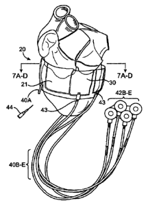
[0089] As can be seen, the regions in which middle layer sections 25 are
not positioned will
form "pockets" between layers 22 and 24 (since middle layer 25 is not present
which prevents
layers 22 and 24 from becoming bonded to one another). These "pockets" defined
by removed
sections of middle layer 25 form the tillable chambers 30 in the band.
[0090] As can also be seen in Fig. 9, the distal ends 41 of filling tubes
40 may be inserted into
the removed sections in middle layer 25. As a result, the distal ends 41 of
filing tubes 40 are
inserted within tillable chambers 30, while the bonding of layers 22 and 24
together secure in
position the remaining end portion of filing tubes 40. A bonding tab 46 can be
used to bind distal
end 41 in position against layer 22 if needed to form a fluid tight chamber
that communicates with
tubing 40.
19
CA 02701453 2010-03-31
WO 2009/032307 PCT/US2008/010421
[0091] Fig. 10 is an illustration of a second system for manufacturing the
present invention
using one layer of material folded on top of itself with a second layer of
material inserted
therebetween.
[0092] In this second method of making the invention, a single layer of
material 23 is used to
form both inner layer 22 and outer layer 24. As can be seen, the single layer
of material 23 is
simply folded over upon itself. An advantage of this particular method of
fabricating band 20 is
that it avoids having to use two separate materials to for layers 22 and 24.
This method also
eliminates the creation of a seal all around the fillable chambers, so that
fillable chambers 30
might be larger.
[0093] The method forming the device in Fig. 10 is similar to that set
forth above with respect
to forming the device of Fig. 9. Specifically, layer 23 is bonded, fused,
cross-linked or adhered
onto itself with removed sections in middle layer 25 forming the resulting
finable chambers 30.
Similarly as well, the distal ends 41 of filling tubes 40 may be inserted in
the removed sections of
middle layer 25. As a result, the distal ends 41 of filing tubes 40 are
inserted within finable
chambers 30, while the bonding of layer 23 onto itself secures in position the
remaining end
portion of filing tubes 40.
[0094] Fig. 11 is an illustration of a third system for manufacturing the
present invention
using a tube 27 of extruded non-cured or non-vulcanized silicon rubber. As
tube 27 is extruded,
regions 28 are pinched onto itself and are thus bound together. The regions of
tube 27 that are not
pinched together form the fillable chambers 30A, 30B and 30C. Tube 27 is
extruded, and then
separated along lines 29 into separate devices fillable chambers 30A, 30B and
30C, etc. Note: line
29 may simply be a line passing through a region of tube 27 that has been
bound onto itself. As
such, the ends of the separate devices 10A, 10B, etc. can be sealed.
Thereafter, the distal ends 41
of finable tubes 40 can be poked through side holes in band 20 and inserted
into the separate
fillable chambers 30. Thereafter, fillable tubes 40 can be adhesively bound
into position, for
example with a non-vulcanized silicone rubber tab 45 being rolled around,
pressed in place, and
CA 02701453 2010-03-31
WO 2009/032307 PCT/US2008/010421
heated to bond tubing 40 in position such that the tubing remains in fluid
communication with
finable chambers 30.
[0095] Fig. 12 shows an alternate embodiment of the invention in which
device 100
comprises a plurality of pockets 102 into which finable chambers 104 are
received. Each finable
chamber 104 has its own dedicated filling tube 106. Device 100 operates in a
Manner similar to
device 10 as described above, with the only difference being that each
tillable chamber is not
integrally formed into band 20 as depicted in the previous embodiments, but
the equivalent
function of chamber 30 is now accomplished by the combination of a pocket 102
into which a
separate fillable chamber 104 is inserted. These pockets into which finable
chambers 104 are
received may simply be formed by bonding or attaching layers 22 and 24 along
the sides and
bottom edges of each pocket. Each fillable chamber is then bonded into place
or layers 22 and 24
are bonded together to entrap each chamber in place.
[0096] Lastly, Fig. 13 shows a close-up view of the blunt needle port 44
that can be applied to
the end of any of the tubing 40. (For example, as illustrated as tubing 40A in
Fig. B. The blunt
needle port 44 may be formed by injecting room temperature vulcanized (RTV)
silicone rubber
approximately half-way into a short piece of silicone rubber tubing. The RTV
cures and then the
first insertion of a blunt needle tears a slit in the RTV section creating a
sealable slit and port. The
section of tubing absent of RTV acts as a pilot to help locate, hold, seal,
and guide the insertion of
a blunt hypodermic needle. This blunt needle port 44 is then bonded into
tubing 40 using RTV
silicone rubber.
Device Size
[0097] Although the size of the device depends on the purpose for which it
is being
implanted, it is contemplated that the device will be wide enough (measured
from the top edge,
i.e. the atrium edge, to the outside of the second or bottom edge, i.e. the
apex edge) to provide
efficient support to the atrio-ventricular grove. Accordingly, in one
embodiment, the device is
21
CA 02701453 2010-03-31
WO 2009/032307 PCT/US2008/010421
between 2 and 5 centimeters wide. In other embodiments, the device may be
adapted to provide
support over a larger area of the heart. This would provide specifically for
reinforcement of areas
of scar or muscular weakness as in dyskinetic infracted areas of the
myocardium.
[0098] As shown in Fig. 1, the distance between the base and the bottom of
the apex of the
heart can be expressed as distance "X". Because the focus of the device of the
present invention is
base stabilization, it is generally preferred that the width of the device be
less than or equal to 1/2
X, and be adapted for placement around the top half of the distance X, i.e.
closer to the A-V
Groove than the bottom of the apex.
Device Attachment
[0099] The device may be attached to the outside of the base of the heart
by any known
method. For example, attachment may be biological, chemical or mechanical.
Biological
attachment may be brought about by the interaction of the device with the
surrounding tissues and
cells, and can be promoted by providing appropriate enhancers of tissue
growth. Alternatively,
chemical attachment may be provided by supplying a mechanism for chemical
attachment of the
device, or portions thereof, to the external surface of the heart. In yet
another embodiment, the
rigidity and tightness of the device around the heart may provide for
sufficient mechanical
attachment due to the forces of the heart against the device without the need
for other means of
attachment.
[00100] In other alternate optional embodiments, the device instead further
comprises
attachment members, such as tabs. Specific anchor points or loops made of any
biocompatible and
implantable material may be attached to the edges or to the center portion or
both to facilitate
anchoring. Suitable materials include, inter alia, polyester, polypropylene or
complex polymers.
Alternative attachment members may comprise suture materials, protrusions that
serve as sites for
suturing or stapling, as well as other structural members that facilitate
attachment to the surface of
the heart.
22
CA 02701453 2010-03-31
WO 2009/032307 PCT/US2008/010421
Implantation
[00101] The BACETm system may be implanted through a conventional midline
total
stemotomy, sub maximal stemotomy or partial upper or lower stemotomy.
Alternatively, the
device may be implanted through a thoracotomy incision, or .a Video Assisted
Thoracoscopic
(VAT) approach using small incisions. The BACEn1 system can also be implanted
by a sub-
costal incision as in the Sub-Costal Hand-Assisted Cardiac Surgery (SHACS).
Additionally, the
BACE.174 system may be implanted with sutures onto epicardium or clips,
staples, or adhesive
material that can secure the device on the heart accurately. The device may
also be implanted
using robotic placement of the device along the posterior aspects of the base
of the heart.
[00102] The method of implantation and the adequacy of the external
annuloplasty can be
dynamically assessed by intra-operative trans-esophageal echocardiography,
epicardial
echocardiography or trans-thoracic echocardiography. The size of the device is
assessed based on
external circumference measurements of the cardiac base in the fully loaded
beating heart state.
EXPERIMENTAL RESULTS:
[00103] The device was tested with good results with 4 fluid chambers around
the mitral valve
side of the heart. The fluid chambers were filled one at a time with contrast
media (fluid visible
under fluoroscopy), and were thus visible under fluoroscopy. Saline was first
extracted from the
chambers that was present during implantation from priming them. Next, about
4cc of contrast
media was injected into each chamber and a fluoroscopy picture was taken. The
diameter across
the mitral valve was measured before and after filling the chambers. The
measurement before was
3.73 cm and then it reduced to 3.02 cm. This test shows that the mitral valve
annulus can be
reduced in diameter using the present invention.
23