Note: Descriptions are shown in the official language in which they were submitted.
4
CA 02705446 2010-05-11
PERIPROSTHETIC FRACTURE REPAIR
BACKGROUND
[0001] The use of fixation plates to treat periprosthetic
fractures has been limited
by the interference of a prosthetic within the medullary canal poses to the
insertion of
screws, pegs, nails or other fixation devices therethrough.
SUMMARY OF THE INVENTION
[0002] The present invention is directed to a device for
treating fractures,
comprising: a plate receiving structure including on a bone facing side
thereof a recess
sized and shaped to receive a fixation plate anchored in a desired position on
a bone;
a mating structure sized and located to engage a corresponding structure of
the fixation
plate to prevent relative movement between the fixation plate and the device;
and at
least one leg projecting laterally away from the recess, a first one of the at
least one leg
including a first fixation element receiving hole extending therethrough, the
first leg
being positioned and oriented so that, when the device is received on a
fixation plate
anchored to the bone in the desired position, the first fixation element
receiving hole is
aligned to pass a fixation element inserted therethrough into the bone without
passing
through a longitudinal axis of the medullary canal. Those skilled in the art
will
understand that medullary canals are generally neither straight nor concentric
with the
bone. Thus the axis of the medullary canal, as that term is used in this
application,
refers to a curve Connecting points in the center of the medullary canal along
the length
of the bone.
[0003] The present invention also relates to a kit for treating
fractures,
comprising:
a lateral fixation device comprising: a longitudinal fixation device
receiving structure including on a bone facing side thereof a recess sized and
shaped
1753016.1 1
CA 02705446 2010-05-11
to receive a fixation plate which, when anchored in a desired position on a
bone,
extends substantially parallel to a longitudinal axis of the medullary canal;
a mating
structure sized and located to engage a corresponding structure of the
fixation plate to
prevent relative movement between the fixation plate and the device; and a
plurality of
legs projecting laterally away from the recess, at least first and second ones
of the legs
including fixation element receiving holes extending therethrough; and
instructions for performing the following steps: coupling a first end of a
longitudinal fixation device to a bone on a first side of a fracture so that
the longitudinal
fixation device extends along the bone substantially parallel to a
longitudinal axis of the
medullary canal; coupling the lateral fixation device to a portion of the
longitudinal
fixation device extending over a portion of the bone on a second side of the
fracture by
mating the longitudinal fixation device within the recess of the lateral
fixation device;
and coupling the lateral fixation device to the bone by inserting a fixation
element
through a hole formed in a portion thereof separated from the longitudinal
fixation
device by a selected distance around a portion of a circumference of the bone.
[0004] The foregoing and other objects, advantages and features of the
present
invention will become more apparent upon reading of the following non
restrictive
description of illustrative embodiments thereof, given by way of example only
with
reference to the accompanying drawings in which:
BRIEF DESCRIPTION OF THE DRAWINGS
[0005] Figure 1 is a side view of a fracture located on a distal end of a hip
prosthesis;
Figure 2 is a side view of a fracture located distal to a hip prosthesis;
Figure 3 is a side view of a fracture located near to a knee prosthesis;
Figure 4 is a perspective view of an embodiment of a fixation kit according to
the invention;
Figure 5 is a side view of the fixation kit of Figure 4;
Figure 6 is a front view of the fixation kit of Figure 4;
Figure 7 is a perspective view of a first embodiment of an attachment plate
1753016.1 2
CA 02705446 2010-05-11
WO 2009/064643
PCT/US2008/082470
according to the invention;
Figure 8 is a top view of the attachment plate of Fig. 7;
Figure 9 is a front view of the attachment plate of Fig. 7;
Figure 10 is a perspective view of a second embodiment of an attachment
plate according to the invention;
Figure 11 is cross-sectional view of the fixation kit of Fig. 4;
Figure 12 is a perspective view of an embodiment of an aiming block
according to the invention;
Figure 13A is a side view of an embodiment of a fixation device according to
the invention;
Figure 13B is a cross-sectional view of the fixation device of Fig. 13A;
Figure 13C is a top view of the fixation device of Fig. 13A,
Figure 13D is a front view of a distal portion of the fixation device of Fig.
13A;
Figure 14A is a side view of a second embodiment of a fixation device
according to the invention;
Figure 14B is a cross-sectional view of the fixation device of Fig. 14A;
Figure 14C is a top view of the fixation device of Fig. 14A;
Figure 14D is a front view of a distal portion of the fixation device of Fig.
14A;
Figure 15A is a cross-sectional view of a third embodiment of a fixation
device
according to the invention;
Figure 15B is a top view of the fixation device of Fig. 15A;
Figure 16A is a cross-sectional view of a fourth embodiment of a fixation
device according to the invention;
Figure 16B is a top view of the fixation device of Fig. 16A;
Figure 17A is a cross-sectional view of a fifth embodiment of a fixation
device
according to the invention;
Figure 17B is a top view of the fixation device of Fig. 17A;
Figure 18A is a cross-sectional view of a sixth embodiment of a fixation
device
according to the invention; and
-3-
CA 02705446 2010-05-11
WO 2009/064643
PCT/US2008/082470
Figure 18B is a top view of the fixation device of Fig. 18A.
DETAILED DESCRIPTION
[0006] The present invention may be further understood with reference to the
following description and to the appended drawings, wherein like elements are
referred
to with the same reference numerals. The present invention relates to devices
for
treatment of fractures. In particular, the invention relates to improved
methods and
systems for repairing periprosthetic fractures. Although exemplary embodiments
of the
present invention will be discussed with reference to knee and hip prostheses,
the
present invention may be successfully implemented in any long bone including a
prosthetic device inserted into its medullary canal. In addition, as would be
understood
by those skilled in the art, the present invention may be used for the
treatment of
fractures around nails and for "conventional" fractures in patients with poor
bone quality.
The present invention allows the user to apply standard fixation plates while
placing
screws and/or pins therethrough into the bone along paths selected to: 1)
avoid any
prosthesis in the medullary canal; 2) align the screws/pins non-parallel to
one another to
improve purchase in the bone; and/or 3) maximize the length of cancellous bone
through which the screws/pins pass.
[0007] Periprosthetic fractures may occur intraoperatively (during
implantation or
replacement of a prosthetic), or postoperatively (e.g., as a result of stress
or trauma to
the bone in which the prosthetic was previously implanted). As would be
understood by
those skilled in the art, fractures have been effectively treated by
stabilizing the bone
using fixation plates (e.g., dynamic compression plates (DCPs), locking
compression
plates (LCPs), etc.), which typically comprise a metal plate including a
plurality of holes
through which anchoring of screws or other fixation devices are inserted into
underlying
bone tissue. Periprosthetic fractures are more difficult to treat than
ordinary fractures
because a prosthesis extending within the medullary canal may interfere with
the proper
-4-
CA 02705446 2010-05-11
WO 2009/064643
PCT/US2008/082470
coupling of a fixation plate across the fracture by preventing the fixation
devices from
being inserted through the bone across the medullary canal. For example, hip
prostheses may interfere with certain fractures of the femur. These hip
prostheses
often include a ball joint and a stem which is inserted into the medullary
canal of the
femur. As the femur absorbs significant stresses with each step, to adequately
couple a
fixation plate thereto, it is desired to maximize the purchase of the fixation
devices in the
femur.
[0008] In determining a course of treatment, the needs of the patient must be
considered. For example, an active 55 year old with a periprosthetic fracture
will likely
have functional demands different from those of a sedentary 85 year old.
Important
factors to consider include the location of the fracture, how well-fixed the
prosthesis is,
and the quality of the femoral bone stock. The Vancouver classification
divides
periprosthetic hip fractures into three categories: Type A fractures are
trochanteric (i.e.,
disposed at or near the greater or lesser trochanters); Type B fractures occur
around
the stern of the prosthesis; and Type C fractures occur so far from the stem
that the
fracture may be treated as a general fracture (i.e., the prosthesis may be
ignored). Of
these fractures, Type B is the most common. As used in the following
descriptions of
exemplary embodiments of the invention, the term "distal" refers to a
direction away
from the end of the bone through which the prosthesis is inserted into the
medullary
canal. Thus, the distal end of a hip prosthesis is that which is located
furthest from the
pelvis and the proximal end is that which is nearest to the pelvis. Fig. 1
shows an
example of a Type B fracture 50 located along a distal portion 110 of a hip
prosthesis
100. As discussed above, the present invention may also be used to treat
general
fractures (e.g., Type C fractures). An example of a Type C fracture is shown
in Fig. 2.
In particular, Fig. 2 shows a fracture 52 located distal of the hip prosthetic
100.
However, those skilled in the art will understand that the apparatus according
to the
present invention may also be used to treat Type A fractures as well as
similar fractures
of other bones. For example, Fig. 3 shows a treatable fracture 54 located near
a knee
-5-
CA 02705446 2010-05-11
WO 2009/064643
PCT/US2008/082470
prosthetic 130.
[0009] Regardless of how the fracture is classified, complications common to
each of
the fracture types may make proper treatment critical while creating
difficulties,
rendering aspects of standard fracture treatment unsuitable. For example,
standard
fixation plates are typically fixed by inserting one or more fixation devices
(e.g., bone
screws) substantially diametrically through the bone. Thus, these fixation
devices pass
through the periosteum and compact bone adjacent to the fixation plate,
through the
medullary canal and then into the compact bone on the opposite side of the
medullary
canal. When a prosthesis occupies the medullary canal, inserting a screw
directly
therethrough is no longer possible and inserting the screw through only that
portion of
compact bone between the fixation plate and the medullary canal often does not
provide
sufficient anchorage. Thus, it is desirable to maximize the length of the
screw within the
compact bone. Accordingly, exemplary embodiments of fracture repair devices
according to the present invention, as will be described below, enable the
anchoring of
screws or other fixation devices along extended paths through compact bone
without
contacting the prosthesis occupying the medullary canal. Furthermore, the
devices and
methods according to the present invention may also allow the user to
customize the
configuration and location of the fixation plate to achieve a desired fracture
treatment.
[0010] Exemplary embodiments of a fixation kit according to the present
invention will
now be described with reference to fracture repair devices designed to work in
conjunction with any conventional fixation plate such as, for example, locking
compression plates (LCPs). Exemplary embodiments of the fracture repair
devices may
be utilized in conjunction with any number of different types of LCPs or other
fixation
plates including, for example, an LCP broad curved plate, an LCP broad plate,
an LCP
Distal Femur (DF) plate, an LCP Less Invasive Stablization System (LISS)
plate, an
LCP proximal femur plate, an LCP proximal femur with hook plate, an LCP
condylar
plate, etc. As will be understood by those skilled in the art, the exemplary
fracture
-6-
CA 02705446 2010-05-11
WO 2009/064643
PCT/US2008/082470
repair devices may also be used with other conventional fixation plates in
addition to
LCPs. Thus, the fixation plate may be selected to fit a specific situation in
the same
manner as would be done if there were no prosthesis or other reason for
avoiding the
insertion of fixation devices through the axis of the medullary canal.
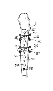
[0011] Figs. 4 - 6 show a fixation kit including an LCP 500 and two fracture
repair
devices 200 in place on a femur 10. An intermedullary prosthesis 100 (shown in
hidden
view) has been inserted into the medullary canal of the femur 10 and the LCP
500
extends proximally from a distal end 510 on a portion of the femur 10 distal
of a distal
end of the prosthesis 100 across a fracture to a proximal end 520. Two devices
200 are
received over proximal and medial portions of the LCP 500 as these portions of
the LCP
500 overlay the prosthesis 100. As the distal end 510 extends distally beyond
the distal
end of the prosthesis 100, it may be secured to the femur in any conventional
manner
(e.g., by one or more bone screws inserted straight through the medullary
canal and the
compact bone on either side thereof). As would be understood by those skilled
in the
art, the size, configuration and/or location of the devices 200 may vary
depending on
anatomy, fracture location and the position and/or size of a prosthesis
relative to the
LCP 500. For example, devices 200 may be placed only where the LCP overlays a
prosthesis, on both sides of a fracture regardless of location of the fracture
relative to
the LCP 500 or in any other desired arrangement so long as the required bond
between
the LCP 500 and the underlying bone is established. Thus, one or more
additional
devices 200 may be attached to the distal end 510 of the LCP 500 or at any
other
locations to provide further stabilization. The LCP 500 of Figs. 4 - 6 extends
distally
beyond the distal end of the prosthesis 100. However, in other embodiments a
shorter
LCP may be selected with devices 200 providing support at all points along the
length
thereof. Those skilled in the art will understand that the number and the
location of the
devices 200 may be determined according to physician preference. Therefore, in
some
embodiments the LCP 500 may be coupled to only a single device 200 (e.g., by
centering the device 200 over the fracture) supplemented as desired by
additional
-7-
CA 02705446 2010-05-11
WO 2009/064643
PCT/US2008/082470
fixation devices including, for example, screws inserted only through the
portion of
compact bone adjacent to the LCP 500. Each device 200 is coupled to the LCP
500 via
a screw 227 and includes one or more screws 257 or other fixation devices that
anchor
the device 200 to the bone 10.
[0012] As shown in Fig. 5, a distal portion of the LCP 500 extends beyond the
prosthesis 100 and is anchored directly into the bone through the medullary
canal as is
done in the manner of general fractures, when no prosthesis is present. The
distal
portion may be anchored using any number of screws 527 spaced in accordance
with
physician preference. Portions of the LCP 500 that extend along the length of
the
prosthesis 100 either contain no screws that enter the medullary canal (e.g.,
portions
not coupled to a device 200) or are secured via a device 200, which is screwed
(via the
screws 257) at an angle into the bone 10 to avoid the prosthesis 100.
[0013] The device 200 shown in Fig. 7 - 9 is shaped for use in conjunction
with the
LCP 500 of Figs. 4 - 6. Of course, those skilled in the art will understand
that any
number of varieties of devices 200 may be formed for use in conjunction with
any of a
variety of different fixation plates of different sizes and shapes. The device
200 includes
a locking attachment plate 202 formed of a substantially rigid biocompatible
material
such as, for example, plastic, medical-grade steel or titanium as would be
understood
by those skilled in the art. The attachment plate 202 includes a body 210
including, on
a bone-facing side thereof, a recess contoured to receive the LCP 500.
Sidewalls 215
of the recess may preferably be shaped to substantially conform to the
contours of the
LCP 500. Although the sidewalls 215 need not form a tight fit against
corresponding
sides of the LCP 500, some embodiments may include sidewalls that snap-fit or
otherwise couple to the LCP 500. The body 210 may also include a coupling
feature
that corresponds to a corresponding feature on the LCP 500. For example, the
body
210 may include a centrally located screw hole 225 that corresponds to a
coupling
arrangement (e.g., a threaded bore) of the LCP 500. Thus, the body 210 may be
-8-
CA 02705446 2010-05-11
WO 2009/064643
PCT/US2008/082470
coupled to the LCP 500 by aligning the attachment plate 202 over to the LCP
500 and
inserting a screw (e.g., the screw 227) or other fixation device through the
hole 225 and
into the threaded bore. The bore may pass through the entire body of the LCP
500,
enabling the screw 227 to extend past a bone-facing surface of the LCP 500.
Thus, in
some embodiments, the screw 227 may be driven into the compact bone on the
side of
the medullary canal facing the LCP 500 without contacting the underlying
intermedullary
prosthesis. However, in other embodiments, the screw 227 may not extend past
the
LCP 500, serving only to couple the LCP 500 and the device 200. Alternatively,
as
would be understood by those skilled in the art, a fracture repair device
according to the
invention may include a projection aligned to mate with a corresponding recess
in an
LCP (e.g., the threaded bore), a recess aligned to mate with a corresponding
projection
of an LCP or a combination of such recess/projection matings. As discussed
above, the
sidewalls 215 may be snap-fit onto the LCP 500. Other coupling arrangements,
such
as, for example, friction-fitting, adhesives, bolts, etc. may also be used to
couple the
LCP 500 and the attachment plate 202 as would be understood by those skilled
in the
art.
[0014] The attachment plate 202 includes one or more arms 220 extending
laterally
from the sidewalls 215, away from the body 210. Each of the sidewalls 215
includes an
arm 220 extending from each end thereof and each of the arms 220 includes a
first and
a second screw hole 222, 224, respectively, extending therethrough. However,
those
skilled in the art will understand that the number of arms per sidewall may
vary. The
arms 220 may be formed integrally with the body 210 or attached separately.
Each of
the arms 220 is preferably oriented such that a bone-facing surface of the arm
220 is
generally follows the contours of a bone on which the arm 220 is to be
mounted.
Optionally, the arms may be formed of a material which may be bent by a user
into a
desired configuration to customize the arms 220 to the anatomy of each
patient. As
shown in Fig. 9, when viewed in a plane substantially perpendicular to a
longitudinal
axis of the medullary canal, each of the arms 220 extends along a curve
substantially
-9-
CA 02705446 2010-05-11
WO 2009/064643
PCT/US2008/082470
approximating the shape of an outer surface of a bone on which it is to be
mounted.
[0015] As seen in Figs. 7 - 9, each arm 220 also extends away from the
corresponding side wall 215 at an angle within a plane of the body 210. Those
skilled in
the art will understand that the selection of this angle and any change in
this angle
between the first and second screw holes 222, 224 allow for the application of
additional
screws at different angles and/or at different locations or, for example, to
increase the
area over which the attachment forces are applied to the bone. As indicated
above,
each of the arms 220 includes a first screw hole 222 adjacent to the
corresponding side
wall 215 and a second screw hole 224 extending laterally away from the first
screw hole
222. Although each of the arms 220 is shown extending at substantially the
same
angle, those skilled in the art will understand that the arms 220 may extend
at different
angles to accommodate varying bone structure, LCP shapes, etc. and may include
the
same number or different numbers of screw holes.
[0016] As described above and as shown in Fig. 9, the arms 220 preferably
curve to
enable the arms 220 to wrap around the bone 10 in a substantially conforming
manner.
A degree to which the arms 220 encircle the bone 10 may vary depending on the
curvature and the angle of each arm 220 in relation to bone physiology. Thus,
the arms
220 may produce a tighter fit when mounted to wide portions of the bone 10,
while
providing a looser fit when mounted to narrow bone portions. In addition, the
arms 220
may flex, allowing the arms 220 to be mounted closer to the bone 10.
[0017] Each of the first and second screw holes 222, 224, respectively, may be
threaded to match a threading of a locking head of a screw 257 or may be
otherwise
suited to receive the particular fixation device to be employed with the
device 200. As
would be understood by those skilled in the art, the number of screw holes in
each arm
220 may vary based on factors such as LCP shape, bone anatomy, desired degree
of
stabilization, etc. As shown in Fig. 10, an exemplary attachment plate 302 is
-10-
CA 02705446 2015-03-18
substantially similar to the plate 202 of Figs. 7 - 9 except that each of the
arms 320
has only one screw hole 250 extending therethrough. It will be understood by
those of
skill in the art that the attachment plate 302 may be used according to the
device 200
in substantially the same manner as the attachment plate 202. In some
embodiments,
one or more arms may not include any holes at all or may contain more than two
screw holes.
[0018] Each of the holes 250 defines an angle of insertion for the screw 257
selected so that when the attachment plate 202 is mounted onto the bone 10,
the
screw 257 passes through the bone 10 without diametrically passing through the
medullary canal, thereby avoiding contact with the prosthesis 100. Those
skilled in the
art will understand that the angle is preferably selected to maximize the
length of the
screw 257 received in the bone 10. Those skilled in the art will also
understand that
some or all of the screw holes for any of the attachment plates according to
the
invention may be variable angle locking holes allowing for locking screws to
be
inserted therethrough and locked to the plate at multiple angles relative to
the
attachment plate. For example, any or all of the screw holes 222, 224 and 250
may be
formed substantially in accord with the description in U.S. Patent Application
Publication No. 2005/0165400 filed by Fernandez, July 28, 2005.
[0019] For example, the screws may have a head shaped like a sphere and
threaded with a substantially constant pitch substantially equal to a pitch of
a threaded
shank of the screw. In addition, an insertion/extraction hole may be cut in
the head for
the connection of an insertion/extraction tool. The thread cut in the screw
head may
have a double entry maintaining substantially the same pitch as that of the
thread of
the shank. Of course, as would be understood by those skilled in the art, the
thread
profile may vary according to the requirements and according to the mechanical
properties of the material of which the screw is formed.
-11-
685226 1
CA 02705446 2010-05-11
WO 2009/064643
PCT/US2008/082470
[0020] This allows the screw to be inserted into a properly designed screw
hole at any
angle within a wide range without affecting the position of the thread of the
screw head
with respect to walls of the screw hole.
[0021] Specifically, such a screw hole may be formed in a spherical shape,
with
edges thereof at both ends of the hole removed in a frusto-conical shape. That
is, the
screw hole may include two frusto-conical portions extending toward one
another from
opposite surfaces of the plate and connected at tips of the cones through a
partial
sphere. The inner wall of each screw hole has a small number of isolated
protrusions
such as pegs or spikes (e.g., between two and thirty) designed to lock against
the
threaded spherical head of the screws when the screws are driven in through
the screw
holes. The protrusions may, for example, be somewhat flattened with a width
bigger
than its length.
[0022] Once such a screw has been driven into such a screw hole, the spherical
shape of the screw head allows it to lock against the protrusions without
regard to
whether the screw extends perpendicular or at a tilt relative to an axis of
the screw hole.
The angle at which of the screw is locked may then be varied by as much as 20
relative to the axis of the screw hole.
[0023] In use, a physician may begin treatment by selecting an LCP 500 of
appropriate size and shape, taking into account the width of the bone 10, the
location of
the fracture and other factors as would be understood by those skilled in the
art. The
LCP 500 is then aligned over the bone 10 to extend across the fracture in a
position
selected to stabilize the portions of the bone on both sides thereof. The
physician then
has the option of initially securing the distal portion of the LCP 500 to the
portion of the
bone not including a prosthesis within the medullary canal or of selecting one
or more
attachment plates 202 to achieve the desired coupling of the LCP 500 and the
proximal
-12-
CA 02705446 2010-05-11
WO 2009/064643
PCT/US2008/082470
portion of the bone. If securing of the distal portion first is desired, the
physician drives
the screws 527 directly into the bone 10 in the same manner as would be used
for a
fracture where no prosthesis was present. Thereafter, the physician may slide
the
selected plate 202 over the proximal portion of the LCP 500 to the desired
alignment
and attach the plate 202 to the LCP 500. Alternatively, as would be understood
by
those skilled in the art, the physician may attach the attachment plate to the
bone in a
desired location before attaching the LCP 500 to either the plate 202 or any
portion of
the bone and then slide the LCP 500 through the recess into place between the
bone 10
and the attachment plate 202.
[0024] The physician may choose to couple the attachment plate 202 to the LCP
500
before attaching the attachment plate 202 to the bone 10. The attachment plate
202 is
positioned over a desired location of the LCP 500. As shown in Figs. 5 and 6,
the LCP
500 includes multiple attachment arrangements comprising attachment sites 507
located along the entire length thereof. The attachment sites 507 are spaced
apart,
either uniformly or at different distances. For example, certain lengths of
the LCP 500
may include more attachment sites (i.e., tighter spacing) than others. The
attachment
sites 507 may correspond to anchoring locations of the screws 227. That is,
the
attachment arrangements may be the same as the holes through which the screws
527
may be driven although, as would be understood, the screws are used to couple
the
attachment plate 202 to the LCP 500 will be shorter than those used to
directly couple
the LCP 500 to the bone so as to avoid interference with the intermedullary
prosthesis.
[0025] After positioning over the LCP 500 substantially flush with the bone
10, the
attachment plate 202 is coupled to the LCP 500 by either driving the screw 227
to a
depth beyond the bone-facing surface of the LCP 500 (i.e., into the bone 10)
or to a
depth within the body of the LCP 500. As an alternative to coupling the
attachment
plate 202 during treatment, the coupling may occur prior to introduction of
the LCP 500
into the patient. The attachment plate 202 is then anchored to the bone 10 by
-13-
CA 02705446 2010-05-11
WO 2009/064643
PCT/US2008/082470
individually driving each screw 257 into the bone 10 at an angle selected by
the
physician (e.g., to maximize a length of the path the screw travels through
the compact
bone without entering the medullary canal). The bone 10 beneath each hole 250
is
drilled out to a desired depth (e.g., a maximum depth of penetration of the
compact
bone without contacting the prosthesis 100). As an alternative to drilling,
the screws
257 may be self-tapping. As would be understood by those skilled in the art,
the
maximum depth to which the screws 257 may be driven is a function of known
factors
such as, for example, bone anatomy and the available insertion angles.
[0026] As would be understood by those skilled in the art, an aiming device
such as
an aiming block may be used to facilitate accurate drilling of the bone 10.
Fig. 12 shows
an exemplary embodiment of an aiming block 400 in an operative position. The
aiming
block 400, which may be placed over the attachment plate 202 or over the
combined
attachment plate-LCP, includes a body portion 410 including a hole 425
matching the
hole 225. Although the hole 425 is not strictly required, including the hole
425 facilitates
visual confirmation that the aiming block 400 has been placed correctly over
the
attachment plate 202. Once the attachment plate 202 has been positioned at a
desired
location, the aiming block 400 is placed on top of the attachment plate 202
and aligned
therewith. The aiming block 400 includes one or more shafts 450 corresponding
to the
first hole 222 and/or the second hole 224. The shafts 450 are positioned at
desired
angles to form a drilling template. A plurality of aiming blocks 400 with
different shaft
configurations may be available for use, enabling the drilled holes to be
oriented at any
desired angle. A drilling tool is inserted through a hole 455 located at one
end of the
shaft 450 and guided through an opening at the opposite end of the shaft 450
into either
of the first hole 222 and the second hole 224 and, subsequently, into the bone
10. After
reaching the desired drilling depth, the drilling tool is withdrawn from the
shaft and
additional holes may be created by inserting the drilling tool into further
shafts 450.
When all the holes have been drilled, the aiming block 400 is removed and the
operating site is cleared of bone debris before inserting the screws 257.
-14-
CA 02705446 2010-05-11
WO 2009/064643
PCT/US2008/082470
[0027] The screws 257 may then be inserted directly into the first and/or the
second
holes 222, 224 or guided through the shafts 450 of the aiming block 400. As
shown in
the cross-sectional view of the fixation kit in Fig. 11, taken along line A-A,
a substantial
portion of each screw 257 occupies the bone cortex 15 without interfering with
a
prosthesis 100 within the medullary canal 12. Screws of varying length may be
provided as part of the fixation kit to take advantage of the maximum
allowable insertion
depth. Thus, the screws may be selected to extend from one side of the bone 10
to an
opposing side. As seen in Fig. 11, the screws 257 may also occupy a portion of
a
medullary canal 12 without passing diametrically therethrough or contacting
the
prosthesis 100. As the screws 257 travel toward their resting positions, the
arms 220
may be drawn toward the bone 10 by pressure exerted by head portions of the
screws
257. Although a close fit is desired for stability, it may also be desirable
not to
excessively constrict the bone 10 or the blood supply thereto by drawing the
arms 220
too tightly thereagainst. Thus, a small gap 60 may be left between the arms
220 and
the bone 10. The gap 60 promotes blood flow and reduces the amount of bone
compressed by the attachment plate 202. The gap 60 may be achieved by forming
the
arms 220 with sufficient curvature such that a, bone-facing surface of the
arms 220 is
substantially concave. The gap 60 may also be a function of the extent to
which the
screws 257 are driven into the bone 10. If a smaller gap is desired, more of
the screw
257 can be driven in. Similarly, less driving will result in a larger gap.
Thus, the length
of a shaft portion 259 of each screw 257 that is exposed within the gap 60 is
variable.
Some screws 257 may be driven entirely into the bone 10 while other screws 257
may
form large gaps.
[0028] Exemplary embodiments of fixation devices that may be used in
conjunction
with the device 200 will now be described. Figs. 13A - 13D show an exemplary
embodiment of a screw 557 according to the present invention. As shown in the
side
view of Fig. 13A, the screw 557 includes a conical body comprising a head 552
and a
-15-
CA 02705446 2010-05-11
WO 2009/064643
PCT/US2008/082470
shaft 554 including a plurality of threads 555. The shaft 554 also includes
one or more
slots 559 extending substantially the entire length thereof. The conical body
of the
screw 557 tapers from the head 552 toward the shaft 554. Fig. 13B shows a
cross-
section of the screw 557, taken along line B-B. As shown, a portion of the
head 552
includes a recess 532. In the exemplary embodiment, the recess 532 is hex-
shaped.
The shape of the recess 532 is shown more clearly in the top view of the head
552
illustrated in Fig. 13C. Although the exemplary embodiment utilizes a hex-
shaped
recess, other shapes (e.g., stars or triangles) may be utilized in other
embodiments.
Fig. 13D shows a front view of a distal tip 590 of the screw 557. As shown in
Fig. 13D,
the slots 559 are equidistantly spaced about the perimeter of the distal tip
590. As
would be understood by those skilled in the art, the screws may be formed of
stainless
steel, titanium or a suitable biocompatible polymer.
[0029] Figs. 14A - 14D show an exemplary embodiment of a screw 657 according
to
the present invention. The screw 657 includes a cylindrical body comprising a
head 652
and a shaft 654 including a plurality of threads 655. The shaft 654 also
includes one or
more slots 659 extending substantially the entire length thereof. Fig. 14B
shows a
cross-section of the screw 657. A portion of the head 652 includes a recess
632. As
shown in the top view of the head 652 illustrated in Fig. 13C, the recess 632
is hex-
shaped. However, other shapes are also possible. Fig. 14D shows a front view
of a
distal tip 690 of the screw 657. As shown in Fig. 13D, the slots 659 are
equidistantly
spaced about the perimeter of the distal tip 690.
[0030] Figs. 15A and 15B show an exemplary embodiment of a screw 757 according
to the present invention. The screw 757 includes an outer member 752 and an
inner
member 754 that couples to the outer member 752. The inner member 754 extends
substantially the entire length of the outer member 752 and includes a hex-
shaped
recess 732. An inner wall 710 of the outer member 752 defines an interface
shaped to
mate with the inner member 754 using friction-fitting. However, the inner
member 754
-16-
CA 02705446 2010-05-11
WO 2009/064643
PCT/US2008/082470
may be coupled to the outer member 752 in any number of ways including, for
example,
screwing in a direction opposite to that of threads 75 running along an outer
surface of
the outer member 752.
[0031] Figs. 16A and 16B show an exemplary embodiment of a peg 857 according
to
the present invention. The peg 857 includes an outer member 852 and an inner
member 854 that couples to the outer member 852. The inner member 854 extends
substantially the entire length of a head portion 856 of the outer member 852
and
includes a hex-shaped recess 832. A distal end 834 of the inner member 854 is
shaped
to conform to the contours of the head 856. An inner wall 810 of the outer
member 852
defines an interface shaped to mate with the inner member 854 using friction-
fitting.
However, the inner member 854 may be coupled to the outer member 852 in any
number of ways including, for example, screwing.
[0032] Figs. 17A and 17B show an exemplary embodiment of a peg 957 according
to
the present invention. The peg 957 includes an outer member 952 and an inner
member 954 that couples to the outer member 952. The inner member 954 extends
substantially the entire length of the outer member 952 and includes a hex-
shaped
recess 932. The outer member 952 includes a head portion 956 and a shaft
portion 958
having a diameter less than that of the head 956. An inner wall 910 of the
outer
member 952 defines an interface shaped to mate with the inner member 954 using
friction-fitting. However, the inner member 954 may be coupled to the outer
member
952 in any number of ways including, for example, screwing.
[0033] Figs. 18A and 18B show an exemplary embodiment of a screw 1057
according
to the present invention. The screw 1057 includes an outer member 1052 and an
inner
member 1054 that couples to the outer member 1052. The inner member 1054
extends
substantially the entire length of a head portion 1056 of the outer member
1052 and
includes a hex-shaped recess 1032. The outer member 1052 includes a head
portion
-17-
CA 02705446 2010-05-11
WO 2009/064643
PCT/US2008/082470
1056 and a shaft portion 1058 having a diameter greater than that of the head
1056. A
distal end 1034 of the inner member 1054 is conically shaped. An inner wall
1010 of
the outer member 1052 defines an interface shaped to mate with the inner
member
1054 using friction-fitting. However, the inner member 1054 may be coupled to
the
outer member 1052 in any number of ways including, for example, screwing in a
direction opposite to that of threads 85 running along an outer surface of the
outer
member 1052.
[0034] The present invention has been described with reference to specific
exemplary
embodiments. Those skilled in the art will understand that changes may be made
in
details, particularly in matters of shape, size, material and arrangement of
parts.
Accordingly, various modifications and changes may be made to the embodiments.
The specifications and drawings are, therefore, to be regarded in an
illustrative rather
than a restrictive sense.
-18-