Note: Descriptions are shown in the official language in which they were submitted.
CA 02707755 2010-06-30
=
1
Methods And Systems For Cardiac Valve Delivery
Field Of The Invention
The present invention relates generally to methods and systems for
cardiovascular surgery.
Background Of The Invention
Various surgical techniques may be used to repair a diseased or damaged
heart valve, such as annuloplasty (contracting the valve annulus),
quadrangular
resection (narrowing the valve leaflets), commissurotomy (cutting the valve
commissures to separate the valve leaflets), or decalcification of valve and
annulus
tissue. Alternatively, the diseased heart valve may be replaced by a
prosthetic valve.
Where replacement of a heart valve is indicated, the dysfunctional valve is
typically
removed and replaced with either a mechanical or tissue valve.
A number of different strategies have been used to repair or replace a
defective
heart valve. Open-heart valve repair or replacement surgery is a long and
tedious
procedure and involves a gross thoracotomy, usually in the form of a median
sternotomy. In this procedure, a saw or other cutting instrument is used to
cut the
sternum longitudinally and the two opposing halves of the anterior or ventral
portion of
the rib cage are spread apart. A large opening into the thoracic cavity is
thus created,
through which the surgeon may directly visualize and operate upon the heart
and
other thoracic contents. The patient must typically be placed on
cardiopulmonary
bypass for the duration of the surgery.
Open-chest valve replacement surgery has the benefit of permitting the direct
implantation of the replacement valve at its intended site. This method,
however, is
highly invasive and often results in significant trauma, risk of
CA 02707755 2010-06-30
2
complications, as well as an extended hospitalization and painful recovery
period
for the patient.
Minimally invasive valve replacement procedures have emerged as an
alternative to open-chest surgery. Wikipedia Encyclopedia defines a minimally
invasive medical procedure as one that is carried out by entering the body
through
the skin or through a body cavity or anatomical opening, but with the smallest
damage possible to these structures. Two types of minimally invasive valve
procedures that have emerged are percutaneous valve procedures and trans-
apical valve procedures. Percutaneous valve procedures pertain to making small
incisions in the skin to allow direct access to peripheral vessels or body
channels
to insert catheters. Trans-apical valve procedures pertain to making a small
incision in or near the apex of a heart to allow valve access. The distinction
between percutaneous valve procedures and minimally invasive procedures is
also highlighted in a recent position statement of the Society of Thoracic
Surgeons
(STS), the American Association for Thoracic Surgery (AATS), and the Society
for
Cardiovascular Angiography and Interventions (SCAI; Vassiliades Jr. TA, Block
PC, Cohn LH, Adams DH, Borer JS, Feldman T, Holmes DR, Laskey WK, Lytle
BW, Mack MF, Williams DO. The clinical development of percutaneous heart
valve technology: a position statement by the Society of Thoracic Surgeons
(STS), the American Association for Thoracic Surgery (AATS), and the Society
for
Cardiovascular Angiography and Interventions (SCAI). J Thorac Cardiovasc Surg.
2005; 129:970-6). Because
minimally invasive approaches require smaller
incisions, they generally allow for faster patient recovery with less pain and
bodily
trauma. This, in turn, reduces the medical costs and the overall disruption to
the
life of the patient.
The use of minimally invasive approaches, however, introduces new
complexities to surgery. An
inherent difficulty in the minimally invasive
percutaneous approach is the limited space that is available within the
vasculature. Unlike open heart surgery, percutaneous heart surgery offers a
surgical field that is only as large as the diameter of the blood vessel used
for
access. Consequently, the introduction of tools and prosthetic devices becomes
a
great deal more complicated as compared to open-chest surgeries. The device
must be dimensioned and configured to permit it to be introduced into the
vasculature, maneuvered therethrough, and positioned at a desired location.
This
may involve passage through significant convolutions, at some distance from
the
initial point of introduction, before placement can be made at the intended
site.
CA 02707755 2010-06-30
3
Andersen et al. describe a valve prosthesis implanted in a body channel by a
way of catheterization in U.S. Patent Nos. 5,411,442; 5,840,081; 6,168,614;
and
6,582,462; and U.S. Patent Application No. 2003/0036795. Catheters are hollow
flexible tubes which can be passed inside blood vessels to the heart for
diagnostic
and treatment purposes. The delivery of catheter expanded valves through body
channels such as that described by Andersen et al. is thus dependent on
instruments of sufficiently small diameters, as well as adequate length and
flexibility
to navigate blood vessels.
Minimally invasive trans-apical valve replacement procedures have emerged
as an alternative to both open-chest surgery and percutaneous valve surgeries.
Bergheim et al. present improved methods and systems for cardiac valve
delivery
in U.S. Patent Application Nos. 2007/0027534 and 2005/0240200. Methods and
systems for the repair, removal, and/or replacement of heart valves through
the
apex of the heart are described. This is an improvement over minimally
invasive
percutaneous approaches attempting insertion into the vasculature as the trans-
apical approach is not limited by the space that is available within the
vasculature.
Trans-apical delivery is also closer to the heart than catheter-based
procedures.
In-vivo studies have shown that catheter-based valve delivery
instrumentation may not be well adapted for trans-apical procedures. When
inserting balloon catheters, as described in U.S. Patent No. 6,582,462 and
U.S.
Patent Application No. 2005/0240200, it is difficult to steer the balloon and
the
valve into position resulting from the lack of rigidity and the inherent
flexibility of
catheters. This is especially true in minimally invasive trans-apical
valve
procedures. By their very nature, catheters are designed to be long, flexible
and
bendable to navigate long distances through the vasculature. Catheters are
also
frequently susceptible to twisting. As a result, catheters are typically thin
and made
of flexible materials such as plastics or polymers. Catheters are also
designed to
be disposed on guidewires to better direct the catheter to the correct
location.
Even so, it is difficult to steadily and accurately deliver tools and devices
over long
distances. This is especially true in high flow situations such as a beating
heart and
in places offering the catheters a substantial amount of space to move within.
Correct and accurate placement of a heart valve requires both accurate
longitudinal
positioning as well as rotational positioning. It is important to correctly
place the
valve as much as possible into a position that mimics that of the native valve
to
maximize durability and function. It is also important to prevent
CA 02707755 2010-06-30
=
4
placement of the valve in a manner that blocks the left and right coronary
outflow
(as in the case of the aortic valve). There is hence a need to accurately
maneuver
and steer the valve during implantation. There is also a need for a device
that is
more suitable for delivering valves during trans-apical procedures.
During balloon-inflation of a flexible leaflet valve, such as a stented tissue
valve, it is desired that the valve remain on the balloon until it is firmly
positioned at
the site of implantation. In the case of balloon-expandable valves, there is
hence a
need for devices designed to make sure the valve stays on the balloon during
inflation.
Bergheim further presents methods and assemblies for distal embolic
protection in U.S. Patent Application No. 2005/0119688. Here, Bergheim
describes
distal embolic protection assemblies for use during trans-apical valve
surgery. In
order to accommodate a distal embolic protection assembly alongside other
valve
insertion and replacement devices, it is important that the distal embolic
protection
assembly collapses down to a substantially small diameter to minimize the
space it
occupies.
Macoviak et al. present a filter catheter used to capture potential emboli
within the aorta during heart surgery and cardiopulmonary bypass in U.S.
Patent
Application No. 2002/0161394. The filters described by Macoviak are adapted
for
use during cardiopulmonary bypass, and not during beating heart surgery. The
filters described by Macoviak are also intended to be inserted through the
femoral
artery and further fail to incorporate a temporary valve, useful for capturing
large
amounts of debris while performing beating heart surgeries. There is hence a
need
for a filter system better suited for percutaneous and trans-apical valve
surgeries.
Accordingly, while open-heart surgery produces beneficial results for many
patients, numerous others who might benefit from such surgery are unable or
unwilling to undergo the trauma and risks of current techniques. Therefore,
what is
needed are methods and devices for performing heart valve repair and
replacement as well as other procedures within the heart and great vessels of
the
heart that provide greater ease of access to the heart valves than the current
minimally invasive techniques, while at the same time reducing the trauma,
risks,
recovery time and pain that accompany more invasive techniques.
Summary Of The Invention
The present invention provides methods and systems for performing
cardiovascular surgery, wherein access to the heart or great vessels is
provided
CA 02707755 2012-08-22
=
through the heart muscle. In preferred embodiments, access is provided through
the apical area of the heart. The apical area of the heart is generally the
blunt
rounded inferior extremity of the heart formed by the left and right
ventricles. In
normal healthy humans, it generally lies behind the fifth left intercostal
space from
5 the mid-sternal line.
The unique anatomical structure of the apical area permits the introduction
of various surgical devices and tools into the heart without significant
disruption of
the natural mechanical and electrical heart function. Because the methods and
systems of the present invention permit direct access to the heart and great
vessels through the apex, they are not limited by the size constraints which
are
presented by minimally invasive percutaneous valve surgeries. While access to
the heart through peripheral (e.g. femoral, jugular, etc) vessels in
percutaneous
methods are limited to the diameter of the vessel (approximately 1 to 8 mm),
access to the heart through the apical area is significantly larger
(approximately 1
to 25 mm or more). Thus, apical access to the heart permits greater
flexibility with
respect to the types of devices and surgical methods that may be performed in
the
heart and great vessels.
Accordingly, it is one aspect of this invention to provide methods and
devices for the repair, removal, and/or replacement of valves or their valve
function by access through the heart muscle, particularly through the apical
area
of the heart. It should be noted that while reference is made herein of trans-
apical
procedures, it is intended for such procedures to encompass access to the
heart
through any wall thereof, and not to be limited to access through the apex
only.
While the apical area is particularly well suited for the purposes of the
present
invention, for certain applications, it may be desirable to access the heart
at
different locations, all of which are within the scope of the present
invention.
In one embodiment of the present invention, a method for delivering a
prosthesis to a target site in or near a heart is provided. The method
comprises
introducing a delivery system into the heart, preferably at or near the apex
of the
heart, wherein a prosthesis is disposed on the delivery member attached to the
delivery system, advancing the prosthesis to the target site, and disengaging
the
prosthesis from the delivery member at the target site for implantation. In
another
embodiment of the current invention-, a method for delivering a prosthesis to
a pre-
existing man-made valve within or near a heart is provided.
The present invention also provides an implant system for delivering a
prosthesis to a target site in or near a heart. In one embodiment of the
present
invention, the implant system comprises a delivery system, an' access system,
CA 02707755 2012-08-22
6
and a prosthesis. In one embodiment of the present invention, the access
system
is a trocar, cannula, or other suitable device to penetrate the heart,
preferably at
or near the apex of the heart; and the delivery system is substantially rigid
and
movably disposed within the trocar, wherein a prosthetic valve is disposed on
the
delivery member attached to the delivery system. In one embodiment of the
present invention, the delivery system is termed a ScapusTM system. The term
"ScapusTM" denotes a slender or elongated rod shaped support structure that is
substantially rigid. The term substantially rigid implies structural stability
to
withstand fluid pressures and other forces without unintended deformation. On
the other hand, the ScapusTM may encompass junctions or other means of
controlled bending to allow for directional control by the operator at
predetermined
points along the length of the ScapusTM. In one embodiment of the current
invention, the delivery system comprises a ScapusTM and a delivery member.
The delivery system may be used to deliver a variety of prosthetic heart
valves, including stented and stentless tissue valves. In one embodiment of
the
present invention, the delivery member comprises a mechanical or inflatable
expanding member to facilitate implantation of the prosthetic valve at the
target
site. In another embodiment of the present invention, the delivery member is a
balloon. In another embodiment of the present invention, the delivery member
is
a device used to expand folded valves. In yet another embodiment of the
present
invention, the delivery member may .comprise an inflatable balloon member,
whose distal and proximal ends have substantially larger cross-sectional areas
than the portion of the balloon covered by the prosthesis, to prevent
prosthesis
migration. In a further embodiment of the present invention, the delivery
system
may comprise a duct or perfusion tube to allow blood flow through the delivery
member during the procedure.
It is a further aspect of the current invention to provide systems and
methods for converting a catheter into a ScapusTM delivery system. In one
embodiment of the current invention, a substantially thin, stiff guide-stick
is
inserted into the catheter to give it similar characteristics as a ScapusTM.
In
another embodiment of the current invention, a substantially thin, stiff guide-
sleeve slides on the outside of a catheter to give it similar characteristics
as a
ScapusTm.
The delivery systems described herein may be used to deliver prosthetic
valves to all four valves of the heart including the aortic valve, mitral
valve,
tricuspid valve, and pulmonary valve. Different anatomical features for the
different heart valves (bicuspid vs. tricuspid valves) may call for different
design
CA 02707755 2010-06-30
7
heart valves. Therefore, in one embodiment of the present invention, the
prostheses are designed to match the anatomy of the target valve position. In
another embodiment of the current invention, the prosthesis is composed of a
tissue valve mounted in a stent.
One group of patients that will benefit from a trans-apical procedure is
patients who have had previous valve replacements, and where replacement
valves are failing. Rather than performing yet another open-chest procedure,
many of these patients may be candidates for trans-apical valve replacements.
This is especially the case for older patients who may not tolerate the stress
of a
new open-chest procedure. For these patients, who have a failing valve, one
may
seat the new trans-apical delivered prosthesis inside the failing valve.
Therefore,
in one embodiment of the present invention, the new prosthesis matches the
configuration of the failing valve. Some patients who have had previous valve
replacements, and whose valve replacement valves are failing may also be
candidates for percutaneous valve procedures. For these patients, who have a
failing valve, one may seat the new percutaneously delivered prosthesis inside
the
failing valve.
The present invention also provides for devices and methods for providing
distal embolic protection and a temporary valve. In one embodiment of the
present invention, the distal embolic protection system provides a filter
member
for trapping embolic material that concurrently functions as a temporary
valve.
The filter and temporary valve assembly prevents flush back of embolic
material
and debris, while still allowing fluid flow into the filter during surgery.
The valve-
filter combination may be compressed and expanded to allow entry into small
blood vessels or other body cavities. In one embodiment of the present
invention,
the filter assembly is implanted in the heart or great vessel of the heart,
downstream from the surgical site.
In one embodiment of the present invention, a valvuloplasty balloon is
inflated to increase the effective orifice area of a heart valve. In another
embodiment of the present invention, the valvuloplasty balloon slides over the
guide wire or actuation sleeve connected to the distal embolic protection
device.
Since a transapical procedure does not provide direct line of sight,
sufficient imaging of the heart, valves, and other structures is important to
provide
diagnostics, guidance and feed-back during the procedure. A Scapus TM delivery
system may be of a larger diameter than that of a catheter and is thus better
suited for containing imaging transducers. Thus in one embodiment of the
present invention, an imaging transducer is placed onto the delivery system.
In
CA 02707755 2010-06-30
= =
8
another embodiment of the present invention, an external imaging transducer
may
be provided to view the operating field. Imaging systems may be used at any
time
or throughout the duration of the surgery. Imaging systems are well-known to
those
skilled in the art and include transesophageal echo, transthoracic echo,
intravascular ultrasound imaging (IVUS), intracardiac echo (ICE), or an
injectable
dye that is radiopaque. Cinefluoroscopy may also be utilized.
In another embodiment of the present invention, a positioning balloon is
used to help position the ScapusTM correctly such that the new prosthesis (or
alternatively other tools) land in the proper location.
In yet another embodiment of the present invention, the method and system
may further comprise means to remove at least a portion of the patient's heart
valve
by a cutting tool that is disposed on the delivery system.
In a further embodiment of the present invention, the methods and devices
of the present invention may be adapted to provide a valve decalcification
system,
wherein the delivery system is capable of providing a dissolution solution to
the
treatment site by access through the apical area of the heart. The delivery
system
may be a catheter or a ScapusTM that is configured with means to both
introduce
and remove the dissolution solution to the treatment site. The delivery system
may
also provide means for isolating the treatment site to prevent the dissolution
solution from entering into the patient's circulatory system. Such means for
isolating the treatment site may include a barrier, such as a dual balloon
system on
the catheter that inflates on both sides of the treatment site.
The present invention provides methods and systems for creating a calcified
animal model for use in the development and testing of cardiac valves.
Although many of the above embodiments are referenced with respect to the
aortic valve in the heart, the current invention may also be utilized for
procedures
related to the mitral valve, tricuspid valve, and the pulmonary valve.
According to another aspect of the present invention, there is provided an
intra-vascular embolic protection system consisting of a distal and proximal
end, so
as to operate, when expanded, as a temporary valve and prevent flush back of
blood, embolic material and other debris comprising:
a compressible frame located at the proximal end of the intra-vascular
embolic protection system that makes a seal with the surrounding vasculature;
a first compressible porous bag, having an open filter mouth at its proximal
end and connected to the compressible frame, and whose distal end is open; and
a second compressible porous bag enveloping the first compressible porous
CA 02707755 2012-08-22
8a
bag, having an open filter mouth at its proximal end and connected to the
compressible frame, and whose distal end is open.
The above aspects and other objects of aspects, features and advantages of
the present invention will become apparent to those skilled in the art from
the
following description of the preferred embodiments taken together with the
accompanying figures.
According to a further aspect, there is provided an intra-vascular embolic
protection system having a distal end and proximal end, so as to operate, when
expanded, as a temporary valve and prevent flush back of blood, embolic
material
and other debris, the embolic protection system comprising:
a compressible frame located at the proximal end of the intra-vascular
embolic protection system that makes a seal with the surrounding
vasculature;
a first compressible porous bag, having an open filter mouth at its
proximal end and connected to the compressible frame; and
a second compressible porous bag enveloping the first compressible
porous bag, having an open filter mouth at its proximal end and connected to
the compressible frame.
Brief Description of the Drawings
FIG. 1 is a partial front view of a patient's chest showing a prosthesis
introduced into the apex of the heart through the fifth intercostal space
using an
implant system.
CA 02707755 2010-06-30
9
FIG. 2 depicts a trocar penetrating the apex of the heart and into the left
ventricle.
FIG. 3 shows two independent balloon delivery members contained on the
Scapus TM delivery system for providing both valvuloplasty and valve delivery.
FIG. 4 shows a prosthetic valve disposed onto a "dog-bone" shaped
balloon.
FIG. 5 shows a ScapusTM delivery system and a distal embolic protection
assembly.
FIG. 6 shows a ScapusTM delivery system and a distal embolic protection
assembly.
FIG. 7 shows the distal embolic protection system positioned in the aorta
and inserted through the femoral artery.
FIG. 8 shows a prosthetic valve implanted in the heart.
FIG. 9 shows a ScapusTM delivery system.
FIG. 10 shows a Scapus TM delivery system.
FIG. 11 shows a close-up of a balloon delivery member of a ScapusTM
delivery system.
FIG. 12 shows a distal embolic protection subsystem.
FIG. 13 shows a temporary valve distal embolic protection system.
FIG. 14 shows a dual balloon system for providing a valve decalcification
system.
FIG. 15 shows an exploded view of a heart valve implanted inside a
previously implanted heart valve.
FIG. 16 shows an a heart valve implanted inside a previously implanted
heart valve.
Detailed Description Of The Preferred Embodiments
FIGS. 1 through 16 show embodiments of the methods and systems of the
present invention for the repair, removal, and/or delivery of prosthetic
valves, and
also for providing distal embolic protection and a temporary valve during
cardiovascular procedures.
Valve Delivery Method and Implantation System
FIG. 1 is a partial front view of the chest 11 of a patient 10 and shows the
position of a surgical tool 29 in relation to other anatomical landmarks, such
as the
sternum 13, xiphoid 14, ribs 15, and heart 12. A surgical tool 29 is depicted
as
entering the body cavity through the fifth intercostal space 16 and through
the
CA 02707755 2010-06-30
apex of the heart 12. The surgical tool 29 is seen inserted through an access
system 31. The surgical tool 29 may contain devices or systems used for
surgical
procedures in or on the heart or the greater vessels of the heart. In one
embodiment of the current invention, the surgical tool 29 is a delivery
system. In
5 another embodiment of the current invention, the surgical tool 29 may be
a distal
embolic protection device. The surgical tool 29 may enter the body cavity
through
various other locations 17A, 17B and 17C in the chest 11. In another
embodiment
of the current invention, the surgical tool 29 may be a plurality of devices.
In one
embodiment of the current invention, the surgical tool 29 is both a delivery
system
10 and a distal embolic protection system.
In one embodiment of the present invention, the implant system comprises
an access system, delivery system, and a prosthesis. In one embodiment of the
current invention, the prosthesis is a heart valve prosthesis. In
another
embodiment of the current invention, the access system 31 is a trocar,
cannula, or
other suitable device for penetrating the apex 18 of the heart 12. In another
embodiment of the current invention, the delivery system is composed of a
delivery member, wherein the prosthetic valve is disposed on the delivery
member. In another embodiment of the current invention, the delivery system is
substantially rigid. In yet another embodiment of the current invention, the
substantially rigid support structure of the delivery system ,is called a
ScapusTM.
Inherent in its definition, the term ScapusTM implies a rigid support
structure with
other devices, tools, and assemblies attached to it. In one embodiment of the
current invention, the delivery member of the delivery system is attached to
the
Scopus .
The delivery system described in the current invention presents major
advances over the use of catheters as delivery systems for procedures in close
vicinity of the heart. By their very nature, catheters are designed to be
flexible to
navigate long distances. Catheters must also be able to twist and bend to move
through bends in the vasculature, such as those encountered in percutaneous
procedures. Catheters are also designed to be disposed on guidewires to better
direct the catheter to the correct location. Even with the use of guidewires,
it is
difficult to steadily and accurately deliver tools and devices over long
distances.
This is especially true in high flow situations such as a beating heart
procedure.
Correct and accurate placement of a heart valve requires both accurate
longitudinal positioning as well as rotational positioning. It is important
to
correctly place the valve as much as possible into a position that mimics that
of
the native valve to maximize durability and function. It is also important to
prevent
CA 02707755 2010-06-30
" e
11
placement of the valve in a manner that blocks the left and right coronary
outflow
(as in the case of the aortic valve).
Accurate delivery of cardiac valves in trans-apical procedures requires
accurate and precise longitudinal and rotational positioning.
Longitudinal
positioning implies positioning along the length of the aorta. Rotational
positioning
implies rotational positioning around the lengthwise direction of the aorta.
The
route from the apex of the heart to all four cardiac valves is also a
substantially
straight line, meaning that the maneuvering features such as bending,
twisting,
and torsion of a catheter are not typically desired. In fact,
the inherent
maneuvering features of a catheter are disadvantageous in this procedure as it
allows bending and torsion and is not able to hold the delivery member in
place
during valve implantation. The blood flow and pressure inherent in a beating
heart
procedure in combination with a catheter delivery system therefore does not
allow
accurate and precise delivery of prosthetic valves.
An object of the present invention is therefore to provide a delivery system
that is substantially rigid to resist any unintended bending and torsion. A
ScaPUSTM, in contrast to a catheter, provides sufficient rigidity to
accurately and
precisely deliver a prosthesis during a beating heart procedure. A ScapusTM
delivery system is designed not twist or bend unless intended by the operator.
The ScapusTM of the present invention can incorporate junctions or other means
of bending at predetermined points to allow the operator to adjust the
direction or
angle of the delivery path in a controlled fashion.
In one embodiment of the present invention, the ScapusTM provides rigid
support between the operator and the distal portion of the delivery system
located
in the heart. In contrast to catheter delivery systems, a Scapus TM delivery
system
may incorporate a larger cross-sectional area since access through the heart
walls provides a larger access port diameter (in some instances up to 25 mm or
more) compared with the vasculature (0 to 8 mm or less).
In one embodiment of the current invention, the ScapusTM is made of a
material that substantially resists bending and torsion. One example of such a
material is stainless steel or substantially strong polymer plastics.
In one embodiment of the current invention, the Scapus TM is a solid rod. In
yet another embodiment of the current invention, the ScapusTM is a hollow rod.
A
ScapusTM may contain one or more lumens for moving fluid. A ScapusTM may
also contain actuating members such as rods, wires, guidewires, or catheters.
A
ScapusTM may also conduct or transmit electricity or electrical signals and
may
also transmit light or light signals. A ScapusTM may also transmit radiation
or
CA 02707755 2010-06-30
12
other forms of energy such as ultrasound, ultraviolet light, infrared light,
or gamma
rays
A catheter used for percutaneous valve procedures are typically longer than
50cm to navigate through the vasculature. By contrast, the ScapusTM length can
be
less than 50cm. In preferred embodiments of the present invention, the length
of
the ScapusTM can be about 15-30 cm in total, of which about 10 cm may be
inserted into heart, and the remaining length left outside.
The methods and systems of the present invention may be used to implant a
variety of heart valve prosthesis known in the art, including stented and
stentless
tissue valves. The methods and systems of the present invention may also be
used to implant a variety of stents. In one embodiment of the present
invention, the
prosthetic delivery member is located towards the distal end of the delivery
system.
Stented valves may be expandable by mechanical or balloon expansion devices,
or
they may be self-expanding. Self-expanding stents may be constructed from
elastic materials such as memory shaped metal alloys. An example of a memory
shaped metal alloy is that of Nitinol. The valves are expanded using the valve
expansion member located on the delivery system. In one embodiment of the
present invention, the delivery member is a mechanically actuated device used
to
expand stented valves. In another embodiment of the current invention, the
delivery member is a balloon expansion device. In another embodiment of the
present invention, the delivery member is a balloon used for radial expansion.
In
yet another embodiment of the current invention, the delivery member contains
a
self-expandable heart valve. There are numerous methods and systems for
releasing a self-expandable heart valve. One example is U.S. Patent No.
6,682,558.
Stented valves may also be expandable by unfolding the valve. The valve
may be unfolded by using a balloon or mechanical expansion device.
Alternatively,
the folded valves may be self-expanding.
Self-expanding stents may be
constructed from elastic materials such as memory shaped alloys. The valves
are
expanded using the valve expansion member located on the delivery system. In
one embodiment of the present invention, the delivery member is a mechanically
actuated device used to expand stented valves that have been folded. In
another
embodiment of the current invention, the delivery member is a balloon,
expansion
device. In such an embodiment, the balloon and stented valve have been folded
together. When inflated, the balloon and stented valve return to their
original shape.
When unfolding a stented valve using a mechanical expansion device or a
balloon,
the stent making up the stented valve is typically
CA 02707755 2010-06-30
13
made from a non-memory shaped alloy. Examples of suitable materials include
stainless steel, polymers, plastics, and non-memory shaped metals. In another
embodiment of the present invention, the delivery member is used to unfold
stented
valves made from memory shaped alloys. In one embodiment of the present
invention, the delivery member consists of a hollow tube in which the stented
valve
is placed into and a plate or actuating mechanism just proximal to the valve
used to
push out the valve out of the hollow tube.
Alternatively, the methods and devices of the present invention may also be
used to implant a stentless prosthetic heart valve. In one embodiment of the
present invention, the delivery member is adapted to position the tissue valve
at the
target site and the deliver member further comprises a means to suture or
staple
the tissue valve to the valve annulus.
Examples of suitable prosthetic valves are disclosed in the following
commonly owned patents: U.S. Patent Nos. 6,682,559; 5,480,424; 5,713,950;
5,824,063; 6,092,529; 6,270,526; 6,673,109; 6,719,787; 6,719,788; and
6,719,789.
Examples of other valve assemblies suitable for use in connection with the
present
invention are described in U.S. Patent Nos. 5,411,552; 6,458,153; 6,461,382;
and
6,582,462. Yet another valve suitable for use in connection with the present
invention is disclosed in U.S. Patent Application No. 2005/0075731.
Access systems suitable for use in connection with the present invention
typically comprise a hollow lumen and a first and second ends. In one
embodiment
of the present invention, the access system 31 is a trocar. The first end
comprises
a means for penetrating the heart tissue and the second end comprises a port
through which the valve delivery system may be introduced into the hollow
lumen of
the trocar and into the heart. FIG. 2 depicts an access system 31 penetrating
through the apex 18 of the heart 12. The moving direction of the access system
31
is indicated by the arrow 19. The access system 31 can enter either the right
ventricle 20 or the left ventricle 21. To access the aortic or mitral valve,
the trocar
31 would preferably pass through the left ventricle 21. This yields direct
access to
the aortic or mitral valve. To access the pulmonary or tricuspid valve, the
trocar 31
would preferably pass through the right ventricle 20.
In another embodiment of the present invention, the access system 31
further comprises a valve disposed within its lumen. The valve is designed to
reduce significant backflow of blood out of the heart 12 after the access
system 31
is inserted into the beating heart 12, while at the same time permitting the
CA 02707755 2010-06-30
' =
14
introduction of the delivery member and other surgical devices in through the
access system 31. Other suitable access systems 31 and devices are well known
in the art and are disclosed in U.S. Patent Nos. 5,972,030; 6,269,819;
6,461,366;
6,478,806; and 6,613,063.
In one embodiment of the present invention, the operator places a
pursestring suture on the apex 18 of the heart 12 to create a seal around the
access system 31. Another embodiment of the present invention allows the use
of
the Scapus TM delivery system without an access system 31. It is contemplated
that the physician becomes familiar with the advantages of the present
invention
and thus may find it unnecessary to use a trocar. In the latter case, the
distal
embolic protection system and the delivery system is placed directly through
an
incision in the apex 18 or other area of the heart wall. In another embodiment
of
the current invention, a delivery sleeve or delivery sheath is placed on the
delivery
system.
In one embodiment of the present invention, an off-the-shelf valvuloplasty
balloon catheter is introduced through the access system 31 into the apex 18
of the
heart 12, positioning the balloon of the catheter within the valve and valve
annulus.
Valvuloplasty balloons are well known to anyone skilled in the art. Once the
balloon is placed within the valve, it may be inflated to widen a stiff or
narrowed
heart valve (stenotic heart valve) improving blood flow through the heart and
to the
rest of the body. Previous methods for performing valvuloplasty required the
insertion of a catheter typically through the femoral artery or femoral vein
which is
then guided through the heart and positioned through the diseased heart valve.
The methods and devices of this present invention, however, provide a more
direct
route to the valve to be treated.
In another embodiment of the present invention, the delivery member of the
delivery system described in the current invention is used to valvuloplasty
the
diseased valve. In such an embodiment, the delivery member of the delivery
system is first guided to the diseased heart valve and positioned within the
valve
and valve annulus. After expanding the valve orifice, the delivery system is
withdrawn from the access system 31 and a new prosthetic valve is placed onto
the
valve delivery system. The valve delivery system is further introduced through
the
access system 31 and the delivery member moved into position within the valve
orifice to expand and implant the valve.
In yet another embodiment of the present invention, two independent
delivery members are contained on the delivery system. Such a system is shown
in
FIG. 3. Here, the delivery system 67 includes a ScapusTM 46, a perfusion tube
CA 02707755 2010-06-30
49, and two independently operated balloon delivery members 90 and 91. Such a
configuration allows the delivery system 67 to be used both for valvuloplasty
and
valve delivery. In such an embodiment, the most distal delivery member 91 is
first
guided to the diseased heart valve and positioned within the valve and valve
5 annulus. After expanding the valve orifice, the delivery system 67 is
moved such
that the second most proximal delivery member 90, onto which the prosthetic
valve is placed, is moved within the valve and valve annulus to expand and
implant the valve. In a further embodiment of the present invention, no
perfusion
tube 49 is present and the balloons 90 and 91 are in intimate contact with the
10 Scapus TM 46. The use of two balloons 90 and 91 as shown in Figure 3 is
not only
practical in trans-apical .valve procedures, but also in percutaneous valve
procedures. Thus, in one embodiment of the present invention, the ScapusTM 46
shown in Figure 3 is a catheter. In a further embodiment of the foregoing
embodiment, the catheter is a multilumen catheter.
15 Balloon Systems and Implantation Methods Thereof
Regardless of the type of valve delivery member utilized, it is important that
the prosthetic valve remain securely attached to the delivery member during
implantation. This is especially true if the operator accidentally or
intentionally
lowers the pressure in the ballon (via a syringe, etc). Thus, the present
invention
further provides balloons that are shaped such that the distal and proximal
ends of
the balloon, not covered by the prosthetic valve, are larger in area, and thus
prevents migration of the valve. Such a balloon may take the shape of a "dog-
bone".
FIG. 4 shows a balloon 50 delivery member whose proximal end 70 and
distal end 71 have a larger cross sectional area than the middle portion of
the
balloon in intimate contact with the prosthetic valve 100. FIG. 4 also shows a
perfusion tube 49 extending through the balloon from the proximal end 70 to
the
distal end 71 of the balloon delivery member 50 allowing fluid to flow through
the
length of the balloon delivery member 50. In one embodiment of the present
invention, the balloon delivery member 50 does not contain a perfusion tube
49.
The orientation of the prosthetic valve 100 on the balloon delivery member 50
shown in FIG. 4 in relation to the proximal end 70 and distal end 71 of the
balloon
delivery member 50 depends on the= implantation method in relation to the
blood
flow direction through the native valve. The orientation shown in FIG. 4 is
preferred for apical implantation. In another embodiment of the present
invention,
CA 02707755 2010-06-30
=
16
the prosthetic valve 100 is oriented the opposite direction on the balloon
delivery
member 50.
In one embodiment of the present invention, the distal end 71 and proximal
end 70 of the balloon delivery member 50 has a material coating that has a
larger
coefficient of friction with the prosthetic valve as opposed to the middle
portion of
the balloon delivery member 50. In the case of a balloon delivery member 50,
an
example of a material that has a larger coefficient of friction with a
prosthetic valve
as compared to the balloon is cloth. Increasing the roughness in the plastic
making up the balloon will also increase the coefficient of friction with the
prosthetic valve.
The "dog-bone" shape balloon delivery member 50 described herein is not
limited to Scapus TM delivery systems. Such balloons can be utilized in any
type of
stent delivery. Thus, in one embodiment of the present invention, the "dog-
bone"
balloon delivery member 50 described herein may be utilized in any type of
stent
or prosthetic valve delivery system. In one embodiment of the present
invention,
the "dog-bone" balloon delivery member 50 is utilized on a catheter valve
delivery
system, such as those used for percutaneous valve delivery.
Delivery System and Methods
FIG. 5 depicts a delivery system 67 consisting of a ScapusTM 46, balloon
inflation tube 45, proximal balloon delivery member connector 48, distal
balloon
member connector 51, perfusion tube 49, and a balloon delivery member 50. In a
preferred embodiment of the present invention, the proximal balloon delivery
member connector 48 and the distal balloon delivery member connector 51 have
a hole or a plurality of holes allowing blood to flow through the perfusion
tube 49
and hence through the balloon delivery member 50. In another preferred
embodiment, the ScapusTM 46 comprises a substantially rigid solid rod. In one
embodiment of the present invention, the ScapusTM 46 and the balloon inflation
tube 45 are glued or fused together at a plurality of points along the extent
of the
ScapusTM 46. In another embodiment of the present invention, the ScapusTM 46
contains one or more inside lumens. In yet another embodiment of the current
invention, the balloon inflation tube 45 is disposed within the ScapusTM 46.
In
another embodiment of the current invention, the balloon inflation tube 45 is
one
of the internal lumens of the ScapusTM 46. In yet another embodiment of the
current invention, the ScapusTM 46 may be bent in a controlled fashion, using
a
bending force. As used herein, bending force here means bending moment that
can be created by the use of the operators' hands. The ScapusTM 46 cannot be
CA 02707755 2010-06-30
17
bent by the much smaller forces imposed by the blood flow and the beating
heart.
The Scopus TM 46 may further incorporate junctions or other bending means that
allow for operator-controlled bending of the Scopus TM 46 at predetermined
points.
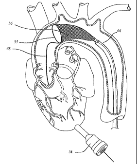
FIG. 5 also shows a distal embolic protection assembly 68. The distal
embolic protection assembly consists of a frame 55 and a porous bags 56. In
one
embodiment of the present invention, the distal inlet portion of the filter
mouth 53
includes a temporary valve.
In one embodiment of the present invention, the delivery system 67 is
inserted through the trocar 31 into the left ventricle 21 and advanced towards
the
native aortic valve of the heart 12. The delivery system 67 may be composed of
a
substantially rigid Scopus TM 46 and a delivery member. The heart valve
prosthesis
100 is disposed around the balloon delivery member 50 and delivered to the
target site for implantation. The length of balloon delivery members 50
suitable for
the purposes of the present invention will depend on the height of the
prosthetic
valve 100 to be implanted.
FIG. 6 shows a delivery system 67 comprising a perfusion tube 49, balloon
delivery member 50, and a ScapusTM 46. Here, the ScapusTM 46 is rigidly
attached to the perfusion tube 49. In one embodiment of the current invention,
the
ScapusTM 46 has a lumen that extends to the balloon delivery member 50 and
serves to inflate and deflate the balloon. The actuation sleeve 43 and
guidewire
41 is loosely disposed within the perfusion tube 49.
In one embodiment of the present invention, the distal embolic protection
assembly 68, actuation sleeve 43 and guidewire 41 within activation sleeve 43
is
movably disposed within the ScapusTM 46 of the delivery system 67 and balloon
delivery member 50 shown in FIG. 6. In yet a further embodiment of the present
invention, the distal embolic protection assembly 68 may be collapsed and
moved
through the ScapusTM 46 and balloon delivery member 50. In one embodiment of
the present invention, the delivery system shown in FIG. 6 is inserted through
the
trocar 31 in two steps: first the distal embolic protection assembly 68;
second the
delivery system 67 and balloon delivery member 50. After having introduced the
trocar 31 through the apex 18 of the heart 12, the distal embolic protection
assembly 68 is moved in a collapsed configuration through the trocar 31 and
the
left ventricle 21 and placed downstream from the aortic valve. Once the distal
embolic protection assembly 68 is in position, the distal embolic protection
assembly 68 is expanded to seal the inside circumference of the aorta.
Expansion takes place by moving the actuation sleeve 43 relative to the
guidewire
41. All circulation through the aorta will hence have to be filtered in the
porous
CA 02707755 2010-06-30
=
18
bag 56 of the distal embolic protection assembly 68. The guidewire 41 and
actuation sleeve 43 extends from the proximal side of the distal embolic
protection
assembly 68 to the outside of the body 10 and is accessible to the operator.
In one
embodiment of the present invention, the actuation sleeve 43 may also be used
as
a guidewire to move the ScapusTM 46 into position. Thus in one embodiment of
the
current invention, the ScapusTM delivery system may be loosely disposed on a
guidewire 41. In yet another embodiment of the present invention, the
perfusion
tube 49 functions as the actuation sleeve 43 to open and collapse the distal
embolic protection catheter.
Distal embolic protection assemblies 68 may be introduced through the apex
18 of the heart 12. Such embodiments are summarized in co owned U.S.
Application No. 2005/0119688. Distal embolic protection assemblies 68 may also
be inserted through arteries such as the femoral artery such as those
disclosed by
Macoviak, et al in U.S. Application No. 2002/0161394. In another embodiment of
the present invention, the distal embolic protection filter assembly 68 is
introduced
through the femoral artery and moved to the aortic arch, positioned just
downstream of the aortic valve as shown in FIG. 7. A delivery sheath 66 is
used to
collapse the filter assembly composed of the filter frame 55 and the porous
bag 56.
In a further embodiment of the current invention, a guidewire 65 is attached
to the
frame 55 on the proximal side of the filter assembly 68 and continues through
the
aortic valve and out through the trocar 31 and out through the body 10. The
guidewire 65 may be used for guiding the delivery system 67 into position
through
the trocar 31 and the apex 18 of the heart. The way the guidewire 65 is
attached to
the mouth of the filter 55 is for illustrational purposes only. Anyone skilled
in the art
will appreciate there are many different ways of attaching a guidewire to the
mouth
55 of the filter and different opening and closing mechanism for the filter.
Other
aortic filter systems described in prior art for femoral artery insertion may
also be
adapted for this procedure.
In one embodiment of the current invention, the delivery sheath 66 shown in
FIG. 7 is a ScapusTM 46 delivery system. The ScapusTM 46 delivery system may
slide across the guidewire. The porous bag 56 may also be inserted and removed
through the delivery system.
Once the distal embolic protection assembly 68 has been placed into
position, the ScapusTM 46 of the delivery system 67 slides over the actuation
sleeve
43 through the apex 18 of the heart 12. In one embodiment of the present
CA 02707755 2010-06-30
19
invention, the delivery system 67 slides over the guidewire 41 or 65,
depending on
the configuration of the distal embolic protection assembly. The balloon
delivery
member 50 is positioned in the aorta and within the aortic valve and aortic
valve
annulus. In one
embodiment of the present invention, the distal embolic
protection system 68 and valve delivery system 67 is inserted through the apex
18
together.
A collapsed replacement heart valve prosthesis 100 is disposed on the
balloon delivery member 50. The delivery system 67 with the attached
replacement prosthetic valve slides over the actuation sleeve and is
introduced
into the port of the access system 31 and through the apex 18 of the heart 12.
The balloon delivery member 50 with the attached heart valve prosthesis 100 is
positioned in the aorta and within the aortic valve and aortic valve annulus.
The
balloon delivery member 50 is expanded by moving fluid through the balloon
inflation tube 45. The balloon inflation tube 45 connects fluid to the balloon
delivery member 50. In one embodiment of the present invention, the device
used to move fluid through the balloon inflation tube 45 is a syringe. The
balloon
delivery member 50 expands in a radial direction when filled with fluid
through the
balloon inflation tube 45 causing the replacement prosthetic valve 100 to
exert
force against the existing valvular leaflets and the walls of the vessel.
In one- embodiment of the present invention, the valve replacement
procedure described herein is done more than once. A repeat procedure may, for
example, be performed in patients who cannot tolerate an open chest surgery.
Once the heart valve prosthesis 100 is implanted, the balloon delivery
member 50 is deflated and the valve delivery system 67 is withdrawn from the
body. The distal embolic protection assembly 68 is further withdrawn from the
body 10. In one embodiment of the present invention, the distal embolic
protection assembly 68 and the valve delivery system 67 are withdrawn from the
body together. In one embodiment of the present invention, a distal embolic
protection assembly 68 is not utilized. In yet another embodiment of the
present
invention, the distal embolic protection assembly 68 is left in the body for
some
time (up to 7 days) after the operation to make sure that the porous bag 56 of
the
distal embolic filter assembly 67 has collected all the debris.
FIG. 8 shows an implanted heart valve prosthesis 100 positioned in the
aortic valve position.
FIGS. 9, 10, and 11 show a ScapusTM delivery system comprising a
SCaPUSTM 46, luer fitting 62, perfusion tube 49 and a dog-bone shaped balloon
delivery member 50. The luer fitting 62 is attached to the proximal side of
the
CA 02707755 2010-06-30
ScapusTM 46 and may be used to direct fluid for opening and closing the
balloon
delivery member 50. The balloon delivery member 50 is tightly disposed around
the perfusion tube 49. The perfusion tube 49 is attached to the ScapusTM 46.
Fluid may flow through the luer fitting 62, through the ScapusTM 46 and into
the
5 balloon delivery member 50 to inflate and deflate the balloon.
It is important to note that although the different inventions described
herein
is typically described in reference to trans-apical valve implantation, they
may also
be used in non-beating heart surgeries. A ScapusTM delivery system, for
example, may also be used in a open surgery situation. Thus, in one embodiment
10 of the current invention, a ScapusTM delivery system is used in non-
beating heart
surgeries. In another embodiment of the current invention, a ScapusTM delivery
system may be used in an open chest surgery or robotic surgery.
Converting a Catheter to a Scapus TM Systems and Methods Thereof
The preferred delivery system for delivering heart valves and tools in a
15 trans-apical
or trans-heart procedure is a ScapusTM delivery system. If a
ScapusTM delivery system is not available, however, one may convert a catheter
into a delivery system that is similar to a Scapus TM delivery system.
In one embodiment of the current invention, a substantially thin, stiff guide
stick is inserted into the catheter to give it similar characteristics as a
ScapusTM.
20 The guide-stick is loosely disposed within the catheter and occupies the
space
that a guidewire would otherwise occupy. But as opposed to a guidewire that
cannot resist bending, a guide-stick is substantially rigid and can resist any
unintended bending and torsion. A guide-stick disposed within a catheter, in
contrast to a catheter by itself, provides sufficient rigidity such that the
resulting
delivery system may more accurately and more precisely deliver a prosthesis
during a beating heart procedure. The resulting delivery system is designed
not to
bend unless intended by the operator. The
resulting delivery system can
incorporate junctions or other means of bending at predetermined points to
allow
the operator to adjust the direction or angle of the delivery path in a
controlled
fashion.
In another embodiment of the current invention, a substantially stiff guide-
sleeve is loosely disposed on the outside of a catheter to give it similar
characteristics as a ScapusTM delivery system. The catheter is loosely
disposed
within the delivery sleeve. The described delivery sleeve is substantially
rigid and
can resist any unintended bending and torsion. A guide-sleeve loosely disposed
on a catheter, in contrast to a catheter by itself, provides sufficient
rigidity such
CA 02707755 2010-06-30
21
that the resulting delivery system may more accurately and more precisely
deliver
a heart valve prosthesis 100 during a beating heart procedure. The resulting
delivery system is designed not to bend unless intended by the operator. The
resulting delivery system can incorporate junctions or other means of bending
at
predetermined points to allow the operator to adjust the direction or angle of
the
delivery path in a controlled fashion.
Method for Valve Crimping and Valve Preparation
In one embodiment of the present invention, the heart valve prosthesis 100
is shipped to the operating room in an expanded configuration. The heart valve
prosthesis 100 is crimped down in diameter using crimpers known to anyone
skilled in the art while the heart valve prosthesis 100 is loosely disposed
around a
delivery member. The crimping process occurs with the operating room or in
vicinity of the operating room. The heart valve prosthesis 100 is further
delivered
to the target site for implantation.
In one embodiment of the current invention, the heart valve prosthesis 100
is shipped to the operating room in a crimped configuration. The heart valve
prosthesis 100 is crimped at the manufacturing facility in a careful,
consistent, and
controlled manner. The heart valve prosthesis 100 may be crimped directly onto
a
delivery member, such as a balloon delivery member 50. Alternatively, the
heart
valve prosthesis 100 may be crimped down to a size such that the internal
diameter of the heart valve prosthesis 100 matches the external diameter of
the
delivery member. The heart valve prosthesis 100 remains in a crimped
configuration until the heart valve prosthesis 100 reaches the operating room.
Crimping the heart valve prosthesis 100 in a controlled environment will
minimize
structural deterioration to the heart valve prosthesis 100 and will simplify
the
procedure in the operating room. When reaching the operating room, the crimped
heart valve prosthesis 100 is disposed around the delivery member, and the
heart
valve prosthesis 100 is further delivered to the target site for implantation.
Imaging Systems
Since a transapical procedure does not provide direct line of sight,
sufficient imaging of the heart, valves, and other structures is important to
provide
diagnostics, guidance and feed-back during the procedure. A ScapusTM delivery
system may be of a larger diameter than that of a catheter and is thus better
suited for containing imaging transducers. Thus in one embodiment of the
present invention, an imaging transducer is placed onto the delivery system.
In
CA 02707755 2010-06-30
=
22
one embodiment of the current invention, the imaging transducer is placed
within
the delivery member. In another embodiment of the present invention, the
imaging
transducer is placed just proximal and/or distal to the delivery member.
An external imaging transducer may be provided to view the operating field
and imaging systems may be used at any time or throughout the duration of the
surgery. The valvuloplasty assembly may include IVUS or other imaging sensors.
Such imaging technology can be used to inspect native valve annulus and size
the
required heart valve prosthesis 100 after valvuloplasty has been completed.
Imaging systems are well-known to anyone skilled in the art and include
transesophageal echo, transthoracic echo, intravascular ultrasound imaging
(IVUS), intracardiac echo (ICE), or an injectable dye that is radiopaque.
Cinefluoroscopy may also be utilized. The placement of imaging probes in
relation
to a balloon delivery member 50 has previously been described in co-owned WO
2005/046528 filed October 6, 2004.
Valve Removal Systems
The present invention also provides a method or system for removing the
native valve with a valve removal device by access through the apical area of
the
heart. By way of example, the valve removal may be accomplished as taught in
co-
pending U.S. Patent Application Nos. 2003/0216764 and 2005/0075724.
In one embodiment of the present invention, the method may further
comprise the step of removing at least a portion of the patient's heart valve
by
means of a cutting tool that is disposed on the Scapus TM. In another aspect
of the
present invention, the cutting tool may be made of an electrically conductive
metal
that provides radiofrequency energy to the cutting tool for enhanced valve
removal.
The high frequency energy ablation is well known in the art.
In another embodiment of the present invention, the delivery member
includes cutting means comprising a plurality of jaw elements, each jaw
element
having a sharp end enabling the jaw element to cut through at least a portion
of the
native valve. In another aspect, the cutting means comprises a plurality of
electrode
elements, wherein radiofrequency energy is delivered to each electrode
element,
enabling the electrode element to cut through at least a portion of the native
valve.
In a further aspect of the present invention, the cutting means comprises a
plurality
of ultrasound transducer elements, wherein ultrasound
CA 02707755 2010-06-30
=
23
energy is delivered to each transducer element enabling the transducer element
to cut through at least a portion of the native valve.
A ScapusTM with a valve removal system disposed on it is introduced
through the apex and positioned substantially in the vicinity of the aortic
valve.
The native valve leaflets and debris (e.g. calcium and valve leaflets) are
removed.
The parts that are not contained by the valve removal systems are caught in
the
distal embolic protection filter.
Distal Embolic Protection System
The present invention also provides for devices and methods for providing
distal embolic protection during the procedure. FIG. 5 and FIG. 6 show
examples
of distal embolic protection assemblies 68 and its relation to the delivery
system
67. It is important that the distal embolic protection filter provides a means
for
trapping embolic material and debris. In one embodiment, it is also desired
that
the distal embolic protection filter provides a temporary valve. The filter
and
temporary valve assembly prevents flush back of blood, embolic material and
debris, while still allowing fluid flow into the filter during surgery. The
temporary
valve may also temporarily do the work of an adjacent heart valve, such as the
aortic valve. Thus in one embodiment of the present invention, the distal
embolic
protection assembly 68 provides a filter member for trapping embolic material
that
concurrently functions as a temporary valve.
Distal embolic= protection assemblies 68 used in both trans-apical and
percutaneous procedures must be compressed and expanded to allow entry into
small blood vessels or other body cavities. Combining both a one-way valve and
a filter basket mechanism requires a significant amount of hardware making it
difficult to compress the filter down sufficiently to be used during trans-
apical and
percutaneous procedures.
FIG. 12 shows a sub-component of a distal embolic protection filter system
that incorporates both a filter and the function of a temporary valve. The
proximal
mouth 73 of the filter consists of a proximal frame 74 that pushes against and
makes a seal with the surrounding vasculature. The proximal frame 74 may, for
example, push and seal against the inner wall of the aorta, causing all emboli
and
debris to flow through the filter assembly. In one embodiment of the present
invention, the proximal frame 74 is made out of a shape memory alloy such as
Nitinol, allowing it to expand into position.
The distal end 76 of the filter sub-assembly is shown open. In other words,
debris not caught in the filter mesh 78 may continue out through the distal
end 76
CA 02707755 2010-06-30
=
=
24
of the filter sub-assembly, moving past the distal frame75. In one embodiment
of
the present invention, the distal frame 75 is made out of a shape memory alloy
such as nitinol, allowing it to maintain an open configuration.
FIG. 13 shows three inter-connected filter sub-assemblies shown in FIG. 12.
Although three sub-assemblies are shown, any number of two or more sub-
assemblies will work. The length from the proximal frame to the distal frame
of each
sub-assembly is slightly different, thus separating the filters meshes of the
different
filter sub-assemblies 78, 79, and 82. The proximal frame 74 is shared by all
the
different filter sub-assemblies.
Thus, in one embodiment of the present invention, a plurality of filter sub-
assemblies are interconnected at the large inlet of the filters, while the
downstream
sides of the sub-assemblies have smaller openings allowing debris to flow
through.
In one embodiment of the current invention, the outermost filter-assembly is
closed
at the downstream end. As such, the device provides less flow restriction as
the
blood flows into the porous bags (i.e. downstream from the aortic valve) as
opposed to the reverse. This means that the device also functions as a one-way
valve.
Valve Decalcification Systems
The formation of atherosclerotic plaques and lesions on cardiovascular
tissue, such as blood vessels and heart valves, is a major component of
cardiovascular disease. A variety of different methods have been developed to
treat cardiovascular diseases associated with calcified atherosclerotic
plaques and
lesions. Such methods include mechanical removal or reduction of the lesion,
such
as bypass surgery, balloon angioplasty, mechanical debridement, atherectomy,
and
valve replacement.
Calcified atherosclerotic plaques and lesions may also be treated by
chemical means which may be delivered to the affected area by various catheter
devices. For example, U.S. Patent No. 6,562,020 by Constantz et al., discloses
methods and systems for dissolving vascular calcified lesions using an acidic
solution. A catheter delivers an acidic fluid to a localized vascular site.
Such a
system may, for example, decalcify a calcified heart valve by applying an
acidic
solution (such as hydrochloric acid, etc.)
The current percutaneous anti-calcification system disclosed by Constantz et
al. is inserted through the femoral artery. Insertion through the femoral
artery is
impractical in the case of a trans-apical procedure as it requires another
incision
CA 02707755 2010-06-30
into the patient. The system by Constantz et al. may be adapted such that the
delivery member controlling and holding the decalcification system is moved
from
the proximal side (i.e. side of the operator as in the case of femoral access)
to the
distal side.
5 Accordingly,
in another embodiment of the present invention, the methods
and devices of the present invention may be adapted to provide a valve
decalcification system, wherein a ScapusTM system is capable of providing the
dissolution solution to the treatment site by access through the apical area
of the
heart. Suitable dissolution solutions are known in the art and are generally
10
characterized as those which are capable of increasing the proton
concentration
at the treatment site to a desired level sufficient to at least partially
dissolve the
mineral component of a calcified atherosclerotic lesion.
A trans-apical delivered ScapusTM system may also provide means for
isolating the treatment site to prevent the dissolution solution from entering
into
15 the
patient's circulatory system. Thus in one embodiment of the current invention
the decalcification systems described and incorporated for reference above is
adapted to be disposed on a Scapus TM as opposed to a catheter. Such means for
isolating the treatment site may include a barrier, such as a dual balloon
system
on the catheter that inflate on both sides of the treatment site.
20 FIG. 14
shows such a delivery system where a multilumen ScapusTM 46
connects to a perfusion tube 49 which in turn connects two balloons, a
proximal
balloon 92 and a distal balloon 93. The two balloons are shown inflated and in
intimate contact with the walls of the aorta 94. In one embodiment of the
present
invention, the perfusion tube 49 is not present and the proximal balloon 92
and the
25 distal
balloon 93 are intimately in contact with the ScapusTM 43. Fluid may flow
through the SCaPUSTM 46 to inflate the proximal balloon 92 and distal balloon
93
as well as provide the dissolution solution to the treatment site confined by
the
proximal balloon 92 and distal balloon 93.
Valve Within Man-Made Valve: Systems And Methods Thereof
It is one objective of the current invention to provide systems and methods
for implanting an expandable heart valve within a target valve located within
a
heart. Such a procedure is beneficial in older or diseased patients who have
previously received a valve implant and who cannot or does not want to undergo
the trauma of another open heart surgery. Implanting an expandable heart valve
within an existing target heart valve allows the use of minimally invasive
CA 02707755 2010-06-30
26
implantation techniques such as percutaneous or trans-apical valve
implantation
techniques.
The current methods and systems are distinctly different from Andersen et
al. disclosed in U.S. Patent No. 6,582,462 who describes the implantation of a
valve in a body channel or the vasculature. Andersen's intent and objective is
to
describe an expandable valve that is placed within a body channel or
vasculature
and uses the intimate contact created within the vasculature, body channel, or
native valve as support to allow implantation. In the present invention, an
expandable heart valve prosthesis 100 is implanted within a previously
implanted
man-made heart valve prosthesis and uses the intimate contact created with the
previously implanted heart valve prosthesis for support. If the
previously
implanted heart valve prosthesis is removed, one will concurrently remove the
expandable heart valve prosthesis 100 located within the previously implanted
heart valve prosthesis.
In one embodiment of the current invention, an expandable heart valve
prosthesis 100 is mounted within a previously implanted heart valve prosthesis
located within a heart. The expandable heart valve prosthesis 100 may be any
valve that can be delivered minimally invasively, such as percutaneous or
trans-
apically delivered valves. In one embodiment of the current invention, the
expandable heart valve prosthesis 100 is a balloon-expandable heart valve. In
another embodiment of the present invention, the expandable heart valve
prosthesis 100 is the 3F EntrataTm heart valve. In another embodiment of the
current invention, the expandable heart valve prosthesis 100 is a self-
expandable
heart valve. In yet another embodiment of the present invention, the
expandable
heart valve prosthesis 100 is a valve expanded using some other mechanical or
actuating means.
The previously implanted heart valve prosthesis may be any valve either
native or man-made. In one embodiment of the current invention, the previously
implanted heart valve prosthesis is a mechanical valve. In another embodiment
of
the present invention, the previously implanted heart valve prosthesis is a
tissue
valve. The previously implanted heart valve prosthesis may also be made out of
polyurethane or be a tissue-engineered valve. In one embodiment of the current
invention, the previously implanted heart valve prosthesis can be an
expandable
heart valve. In yet a further embodiment of the current invention, more than
one
expandable heart valve prosthesis 100 may be implanted within a previously
implanted heart valve prosthesis. As such, multiple minimally invasive heart
valve
deliveries may be conducted without removing the existing valve or existing
CA 02707755 2010-06-30
27
valves. The previously implanted heart valve prosthesis may be an aortic
valve,
mitral valve, pulmonary valve, or a tricuspid valve. The previously implanted
heart
valve prosthesis may also be a a homograft valve or a xenograft valve.
Examples
of previously implanted heart valve prosthesis include, but are not limited
to, the
Edwards Perimount Valve, the Edwards BioPhysio Valve, the Medtronic Hancock
I Valve, the Medtronic Hancock M.O. Valve, the Medtronic Hancock II Valve, the
Medtronic Mosaic Valve, the Medtronic Intact Valve, the Medtronic Freestyle
Valve, the St. Jude Toronto Stentless Porcine Valve (SPV), and the St. Jude
Prima Valve.
The expandable heart valve prosthesis 100 may be made to fit well within
the previously implanted heart valve prosthesis. In one instance, the posts of
the
expandable heart valve prosthesis 100 are coordinated to fit the posts of the
previously implanted heart valve prosthesis. Thus in one embodiment of the
present invention, the posts of the expandable heart valve prosthesis 100
matches the orientation for the posts of the previously implanted heart valve
prosthesis. In one embodiment of the present invention, the inter-post
separation
angles of the expandable heart valve prosthesis 100 add up to 360 . In other
embodiments of the present invention, the inter-post separation angle of the
heart
valve prosthesis 100 is 120 , 120 , and 120 ; or 135 , 120 , and 105'; or 135
,
105 , and 1200; or 120 , 135 , and 105 ; or 120 , 105 , and 135'; or 105 , 135
,
and 120 ; or 105 , 120 , and 135 .
FIG. 15 shows an exploded view of FIG. 16 where a heart valve prosthesis
100 is shown implanted within a previously implanted heart valve prosthesis
101.
In a preferred embodiment, the inflow ring or annulus 105 of the heart valve
prosthesis 100 is aligned with the inflow ring or annulus 115 of the
previously
implanted heart valve prosthesis 101. In another preferred embodiment, the
commissural posts 106 of the heart valve prosthesis 100 is aligned with the
commissural posts 116 of the previously implanted heart valve prosthesis 101.
It should be noted that although reference is made herein to a heart valve
100 implanted into a previously implanted heart valve prosthesis 101 inside
the
aorta, it is intended for such valve procedures to encompass any location
within
the heart 12, and not to be limited to the aorta.
In a preferred embodiment of the current invention, the previously
implanted heart valve prosthesis 101 is the same size or one size larger than
the
expandable heart valve prosthesis 100. For purpose of example, if the
previously
implanted heart valve prosthesis 101 is 27mm, the expandable heart valve
prosthesis 100 is either 25mm or 27mm in size. Thus, in one embodiment of the
CA 02707755 2010-06-30
=
28
current invention, the expandable heart valve prosthesis 100 is the same size
as
the previously implanted heart valve prosthesis 101. In another embodiment of
the current invention, the expandable heart valve prosthesis 100 is larger
than the
previously implanted heart valve prosthesis 101. In another embodiment of the
current invention, the expandable heart valve prosthesis 100 is smaller than
the
previously implanted heart valve prosthesis 101.
Clinical records will specify the exact size used during an earlier implant.
The size of the previously implanted heart valve prosthesis 101 will thus be
known. In-vitro tests will show the best size expandable heart valve
prosthesis
100 for a specific size and type target valve. The optimal size expandable
heart
valve can thus be determined from the clinical records from the previous heart
valve implant. Thus in one embodiment of the present invention, the size of
the
expandable heart valve prosthesis 100 to be used is determined from clinical
records of prior implants.
In one embodiment of the current invention, an expandable stent is
implanted within a previously implanted heart valve prosthesis 101 prior to
implanting the expandable heart valve prosthesis 100. In another embodiment of
the current invention, valvuloplasty is used to expand the orifice of the
previously
implanted heart valve prosthesis 101 before implanting an expandable heart
valve
prosthesis 100 or before implanting an expandable stent..
Any delivery system may be used to deliver the expandable heart valve
prosthesis 100. In one embodiment of the current invention, the delivery
system
is a catheter. In another embodiment of the current invention, the delivery
system
is a ScapusTM. The expandable heart valve prosthesis 100 may be delivered
through any access point to the heart. In one embodiment of the present
invention, the expandable heart valve prosthesis 100 is delivered minimally
invasively. In one embodiment of the present invention, the expandable heart
valve prosthesis 100 is delivered percutaneously. In another embodiment of the
present invention, the expandable heart valve prosthesis 100 is delivered
trans-
apically.
Sutureless Valve Inserter System And Methods Thereof
The benefits of a ScapusTM valve delivery system may also be utilized in
the case of self-expandable valves. As such, the delivery system may be used
for
percutaneous valve delivery, trans-apical valve delivery, trans-heart
delivery. In
addition to these delivery techniques, the ScapusTM delivery system may be
utilized in more invasive cardiac procedures such as open heart procedures.
CA 02707755 2010-06-30
29
The ScapusTM delivery system is well suited for delivering the 3F Enable
Aortic
Heart ValveTM and the other valves described in co-owned U.S. applications
entitled "Minimally Invasive Valve Replacement System" with the following U.S.
Patent Application Nos.: 2005/0075584; 2005/0075712; 2005/0075713;
2005/0075717; 2005/0075718; 2005/0075719; 2005/0075720; 2005/0075726;
2005/0075728; 2005/0075729; 2005/0075730; and 2005/0096738.
Sutureless Valve Inserter System And Methods Thereof
Current tissue heart valve replacements gradually calcify after implanted in
the heart. Such is also the case when implanting replacement heart valves in
animals such as pigs or sheep. In fact, replacement heart valves intended for
human use typically calcify faster when implanted in animals such as pigs or
sheep.
Because of difference in flow dynamics, physiology, and biochemistry, the best
performing commercially available heart valves will typically show signs of
calcification in pigs and sheep within 10-200 days. Standard animal models
used
for pre-clinical valve testing is frequently sheep, but pigs may also be used.
Adolescent sheep have great propensity to calcify bioprosthetic valves.
The fact that cardiac valves places in the heart of certain animal models
calcify quickly may be used as basis for creating calcified animal models for
use in
the development and testing of cardiac valves.
Open heart surgery valve replacement on adolescent sheep typically results
in less than 40% success/survival in the aortic position, owing to the very
small
valve sizes and the use of full bypass. Open heart surgery valve replacement
on
adolescent sheep typically results in more than 80% success/survival in the
mitral
position owing to the larger valve sizes and the use of beating heart, partial
bypass.
Placing replacement valves in the mitral position during animal testing is not
just
used to reduce costs but is also considered a "worst-case" position due to
higher
backpressures. Replacement aortic and mitral valves are therefore frequently
placed in the mitral position during animal studies.
Accordingly, it is one object of the present invention to provide methods and
systems for the creation of a calcified animal model. Commercially available
tissue
valves are first implanted in adolescent sheep. The animals are survived for
10-
200 days and the performance evaluated. Because of the increased propensity
for
calcification in animals, all the valves implanted are expected to be stenotic
and/or
incompetent due to calcification of tissue leaflets. Sheep may be evaluated at
regular intervals using echo.
CA 02707755 2010-06-30
It is another object of the present invention to utilize the calcification
model
described above in the development and testing of cardiac valves. Using either
a
percutaneous or trans-apical implant techniques, place replacement heart
valves
within the calcified valves of adolescent sheep. Survive the test animals for
20
5 weeks (150 +/- 10 days) or as required by regulatory authorities.
Monitor the
replacement valves. Necropsy, pathology, and post mortem histology may be
performed.
The present invention may be divided in two phases:
Create a calcified animal model by replacing the native valves of
10 adolescent sheep with commercially available valves and surviving
the animals for
10-200 days.
11. Implant
minimally invasive valves within the calcified valve orifices
using a minimally invasive valve procedure.
In phase l of the invention, a calcified animal model is created by replacing
15 the native valves of adolescent sheep with commercially available
valves and
surviving the animals. In one embodiment of the present invention, the heart
valve replacement procedure is an on-pump procedure. In another embodiment
of the present invention, the heart valve replacement procedure is a minimally
invasive heart valve procedure, such as a percutaneous heart valve replacement
20 procedure, or a trans-apical valve procedure. In the latter
embodiment, the
method described herein would allow testing of a minimally invasive heart
valve
repeat-procedure. In one embodiment of the current invention, the heart valve
replacement procedure is conducted endoscopically. In yet another embodiment
of the current invention, the heart valve replacement procedure is conducted
using
25 robots.
In one embodiment of the present invention, drugs are utilized to adjust the
rate of calcification. In another embodiment, the valve implanted during Phase
I is
coated with a chemical substance used to adjust the rate of calcification.
Any valve may be implanted during Phase I. In one embodiment of the
30 present invention, the valve implanted is a tissue valve. In another
embodiment of
the present invention, the valve implanted is a mechanical valve. Implanted
heart
valves may include aortic valves, mitral valves, tricuspid valves, or
pulmonary
valves. A replacement valve may not necessarily be implanted in its intended
position. As an example, an aortic valve may be implanted in the mitral
position of
the animal. Thus in one embodiment of the present invention, a replacement
aortic valve is implanted in the mitral position. In one embodiment of the
current
invention, multiple valves are implanted in different positions at the same
time.
CA 02707755 2010-06-30
31
In a preferred embodiment of the current invention, valves are implanted in
animals whose heart physiology and flow dynamics, as well as biochemistry,
match humans as close as possible. Sheep and pigs are thus frequently used for
heart valve testing. Thus, in one embodiment of the current invention, sheep
is
used as the animal model. In another embodiment of the current invention, pigs
are used as the animal model. Any other primate may be used as an animal
model. In one embodiment of the current invention, animals of subclass
eutheria
are used as the animal model. In another embodiment of the current invention,
animals of the suborder anthropoidea (e.g. monkeys and apes).
The age of an animal affects the rate of calcification. Thus, in one
embodiment of the current invention, the age of the animal is the equivalent
of
adolescence. In another embodiment of the current invention, the animals used
are adults.
It is one object of the current invention to survive sufficient animals to the
end of Phase ll such that pre-clinical regulatory requirements are met for
different
regulatory bodies. It is expected that some animals will not survive the valve
replacements during phase I. Further animals may perish during the duration of
Phase I. Further animals will perish during the replacement implants during
Phase II as well as during the duration of Phase II. In one preferred
embodiment
of the present invention, an excess number of animals are used to start Phase
I
such that sufficient numbers of animals are survived all the way through Phase
II.
The exact number of animals needed for the start of Phase I depends on
numerous variables including the operator, the type of animal, the type of
procedures.
It is one object of the current invention to monitor the progression of
calcification and diseases related to implanted valves. Different monitoring
equipment such as ultrasound, MRI, CT, and cinefluoroscopy, are used during
the
course of Phase I and Phase II. In one embodiment of the present invention,
echo is used at 60, 90, and 120 days during Phase I to monitor the implanted
valves.
In one embodiment of the present invention, the animals in Phase I are
survived for 90-120 days. The length of Phase I depends on factors such as
what
type of animal used and what type of tissue valves used. The valves may be
monitored during Phase I. It may be possible to go to Phase II earlier based
upon
in-vivo evaluation.
In Phase II of the invention, valves are implanted within the calcified valve
orifices. Implant procedures include, but are not limited to minimally
invasive
CA 02707755 2010-06-30
32
valve procedures, percutaneous valve procedures, trans-apical valve procedure,
on-pump valve procedure, endoscopic valve procedure, and robotic valve
procedure.
In one embodiment of the present invention, drugs are utilized to adjust the
rate of calcification. In another embodiment, the valve implanted during Phase
11
is coated with a chemical substance used to adjust the rate of calcification.
Any valve may be implanted during Phase 11. Tissue valves include, but
are not limited to tissue valve, mechanical valves, aortic valves, mitral
valves,
tricuspid valves, pulmonary valves. A replacement valve may not necessarily be
implanted in its intended position. As an example, an aortic valve may be
implanted in the mitral position of the animal. Thus in one embodiment of the
present invention, a replacement aortic valve is implanted in the mitral
position.
In one embodiment of the present invention, the animals in Phase I are
survived for 20 weeks (150 +1- 10 days). . The length of Phase 11 depends on
guidelines provided by regulatory bodies.
In one embodiment of the current invention, the minimally invasive valve
delivery is conducted using a SCAPUSTM delivery system. In one embodiment of
the present invention, the valve utilized is the 3F Therapeutics, Inc.
EntrataTM
valve. In one embodiment of the present invention, a balloon is used in the
inferior vena cava to regulate pressure during the procedure.
In one embodiment of the current invention, the replacement valve in
Phase 11 is seated within the calcified valve orifice of the valve replacement
conducted in Phase l. ln another embodiment of the present invention, the
replacement valve in Phase II is seated just upstream from the calcified valve
orifice of the valve replacement conducted in Phase 1. In another embodiment
of
the present invention, the replacement valve in Phase 11 is seated just
downstream from the calcified valve orifice of the valve replacement conducted
in
Phase).
Obviously, numerous variations and modifications can be made within
departing from the spirit of the present invention. Therefore, it should be
clearly
understood that the forms of the present invention described above and shown
in
the figures of the accompanying drawings are illustrative only and are not
intended to limit the scope of the present invention.