Note: Descriptions are shown in the official language in which they were submitted.
CA 02708147 2010-06-04
WO 2009/076164 PCT/US2008/085522
CANCELLOUS BONE IMPLANT FOR CARTILAGE REPAIR
RELATED APPLICATION
This application claims priority to United States Provisional Patent
Application No.
60/996,800 filed December 5, 2007, which is incorporated by reference herein
in its entirety.
STATEMENT REGARDING FEDERALLY SPONSORED RESEARCH OR
DEVELOPMENT
Not applicable.
REFERENCE TO SEQUENCE LISTING, A TABLE OR A COMPUTER PROGRAM
LISTING COMPACT DISC APPENDIX
None.
BACKGROUND OF THE INVENTION
1. Field of Invention
The present invention is generally directed toward an allograft cartilage
repair implant
and is more specifically directed toward a two piece allograft cancellous bone
implant having a
mineralized cancellous bone base member defining a central blind bore and a
bore transverse to
the central bore intersecting the central bore and a demineralized cancellous
cap member
mounted to the base member. The cap member has a cylindrical top section and a
stem
extending from the top section which has a transverse bore cut therethrough
and is placed in the
central bore of the base member. A pin is mounted in the transverse bore of
the base member
through the stem transverse bore. In an alternate embodiment the cap member
defines a central
blind bore with a bone transverse to the central bore intersecting the central
bore. The base
member has a cylindrical bottom section and a stem extending from the bottom
section which
has a transverse bore cut therethrough which is placed in the central bore of
the cap member to
receive a pin. The implant is shaped for an interference fit implantation in a
bore cut in a
shoulder, knee, hip, or ankle joint to remove a cartilage defect area.
2. Description of the Prior Art
Articular cartilage injury and degeneration present medical problems to the
general
population which is constantly addressed by orthopedic surgeons. Every year in
the United
States, over 500,000 arthroplastic or joint repair procedures are performed.
These include
1
CA 02708147 2010-06-04
WO 2009/076164 PCT/US2008/085522
approximately 125,000 total hip and 150,000 total knee arthroplasties and over
41,000 open
arthroscopic procedures to repair cartilaginous defects of the knee.
In the knee joint, the articular cartilage tissue forms a lining which faces
the joint cavity
on one side and is linked to the subchondral bone plate by a narrow layer of
calcified cartilage
tissue on the other side (see Figure 1). Articular cartilage (hyaline
cartilage) consists primarily
of extracellular matrix with a sparse population of chondrocytes distributed
throughout the
tissue. Articular cartilage is composed of chondrocytes, type II collagen
fibril meshwork,
proteoglycans and water. Active chondrocytes are unique in that they have a
relatively low
turnover rate and are sparsely distributed within the surrounding matrix. The
collagens give the
tissue its form and tensile strength and the interaction of proteoglycans with
water give the tissue
its stiffness to compression, resilience and durability. The hyaline cartilage
provides a low
friction bearing surface over the bony parts of the joint. If the lining
becomes worn or damaged,
resulting in lesions, joint movement may be painful or severely restricted.
Whereas damaged
bone typically can regenerate successfully, hyaline cartilage regeneration is
quite limited because
of its limited regenerative and reparative abilities.
Articular cartilage lesions generally do not heal, or heal only partially
under certain
biological conditions due to the lack of nerves, blood vessels and a lymphatic
system. The
limited reparative capabilities of hyaline cartilage usually results in the
generation of repair
tissue that lacks the structure and biomechanical properties of normal
cartilage. Generally, the
healing of the defect results in a fibrocartilaginous repair tissue that lacks
the structure and
biomedical properties of hyaline cartilage and degrades over the course of
time. Articular
cartilage lesions are frequently associated with disability and with symptoms
such as joint pain,
locking phenomena and reduced or disturbed function. These lesions are
difficult to treat
because of the distinctive structure and function of hyaline cartilage. Such
lesions are believed
to progress to severe forms of osteoarthritis. Osteoarthritis is the leading
cause of disability and
impairment in middle-aged and older individuals, entailing significant
economic, social and
psychological costs. Each year, osteoarthritis accounts for as many as 39
million physician visits
and more than 500,000 hospitalizations. By the year 2020, arthritis is
expected to affect almost
60 million persons in the United States and to limit the activity of 11.6
million persons.
There are many current therapeutic methods being used. None of these therapies
has
resulted in the successful regeneration of hyaline-like tissue that withstands
normal joint loading
2
CA 02708147 2010-06-04
WO 2009/076164 PCT/US2008/085522
and activity over prolonged periods. Currently, the techniques most widely
utilized clinically for
cartilage defects and degeneration are not articular cartilage substitution
procedures, but rather
lavage, arthroscopic debridement, and repair stimulation. The direct
transplantation of cells or
tissue into a defect and the replacement of the defect with biologic or
synthetic substitutions
presently accounts for only a small percentage of surgical interventions. The
optimum surgical
goal is to replace the defects with cartilage-like substitutes so as to
provide pain relief, reduce
effusions and inflammation, restore function, reduce disability and postpone
or alleviate the need
for prosthetic replacement.
Lavage and arthroscopic debridement involve irrigation of the joint with
solutions of
sodium chloride, Ringer or Ringer and lactate. The temporary pain relief is
believed to result
from removing degenerative cartilage debris, proteolytic enzymes and
inflammatory mediators.
These techniques provide temporary pain relief, but have little or no
potential for further healing.
Repair stimulation is conducted by means of drilling, abrasion arthroplasty or
microfracture. Penetration into the subchondral bone induces bleeding and
fibrin clot formation
which promotes initial repair, however, the tissue formed at the cartilage
interface is fibrous in
nature and not durable. Pain relief is temporary as the tissue exhibits
degeneration, loss of
resilience, stiffness and wear characteristics over time.
The periosteum and perichondrium have been shown to contain mesenchymal
progenitor
cells capable of differentiation and proliferation. They have been used as
grafts in both animal
and human models to repair articular defects. Few patients over 40 years of
age obtain good
clinical results, which most likely reflect the decreasing population of
osteochondral progenitor
cells with increasing age. There have also been problems with adhesion and
stability of the
grafts, which result in their displacement or loss from the repair site.
Transplantation of cells grown in culture provides another method of
introducing a new
cell population into chondral and osteochondral defects. CARTICEL is a
commercial process
to culture a patient's own cartilage cells for use in the repair of cartilage
defects in the femoral
condyle marketed by Genzyme Biosurgery in the United States and Europe. The
procedure uses
arthroscopy to take a biopsy from a healthy, less loaded area of articular
cartilage of the patient.
Enzymatic digestion of the harvested tissue releases the cells that are sent
to a laboratory where
they are grown for a period ranging from 2-5 weeks. Once cultivated, the cells
are injected
during a more open and extensive knee procedure into areas of defective
cartilage where it is
3
CA 02708147 2010-06-04
WO 2009/076164 PCT/US2008/085522
hoped that they will facilitate the repair of damaged tissue. An autologous
periosteal flap with a
cambium layer is used to seal the transplanted cells in place and act as a
mechanical barrier.
Fibrin glue is used to seal the edges of the flap. This technique preserves
the subchondral bone
plate and has reported a high success rate. Proponents of this procedure
report that it produces
satisfactory results, including the ability to return to demanding physical
activities, in more than
90% of patients and those biopsy specimens of the tissue in the graft sites
show hyaline-like
cartilage repair. More work is needed to assess the function and durability of
the new tissue and
determine whether it improves joint function and delays or prevents joint
degeneration. As with
the perichondrial graft, patient/donor age may compromise the success of this
procedure as
chondrocyte population decreases with increasing age. Disadvantages to this
procedure include
the need for two separate surgical procedures, potential damage to surrounding
cartilage when
the periosteal patch is sutured in place, the requirement of demanding
microsurgical techniques,
and the expensive cost of the procedure resulting from the cell cultivation
which is currently not
covered by insurance.
Another procedure known as osteochondral transplantation or mosaicplasty
involves
excising all injured or unstable tissue from the articular defect and creating
cylindrical holes in
the base of the defect and underlying bone. These holes are filled with
autologous cylindrical
plugs of healthy cartilage and bone in a mosaic fashion. The filler
osteochondral plugs are
harvested from a lower weight-bearing area of lesser importance in the same
joint. This
technique can be performed as arthroscopic or open procedures. Reports of
results of
osteochondral plug autografts in a small number of patients indicate that they
decrease pain and
improve joint function, however, long-term results have not been reported.
Factors that can
compromise the results include donor site morbidity, effects of joint
incongruity on the opposing
surface of the donor site, damage to the chondrocytes at the articular margins
of the donor and
recipient sites during preparation and implantation, and collapse or settling
of the graft over time.
The limited availability of sites for harvest of osteochondral autografts
restricts the use of this
approach to treatment of relatively small articular defects and the healing of
the chondral portion
of the autograft to the adjacent articular cartilage remains a concern.
Transplantation of large allografts of bone and overlying articular cartilage
is another
treatment option that involves a greater area than is suitable for autologous
cylindrical plugs, as
well as for a non-contained defect. The advantages of osteochondral allografts
are the potential
4
CA 02708147 2010-06-04
WO 2009/076164 PCT/US2008/085522
to restore the anatomic contour of the joint, lack of morbidity related to
graft harvesting, greater
availability than autografts and the ability to prepare allografts in any size
to reconstruct large
defects. Clinical experience with fresh and frozen osteochondral allografts
shows that these
grafts can decrease joint pain, and that the osseous portion of an allograft
can heal to the host
bone and the chondral portion can function as an articular surface. Drawbacks
associated with
this methodology in the clinical situation include the scarcity of fresh donor
material and
problems connected with the handling and storage of frozen tissue. Fresh
allografts carry the
risk of immune response or disease transmission. Musculoskeletal Transplant
Foundation (MTF)
has preserved fresh allografts in a media that maintains a cell viability of
50% for 35 days for use
as implants. Frozen allografts lack cell viability and have shown a decreased
amount of
proteoglycan content which contribute to deterioration of the tissue.
A number of United States Patents have been specifically directed towards bone
plugs
which are implanted into a bone defect. Examples of such bone plugs are U.S.
Patent Number
4,950,296 issued August 21, 1990 which discloses a bone graft device
comprising a cortical shell
having a selected outer shape and a cavity formed therein for receiving a
cancellous plug, which
is fitted into the cavity in a manner to expose at least one surface; U.S.
Patent Number 6,039,762
issued March 21, 2000 discloses a cylindrical shell with an interior body of
deactivated bone
material; and U.S. Patent Number 6,398,811 issued June 4, 2002 directed toward
a bone spacer
which has a cylindrical cortical bone plug with an internal through-going bore
designed to hold a
reinforcing member. U.S. Patent Number 6,383,221 issued May 7, 2002 discloses
an
intervertebral implant having a substantially cylindrical body with a through-
going bore
dimensioned to receive bone growth materials.
U.S. Patent Number 6,379,385 issued April 30, 2002 discloses an implant base
body of
spongious bone material into which a load carrying support element is
embedded. The support
element can take the shape of a diagonal cross or a plurality of cylindrical
pins. See also, U.S.
Patent Number 6,294,187 issued September 25, 2001 which is directed to a load
hearing
osteoimplant made of compressed bone particles in the form of a cylinder. The
cylinder is
provided with a plurality of through-going bores to promote blood flow through
the osteoimplant
or to hold a demineralized bone and glycerol paste mixture. U.S. Patent Number
6,096,081
issued August 1, 2000 shows a bone dowel with a cortical end cap or caps at
both ends, a brittle
cancellous body and a through-going bore.
CA 02708147 2010-06-04
WO 2009/076164 PCT/US2008/085522
The use of implants for cartilage defects is much more limited. Aside from the
fresh
allograft implants and autologous implants, U.S. Patent Number 6,110,209
issued November 5,
1998 shows the use of an autologous articular cartilage cancellous bone paste
to fill arthritic
defects. The surgical technique is arthroscopic and includes debriding
(shaving away loose or
fragmented articular cartilage), followed by morselizing the base of the
arthritic defect with an
awl until bleeding occurs. An osteochondral graft is then harvested from the
inner rim of the
intercondylar notch using a trephine. The graft is then morselized in a bone
graft crusher, mixing
the articular cartilage with the cancellous bone. The paste is then pushed
into the defect and
secured by the adhesive properties of the bleeding bone. The paste can also be
mixed with a
cartilage stimulating factor, a plurality of cells, or a biological glue. All
patients are kept non-
weight bearing for four weeks and used a continuous passive motion machine for
six hours each
night. Histologic appearance of the biopsies has mainly shown a mixture of
fibrocartilage with
hyaline cartilage. Concerns associated with this method are harvest site
morbidity and
availability, similar to the mosaicplasty method and retention of the implant
in the prepared
cartilage defect space.
U.S. Patent Number 6,379,367 issued April 30, 2002 discloses a plug with a
base
membrane, a control plug, and a top membrane which overlies the surface of the
cartilage
covering the defective area of the joint.
U.S. Patent Number 7,067,123 issued June 27, 2006 is directed toward cartilage
defect
filler material comprising cartilage pieces ranging from 0.01 mm to 1.0 mm in
size in a
biological carrier which can be phosphate buffered saline, hyaluronic acid and
its derivatives as
well as other carriers together with allogenic chondrocytes including an
additive which can be
growth factors.
SUMMARY OF THE INVENTION
A cartilage repair allograft construct implant assembly is formed with a
cylindrical
mineralized cancellous bone base member and a demineralized cancellous cap
member mounted
to the base member. The cap member is preferably formed with a cylindrical top
portion and a
stem extending therefrom. The cap member is infused with a cartilage paste
having small
cartilage pieces ranging from about 10 to about 212 microns in size, a carrier
and a FGF-2
variant growth factor and the stem of the cap member is mounted in a central
bore cut in the base
6
CA 02708147 2010-06-04
WO 2009/076164 PCT/US2008/085522
member and held in place by a pin inserted into a transverse bore in the base
member which is
aligned with a transverse bore formed in the cap member stem. An alternative
embodiment uses
an inverted design. The construct is used for replacing articular cartilage
defects and is placed in
a bore which has been cut into the patient to remove the lesion defect area.
Each allograft
construct can support the addition of a variety of chondrogenic stimulating
factors including, but
not limited to morselized allogeneic cartilage, growth factors (e.g., FGF-2,
FGF-5, FGF-7, FGF-
9, FGF-11, FGF-21, IGF-1, TGF-(3, BMP-2, BMP-7, PDGF, VEGF) and variants
thereof.
It is an object of the invention to provide an allograft implant for joints
which provides
pain relief, restores normal function and will postpone or alleviate the need
for prosthetic
replacement.
It is also an object of the invention to provide a cartilage repair implant
which is easily
placed in a cartilage defect area by the surgeon using a minimally invasive
technique.
It is still another object of the invention to provide a cartilage repair
allograft implant
which has load bearing capabilities.
It is further an object of the invention to provide an allograft implant
procedure which is
applicable for osteochondral defects.
It is yet another object of the invention to provide a cartilage repair
implant which
facilitates growth of hyaline cartilage in the cartilage defect area.
It is an additional object of the invention to provide a cancellous construct
which is
treated with chondrogenic stimulating factors.
These and other objects, advantages, and novel features of the present
invention will
become apparent when considered with the teachings contained in the detailed
disclosure along
with the accompanying drawings.
BRIEF DESCRIPTION OF THE DRAWINGS
The present invention will be further explained with reference to the attached
drawings,
wherein like structures are referred to by like numerals throughout the
several views. The
drawings shown are not necessarily to scale, with emphasis instead generally
being placed upon
illustrating the principles of the present invention.
7
CA 02708147 2010-06-04
WO 2009/076164 PCT/US2008/085522
Figure I is an anatomical illustration of a knee joint having articular
cartilage in which a
lesion has formed;
Figure 2 is an exploded perspective view of a multi-piece cancellous construct
produced
in accordance with an exemplary embodiment of the present invention;
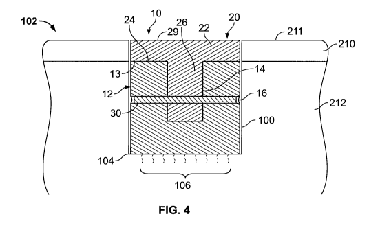
Figure 3 is a top perspective view of the multi-piece construct of Figure 2,
as assembled;
Figure 4 is a cross-sectional view of the multi-piece construct of Figure 2
which has been
placed in a bore of a cartilage defect area in a patient according to a method
performed in
accordance with the present invention;
Figure 5 is an exploded perspective view of the multi-piece cancellous
construct of
Figure 2 incorporating a pin assembly; and
Figure 6 is an exploded perspective view of a multi-piece cancellous construct
produced
in accordance with another embodiment of the present invention.
DESCRIPTION OF THE INVENTION
The term "tissue" is used in the general sense herein to mean any
transplantable or
implantable tissue, the survivability of which is improved by the methods
described herein upon
implantation. In particular, the overall durability and longevity of the
implant are improved, and
host-immune system mediated responses, are substantially eliminated.
The terms "transplant" and "implant" are used interchangeably to refer to
tissue, material
or cells (xenogeneic or allogeneic) which may be introduced into the body of a
patient.
The terms "autologous" and "autograft" refer to tissue or cells which
originate with or are
derived from the recipient, whereas the terms "allogeneic" and "allograft"
refer to cells and
tissue which originate with or are derived from a donor of the same species as
the recipient. The
terms "xenogeneic" and "xenograft" refer to cells or tissue which originate
with or are derived
from a species other than that of the recipient and the best mode and
preferred embodiment is
shown in Figures 2-5.
The present invention is directed towards a sterile cartilage repair construct
constructed
of cancellous bone taken from allogenic or xenogenic bone sources.
The construct is preferably derived from dense allograft cancellous bone that
may
originate from the proximal or distal femur, proximal or distal tibia,
proximal humerus, talus,
calcaneus, patella, or ilium.
8
CA 02708147 2010-06-04
WO 2009/076164 PCT/US2008/085522
The biphasic design of the scaffold is configured to provide one phase that
allows for
healing of the cartilage region and another distinct phase that allows for
healing of the
underlying subchondral bone. The thickness of the top section of the cap
member is designed to
match or slightly exceed the thickness of the patient's cartilage region. The
porous structure of
the demineralized cancellous bone in the cap member allows the incorporation
and retention of a
paste-like matrix of cartilage particles in this region. This cartilage-
derived matrix provides the
environment and necessary biochemical cues to elicit a healing response from
the cells that have
infiltrated the scaffold from the surrounding host tissue and bleeding bone.
The sponginess of
the cap member enables the top surface of the implant to conform to the
natural curvature of the
joint surface. This conformability of the top of the scaffold permits
treatment of large diameter
defects without the risk of a proud edge of the implant causing damage to the
opposing joint
surface during articulation. The base member is similar in structure and
composition to the
surrounding subchondral bone and is designed to provide mechanical support to
the cap member
creating a load-bearing scaffold, and also to allow a press-fit into the
defect. In addition, the
porous nature of the base member enables the bleeding bone to permeate rapidly
throughout the
scaffold providing the host cells necessary for healing. While the scaffold is
preferably
constructed with allograft bone, it is also envisioned that the same can be
constructed of
xenograft bone when the same is properly treated.
Cancellous tissue is first processed into blocks and then milled into the
desired shapes for
the various components of the invention. In a preferred embodiment, the
bicomponent implant
assembly 10 is milled using a lathe to form a mineralized cancellous bone base
member 12
having a cylindrical shape and a diameter varying between 6-30 mm and a
demineralized cap
member 20. The base member 12 has a top planar surface 13 and defines a
central blind bore 14
cut in and along the central axis of the base member 12. The base member 12
additionally has a
through-going transverse bore 16 cut through the diameter which intersects the
central bore 14.
A demineralized cancellous bone cap member 20 is formed with a cylindrical or
disc shaped top
section 22 having a thickness similar or greater than the thickness of human
articular cartilage,
namely about 1.5 mm to about 6.0 mm. The cap member 20 is fully demineralized
(<0.5%
residual calcium wt/wt) and treated with chemical soaks to be non-
osteoinductive. The cap
member 20 includes a top section 22 having a planar bottom seating surface 24
which sits on the
top planar surface 13 of the base member 12. The top section 22 may have the
same diameter as
9
CA 02708147 2010-06-04
WO 2009/076164 PCT/US2008/085522
the base member 12 or be of a greater diameter than the base member 12. An
integral stem 26
extends perpendicularly outward from the top section 22 and has a diameter
smaller than the
base member central blind bore 14 so that it fits in the bore 14 of the base
member 12. A
through-going bore 28 ranging from 1.5 mm to about 3.0 mm in diameter is cut
through the mid-
section of the stem 26 and when the planar seating surface 24 rests on the top
planar surface 13
of the base member 12, the cap member 20 is rotated until the stem bore 28 is
aligned with the
transverse bore 16 of the base member 12 providing a straight axially aligned
combined bore
extending through the base member 12 and the stem 26. If desired, the bore 28
and the bore 16
can be angled to provide an angled combined bore through the base member 12
and the stem 26.
A cylindrical cancellous bone pin 30 or bone pin assembly 31 is inserted into
the axially aligned
combined bores 16, 28 to hold the two pieces (i.e., the base member 12 and the
cap member 20)
in a fixed relationship.
If the implant assembly 10 has a large diameter, multiple pin sections can be
used as
shown in Figure 5 to form the bone pin assembly 31. Multiple cancellous pins
32, 34 and 36 are
used in sequence to attach the cap member 20 to the base member 12. In this
configuration, one
pin 32 is inserted into one end of the stem bore 28 through the transverse
bore 16, a second
longer pin 34 is inserted into the opposite end of the stem bore 28 while the
pin 32 is held in
place and a third shorter pin 36 is inserted into the stem bore 28 from the
same side as the second
pin 34. While the bone pin is preferably constructed of cancellous bone or
cortical bone, other
biocompatible materials such as a ceramic, metal such as surgical steel or a
biocompatible
polymer can be used.
In an alternate embodiment as shown in Figure 6 which is an inverted design of
the
embodiment shown in Figures 2-5, a cylindrically shaped base member 112 is
stepped at 118 to
form a stem 114 having a transverse bore 116 extending through the diameter of
the stem 114,
with the end surface 119 of the stem 114 being planar to fit against the end
surface of bore 124 of
the cap member 120. The cap member 120 is cylindrical with a blind bore 124
cut therein to
receive the stem 114 and has a transverse bore 122 which intersects the blind
bore 124. When
the cap member 120 is rotated around the stem 114, the bores 122 and 116 are
axially aligned to
receive a pin 130 (or a pin assembly as shown in Figure 5) holding the two
pieces of the implant
together in a fixed relationship. The top surface 129 of cap member 120 is
substantially planar
CA 02708147 2010-06-04
WO 2009/076164 PCT/US2008/085522
or slightly curved to correspond with the surrounding cartilage area 210 of
the patient forming a
smooth continuous surface.
The cap member 20/120 is preferably constructed of cancellous bone and is
demineralized in dilute acid such as HCL until the bone contains less than
0.5% wt/wt residual
calcium. If desired, the cap member 20/120 can be treated so that a section of
the stem 26/114 is
left mineralized. Subsequently, the resultant demineralized tissue form of the
cap member
20/120 is predominantly Type I collagen, which is sponge-like in nature with
an elastic quality.
Following decalcification, the tissue is further cleaned, brought to a
physiological pH level of
about 7.0 and treated with chemical soaks of hydrogen peroxide for about 1
hour with ultrasonic
so that the cancellous tissue is nonosteoinductive. Alternatively, this
inactivation of inherent
osteoinductivity of the demineralized cancellous bone may be accomplished via
chemical or
thermal treatment or by high energy irradiation.
The demineralized cap member 20/120 is infused with a matrix of minced
cartilage putty
or gel consisting of minced or milled allograft cartilage pieces having a size
ranging from about
microns to about 212 microns that have been reconstituted in saline. The
cartilage particles
are preferably allograft cartilage derived from hyaline, fibrous or a
combination of hyaline and
fibrous cartilage. However, it is also envisioned that autograft or xenograft
cartilage may be
used. The cartilage particles have been previously lyophilized so that their
water content ranges
from 0.1 % to 8.0% with the cartilage pieces ranging from about 20% to about
40% by weight of
the infusion matrix, preferably 22% and mixed with a carrier which can have a
composition of
one or more of the following: phosphate buffered saline, saline sodium
hyaluronate solution
(HA) (molecular weight ranging from 7.0 x 10⁵ to 1.2 x 10⁶) or other suitable
bioabsorbable
carrier such as hyaluronic acid and its derivatives, gelatin, collagen,
chitosan, alginate, Dextran,
carboxymethylcellulose (CMC), hydroxypropyl methylcellulose, or other
polymers, the carrier
ranging from ranging from about 75% to about 60% by weight. The preferred
carrier is
phosphate buffered saline at about 22% w/w. Another carrier which can be used
is sterile water.
In a most preferred embodiment, morselized cartilage particles having a size
less than
212 microns, preferably ranging from about 10 to about 212 microns, are
combined with a
phosphate buffered saline carrier and a preferred fibroblast growth factor
such as FGF-2 variant
(FGF-2v) in a dosage of 10 -5000 micrograms per cubic cm. This combination is
infused into
the cap member 20/120. The preferred fibroblast growth factor FGF-2v is
described in U.S.
11
CA 02708147 2010-06-04
WO 2009/076164 PCT/US2008/085522
Patent Application Publication Number 20050148511 filed November 5, 2004 which
is
incorporated by reference herein and discloses a variant of FGF-2 having at
least one amino acid
substitution in the beta 8-beta 9 loop, the variant is characterized in having
at least one of the
following attributes compared to the corresponding wild type FGF-2: enhanced
specificity for
one receptor subtype; increased biological activity mediated by at least one
receptor subtype with
equivalent or reduced activity mediated through another receptor subtype;
enhanced affinity to at
least one receptor subtype; and increased cell proliferation mediated through
one receptor
subtype. The demineralized portion will contain approximately 0.1 - 1.0 g/cc
of cartilage paste.
The outer diameter of the assembled implant ranges from between 6 - 30 mm and
its
overall height ranges between 8 - 20 mm.
If desired, the open cancellous structure of the cap member 20 may
additionally be
loaded with the cartilage pieces and carrier noted above and/or one or more
chondrogenic growth
factor additives namely recombinant or native or variant growth factors of FGF-
2, FGF-5,
FGF-7, FGF-9, FGF-11, FGF-21, TGF-0, BMP-2, BMP-4, BMP-7, PDGF, VEGF, and a
bioactive peptide such as Nell-1 or TP508. Additional growth factors which can
be added are
insulin-like growth factor-1 (IGF-1), hepatocyte growth factor and platelet-
derived growth
factor. Other additives can include human allogenic or autologous
chondrocytes, human
allogenic cells, human allogenic or autologous bone marrow cells, human
allogenic or
autologous stem cells, demineralized bone matrix, insulin, insulin-like growth
factor-1,
interleukin-1 receptor antagonist, hepatocyte growth factor, platelet-derived
growth factor,
Indian hedgehog, parathyroid hormone-related peptide, viral vectors for DNA
delivery,
nanoparticles, or platelet-rich plasma. This design enables the fabrication of
an implant that
possesses a relatively uniform substantially demineralized top section that is
distinct from the
mineralized base section.
The sterile implant 10 is placed in a defect area bore 100 which has been cut
in the lesion
area of the bone 102 of a patient with the top surface 29 of the cap member
top section 22 being
slightly proud, slightly below, or substantially flush with the surface 211 of
the original cartilage
210 surrounding the defect bone area remaining at the area being treated (see
Figure 4). The
base member 12 and the cap member 20 are force fit into the bore 100 defining
the defect area.
The diameter of the base member 12 is preferably greater than the diameter of
the bore 100 prior
to insertion into the bore 100. The implant 10 has a length which can he the
same as the depth of
12
CA 02708147 2010-06-04
WO 2009/076164 PCT/US2008/085522
the defect bore 100 or more or less than the depth of the bore 100. If the
height of the implant 10
is the same as the depth of the bore 100, the base of the implant 10 is
supported by the bottom
surface of the bore 100 and the top surface 29 of the cap member 20 is
substantially level with
the surrounding articular cartilage to form a smooth continuous surface and to
be load bearing.
With such load bearing support the graft surface is not damaged by weight or
bearing loads
which can cause micromotion interfering with the graft interface producing
fibrous tissue
interfaces and subchondral cysts.
The invention disclosure also describes the method of treatment of either
primary focal
lesions in articular cartilage or backfill site defects with the biphasic
scaffold. During the
treatment of a primary defect, the lesion is first prepared by measuring the
defect and coring out
the damaged region with a flat-bottom drill. The diameter of the chosen
scaffold will be slightly
larger than the diameter of the cored defect in order to create a press-fit.
The base of the scaffold
will be trimmed to match the depth of the defect and the edges of the base may
be chamfered to
facilitate insertion. The implant will then be inserted in a dry state into
the defect site by using a
tamp and a mallet or other insertion device. The implant is positioned such
that its top surface is
either flush, slightly proud, or slightly lower to the surface of the adjacent
cartilage. The scaffold
is re-hydrated by the bleeding bone from the surrounding host tissue in situ.
During treatment of a backfill defect site, the defect will be created when an
osteochondral plug is removed from a non-weight bearing region of the
patient's own joint and
transferred to a primary defect site. After the backfill site is prepared, the
biphasic scaffold will
be selected for a press-fit with the defect and will be trimmed to match the
depth of the defect.
The edges of the base of the scaffold may be chamfered to facilitate
insertion. The scaffold will
then be implanted in a similar manner for treatment of a primary defect.
In operation, the lesion or defect is removed by cutting a blind bore 100
removing the
cartilage 210 having a lesion and the subchondral bone 212 beneath the
cartilage defect of the
patient. The base 104 of the bore 100 is then micro-fractured 106 to cause
bleeding. The
implant 10 is then force fit in the bore 100 in an interference fit with the
surrounding walls of the
bore with the top surface 29 of the cap member section 22 being aligned with
the top surface 211
of the cartilage 210 surrounding the implant area of the patient.
If desired, suitable organic glue material can be used to keep the implant
components
additionally secured together. Suitable organic glue material can be found
commercially, such as
13
CA 02708147 2010-06-04
WO 2009/076164 PCT/US2008/085522
for example; TISSEEL or TISSUCOL (fibrin based adhesive; Immuno AG,
Austria),
Adhesive Protein (Sigma Chemical, USA), Dow Corning Medical Adhesive B (Dow
Coming,
USA), fibrinogen thrombin, clastin, collagen, casein, albumin, keratin and the
like.
The principles, preferred embodiments and modes of operation of the present
invention
have been described in the foregoing specification. However, the invention
should not be
construed as limited to the particular embodiments which have been described
above. Instead,
the embodiments described here should be regarded as illustrative rather than
restrictive.
Variations and changes may be made by others without departing from the scope
of the present
invention as defined by the following claims:
14