Note: Descriptions are shown in the official language in which they were submitted.
CA 02711955 2015-05-19
INTERSPINOUS SPACER
[0001]
BACKGROUND
[0002] With spinal
stenosis, the spinal canal narrows and pinches the spinal cord and
nerves, causing pain in the back and legs. Typically, with age, a person's
ligaments
may thicken, intervertebral discs may deteriorate and facet joints may break
down¨
all contributing to the condition of the spine characterized by a narrowing of
the
spinal canal. Injury, heredity, artindtis, changes in blood flow and other
causes may
also contribute to spinal stenosis.
CA 02711955 2015-05-19
t00031 Doctors have been at the forefront with various treatments of the
spine
including medications, surgical techniques and implantable devices that
alleviate and
substantially reduce debilitating pain associated with the back. In one
surgical
technique, a spacer is implanted between adjacent spinous processes of a
patient's
2
CA 02711955 2010-07-12
WO 2009/091922 PCT/US2009/031150
spine. The implanted spacer opens the foramen and spinal canal, maintains the
desired distance between vertebral body segments, and as a result, avoids
impingement of nerves and relieves pain. For suitable candidates, an
implantable
interspinous spacer may provide significant benefits in terms of pain relief.
However,
there is a need for an implantable interpsinous spacer for patients with
adjacent
spinous processes that are not aligned such as in patients suffering with
scoliosis.
Scoliosis is the lateral or sideways curvature caused by congenital,
neuromuscular,
idiopathic, syndromic or postural conditions. An example of a scoliotic spine
is
shown in FIG. 12.
[0004] Any surgery is an ordeal. However, the type of device and how it
is
implanted has an impact. For example, one consideration when performing
surgery to
implant an interspinous spacer is the size of the incision that is required to
allow
introduction of the device. Small incisions and minimally invasive techniques
are
quick and generally preferred as they affect less tissue and result in
speedier recovery
times. As such, there is a need for interspinous spacers that work well with
surgical
techniques that are minimally invasive for a patient with misaligned spinous
processes such as patients with scoliosis. The present invention sets forth
such a
spacer.
SUMMARY
[0005] According to one aspect of the invention, an implant configured
for placement
between adjacent spinous processes in a spinal motion segment with a scoliotic
curve
and configured to laterally stabilize the spacer with respect to said adjacent
spinous
processes is provided.
[0006] An implant for placement between adjacent spinous processes in a
spinal
motion segment is provided. The implant includes a body defining a
longitudinal
passageway through at least a portion of the body. A first arm connected to
the body
and capable of rotation with respect to the body. The first arm has a first
pair of
extensions and a first bridge defining a spinous process receiving portion for
seating a
first spinous process therein. The first arm has a first proximal caming
surface. The
3
CA 02711955 2010-07-12
WO 2009/091922 PCT/US2009/031150
implant further includes a second arm connected to the body and capable of
rotation
with respect to the body. The second arm has a second pair of extensions and a
second bridge defining a spinous process receiving portion for seating a
second
spinous process therein. The second arm has a second proximal caming surface.
The
implant further includes an actuator connected to the body. The actuator is
configured such that the actuator is disposed inside the body and configured
to move
relative to the body and contact the caming surfaces of the arms to rotate
them from a
first configuration in which the arms are substantially parallel to the
longitudinal axis
of the body to a second configuration in which the first arm seats the first
spinous
process and the second arm seats the second spinous process. At least one of
the first
arm and second arm is configured to seat the spinous processes of a spinal
motion
segment with a scoliotic curve.
[0007] An implant for placement between adjacent spinous processes in a
spinal
motion segment is provided. The implant includes a body defining a
longitudinal
axis. A first arm is connected to the body and has a first pair of extensions
defining a
spinous process receiving portion for seating a superior spinous process
therein. The
implant includes a second arm connected to the body. The second arm has a
second
pair of extensions defining a spinous process receiving portion for seating an
inferior
spinous process therein. One extension of the first pair and one extension of
the
second pair that are adjacent to each other on the same side of the spacer are
both
shorter than the other of the extensions.
[0008] An implant for placement between adjacent spinous processes in a
spinal
motion segment is provided. The implant includes a body defining a
longitudinal
axis. A first arm is connected to the body having a first pair of extensions
defining a
spinous process receiving portion for seating a superior spinous process
therein. A
second arm is connected to the body. The second arm has a second pair of
extensions
defining a spinous process receiving portion for seating an inferior spinous
process
therein. The distance between the first pair of extensions is greater than the
distance
between the second pair of extensions to accommodate a generally wider lower
or
caudal end of a superior spinous process relative to a generally narrower
upper or
cephalad end of an inferior spinous process.
4
CA 02711955 2015-05-19
[0009] An implant for placement between adjacent spinous processes in
a spinal
motion segment is provided. The implant includes a body defining a
longitudinal axis. A first
arm is connected to the body and configured to laterally stabilize the body
with respect to a
first spinous process when in a deployed configuration. A second arm is
connected to the
body and configured to laterally stabilize the body with respect to a second
spinous process
when in a deployed configuration. The first and second arms are configured for
placement
between adjacent spinous processes in which at least one of the adjacent
spinous processes
has a projection in a coronal plane that is angled with respect to the
sagittal plane.
[0009a] According to one aspect of the present invention, there is
provided an implant
for placement between adjacent spinous processes in a spinal motion segment,
the implant
comprising: a body defining a longitudinal axis; a first arm connected to the
body and
configured to laterally stabilize the body with respect to a first spinous
process when in a
deployed configuration, wherein the first arm includes a pair of first
extensions configured to
receive the first spinous process between the first extensions; and a second
arm connected to
the body and configured to laterally stabilize the body with respect to a
second spinous
process when in a deployed configuration; a first bridge connected between the
first
extensions to define a first spinous process receiving portion, wherein the
first pair of
extensions are substantially parallel to a sagittal plane of the implant and
the first bridge is
angled with respect to the sagittal plane of the implant; and wherein the
first and second arms
are configured for placement between adjacent spinous processes in which at
least one of the
adjacent spinous processes has a projection in a coronal plane that is angled
with respect to
the sagittal plane.
[0009b] According to another aspect of the present invention, there is
provided an
implant for placement between adjacent spinous processes in a spinal motion
segment, the
implant comprising: a body defining a longitudinal axis; a first arm connected
to the body and
configured to laterally stabilize the body with respect to a first spinous
process when in a
deployed configuration, wherein the first arm includes a pair of first
extensions configured to
receive the first spinous process between the first extensions; and a second
arm connected to
the body and configured to laterally stabilize the body with respect to a
second spinous
process when in a deployed configuration, wherein the second arm includes a
pair of second
5
CA 02711955 2015-05-19
extensions configured to receive the second spinous process between the second
extensions; a
first bridge connected between the first pair of extensions to define a first
spinous process
receiving portion; a second bridge connected between the second pair of
extensions to define
a second spinous process receiving portion; and wherein the first and second
arms are
configured for placement between the spinous processes in which at least one
of the adjacent
spinous processes has a projection in a coronal plane that is angled with
respect to a sagittal
plane of the implant, wherein the pair of first extensions and the pair of
second extensions are
substantially parallel to the sagittal plane of the implant and the first and
second bridges are
angled with respect to the sagittal plane of the implant.
[0009c] According to another aspect of the present invention, there is
provided an
implant for placement between adjacent spinous processes in a spinal motion
segment, the
implant comprising: a body defining a longitudinal axis; a first arm connected
to the body and
configured to laterally stabilize the body with respect to a first spinous
process when in a
deployed configuration, wherein the first arm includes a pair of first
extensions at a first fixed
spaced apart relationship relative to one another and configured to receive
the first spinous
process between the first extensions; and a second arm connected to the body
and configured
to laterally stabilize the body with respect to a second spinous process when
in a deployed
configuration, wherein the second arm includes a pair of second extensions at
a second fixed
spaced apart relationship relative to one another and configured to receive
the second spinous
process between the second extensions; wherein the first and second arms are
configured for
placement between adjacent spinous processes in which at least one of the
adjacent spinous
processes has a projection in a corona' plane that is angled with respect to a
sagittal plane of
the implant, wherein each of the first extensions is permanently angled with
respect to the
sagittal plane of the implant and each of the second extensions is permanently
angled with
respect to the sagittal plane of the implant.
[0009d1 According to another aspect of the present invention, there is
provided an
implant for placement between adjacent spinous processes in a spinal motion
segment, the
implant comprising: a body defining a longitudinal axis; a first arm connected
to the body and
configured to laterally stabilize the body with respect to a first spinous
process when in a
deployed configuration, wherein the first arm includes a pair of first
extensions configured to
5a
CA 02711955 2015-05-19
receive the first spinous process between the first extensions; and a second
arm connected to
the body and configured to laterally stabilize the body with respect to a
second spinous
process when in a deployed configuration, wherein the second arm includes a
pair of second
extensions configured to receive the second spinous process between the second
extensions; a
first bridge connected between the first pair of extensions to define a first
spinous process
receiving portion; a second bridge connected between the second pair of
extensions to define
a second spinous process receiving portion; and wherein the first and second
arms are
configured for placement between adjacent spinous processes and which at least
one of the
adjacent spinous processes has a projection in a coronal plane that is angled
with respect to a
sagittal plane of the implant, wherein the pair of first extensions is angled
with respect to the
sagittal plane of the implant and the pair of second extensions is angled with
respect to the
sagittal plane of the implant and the first and second bridges are
perpendicular to their
respective extensions to which they are connected.
[0009e] According to another aspect of the present invention, there is
provided an
implant for placement between a first spinous process and a second spinous
process in a spinal
motion segment, the implant comprising: a body defining a sagittal plane; a
first arm rotatably
coupled to the body and configured to rotate relative to the body about a
first axis of rotation
generally perpendicular to the sagittal plane such that the first arm moves
from an undeployed
configuration to a deployed configuration, the first arm is configured to
laterally stabilize the
body with respect to the first spinous process when in the deployed
configuration, the first
arm comprising a first pair of extensions at a first permanent generally
parallel arrangement
and configured to receive the first spinous process when the first arm moves
from the
undeployed configuration to the deployed configuration; and a second arm
rotatably coupled
to the body and configured to rotate relative to the body about a second axis
of rotation
generally perpendicular to the sagittal plane such that the second arm moves
from an
undeployed configuration to a deployed configuration, the second arm is
configured to
laterally stabilize the body with respect to the second spinous process when
in the deployed
configuration; wherein the first and second arms are configured for placement
between the
first and second spinous processes in which the first pair of extensions are
oriented at a
permanent non-parallel angle with respect to the sagittal plane in the
deployed configuration.
5b
CA 02711955 2015-05-19
[000911 According to another aspect of the present invention, there is
provided an implant
for placement between adjacent vertebrae in a spinal motion segment, the
implant comprising: a
body defining a sagittal plane; a first arm connected to the body, the first
arm having a first pair of
substantially parallel extensions defining a first spinous process receiving
portion for seating a
superior spinous process therein when the first arm rotates relative to the
body about a first axis
that is substantially perpendicular to the sagittal plane; and a second arm
connected to the body,
the second arm having a second pair of substantially parallel extensions
defining a second spinous
process receiving portion for seating an inferior spinous process therein when
the second arm
rotates relative to the body about a second axis that is substantially
perpendicular to the sagittal
plane; wherein a distance between the first pair of extensions is greater than
a distance between
the second pair of extensions; and wherein when the first and second spinous
process receiving
portions seat the corresponding superior and inferior spinous process, each of
the first and second
pairs of extensions is oriented at an acute angle with respect to the sagittal
plane,
[0009g] According to yet another aspect of the present invention, there
is provided an
implant for placement between adjacent spinous processes in a spinal motion
segment, the
implant comprising: a body defining a longitudinal axis; a first arm connected
to the body,
the first arm having a first pair of extensions defining a spinous process
receiving portion,
wherein the first pair of extensions move superiorly and posteriorly as the
first arm rotates
relative to the body to seat a superior spinous process in the spinous process
receiving portion
when the longitudinal axis extends in an anterior-posterior direction; and a
second arm
connected to the body, the second arm having a second pair of extensions
defining a spinous
process receiving portion, wherein the second pair of extensions move
inferiorly and
posteriorly as the second arm rotates relative to the body to seat an inferior
spinous process in
the spinous process receiving portion defined by the second pair of
extensions; wherein one
extension of the first pair and one extension of the second pair that are
adjacent to each other
are both shorter than the other of the extensions; and wherein the first and
second arms are
movable from an undeployed configuration in which the first and second arms
are generally
aligned with the longitudinal axis to a deployed configuration in which the
first and second
arms extend laterally away from the body at an angle generally perpendicular
to the
longitudinal axis.
5c
CA 02711955 2015-05-19
BRIEF DESCRIPTION OF THE DRAWINGS
[0010] FIG. la is a perspective view of a spacer according to the
present invention.
[00111 FIG. lb is a side view of a spacer according to the present
invention.
[0012] FIG. lc is a top view of a spacer according to the present
invention.
[0013] FIG. I d is a cross-sectional view of a spacer taken along line A-A
of FIG. 1 c
according to the present invention.
[0014] FIG. le is an end view of a spacer according to the present
invention.
[0015] FIG, If is an exploded view of a spacer according to the
present invention.
[0016] FIG. 2a is a perspective view of a half of a body of a spacer
according to the
present invention.
[0017] FIG. 2b is a side view of half of a body of a spacer according
to the present
invention.
[0018] FIG. 2c is a perspective view of a half of a body of a spacer
according to the
present invention.
[0019] FIG. 2d is a side view of half of a body of a spacer according to
the present
invention.
[0020] FIG. 3a is a perspective view of a superior wing of a spacer
according to the
present invention.
[0021] FIG. 3b is a top view of a superior wing of a spacer according
to the present
invention.
5d
CA 02711955 2010-07-12
WO 2009/091922 PCT/US2009/031150
[0022] FIG. 3c is a side view of a superior wing of a spacer according to
the present
invention.
[0023] FIG. 3d is a perspective view of an inferior wing of a spacer
according to the
present invention.
[0024] FIG. 3e is a bottom view of an inferior wing of a spacer according
to the
present invention.
[0025] FIG. 3f is a side view of an inferior wing of a spacer according to
the present
invention.
[0026] FIG. 4a is a side view of a spacer according to the present
invention.
[0027] FIG. 4b is a side view of a spacer with wings partially deployed
according to
the present invention.
[0028] FIG. 4c is a side view of a spacer with wings in a deployed
configuration
according to the present invention.
[0029] FIG. 4d is a side view of a spacer with wings in a deployed and
extended
configuration according to the present invention.
[0030] FIG. 5a is a cross-sectional view of a spacer with wings in a
partially deployed
configuration according to the present invention.
[0031] FIG. 5b is a cross-sectional view of a spacer with wings in a
deployed
configuration according to the present invention.
[0032] FIG. 5c is a cross-sectional view of a spacer with wings in a
deployed and
extended configuration according to the present invention.
[0033] FIG. 6a is a semi-transparent view of a spacer with wings partially
deployed
according to the present invention.
[0034] FIG. 6b is a semi-transparent view of a spacer with wings in a
deployed
configuration according to the present invention.
[0035] FIG. 6c is a semi-transparent view of a spacer with wings in a
deployed and
extended configuration according to the present invention.
[0036] FIG. 7 is a partial cross-sectional view of a spacer according to
the present
invention located between two adjacent spinous processes.
[0037] FIG. 8 is a cross-sectional view of a spacer according to the
present invention
located between two adjacent spinous processes.
6
CA 02711955 2010-07-12
WO 2009/091922 PCT/US2009/031150
[0038] FIG. 9 is a cross-sectional view of a spacer according to the
present invention
located between two adjacent spinous processes.
[0039] FIG. 10 is a partial view of a spacer according to the present
invention.
[0040] FIG. 11 is a partial view of a spacer and driving tool according to
the present
invention.
[0041] FIG. 12 is a posterior view of part of a spine with a scoliotic
curve.
[0042] FIG. 13a is a side view of a spacer connected to an insertion
instrument
according to the present invention.
[0043] FIG. 13b is a side view of a spacer in a partially deployed
configuration
connected to an insertion instrument according to the present invention.
[0044] FIG. 13c is a side view of a spacer in a deployed configuration
connected to
an insertion instrument according to the present invention.
[0045] FIG. 13d is a side view of a spacer in a deployed and extended
configuration
connected to an insertion instrument according to the present invention.
[0046] FIG. 14 is a perspective view of a spacer in a deployed
configuration
according to the present invention implanted between adjacent spinous
processes of
two vertebral bodies.
DETAILED DESCRIPTION
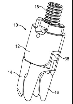
[0047] With reference to FIGs. la-if, various views of a spacer 10
according to the
present invention are shown. The spacer 10 includes a body 12, a superior
extension
member, arm or wing14, an inferior extension member, arm or wing 16, and an
actuator assembly 18.
[0048] Turning now to FIGs. 2a-2d, the body will now be described. The
body 12 is
shown to have a clamshell construction with a left body piece 20 (shown in
FIGs. 2a
and 2b) joined to a right body piece 22 (shown in FIGs. 2c and 2d) to capture
arms
14, 16 inside. With the right and left body pieces 20, 22 joined together, the
body 12
is generally cylindrical. The spacer body 12 has a cross-sectional size and
shape that
allows for implantation between adjacent spinous processes and facilitates
delivery
into a patient through a narrow port or cannula.
7
CA 02711955 2010-07-12
WO 2009/091922 PCT/US2009/031150
[0049] The inside of the body 12 defines an arm receiving portion 24 and
an actuator
assembly receiving portion 26 with features formed in each of the left and
right body
pieces 20, 22 that together define the arm and actuator assembly receiving
portions
24, 26. In one variation, the arm receiving portion 24 includes slots 28 that
receive
pins formed on the arms 14, 16 such that the pins rotate and/or translate
inside the
slots 28. The actuator assembly receiving portion 26 includes a threaded
passageway
30. Other features include a tongue and groove for mating with the opposite
clamshell.
[0050] The outside of the body 12 defines a ledge 32 along at least a
portion of the
periphery. Notches 34 are formed at opposite locations and are configured for
pronged attachment to a spacer delivery instrument. When joined together, the
left
and right body pieces 20, 22 define a proximal opening 36 (as also seen in
FIG. le)
and a distal opening 38 (as also seen in FIG. la) in the body 12. A
longitudinal
scallop (not shown) extending from the proximal end of the spacer to the
distal end is
formed to facilitate placement of the spacer 10 between and to conform to the
anatomy of adjacent spinous processes. In one variation, two oppositely
located
londigutinal scallops are formed in the outer surface of the body 12 such
that, when
implanted in a patient's spine, one scallop faces the superior spinous process
and the
other scallop faces the inferior spinous process. In one variation, the
distance
between oppositely located longitudinal scallops is approximately 8.0
millimeters
imparting the spacer 10 with a low profile advantageous for insertion between
closely
spaced or "kissing" spinous processes.
[0051] Turning now to FIGs. 3a-3c, the superior arm 14 is shown and in
FIGs. 3d-3f,
the inferior arm 16 is shown. The superior and inferior arms 14, 16 include
pins 40
for mating with the body 12, in particular, for mating with the slots 28 of
the arm
receiving portion 24. Each of the superior and inferior arms 14, 16 includes
at least
one caming surface 41, 43, respectively, for contact with the actuator
assembly 18.
The superior and inferior arms 14, 16 include elongated superior extensions
42a, 42b
and elongated inferior extensions 44a, 44b, respectively. Extensions 42a and
44a are
located on the left adjacent to the left body piece 20 and extensions 42b and
44b are
located on right adjacent to the right body piece 22. Superior extensions 42a,
42b
8
CA 02711955 2010-07-12
WO 2009/091922 PCT/US2009/031150
extend substantially parallel to each other in both an undeployed
configuration and in
a fully-deployed configuration as do inferior extensions 44a, 44b. Extending
between
extensions 42a, 42b is a strut, bridge, bracket or saddle 46 that forms a
superior
substantially U-shaped configuration that is sized and configured to receive a
superior
spinous process. As seen in FIG. 3c, the anterior face of the superior
extensions 14
includes a slight concavity or curvature 45 for conforming to the bony anatomy
of the
superior spinous process and or lamina. Extending between inferior extensions
44a,
44b is a strut, bridge, bracket or saddle 48 that forms an inferior
substantially U-
shaped configuration that is sized and configured to receive an inferior
spinous
process of a spinal motion segment. As seen in FIG. 3f, the anterior face of
the
inferior extensions 16 includes a slight convexity or curvature 47 for
conforming to
the bony anatomy of the inferior spinous process and/or lamina. In one
variation, the
length of the saddle 46 of the superior arm 14 is approximately 9.0
millimeters and
the length of the saddle 48 of the inferior arm 16 is approximately 7.0
millimeters.
Also, the tip-to-tip distance of the superior extensions 42a, 42b is
approximately 10.0
millimeters and the tip-to-tip distance of the inferior extensions 44a, 44b is
approximately 9.0 millimeters. In sum, the seat comprising the saddle 46 and
superior extensions 42a, 42b formed by the superior arm 14 is larger than the
seat
comprising the saddle 48 and inferior extensions 44a, 44b formed by the
inferior arm
16. The larger superior seat of the spacer conforms closely to a wider lower
end of
the spinous process and the smaller inferior seat of the spacer conforms
closely to a
narrower upper end of the adjacent inferior spinous process when the spacer 10
is
inserted between adjacent spinous processes as spinous processes are naturally
narrower on top and wider on the bottom and thereby providing greater lateral
stability to the spacer with respect to the spinous processes.
[0052] The superior and inferior arms 14, 16 are movably or rotatably
connected to
the body 12, for example by hinge means or the like to provide rotational
movement
from an undeployed configuration to a deployed configuration that arcs through
about
a 90 degree range or more with respect to the body 12. The arms 14, 16 are
rotationally movable between at least an undeployed, collapsed or folded state
(as
shown in FIGs. la-le) and at least a fully deployed state (as shown in FIGs.
4c, Sc
9
CA 02711955 2010-07-12
WO 2009/091922 PCT/US2009/031150
and 6c). In the undeployed state, the arm pairs 14, 16 are aligned generally
or
substantially axially (i.e., axially with the longitudinal axis defined by the
body 12 or
to the translation path into the interspinous space of the patient) to provide
a minimal
lateral or radial profile. The longitudinal axis X of the spacer 10 and body
12 is
shown in FIG. lc. In the deployed state, the arm pairs 14, 16 are positioned
generally
or substantially transverse to the collapsed position (i.e., transverse to the
longitudinal
axis defined by the body 12 or to the translation path into the interspinous
space of the
patient). In the deployed state, the arm pairs 14, 16 are positioned such that
each of
the U-shaped saddles are in a plane (or individual planes) or have a
substantially U-
shaped projection in a plane that is generally or substantially transverse to
the
longitudinal axis X defined by the body 12 or to the collapsed position or to
the
implantation path into the interspinous space of the patient. In one
variation, the
spacer 10 is configured such that the arms 14, 16 are linearly moveable or
translatable
within the same transverse plane from the deployed state (such as the state
shown in
FIGs. 4c, 5b and 6b) to and from an additionally extended state or second
deployed
state (such as the state shown in FIGs. 4d, 5c and 6c) characterized by an
additional
translation of at least one of the arms 14, 16 with respect to the body 12
along the
direction of the arrows in FIG. 4d and 6c away from or towards the body 12.
More
specifically, the arms 14, 16 can be extended in the general vertical or
lateral direction
along an axis along the general length of the spine wherein the arms 14, 16
are
extended away from each other and away from the body 12 as denoted by the
arrows
in FIG. 4d. The arms 14, 16 can be un-extended in a direction towards each
other and
towards the body 12 for un-deployment or repositioning of the spacer 10 and
shown
by the arrows in FIG. 6c. This extended feature advantageously allows for the
most
minimally invasive configuration for the spacer without compromising the
ability of
the spacer 10 to seat and contain the spinous processes or to laterally
stabilize the
spacer relative to the spinous processes in between levels where the anatomy
of the
spinous processes is such that the interspinous process space increases in the
anterior
direction of the patient or without compromising the ability of the spacer to
provide
adequate distraction. The arms 14, 16 are connected to the body 12 and/or to
each
other in a manner that enables them to be moved simultaneously or
independently of
CA 02711955 2010-07-12
WO 2009/091922 PCT/US2009/031150
each other, as well as in a manner that provides passive deployment and/or
vertical
extension or, alternatively, active or actuated deployment and/or vertical
extension.
[0053] Turning back to FIG. if, the actuator assembly 18 will now be
described. The
actuator assembly 18 includes an actuator 48, shaft 50 and retainer 52. The
actuator
48 includes a distal end 54 and a proximal end 56 and at least two bearing
surfaces
58. The bearing surfaces 58 angle towards each other from the proximal end 56
to the
distal end 54. The proximal end 56 of the actuator 48 includes a shaft
receiving
portion 60 configured to receive the shaft 50. In one variation, the shaft 50
is
integrally formed with the actuator 48. The distal end 54 of the actuator 48
is further
configured to engage the superior and inferior arms 14, 16 such that forward
translation of the actuator 48 relative to the body 12 effects deployment of
the arms
into at least one deployed configuration. The actuator assembly 18 is at least
partially
disposed inside the body 12 and is configured to move with respect to the body
12.
[0054] Still referencing FIG. 1, the shaft 50 is substantially cylindrical
in shape and
includes a threaded outer surface for engagement with the threaded inner
surface of
the actuator assembly receiving portion 26 of the body 12. The threads on the
inner
surface of the body 12 are formed by the conjunction of both left and right
body
pieces 20, 22. The proximal end of the shaft 50 includes a hex socket 62 for
receiving
a driving tool. The distal end of the shaft 50 includes an actuator engagement
portion
64 configured to connect to the actuator 48. The actuator engagement portion
64 as
shown in FIG. 1 is a projection that slides into a channel 66 on the actuator
48. Once
inserted into the channel 66, movement of the shaft 50 solely along the
longitudinal
axis of the spacer 10 will not release the shaft 50 from the actuator 48.
[0055] Still referencing FIG. 1, the retainer 52 is a circular ring
preferably made of
metal such as steel or titanium. The retainer 52 fits into a recess 68 formed
on the
inner surface of the body 12. When pressed into the recess 68, the retainer 52
secures
the actuator 48 inside the passageway 30 of the body 12.
[0056] Assembly of the spacer 10 with reference to FIGs. la-if will now be
described. The arms 14, 16 are disposed in the arm receiving portion 24 of one
body
piece. The other of the left or right body piece 20, 22 is securely
connected/welded to
the one body piece thereby capturing the arms 14, 16 inside the arm receiving
portion
11
CA 02711955 2010-07-12
WO 2009/091922 PCT/US2009/031150
24 such that the arms 14, 16 are capable of at least rotational movement with
respect
to the body 12 and in one variation, capable of rotational movement and
translation
with respect to the body 12. The shaft 50 is connected to the actuator 48 and
together
inserted and threadingly connected into the passageway 30 of the body 12. The
retainer 52 is passed over the proximal end of the shaft 50 and snapped into
the recess
68 of the body 12 to secure the actuator assembly 18 inside the body 12 such
that the
actuator assembly 18 is capable of threaded translational movement with
respect to
the body 12.
[0057] To deliver and deploy the spacer 10 within the patient, the spacer
10 is
releasably attached to a delivery instrument (not shown) at the proximal end
of the
spacer 10 via notches 34. The spacer 10 is provided or otherwise placed in its
undeployed state as illustrated in FIG. 4a. In the undeployed state and
attached to a
delivery instrument, the spacer 10 is inserted into a port or cannula which
has been
operatively positioned in an interspinous space within a patient's back and
the outside
of the patient via a minimally invasive incision. In some circumstances it may
not be
necessary to use a cannula where the device is inserted through a larger
opening in the
skin. Where a cannula is employed, the spacer 10 is then advanced through the
cannula to within the targeted interspinous space between two adjacent spinous
processes. The spacer 10 is advanced beyond the end of the cannula or,
alternatively,
the cannula is pulled proximately to uncover the spacer 10 within. A driver
such as a
hex-shaped tool is inserted into the hex socket 62 of the spacer 10 and turned
to
advance the shaft 50 of the actuator assembly 18. As the shaft 50 advances
within the
passageway 30, the bearing surfaces 58 of the actuator 48 contact the superior
and
inferior caming surfaces 41, 43 of the superior and inferior arms 14, 16
forcing the
arms 14, 16 to rotate about their pins 40 with respect to the body 12. The
arms 14, 16
rotate through an arc of approximately 90 degrees into the deployed
configuration in
which the superior and inferior extensions 42a, 42b, 44a, 44b are
substantially
perpendicular to the longitudinal axis of the spacer 10 as shown in FIGs. 4c
and 4d.
In one variation, continued advancement of the actuator assembly 18 forces the
arms
14, 16 outwardly in the direction of the arrows in FIG. 4d. Such outward
translation
is guided by the length and shape of the slots 28. Once deployed, the superior
arm 14
12
CA 02711955 2010-07-12
WO 2009/091922 PCT/US2009/031150
seats the superior spinous process and the inferior arm 16 seats the adjacent
inferior
spinous process.
[0058] Referring now to FIGs. 4a-4d, the spacer 10 is shown in a closed,
undeployed
configuration (FIG. 4a), a partially deployed configuration or otherwise
intermediary
configuration (FIG. 4b), a deployed configuration (FIG. 4c) and a deployed and
extended configuration (FIG. 4d). In FIGs. 4a-4d, the sagittal plane of the
spacer 10
corresponds to the plane of the paper that bisects the spacer 10. In moving
from an
undeployed to a deployed configuration, the actuator assembly 18 and, in
particular,
the shaft 50 of the actuator assembly moves distally with respect to the body
to a
position flush or almost flush with the proximal end of the body 12 or to a
position
completely inside the body 12 disappearing from sight providing a low profile
for the
spacer 10 along the longitudinal axis of the body 12.
[0059] Turning now to the cross-sectional views of the spacer 10 in FIGs.
5a-5c, as
the shaft 50 advances within the passageway 30, the bearing surfaces 58 of the
actuator 48 contact the superior and inferior caming surfaces 41, 43 of the
superior
and inferior arms 14, 16 turning the arms 14, 16 into rotation with respect to
the body
12. Upon rotation, the bearing surfaces 58 of the actuator 48 slide with
respect to the
superior and inferior caming surfaces 41, 43 of the superior and inferior arms
14, 16.
The arms 14, 16 rotate through an arc of approximately 90 degrees with respect
to the
body 12 into the deployed configuration (FIG. 5b) in which the superior and
inferior
extensions of the arms 14, 16 are substantially perpendicular to the
longitudinal axis
of the spacer 10 as shown in FIGs. 5b and with further actuation into a
deployed and
extended configuration as shown in FIG. Sc in which the arms 14, 16 have
extended
outwardly away from the body 12. The arms 14, 16 have a substantially U-shaped
projection in a plane perpendicular to the longitudinal axis of the spacer 10
or a
substantially U-shaped projection in a plane perpendicular to the longitudinal
axis of
the spacer 10.
[0060] Turning now to the semi-transparent views of the spacer 10 in FIGs.
6a-6c, the
rotation of the pins 40 of the arms 14, 16 in the openings 28 of the body 12
is shown
in moving from the configuration of FIG. 6a to the configuration of FIG. 6c.
The
translation of the pins 40 of the arms 14, 16 in the elongated portion of the
slots 28 of
13
CA 02711955 2010-07-12
WO 2009/091922 PCT/US2009/031150
the body 12 is shown in moving from the deployed configuration of FIG. 6b to
the
deployed and extended configuration of FIG. 6c in the direction of the arrows
in FIG.
6c. Such outward translation with respect to the body 12 is guided by the
length and
shape of the slots 28. Reverse rotation of the spindle 86 moves the shaft 50
proximally with respect to the body 12 allowing the arms to close to any
intermediary
configuration between a deployed, configuration and an undeployed, closed
configuration. This feature advantageously permits the surgeon to deploy and
undeploy the spacer as needed to ease installation and positioning of the
spacer with
respect to patient anatomy.
[0061] Any of the spacers disclosed herein are configured for implantation
employing
minimally invasive techniques including through a small percutaneous incision
and
through the supraspinous ligament. Implantation through the supraspinous
ligament
involves selective dissection of the supraspinous ligament in which the fibers
of the
ligament are cut, separated or spread apart from each other in a manner to
maintain as
much of the ligament intact as possible such as cutting, separating or
spreading in a
direction parallel to the orientation of the ligament fibers. This approach
avoids
crosswise dissection or cutting of the ligament and thereby reduces the
healing time
and minimizes the amount of instability to the affected spinal segment. While
this
approach is ideally suited to be performed through a posterior or midline
incision, the
approach may also be performed through one or more incisions made laterally of
the
spine with or without affect to the supraspinous ligament. Of course, the
spacer may
also be implanted in a lateral approach that circumvents the supraspinous
ligament
altogether.
[0062] Other variations and features of the various mechanical spacers are
covered by
the present invention. For example, a spacer may include only a single arm
which is
configured to receive either the superior spinous process or the inferior
spinous
process or laterally stabilize the body of the spacer with respect to the
superior
spinous process and/or with respect to the inferior spinous process. The
surface of the
spacer body opposite the side of the single arm may be contoured or otherwise
configured to engage the opposing spinous process wherein the spacer is sized
to be
securely positioned in the interspinous space and provide the desired
distraction of the
14
CA 02711955 2010-07-12
WO 2009/091922 PCT/US2009/031150
spinous processes defining such space. The additional extension of the arm(s)
subsequent to their initial deployment in order to seat or to effect the
desired
distraction between the vertebrae may be accomplished by expanding the body
portion of the device instead of or in addition to extending the individual
extension
members 14, 16.
[0063] The extension arms of the subject device may be configured to be
selectively
movable subsequent to implantation, either to a fixed position prior to
closure of the
access site or otherwise enabled or allowed to move in response to normal
spinal
motion exerted on the device after deployment. The deployment angles of the
extension arms may range from less than 90 degrees (relative to the
longitudinal axis
defined by the device body) or may extend beyond 90 degrees. Each extension
member may be rotationally movable within a range that is different from that
of the
other extension members. Additionally, the individual superior and/or inferior
extensions 42a, 42b, 44a, 44b may be movable in any direction relative to the
strut or
bridge extending between an arm pair or relative to the device body in order
to
provide shock absorption and/or function as a motion limiter, or serve as a
lateral
adjustment particularly during lateral bending and axial rotation of the
spine. The
manner of attachment or affixation of the extensions to the arms may be
selected so as
to provide movement of the extensions that is passive or active or both. In
one
variation, the saddle or distance between extensions 42a and 42b or between
44a and
44b can be made wider to assist in seating the spinous process and then
narrowed to
secure the spinous process positioned between extensions 42a and 42b or
between 44a
and 44b. Spacers having different arm 14, 16 configurations will now be
discussed.
[0064] Turning now to FIGs. 7-11, there is shown another variation of the
spacer 10
according to the present invention wherein like numerals are used to describe
like
parts. The spacer 10 of FIGs. 7-11 is adapted for implantation into patients
with
adjacent spinous processes that are misaligned such as patients with scoliosis
where
the spine curves laterally forming an S-shaped or C-shaped curve. With
reference to
FIG. 12, there is shown a scoliotic spine. Cobb's angle is a measurement used
for
evaluation of curves in scoliosis on an anterior-posterior projection of the
spine as
shown in FIG. 12. When assessing a curve of the spine, the apical vertebra is
first
CA 02711955 2010-07-12
WO 2009/091922 PCT/US2009/031150
identified. The apical vertebra is the most likely displaced and rotated
vertebra with
the least tilted end plate. The end/transitional vertebra are then identified
through the
curve above and below. The end vertebrae are the most superior and inferior
vertebrae
which are least displaced and rotated and have the maximally tilted end plate.
As
shown in FIG. 12, a line is drawn along the superior end plate of the superior
end
vertebra and a second line drawn along the inferior end plate of the inferior
end
vertebra. If the end plates are indistinct the line may be drawn through the
pedicles.
The angle between these two lines (or lines drawn perpendicular to them) is
measured
as the Cobb angle. In S-shaped scoliosis where there are two contiguous curves
the
lower end vertebra of the upper curve will represent the upper end vertebra of
the
lower curve. Because the Cobb angle reflects curvature only in a single plane
and
fails to account for vertebral rotation it may not accurately demonstrate the
severity of
three dimensional spinal deformity. Generally, a Cobb angle of 10 is regarded
as a
minimum angulation to define scoliosis. In a normal spine the spinous
processes of
the spine are substantially aligned and lie in one plane, which for practical
purposes
will be defined as a sagittal plane. In particular, the projection of the
spinous
processes on a coronal plane will be substantially aligned with the sagittal
plane. In a
scoliotic spine, the spinous processes are angle with respect to the sagittal
plane. In
particular, the anterior-posterior projection of the spinous processes on a
coronal
plane will show at least one spinous process angled with respect to the
sagittal plane.
[0065] FIG. 7 shows an anterior-posterior view of a partially cross-
sectioned superior
spinous process 108 and an adjacent inferior spinous process 110 between which
the
spacer 10 is implanted in a portion of a spine showing a scoliotic curve C
convex to
the left. The spacer 10 of FIG. 7 includes superior and inferior arms 14, 16
adapted to
a scoliotic curve C that is convex to the left. The remaining components of
the spacer
such as the body 12 and actuator assembly 18 are similar if not identical to
the
same components described above with respect to FIGs. 1-6.
[0066] The superior and inferior arms 14, 16 include elongated superior
extensions
42a, 42b and elongated inferior extensions 44a, 44b respectively. Extensions
42a and
44a are located on the left and extensions 42b and 44b are located on the
right.
Superior extensions 42a, 42b extend substantially parallel to each other in
both an
16
CA 02711955 2010-07-12
WO 2009/091922 PCT/US2009/031150
undeployed configuration and fully deployed configuration as do inferior
extensions
44a, 44b. As shown, extensions 42a, 42b, 44a, 44b are substantially parallel
to the Y
axis.
[0067] Extending between superior extensions 42a, 42b is a strut, bridge,
bracket or
saddle 46 that, together with superior extensions 42a, 42b, form a superior
receiving
portion or seat that is sized and configured to laterally stabilize the body
12 with
respect to the superior spinous process 108 and in one variation configured to
receive
at least a portion of a superior spinous process 108. In previous embodiments
described above, when in the fully deployed configuration, the bridge 46 is
substantially perpendicular to the superior extensions 42a, 42b and
substantially
parallel to the X-Z plane where Z corresponds to the longitudinal axis of the
spacer 10
extending into and out of the page. In the embodiment shown in FIG. 7, the
bridge 46
is angled with respect to the superior extensions 42a, 42b to adapt to the
convex left
scoliotic curve C. The angled bridge 46 is integrally formed with the superior
arm 14
or alternatively, the bridge 46 is a wedge-shaped insert adapted to modify a
spacer 10
into a spacer 10 having an angled bridge 46. The plane of the bridge 46 in the
transverse or X-Y plane forms an angle 0 with the Y-Z plane that is between 0
and 90
degrees, preferably between 5 and 60 degrees.
[0068] The Y-Z plane, where Z corresponds to the longitudinal axis of the
spacer 10
extending into and out of the page, is the sagittal plane of the spacer 10 and
it may or
may not correspond to the sagittal plane of the patient's body or spine. FIG.
7 shows
the superior spinous process 108 and inferior spinous process 110 angled with
respect
to the sagittal plane with extensions 42 and 44 being substantially parallel
to the
sagittal plane.
[0069] Extending between inferior extensions 44a, 44b is a strut, bridge,
bracket or
saddle 48 that, together with inferior extensions 44a, 44b, form an inferior
receiving
portion that is sized and configured to laterally stabilize the body 12 with
respect to
the inferior spinous process 110 and in one variation configured to receive at
least a
portion of an adjacent inferior spinous process 110. In previous embodiments
described above, when in the fully deployed configuration, the bridge 48 is
substantially perpendicular to the inferior extensions 44a, 44b and
substantially
17
CA 02711955 2010-07-12
WO 2009/091922 PCT/US2009/031150
parallel to the X-Z plane where Z corresponds to the longitudinal axis of the
spacer 10
extending into and out of the page. In the embodiment shown in FIG. 7, the
bridge 48
is angled with respect to the inferior extensions 44a, 44b or angle with
respect to the
sagittal plane to adapt to the convex left scoliotic curve C. The angled
bridge 48 is
integrally formed with the inferior arm 16 or alternatively, the bridge 48 is
a wedge-
shaped insert adapted to modify a spacer 10 into a spacer 10 having an angled
bridge
48. The plane of the bridge 48 in the transverse or X-Y plane forms an angle 0
with
the Y-Z plane or sagittal plane that is between 0 and 90 degrees, preferably
between 5
and 60 degrees.
[0070] As shown in FIG. 7, the angled bridges 46, 48 conform the spacer 10
to the
scoliotic curve such that the superior and inferior spinous processes 108, 110
are
seated in the superior and inferior arms 14, 16, or receiving portion of those
arms,
respectively, when in the deployed configuration. In another variation, the
right
superior extension 42b is slightly shorter in length relative to the left
superior
extension 42a to better accommodate the angled superior spinous process in a
convex
left scoliotic curve as shown in FIG. 7. Also, the right inferior extension
44b is
slightly shorter in length relative to the left inferior extension 44a to
better
accommodate the angled inferior spinous process in the convex left scoliotic
curve.
Furthermore, only one of the bridges 46,48 need be angled.
[0071] Turning now to FIG. 8, there is shown another variation of the
spacer 10
according to the present invention wherein like numerals are used to describe
like
parts. The spacer 10 of FIG. 8 is adapted for implantation into patients with
adjacent
spinous processes that are misaligned such as patients with scoliosis where
the spine
curves laterally forming an S-shaped or C-shaped curve. FIG. 8 shows a
superior
spinous process 108 and an adjacent inferior spinous process 110 between which
the
spacer 10 is implanted in a portion of a spine showing a scoliotic curve C
convex to
the right. The spacer 10 of FIG. 8 includes superior and inferior arms 14, 16
configured to a scoliotic curve C that is convex to the right. The remaining
components of the spacer 10 such as the body 12 and actuator assembly 18 of
the
spacer 10 are similar if not identical to the same components described above
with
respect to FIGs. 1-6.
18
CA 02711955 2010-07-12
WO 2009/091922 PCT/US2009/031150
[0072] The superior and inferior arms 14, 16 include elongated superior
extensions
42a, 42b and elongated inferior extensions 44a, 44b, respectively. Extensions
42a and
44a are located on the left and extensions 42b and 44b are located on the
right.
Superior extensions 42a, 42b extend substantially parallel to each other in
both an
undeployed configuration and fully deployed configuration as do inferior
extensions
44a, 44b.
[0073] Still referencing FIG. 8, extending between superior extensions
42a, 42b is a
strut, bridge, bracket or saddle 46 that, together with superior extensions
42a, 42b,
form a superior receiving portion that is sized and configured to laterally
stabilize the
body 12 with respect to the superior spinous process 108 and in one variation
receive
a superior spinous process 108. As shown, extensions 42a, 42b, 44a, 44b are
substantially parallel to the Y-Z plane. In previous embodiments described
above, the
bridge 46 is substantially perpendicular to the superior extensions 42a, 42b
and
substantially parallel to the X-Z plane where Z corresponds to the
longitudinal axis of
the spacer 10 extending into and out of the page. In the embodiment shown in
FIG. 8,
the bridge 46 is angled with respect to the superior extensions 42a, 42b to
adapt to the
convex right scoliotic curve C. The angled bridge 46 is integrally formed with
the
superior arm 14 or alternatively, the bridge 46 is a wedge-shaped insert
adapted to
modify a spacer 10 into a spacer 10 having an angled bridge 46. The plane of
the
bridge 46 in the transverse or X-Y plane forms an angle 0 with the Y-Z plane
or
sagittal plane that is between 90 and 180 degrees, preferably between 120 and
175
degrees.
[0074] Extending between inferior extensions 44a, 44b is a strut, bridge,
bracket or
saddle 48 that, together with inferior extensions 44a, 44b, form an inferior
receiving
portion that is sized and configured to laterally stabilize the body 12 with
respect to
the inferior spinous process 110 and in one variation to receive an adjacent
inferior
spinous process 110. In previous embodiments described above, the bridge 48 is
substantially perpendicular to the inferior extensions 44a, 44b and
substantially
parallel to the X-Z plane where Z corresponds to the longitudinal axis of the
spacer 10
extending into and out of the page. In the embodiment shown in FIG. 8, the
bridge 48
is angled with respect to the inferior extensions 44a, 44b to adapt the spacer
10 to the
19
CA 02711955 2010-07-12
WO 2009/091922 PCT/US2009/031150
convex right scoliotic curve C. The angled bridge 48 is integrally formed with
the
inferior arm 16 or alternatively, the bridge 48 is a wedge-shaped insert
adapted to
modify a spacer 10 into a spacer 10 having an angled bridge 48. The plane of
the
bridge 48 in the transverse or X-Y plane forms an angle 0 with the Y-Z plane
that is
between 90 and 180 degrees, preferably between 120 and 175 degrees.
[0075] As shown in FIG. 8, the angled bridges 46, 48 conform to the
scoliotic curve
such that the superior and inferior spinous processes 108, 110 are seated in
the
superior and inferior arms 14, 16, respectively, when in the deployed
configuration.
In another variation, the left superior extension 42a is slightly shorter in
length
relative to the right superior extension 42b to better accommodate the angled
superior
spinous process in a convex right scoliotic curve as shown in FIG. 8. Also,
the left
inferior extension 44a is slightly shorter in length relative to the right
inferior
extension 44b to better accommodate the angled inferior spinous process in a
convex
right scoliotic curve.
[0076] Turning now to FIG. 9, there is shown another variation of the
spacer 10
according to the present invention wherein like numerals are used to describe
like
parts. The spacer 10 of FIG. 9 is adapted for implantation into patients with
adjacent
spinous processes that are misaligned such as patients with scoliosis where
the spine
curves laterally forming an S-shaped or C-shaped curve. FIG. 9 shows a
superior
spinous process 108 and an adjacent inferior spinous process 110 between which
the
spacer 10 is implanted in a portion of a spine showing a scoliotic curve C
convex to
the left. The spacer 10 of FIG. 9 includes superior and inferior arms 14, 16
adapted to
a scoliotic curve C that is convex to the left in which the superior and
inferior arms
14, 15 are angled. The spacer 10 may also be configured with superior and
inferior
arms 14, 16 adapted to a scoliotic curve C that is convex to the right in
which the
superior and inferior arms, 14, 15 are angled in the opposite direction. The
remaining
components such of the spacer 10 as the body 12 and actuator assembly 18 of
the
spacer 10 are similar if not identical to the same components described above
with
respect to FIGs. 1-6.
[0077] Still referencing FIG. 9, the superior and inferior arms 14, 16
include
elongated superior extensions 42a, 42b and elongated inferior extensions 44a,
44b
CA 02711955 2010-07-12
WO 2009/091922 PCT/US2009/031150
respectively. Extensions 42a and 44a are located on the left and extensions
42b and
44b are located on the right. Superior extensions 42a, 42b extend
substantially
parallel to each other in both an undeployed configuration and fully deployed
configuration as do inferior extensions 44a, 44b.
[0078] In the variation of FIG. 9, the superior extensions 42a, 42b are
angled such
that the superior extensions 42a, 42b form an angle 0 with respect to the Y-Z
plane or
sagittal plane when in the deployed configuration where Z corresponds to the
longitudinal axis of the spacer 10 extending into and out of the page. The
angle 0 is
between 0 and 90 degrees, preferably between 5 and 75 degrees. Likewise,
inferior
extensions 44a, 44b are also angled such that the inferior extensions 44a, 44b
form an
angle 0 with the Y-Z plane when in the deployed configuration. The angle 0 is
between 0 and 90 degrees, preferably between 5 and 75 degrees. The superior
arm 14
extensions 42a, 42b need not have the same angle 0 as the inferior arm 16
extensions
44a, 44b.
[0079] Still referencing FIG. 9, extending between superior extensions
42a, 42b is a
strut, bridge, bracket or saddle 46 that, together with superior extensions
42a, 42b,
form a superior receiving portion that is sized and configured laterally
stabilize the
body 12 with respect to the superior spinous process 108 and in one variation
to
receive a superior spinous process 108. The bridge 46 is substantially
perpendicular
to the superior extensions 42a, 42b. In the embodiment shown in FIG. 10, the
plane
of the bridge 46 in the X-Y plane is angled with respect to the X-Z plane or
sagittal
plane by the angle 0 that is between 0 and 90 degrees, preferably between 5
and 75
degrees to adapt to the scoliotic curve convex to the left.
[0080] Extending between inferior extensions 44a, 44b is a strut, bridge,
bracket or
saddle 48 that, together with inferior extensions 44a, 44b, form an inferior
receiving
portion that is sized and configured to laterally stabilize the body 12 with
respect to
the inferior spinous process 110 and in one variation to receive an adjacent
inferior
spinous process 110. The bridge 48 is substantially perpendicular to the
inferior
extensions 44a, 44b. In the embodiment shown in FIG. 9, the plane of the
bridge 48
in the X-Y plane is angled with respect to the X-Z plane by an angle 0 that is
between
0 and 90 degrees, preferably between 5 and 75 degrees to adapt to the
scoliotic curve
21
CA 02711955 2010-07-12
WO 2009/091922 PCT/US2009/031150
convex to the left. As shown in FIG. 9, the angled bridges 46, 48 conform to
the
scoliotic curve such that the superior and inferior spinous processes 108, 110
are
received in the superior and inferior arms 14, 16, respectively, when in the
deployed
configuration.
[0081] Turning now to FIGs. 10 and 11, there is shown a partial anterior-
posterior
view of a spacer 10 illustrating a portion of the body 12 and an inferior arm
16. The
spacer 10 of FIG. 10 includes at least one arm that articulates in the
direction of the
arrows to accommodate a convex right or convex left scoliotic curve of varying
degrees. Only the inferior arm is shown in FIGs. 10 and 11. The angle 0 that
the
bridge 48 in the X-Y plane makes with respect to the Y-Z plane or sagittal
plane
where Z corresponds to the longitudinal axis of the spacer 10 extending into
and out
of the page is adjusted and locked by a driving tool 112 shown in FIG. 11 and
configured to angulate the superior arm 14 and/or inferior arm 16 as desired
so that
the superior arm 14 seats the superior spinous 108 process and the inferior
arm 16
seats the inferior spinous process 110.
[0082] The spacer 10 of FIGs. 7-11 are delivered and deployed within the
patient in
the same manner as described above with respect to FIGs. 1-6. The spacers 10
of
FIGs. 9-11 that are angled before delivery into the patient require the
clinician to
angle the spacer 10 during delivery into the interspinous space. For example,
when in
the undeployed configuration, spacer 10 of FIG. 9 or the spacer 10 of FIGs. 10
and 11
that is angled before delivery, requires insertion first along a path parallel
to the
superior and inferior extensions 42a, 42b, 44a, 44b. The spacer 10 is then
turned such
that the body 12 trailing the extensions is oriented parallel to the same path
so that the
extensions conform to the scoliotic curvature. Otherwise, the delivery and
deployment of the spacer 10 proceeds as described herein.
[0083] The spacer 10 is provided or otherwise placed in its undeployed,
closed state
in juxtaposition to the insertion instrument 80 and connected thereto as shown
in FIG.
13a. The longitudinal axis of the insertion instrument 80 is advantageously
aligned
with the longitudinal axis of the spacer 10 as shown. The delivery instrument
80
includes a first subassembly 102 to releasably clamp to the body 12 of the
spacer 10
at a distal end of the insertion instrument 80. The first subassembly 102
includes an
22
CA 02711955 2010-07-12
WO 2009/091922 PCT/US2009/031150
inner clamp shaft (not shown) having flexible prongs 126 at the distal end
configured
for attachment to the body 12 of the spacer 10 and, in particular, for
insertion into the
notches 34 of the spacer body 12. The first subassembly 102 includes an outer
shaft
112 located over the inner clamp shaft and configured for relative motion with
respect
to one another via a control 114 located at the handle assembly 106. The
control 114
is threaded to the outer shaft 112 such that rotation of the control 114 moves
the outer
shaft 112 along the longitudinal axis of the insertion instrument 80 over the
inner
clamp shaft to deflect and undeflect the prongs 126 to connect or disconnect
the
instrument 80 to or from the body 12. The first control 114 is activated at
the handle
of the insertion instrument 80 such that the first subassembly 102 is
connected to the
body 12 of the spacer 10. The first control 114 is rotated in one direction to
advance
the outer shaft 112 over the inner clamp shaft (not shown) deflecting the
prongs 126
inwardly into the notches 34 on the body 12 of the spacer 10 to secure the
spacer
body 12 to the instrument as shown in FIG. 13a. Reverse rotation of the
control 114
reverses the direction of translation of the outer shaft 112 to release the
prongs 126
from the notches 34 and, thereby, release the spacer 10 from the instrument
80.
[0084] Still referencing FIG. 13a, the insertion instrument 80 includes a
second
subassembly 104 that is configured to connect to the actuator assembly 18 of
the
spacer 10. In particular, the second subassembly 104 includes means located at
the
distal end of the second subassembly 104 to activate the actuator assembly 18.
In one
variation, the second subassembly 104 is a pronged driver having an elongated
shaft
that is configured to be insertable into the notches of a spindle. In another
variation,
the second subassembly 104 is an elongated shaft with hexagonally-shaped tip
configured to be insertable into a corresponding hexagonally shaped socket 62
of the
shaft 50. The second subassembly 104 is insertable at the proximal end of the
instrument 80 and extends through the handle assembly 106 and through the
inner.
The removable driver 104 is rotatable with respect to the instrument 80 to
rotate the
shaft 50 and arrange the spacer 10 to and from deployed and undeployed
configurations.
[0085] To deliver and deploy the spacer 10 within the patient, the spacer
10 is
releasably attached to a delivery instrument 80 at the proximal end of the
spacer 10 as
23
CA 02711955 2010-07-12
WO 2009/091922 PCT/US2009/031150
shown in FIG. 13a. A small midline or lateral-to-midline incision is made in
the
patient for minimally-invasive percutaneous delivery. In one variation, the
supraspinous ligament is avoided. In another variation, the supraspinous
ligament is
split longitudinally along the direction of the tissue fibers to create an
opening for the
instrument. Dilators may be further employed to create the opening. In the
undeployed state with the arms 14, 16 in a closed orientation and attached to
a
delivery instrument 80, the spacer 10 is inserted into a port or cannula, if
one is
employed, which has been operatively positioned to an interspinous space
within a
patient's back and the spacer is passed through the cannula to the
interspinous space
between two adjacent vertebral bodies. The spacer 10 is advanced beyond the
end of
the cannula or, alternatively, the cannula is pulled proximately to uncover
the spacer
connected to the instrument 80. Once in position, the second assembly 104 is
inserted into the instrument 80 if not previously inserted to engage the
actuator and is
rotated to rotate the shaft 50. The rotating shaft 50 advances the actuator 48
to begin
deployment the spacer 10. Rotation in one direction, clockwise, for example,
threadingly advances the shaft 50 which then results in the actuator 48
contacting the
superior and inferior caming surfaces 41, 43 of the superior and inferior arms
14, 16
to begin their deployment. FIG. 13b illustrates the superior arm 14 and the
inferior
arm 16 in a partially deployed position with the arms 14, 16 rotated away from
the
longitudinal axis. The position of the arms 14, 16 in FIG. 13b may be
considered to
be one of many partially deployed configurations or intermediary
configurations that
are possible and from which the deployment of the arms 14, 16 is reversible
with
opposite rotation of the second assembly 104. With further advancement, the
arms
14, 16 rotate through an arc of approximately 90 degrees into the deployed
configuration in which the superior and inferior extensions are substantially
perpendicular to the longitudinal axis of the spacer 10 as shown in FIG. 13c.
[0086] Turning to FIG. 13c, there is shown an insertion instrument 80
connected to a
spacer 10 in a first deployed configuration in which the arms 14, 16 are
approximately 90 degrees perpendicular to the longitudinal axis or
perpendicular to
the initial undeployed configuration. Continued rotation of second assembly
104
rotates the shaft 50 further distally with respect to the body 12 of the
spacer 10
24
CA 02711955 2010-07-12
WO 2009/091922 PCT/US2009/031150
pushing the bearing surfaces 58 further against the superior and inferior
camming
surfaces 41, 43. While in the first deployed configuration of FIG. 13c, the
clinician
can observe with fluoroscopy the positioning of the spacer 10 inside the
patient and
then choose to reposition the spacer 10 if desired. Repositioning of the
spacer 10 may
involve undeploying the arms 14, 16 by rotating the shaft 50 via the second
assembly
104 to rotate the arms into any one of the many undeployed configurations and
then
moving the delivery instrument while connected to the spacer into a new
position.
The spacer wings may then be re-deployed into the desired location. This
process can
be repeated as necessary with or without undeployment of the wings until the
clinician has achieved the desired positioning of the spacer in the patient.
Of course,
inspection of the spacer 10 may be made via fluoroscopy while the spacer 10 is
in an
intermediate or partially deployed configuration such as that of FIG. 13b.
[0087] Even further advancement of the actuator shaft 50 via rotation of
the second
subassembly 104 from the first deployed configuration results in the spacer 10
assuming a second deployed configuration shown in FIG. 13d, if the spacer 10
is so
configured as to allow a second deployed configuration. The second deployed
configuration is an extended configuration as described above in which the
superior
and inferior arms 14, 16 extend transversely with respect to the longitudinal
axis
outwardly in the direction of the arrows in FIG. 4d. The spacer 10 is
configured such
that the outward translation of the arms 14, 16 follows the rotation into 90
degrees
and is guided by the length and shape of the openings 28 in which the arms 14,
16
move. Once deployed, the superior arm 14 seats the superior spinous process
and the
inferior arm 16 seats the adjacent inferior spinous process. Such extension
may also
provide some distraction of the vertebral bodies.
[0088] Following deployment, the second assembly 104 may be removed.
Control
114 is rotated in the opposite direction to release the body 12 from the
instrument 80.
The insertion instrument 80, thus released from the spacer 10, is removed from
the
patient leaving the spacer 10 implanted in the interspinous process space as
shown in
FIG. 14. In FIG. 14, the spacer 10 is shown with the superior arm 14 seating
the
superior spinous process 138 of a first vertebral body 142 and the inferior
arm 16
seating the inferior spinous process 140 of an adjacent second vertebral body
144
CA 02711955 2010-07-12
WO 2009/091922 PCT/US2009/031150
providing sufficient distraction to open the neural foramen 146 to relieve
pain. As
mentioned above, the shape of the superior arm 14 is such that a superior
concavity or
curvature 45 is provided to conform to the widening of the superior spinous
process
138 in an anterior direction of the patient toward the superior lamina 148
going in the
anterior direction. In general, the superior arm 14 is shaped to conform to
anatomy in
the location in which it is seated. Likewise, as mentioned above, the shape of
the
inferior arm 16 is such that an inferior convexity or curvature 47 is provided
to
conform to the widening of the inferior spinous process 140 in an anterior
direction
toward the inferior lamina 150. The supraspinous ligament 152 is also shown in
FIG.
14. In FIG. 14, the lateral direction is into and out of the page and the
superior 14 and
inferior arms 14, 16 are configured to laterally stabilize the spacer 10 with
respect to
the adjacent spinous processes 138, 140.
[0089] The spacer 10 is as easily and quickly removed from body of the
patient as it
is installed. The instrument 80 is inserted into an incision and reconnected
to the
spacer 10. The shaft 50 is rotated in the opposite direction via a driver 104
to fold the
arms 14, 16 into a closed or undeployed configuration. In the undeployed
configuration, the spacer 10 can be removed from the patient along with the
instrument 80 or, of course, re-adjusted and re-positioned and then re-
deployed as
needed with the benefit of minimal invasiveness to the patient.
[0090] Any of the spacers disclosed herein are configured for implantation
employing
minimally invasive techniques including through a small percutaneous incision
and
through the supraspinous ligament. Implantation through the supraspinous
ligament
involves selective dissection of the supraspinous ligament in which the fibers
of the
ligament are separated or spread apart from each other in a manner to maintain
as
much of the ligament intact as possible. This approach avoids crosswise
dissection or
cutting of the ligament and thereby reduces the healing time and minimizes the
amount of instability to the affected spinal segment. While this approach is
ideally
suited to be performed through a posterior or midline incision, the approach
may also
be performed through one or more incisions made laterally of the spine with or
without affect to the supraspinous ligament. Of course, the spacer may also be
26
CA 02711955 2015-05-19
implanted in a lateral approach that circumvents the supraspinous ligament
altogether
as well as in open or mini-open procedures.
[0091] All publications mentioned herein are incorporated herein by
reference to
disclose and describe the methods and/or materials in connection with which
the
publications are cited. The publications discussed herein are provided solely
for their
disclosure prior to the filing date of the present application. Nothing herein
is to be
construed as an admission that the present invention is not entitled to
antedate such
publication by virtue of prior invention. Further, the dates of publication
provided
may be different from the actual publication dates which may need to be
independently confirmed,
[0092] The preceding merely illustrates the principles of the invention. It
will be
appreciated that those skilled in the art will be able to devise various
arrangements
which, although not explicitly described or shown herein, embody the
principles of
the invention and are included within its scope. Furthermore, all examples
and conditional language recited herein are principally intended to aid the
reader in
understanding the principles of the invention and the concepts contributed by
the
Inventors to furthering the art, and are to be construed as being without
limitation to
such specifically recited examples and conditions. Moreover, all statements
herein
reciting principles, aspects, and embodiments of the invention as well as
specific
examples thereof, are intended to encompass both structural and functional
equivalents thereof. Additionally, it is intended that such equivalents
include both
currently known equivalents and equivalents developed in the future, i.e., any
elements developed that perform the same function, regardless of structure.
The
scope of the present invention, therefore, is not intended to be limited to
the
exemplary embodiments shown and described herein. Rather, the scope of
present invention is embodied by the appended claims.
27