Note: Descriptions are shown in the official language in which they were submitted.
CA 02716151 2010-09-30
DISSECTION TIP AND INTRODUCER FOR SURGICAL INSTRUMENT
BACKGROUND
1. Technical Field
[0001] The present disclosure relates generally to an apparatus for the
application of
surgical fasteners to tissue. The present application also relates to flexible
guide for use with a
surgical fastener applying apparatus to facilitate the separation of tissue
and access to internal
anatomical structures.
2. Background of the Related Art
[0002] In an effort to reduce trauma and recovery time, many surgical
procedures are
performed through small openings in the skin, such as an incision or a natural
body orifice.
Generally, such procedures are referred to as "endoscopic," unless performed
on the patient's
abdomen, in which case the procedure is referred to as "laparoscopic."
Throughout the present
disclosure, the term "minimally invasive" should be understood to encompass
both endoscopic
and laparoscopic procedures.
[0003] During the course of minimally invasive surgical procedures, a surgical
fastener applying
apparatus is often employed to connect adjacent sections of tissue. Many
varieties of such apparatus
are known in the art, some of which are specifically adapted for use in
particular surgical
procedures including, but not limited to, end-to-end anastomosis, circular end-
to-end
anastomosis, open gastrointestinal anastomosis, endoscopic gastrointestinal
anastomosis, and
CA 02716151 2010-09-30
transverse anastomosis. Examples of suitable surgical fastener applying
apparatus are disclosed
in U.S. Pat. Nos. 5,915,616; 6,202,914; 5,865,361; and 5,964,394. Typically,
these surgical
fastener applying apparatus include a first member that is movable relative to
a second member
such that target tissue is positionable therebetween to facilitate grasping
and/or clamping of the
target tissue.
[0004] Linear surgical fastener applying apparatus generally include two
elongated jaw
members, one of which includes a surgical fastener cartridge housing a
plurality of surgical
fasteners that are arranged in two or more linear rows, and the other of which
includes an anvil
component with a plurality of fastener forming pockets that are configured and
dimensioned to
receive and form the surgical fasteners upon ejection of the fasteners from
the surgical fastener
cartridge. Typically, the surgical fastener applying apparatus will also
include a knife that is
movable between the linear rows of surgical fasteners such that the tissue
being joined and/or
sealed is simultaneously, or nearly simultaneously, cut upon actuation of the
surgical fastener
applying apparatus. Given this capability, surgical fastener applying
apparatus of the linear
variety are commonly used during surgical procedures to simultaneously seal
and cut target
tissue, e.g., a patient's vasculature, organs, or the like.
[0005] It is not uncommon that certain collateral tissues, e.g., vasculature
or other
connective tissues, adhere to, or are otherwise joined with, the target
tissue. Accordingly, a
surgical fastener applying instrument including structure capable of
separating the target tissue
from these adherent collateral tissues would be desirable to facilitate
isolation of the target tissue
before continuing with the surgical procedure.
SUMMARY
2
CA 02716151 2010-09-30
[0006] In one aspect of the present disclosure, a surgical fastener applying
apparatus is
disclosed that includes an elongate body portion with proximal and distal
ends, an end effector
that is positioned at the distal end of the elongate body portion, and an
introducer member.
[0007] The end effector includes a first jaw that is movably coupled to a
second jaw such
that target tissue is positionable therebetween. The first jaw of the end
effector includes an anvil
component, and the second jaw of the end effector includes a surgical fastener
cartridge that is
configured and dimensioned to retain a plurality of surgical fasteners
therein. The first jaw
terminates in a tapered, rigid tip having engagement structure.
[0008] The introducer member includes proximal and distal portions, and is
configured
and dimensioned for releasable connection with the end effector. The
introducer member is at
least partially formed from a flexible material, and is configured and
dimensioned to separate the
target tissue from collateral tissue to positioning of the target tissue
between the first and second
jaws of the end effector. The introducer has attachment structure at the
proximal portion
corresponding in configuration and dimensions to the engagement structure of
the first jaw to
facilitate releasable connection of the introducer member with the first jaw.
[0009] In one embodiment of the surgical fastener applying apparatus, it is
envisioned
that the proximal portion of the introducer member may be formed from a first
material, whereas
the distal portion of the introducer member may be formed from a second,
different material with
a lower durometer.
[0010] The proximal portion of the introducer member may be a separate part
attached to
the distal portion of the introducer. The proximal portion can be a conical
member having ridges
for frictionally engaging the distal portion.
3
CA 02716151 2010-09-30
[0011] In certain embodiments, the first jaw includes an anvil component and
the second
jaw includes a surgical fastener cartridge. The cartridge is configured and
dimensioned to retain
a plurality of surgical fasteners therein. The second jaw member may be
pivotably mounted with
respect to the first jaw member. In certain embodiments, the engagement
structure is formed on
the tip of the anvil component.
[0012] In order to facilitate connection of the introducer member to the end
effector, it is
envisioned that the proximal portion of the introducer member may include a
hollow that is
configured and dimensioned to at least partially receive one of the first jaw
and the second jaw.
To enhance the connection between the introducer member and the end effector,
the engagement
structure included on the end effector and the attachment structure included
on the introducer
member may be configured and dimensioned for connection in snap-fit
arrangement. For
example, the engagement structure provided on the end effector may include one
or more
recesses, and the attachment structure may include one or more detents that
are configured and
dimensioned for releasable positioning within the recess(es). When included,
the recess(es) and
the detent may be configured and dimensioned to provide the clinician with an
audible, or tactile,
indication upon successful connection of the introducer member to the end
effector.
[0013] In certain embodiments, the introducer member is longer than a length
of the first
jaw member.
[0014] In another aspect of the present disclosure, a surgical fastener
applying apparatus
is disclosed that includes an elongate body portion with proximal and distal
ends, an end effector
that is positioned at the distal end of the elongate body portion, a connector
member with
proximal and distal ends that is configured and dimensioned for releasable
engagement with the
4
CA 02716151 2010-09-30
end effector, and an introducer member with proximal and distal portions that
is configured and
dimensioned for releasable engagement with the connector member. One of the
first jaw and the
second jaw terminates in a dissector tip and the connector member is engaged
with the end
effector at the dissector tip.
[0015] The end effector includes a first jaw that is movably coupled to a
second jaw such
that target tissue is positionable therebetween. For example, the first jaw
may include an anvil
component, and the second jaw may include a surgical fastener cartridge that
is configured and
dimensioned to retain a plurality of surgical fasteners.
[0016] The proximal end of the connector member may include first attachment
structure, and the distal end of the connector member may include second
attachment structure.
To facilitate attachment of the connector member to the end effector, it is
envisioned that at least
one of the first and second jaws of the end effector may include first
engagement structure, e.g.,
on the anvil component, that corresponds in configuration and dimensions to
the first attachment
structure included at the proximal end of the connector member. For example,
the dissector tip
includes the first engagement structure. Similarly, to facilitate attachment
of the connector
member to the introducer member, it is envisioned that the proximal end of the
introducer
member may include second engagement structure for engaging the second
attachment structure.
For example, it is contemplated that the first attachment structure and the
first engagement
structure may be configured and dimensioned for connection in snap-fit
arrangement.
Additionally, or alternatively, the first attachment structure and the first
engagement structure
may be configured and dimensioned to provide the clinician with an audible, or
tactile, indication
upon successful connection of the connector member and the end effector.
CA 02716151 2010-09-30
[0017] In certain embodiments, the first jaw includes an anvil component and
the second
jaw includes a surgical fastener cartridge that retains a plurality of
surgical fasteners. The
dissector tip may form the terminal end of the first jaw. The first engagement
structure may be
formed on the anvil component..
[0018] The introducer member is configured and dimensioned to separate target
tissue
from collateral tissue prior to positioning of the target tissue between the
first and second jaws of
the end effector. To facilitate connection of the introducer member and the
connector member, it
is envisioned that the proximal portion of the introducer member may include a
hollow that is
configured and dimensioned to at least partially receive the second attachment
structure of the
connector member. It is further envisioned that the proximal end of the
introducer member may
be at least partially formed from a flexible material such that receipt of the
second attachment
structure of the connector member causes outward expansion of the proximal end
of the
introducer member to create an interference fit between the introducer member
and the connector
member. In certain embodiments, the connector member defines ridges for
frictionally engaging
the proximal portion of the introducer member.
[0019] In yet another aspect of the present disclosure, a flexible guide is
disclosed for use
with a surgical fastener applying apparatus that includes an end effector with
first and second
movable jaws, and the flexible guide is configured and dimensioned to separate
target tissue
from collateral tissue prior to positioning of the target tissue between the
first and second jaws.
The flexible guide is longer than each of the first and second jaws, and has a
connector member
configured and dimensioned for releasable, contemporaneous engagement with a
dissector tip of
the end effector and with an introducer member such that the connector member
is positionable
6
CA 02716151 2010-09-30
between the end effector and the introducer member. The connector member is
formed from a
biocompatible material that is relatively more rigid than the introducer
member.
[0020] In one embodiment, it is envisioned that the connector member may
include a
proximal end with first attachment structure and a distal end with second
attachment structure.
In this embodiment, the first attachment structure corresponds in
configuration and dimensions
to first engagement structure associated with at least one of the first and
second jaws of the end
effector, and the second attachment structure corresponds in configuration and
dimensions to
second engagement structure associated with the introducer member. The
introducer member
may be at least partially formed from a flexible material.
[0021] These and other features of the presently disclosed surgical fastener
applying
apparatus, introducer member, and connector member will become more readily
apparent to
those skilled in the art through reference to the detailed description of
various embodiments of
the present disclosure that follows.
BRIEF DESCRIPTION OF THE DRAWINGS
[0022] Various embodiments of the present disclosure are described herein
below with
references to the drawings, wherein:
[0023] FIG. 1 is a side, perspective view of a surgical fastener applying
apparatus
including an end effector at a distal end thereof that is configured and
dimensioned for releasable
connection to an introducer member in accordance with one embodiment of the
present
disclosure;
7
CA 02716151 2010-09-30
[0024] FIG. 2 is a partial, perspective view of the end effector seen in FIG.
1 with parts
separated illustrating an anvil component and a surgical fastener cartridge;
[0025] FIG. 3 is a side, plan view of the anvil component illustrating an
anvil plate and
an anvil cover;
[0026] FIG. 4 is a side, perspective view of a surgical fastener for use with
the presently
disclosed surgical fastener applying apparatus;
[0027] FIG. 5 is a schematic, enlarged view of the area of detail indicated in
FIG. 2
illustrating a tissue contacting surface of the anvil plate and a plurality of
pockets formed therein;
[0028] FIG. 6 is a partial, longitudinal, cross-sectional view taken along
line 6-6 in FIG.
5;
[0029] FIG. 7 is a side, perspective view of the surgical fastener shown in
FIG. 4
exhibiting a standard "B" shaped configuration subsequent to formation through
engagement
with the pockets formed in the tissue contacting surface of the anvil plate
seen in FIGS. 5 and 6;
[0030] FIG. 8 is a side, plan view illustrating an introducer member attached
to an end
effector;
[0031] FIG. 9 is a side, plan view of the introducer member seen in FIG. 8
shown
attached to the end effector, and positioned between target tissue and
collateral tissue;
[0032] FIG. 10 is a top view illustrating one embodiment of the presently
disclosed
introducer member prior to attachment to the end effector;
8
CA 02716151 2010-09-30
[0033] FIG. 11 is a longitudinal, cross-sectional view of an embodiment of a
presently
disclosed introducer member;
[0034] FIG. 12 is a side, plan view illustrating another introducer member
before
attachment to an end effector;
[0035] FIG. 13 is a partial, rear, perspective view illustrating a proximal
end of the
presently disclosed introducer member according to an embodiment of the
present disclosure;
[0036] FIG. 14 is a partial, longitudinal, cross-sectional view of the
proximal end of the
introducer member shown in FIG. 13;
[0037] FIG. 15 is a top, perspective view of one embodiment of a dissector
portion or
dissector tip included on the anvil component of the end effector;
[0038] FIG. 16 is a partial, rear, perspective view illustrating a proximal
end of the
presently disclosed introducer member according to another embodiment of the
present
disclosure;
[0039] FIG. 17 is a top, perspective view illustrating an alternative
embodiment of the
dissector portion for use with the introducer member shown in FIG. 16;
[0040] FIG. 18 is a partial, bottom, perspective view illustrating the end
effector of the
surgical fastener applying apparatus, one embodiment of the presently
disclosed introducer
member, and a connector member that is positionable between the end effector
and the
introducer member;
[0041] FIG. 19 is an enlarged view of the area of detail indicated in FIG. 18;
9
CA 02716151 2010-09-30
[0042] FIG. 20 is a partial, top, plan view illustrating another embodiment of
the
presently disclosed introducer member shown separated from the end effector
according to an
embodiment of the present disclosure;
[0043] FIG. 21 is a partial, top, plan view illustrating an alternative
embodiment of the
introducer member shown in FIG. 20;
[0044] FIG. 22 is a partial, top, schematic view illustrating one embodiment
of the
dissector portion of the anvil component including a tip that is secured
between the anvil plate
and the anvil cover;
[0045] FIG. 23 is a partial, side, schematic view of the dissector portion
seen in FIG. 22;
[0046] FIG. 24 is a partial, bottom, schematic view of the dissector portion
seen in FIG.
22;
[0047] FIG. 25 is a partial, top, schematic view illustrating an alternative
embodiment of
the tip of the dissector portion shown separated from the remainder of the
anvil component;
[0048] FIG. 26 is a partial, side, schematic view illustrating the tip seen in
FIG. 25
following connection with the anvil component;
[0049] FIG. 27 is a partial, bottom view illustrating the tip seen in FIG. 25
following
connection with the anvil component;
[0050] FIG. 28 is a longitudinal, cross-sectional view of another embodiment
of the
presently disclosed introducer member;
CA 02716151 2010-09-30
[0051] FIG. 29 is a partial, side, plan view illustrating another embodiment
of the
dissector portion of the anvil component incorporating a roughened surface;
[0052] FIG. 30 is a partial, side, plan view illustrating an alternative
embodiment of the
dissector portion shown in FIG. 29;
[0053] FIG. 31 is a partial, top view of the anvil component including an
alternative
embodiment of the presently disclosed introducer member;
[0054] FIG. 32 is a partial, side view of the anvil component shown in FIG. 32
illustrating the introducer member in a retracted position;
[0055] FIG. 33 is a partial, side view of the anvil component shown in FIG. 32
illustrating the introducer member in an advanced position;
[0056] FIGS. 34 and 35 are partial, top views of the anvil component including
an
another embodiment of the presently disclosed introducer member that is
pivotably connected
thereto;
[0057] FIG. 36 is a partial, top view of the anvil component including an
alternative
embodiment of the presently disclosed introducer member;
[0058] FIG. 37 is a partial, side, schematic view of the anvil component and
the
introducer member shown in FIG. 36;
[0059] FIG. 38 is a partial, side, schematic view of an alternative embodiment
of the
anvil component and the introducer member shown in FIG. 36;
11
CA 02716151 2010-09-30
[0060] FIG. 39 is a partial, top view of the anvil component including an
alternative
embodiment of the presently disclosed introducer member;
[0061] FIG. 40 is a partial, side view of the anvil component and the
introducer member
shown in FIG. 39;
[0062] FIG. 41 is a partial, side view of the anvil component including yet
another
embodiment of the presently disclosed introducer member;
[0063] FIG. 42 is a cross-sectional view of the anvil component and the
introducer
member taken along line 42-42 in FIG. 41;
[0064] FIG. 43 is a partial, top, schematic view of the anvil component and
the
introducer member according to another embodiment of the present disclosure;
[0065] FIG. 43A is a side view of another embodiment of the presently
disclosed
introducer member for use with the anvil component seen in FIG. 43;
[0066] FIG. 44 is a partial, longitudinal, cross-sectional view of the
presently disclosed
anvil component and introducer member according to another embodiment of the
present
disclosure;
[0067] FIG. 45 is an enlarged view of the area of detail indicated in FIG. 44;
[0068] FIG. 46 is a side, schematic view of another embodiment of the
presently
disclosed introducer member;
[0069] FIG. 47 is a partial, top, cross-sectional view illustrating another
embodiment of
the presently disclosed introducer member;
12
CA 02716151 2010-09-30
[0070] FIG. 48 is a partial, top, cross-sectional view illustrating an
alternative
embodiment of the introducer member seen in FIG. 47;
[0071] FIG. 49 illustrates an end-to-end anastomosis device for use with an
alternative
embodiment of the presently disclosed introducer member;
[0072] FIG. 50 illustrates a surgical fastener applying instrument for use
with one
embodiment of the presently disclosed introducer member; and
[0073] FIG. 51 illustrates a transverse anastomosis fastener applying
instrument for use
with another embodiment of the presently disclosed introducer member.
DETAILED DESCRIPTION OF THE EMBODIMENTS
[0074] Various embodiments of the presently disclosed surgical fastener
applying
apparatus, introducer member, connector member, and methods of using the same
will now be
described in detail with reference to the drawings wherein like references
numerals identify
similar or identical elements. In the drawings, and in the following
description, the term
"proximal" should be understood as referring to the end the surgical fastener
applying apparatus,
introducer member, or connector member discussed below, or component thereof,
that is closer
to the clinician during proper use, while the term "distal" should be
understood as referring to the
end that is further from the clinician, as is traditional and conventional in
the art. In addition, the
term "surgical fastener" should be understood to include any substantially
rigid structure formed
of a biocompatible material that is suitable for the intended purpose of
joining tissue together,
including but not being limited to surgical staples, clips, and the like.
Moreover, the term
"tissue" should be understood as referring to any human or animal tissue,
artery, vein, organ, or other
13
CA 02716151 2010-09-30
such anatomical structure found within the body. Specifically, use of the term
"target tissue" herein
below identifies the tissue that is the target, or subject, of the surgical
procedure, whereas use of the
term "collateral tissue" refers to any tissue surrounding the target tissue
that is not the subject of the
surgical procedure.
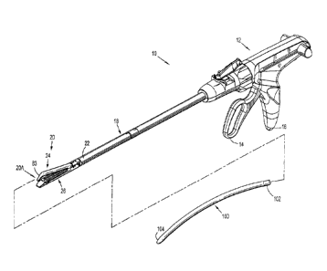
[0075] FIG. 1 illustrates a surgical fastener applying apparatus 10, of either
the re-usable
or disposable variety, that includes a handle assembly 12 with a movable
handle 14 and a
stationary handle 16, an elongated shaft 18 that extends distally from the
handle assembly 12, an
end effector 20 that is positioned at a distal end 22 of the elongated shaft
18, and an introducer
member 100 that is releasably connectable to the end effector 20.
[0076] In various embodiments, it is envisioned that the handle assembly 12
may include
motor-driven, hydraulic, ratcheting, or other such mechanisms to facilitate
actuation of the
surgical fastener applying apparatus 10.
[0077] In general, the end effector 20 is adapted to clamp, fasten together,
and sever
adjacent tissue segments along a cut-line. During use, the surgical fastener
applying apparatus
is approximated and fired similarly to, and in accordance with, other known
surgical fastener
applying apparatus. A discussion of the approximation and firing of surgical
fastener applying
apparatus 10, including the components and interaction of the handle assembly
12 and included
drive assembly, is provided below. However, additional details regarding
approximation and
firing of surgical fastener applying apparatus 10 may also be obtained through
reference to
commonly assigned U.S. Pat. No. 5,865,361, currently assigned to Tyco
Healthcare Group LP.
[0078] Referring now to FIGS. 2-6 as well, the end effector 20 includes a
first jaw 24 that
is pivotally coupled to a second jaw 26 to facilitate approximation thereof,
and is adapted to
14
CA 02716151 2010-09-30
clamp, fasten together, and sever adjacent tissue segments along a cut-line.
The first jaw 24 of
the end effector 20 includes an anvil component 28 comprising an anvil plate
30 (FIGS. 2, 3) and
an anvil cover 32 (FIG. 3), and the second jaw 26 includes a surgical fastener
cartridge 34 (FIG.
2) that is loaded with a plurality of surgical fasteners 36 (FIGS. 2, 4).
Pivoting the movable
handle 14 (FIG. 1) towards the stationary handle 16 approximates the first jaw
24 and the second
jaw 26. After the jaws 24, 26 are approximated, i.e., brought into close
operative alignment,
continued pivoting of the movable handle 14 ejects the plurality of surgical
fasteners 36 (FIGS.
2, 4) from the surgical fastener cartridge 34 (FIG. 2) such that the plurality
of surgical fasteners
36 are driven into the anvil plate 30, thus being formed into completed
surgical fasteners, as
described below. Further details regarding ejection of the surgical fasteners
36 are also provided
below.
[0079] It is envisioned that the surgical fastener cartridge 34 may be
removable and
replaceable with another loaded cartridge. In other embodiments, the present
disclosure
contemplates that the end effector 20 may constitute a component of a
removable and
replaceable loading unit for the surgical fastener applying apparatus 10.
[0080] With reference now to FIG. 4, each surgical fastener 36 loaded into the
surgical
fastener cartridge 34 (FIG. 2) includes two legs 38 that are connected by a
backspan 40
extending therebetween. The legs 38 extend from the backspan 40 to penetrating
ends 42 that
are configured and dimensioned to facilitate passage of the legs 38 through
tissue. The
dimensions of the backspan 40 and the legs 38 can be varied such that the
surgical fastener 36
may be used to fasten tissue with varying attributes, such as tissues of
different thickness, or
tissues including scar tissue.
CA 02716151 2010-09-30
[0081] The legs 38 and the backspan 40 may define a cross-section having any
suitable
geometric configuration including, but not limited to, rectangular, oval,
square, triangular,
trapezoidal, etc. The legs 38 and the backspan 40 may exhibit the same
geometrical
configuration, as shown in FIG. 4, or alternatively, the legs 38 and the
backspan 40 may exhibit
different geometrical configurations. For example, the legs 38 may exhibit a
rectangular cross-
section, whereas the backspan 40 may exhibit an oval cross-section.
[0082] The penetrating ends 42 of the legs 38 may be tapered to facilitate the
penetration
of tissue, or alternatively, the penetrating ends 42 may not include a taper.
In various
embodiments, it is envisioned that the penetrating ends 42 may define either a
conical surface, or
flat surface.
[0083] Prior to formation, the legs 38 of each surgical fastener 36 may extend
from the
backspan 40 such that they are substantially parallel. In the alternative,
however, the legs 38
may converge or diverge from the backspan 40.
[0084] With reference now to FIGS. 1-3 and 6 in particular, the anvil
component 28
(FIG. 3) will be discussed. As mentioned above, the anvil component 28
includes the anvil plate
30 and the anvil cover 32. The anvil plate 30 is an elongated member with a
tissue contacting
surface 44 (FIG. 5) that includes a plurality of pockets 46 formed therein.
Each of the pockets 46
is positioned to receive and deform the legs 38 (FIG. 4) of a surgical
fastener 36 to achieve a
formed configuration. More particularly, each pocket 46 formed in the anvil
component 28
includes two forming surfaces 48 (FIG. 5) that extend into the anvil component
28, i.e., away
from the tissue contacting surface 44, to define a depth "D," as best seen in
FIG. 6. Upon
engagement of the legs 38 with the forming surfaces 48, the forming surfaces
48 guide the legs
16
CA 02716151 2010-09-30
38 inwardly in the direction of arrows "A" (FIG. 5) to facilitate deformation
of the surgical
fastener 36 into a standard "B" shaped configuration (FIG. 7). In an
alternative embodiment, it
is envisioned that the pockets 46 formed in the tissue contacting surface 44
of the anvil plate 30
may be configured and dimensioned to deform the surgical fastener 36 so as to
achieve a single-
loop configuration or other shape upon formation. It should be appreciated
that additional
configurations and dimensions for the pockets 46 are also contemplated herein
such that the
surgical fasteners 36 may exhibit other configurations upon formation.
[0085] The pockets 46 are arranged into rows disposed on opposite sides of a
slot 50
(FIGS. 2, 5) extending through the anvil plate 30. The slot 50 is configured
to accommodate
movement of a knife 52 (FIG. 2), or other such cutting element, in order to
facilitate severing of
tissue along a cut-line. Although the slot 50 is depicted as extending
longitudinally through the
anvil plate 30, in alternative embodiments, it is envisioned that the slot 50
may define a
configuration that is angled, arcuate, or shaped otherwise. The slot 50 may
extend along a
centerline of the anvil plate 30, as shown in the embodiment illustrated in
FIGS. 2 and 5, or
alternatively, the slot 50 may be offset from the centerline of the anvil
plate 30.
[0086] In the embodiment of the anvil plate 30 seen in FIGS. 2 and 5, the
pockets 46
formed in the tissue contacting surface 44 are arranged into a pair of inner
rows 54A, a pair of
intermediate rows 54B, and a pair of outer rows 54C (FIG. 4). The pair of
inner rows 54A are
spaced laterally outward of the slot 50 and are closest thereto, the pair of
intermediate rows 54B
are spaced laterally outward from the pair of inner rows 54A, and the pair of
outer rows 54C are
spaced laterally outward from the pair of intermediate rows 54B and are
furthest from the slot
50. While the anvil plate 30 is depicted as including three pairs of rows,
i.e., the respective pairs
of inner, intermediate, and outer rows 54A, 54B, 54C, alternative embodiments
of the anvil plate
17
CA 02716151 2010-09-30
30 including fewer and greater numbers of rows of pockets 46 are not beyond
the scope of the
present disclosure.
[00871 Referring now to FIG. 3, the anvil cover 32 includes an outer surface
56 and is
fixed relative to the anvil plate 30 such that there is no relative movement
therebetween. For
example, it is envisioned that the anvil cover 32 may be secured to the anvil
plate 30 in a snap-fit
arrangement, via one or more welds, or in any other manner suitable for the
intended purpose of
establishing a secured connection therewith.
[00881 Second jaw 26 includes a surgical fastener cartridge 34 and a channel
66. The
surgical fastener cartridge 34 will now be described with reference to FIG. 2.
The surgical
fastener cartridge 34 includes a cartridge body 58 with a pair of sidewalls
60, a bottom wall 62,
and a top wall 64, and resides in the channel 66 of the second jaw 26. In the
illustrated
embodiment, the cartridge body 58 includes a slot 68 extending therethrough
that is configured
to accommodate longitudinal movement of the knife 52. As discussed above with
respect to the
anvil component 28, while the slot 68 is depicted as extending longitudinally
through the
surgical fastener cartridge 34, in alternative embodiments, the slot 68 may
define a configuration
that is angled, arcuate, or shaped otherwise. The position of the slot 68
corresponds to that of the
slot 50 extending through the anvil plate 30 such that the slot 68 aligns with
the slot 50. The slot
68 may extend along a centerline of the surgical fastener cartridge 34, as
seen in FIG. 3, or
alternatively, the slot 68 may be spaced therefrom.
[00891 The top wall 64 of the cartridge body 58 includes a substantially
planar
configuration that extends in substantially parallel relation to the tissue
contacting surface 44
(FIG. 5) of the anvil plate 30, as well as a plurality of retention slots 68.
The retention slots 68
18
CA 02716151 2010-09-30
are arranged into rows corresponding in position to the rows of pockets 46
(FIG. 5) formed in the
tissue contacting surface 44 of the anvil plate 30. Accordingly, in the
particular embodiment of
the surgical fastener cartridge 34 seen in FIG. 2, the retention slots 68 are
arranged into a pair of
inner rows 72A, a pair of intermediate rows 72B, and a pair of outer rows 72C,
each of which is
disposed on opposite sides of the slot 68. The pair of inner rows 72A are
spaced laterally
outward of the slot 68 and are closest thereto, the pair of intermediate rows
72B are spaced
laterally outward from the pair of inner rows 72A, and the pair of outer rows
72C are spaced
laterally outward from the pair of intermediate rows 72B and are furthest from
the slot 68. While
the surgical fastener cartridge 34 is depicted as including three pairs of
rows, i.e., the respective
inner, intermediate, and outer rows 72A, 72B, 72C, alternative embodiments of
the surgical
fastener cartridge 34 including fewer and greater numbers of rows of fastener
retention slots 68
are not beyond the scope of the present disclosure.
[0090] Each fastener retention slot 68 is configured and dimensioned to
receive a surgical
fastener 36 (FIGS. 2, 4), as well as a correspondingly dimensioned pusher 74
(FIG. 2) positioned
therein. During use, the pushers 74 are driven upwardly, i.e. towards the top
wall 64 of the
cartridge body 58, by a sled 76 (FIG. 3) into engagement with the surgical
fasteners 36 to
thereby eject the surgical fasteners 36 from the retention slots 68, as
discussed in further detail
below. As the surgical fasteners 36 exit the fastener retention slots 68, they
are deployed in
rows, e.g., inner, intermediate, and outer rows in the illustrated embodiment,
on opposite sides of
the cut-line created in the tissue.
[0091] With reference now to FIGS. 1 and 8-10, one embodiment of the presently
disclosed introducer member, which is identified by the reference character
100, will be
discussed. The introducer member 100 includes respective proximal and distal
ends 102, 104, and is
19
CA 02716151 2010-09-30
formed from a pliable, biocompatible material, including but not limited to
polymeric materials, such
as rubbers or plastics. In one particular embodiment, it is envisioned that
the introducer member
100 may be entirely formed from a flexible material. Alternatively, however,
it is envisioned
that the introducer member 100 may include portions of increased rigidity
formed from a higher
durometer material to provide additional structure to the introducer member
100, and/or assist in
the separation of tissue.
[00921 The open proximal end 102 of the introducer member 100 is configured
and
dimensioned to facilitate placement over a distal end 20A of the end effector
20. For example, as
illustrated in FIGS. 8 and 9, the proximal end 102 of the introducer member
100 may be
configured and dimensioned for placement about the anvil component 28, or
alternatively, the
proximal end 102 may be configured and dimensioned for placement about the
surgical fastener
cartridge 34. To facilitate connection and disconnection of the introducer
member 100 to the end
effector 20, it is envisioned that the proximal end 102 of the introducer 100
may include radiused
edges "E," as shown in FIG. 10.
[00931 In one embodiment, it is envisioned that the introducer member 100 may
be
operatively connected to the handle assembly 12 (FIG. 1) of the surgical
fastener applying apparatus
such that the introducer member 100 is steerable by the clinician. For
example, the introducer
member 100 may be operatively connected to the handle assembly 12 via one or
more flexible
members (not shown), such as cables, guidewires, or the like, that are
attached at various points along
the length of the introducer member 100 such that manipulation of the flexible
member(s) will cause
corresponding movement of the introducer member 100. For example, the flexible
member(s) may be
attached to the introducer member 100 such that proximal retraction of the
flexible member(s) causes
bending or flexing of the introducer member 100 at a predetermined location,
thereby allowing the
CA 02716151 2010-09-30
clinician to selectively reconfigure the introducer member 100, and
effectively steer the introducer
member 100 between the target tissue "T" (FIG. 10) and the collateral tissue
"C."
[0094] The introducer member 100 is configured, dimensioned, and adapted to
guide the end
effector 20 into position between the target tissue "T" (FIG. 10) that is the
subject of the surgical
procedure, and any collateral tissue "C" surrounding the target tissue "T." To
this end, the introducer
member 100 may include a partially or wholly curved configuration that defines
an arc
substantially in the range of approximately 5 to approximately 90 . In one
embodiment, for
example, as shown in FIGS. 8 and 9, the introducer member 100 curves from the
anvil component 28
towards the surgical fastener cartridge 34, and is longer than the jaws, so
that the target tissue is
approached at an angle and directed between the jaws of the surgical fastener
applying apparatus. It
should be appreciated, however, that the introducer member 100 may curve in
any direction suitable for
the intended purpose of guiding the instrument into position and/or
facilitating separation of the target
tissue "T" from any collateral tissue "C."
[0095] Additionally, the introducer member 100 includes an elongate profile
that tapers
towards the distal end 104, as seen in FIGS. 1 and 9-11. Including a tapered
profile allows the
distal end 104 of the introducer member 100 to define a reduced cross-
sectional area when
compared to more proximal portions thereof in order to facilitate advancement
of the introducer
member 100 through the patient's tissue. Additionally, the reduced cross-
sectional area at the
distal end 104 of the introducer member 100 facilitates access to internal
spaces and tissues that
would otherwise be inaccessible given the larger dimensions of the end
effector 20 (FIG. 1), and
reduces the effect that distal advancement of the surgical fastener applying
apparatus 10 has
upon the patient's internal tissues. For example, in one embodiment, it is
envisioned that the
distal end 104 of the introducer member 100 may have a transverse dimension in
the range of
21
CA 02716151 2010-09-30
approximately 2 mm to approximately 6 mm, although dimensions that are both
larger and
smaller are not beyond the scope of the present disclosure.
[0096] The introducer member 100 may have any cross-sectional configuration
suitable
for the intended purpose of atraumatically separating the target tissue "T"
(FIG. 10) from any
collateral tissue "C" in order to facilitate access to a patient's internal
tissues and organs with the
surgical fastener applying apparatus 10 (FIG. 1). For example, the introducer
member 100 may
have an oval cross-sectional configuration (see FIG. 13), a substantially
rounded cross-sectional
configuration, or a polygonal cross-sectional configuration.
[0097] Referring now to FIGS. 1-10, use and operation of the surgical fastener
applying
apparatus 10 (FIG. 1) will be discussed in connection with the introducer
member 100. As
mentioned above and seen in FIG. 1, the handle assembly 12 includes a movable
handle 14. The
movable handle 14 is operatively connected to an actuation shaft (not shown),
which receives the
proximal end of a control rod such that linear advancement of the actuation
shaft causes
corresponding linear advancement of the control rod. The control rod is
further engagable with
an axial drive assembly including an elongated drive beam 78 (FIG. 2) having a
distal end that is
configured and dimensioned to support the knife 52. The sled 76, as discussed
above, drives the
pushers 74 upwardly, driving the fasteners 36 against the pockets 46 of the
anvil component 28.
The knife 52 is positioned on the drive beam 78 to translate behind the sled
76. The drive beam
78 includes an upper flange 79A that is configured and dimensioned to move
along a top surface
of the anvil plate 30, and a lower flange 79B that is configured and
dimensioned for movement
along the outer side of the channel 66 while the drive beam moves through the
slot 50 of the
anvil component and the slot 68 of the channel 66.
22
CA 02716151 2010-09-30
[0098] Initially, the introducer member 100 is connected to the end effector
20 (FIG. 1, 8,
9), i.e., the introducer member 100 is positioned about the anvil component 28
or the surgical
fastener cartridge 34. Thereafter, the assembly of the surgical fastener
applying apparatus 10
(FIG. 1) and the introducer member 100 can be manipulated such that the distal
end 104 of the
introducer member 100 is positioned between the target tissue "T' (FIG. 10)
and the collateral tissue
"C." During manipulation of the introducer member 100, the flexible material
comprising the introducer
member 100 allows the introducer member 100 to gently urge the collateral
tissue "C" away from the
target tissue "T," thereby establishing and/or dilating a pathway along which
the end effector 20 can
travel.
[0099] To facilitate positioning of the target tissue "T" (FIG. 9) between the
anvil component
28 and the surgical fastener cartridge 34, the distal end 104 of the
introducer member 100 can be
withdrawn, or pulled proximally, e.g., via the aforementioned flexible
member(s) (not shown), if
included. Manipulating the introducer member 100 in this manner guides the
target tissue "T" into
position between the anvil component 28 and the surgical fastener cartridge
34, while simultaneously
protecting the collateral tissue "C" from undesirable contact with any
components of the surgical
fastener applying apparatus 10.
[00100] After confirming that that the target tissue "T" has been positioned
as desired between
the anvil component 28 and the surgical fastener cartridge 34, and confirming
that the collateral
tissue "C" is not located in a position that will result in damage to the
target tissue "T" or the collateral
tissue "C" upon actuation of the surgical fastener applying apparatus 10 (FIG.
1), the introducer member
100 can be removed from the end effector 20. Thereafter, the jaws 24, 26 (FIG.
1) are approximated
using the handle assembly 12 to clamp the target tissue "T" (FIG. 9)
therebetween, and apply a
compressive force thereto. Specifically, manipulation of the movable handle 14
advances the
23
CA 02716151 2010-09-30
actuation shaft to effectuate corresponding advancement of the control rod. In
particular
embodiments, the actuation shaft includes a toothed rack defined thereon, and
the movable
handle 14 has a ratcheting pawl mounted thereto for incrementally engaging and
advancing the
actuation shaft. The pawl may be mounted on a pivot pin and a coiled torsion
spring may be
provided to bias the pawl into engagement with the toothed rack. The control
rod is connected at
its distal end to the axial drive assembly, which includes the aforementioned
drive beam 78 (FIG.
2), such that distal movement of control rod effects distal movement of the
drive beam 78, which
in turn, forces the anvil component 28 (FIG. 2) towards the surgical fastener
cartridge 34.
Specifically, the control rod advances the drive beam 78 distally such that
the upper flange 79A
traverses the top surface of the anvil plate 30, and the lower flange 79B
traverses the channel 66
extending through the surgical fastener cartridge 34.
[00101] With the tissue securely clamped between the jaws 24, 26 (FIGS. 1, 2),
the
surgical fastener applying apparatus 10 is fired to eject the surgical
fasteners 36 (FIG. 2). The
surgical fastener applying apparatus 10 is approximated and fired similarly
to, and in accordance
with other known surgical fastener applying apparatus, such as, for example,
the surgical fastener
applying apparatus disclosed in commonly assigned U.S. Pat. No. 5,865,361,
which is currently
assigned to Tyco Healthcare Group LP. Specifically, the movable handle 14 is
manipulated to
cause advancement of the drive assembly, which causes the sled 76 (FIG. 2) to
traverse the
cartridge body 58 and engage the pushers 74 to thereby eject the plurality of
surgical fasteners 36
from the surgical fastener cartridge 34. During distal advancement of the sled
76, angled leading
surfaces thereof sequentially contact cam surfaces included on the pushers 74.
The interaction
between the leading surfaces of the sled 76 and the cam surfaces of the
pushers 74 urges the
pushers 74 towards the top wall 64 of the cartridge body 58 to eject the
surgical fasteners 36
24
CA 02716151 2010-09-30
from the retention slots 68 formed in the cartridge body 58. Sequential firing
of the surgical
fasteners 36 continues until the sled 76 is advanced to the distal end of the
surgical fastener
cartridge 34, at which time all of the surgical fasteners 36 housed within the
surgical fastener
cartridge 34 will have been ejected.
[00102] During ejection, the plurality of surgical fasteners 36 pass through
the retention
slots 68 through the target tissue "T," and into engagement with the pockets
46 (FIGS. 2, 5)
defined in the tissue contacting surface 44 of the anvil component 28.
Engagement of the
surgical fasteners 36 with the pockets 46 forms the surgical fasteners 36 to
thereby connect
adjacent portions of the target tissue "T" (FIG. 9).
[00103] Referring now to FIGS. 11-48, alternative embodiments of the presently
disclosed
introducer member will be discussed. Each embodiment of the introducer member
discussed
herein below is similar to the aforedescribed introducer member 100, and
accordingly, will only
be discussed with respect to any differences therefrom.
[00104] With specific reference to FIGS. 11-14, an embodiment of the
introducer member
identified by the reference character 200 will be discussed. The introducer
member 200 may be
entirely or partially hollow or solid, and includes an open proximal end 202,
and a closed distal end
204. When configured as a solid member, interior surfaces of the open proximal
end 202 may be shaped
to abut exterior surfaces of the anvil component 28 (FIG. 3) to provide
stability and support for the
introducer member 200.
[00105] To facilitate distal advancement and navigation of the introducer
member 200 between
the target tissue "T" (FIG. 9) and the collateral tissue "C," it is envisioned
that the introducer member
CA 02716151 2010-09-30
200 may include a dual taper. Specifically, the introducer member 200 may
include a tapered profile
that decreases in cross-sectional height and width from the proximal end 202
to the distal end 204.
[00106] The open proximal end 202 of the introducer member 200 is configured,
dimensioned,
and adapted to facilitate slidable engagement and disengagement with the end
effector 20 (FIGS. 1,
12). For example, the open proximal end 202 of the introducer member 200 may
be configured and
dimensioned to frictionally engage the surgical fastener cartridge 34, as
shown in FIG. 13, or
alternatively, the open proximal end 202 of the introducer member 200 may be
configured and
dimensioned to frictionally engage the anvil component 28 (FIG. 3), e.g., in a
manner similar to that
discussed above with respect to the introducer member 100 (FIGS. 1-11).
[00107] To ensure proper operation of the end effector 20 (FIGS. 1, 12)
without having to
remove the introducer member 200 prior to actuation of surgical fastener
applying apparatus 10 (FIG. 1),
it is envisioned that the portion of the introducer member 200 positioned
between the anvil component 28
(FIG. 12) and the surgical fastener cartridge 34 may have a thickness less
than the designed gap between
the anvil component 28 and the surgical fastener cartridge 34, thus inhibiting
interference with the anvil
component 28 upon actuation of the surgical fastener applying apparatus 10.
Additionally, or
alternatively, it is envisioned that the proximal end 202 of the introducer
member 200 may include a
cutout (not shown) that is positionable between the anvil plate 30 and the
surgical fastener cartridge 34.
[00108] To facilitate engagement with, and disengagement from, the end
effector 20 (FIGS. 1,
12), it is envisioned that the introducer member 200 may incorporate an
adhesive, or alternatively, that
the introducer member 200 may include structure that is configured and
dimensioned for engagement
with corresponding structure formed on the end effector 20. For example, the
introducer member 200
may include structure, e.g., projections 206 (FIG. 13), formed on opposing
surfaces within the proximal
26
CA 02716151 2010-09-30
end 202 that is configured and dimensioned for engagement with corresponding
structure on the end
effector 20, e.g., with notches 80 formed in the sidewalls 60 of the cartridge
body 58. Typically, the
notches 80 formed in the sidewalls 60 cartridge body 58 are configured and
dimensioned for engagement
with corresponding structure (not shown) on the second jaw 26 (FIG. 1) to
assist in secured placement
of the surgical fastener cartridge 34. In this embodiment, during use, the
introducer member 200 is
positioned about the distal end of the surgical fastener cartridge 34 until
the projections 206 within the
proximal end 202 of the introducer member 200 engage the notches 80. To remove
the introducer
member 200, the clinician applies a force sufficient to deform the proximal
end 202 of the introducer
member 200 such that the proximal end 202 flexes outwardly, thereby
disengaging the projections 206
from the notches 80. Subsequently, the introducer member 200 can be removed
from the surgical
fastener cartridge 34.
1001091 It is envisioned that that the introducer member 200 may include one
or more
recessed portion(s), cutouts, or the like (not shown) to inhibit interference
with movable structure
of the end effector 20, such as a flange 81 (FIG. 12) formed on the surgical
fastener cartridge 34.
In the illustrated embodiment of the surgical fastener cartridge 34, the
flange 81 is configured and
dimensioned for longitudinal, slidable movement towards the distal end of the
surgical fastener
cartridge 34 as the anvil component 28 is progressively clamped to balance
clamping forces generated
within the end effector 20.
[001101 Although each embodiment of the presently disclosed introducer has
been
discussed hereinabove as being a removable component of the surgical fastener
applying
apparatus 10 (FIG. 1), it is also envisioned that either of the introducer
members 100, 200 may
be integrally formed with the end effector 20, such that the introducer
members 100, 200 are not
detachable from the end effector 20.
27
CA 02716151 2010-09-30
[00111] As discussed above with respect to the introducer member 100 (FIGS. 1-
10), it is
envisioned that the introducer member 200 may be operatively connected to the
handle assembly 12
(FIG. 1) of the surgical fastener applying apparatus 10 via a linkage, e.g.,
cables, bands, or the like, to
facilitate selective reconfiguration of the introducer member 200.
[00112] In addition to being steerable, it is envisioned that the introducer
member 200 may be
biased towards a curved configuration (FIG. 12) relative to the component of
the end effector 20
(FIGS. 1, 12) to which the introducer member 200 is attached, e.g., the anvil
component 28 (FIG. 12)
or the surgical fastener cartridge 34, or alternatively, that the introducer
member 200 may be biased
towards a linear position (FIG. 12)
[00113] Referring now to FIGS. 13-15, an embodiment of the presently disclosed
introducer, which is identified by the reference character 300, will be
discussed. During certain
procedures, certain vasculature or other adherent connective, joined or other
tissue, adheres or is
joined with the target tissue, i.e., the tissue to be stapled and severed. If
such tissue must be
separated before accessing the target tissue, separating the tissue and
guiding the surgical
fastening apparatus into place so that the target tissue is directed between
the jaws, while the
collateral tissue is avoided, it is advantageous to include a dissector
portion or dissector tip 85 on
one of the jaws. In a certain preferred embodiment, the dissector tip 85 is a
rigid, tapered tip that
is integral with or attached to the anvil component of the surgical fastening
apparatus. Such a
dissector tip 85 can be seen in FIG. 16. The dissector tip 85 is desirably
curved or angled so as
to approach the surgical fastener cartridge 34 so that when tissue is
approached at an angle, the
tissue is directed between the jaws.
28
CA 02716151 2010-09-30
[001141 The introducer 300 includes a proximal end 302 defining a hollow 304
that is
configured and dimensioned to at least partially receive the dissector tip of
the anvil component
28 (FIGS. 2, 3). As illustrated in FIGS. 13-15, for example, the hollow 304 is
configured and
dimensioned to receive a portion of the anvil component 28, and more
specifically, an optional
dissector portion 83 or tip 85 of the anvil component 28 (FIGS. 1, 3, 15). The
introducer 300
provides a flexible, longer guide for the surgical fastener apparatus and
extends the access of the
apparatus, while more gradually guiding the apparatus toward the target
tissue. The introducer
300 is flexible, in contrast to the rigid dissector tip, to give the user more
options in how to
approach the tissue. If the user prefers, the introducer 300 can be attached
to the dissector tip, or
removed so that the dissector tip can be used to approach, access and separate
tissue while
guiding the tissue into place between the jaws.
[001151 To facilitate connection between the introducer member 300 and the end
effector
20 (FIG. 1), the proximal end 302 of the introducer member 300 includes
attachment structure
306 (FIG. 13) that is configured and dimensioned for releasable connection
with corresponding
engagement structure 82 (FIG. 15) included on the dissector portion of the end
effector 20 (FIG.
1), e.g., on the dissector portion 83 or tip 85 in the embodiment shown in
FIG. 16. To establish,
and maintain, connection with the end effector 20, it is envisioned that the
attachment structure
306 included at the proximal end 302 of the introducer member 300 may be
formed from a
material different than that comprising the remainder of the introducer member
300. In one
specific embodiment, for example, it is envisioned that the attachment
structure 306 may be
formed from a material having a higher durometer than that of the material
comprising the
remainder of the introducer member 300.
29
CA 02716151 2010-09-30
[00116] With continued reference to FIGS. 13-15, in one embodiment of the
present
disclosure, the attachment structure 306 (FIGS. 13, 14) and the engagement
structure 82 (FIG.
15) are configured and dimensioned for engagement in a snap-fit manner. More
specifically, the
attachment structure 306 includes one or more raised protrusions 308 that
extend inwardly into
the hollow 304, and the engagement structure 82 includes one or more
recess(es) 84 that
correspond in configuration and dimensions to the protrusion(s) 308. It is
contemplated that the
particular configuration and dimensions of the protrusion(s) 308 and the
recess(es) 84 may be
such that the clinician is provided with an audible, or tactile, indication
upon successful
connection of the introducer member 100 with the end effector 20.
[00117] The protrusion(s) 308 and the recess(es) 84 may have any geometrical
configuration suitable for the intended purpose of establishing a releasable
connection between
the introducer member 300 and the end effector 20 (FIG. 1). For example, it is
contemplated that
the protrusion(s) 308 may be configured as one or more raised ribs 310 (FIG.
13), and that the
recess(es) 84 may be configured as one or more corresponding linear channels
85 (FIG. 15).
Alternatively, the protrusion(s) 308 may be configured as one or more
hemispherical
protuberances 312 (FIG. 16), and the recess(es) 84 may be configured as one or
more
corresponding cavities 92 (FIG. 17).
[00118] Referring now to FIGS. 1 and 13-15, a method of fastening tissue with
the
surgical fastener applying apparatus 10 (FIG. 1) will be discussed in
connection with the
introducer member 300. Prior to inserting the surgical fastener applying
apparatus 10 (FIG. 1)
into the patient, the introducer member 300 (FIGS. 14, 15) is connected to the
end effector 20
(FIG. 1), e.g., to the dissector portion 83 (FIGS. 1, 3, 16) of the anvil
component 28 (FIG. 3).
Upon connection, the attachment structure 306 (FIGS. 14, 15) included at the
proximal end 302
CA 02716151 2010-09-30
of the introducer member 300 mates with the corresponding engagement structure
82 (FIG. 16)
included on the dissector portion 83 (FIG. 16), or other component of the end
effector 20.
[001191 Following assembly, the surgical fastener applying apparatus 10 (FIG.
1) is
inserted into, and advanced distally through, the patient's tissue. During
distal advancement, the
introducer member 300 atraumatically separates the patient's internal tissues
and organs to
provide the clinician with access to the target tissue "T" (FIG. 9). When the
target tissue "T" is
reached, the clinician may elect to remove the introducer member 300, e.g.,
through an incision,
or alternatively, the introducer member 300 may be left in place. When left in
place, the flexible
material comprising the introducer member 300 allows the introducer member 300
to bend and
flex during manipulation to accommodate movement of the introducer member 300
without
damaging the patient's internal tissues. The clinician then orients the
surgical fastener applying
apparatus 10 (FIG. 1) such that the target tissue "T" (FIG. 9) is disposed
between the open jaws
24, 26 (FIG. 1) of the end effector 20. After the target tissue "T" is
positioned as desired, the
jaws 24, 26 are approximated using the handle assembly 12 to clamp the target
tissue "T"
therebetween, and the surgical fastener applying apparatus 10 is fired to
thereby eject the
surgical fasteners 36 (FIG. 2) from the surgical fastener cartridge 34, as
discussed above. After
passing through the target tissue "T," the surgical fasteners 36 are formed
through engagement
with the anvil plate 30 to achieve a formed configuration, and thereby attach
adjacent portions of
the target tissue "T." During ejection of the surgical fasteners 36 from the
surgical fastener
cartridge 34, the target tissue "T" is simultaneously, or nearly
simultaneously, severed by the
aforementioned knife 52 (FIG. 2) which moves in concert with the drive beam 78
in a position
behind, i.e., proximally of, the sled 76.
31
CA 02716151 2010-09-30
[00120] Referring now to FIGS. 18 and 19, in an alternative embodiment of the
present
disclosure, a connector member 400 is provided to connect the surgical
fastener applying
apparatus 10 (FIG. 1), e.g., the anvil component 28, with the introducer
member 300 discussed in
connection with FIGS. 14 and 15.
[00121] The connector member 400 is formed from a biocompatible material
suitable for
contact with a patient's internal tissue during a surgical procedure, e.g.,
polymeric materials or
stainless steel, and includes respective proximal and distal ends 402, 404.
The connector
member 400 is configured and dimensioned for releasable engagement with the
anvil component
28, or other such component of the end effector 20, as well as the introducer
member 300, to
facilitate operative attachment of the introducer member 300 to the end
effector 20. Specifically,
the proximal end 402 of the connector member 400 is engagable with the end
effector 20, (FIG.
1), e.g., the dissector portion 83 of the anvil component 28, as shown in
FIGS. 18 and 19, such
that the connector member 400 extends distally from the anvil component 28,
and the distal end
404 of the connector member 400 is engagable with the proximal end 302 of the
introducer
member 300, such that the introducer member 300 extends distally from the
connector member
400.
[00122] As seen in FIG. 19, in one embodiment of the connector member 400, the
proximal end 402 thereof includes an internal space 406 that is configured and
dimensioned to at
least partially receive the dissector portion 83 of the anvil component 28. To
facilitate
connection between the connector member 400 and the anvil component 28, the
proximal end
402 of the connector member 400 includes proximal attachment structure 408
that is configured
and dimensioned for releasable connection with the aforedescribed engagement
structure 82
included on the dissector portion 83, which corresponds in configuration and
dimensions thereto.
32
CA 02716151 2010-09-30
Although illustrated as part of the dissector portion 83 in FIGS. 18 and 19,
as discussed above, it
should be appreciated that the engagement structure 82 may be associated with
any other portion
of the anvil component 28 (or the surgical fastener cartridge 34 (FIG. 2), in
alternative
embodiments of the present disclosure).
[00123] In the embodiment of the connector member 400 shown in FIGS. 18 and
19, the
proximal attachment structure 408 is illustrated as including one or more
raised protrusions 410
extending inwardly into the internal space 406 that is/are configured and
dimensioned for
engagement with the recess(es) 84 formed on the dissector portion 83 of the
anvil component 28.
In alternative embodiments of the present disclosure, however, the proximal
attachment structure
408 and the engagement structure 82 may assume any configuration or dimensions
suitable for
the intended purpose of creating a releasable connection between the connector
member 400 and
the end effector 20 (FIG. 1). As discussed above with respect to the
introducer member 100
(FIGS. 1-11), it is contemplated that the particular configuration and
dimensions of the
attachment structure 408 and the engagement structure 32 may be such that the
clinician is
provided with an audible, or tactile, indication upon successful connection of
the connector
member 400 to the end effector 20.
[00124] To facilitate engagement between the connector member 400 and the
introducer
member 100, the distal end 404 of the connector member 400 includes distal
attachment
structure 412 that is configured and dimensioned for partial or complete
reception by the hollow
304 (FIG. 19) included at the proximal end 302 of the introducer member 300.
[00125] In one embodiment, it is envisioned that the distal attachment
structure 412 may
include one or more protrusions (not shown), e.g., having an annular
configuration, that are
33
CA 02716151 2010-09-30
configured and dimensioned for engagement with one or more recesses (not
shown) formed in
the hollow 304 at the proximal end 302 of the introducer member 300 such that
the distal
attachment structure 412 is engagable with the proximal end 302 of the
introducer member 300
in a snap-fit, or friction-fit, arrangement. As mentioned in connection with
the attachment
structure 408 formed at the proximal end 402 of the connector member and the
engagement
structure 82 formed on the end effector 20 (FIG. 1), the distal attachment
structure 412 and the
hollow 304 may be configured and dimensioned so as to provide the clinician
with an audible, or
tactile, indication upon successful connection of the connector member 400 to
the introducer
member 300.
[001261 Alternatively, however, as seen in FIGS. 18 and 19, the distal
attachment structure
412 may include a series of tapered portions 414 that progressively decrease
in size to define
ridges 416 at the location where adjacent tapered portions 414 intersect. In
this embodiment, as
the distal attachment structure 412 is received by the hollow 304, the
flexible material
comprising the introducer member 300 allows the tapered portions 414 to expand
the proximal
end 302 of the introducer member 300 outwardly such that an interference fit
is created between
the proximal end 302 of the introducer member and the distal attachment
structure 412 of the
connector member 400. After expansion, the ridges 416 engage the inner surface
of the hollow
304 in a manner which maintains the position of the introducer 300 relative to
the connector
member 400 until disconnection is desired.
[001271 Referring now to FIGS. 1, 18, and 19, a method of fastening tissue
with the
surgical fastener applying apparatus 10 (FIG. 1) will be discussed in
connection with the
introducer member 300 and the connector member 400. Prior to inserting the
surgical fastener
applying apparatus 10 (FIG. 1) into the patient's tissue, the proximal end 402
of the connector
34
CA 02716151 2010-09-30
member 400 is attached to the end effector 20, e.g., the dissector portion 83
of the anvil
component 28, and the introducer member 300 is connected to the distal end 404
of the
connector member 400. Upon connection, the proximal attachment structure 408
of the
connector member 400 mates with the corresponding engagement structure 82
included on the
dissector portion 83, and the distal attachment structure 412 of the connector
member 400 is
inserted into the hollow 304 defined in the proximal end 302 of the introducer
member 300 such
that the ridges 416 engage an inner surface thereof to maintain the relative
positions of the
introducer member 300 and the connector member 400.
[00128] After assembly, the surgical fastener applying apparatus 10 (FIG. 1)
is inserted
into, and advanced distally through, the patient's tissue, during which time
the introducer
member 300 atraumatically separates the target tissue "T" (FIG. 9) from any
collateral tissue "C"
to provide access to the target tissue "T" with the surgical fastener applying
apparatus 10.
[00129] When the target tissue "T" is reached, the clinician may elect to
remove the
introducer member 300 and the connector member 400, or leave them in place, as
mentioned
previously. The clinician can then proceed with the grasping, fastening, and
cutting of the target
tissue "T" in accordance with the discussion above.
[00130] Referring now to FIG. 20, another embodiment of the presently
disclosed
introducer member, which is identified by the reference character 500, will be
discussed. The
introducer 500 includes a proximal end 502 and a distal end 504. The proximal
end 502 of the
introducer member 500 is formed from an at least partially resilient material,
and includes a Y-
shaped configuration with respective first and second branches 506, 508. The
branches 506, 508
are configured and dimensioned to facilitate releasable engagement with the
end effector 20 of
CA 02716151 2010-09-30
the surgical fastener applying apparatus 10 (FIG. 1), e.g., with either the
anvil component 28
(FIGS. 2, 3) or the surgical fastener cartridge 34 (FIG. 2). Specifically, the
branches 506, 508 of
the introducer member 500 include attachment structure 510 that is configured
and dimensioned
for releasable connection with the engagement structure 82 included on the end
effector 20.
While the engagement structure 82 is shown as formed on the anvil component 28
of the end
effector 20 in the illustrated embodiment, it should be appreciated that the
attachment structure
510 may alternatively be configured and dimensioned for engagement with
structure formed on
the surgical fastener cartridge 34 (FIG. 2). Additionally, although the
branches 506, 508 of the
introducer member 500 are illustrated as being configured and dimensioned to
facilitate
connection with engagement structure 82 that is positioned on side portions 87
of the end
effector 20, in alternative embodiments, it is envisioned that the engagement
structure 82 may be
included on upper and lower portions of the end effector 20, and that the
branches 506, 508 of
the introducer member 500 may be configured and dimensioned accordingly.
[00131] In the embodiment of the introducer 500 illustrated in FIG. 20, the
attachment
structure 510 included on the branches 506, 508 includes a pair of protrusions
512 that are
configured as arcuate detents 514, and the engagement structure 82 included on
the anvil
component 28 is configured as recess(es) 84 of corresponding configuration and
dimensions. It
should be appreciated, however, that in alternative embodiments of the present
disclosure, the
attachment structure 510 and the engagement structure 82 may assume other
geometrical
configurations without departing from the scope of the present disclosure.
[00132] The distal end 504 of the introducer member 500 includes an elongate
profile that
is configured and dimensioned to facilitate atraumatic distal advancement of
the introducer
member 500 through the patient's tissue. The distal end 504 includes a
flexible, resilient
36
CA 02716151 2010-09-30
material that allows the distal end 504 to bend and flex when necessary so as
to permit the
clinician to navigate the introducer member 500 around the target tissue "T"
(FIG. 9) and the
collateral tissue "C" without causing unnecessary trauma.
[00133] Upon application of the introducer member 500 to the anvil component
28, the
attachment structure 510 rides along an outer surface of the end effector 20,
which causes the
branches 506, 508 to splay slightly outward in the direction indicated by
arrows 1. However,
upon positioning of the attachment structure 510 within the engagement
structure 82, the resilient
material comprising the proximal end 502 allows the branches 506, 508 to
return to their normal
configuration, whereby the branches 506, 508 are caused to engage the end
effector 20 in press-
fit arrangement. To disconnect the introducer 500 from the end effector 20,
the clinician applies
a predetermined force to the introducer member 500 to separate the attachment
structure 510
from the engagement structure 82. For example, the clinician can pull the
introducer member
500 distally, or rotate the introducer member 500 relative to the end effector
20, to force the
detents 514 from the recess(es) 84. more
[00134] In an alternative embodiment of the introducer member 500, which is
shown in
FIG. 21, it is envisioned that the branches 506, 508 of the introducer member
500 may further
include pivot members 516, in addition to the attachment structure 510, to
facilitate separation of
the introducer 500 from the end effector 20. In this embodiment, when
disengagement of the
introducer 500 from the end effector 20 is desired, a force is applied to the
branches 506, 508
about the pivot members 516 that is directly inwardly, i.e., in the direction
identified by arrows
2. This application of force to the branches 506, 508 about the pivot members
516 causes the
branches 506, 508 to bend about the pivot members 516 such that more proximal
portions of the
branches 506, 508 splay outwardly, i.e., in the direction indicated by arrows
1, thereby releasing
37
CA 02716151 2010-09-30
the attachment structure 510 from the engagement structure 82, and allowing
for removal of the
introducer member 500.
[00135] Referring again to FIG. 20, it is envisioned that the proximal end 502
of the
introducer member 500, including the attachment structure 510, may be formed
from a first
material, whereas the distal end 504 of the introducer member 500 may be
formed from a
second, different material. For example, the first material may have a higher
durometer than the
second material. Incorporating a material with a higher durometer at the
proximal end 502 of the
introducer 500, as opposed to a more compliant material, facilitates a more
secure connection
between the attachment structure 510 and the engagement structure 82. The
material comprising
the distal end 504 of the introducer 500, while more flexible than the
material comprising the
proximal end 502, is sufficiently rigid to facilitate separation of the target
tissue "T" (FIG. 9)
from the collateral tissue "C" in the manner discussed above.
[00136] In one embodiment of the disclosure, it is envisioned that the distal
end 504 of the
introducer member 500 may incorporate a material having a highly visible
color, such as red,
green, yellow, etc., in order to increase visibility of the introducer member
500 within an internal
workspace.
[00137] Referring now to FIGS. 22-24, in one embodiment of the present
disclosure, it is
envisioned that the dissector portion 83 of the anvil component 28 may
comprise a tip 600 that is
secured between the anvil plate 30 (FIGS. 23, 24) and the anvil cover 32 (FIG.
22). As seen in
FIG. 23, the tip 600 includes an arcuate distal portion 602 that is configured
and dimensioned to
facilitate separation of the target tissue "T" (FIG. 9) from any collateral
tissue "C." The tip 600
38
CA 02716151 2010-09-30
may be formed from any suitable biocompatible material, including but not
limited to polymeric
materials, through any suitable process, such as molding.
[00138] The tip 600 is secured to the anvil plate 30 via a post 604, rivet, or
the like, that
extends through corresponding openings (not shown) formed in the tip 600 and
the anvil plate
30. Following placement of the post 604 through the tip 600 and the anvil
plate 30, the anvil
cover 32 (FIG. 22) is secured to the anvil plate 30 in the aforedescribed
manner, e.g., via one or
more welds.
[00139] Referring now to FIGS. 25-27, an alternative embodiment of the tip,
which is
identified by the reference character 600', will be discussed. The tip 600' is
substantially similar
to the tip 600 discussed with respect to FIGS. 22-24, and accordingly, will
only be described
with respect to any differences therefrom.
[00140] The tip 600'is inserted into the anvil component 28 through an opening
92 (FIG.
25) formed in a distal end 28A thereof. As discussed above with respect to the
tip 600 (FIGS.
22-24), when properly positioned relative to the anvil component 28, the tip
600' is oriented
between the anvil plate 30 (FIGS. 26, 27) and the anvil cover 32 (FIG. 25). In
lieu of the post
604 (FIG. 24) discussed above, or in addition thereto, the tip 600' includes a
tab 606 that is
configured and dimensioned for positioning within the slot 50 formed in the
anvil plate 30 to
inhibit inadvertent disconnection of the tip 600' from the anvil component 28.
Specifically, the
tab 606 is configured and dimensioned for engagement with a distal wall 93
(FIGS. 25, 26)
defined by the slot 50. Engagement of the tab 606 and the distal wall 93 of
the slot 50 prevents
distal movement of the tip 600' relative to the anvil component 28 beyond a
predetermined
location.
39
CA 02716151 2010-09-30
[00141] To further facilitate separation of the target tissue "T" (FIG. 9)
from any collateral
tissue "C," it is envisioned that the tip 600 (FIGS. 22-24), 600' (FIGS. 25-
27) may be used in
conjunction with any embodiment of the presently disclosed introducer member
described
herein, such as, for example, the introducer member identified by the
reference character 700 in
FIG. 28.
[00142] The introducer member 700 includes a proximal end 702 defining a
hollow 704
that is configured and dimensioned to facilitate attachment with the distal
end 28A of the anvil
component 28 (see FIG. 25). It should be appreciated, however, that the hollow
704 may be
configured and dimensioned for engagement with the surgical fastener cartridge
34 (FIG. 2) in
alternative embodiments of the introducer member 700. To facilitate attachment
of the
introducer member 700 to the anvil component 28, the introducer member 700
includes
attachment structure 706 that is positioned on an internal wall 708 of the
proximal end 702. The
attachment structure 706 extends into the hollow 704, and includes a plurality
of protrusions 710.
Although the protrusions 710 are illustrated as including a configuration
defining a plurality of
teeth 712, it should be appreciated that alternative configurations for the
protrusions 710 are not
beyond the scope of the present disclosure, e.g., protrusions with an arcuate
configuration.
[00143] To further enhance the connection between the introducer member 700
and the
anvil component 28, it is contemplated that the anvil component 28, e.g., the
dissector portion
83, may include a roughened surface 88. While the roughened surface 88 has
been illustrated as
formed on the anvil component 28 in the illustrated embodiment, the roughened
surface 88
alternatively be formed on the surgical fastener cartridge 34 (FIG. 2) in
order to enhance the
connection between the introducer member 700 and the surgical fastener
cartridge 34, when
desired. The roughened surface 88 may include any structure suitable for the
intended purpose
CA 02716151 2010-09-30
of facilitating attachment with the proximal end 702 (FIG. 28) of the
introducer member 700.
For example, the roughened surface 88 may include a surface coating 89, as
shown in FIG. 29, or
alternatively, the roughened surface 88 may include a glass-bit surface finish
90, as shown in
FIG. 30.
[00144] To facilitate insertion of the anvil component 28 into the hollow 704
defined in
the proximal end 702 of the introducer member 700, it is envisioned that the
anvil component 28
may include a polished tip 91, as also shown in FIG. 30. The polished tip 91
serves to reduce
friction between the anvil component 28 and the proximal end 702 of the
introducer member 700
in order to allow for sufficient advancement of the anvil component 28 into
the hollow 704 to
facilitate engagement of the attachment structure 706 with the roughened
surface 88.
[00145] With reference now to FIGS. 31-33, an embodiment of the presently
disclosed
introducer member is illustrated, which is identified by the reference
character 800. The
introducer member 800 includes a proximal end 802 and a distal end 804, and is
configured and
dimensioned for longitudinal movement relative to the end effector 20 (FIG.
1), e.g., the anvil
component 28. More specifically, the introducer member 800 is configured and
dimensioned for
slidable movement through a slot 94 formed in the anvil cover 32 such that the
introducer
member 800 is selectively repositionable between a retracted position (FIGS.
31, 32) and an
advanced position (FIG. 33). In the retracted position, the distal end 804 of
the introducer
member 800 is positioned proximally of a distal-most tip 95 of the anvil
component 28, and in
the advanced position, the distal end 804 of the introducer member 800 is
positioned distally of
the distal-most tip 95 of the anvil component 28 to facilitate separation of
the target tissue "T"
(FIG. 9) from any collateral tissue "C."
41
CA 02716151 2010-09-30
[00146] To facilitate movement between the retracted and advanced positions,
it is
envisioned that the introducer member 800 may include a manual member 806 that
is configured
and dimensioned for engagement by the clinician. In one embodiment of the
introducer member
800, the manual member 806 extends upwardly from the introducer member 800,
and may be
positioned at the proximal end 802 of the introducer member 800, as shown in
FIGS. 31-33, or in
any other suitable location. Alternatively, the manual member 806 may
constitute a roughened
surface (not shown) which does not extend upwardly from the introducer member
800, but
rather, extends along the plane of the anvil cover 32 (FIG. 3).
[00147] In order to increase the clinician's ability to manipulate the
introducer member
800, it is envisioned that the introducer member 800 may be connected to the
end effector 20
(FIG. 1) so as to allow for lateral movement thereof, e.g., side-to-side, as
well as longitudinal
movement. For example, as shown in FIGS. 34 and 35, the introducer member 800
may be
connected to the anvil component 28 via a pivot member 808 such that the
introducer member
800 is movable in the directions indicated by arrows 1 and 2. Allowing for
both longitudinal and
lateral movement provides the clinician with increased access to internal
spaces, which allows
the clinician to more accurate place the introducer member 800during
navigation around the
patient's internal tissues.
[00148] FIGS. 36-38 illustrate another embodiment of the introducer member,
which is
identified by the reference character 900. The introducer member 900 includes
a pair of flexible
wings 902 with feet 904 that are configured and dimensioned to securely engage
the anvil
component 28, e.g., the anvil cover 32. For example, it is envisioned that the
feet 904 may be
configured and dimensioned to engage the anvil component 28 in snap-fit
relation. The
introducer member 900 further includes a rail 906 (FIGS. 37, 38) that is
configured and
42
CA 02716151 2010-09-30
dimensioned for positioning within the slot 50 formed in the anvil plate 30.
The rail 906
includes a wall 908 that is configured and dimensioned for engagement with the
distal wall 93
defined by the slot 50 to inhibit distal movement of the introducer member 900
relative to the
anvil component 28 beyond a predetermined location in order to reduce the
likelihood of
inadvertent disconnection therefrom. After the anvil component 28 is in the
desired position
adjacent the target tissue "T" (FIG. 9), prior to firing of the surgical
fastener applying apparatus
(FIG. 1), the introducer member 900 can be removed from the anvil component 28
by
disengaging the feet 904 from the anvil cover 32. For example, the wings 902
can be deformed
laterally outward, e.g., by rotating the introducer member 900 relative to the
anvil component 28.
[00149] FIGS. 39 and 40 illustrate another embodiment of the presently
disclosed
introducer that is identified by the reference character 1000. The introducer
member 1000 is
mounted to the anvil component 28 by a pivot member 1002 that extends through
the introducer
1000 and one or more pivot plates 1004 that are secured to an outer surface of
the end effector
20. The pivot member 1002 may be any member suitable for the intended purpose
of facilitating
lateral pivotal movement of the introducer member 1000, i.e., side-to-side in
the direction
indicated by arrows 1 (FIG. 39), including but not limited to screws, rivets,
or the like.
[00150] It is envisioned that the introducer member 1000 may be utilized in
connection
with end effectors 20 of various configurations. For example, it is envisioned
that the introducer
member 1000 may be employed in connection with an anvil component 28 having a
substantially
linear configuration (see FIG. 41), or alternatively, with an anvil component
28 that includes the
aforementioned dissector portion 83, as shown in FIG. 40.
43
CA 02716151 2010-09-30
[001511 Another embodiment of the introducer member, which is identified by
the
reference character 1100, is illustrated in FIGS. 41 and 42. The introducer
member 1100
includes a proximal end 1102 with a collar 1104 that is configured and
dimensioned for
positioning about the anvil cover 32, and a distal portion 1106 that is
configured and
dimensioned to facilitate separation of the target tissue "T" (FIG. 9) from
any collateral tissue
"C." The collar 1104 is configured and dimensioned to engage the anvil
component 28 such that
relative longitudinal movement therebetween is substantially inhibited. For
example, it is
envisioned that the collar 1104 may comprise an at least partially resilient
material allowing the
collar 1104 to be deformed outwardly upon association with the anvil component
28 in order to
facilitate a snap-fit connection with the anvil cover 32.
[001521 To further enhance stability of the connection between the introducer
1100 and
the anvil component 28, it is envisioned that the distal portion 1106 of the
introducer member
1100 may include one or more tabs 1108, snaps, protrusions, or the like that
are configured and
dimensioned for engagement with corresponding structure in the anvil cover 32,
as shown in
FIG. 41.
[001531 With reference now to FIG. 43, another embodiment of the introducer
member,
which is identified by the reference character 1200, will be discussed. The
introducer member
1200 includes a proximal end 1202 incorporating attachment structure 11204, a
distal end 1206
including an atraumatic tip portion 1208, and an elongate body portion 1210.
As illustrated in
FIG. 43, the elongate body portion 1210 of the introducer member 1200 may
include a tapered
configuration to facilitate separation of the target tissue "T" (FIG. 9) from
any collateral tissue
C
44
CA 02716151 2010-09-30
[001541 The proximal end 1202 of the introducer member 1200 is configured and
dimensioned for insertion into the opening 92 formed at the distal end 28A of
the anvil
component 28 such that the introducer member 1200 is positionable between the
anvil cover 32
(FIG. 3) and the anvil plate 30. To facilitate attachment of the introducer
member 1200 to the
anvil component 28, it is envisioned that the proximal end 1202 of the
introducer member 1200
and the distal end 28A of the anvil component 28 may include corresponding
structure that is
configured and dimensioned for mating engagement. For example, in the
embodiment shown in
FIG. 43, it is envisioned that the attachment structure 1204 included at the
proximal end 1202 of
the introducer member 1200 may be configured and dimensioned for connection
with
corresponding engagement structure 96 formed within the opening 92 at the
distal end 28A of
the anvil component 28 in a press-fit arrangement. In the illustrated
embodiment, the attachment
structure 1204 is depicted as including a plurality of protrusions 1212, and
the engagement
structure 96 is depicted as including recesses 97 of corresponding
configuration and dimensions.
It should be appreciated, however, that alternative configurations for the
attachment structure
1204 and the engagement structure 96 are not beyond the scope of the present
disclosure.
[001551 Although the introducer member 1200 is illustrated as including a
configuration
similar to that of the dissector portion 83 (FIGS. 1, 3, 15), in alternative
embodiments of the
present disclosure, it is envisioned that the introducer member 1200 may
assume other
configurations. For example, as seen in FIG. 43A, the introducer member 1200
may include a
more elongate configuration similar to that of the introducer member 100
discussed above with
respect to FIGS. 1-10.
[001561 Referring now to FIG. 44, in one embodiment, the attachment structure
1204
included at the proximal end 1202 of the introducer member 1200 may include a
stem 1214 with
CA 02716151 2010-09-30
a head portion 1216 attached thereto that is configured and dimensioned for
positioning within
the opening 92 (FIG. 43) formed at the distal end 28A of the anvil component
28. To facilitate
retention of the attachment structure 1204 within the opening 92, the
engagement structure 96
included at the distal end 28A of the anvil component 28 may include a pair of
shoulders 98
defining a gap "G" (FIG. 45) therebetween. The transverse dimension of the gap
"G" is larger
than the transverse dimension of the stem 1214, but smaller than the
transverse dimension of the
head portion 1216, in order to prevent inadvertent disconnection of the
introducer member 1200
from the anvil component 28.
[001571 To facilitate advancement of, and control over, the introducer member
1200
during manipulation within an internal workspace, it is envisioned that the
distal end 28A of the
anvil component 28 may be formed from a first material, e.g., a rigid
material, such as solid or
sheet metal, and that the introducer member 1200 may be formed from a second,
more flexible
material, such as plastic or rubber. To further enhance stability of the
connection between the
introducer member 1200 and the anvil component 28, it is envisioned that the
attachment
structure 1204 included at the proximal end 1202 of the introducer member 1200
may be heat-
staked to the distal end 28A of the anvil component 28.
[001581 FIG. 46 illustrates another embodiment of the presently disclosed
introducer
member, which is identified by the reference character 1300, that includes a
proximal portion
1302 and a distal portion 1304. The proximal portion 1302 of the introducer
member 1300
includes an outer layer 1306 and an inner layer 1308 which collectively define
a cavity 1310
therebetween, as well as a hollow 1312 that is configured and dimensioned to
receive the end
effector 20 (FIG. 1), e.g., the anvil component 28 (FIG. 2, 3) or the surgical
fastener cartridge 34
(FIG. 2). The introducer member 1300 further includes a passage 1314 extending
longitudinally
46
CA 02716151 2010-09-30
therethrough that is in fluid communication with the cavity 1310 and a port
1316 included on the
distal portion 1304 of the introducer member 1300. The passage 1314 allows for
the
communication of a fluid, such as air or saline, through the port 1316 and
into the cavity 1310 to
thereby fill and inflate the cavity 1310. It is envisioned that the port 1316
may include a seal
(not shown), valve, or the like to inhibit the escape of fluid from the cavity
1310 and/or the port
1316 until such time that deflation of the cavity 1310 is desired.
[00159] Prior to inflation of the cavity 1310, the hollow 1312 at the proximal
end 1302 of
the introducer member 1300 defines a transverse dimension that is smaller than
the transverse
dimension defined by the component of the end effector 20 to which the
introducer member
1300 is to be attached, e.g., a transverse dimension that is smaller than that
of the anvil
component 28 or the surgical fastener cartridge 34. However, upon inflation of
the cavity 1310,
as the cavity 1310 fills with fluid, the transverse dimension of the hollow
1312 is increased such
that the end effector 20, or component thereof, is positionable within the
hollow 1312.
Following connection of the proximal end 1302 of the introducer member 1300 to
the end
effector 20, the clinician can optionally deflate the cavity 1310 by draining
the inflation fluid. It
is also contemplated herein that the fluid within the cavity 1310 may exert
pressure upon the
outer layer 1306 and the inner layer 1308, and that this pressure may be
transmitted to an outer
surface of the end effector 20 (FIG. 1), thus assisting in retention of the
proximal portion 1302 of
the introducer member 1300 about the end effector 20.
[00160] In order to accommodate expansion and contraction of the cavity 1310,
it is
envisioned that either or both of the outer layer 1306 and the inner layer
1308 comprising the
proximal portion 1302 of the introducer member 1300 may be formed from a
resilient material,
e.g., materials with a relatively low durometer, such as shrink tubing.
47
CA 02716151 2010-09-30
[00161] FIG. 47 illustrates another embodiment of the presently disclosed
introducer
member, which is identified by the reference character 1400. The introducer
member 1400
includes a proximal portion 1402 including a selectively deformable section
1404, and a distal
portion 1406 that is positionable about the deformable section 1404.
[00162] The deformable section 1404 includes a resilient member 1408 that is
formed
from a deformable, e.g., malleable, material that can be manually reconfigured
by the clinician in
order to assume a desired geometrical configuration. For example, it is
envisioned that the
resilient member 1408 may include a flexible shaft 1410 that is connected,
either directly or
operatively, to the end effector 20, as shown in FIG. 47. In the illustrated
embodiment the
flexible shaft 1410 is connected to the end effector 20 via attachment
structure 1412 formed,
either integrally or releasably, with the end effector 20, e.g., between the
anvil plate 30 (FIG. 3)
and the anvil cover 32.
[00163] The distal portion 1406 of the introducer member 1400 is formed from a
material
that is at least partially resilient in order to facilitate atraumatic
advancement of the introducer
member 1400 during a surgical procedure, such that introducer member 1400 can
be effectively
navigated about the target tissue "T" (FIG. 9) and any collateral tissue "C"
without causing
unnecessary trauma. The distal portion 1406 includes an internal cavity 1414
that is configured
and dimensioned to receive the deformable section 1404. The distal portion
1406 may be
attached to the proximal portion 1402 in any suitable manner, such as, for
example, through the
incorporation of a resilient material. Incorporating a resilient material
allows the distal portion
1406 to be forced over the deformable section 1404 and/or the attachment
structure 1412 such
that the proximal portion 1402 and the distal portion 1406 engage one another
in a force-fit
arrangement. Alternatively, it is envisioned that the proximal portion 1402
and the distal portion
48
CA 02716151 2010-09-30
1406 may include corresponding structure that is configured and dimensioned
for releasable,
mating engagement, such as that described in connection with several of the
embodiments of the
introducer member discussed herein above.
[00164] Although the introducer member 1400 is illustrated as including an
elongate
configuration similar to that of the introducer member 100 discussed above
with respect to FIGS.
1-10, in alternative embodiments of the present disclosure, it is envisioned
that the introducer
member 1400 may assume other configurations. For example, as seen in FIG. 49,
the introducer
member 1200 may include a configuration similar to that of the dissector
portion 83 (FIGS. 1, 3,
15).
[00165] While each embodiment of the presently disclosed introducer member has
been
discussed and illustrated in connection with the end effector 20 (FIG. 1),
which is adapted for use
in laparoscopic procedures for performing surgical anastomotic fastening of
tissue, the introducer
member may be adapted for use with any surgical instrument suitable for the
intended purpose of
applying the plurality of surgical fasteners 36 (FIG. 2) to a section of
tissue, and thereafter
severing the tissue along a cut-line.
[00166] The presently disclosed introducer member may alternatively be adapted
for use
with a surgical stapling apparatus 3000 (FIG. 49), such as that disclosed in
commonly assigned
U.S. Pat. No. 7,334,717, currently assigned to Tyco Healthcare Group LP. The
surgical stapling
apparatus 3000 includes a cartridge receiving half-section 3002, which
accommodates a plurality
of surgical fasteners, and an anvil half-section 3004. The half-sections 3002,
3004 are pivotally
connected via handles 3006, 3008 for approximation during use.
49
CA 02716151 2010-09-30
[00167) Following approximation of the half-sections 3002, 3004, the surgical
fastener
applying apparatus 3000 is fired by driving a firing slide 3010 distally
through the advancement
of a firing lever 3012. Distal movement of the firing slide 3010 causes a
plurality of cam bars to
engage camming surfaces that interact with a plurality of pushers to expel the
plurality of
surgical fasteners from the cartridge receiving half-section 3002. The
surgical fasteners are
positioned on either side of a track which guides a knife during longitudinal
movement to
thereby sever tissue along a cut-line.
[001681 It is also envisioned that the presently disclosed introducer member
may be
adapted for use with a transverse anastomosis fastening instrument 4000 (FIG.
50), such as that
disclosed in commonly owned U.S. Pat. No. 5,964,394, currently assigned to
United States
Surgical Corporation. The surgical fastener applying apparatus 4000 includes
an approximation
lever 4001, a movable handle 4002, an elongated portion 4004 that extends
distally from the
handle 4002, and an arm 4006 that extends from a distal end 4008 of the
elongated portion 4004.
The surgical fastener applying apparatus 4000 further includes a end effector
4010 that includes
an anvil 4012 that is orthogonally affixed to the arm 4006, and a surgical
fastener cartridge
receiver 4014 that is operatively coupled to the distal end 4008 of the
elongated portion 4004 for
retention of the surgical fastener cartridge 200.
[001691 Prior to firing of the surgical fastener applying apparatus 4000, the
approximation
lever 4001 is actuated to distally advance a drive member that is operatively
connected to the
surgical fastener cartridge 200 to move the surgical fastener cartridge 200
towards the anvil
4012, which remains stationary, and capture tissue therebetween. Thereafter,
the handle 4002 is
moved to advance a pusher bar distally through the elongated portion 4004 to
cause
corresponding movement of a head portion included at the distal end of the
pusher bar. The head
CA 02716151 2010-09-30
portion includes a plurality of fingers extending distally therefrom that are
configured and
dimensioned to engage the cartridge assembly to thereby discharge the
plurality of surgical
fasteners retained therein. Upon discharge, the surgical fasteners are driven
through the tissue
and into the anvil 4012 for formation.
[001701 Additionally, it is envisioned that the presently disclosed introducer
member may
be adapted for use with any of the other surgical fastener applying apparatus
discussed in
commonly owned U.S. Pat. Nos. 6,045,560; 5,964,394; 5,894,979; 5,878,937;
5,915,616;
5,836,503; 5,865,361; 5,862,972; 5,817,109; 5,797,538; and 5,782,396.
[001711 In certain embodiments of the present disclosure, it is envisioned
that the
disclosed surgical fastener applying apparatus may include a plurality of cam
bars for interacting
with the pushers to deploy the surgical fasteners. For example, the apparatus
disclosed in
commonly owned U.S. Pat. No. 5,318,221, includes a cam bar adapter that holds
a plurality of
cam bars and a knife, as well as a channel that is advanced through operation
of the handle of the
apparatus, which drives the cam bars and knife forward. To clamp the anvil and
the surgical
fastener cartridge together, the apparatus further includes a clamp tube that
is movable to
surround the proximal end of the anvil.
[001721 As another example, the apparatus disclosed in U.S. Pat. No.
5,782,396, includes
an actuation sled and an elongated drive beam that is advanced distally
through operation of the
handle of the apparatus, driving the actuation sled forward. In this
apparatus, the distal end of
the drive beam engages the anvil and the channel that supports the surgical
fastener cartridge as
the drive beam travels distally to deploy the staples and clamp the anvil and
surgical fastener
cartridge together.
51
CA 02716151 2010-09-30
[00173] Persons skilled in the art will understand that the devices and
methods specifically
described herein and illustrated in the accompanying figures are non-limiting
exemplary
embodiments, and that the description, disclosure, and figures should be
construed merely
exemplary of particular embodiments. It is to be understood, therefore, that
the present
disclosure is not limited to the precise embodiments described, and that
various other changes
and modifications may be effected by one skilled in the art without departing
from the scope or
spirit of the disclosure. Additionally, it is envisioned that the elements and
features illustrated or
described in connection with one exemplary embodiment may be combined with the
elements
and features of another without departing from the scope of the present
disclosure, and that such
modifications and variations are also intended to be included within the scope
of the present
disclosure. Accordingly, the invention is not to be limited by what has been
particularly shown
and described, except as indicated by the appended claims.
52