Note: Descriptions are shown in the official language in which they were submitted.
CA 02722366 2010-10-22
WO 2009/132187
PCT/US2009/041536
1
STENTED HEART VALVE DEVICES
TECHNICAL FIELD
The present invention relates generally to devices and methods for
repair of heart valves, and more particularly to prosthetic heart valves for
use in
replacement of the mitral valve.
One of the two atrio-ventricular valves in the heart is the mitral valve,
which is located on the left side of the heart and which forms or defines a
valve
annulus and valve leaflets. The mitral valve is located between the left
atrium and
the left ventricle, and serves to direct oxygenated blood from the lungs
through the
left side of the heart and into the aorta for distribution to the body. As
with other
valves of the heart, the mitral valve is a passive structure in that it does
not itself
expend any energy and does not perform any active contractile function.
The mitral valve includes two moveable leaflets that open and close in
response to differential pressures on either side of the valve. Ideally, the
leaflets
move apart from each other when the valve is in an open position, and meet or
"coapt" when the valve is in a closed position. However, problems can develop
with valves, which can generally be classified as either stenosis, in which a
valve
does not open properly, or insufficiency (also called regurgitation), in which
a
valve does not close properly. Stenosis and insufficiency may occur
concomitantly in the same valve. The effects of valvular dysfunction vary,
with
mitral regurgitation or backflow typically having relatively severe
physiological
consequences to the patient. Regurgitation, along with other abnormalities of
the
mitral valve, can increase the workload placed on the heart. The severity of
this
increased stress on the heart and the patient, and the heart's ability to
adapt to it,
determine the treatment options that are available for a particular patient.
In some
cases, medication can be sufficient to treat the patient, which is the
preferred
option when it is viable; however, in many cases, defective valves have to be
repaired or completely replaced in order for the patient to live a normal
life.
CA 02722366 2010-10-22
WO 2009/132187
PCT/US2009/041536
2
One situation where repair of a mitral valve is often viable is when the
defects present in the valve are associated with dilation of the valve
annulus,
which not only prevents competence of the valve but also results in distortion
of
the normal shape of the valve orifice. Remodeling of the annulus is central to
these types of reconstructive procedures on the mitral valve. When a mitral
valve
is repaired, the result is generally a reduction in the size of the posterior
segment
of the mitral valve annulus. As a part of the mitral valve repair, the
involved
segment of the annulus is diminished (i.e., constricted) so that the leaflets
may
coapt correctly on closing, and/or the annulus is stabilized to prevent post-
operative dilatation from occurring. Either result is frequently achieved by
the
implantation of a prosthetic ring or band in the supra annular position. The
purpose of the ring or band is to restrict, remodel and/or support the annulus
to
correct and/or prevent valvular insufficiency. Such repairs of the valve, when
technically possible, can produce relatively good long-term results.
However, valve repair is sometimes either impossible or undesirable or
has failed, such as in cases where dilation of the valve annulus is not the
problem,
leaving valve replacement as the preferred option for improving operation of
the
mitral valve. In cases where the mitral valve is replaced, the two general
categories of valves that are available for implantation are mechanical valves
and
bioprosthetic or tissue valves. Mechanical valves have been used for many
years
and encompass a wide variety of designs that accommodate the blood flow
requirements of the particular location where they will be implanted. Although
the materials and design features of these valves are continuously being
improved,
they do increase the risk of clotting in the blood stream, which can lead to a
heart
attack or stroke. Thus, mechanical valve recipients must take anti-coagulant
drugs
for life to prevent the formation of thrombus. On the other hand, the use of
tissue
valves provide the advantage of not requiring anti-coagulant drugs, although
they
do not typically last as long as a mechanical valve. Traditionally, either
type of
valve has been implanted using a surgical procedure that involves opening the
patient's chest to access the mitral valve through the left atrium, and sewing
the
new valve in position. This procedure is very invasive, carries risks of
infection
and other complications, and requires a lengthy period of recovery for the
patient.
CA 02722366 2010-10-22
WO 2009/132187
PCT/US2009/041536
3
To simplify surgical procedures and reduce patient trauma, there has
been a recent increased interest in minimally invasive and percutaneous
replacement of cardiac valves. Replacement of a heart valve in this way
typically
does not involve actual physical removal of the diseased or injured heart
valve.
Rather, a replacement valve is delivered in a compressed condition to the
valve
site, where it is expanded to its operational state. One example of such a
valve
replacement system includes inserting a replacement pulmonary valve into a
balloon catheter and delivering it percutaneously via the vascular system to
the
location of a failed pulmonary valve. There, the replacement valve is expanded
by a balloon to compress the native valve leaflets against the right
ventricular
outflow tract, thereby anchoring and sealing the replacement valve. In the
context
of percutaneous, pulmonary valve replacement, U.S. Patent Application
Publication Nos. 2003/0199971 Al and 2003/0199963 Al, both filed by Tower, et
al., describe a valved segment of bovine jugular vein, mounted within an
expandable stent, for use as a replacement pulmonary valve. As described in
the
articles: "Percutaneous Insertion of the Pulmonary Valve", Bonhoeffer, et al.,
Journal of the American College of Cardiology 2002; 39: 1664-1669 and
"Transcatheter Replacement of a Bovine Valve in Pulmonary Position",
Bonhoeffer, et al., Circulation 2000; 102: 813-816, the replacement pulmonary
valve may be implanted to replace native pulmonary valves or prosthetic
pulmonary valves located in valved conduits. Other implantables and implant
delivery devices also are disclosed in published U.S. Patent Application
Publication No. 2003/0036791 Al and European Patent Application No. 1 057
460-Al.
Due to the different physical characteristics of the mitral valve as
compared to the pulmonary valve, percutaneous implantation of a valve in the
mitral position has its own unique requirements for valve replacement. There
is a
continued desire to be able to be able to improve mitral valve replacement
devices
and procedures to accommodate the physical structure of the heart without
causing undue stress during operation of the heart, such as providing devices
and
methods for replacing the mitral valve percutaneously.
CA 02722366 2015-11-06
51749-46
4
SUMMARY
One embodiment of the invention includes a compressible and
expandable stent for implantation into a body lumen, such as for replacement
of
one of the atrioventricular valves. The stent comprises a frame having a
central
annular region, atrial flares extending from one side of the annular region,
and
ventricular flares extending from one portion of the opposite side of the
annular
region. Advantageously, the flares and other features of the stent frames of
the
present invention can be used to create stented valves that can accommodate
large
orifices and orifices having unusual shapes. With regard to placement within
the
relatively large mitral orifice, the stented valves of the invention can be
implanted
in such a way that no migration of the valve occurs and so that the left
ventricular
outflow tract is not obstructed. The stent frames of the invention are self-
expanding and are used with a fabric covering to make a stent assembly. The
valve can be either a pericardial construct or can use an animal valve. The
delivery system used for such a stent assembly can consist of a catheter with
a
sheath at the distal end to maintain the stent assembly in a compressed state
for
delivery.
The invention further includes a method of positioning a valve into a
body lumen, such as one of the atrioventricular valve openings of the heart.
The
method comprises the steps of compressing a stent frame of a stented valve,
wherein the stent frame includes a central annular region, atrial flares, and
ventricular flares. The stented valve is then delivered to the annulus of the
desired
valve area of the patient, which delivery may be performed transapically, for
example. In one method, the valve is accessed through the bottom of the valve.
When the valve is in position, the atrial region or portion of the stent is
released,
and then the delivery system is used to pull the stent valve back against the
annulus to engage the atrial portion of the stent with the annulus. The
ventricular
portion of the stent is then released from the delivery system and the
delivery
system can be retracted from the patient.
CA 02722366 2015-11-06
51749-46
4a
According to an aspect of the present invention, there is provided a stent
frame comprising: an annular portion comprising first and second ends, a
central longitudinal
axis, and a wire portion comprising at least two extending posts and a
generally sinusoidal
structure of peaks and valleys between each of the at least two extending
posts; an atrial
portion extending from the first end of the annular portion, wherein the
atrial portion
comprises a plurality of flares that extend radially outward relative to the
longitudinal axis of
the annular portion; and a ventricular portion extending from the second end
of the annular
portion, wherein the ventricular portion comprises at least one flare that
extends radially
outward relative to the longitudinal axis of the annular portion, the at least
one flare having a
first height; and a first portion defined by the outer periphery of the
annular portion, the first
portion extending from the second end of the annular portion, the first
portion having a second
height that is smaller than the first height.
According to another aspect of the present invention, there is provided a
valve
prosthesis comprising: a stent frame comprising: an annular portion comprising
first and
second ends, a central longitudinal axis, and a wire portion comprising at
least two extending
posts and a generally sinusoidal structure of peaks and valleys between each
of the at least
two extending posts; an atrial portion extending from the first end of the
annular portion,
wherein the atrial portion comprises a plurality of flares that extend
radially outward relative
to the longitudinal axis of the annular portion; and a ventricular portion
extending from the
second end of the annular portion, wherein the ventricular portion comprises:
at least one flare
that extends radially outward relative to the longitudinal axis of the annular
portion, the at
least one flare having a first height; and a first portion defined by the
outer periphery of the
annular portion, the first portion extending from the second end of the
annular portion, the
first portion having a second height that is smaller than the first height;
and a prosthetic valve
comprising a first leaflet attached to two adjacent extending posts within an
interior area of
the stent frame, and a second leaflet attached to two adjacent extending posts
within the
interior area of the stent frame.
According to another aspect of the present invention, there is provided a
stent
frame comprising: an annular portion comprising first and second ends, a
central longitudinal
CA 02722366 2015-11-06
.51749-46
4b
axis, and a generally sinusoidal structure of peaks and valleys; an atrial
portion extending
from the first end of the annular portion and comprising a generally
sinusoidal structure of
peaks and valleys, wherein each of the valleys of the atrial portion extends
from a peak of the
annular portion; a ventricular portion extending from the second end of the
annular portion,
wherein the ventricular portion comprises: at least one flare that extends
radially outward
relative to the longitudinal axis of the annular portion, the at least one
flare having a first
height; and a first portion defined by the outer periphery of the annular
portion, the first
portion extending that extends from the second end of the annular portion, the
first portion
having a second height that is smaller than the first height; and a securing
structure extending
from the atrial portion.
CA 02722366 2010-10-22
WO 2009/132187
PCT/US2009/041536
BRIEF DESCRIPTION OF THE DRAWINGS
The present invention will be further explained with reference to the appended
Figures, wherein like structure is referred to by like numerals throughout the
several views, and wherein:
Figure 1 is a perspective view of one exemplary embodiment of a stent
frame in accordance with the invention;
Figure 2 is a top view of the stent frame of Figure 1;
Figures 3 and 4 are different side views of the stent frame of Figure 1;
Figure 5 is a perspective view of another stent frame including a different
stent frame arrangement than the embodiment of Figures 1-4, and further
illustrating
fabric attached to the wires of the stent;
Figure 6 is a top view of the stent frame of Figure 5;
Figure 7 is a bottom view of the stent frame of Figure 5;
Figures 8 and 9 are different side views of the stent frame of Figure 5;
Figure 10 is a perspective view of another stent frame in accordance with
the invention;
Figure 11 is a top view of the stent frame of Figure 10;
Figures 12 and 13 are different side views of the stent frame of Figure 10;
Figure 14 is a perspective view of the stent frame of Figure 10 with fabric
attached to portions of the stent frame;
Figure 15 is a top view of the stent frame of Figure 14;
Figure 16 is a bottom view of the stent frame of Figure 14;
Figures 17 and 18 are different side views of the stent frame of Figure 14;
Figure 19 is a perspective view of another stent frame in accordance with the
invention;
Figure 20 is a top view of the stent frame of Figure 19;
Figures 21 and 22 are different side views of the stent frame of Figure 19;
Figure 23 is a side view of a pattern for a stent frame of the invention;
CA 02722366 2010-10-22
WO 2009/132187
PCT/US2009/041536
6
Figure 24 is a perspective view of a stent frame of the invention with
fabric attached;
Figure 25 is a schematic sectional view of a portion of a heart with a stent
frame of the invention positioned within the annulus of a mitral valve;
Figure 26 is a schematic front view of a portion of a heart with an
exemplary stent of a transcatheter valve positioned relative to a native valve
annulus;
Figure 27 is a schematic front view of a portion of a heart with an
exemplary stent frame positioned relative to a native valve annulus;
Figure 28 is a perspective view of the stent frame of Figure 27;
Figure 29 is a side view of the stent frame of Figures 27 and 28;
Figure 30 is a perspective view of another exemplary stent frame;
Figure 31 is a side view of the stent frame of Figure 30;
Figure 32 is a perspective view of another exemplary stent frame;
Figure 33 is a side view of the stent frame of Figure 32;
Figure 34 is a perspective view of another exemplary stent frame;
Figure 35 is a top view of the stent frame of Figure 34; and
Figure 36 is a side view of the stent frame of Figure 34.
CA 02722366 2010-10-22
WO 2009/132187
PCT/US2009/041536
7
DETAILED DESCRIPTION
Referring now to the Figures, wherein the components are labeled with
like numerals throughout the several Figures, and initially to Figures 1-4,
one
embodiment of an exemplary stent frame 10 in accordance with the invention is
illustrated. Although the stents of the invention, such as stent frame 10, are
primarily described herein as being used for mitral valve replacement, it is
understood that many of the features of these stents can also be used for
valves in
other areas of the heart. For example, the stents of the invention may be used
in
the replacement of the tricuspid valve, where the configuration of such a
stent may
be identical or slightly different than described herein for replacement of
the
mitral valve due to the different anatomy in that area of the heart. In any
case, the
stents of the invention desirably restore normal functioning of a cardiac
valve, and
are intended for percutaneous implantation to take advantage of the benefits
of
this type of surgery. However, the stents described herein may instead be
implanted using surgical techniques that include minimally invasive methods or
more traditional open-heart surgical methods.
Exemplary embodiments of the stent frames of the invention are shown
and described relative to the figures, such as stent frame 10. These stent
frames
may be fabricated of platinum, stainless steel, Nitinol, or other
biocompatible
metals or combinations of metals. The stent frames of the invention may
alternatively be fabricated using wire stock, or the stent frames may be
produced
by machining or laser cutting the stent from a metal tube, as is commonly
employed in the manufacturing of stents. The number of wires, the positioning
of
such wires, and various other features of the stent can vary considerably from
that
shown in the figures, while remaining within the scope of the invention.
In any case, the stent frames of the invention are preferably
compressible to a relatively small diameter for insertion into a patient, but
are also
at least slightly expandable from this compressed condition to a larger
diameter
when in a desired position in the patient. It is further preferable that the
process of
compressing the stents does not permanently deform the stents in such a way
that
expansion thereof would be difficult or impossible. That is, each stent should
be
CA 02722366 2010-10-22
WO 2009/132187
PCT/US2009/041536
8
capable of maintaining a desired structural integrity after being compressed
and
expanded. In one preferred embodiment of the invention, the wires that make up
each of the stent frames can be formed from a shape memory material, such as a
nickel titanium alloy (e.g., Nitinol). With this material, the stent frame can
be
self-expandable from a contracted state to an expanded state, such as by the
application of heat, energy, or the like, or by the removal of external forces
(e.g.,
compressive forces). The stent frame should be able to be repeatedly
compressed
and expanded without damaging the structure of the stent frame. In addition,
the
stent frame may be laser cut from a single piece of material, as described
above,
or may be assembled from multiple components or wires. For these types of
stent
structures, one example of a delivery system that can be used includes a
catheter
with a retractable sheath that covers the stent and its associated valve
structure
until it is to be deployed, at which point the sheath can be refracted to
allow the
stent frame to expand. Further details of such a delivery process with stent
frames
of the present invention are discussed in further detail below.
The stent frames of the invention will preferably be used as a part of a
stented valve assembly that includes a valve material attached within the
inner
area of the stent frame to form leaflets. These stented valve assemblies of
the
invention may use a preserved native porcine aortic valve or other vessels or
donor species. In order to provide additional valve strength in the relatively
high-
pressure conditions that exist in the mitral valve area of the heart, and/or
to
provide greater flexibility in designing a valve with a particular size and/or
shape,
pericardial valves may alternatively be assembled in a tricuspid or bicuspid
leaflet
configuration.
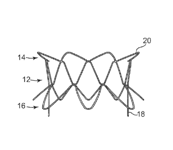
Referring again to Figures 1-4, stent frame 10 generally includes an
annular portion 12, an atrial portion 14 extending from one end of the annular
portion 12, and a ventricular portion 16 extending from the opposite end of
the
annular portion 12. Annular portion 12 includes a wire structure that is
shaped in
a generally sinusoidal configuration around its perimeter. More particularly,
annular portion 12 includes two extending posts 18 on generally opposite sides
of
its perimeter, and a sinusoidal pattern having a generally constant height
between
each of the extending posts 18. This annular portion 12 is shown as being
formed
CA 02722366 2010-10-22
WO 2009/132187
PCT/US2009/041536
9
by a single wire, although it is contemplated that a number of different wires
or
stent frame components may be assembled to make up this annular portion 12. It
is further contemplated that the entire stent frame 10 is cut from a single
sheet of
material such that annular portion 12 is part of an integral structure that
does not
include individual components. The extending posts 18 are shown as having a
greater height than the portion of the annular portion 12 between the posts
18,
where the relative size difference between these parts of the annular portion
12
can be the same or substantially different than shown. In any case, the height
of
each of the extending posts 18 is designed to provide an attachment area for
the
leaflet of a valve that will be attached within the stent frame 10. Thus, this
embodiment of the stent frame 10 that has two extending posts 18 is designed
to
accommodate a bi-leaflet valve; however, it is contemplated that the annular
portion 12 instead can comprise three extending posts 18 to accommodate
attachment of a tri-leaflet valve.
It is further contemplated that the stent frame can alternatively or
additionally include one or more extending posts that extend in the opposite
direction than discussed above relative to the extending posts 18. These
extending
posts can extend toward the atrial portion of the stent rather than the
ventricular
portion of the stent.
Atrial portion 14 includes a wire structure that is shaped to provide a
series of flanges 20 that extend radially outward at an angle around the
periphery
of one end of the annular portion 12. This atrial portion 14 is shown as being
formed by a single wire, although it is contemplated that multiple wires or
stent
frame components may be assembled to make up this atrial portion 14, or that
the
entire stent frame 10 is cut from a single sheet of material such no
individual
wires are used in the construction thereof. As shown, all of the flanges 20
are
generally the same size and shape and extend at generally the same angle from
the
annular portion 12, although it is contemplated that the flanges 20 are
configured
differently from each other. The flanges are provided for engagement with one
side of the annulus in which the stent frame 10 will be implanted, thus, the
flanges
20 can be provided with a number of different configurations to meet the
particular requirements of the locations in which the stent frame may be
CA 02722366 2010-10-22
WO 2009/132187
PCT/US2009/041536
implanted. For example, the atrial portion 14 may have more or less flanges 20
than shown, the flanges 20 can extend at a greater or smaller angle than shown
relative to the generally cylindrical shape of the annular portion 12, the
flanges 20
can be longer or shorter than shown, and the like.
Ventricular portion 16 includes a wire that is arranged to provide a first
portion 22 that extends in generally the same longitudinal or axial direction
as the
annular portion 12 along a portion of its periphery, and at least one flange
24 that
extends radially outward at an angle relative to the annular portion 12. This
ventricular portion 16 is shown as being formed by a single wire, although it
is
contemplated that multiple wires or stent frame components may be assembled to
make up this ventricular portion 16, or that the entire stent frame 10 is cut
from a
single sheet of material such no individual wires are used in the construction
thereof As shown, the first portion 22 of the ventricular portion 16 is a
series of
sinusoidal peaks and valleys that are generally the same size and shape as
each
other, although it is contemplated that they are configured differently from
each
other. This first portion 22 generally follows the outer periphery of the
annular
portion 12 in the axial direction of the stent frame (i.e., there is little to
no flare of
this portion 22 relative to the annular portion 12), where the "peaks" of the
wires
of portion 22 meet the "valleys" of the annular portion 12, such as at an
intersection point 26, for example. Such intersection points can occur around
the
periphery of the stent frame 10. It is further contemplated that the portion
22 can
be flared at least slightly relative to the annular portion 12 in order to
engage with
the aortic leaflet (i.e., the aortic portion of the ventricular flare) without
substantially blocking the left ventricular outflow tract.
The ventricular portion 16 further includes at least one flange 24 that
extends or flares outwardly from the annular portion 12 on one side of the
stent
frame 10. Each flange 24 is provided for particular engagement with an annulus
in which the stent frame will be implanted, such as the posterior side of a
mitral
annulus. In this embodiment, the portion 22 of the ventricular portion 16 does
not
flare outwardly on the anterior side so that it will not obstruct the left
ventricular
outflow tract when implanted in the mitral position. Because the flanges 24
are
provided for engagement with one side of the annulus in which the stent frame
10
CA 02722366 2010-10-22
WO 2009/132187
PCT/US2009/041536
11
will be implanted, the flanges 24 can be provided with a number of different
configurations to meet the particular requirements of the location in which
the
stent frame may be implanted. In particular, the ventricular portion 16 may
have
more or less flanges 24 than shown, the flanges 24 can extend at a greater or
smaller angle than shown relative to the periphery of the annular portion 12,
the
flanges 24 can be longer or shorter than shown, and the like.
As discussed above, the stent frame 10 may comprise a single piece
construction, such as a structure that is cut from a single piece of material,
or may
instead include a series of wires or wire segments that are attached to each
other
around the periphery of the stent frame 10. In either case, the wire portions
of the
annular portion 12, the atrial portion 14, and the ventricular portion 16 may
have
the same thickness or different thicknesses from each other. In one exemplary
embodiment, the annular portion 12 comprises relatively thick wire portions,
while the atrial portion 14 and ventricular portion 16 comprise relatively
thin wire
portions. In such an embodiment, the thickness of the wires that make up the
atrial portion 14 and ventricular portion 16 may be the same or different from
each
other.
Figures 5-9 illustrate a stent assembly 30 in accordance with another
embodiment of the invention. Stent assembly 30 includes a stent frame 32 and a
covering material 34. Stent frame 32 generally includes a central annular
portion
36, an atrial portion 38 extending from one end of the annular portion 36, and
a
ventricular portion 40 extending from the opposite end of the annular portion
36.
Annular portion 36 is similar to the annular portion described above relative
to
Figures 1-4, except that the annular portion 36 has a wire arrangement that
includes two members 42 on generally opposite sides of the annular portion 36
that are somewhat wider than the extending posts 18 of stent frame 10. These
members 42 have a height that is greater than that of the remainder of the
annular
portion 36. The wire between each of the members 42 around the periphery of
the
annular portion 36 is arranged in a generally sinusoidal pattern. The atrial
portion
38 includes a wire that is arranged to provide a series of flanges 44 that
extend
radially outward at an angle from one end of the annular portion 36. All of
the
flanges 44 are generally the same size and shape and extend at generally the
same
CA 02722366 2010-10-22
WO 2009/132187
PCT/US2009/041536
12
angle from the annular portion 36, although it is contemplated that the
flanges 44
are configured differently from each other. Ventricular portion 40 includes a
wire
that is shaped to provide a first portion 46 that extends in generally the
same
longitudinal or axial direction as the annular portion 36 along a portion of
its
periphery, and at least one flange 48 that extends radially outward at an
angle
relative to the annular portion 36. First portion 46 may alternatively be
flared at
least slightly relative to the annular portion 36 in order to engage with the
aortic
leaflet, without substantially blocking the left ventricular outflow tract.
First
portion 46 is arranged as a series of sinusoidal peaks and valleys that are
generally
the same size and shape as each other, although it is contemplated that they
are
different from each other.
The stent frame 32 may include a number of wires or wire portions that
are attached to each other generally as shown in the illustrated
configuration,
where one arrangement could include separate wires for each of the annular
portion 36, the atrial portion 38, and the ventricular portion 40.
Alternatively, the
entire stent frame 32 may be cut from a single sheet of material such that the
stent
frame 32 is an integral structure that does not include individual components.
The
relative sizes and number of wire peaks, valleys, and flanges illustrated for
each of
the portions of the stent frame 32 are exemplary, and the construction can
instead
include different sizes, numbers, and configurations of these components. In
addition, this embodiment of stent frame 32 can include any of the variations
discussed above relative to stent frame 10, including a variation that
includes three
extending members 42 to accommodate the attachment of a tri-leaflet valve
within
the frame instead of the bi-leaflet attachment arrangement shown.
Stent assembly 30 further includes a covering material 34 that is
attached to at least some of the wires of the stent frame 32, and may be
attached to
all of the wires or wire portions of stent frame 32, if desired. The covering
material can be cut before or after attachment to the stent frame 32 to allow
for a
valve structure (not shown) to be attached to the stent frame 32 within the
central
area of the annular portion 36. The covering material 34 can be a knit or
woven
polyester, such as a polyester or PTFE knit, which can be utilized when it is
desired to provide a medium for tissue ingrowth and the ability for the fabric
to
CA 02722366 2010-10-22
WO 2009/132187
PCT/US2009/041536
13
stretch to conform to a curved surface. Polyester velour fabrics may
alternatively
be used, such as when it is desired to provide a medium for tissue ingrowth on
one
side and a smooth surface on the other side. These and other appropriate
cardiovascular fabrics are commercially available from Bard Peripheral
Vascular,
Inc. of Tempe, Arizona, for example. The covering material may be attached to
its respective stent frame by sewing, adhesives, or other attachment methods.
Figures 10-13 illustrate a stent frame 60 in accordance with another
embodiment of the invention that generally includes a central annular portion
62,
an atrial portion 64 extending from one end of the annular portion 62, and a
ventricular portion 66 extending from the opposite end of the annular portion
62.
Annular portion 62 is similar to the annular portion described above relative
to
Figures 1-4 in that it includes a wire portion that is shaped to provide two
extending posts 68 on generally opposite sides of the annular portion 62, and
a
generally sinusoidal pattern between each of its extending posts 68. In this
embodiment, the annular portion 62 further includes V-shaped support members
70 that are arranged with the base of each "V" of the V-shaped members 70
generally coinciding with the base of an extending post 68. These V-shaped
members 70 have a similar configuration to the extending members 42 of stent
frame 32 in that the stent frame 60 includes a combination of extending posts
68
along with V-shaped members 70. These V-shaped members 70 can be used to
provide additional structural integrity to the stent frame 60.
The atrial portion 64 includes a series of flanges 72 that extend radially
outward at an angle from one end of the annular portion 62. All of the flanges
72
are shown as being generally the same size and shape and extend at generally
the
same angle from the annular portion 62, although it is contemplated that at
least
some of the flanges 72 are configured differently from each other. Ventricular
portion 66 includes a wire that is arranged to provide a first portion 74 that
extends in generally the same longitudinal or axial direction as the annular
portion
62 along a portion of its periphery, and at least one flange 76 that extends
radially
outward at an angle relative to the annular portion 62. First portion 74 may
be
flared at least slightly relative to the annular portion 62 in order to engage
with the
aortic leaflet without substantially blocking the left ventricular outflow
tract. First
CA 02722366 2010-10-22
WO 2009/132187
PCT/US2009/041536
14
portion 74 is arranged as a series of sinusoidal peaks and valleys that are
generally
the same size and shape as each other, although it is contemplated that they
are
differently sized and/or shaped from each other.
The stent frame 60 may include a number of wires or wire portions that
are attached to each other generally as shown in the illustrated
configuration,
where one arrangement could include separate wires for each of the annular
portion 62, the atrial portion 64, and the ventricular portion 66. In one
embodiment, the V-shaped members 70 are crimped to other wires of the stent
frame 60. Alternatively, the entire stent frame 60 may be cut from a single
sheet
of material such that the stent frame 60 is an integral structure that does
not
include individual components. The relative sizes and number of wire peaks,
valleys, and flanges illustrated for each of the portions of the stent frame
60 are
exemplary, and the construction can instead include different sizes, numbers,
and
configurations of these components. In addition, this embodiment of stent
frame
60 can include any of the variations discussed above relative to the stent
frames
described herein, including a variation that includes three extending posts 68
to
accommodate the attachment of a tri-leaflet valve within the frame instead of
the
bi-leaflet attachment arrangement shown.
Figures 14-18 illustrate a stent assembly 80 that comprises a stent
frame 82 that is generally similar to the stent frame 60 described above
relative to
Figures 10-13, and further including a covering material 84. As with the
covering
material 34 described above, covering material 84 can similarly include
materials
that facilitate at least some tissue ingrowth. The covering material 84 can be
cut
between extending posts 86 of stent frame 82, such as generally along cut line
88,
to allow for attachment of a valve (not shown) that will be positioned within
the
interior area of the stent frame 82. This stent frame and assembly, along with
many other stents of the invention, may be provided with portions that are
made
of self-expandable materials and other portions that are made of balloon-
expandable materials. With particular reference to Figure 17, for example, the
atrial and ventricular portions may be made of a self-expanding material,
while
the central annular portion may be made of a balloon-expandable material to
allow
for high radial force at the annulus.
CA 02722366 2010-10-22
WO 2009/132187
PCT/US2009/041536
Figures 19-22 illustrate a stent frame 100 in accordance with another
embodiment of the invention that generally includes an annular portion 102, an
atrial portion 104 extending from one end of the annular portion 102, and a
ventricular portion 106 extending from the opposite end of the annular portion
102. Annular portion 102 includes wire or wire portions that cross each other
around the periphery of the stent frame 100 in a series of X-shaped
structures. The
stent frame 100 includes one or more wires shaped to provide a series of
flanges
108 that extend radially outward at an angle from one end of the annular
portion
102. All of the flanges 108 are shown as having generally the same size and
shape and as extending at the same angle from the annular portion 102,
although it
is contemplated that the flanges 108 are configured differently from each
other.
At least some of the flanges 108 also include one or more barbs or extensions
110,
where this illustrated embodiment includes two barbs 110 near the distal tip
of
each of the flanges 108. Each of the barbs 110 preferably extends from its
respective flange 108 in such a way so that when the stent frame 100 is
positioned
relative to the annulus of a valve in which it will be implanted, the barbs
110 will
be engageable with the tissue to which they are adjacent. Thus, as is best
illustrated in Figures 21 and 22, barbs 110 extend downwardly or toward the
annular portion 102 of the stent frame 100 so that they can engage with the
structure of the heart when implanted. It is understood that the barbs 110 can
have
a different size, shape, orientation, positioning, etc. than shown, and that
the each
of the flanges 108 can include more or less than the two barbs 110 shown.
Further, it is contemplated that only some of the flanges 108 include such
barbs
110.
The ventricular portion 106 includes a wire that is shaped to provide
two extending posts 112 on generally opposite sides of the stent frame 100, at
least one flange portion 114 extending radially outward from annular portion
102
on one side of the stent frame 100 between extending posts 112, and a
sinusoidal
wire pattern on the other side of the stent frame 100 between extending posts
112.
At least some of the flanges 114 also include at least one barb 116, where
this
illustrated embodiment includes two barbs 116 near the distal tip of each of
the
flanges 114. Each of the barbs 114 preferably extends from its respective
flange
CA 02722366 2010-10-22
WO 2009/132187
PCT/US2009/041536
16
114 in such a way that when the stent frame 100 is positioned relative to the
annulus of a valve in which it will be implanted, the barbs 116 will be
engageable
with the tissue to which they are adjacent. Thus, as is best illustrated in
Figures
21 and 22, barbs 116 extend upwardly or toward the annular portion 102 of the
stent frame 100. As with the barbs 110 described above, barbs 116 can have a
different size, shape, orientation, positioning, etc. than shown, and each of
the
flanges 114 can include more or less than the two barbs 116 shown. Further, it
is
contemplated that only some of the flanges 114 include barbs 116.
Figure 23 illustrates an exemplary pattern 120 for a stent frame of the
type illustrated above relative to Figures 19-22. This stent frame pattern 120
includes a diamond-shaped pattern that can be cut from a single sheet of
material.
The stent frame pattern 120 can be formed into a tubular shape to make a stent
frame. As shown, this embodiment includes a number of barbs 122 extending
from distal ends of the pattern.
Figure 24 illustrates a stent assembly 130 of the invention, which
includes a stent frame 132 and a covering material 134. As shown, the covering
material 134 is stitched to the stent frame 132 along many of the wires of
this
assembly that are visible. This stent frame 132 includes two extending posts
136
positioned generally across from each other, which are provided as the
commissure posts to which the leaflets of a valve assembly will be attached to
provide a bi-leaflet valve.
Figure 25 schematically illustrates a portion of a heart 140, with an
exemplary stent assembly 141 of the invention positioned therein. In
particular,
heart 140 includes a left atrium 142, a left ventricle 144, a mitral valve 146
and an
aortic valve 148. The broken lines of mitral valve 146 illustrate its native
leaflets
as they would generally be configured prior to implantation of stent assembly
141.
In particular, mitral valve 146 includes a first leaflet 150 on the anterior
side of the
valve, and a second leaflet 152 on the posterior side of the valve. When the
native
mitral valve 146 is operating properly, the native leaflets 150, 152 will
generally
function in such a way that blood flows toward the left ventricle 144 when the
leaflets 150, 152 are in an open position, and so that blood is prevented from
moving toward the left atrium 142 when the leaflets 150, 152 are in a closed
CA 02722366 2010-10-22
WO 2009/132187
PCT/US2009/041536
17
position. However, stent assembly 141 can be positioned in the area of mitral
valve 146 when it is not functioning properly (to replace the mitral valve) in
accordance with the invention, thereby pushing the leaflets 150, 152 out of
the
mitral valve space, such as are shown as displaced leaflets 156 and 158,
respectively.
As shown, stent assembly 141 includes an annular portion 160, an
atrial portion 162 including flares extending from one side of the annular
portion
160 and toward the left atrium 142, and a ventricular portion 164 including
flares
extending from the posterior side of the annular portion 160 and toward the
left
ventricle 144. In order to not block the flow of blood through the aortic
valve
148, the ventricular portion 164 of the stent assembly 142 that is closest to
the
aortic valve 148 does not have flares or has flares that have a minimal
height. In
this way, the stent assembly 142 will not push the leaflet 156 to a position
in
which it will interfere with blood flow through the aortic valve 148 and/or
interfere with the actual movement or functioning of the leaflets of the
aortic
valve 148. However, annular portion 160 preferably has a sufficient length to
provide a suitable area of contact with the annulus of the mitral valve to
help to
maintain it in its desired position.
As stated above, the stent assemblies of the invention can also be
implanted for replacement of the tricuspid valve. In particular, if the stent
assemblies of the invention are positioned within the annulus of a triscuspid
valve,
the atrial flares would be removed or made in such as way that they do not
contact
the apex of the triangle of Koch in order to not disturb the conduction system
(i.e.,
the AV node and bundle of His). In addition, the ventricular flares would not
contact the septal portion of the ventricle in order to not disturb the
conduction
system, wherein these flares can thus be similar to those described above
relative
to stent assemblies for the mitral area. In addition, the ventricular flares
in this
embodiment can generally resemble the posterior flares in an inferior and
anterior
direction (e.g., approximately 2/3 of the flares).
Stent frames of the type described above can be assembled into a
stented valve assembly in accordance with the methods of the invention
described
herein, although such valves are not shown in the Figures. One exemplary
CA 02722366 2010-10-22
WO 2009/132187
PCT/US2009/041536
18
method for assembling a stented valve generally first includes preparation of
a
porcine aortic valve, then a subsequent mounting or attachment of the prepared
porcine valve to the stent frame using a variety of mounting or attachment
techniques. Bi-leaflet, tri-leaflet, and other variations of valve assemblies
can be
attached within the stent frames described herein.
The various flange portions described above relative to the atrial
portions and ventricular portions of the stent frames are generally shown as
being
V-shaped or U-shaped. However, the flange portions may instead be semi-
circular, rectangular, oblong, or the like, and may be considerably smaller or
larger than shown. In yet another variation, a different flange structure that
is
more continuous around the periphery of the annular portion of the stent frame
can be used (i.e., the flange structure does not comprise a series of adjacent
flanges but instead comprises more of a continuous flared structure at one or
both
ends of the stent frame). In any case, the flange portion(s) are preferably
configured to be a shape and size that can provide an anchoring function for
the
stent assembly when it is positioned to replace a valve. For example, if stent
assembly were positioned within the mitral valve annulus any flange portions
that
extend from the stent assembly on the atrial side can provide interference
with the
walls of the left atrium, thereby inhibiting motion of the stent assembly.
The atrial flares or flange portions can also incorporate features that
enable the stent to be sewn in place as part of an atrial incision closure
using
various means, such as clips, sutures, and the like. In addition, if the
atrial flares
or flange portions of a stent progress further upward toward the top of the
atrium,
the result can provide enhanced stabilization of the prosthesis. One example
of a
configuration of a stent frame 180 that provides such a stabilization feature
is
illustrated in Figure 26. This and other stent frames comprising stabilization
features can engage the native anatomy of the atrium for stable position and
fixation of a replacement valve. This concept can be applicable to
transcatheter or
minimally invasive replacement of an insufficient or stenotic mitral or
tricuspid
valve. Such stent frames generally include a stent inflow aspect member or
members that extend beyond the native valve annulus to match the curvature of
the atrium. These members can have a variety of shapes and configurations, but
CA 02722366 2010-10-22
WO 2009/132187
PCT/US2009/041536
19
generally all function to prevent antegrade and/or retrograde migration of the
valve assembly. The degree of protrusion into the atrium can vary, but can
advantageously extend past the inflection point of the radius of curvature.
The
members can also extend all the way to the top of the atrium, if desired. The
members can be discrete or joined at the top of the atrium to generally match
the
shape of the anatomy. Various covering materials can be used to cover or
partially cover the stabilization portion of the stent frame, including
materials such
as fabric, polymer, tissue, or other biocompatible materials. The material can
further be chosen to enhance in-growth and/or to reduce abrasion and trauma,
if
desired.
In the exemplary embodiment of Figure 26, a stent frame 180 is shown
as positioned relative to the annulus 182 of a native valve, and a hoop or
series of
hoops 184 (indicated by the broken line in atrium 186) extends from a top end
of
the stent frame 180 into the atrium 186, which provides additional
stabilization of
the stent and can help to minimize stent migration. Referring still to Figure
26, a
schematic view of a portion of a heart is shown, including the left ventricle
188,
atrium 186, papillary muscles 190, and the annulus 182 of the native valve. A
valve comprising a stent frame 180 of the invention is shown as positioned so
that
its annulus 192 is at least slightly higher than the annulus 182 of the native
valve.
Two exemplary leaflets 194 are shown as extending from the frame 180 at the
position of its annulus 192. This positioning of the stent frame 180 can
reduce its
protrusion into the left ventricle 188, which can thereby minimize contact and
rubbing of the stent frame 180 on the wall of the left ventricle 188 and
papillary
muscles 190. The position of the stent frame 180 can also reduce the potential
for
erosion, arrhythmias and ischemia.
Figures 27-29 illustrate another embodiment of a stent frame 200
providing the features described above for positioning and fixation relative
to a
native valve annulus. Figure 27 shows this stent frame 200 positioned relative
to
an atrium 202 and ventricle 204. Stent frame 200 includes an annular portion
206,
an atrial portion 208, a ventricular portion 210, and a securing portion 212
extending from the atrial portion 208. Securing structure 212 generally
includes a
series of wires arranged in petals or another configuration that extends from
the
CA 02722366 2010-10-22
WO 2009/132187
PCT/US2009/041536
peaks of the wires of the atrial portion 208. The petals are attached at their
distal
ends to a disc 214 or other structure that maintains the wires in a dome-type
shape,
as shown. The ventricular portion 210 can include any of the features
described
above relative to the ventricular end of the stent frames, wherein this
particular
embodiment shows a ventricular portion having flares that extend outwardly
relative to a central longitudinal axis of the stent frame 200. The annular
portion
206 further includes two extending posts 216 that are at least somewhat taller
or
longer than the height of the structure of the annular portion between the
extending posts.
Figures 30 and 31 illustrate another embodiment of a stent frame 220
that also includes an atrial portion 224 comprising a series of flares that
are curved
at least slightly toward a central longitudinal axis of the stent frame. The
frame
220 further includes at least two support wires 222 that form an additional
securing structure of this embodiment. As shown, this exemplary embodiment
illustrates two wires 222, each of which extends between two atrial flares on
opposite sides of the frame, thereby helping to maintain the flares in this
configuration and providing a dome-shaped support structure. However, it is
contemplated that the stent frame 220 instead includes more or less than two
wires. Further, it is contemplated that wires extend from only some of the
flares
of the atrial portion 224, as shown, or that all of the flares of the atrial
portion 224
are connected to another flare with a support wire 222. In yet another
embodiment, which is illustrated in Figures 32 and 33, a stent frame 240
includes
an atrial portion 242 having multiple flares that are curved somewhat toward a
central longitudinal axis of the stent frame 240. However, this exemplary
embodiment does not also include any additional connecting wires between the
flares.
Figures 34-36 illustrate yet another embodiment of a stent frame 260
that includes an atrial portion comprising flares 262 and a series of wires
266
extending from the flares 262 toward a central longitudinal axis of the stent
frame.
The wires 266 are arranged as petals or another configuration that extends
from
the peaks of the wires of the atrial portion. The wires 266 are attached at
their
distal ends to a structure 264 that maintains the wires in a dome-type shape,
as
CA 02722366 2010-10-22
WO 2009/132187
PCT/US2009/041536
21
shown. The ventricular portion of the stent frame 260 can include any of the
features described above relative to the ventricular end of the stent frames,
wherein this particular embodiment shows a ventricular portion having flares
that
extend outwardly relative to a central longitudinal axis of the stent frame.
The
annular portion further includes two extending posts 268 that are at least
somewhat taller or longer than the height of the structure of the annular
portion
between the extending posts.
Any of the embodiments of stent assemblies described herein relative
to the invention may include a gasket or other member around its exterior to
provide for sealing against paravalvular leakage and to facilitate pannus in-
growth
for stabilization of the stent. Such a gasket or other member may
alternatively or
additionally be positioned on the interior portion of the stent or on the
underside
of a cuff provided on the stent.
In addition, it is contemplated that the ventricular flares associated
with the stented valves of the invention can house biologics to target
infarcts
(stem cells, genes, proteins, etc.), which are often located posterior-
inferiorly in
patients with ischemic mitral regurgitation. The areas of a the stented valves
of
the invention used for anchoring could also be seeded with cells or biologics
to
promote ingrowth for quick incorporation into the surrounding tissue. This
could
aid in eliminating paravalvular leakage and in eliminating migration or
embolization of the prosthesis. In one example for a mitral valve replacement,
the
atrial and annular portions can include pro-ingrowth biologics and the
ventricular
portion can include therapeutic biologics and/or pro-ingrowth biologics.
The stent assemblies of the present invention may be positioned within
the desired area of the heart via entry in a number of different ways. In one
example, the stent assembly may be inserted transatrially, where entry may be
done either percutaneously or in a minimally invasive technique on a beating
heart
in which access is through the side of the heart, or even through a standard
open
heart valve replacement procedure using heart-lung bypass and sternotomy where
the described device would be used as an alternative to the standard
replacement.
In another example, the stent assembly may be inserted transapically, where
entry
again may be done either percutaneously or in a minimally invasive technique
on
CA 02722366 2015-11-06
51749-46
22
a beating heart in which access is through the side of the heart. In yet
another
example, the stent assembly may be inserted transeptally, where entry can be
done
percutaneously.
The invention further includes a method of positioning a valve into a
body lumen, such as one of the atrioventricular valve openings of the heart.
The
method comprises the steps of compressing a stent frame of a stented valve,
wherein the stent frame includes an annular region, an atrial portion
extending
from one end of the annular region, and a ventricular portion extending from
the
other end of the annular region. A sheath or other component of a delivery
system
can be slid or otherwise positioned over the compressed stented valve to keep
it
from expanding and to minimize interference between the stented valve and the
vasculature through which it will be traveling. The stented valve is then
delivered
to the annulus of the desired valve area of the patient, which delivery may be
performed transapically, for example. In one method, the valve is accessed
through the bottom of the valve. When the valve is in position, the atrial
region or
portion of the stent is released, such as by retracting the sheath of the
delivery
system by a sufficient amount that this portion of the stented valve is
exposed.
Due to the self-expanding properties of the stent frame, the atrial portion
will
expand outwardly relative to the sheath in which it was enclosed. The delivery
system is then used to pull the stent valve back against the annulus to engage
the
atrial portion of the stent with the annulus. The sheath of the delivery
system can
then be further refracted to release the ventricular portion of the stent
frame from
the delivery system. Due to the self-expanding properties of the stent frame,
the
ventricular portion will expand outwardly relative to the sheath in which it
was
enclosed. The delivery system can then be retracted from the patient.
The present invention has now been described with reference to several
embodiments thereof. The foregoing detailed
description and examples have been given for clarity of understanding only. No
unnecessary limitations are to be understood therefrom. It will be apparent to
those skilled in the art that many changes can be made in the embodiments
CA 02722366 2010-10-22
WO 2009/132187
PCT/US2009/041536
23
described without departing from the scope of the invention. Thus, the scope
of
the present invention should not be limited to the structures described
herein.