Note: Descriptions are shown in the official language in which they were submitted.
CA 02734592 2011-03-16
-1-
OPTIMAL IOL SHAPE FACTORS FOR HUMAN EYES
This is a division of co-pending Canadian Patent Application No. 2,567,049
filed on April 4, 2006.
Background
The present invention relates generally to ophthalmic lenses, and more
particularly, to intraocular lenses (IOLs) having optimal shape factors.
Intraocular lenses are routinely implanted in patients' eyes during cataract
surgery to replace the clouded natural lens. The post-operative performance of
such
IOLs, however, can be degraded due to a variety of factors. For example,
aberrations
introduced as a result of misalignment of the implanted IOL relative to the
cornea,
an/or the inherent aberrations of the eye, can adversely affect the lens's
optical
performance.
Accordingly, there is a need for improved IOLs that can provide a more robust
optical performance.
Summary
In accordance with one aspect of the present invention there is provided a
method of designing an ophthalmic lens, comprising: defining an error function
indicative of variability in performance of a lens in a patient population
based on
estimated variability in one or more biometric parameters associated with that
population; and selecting a shape factor for the lens that reduces said error
function
relative to a reference value.
CA 02734592 2011-03-16
-2-
In another aspect, the optic is formed of a biocompatible polymeric material.
By
way of example, the optic can be formed of a soft acrylic polymeric material.
Other
examples of suitable materials include, without limitation, hydrogel and
silicone
materials.
In another aspect, at least one surface of the optic can be characterized by
an
aspheric base profile (i.e., a base profile that exhibits deviations from
sphericity). By
way of example, the base profile can be characterized by a conic constant in a
range of
about -73 to about -27.
In a related aspect, the aspheric profile of the lens surface can be defined
in
accordance with the following relation:
cr2
Z =
1+ 1-(1+k)c2r2
wherein,
c denotes the curvature of the surface at its apex (at its intersection with
the
optical axis),
r denotes the radial distance from the optical axis, and
k denotes the conic constant,
wherein
c can be, e.g., in a range of about 0.0152 mni 1 to about 0.0659 mm'',
r can be, e.g., in a range of about 0 to about 5, and
k can be, e.g., in a range of about -1162 to about -19 (e.g., in a range of
about -
73 to about -27).
In a related aspect, the optic of the above lens can have a shape factor in a
range
of about 0 to about 2.
In some embodiments in which one or more surfaces of the ophthalmic lens
exhibit asphericity, the shape factor of the lens (e.g., an IOL) can be
selected as a
function of that asphericity so as to optimize the lens's optical performance.
By way of
example, in one aspect, the invention provides an ophthalmic lens having an
optic with
an anterior surface and a posterior surface, where at least one of the
surfaces exhibits an
ashperical profile characterized by a conic constant in a range of about -73
to about -27.
The optic exhibits a shape factor in a range of about -0.5 to about 4.
CA 02734592 2011-03-16
-3-
In a related aspect, an ophthalmic lens having an optic with a shape factor in
a
range of about 0 to about 2 includes at least one aspherical surface
characterized by a
conic constant in a range of about -73 to about -27.
In other aspects, an intraocular lens adapted for implantation in an eye
having a
corneal radius equal to or less than about 7,1 mm is disclosed, which includes
an optic
having an anterior surface and a posterior surface. The optic exhibits a shape
factor in a
range of about -0.5 to about 4. In a related aspect, the optic exhibits a
shape factor in a
range of about +0.5 to about 4, or in a range of about I to about 3.
to In another aspect, the invention provides an intraocular lens adapted for
implantation in an eye having a corneal radius in a range of about 7,1 mm to
about 8.6
mrn, which includes an optic having an anterior surface and a posterior
surface. The
optic exhibits a shape factor in a range of about 0 to about 3. In a related
aspect, the
optic exhibits a shape factor in a range of about +0,5 to about 3, or in a
range of about 1
to about 2.
In another aspect, an intraocular lens adapted for implantation in an eye
having a
corneal radius equal to or greater than about 8.6 is disclosed, which includes
an optic
having an anterior surface and a posterior surface. The optic exhibits a shape
factor in a
range of about 0,5 to about 2. In a related aspect, the optic exhibits a shape
factor in a
range of about 1 to about 2.
In another aspect, the invention provides an intraocular lens adapted for
implantation in an eye having an axial length equal to or less than about 22
mm, which
includes an optic having an anterior surface and a posterior surface, The
optic can have
a shape factor in a range of about 0 to about 2, or in a range of about 0,5 to
about 2.
In other aspects, the invention discloses methods for selecting an ophthalmic
lens
for implantation in a patient's eye based on one or more ocular biometric
parameters of
the patient. For example, a method of correcting vision is disclosed that
includes
selecting an IOL, which comprises an optic exhibiting a shape factor in a
range of about
- 0.5 to about 4 (or in a range of about +0.5 to about 4), for implantation in
an eye
having a corneal radius that is equal to or less than about 7.1 mm
CA 02734592 2011-03-16
-4-
In another aspect, a method of correcting vision is disclosed that includes
selecting an IOL, which comprises an optic exhibiting a shape factor in a
range of about
0 to about 3 (or in a range of about 0.5 to about 3), for implantation in an
eye having a
corneal radius in a range of about 7.1 nun to about 8.6 mm.
In yet another aspect, a method of correcting vision is disclosed that
includes
selecting an IOL, which comprises an optic exhibiting a shape factor in a
range of about
0.5 to about 2, for implantation in an eye having a corneal radius that is
equal to or
greater than about 8.6 mm.
to In another aspect, a method of corrected vision is disclosed that includes
selecting an IOL, which comprises an optic exhibiting a shape factor in a
range of about
0 to about 2 (or in a range of about 0.5 to about 2), for implantation in an
eye having an
axial length equal to or less than about 22 mm.
In another aspect, a method of designing an ophthalmic lens is disclosed that
includes defining an error function, which is indicative of variability in
performance of a
lens in a patient population, based on estimated variability in one or more
biometric
parameters associated with that population, and selecting a shape factor for
the lens that
reduces the error function relative to a reference value. In a related aspect,
the error
function can further include an estimated error in optical power correction
provided by
the lens and/or an estimated aberration error.
In a related aspect, the error function (RxError) can be defined in accordance
with the following relation:
RxError = 4Biometric2 + DIOLPower2 + hAberration2
wherein,
E Biometric denotes variability due to biometric data errors,
AIOLPower denotes variability due to optical power correction errors, and
t%Aberration denotes variability due to aberration contributions.
CA 02734592 2011-03-16
-5-
In another aspect, the ABiometric can be defined in accordance with the
following relation:
AB1ometric = Ak2 + ML2 + AACD2
wherein,
Ak denotes error in keratometric measurements,
AAL denotes error in axial length measurements, and
AACD denotes error in anterior chamber depth measurements.
In another aspect, the Mberratlon can be defined in accordance with the
following relation:
AAberration = Mstig2 + ASA2 + AOtherz
wherein,
AAstig represents variability due to astigmatic aberration,
ASA represents variability due to spherical aberration, and
AOther represents variability due to other aberrations.
In a further aspect, the AIOLPower can be defined in accordance with the
following relation:
AIOLPower = AIOLStep2 + AIOLTol2 + AELP2
wherein,
AIOLStep represents variability caused by difference between a power
correction provided by the lens and a power correction needed by a patient,
AIOLTol represents manufacturing power tolerance, and
AELP represents variability in a shift of the lens effective position within
the
eye.
Further understanding of the invention can be obtained by reference to the
following detailed description, in conjunction with the associated drawings,
which are
discussed briefly below.
CA 02734592 2011-03-16
-6-
Brief Description of the Drawings
FIGURE 1 is a schematic side view of an IOL in accordance with one
embodiment of the invention,
FIGURE 2 presents simulated magnitude of different aberration types
(spherical,
defocus, coma and astigmatic aberrations) exhibited by an IOL as a function of
its shape
factor for a 1.5 mm decentration,
FIGURE 3 presents simulation results for aberrations exhibited by an IOL due
to
tilt as a function of the JOL's shape factor,
FIGURE 4A presents graphically calculated spherical aberration exhibited by a
model eye characterized by an average anterior chamber depth in which an 1OL
is
incorporated, as a function of the IOL's shape factor,
FIGURE 4B presents graphically calculated MTFs at 50 lp/mm and 100 lp/mm
for a model eye characterized by an average anterior chamber depth in which an
IOL is
incorporated, as a function of the IOL's shape factor,
FIGURE 5A depicts simulated MTFs at 50 lp/mm and 100 lp/mm for a model
eye characterized by a small anterior chamber depth in which an IOL is
incorporated, as
a function of the IOL's shape factor,
FIGURE 5B depicts simulated spherical aberration exhibited by a model eye
characterized by a small anterior chamber depth in which an TOL is
incorporated, as a
function of the IOL's shape factor,
FIGURE 6A depicts simulated spherical aberration exhibited by a model eye
characterized by a large anterior chamber depth in which an IOL is
incorporated, as a
function of the IOL's shape factor,
CA 02734592 2011-03-16
-7-
FIGURE 6B depicts simulated MTFs at 50 lp/mm and 100 lp/mm for a model
eye characterized by a large anterior chamber depth in which an IOL is
incorporated, as
a function of the IOL's shape factor,
FIGURE 7A depicts graphically simulated spherical aberrations exhibited by a
plurality of model eyes having different corneal asphericities in which an IOL
is
incorporated, as a function of the IOL's shape factor,
FIGURE 7B depicts graphically simulated MTF as 50 lp/mm obtained for model
eyes having different corneal asphericities in which an JOL is incorporated,
as a function
of the IOL's shape factor,
FIGURE 7C depicts graphically simulated MTF at 100 lp/mm obtained for
model eyes having different corneal asphericities in which an IOL is
incorporated, as a
function of the IOL's shape factor,
FIGURE 8A depicts simulated spherical aberration exhibited by two model eyes
characterized by different corneal radii as a function of the shape factor of
an IOL
incorporated in the models,
FIGURE 8B depicts simulated MTF at 50 lp/mm exhibited by two model eyes
characterized by different corneal radii as a function of the shape factor of
an IOL
incorporated in the models,
FIGURE 8C depicts simulated MTF at 100 lp/mm exhibited by two model eyes
characterized by different corneal radii as a function of the shape factor of
an IOL
incorporated in the models,
FIGURE 9A depicts simulated spherical aberration exhibited by a plurality of
model eyes having different axial lengths. as a function of the shape factor
of an IOL
incorporated in the models,
CA 02734592 2011-03-16
-8-
FIGURE 9B depicts simulated MTFs at 50 lp/mm exhibited by a plurality of
model eyes having different axial lengths as a function of the shape factor of
an IOL
incorporated in the models,
FIGURE 9C depicts simulated MTFs at 100 lp/mm exhibited by a plurality of
model eyes having different axial lengths as a function of the shape factor of
an IOL
incorporated in the models,
FIGURE 10 is a schematic side view of a lens according to one embodiment of
the invention having an aspheric anterior surface,
FIGURE 11 presents a plurality of graphs depicting the sag of an aspheric
surface of two lenses in accordance with the teachings of the invention having
different
shape factors, and
FIGURE 12 graphically presents Monte Carlo simulation results for optical
performance of a plurality of IOLs as a function of manufacturing tolerances.
Detailed Description of the Preferred Embodiments
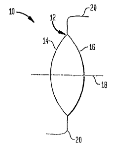
FIGURE 1 schematically depicts an IOL 10 in accordance with one embodiment
of the invention having an optic 12 that includes an anterior surface 14 and a
posterior
surface 16. In this embodiment, the anterior and posterior surfaces 14 and 16
are
symmetrically disposed about an optical axis 18, though in other embodiments
one or
both of those surfaces can exhibit a degree of asymmetry relative to the
optical axis.
The exemplary IOL 10 further includes radially extending fixation members or
haptics
20 that facilitate its placement in the eye. In this embodiment, the optic is
formed of a
soft acrylic polymer, commonly known as Acrysof, though in other embodiments,
it can
be formed of other biocompatible materials, such as silicone or hydrogel, The
lens 10
provides a refractive optical power in a range of about 6 to about 34 Diopters
(D), and
preferably in a range of about 16 D to about 25 D.
CA 02734592 2011-03-16
-9-
In this exemplary embodiment, the lens 10 has a shape factor in a range of
about
0 to about 2. More generally, in many embodiments, the shape factor of the
lens 10 can
range from about -0.5 to about 4. As known in the art, the shape factor of the
lens 10
can be defined in accordance with the following relation:
Shape Factor (X) Cl +C2 Eq (1)
C, - C2
wherein C, and C2 denote, respectively, the curvatures of the anterior and
posterior
surfaces.
The shape factor of the IOL 10 can affect the aberrations (e.g., spherical
and/or
astigmatic aberrations) that the lens can introduce as a result of its tilt
and decentration,
e.g., when implanted in the subject's eye or in a model eye. As discussed in
more detail
below, aberrations caused by a plurality of IOLs with different shape factors
were
theoretically studied as a function of tilt and decentration by utilizing a
model eye.
Those studies indicate that IOLs having a shape factor in a range of about 0
to about 2
introduce much reduced aberrations as a result of tilt and decentration.
More particularly, to study the effects of an IOL's shape factor on
aberrations
induced by its tilt and decentration, a hypothetical eye model having optical
properties
(e.g., corneal shape) similar to those of an average human eye was employed.
The radii
of optical surfaces and the separations between optical components were chosen
to
correspond to mean values of those parameters for the human population. The
refractive
indices of the optical components were chosen to provide selected refractive
power and
chromatic aberrations. Further, the anterior corneal surface of the model was
selected to
have an ashperical shape. An IOL under study replaced the natural lens in the
model.
Table I below lists the various design parameters of the model eye;
CA 02734592 2011-03-16
-10-
Table 1
Surface Type Radius Thickness Class Diameter Conic Constant
(mm) (mm) (mm)
OBJ Standard Infinity Infinity 0.000 0.000
I Standard Infinity 10.000 5.000 0.000
2 Standard 7.720 0.550 Cornea 14.800 -0.260
3 Standard 6.500 3,050 Aqueous 12.000 0.000
STO Standard Inf inity 0,000 Aqueous 10.000 0.000
Standard 10.200 4,000 Lens 11.200 -3.]32
6 Standard -6.000 16.179 Vitreous 11.200 -1.000
IMA Standard -12.000 24.000 0.000
An optical design software marketed as Zemax (version March 4, 2003, Zemax
5 Development Corporation, San Diego, CA) was utilized for the simulations of
the
optical properties of the model eye. A merit function was defined based on the
root-
mean-square (RMS) wavefront aberration, that is, the RMS wavefront deviation
of an
optical system from a plane wave. In general, the larger the RMS wavefront
error, the
poorer is the performance of the optical system. An optical system with an RMS
wavefront error that is less than about 0.071 waves is typically considered as
exhibiting
a diffraction-limited optical performance.
The effects of misalignment (tilt and/or decentration) of an IOL on its
optical
performance for a number of different shape factors was simulated by placing
the IOLs
in the above model eye and utilizing the Zemaxo software, For these
simulations, the
IOL was assumed to have spherical surfaces so as to investigate the effects of
the shape
factor alone (as opposed to that of the combined shape factor and
asphericity). To
simulate the scotopic viewing conditions for old patients, a 5 mm entrance
pupil was
chosen. The following misalignment conditions were considered: 1.5 mm IOL
decentration and a 10-degree IOL tilt. These two conditions represent the
extreme cases
of IOL misalignments.
FIGURE 2 presents the simulated magnitude of different aberration types
(spherical aberration, defocus, coma and astigmatism) as a function of the
shape factor
for 1.5 mm decentration of the IOL. These simulations indicate that IOLs with
a shape
factor in a range of about 0 to about 2 exhibit much lower aberrations as a
result of the
CA 02734592 2011-03-16
-I1-
decentration, For example, an IOL with a shape factor of about 1 introduces a
defocus
aberration of 0.07 D compared to a defocus aberration of 0.32 D introduced by
an IOL
having a shape factor of -1.
FIGURE 3 presents the simulation results for aberrations introduced as a
result
of the IOL's tilt. These results indicate that the defocus and astigmatic
aberrations are
not significantly influenced by the JOL's shape factor while the coma and
spherical
aberrations exhibit even stronger dependence on the shape factor than their
dependence
in case of the IOL's decentration. Again, the IOLs with shape factors in a
range of about
0 to 2 exhibit a stable performance.
In other aspects, it has been discovered that certain biometric parameters of
the
eye (e.g., corneal radius and axial length) can be considered while selecting
the shape
factor of an IOL for implantation in the eye to provide enhanced performance
of the
lens. As discussed in more detail below, in some embodiments, optimal IOL
shape
factors are provided for different eye populations, e.g., average human eye
(eyes with
average values for certain biometric parameters), and other populations
characterized by
extreme values for those parameters.
The biometric parameters of the above eye model were varied to simulate the
performance of a plurality of IOLs having different shape factors for
different eyes. For
an average human eye, a corneal radius (r) of 7.72 mm, a corneal asphericity
(Q) of -
0.26, an anterior chamber depth (ACD) of 4.9 mm, and an axial length (AL) of
24.4 mm
were assumed. To investigate human eyes with extreme large or small biometric
values,
the anterior chamber depth was varied from 4.3 mm to 5.5 mm, the corneal
asphericity
was varied from -0.50 to 0, the corneal radius was varied from 7.10 mm to 8,60
mm, and
the axial length was varied from 22.0 mm to 26.0 mm. These ranges are
sufficiently
broad to cover the values exhibited by the majority of the population. The
optical
performance of the IOLs was evaluated based on two criteria: calculated wave
aberration and modulation transfer function (MTF). As known to those having
ordinary
skill in the art, the MTF provides a quantitative measure of image contrast
exhibited by
an optical system, e.g., a system formed of an IOL and the cornea. More
specifically,
the MTF of an imaging system can be defined as a ratio of a contrast
associated with an
image of an object formed by the optical system relative to a contrast
associated with the
object.
CA 02734592 2011-03-16
-12-
Table 2 below presents the simulation results of the optical performance of
IOLs
having shape factors in a range of about -2 to about 4 for an eye having an
average
anterior chamber depth (ACD) of 4.9 mrn, a cornea! radius of 7,72 mm, a comeal
asphericity of -0.26, and an axial length (AL) of 24.4 mm, at a pupil size of
5 nun
Table 2.
Shape Factor (X) Spherical Aberration (SA) MTF at 50lp/mrn MTF at 100 Ip/mm
-2 0.478 0.037 0.095
-1.5 0.386 0.117 0.051
-1 0.307 0.212 0.011
-0.5 0.244 0.331 0.016
0 0.195 0.455 0,128
0,5 0.162 0.555 0.250
1 0.142 0.615 0.334
1,5 0,134 0.637 0.366
2 0.138 0.625 0.348
3 0.174 0.516 0.199
4 0.239 0.340 0.021
For graphical presentation of the information in Table 2, FIGURES 4A and 4B
provide, respectively, the calculated spherical aberration and MTF presented
in Table 1
as a function of IOL's shape factor.
Table 3 below presents the simulation results for the optical performance of a
plurality oflOLs having shape factors in the above range of -2 to 4 at a pupil
size of 5
mm for an eye having a small anterior chamber depth (ACD) of 4.3 mm, but the
same
corneal radius (7.72 mm) and asphericity (-0,26) as well as axial length (24.4
mm) as
that employed in the previous simulation. FIGURES 5A and 513 graphically
depict,
respectively, the calculated spherical aberration (SA) and the MTF presented
in Table 3
as a function of the IOL's shape factor,
CA 02734592 2011-03-16
-13-
Table 3.
Shape Factor (X) Sph. Aberration (waves) MTF at 501p/mm MTF at 100lplmm
-2 0.461 0.047 0.095
-1.5 0.374 0.125 0.042
-1 0.300 0.219 0.014
-0.5 0.240 0.337 0.021
0 0.194 0.457 0.130
0.5 0,161 0.553 0.249
1 0.141 0.613 0.331
1.5 0.133 0.636 0.365
2 0.136 0.627 0.353
Table 4 below presents the simulation results for the optical performance of a
plurality of IOLs having shape factors in the above range of -2 to 4 at a
pupil size of 5
mm for an eye having a large anterior chamber depth (ACD) of 5.5 mm, a corneal
radius
of 7.72 mm, a corneal asphericity of -0.26 and an axial length of 24.4 mm,
Further,
FIGURES 6A and 613 graphically depict, respectively, the calculated spherical
aberration (SA) and the MTF presented in Table 4 as a function of the IOL's
shape
factor.
Table 4.
Shape Factor (X) Sph. Aberration (waves) MTF at 50 ip/mm MTF at 100 lpImm
-2 0.498 0.026 0.093
-1.5 0.399 0.108 0.059
-1 0.316 0,204 0.008
-0.5 0.249 0.325 0,011
0 0.198 0.454 0.125
0.5 0.162 0.556 0.251
1 0,142 0.617 0,336
1.5 0.135 0,637 0.365
2 0,140 0.622 0.342
These simulations indicate that IOLs with shape factors in a range of about -
0,5
to about 4, and particularly those having shape factors in a range of about 0
to about 2,
provide enhanced optical performance. The simulations, however, show that
anterior
CA 02734592 2011-03-16
-14-.
chamber depth does not significantly affect the performance of an IOL.
Although in the afore-mentioned simulations the spherical aberrations were
considered, if the IOL is misaligned relative to the cornea, other aberrations
(e.g.,
defocus, astigmatism and coma) can also be present. The simulations of these
aberrations for average, small and large ACD confirm that the aberrations can
be
minimized by utilizing shape factors in a range about 0 to about 2.
The impact of corneal asphericity (Q) on optimal IOL shape factor was also
investigated by utilizing the aforementioned eye model and calculating
spherical
aberration and MTF for Q = 0 (spherical), Q = -0.26 and Q = -0.50. The more
negative
the Q value, the flatter is the peripheral portion of the cornea. Q = -0.26
corresponds to
the asphericity of the normal human cornea while Q = -0.50 corresponds to the
asphericity of an extremely flat cornea. Table 5 below lists the results of
these
simulations, with FIGURES 7A, 7B and 7C graphically depicting, respectively,
the
simulated spherical aberration, the MTF at 50 lp/mm and the MTF at 100 lp/nun
as a
function of the IOL's shape factor.
Table 5
SA (micron) MTF@501p/mm IMF@I001p/mm
X Q=0 Q=-0.26 Q=-0.50 F05 Q=-0.26 Q=-50 Q=0 Q=-0.26 Q=-0.50
-2 0.609 0.478 0.364 0.037 0.143 0.036 0.095 0.027
-1.5 0.524 0.386 0.264 0.117 0.292 0.084 0.051 0.007
-1 0.451 0.307 0.180 0.212 0,503 0.091 0,011 0.182
-0.5 0.392 0.244 0.112 0.111 0.331 0.702 0.057 0.016 0.463
0 0.347 0.195 0.061 0.159 0.455 0.822 0.016 0.128 0.661
0.5 0.315 0.162 0.025 0.200 0.555 0.869 0.007 0.250 0.742
1 0.295 0.142 0.005 0.230 0.615 0.879 0.012 0.334 0.759
1.5 0.288 0.134 0.002 0.243 0.637 0.879 0.012 0,366 0.759
2 0.29 0.138 0.003 0.238 0,625 0.879 0.013 0.348 0.759
3 0.321 0.174 0.045 0.189 0.516 0.848 0.004 0,199 0.704
4 0.378 0.239 0.117 0.120 0.340 0.688 0.046 0.021 0,443
CA 02734592 2011-03-16
-15-
The spherical aberration exhibited by a spherical cornea (Q=0) is
significantly
larger than those exhibited by the aspherical corneas (Q = -0.26 and Q = -
0.50), as
expected. As a result, the MTFs associated with Q = 0 are lower than those for
Q
0.26 and Q = -0.50. However, for each of the three cases, the above
simulations
indicate that an optimal IOL shape factor lies in a range of about -0.5 to
about 4, and
preferably in a range of about 0 to about 2.
In another set of simulations, the effect of corneal radius on optimal shape
factor
was investigated. Table 6 below presents the simulation results corresponding
to
spherical aberration as well as MTFs at 50 lplmm and 100 lp/mm obtained for a
plurality
of IOLs having shape factors in a range of about -2 to about 8 by utilizing
the afore-
mentioned eye model and varying the comeal radius. More specifically, the ACD,
Q
and AL were fixed, respectively, at 4.9 mm, -0.26, and 24.4 mm while the
corneal radius
was varied. FIGURE 8A, 8B and 8C graphically depict, respectively, variations
of the
spherical aberration, the MTF at 501p/nun and the MTF at 1001p/mm in these
simulations as a function of the IOL's shape factor for two different radii.
CA 02734592 2011-03-16
-16-
Table 6
r SA(waves) MTF@501p/mm MTF@1001p/mm
X r'7.10 r-7.72 r=8.60 r-7.10 x=7,72 1=8.60 r=7.10 r=7,72 r=8.60
mm mm mm mm mm mm mm mm mm
-2 0.312 0.478 0.856 0.196 0.037 0.086 0.010 0.095 0.031
-1.5 0.282 0.386 0.635 0.245 0.117 0,00 0.015 0.051 0.032
-1 0.255 0.307 0.447 0.297 0.212 0.07 0,002 0.011 0.086
-0,5 0.233 0.244 0,300 0.347 0,331 0.234 0,029 0.016 0.011
0 0.215 0.195 0.195 0.393 0.455 0.468 0.067 0.128 0.139
0.5 0.201 0.162 0,133 0.432 0.555 0.65 0.105 0.250 0,382
1 0.190 0.142 0,111 0.463 0.615 0.711 0.139 0.334 0.476
1.5 0.182 0.134 0,127 0.485 0.637 0.667 0.165 0.366 0.408
2 0,177 0.138 0.174 0.499 0.625 0.528 0.182 0.348 0.210
3 0.175 0.174 0.344 0,503 0.516 0.173 0.188 0.199 0.008
4 0.182 0.239 0.579 0.483 0.340 0.008 0.163 0,021 0.062
0.195 - - 0.444 - - 0.118 - -
6 0.213 - - 0.394 - - 0.067 - -
7 0.234 - - 0.339 - - 0.022 - -
8 0.258 - - 0.285 - - 0.007 - -
These simulations indicate that for a very steep cornea (e.g., a corneal
radius of
5 7.1 mm), the IOL's shape factor has a relatively small impact on the
spherical aberration
and the WE For example, in such a case, for shape factors in a wide range of
about -1
to about 8, good optical performance is observed, though shape factors in a
range of
about 0.5 to about 4 are preferred. However, for a cornea having a large
radius, e.g., a
radius larger than about 8.6 mm, an optimal range of about 0 to about 2 (e.g.,
about 0.5
to to about 2) for the IOL's shape factor is observed. The peak of the IOL's
optical
performance as a function of the shape factor also shifts as the corneal
radius varies from
a small value to a large one. For example, the simulations indicate a peak
performance
at.a shape factor of about 3 for a cornea with a radius of about 7.1 mm and at
a shape
factor of about 1 for a cornea with a radius of about 8.6 mm.
Similar to corneal radius, it was discovered that an optimal shape factor for
an
IOL can vary as a function of the eye's axial length. By way of example, Table
7 below
presents the results of simulations for optical performance of a plurality of
IOLs having
shape factors in a range of -2 to 8 for a plurality of different axial lengths
(ALs). The
CA 02734592 2011-03-16
-17-
model eye utilized for these simulations was characterized by an ACD = 4.9 mm,
a
corneal radius (r) = 7.72 mm, and a corneal asphericity (Q) _ -0.26. The
graphical
representation of these simulations are provided in FIGURE 9A, 9B and 9C for
spherical
aberration, MTF at 50 lp/mm and MTF at 100 lp/mm, respectively.
Table 7
SA (micron) MTF@501p/mm MTF@1001p/mm
X AL=22.0 AL=24.4 AL=26.0 AL=22,0 AL=24.4 AL=26,0 AL=22.0 AL=24.4 AL=26.0
mm mm mm mm mm mm mm mm mm
-2 - 0.478 0.285 - 0.037 0.209 - 0.095 0.021
-1,5 - 0.386 - - 0.117 - 0.051 -
-1 0.609 0.307 0.215 0.000 0.212 0.364 0.078 0.011 0.047
-0.5 - 0.244 - - 0.331 - - 0.016 -
0 0.281 0.195 0.166 0.322 0,455 0.507 0.015 0.128 0.200
0.5 - 0.162 - - 0.555 - - 0.250 -
1 0.168 0.142 0.138 0.591 0.615 0.596 0.284 0.334 0.318
1.5 - 0.134 - - 0.637 - - 0.366 -
2 0.240 0.138 0.127 0.407 0.625 0.629 0,070 0.348 -
3 0.441 0.174 0.132 0.122 0.516 0.616 0.054 0.199 0,345
4 0.718 0.239 0.147 0.011 0.340 0,565 0.030 0.221 0.275
5 - - 0.171 - - 0.488 - - 0.176
6 - - 0.202 - - 0.395 - - 0.075
7 - - 0.237 - - 0.302 - - 0.001
0.274 - - 0.222 - - 0.024
The above simulations indicate that while for a long axial length (e.g., an
axial
length of about 26 mm), IOLs having shape factors over a wide range (e.g., in
a range of
about -I to about 8) provide substantially similar performance, for a short
axial length
(e.g., an axial length of about 22 mm), an optimal IOL shape factor lies in a
range of
about 0 to about 2 (preferably in a range of about 0.5 to about 2). Further,
the peak of
optical performance exhibits a shift as a function of axial length variation.
In some embodiments, an anterior or a posterior surface of the IOL includes an
aspherical base profile selected to compensate for the corneal spherical
aberration.
Alternatively, both anterior and posterior surfaces can be aspherical so as to
collectively
provide a selected degree of compensation for the corneal spherical
aberration. By way
CA 02734592 2011-03-16
-18-
of example, FIGURE 10 shows an IOL 22 according to one embodiment of the
invention that includes an optic having a spherical posterior surface 24 and
an aspherical
anterior surface 26, More specifically, the anterior surface 26 is
characterized by a base
profile that is substantially coincident with a putative spherical profile 26a
(shown by
dashed lines) for small radial distances from an optical axis 28 but deviates
from that
spherical profile as the radial distance from the optical axis increases. In
this
embodiment, the aspherical anterior surface can be characterized by the
following
relation:
cY2
Z= E9 (2)
1+ 1--(l+k)c2r2
wherein,
c denotes the curvature of the surface at its apex (at its intersection with
the
optical axis),
r denotes the radial distance from the optical axis, and
k denotes the conic constant.
In some embodiments, the conic constant k can range from about -1162 to about
-19 (e.g., from about -73 to about -27) and the shape factor of the lens can
range from
about -0.5 to about 4, and more preferably, from about 0 to about 2. To show
the
efficacy of such aspherical IOLs in reducing the corneal spherical
aberrations, two
aspherical IOLs were theoretically designed. The IOLs were assumed to be
formed of
an acrylic polymer commonly known as Acrysof. One of the IOLs was selected to
have
a shape factor of zero (X = 0) while the other was chosen to have a shape
factor of I (X
= 1). The edge thickness for each IOL was fixed at 0.21 mm. For the IOL with X
= 0,
the anterior and posterior radii were set, respectively, at 22.934 mm and -
22.934 mm, the
central thickness was set at 0.577 mm and the anterior surface asphericity
(i.e., the conic
constant) was selected to be -43.656, For the IOL with X =1, the posterior
surface was
selected to be flat while the radius of the anterior surface was set at 11,785
mm The
central thickness of this lens was 0.577 mm and the anterior surface was
assumed to
have an asphericity characterized by a conic constant of -3.594. FIGURE I1
shows the
sag of the anterior surfaces of these exemplary IOLs as a function of radial
distance from
CA 02734592 2011-03-16
-19-
the optical axis.
The simulations of the optical performances of these two IOL designs in the
aforementioned eye model show a reduction of the total RMS wavefront errors to
about
0,000841 waves in case of the IOL having a shape factor that approaches zero
and to
about 0.000046 in case of the IOL having a shape factor of unity.
Another factor that can affect the optical performance of an IOL is its
effective
position. The effective lens position (a g., defined here as the location of
the principal
plane relative to the posterior surface) can vary as a function of the lens's
shape. The
location of the second principal plane (PP2) relative to the apex of the
posterior surface
can be defined by the following relation:
-
PP2 = n,dF, Eq. (3)
n2FL
wherein n1 and n2 denote, respectively, the refractive indices of the IOL and
the
surrounding medium, F1 represents the optical power of the anterior surface
and F2
represents the optical power of the lens, and d is the lens's central
thickness. The
haptics plane (the anchor plane for the implanted IOL) located at the central-
line of the
lens edge can have a distance from the apex of the posterior surface specified
as:
HL = Sag, + ET Eq. (4)
wherein ET denotes the lens's edge thickness and Sage denotes the sag height
of the
posterior surface at the lens's edge. Utilizing the above Equations (3) and
(4), the
location of the second principal point relative to the haptics plane can be
defined as
follows:
APF = Sage + 2T n Eq. (5)
n2 FL
CA 02734592 2011-03-16
-20-
wherein APP2 denotes an offset shift of the principal plane, and the other
parameters
are defined above.
By way of example, the 2"a principal plane shift for the aforementioned IOL
having a shape factor of zero (X = 0) was calculated (by utilizing the above
equations)
across a power range of 0 to about 35 D as +/- 0.03 mm, while the
corresponding shift
for the JUL having a shape factor of unity (X =1) was calculated as +/- 0.15
mm.
To better appreciate the enhanced optical performance provided by the IOLs of
the invention, some of the major factors contributing to the variability of
post-operative
refractive errors can be considered. These factors are generally classified
into three
categories: biometric data errors (ABiometric), IOL power errors (ATOLPower)
and
high-order aberration contributions (Mberration). An overall variability (Rx)
can be
calculated based on these factors by utilizing, e.g., the following relation:
RxError = ABiometric2 + AIOLPower2 + AAberration2 Eq. (6)
The ABiometric can, in turn, be defined in accordance with the following
relation:
ABiometric = Ak2 +AAL2 + AACDZ Eq. (7)
wherein Ak denotes the error in keratometric measurement, AAL denotes the
error in
axial length measurement, and AACD denotes the error in the anterior chamber
depth
measurement. The ATOLPower can be defined in accordance with the following
relation:
ATOLPawer = AIOLStep2 + MOLTol2 + AELP2 Eq. (8)
wherein AIOLStep denotes the variability caused by the use of IOLs whose
optical
powers differ by finite steps for correcting patients' refractive errors that
vary over a
continuous range, AIOLToZ denotes manufacturing power tolerance, and AELP
denotes
the variability in the shift of the JUL effective position across the power
range. Further,
AAberration can be defined in accordance with the following relation:
CA 02734592 2011-03-16
-21-
AAberration = &Astig2 + MSAZ + EOther2 Eq. (9)
wherein Mstig, MA, AOther denote, respectively, astigmatic, spherical and
other
higher order aberrations.
The optical performance of the aforementioned exemplary IOL designs having
shape factors (X) of zero and unity were evaluated based on estimated Rx
variability for
three conditions: (1) uncorrected visual acuity (i.e., in the absence of
corrective
spectacles) with IOL power step of 0.5 D (UCVA), (2) uncorrected visual acuity
with a
refined IOL power step of 0.25 D (UCVA+) and (3) best corrected visual acuity
(i.e.,
utilizing optimal corrective spectacles) (BCVA). The variability due to
biometric
measurements was estimated from information available in the literature. The
focus of
the analysis relates to estimating contributions of the spherical aberration,
errors due to
JOL misalignments, and the 2"d principal plane (PPL) shifts. For comparison
purposes,
a baseline value of 0.65 D was assumed for UCVA and UCVA+ and a baseline value
of
0.33 D was assumed for BCVA, for eyes with spherical IOLs. Table 8 below lists
absolute and percentage reductions in Rx relative to the baseline values for
the two
lOLs:
Table 8
IOLwithX=0 IOLwith X=1
UCVA -0.03D -4,39% 0.00D 0.45%
UCVA+ -0.05D -7.13% -0.01 D -2.16%
L BCVA -0.03 D -8.53% -0.05 D -13.87%
The information presented in Table 8 shows that reductions in Rx variability
are
achieved for both IOLs (X = 0, and X =1), thus indicating improved optical
performance
of those lenses. For the IOL with a vanishing shape factor (X = 0), the visual
benefits
are almost evenly distributed among UCVA, UCVA+ and BCVA while for the other
IOL (X=1), the visual benefit associated with BCVA is more pronounced.
A variety of known manufacturing techniques can be employed to fabricate the
lenses of the invention, The manufacturing tolerances can also affect the
optical
performance of an IOL. By way of example, such tolerances can correspond to
CA 02734592 2011-03-16
-22-
variations of, e.g., surface radii, conic constant, surface decentration,
surface tilt, and
surface irregularity, with tolerances associated with surface asphericity
(conic constant)
generally playing a more important role that others in affecting optical
performance,
Simulations, however, indicate that the IOL's misalignments upon implantation
in the
eye are typically more significant factors in degrading optical performance
than
manufacturing tolerances (e.g., manufacturing errors can be nearly 10 times
less than
misalignment errors). By way of further illustration, the optical performance
of the
aforementioned aspherical lenses with X = 0 and X =1, implanted in the
aforementioned
eye model, was theoretically investigated by employing Monte Carlo
simulations. More
specifically, 500 hypothetical lenses were generated under constraints of
typical
manufacturing tolerances and were randomly oriented relative to the cornea For
example, the tolerances associated with the surface radii, surface
irregularities, and
surface decentration and tilt were assumed to be, respectively, within +/- 0.1
mm, 2
fringes, 0,05 mm and 0.5 degrees. The results of the Monte Carlo simulations
are
summarized in FIGURE 12. More than 50% of the simulated eyes exhibit an RMS
wavefront error that is less than about 0.2 waves (about 0.08 D equivalent
defocus). For
the lens having X =1, about 98% of the simulated eyes show a wavefront error
less than
about 0.3 waves (about 0.12 D).
Those having ordinary skill in the art will appreciate that various changes
can be
made to the above embodiments without departing from the scope of the
invention.