Note: Descriptions are shown in the official language in which they were submitted.
CA 02738221 2011-04-21
=
1
IRRIGATED CATHETER WITH INTERNAL POSITION SENSOR
FIELD OF INVENTION
[0001] The present invention relates to an electrophysiologic
catheter that is
particularly useful for ablation and sensing electrical activity of heart
tissue.
BACKGROUND OF INVENTION
[0002] Electrode catheters have been in common use in medical practice for
many
years. Diagnosis and treatment of cardiac arrythmias by means of electrode
catheters
include mapping the electrical properties of heart tissue and selectively
ablating cardiac
tissue by application of energy. Such ablation can cease or modify the
propagation of
unwanted electrical signals from one portion of the heart to another. The
ablation process
destroys the unwanted electrical pathways by formation of non-conducting
lesions.
Various energy delivery modalities have been disclosed for forming lesions,
and include
use of microwave, laser and more commonly, radiofrequency energies to create
conduction blocks along the cardiac tissue wall.
[0003] In a two-step procedure--mapping followed by ablation--
electrical activity at
points within the heart is typically sensed and measured by advancing a
catheter
containing one or more electrical sensors (or electrodes) into the heart, and
acquiring data
at a multiplicity of points. These data are then utilized to select the tissue
target areas at
which ablation is to be performed.
[0004] In use, the electrode catheter is inserted into a major vein
or artery, e.g., the
femoral artery, and then guided into the chamber of the heart which is of
concern. A
reference electrode is provided, generally taped to the patient's skin. Radio
frequency
(RF) current is applied to the tip electrode, and flows through the
surrounding media, i.e.,
blood and tissue, toward the reference electrode. The distribution of current
depends on
-1-
CA 02738221 2011-04-21
,
=
1
the amount of electrode surface in contact with the tissue, as compared to
blood which
has a higher conductivity than the tissue.
[0005] Heating of the tissue occurs due to its electrical resistivity. The
tissue is
heated sufficiently to cause cellular destruction in the cardiac tissue
resulting in formation
of a lesion within the cardiac tissue which is electrically non-conductive.
During this
process, heating of the electrode also occurs as a result of conduction from
the heated
tissue to the electrode itself. If the electrode temperature becomes
sufficiently high,
possibly above 60 C, a thin transparent coating of dehydrated blood can form
on the
surface of the electrode. If the temperature continues to rise, this
dehydrated layer of
blood can become progressively thicker resulting in blood coagulation on the
electrode
surface. Because dehydrated biological material has a higher electrical
resistance than
tissue, impedance to the flow of electrical energy into the tissue also
increases. If the
impedance increases sufficiently, an impedance rise occurs and the catheter
must be
removed from the body and the tip electrode cleaned.
[0006] In a typical application of RF current, circulating blood
provides some cooling
of the ablation electrode. Another method is to irrigate the ablation
electrode, e.g., with
physiologic saline at room temperature, to actively cool the ablation
electrode instead of
relying on the more passive physiological cooling provided by the blood.
Because the
strength of the RF current is no longer limited by the interface temperature,
current can
be increased. This results in lesions which tend to be larger and more
spherical, usually
measuring about 10 to 12 mm.
[0007] The clinical effectiveness of irrigating the ablation
electrode is dependent
upon the distribution of flow within the electrode structure and the rate of
irrigation flow
through the tip. Effectiveness is achieved by reducing the overall electrode
temperature
and eliminating hot spots in the ablation electrode which can initiate
coagulum formation.
More channels and higher flows are more effective in reducing overall
temperature and
-2-
CA 02738221 2011-04-21
"
1
temperature variations, i.e., hot spots. The coolant flow rate must be
balanced against the
amount of fluid that can be injected into the patient and the increased
clinical load
required to monitor and possibly refill the injection devices during a
procedure. In
addition to irrigation flow during ablation, a maintenance flow, typically a
lower flow
rate, is required throughout the procedure to prevent backflow of blood into
the coolant
passages. Thus, reducing coolant flow by utilizing it as efficiently as
possible is a
desirable design objective.
[0008] Another consideration is the ability to control the exact position
and
orientation of the catheter tip. This is ability is critical and largely
determines the
usefulness of the catheter. It is generally known to incorporate into
electrophysiology
catheters an electromagnetic (EM) tri-axis location/position sensor for
determining the
location of a catheter's distal end. An EM sensor in the catheter, typically
near the
catheter's distal end within the distal tip, gives rise to signals that are
used to determine
the position of the device relative to a frame of reference that is fixed
either externally to
the body or to the heart itself. The EM sensor may be active or passive and
may operate
by generating or receiving electrical, magnetic or ultrasonic energy fields or
other
suitable forms of energy known in the art.
[0009] U.S. Pat. No. 5,391,199, the entire disclosure of which is
incorporated herein
by reference, describes a position-responsive catheter comprising a miniature
sensor coil
contained in the catheter's distal end. The coil generates electrical signals
in response to
externally-applied magnetic fields, which are produced by field-generator
coils placed
outside the patient's body. The electrical signals are analyzed to determine
three-
dimensional coordinates of the coil.
[0010] U.S. Patent No. 6,690,963, the entire disclosure of which is
hereby
incorporated by reference, is directed to a locating system for determining
the location
and orientation of an invasive medical instrument, for example a catheter or
endoscope,
-3-
CA 02738221 2011-04-21
. ,
1
relative to a reference frame, comprising: a plurality of field generators
which generate
known, distinguishable fields, preferably continuous AC magnetic fields, in
response to
drive signals; a plurality of sensors situated in the invasive medical
instrument proximate
the distal end thereof which generate sensor signals in response to said
fields; and a
signal processor which has an input for a plurality of signals corresponding
to said drive
signals and said sensor signals and which produces the three location
coordinates and
three orientation coordinates of a point on the invasive medical instrument.
[0011] Because of the size of the tip electrode and the limited interior
space therein,
the EM sensor is often positioned outside of the tip electrode, proximally
thereof, and
often off axis from the tip electrode which can reduce the accuracy of the
position
sensing capabilities of the sensor. Being outside the tip electrode, the
position sensor is
also exposed to bending stresses and can limit the flexibility and deflection
of the distal
tip section. Moreover, the sensor can be damaged by RF energy during ablation.
[0012] Where the distal tip is irrigated, the efficiency of
irrigated cooling becomes a
significant factor as ablation procedures can last five or six hours resulting
in extensive
fluid-loading in the patient. Conventional irrigated tip electrodes typically
operate with a
flow rate of about 17 ml/minute at below about 30 watts of RF ablation energy
to about
30-50 ml/minute at about 30 watts or greater. The limited space in the distal
tip may also
lead to anchoring of the puller wires to a less desirable location such as a
tubing wall
causing tearing of the tubing wall and/or unintended asymmetrical deflection.
[0013] Accordingly, it is desirable that a catheter be adapted for
mapping and
ablation with improved cooling and position sensing characteristics by
providing a tip
configuration that includes housing in which the position sensor is protected
and is
located both distally and on-axis without inhibiting the flow and dispersion
of irrigation
fluid through the tip. It is also desirable that such a catheter exhibit
symmetrical bi-
-4-
CA 02738221 2011-04-21
. .
1
directional deflection and that the walls of the catheter be damaged from
deflection puller
wires.
SUMMARY OF THE INVENTION
[0014] The present invention is directed to a catheter adapted for
mapping and
ablating heart tissue that carries a position sensor in a distal, on-axis
position in an
irrigated ablation tip electrode. The catheter of the present invention has an
elongated
catheter body and a deflectable section distal the catheter body. The tip
electrode has an
internal configuration that promotes fluid diffusion and dispersion.
[0015] In one embodiment, the tip electrode has a shell wall that
defines a cavity
through which fluid flows and exits via fluid ports formed in the shell wall.
The cavity is
sealed by an internal member extends into the cavity with a baffle portion and
a distal
portion. The distal portion safely houses the position sensor and the baffle
portion
diffuses and disperses fluid entering the tip electrode for a more uniform
flow through the
cavity. The distal portion is configured to provide an annular region that
runs along the
length of the tip electrode to better feed fluid to the more distal fluid
ports on the tip
electrode for more uniform cooling at all locations on the tip electrode.
[0016] In a more detailed embodiment, the baffle portion has a cross-
section
nonconforming to an inner space of the shell so that separate and distinct
axial flow paths
are provided to slow axial momentum of the fluid entering the tip electrode.
For
example, where the inner space of the shell is generally circular, the baffle
portion has a
polygonal (regular or irregular) cross-section upon which fluid impinges when
entering
the cavity of the tip electrode. Additionally, the passage by which fluid
enters the cavity
has an elongated cross-section for more efficient use of space inside the tip
electrode.
-5-
CA 02738221 2011-04-21
1
BRIEF DESCRIPTION OF THE DRAWINGS
[0017] These and other features and advantages of the present
invention will be better
understood by reference to the following detailed description when considered
in
conjunction with the accompanying drawings wherein:
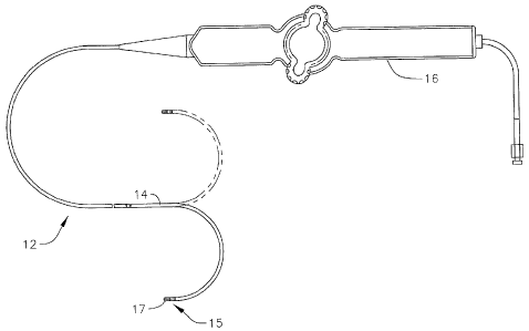
[0018] FIG. 1 is a side view of an embodiment of a catheter of the
present invention.
[0019] FIG. 2A is a side cross-sectional view of the catheter FIG.
1, showing a
junction between a catheter body and a deflectable intermediate section, taken
along a
first diameter.
[0020] FIG. 2B is a side cross-sectional view of the catheter of
FIG. 1, showing a
junction between a catheter body and a deflectable intermediate section, taken
a long a
second diameter generally perpendicular to the first diameter.
[0021] FIG. 2C is a longitudinal cross-section view of the
deflectable intermediate
section of FIGS. 2A and 2B taken along line c--c.
[0022] FIGs. 3A-3C are schematic diagrams of an embodiment of a
control handle
showing the catheter in the neutral and deflected positions.
[0023] FIG. 4 is a top plan view of an embodiment of a control
handle, including a
deflection control assembly.
[0024] FIG. 5 is a partial side perspective view of an embodiment of a
deflection arm
and a tension adjustment knob as mounted on a control handle.
[0025] FIGs. 6A and 6B are perspective top and bottom views of an
embodiment of a
rocker member as used in a deflection control assembly.
[0026] FIG. 7 is a side elevational view of an embodiment of a
pulley as used in a
deflection control assembly.
[0027] FIG. 8 is an exploded perspective view of an embodiment of a
tip electrode
assembly.
-6-
CA 02738221 2011-04-21
'
1
[0028] FIG. 9 is a cross sectional perspective view of an embodiment
of a tip
electrode assembly.
[0029] FIG. 9A is a longitudinal cross sectional view of the tip electrode
assembly of
FIG. 9, taken along line a--a
[0030] FIG. 9B is a longitudinal cross sectional view of the tip
electrode assembly of
FIG. 9, taken along line b--b
[0031] FIG. 9C is a longitudinal cross sectional view of the tip
electrode assembly of
FIG. 9, taken along line c--c
[0032] FIG. 9D is a longitudinal cross sectional view of the tip
electrode assembly of
FIG. 9, taken along line d--d
[0033] FIG. 9E is a longitudinal cross sectional view of the tip
electrode assembly of
FIG. 9, taken along line e--e
[0034] FIG. 9F is a longitudinal cross sectional view of the tip electrode
assembly of
FIG. 9, taken along line f--f
[0035] FIG. 10 is an exploded perspective view of an alternate
embodiment of a tip
electrode assembly.
[0036] FIG. 10A is an end cross-sectional view of an alternate
embodiment of an
internal member.
[0037] FIG. 10B is an end cross-sectional view of another alternate
embodiment of
an internal member.
DETAILED DESCRIPTION OF THE INVENTION
[0038] FIG. 1 illustrates an embodiment of a catheter 10 with improved
position
sensing and cooling capabilities. The catheter has an elongated catheter body
12 with
proximal and distal ends, an intermediate deflectable section 14 at the distal
end of the
catheter body 12, and a distal section 15 with an irrigated mapping and
ablation tip
-7-
CA 02738221 2011-04-21
. .
1
electrode 17. The catheter also includes a control handle 16 at the proximal
end of the
catheter body 12 for controlling bi-directional deflection of the intermediate
section 14.
Advantageously, the tip electrode 17 houses an electromagnetic position sensor
in a distal
and on-axis location while shielding the sensor from RF ablation and bending
stresses.
The tip electrode is also configured to promote turbulent flow and dispersion
of irrigation
fluid for increased thermal transfer from the shell to the fluid and thus with
lower flow
rates resulting in lower fluid load in the patient. Fluid, e.g., saline or
heparinized saline,
can be delivered to the ablation site from the tip electrode to cool tissue,
reduce
coagulation and/or facilitate the formation of deeper lesions. It is
understood that other
fluids can be delivered as well, including any diagnostic and therapeutic
fluids, such as
neuroinhibitors and neuroexcitors.
[0039] With
reference to FIGS. 2A and 2B, the catheter body 12 comprises an
elongated tubular construction having a single, axial or central lumen 18. The
catheter
body 12 is flexible, i.e., bendable, but substantially non-compressible along
its length.
The catheter body 12 can be of any suitable construction and made of any
suitable
material. A presently preferred construction comprises an outer wall 20 made
of
polyurethane or PEBAX. The outer wall 20 comprises an imbedded braided mesh of
stainless steel or the like to increase torsional stiffness of the catheter
body 12 so that,
when the control handle 16 is rotated, the intermediate section 14 of the
catheter 10 will
rotate in a corresponding manner.
[0040] The outer
diameter of the catheter body 12 is not critical, but is preferably no
more than about 8 french, more preferably 7 french. Likewise the thickness of
the outer
wall 20 is not critical, but is thin enough so that the central lumen 18 can
accommodate
puller members (e.g., puller wires), lead wires, and any other desired wires,
cables or
tubings. If desired, the inner surface of the outer wall 20 is lined with a
stiffening tube 22
to provide improved torsional stability. A disclosed embodiment, the catheter
has an
-8-
CA 02738221 2011-04-21
1
outer wall 20 with an outer diameter of from about 0.090 inch to about 0.94
inch and an
inner diameter of from about 0.061 inch to about 0.065 inch.
[0041] Distal ends of the stiffening tube 22 and the outer wall 20 are
fixedly attached
near the distal end of the catheter body 12 by forming a glue joint 23 with
polyurethane
glue or the like. A second glue joint 25 is formed between proximal ends of
the stiffening
tube 20 and outer wall 22 using a slower drying but stronger glue, e.g.,
polyurethane.
[0042] Components that extend between the control handle 16 and the
deflectable
section 14 pass through the central lumen 18 of the catheter body 12. These
components
include lead wires 40 for the tip electrode 17 and ring electrodes 21 on the
tip section, an
irrigation tubing 38 for delivering fluid to the tip section 15, a cable 48
for the position
location sensor 46, a pair of puller wires for deflecting the intermediate
section 14, and a
pair of thermocouple wires 41, 45 to sense temperature at the distal tip
section 15. Glue
joint 28 affixes the proximal portion of the components inside the stiffening
tube.
[0043] Illustrated in FIGS. 2A, 2B and 2C is an embodiment of the
intermediate
section 14 which comprises a short section of tubing 19. The tubing also has a
braided
mesh construction but with multiple off-axis lumens, for example lumens 26,
27, 30 and
32. Each of diametrically opposing first and second lumens 26 carries a puller
wire 36
for bi-directional deflection. A third lumen 30 carries the lead wires 40, the
thermocouple wires 41 and 45, and the sensor cable 48. A fourth lumen 32
carries the
irrigation tubing 38.
[0044] The tubing 19 of the intermediate section 14 is made of a
suitable non-toxic
material that is more flexible than the catheter body 12. A suitable material
for the tubing
19 is braided polyurethane, i.e., polyurethane with an embedded mesh of
braided stainless
steel or the like. The size of each lumen is not critical, but is sufficient
to house the
respective components extending therethrough.
-9-
CA 02738221 2011-04-21
1
[0045] A means for attaching the catheter body 12 to the
intermediate section 14 is
illustrated in FIGs. 2A and 2B. The proximal end of the intermediate section
14
comprises an outer circumferential notch 24 that receives an inner surface of
the outer
wall 20 of the catheter body 12. The intermediate section 14 and catheter body
12 are
attached by glue 29 or the like.
[0046] If desired, a spacer (not shown) can be located within the
catheter body
between the distal end of the stiffening tube (if provided) and the proximal
end of the
intermediate section. The spacer provides a transition in flexibility at the
junction of the
catheter body and intermediate section, which allows this junction to bend
smoothly
without folding or kinking. A catheter having such a spacer is described in
U.S. Pat. No.
5,964,757, the disclosure of which is incorporated herein by reference.
[0047] Each puller wire 36 is preferably coated with Teflon®
The puller wires
36 can be made of any suitable metal, such as stainless steel or Nitinol and
the Teflon
coating imparts lubricity to the puller wire. The puller wire preferably has a
diameter
ranging from about 0.006 to about 0.010 inch.
[0048] As shown in FIG. 2B, a portion of each puller wire 36
extending through the
catheter body 12 passes through a compression coil 35 in surrounding relation
to its
puller wire 36. The compression coil 35 extends from the proximal end of the
catheter
body 12 to the proximal end of the intermediate section 14. The compression
coil 35 is
made of any suitable metal, preferably stainless steel, and is tightly wound
on itself to
provide flexibility, i.e., bending, but to resist compression. The inner
diameter of the
compression coil is preferably slightly larger than the diameter of the puller
wire 36.
Within the catheter body 12, the outer surface of the compression coil 35 is
also covered
by a flexible, non-conductive sheath 39, e.g., made of polyimide tubing. As
shown in
FIGS. 2B and 2C, a portion of each puller wire 36 extending through the
intermediate
section 14 is covered by a nonconductive protective sheath 47.
-10-
CA 02738221 2011-04-21
. ,
1
[0049] Proximal ends of the puller wires 36 are anchored in the
control handle 16.
Distal ends of the puller wires 36 are anchored in the tip section 15 as
described further
below. Separate and independent longitudinal movement of the puller wire 36
relative to
the catheter body 12 which results in deflection of the intermediate section
14 and tip
section 15 is accomplished by suitable manipulation of the control handle 16.
[0050] In the illustrated embodiment, the control handle 16 has a
deflection assembly
60 (FIG. 4) with a deflection arm 62 (FIG. 5), and a rotatable or rocker
member 64
(FIGS. 6A and 6B) supporting a pair of pulleys 66 (FIG. 7) that act on the
puller wires 36
to deflect the intermediate section 14 and thus the tip section 15. The
deflection arm 62
and the rocker member 64 are rotationally aligned and coupled such that
rotation of the
deflection arm 62 by a user rotates the rocker member 64. As the rocker member
64 is
rotated by means of the deflection arm (represented by line 62), the pulleys
66 are
displaced from a neutral position (FIG. 3A) with one pulley 66 drawing a
puller wire 36
on one side of the catheter against its anchored proximal end 37 for
deflecting the section
14 toward that side (FIGS. 3B and 3C). Components such as the lead wires,
irrigating
tubing and sensor cable can extend through the rocker member 64 within a
protective
tubing 68. A deflection tension knob 67 (FIG. 5) enables the user to adjust
the ease by
which the deflection arm 62 can be rotated. A suitable deflection assembly and
control
handle are described in co-pending U.S. Application Serial No. 12/346,834,
filed
December 30, 2008, entitled DEFLECTABLE SHEATH INTRODUCER, the entire
disclosure of which is hereby incorporated by reference. Other suitable
deflection
assemblies are described in co-pending U.S. Application Serial No. 12/211,728,
filed
September 16, 2008, entitled CATHETER WITH ADJUSTABLE DEFLECTION
SENSITIVITY, and U.S. Application Serial No. 12127704, filed May 27, 2008,
entitled
STEERING MECHANISM FOR BI-DIRECTIONAL CATHETER, the entire
disclosures of both of which are hereby incorporated by reference.
-11-
CA 02738221 2011-04-21
=
1
[0051] At the distal end of the intermediate section 14 is the tip
section 15 that
includes the tip electrode 17 and a relatively short piece of connector tubing
53 between
the tip electrode 17 and the intermediate section 14. In the illustrated
embodiment of
FIGS. 8 and 9, three ring electrodes 21 are mounted on the tubing 53 and the
tubing 53
has a single lumen which allows passage of the tip electrode lead wire 40T,
the
electromagnetic sensor cable 48, thermocouple wires 41 and 45, and the
irrigation tubing
38 into the tip electrode 17. The single lumen of the connector tubing 53
allows these
components to reorient themselves as needed from their respective lumens in
the
intermediate section 14 toward their location within the tip electrode 17.
[0052] The tip electrode 17 defines a longitudinal axis 50 and is of
a two piece
configuration that includes an electrically conductive shell or dome 51 and
internal
member or housing 52. The shell is generally cylindrical configuration. It has
a
narrower open neck portion 56 that is proximal of a wider distal portion 54.
The distal
portion has an atraumatic distal end 72 with a flat distal surface and a
rounded
circumferential edge. The distal portion has an inner wall 58 that defines a
generally
cylindrical cavity 70 within the shell. The proximal neck portion 56 is
aligned and on
axis with the longitudinal axis 50. It is understood that the neck portion 56
need not be
narrower than the distal portion 54. Indeed, the two portions may have the
same
diameter, except the distal portion 54 is exposed whereas the neck portion 56
is covered
by the connector tubing 43.
[0053] The shell 51 is constructed of a biocompatible metal,
including a
biocompatible metal alloy. A suitable biocompatible metal alloy includes an
alloy
selected from stainless steel alloys, noble metal alloys and/or combinations
thereof. In
one embodiment, the shell is constructed of an alloy comprising about 80%
palladium
and about 20% platinum by weight. In an alternate embodiment, the shell is
constructed
of an alloy comprising about 90% platinum and about 10% iridium by weight. The
shell
-12-
CA 02738221 2011-04-21
1
51 can formed by deep-drawing manufacturing process which produces a
sufficiently thin
but sturdy wall that is suitable for handling, transport through the patient's
body, and
tissue contact during mapping and ablation procedures. A deep drawn shell is
also
suitable for electrical discharge machining (EDM) process to form a large
plurality of
through-holes or ports 74 in the distal portion 54 that allow communication
between the
cavity 70 and outside the shell 51. In a disclosed embodiment, the shell has a
wall
thickness ranging between about 0.002" and 0.005", preferably between about
0.003" and
0.004", and the wall has a plurality of holes ranging between about 21 and
140,
preferably between about 33 and 60, more preferably between about 33 and 57,
where a
diameter of each hole can range between about 0.002" and 0.010", preferably
between
about 0.003" and 0.004", and preferably about 0.004 inch in diameter.
[0054] The internal member 52 is configured to protect and
encapsulate the sensor 46
in a distal and centered location within the cavity 70 so that the sensor is
distal and
centered in the tip electrode for optimum performance. That is, the more
centered the
sensor is in the tip electrode and the closer the sensor is to the distal end
of the tip
electrode, the more accurate is the data provided by the sensor. In the
illustrated
embodiment, the entirety of the internal member 52 is received in the shell
51.
[0055] The internal member 52 has an elongated configuration that is
aligned and on-
axis with the longitudinal axis 50 of the tip section 15. Advantageously, the
internal
member has a tubular distal portion 80, a baffle mid-portion 81, a stem
portion 82, and a
proximal base portion 83. Extending through the entire length of the internal
member is
an on-axis passage 84 to receive the sensor 46 and the sensor cable 48. In a
disclosed
embodiment, the tubular distal portion 80 is situated generally in the cavity
70 of the
shell, and the baffle, stem and base portions 81, 82, 83 are situated
generally in the neck
portion 56 of the shell. That is, the two piece configuration allows the
internal member
52 to be inserted and received in shell 51, where the tubular distal portion
80 extends in
-13-
CA 02738221 2011-04-21
. ,
1
the distal portion 54 of the shell 51, and the proximal remainder (the baffle
mid-portion
81, the stem portion 82 and the base portion 83) extends in the neck portion
56 of the
shell 51.
[0056] The base portion 83 of the internal member 52 has a circular
cross section
(FIG. 90 that is adapted for a snug fit with the neck portion 56 of the shell
to form a
fluid-tight seal at the proximal end of the tip electrode 17.. The base
portion can have a
thickness ranging between about 0.003" to 0.004".
[0057] Distal the base portion is the narrowed stem portion 82 which
creates an open
annular gap 88 within the shell 51 between the base portion 83 and the baffle
mid-portion
81 (FIG. 9e). The width of the stem portion can range between about 0.090" to
0.110".
[0058] The illustrated embodiment of the baffle mid-portion 81
includes an
equilateral triangular cross-section (FIG. 9d) with three edges 90 spanning
between three
truncated corners 92 that are in circumferential contact with the neck portion
56 of the
shell 51. This contact advantageously enables a snug and on-axis (or centered)
fit
between the shell 51 and the internal member 52. The triangular cross-section
also
advantageously creates different axial flow paths or channels 94 for fluid
passing into the
tip electrode 17. The fluid flowing into the cavity 70 of the shell 51 is
separated into
distinct flow paths by the baffle mid-portion 81. These flow paths facilitate
dispersion of
fluid entering the tip electrode 14 at the base portion. It is understood that
the cross-
section of the baffle portion 81 need not be limited to a triangular
configuration, but
could be polygonal, including quadrilateral or pentagonal, so long as multiple
flow paths
are formed and turbulence is generated without significant drop in fluid
pressure. The
length of the baffle portion between its distal and proximal end can range
between about
0.050" to 0.200".
[0059] The tubular portion 80 has a length and an inner diameter so
that it can receive
the sensor 46 in its entirety and leave a gap 100 between the distal end of
the tubular
-14-
CA 02738221 2011-04-21
1
portion and a distal end of the sensor. A conventional sensor has a diameter
about lmm
and a length about 5 mm. The gap 100 is filled by a sealant 101 (FIG. 9A),
such as
polyurethane, so that the sensor is effectively fixed, sealed and protected in
the tubular
portion 80. The tubular portion has a length that ranges between about 60% to
90% of
the length of the cavity, and preferably about 80%. In an alternate
embodiment, the
tubular portion is a separate component from the baffle portion and is sealed
to the latter.
The baffle portion, 81, must be made of electrically conductive material, but
the tubular
portion can be made of plastic such as polyimide. The tubular portion has an
outer
diameter that ranges between about 25% and 40% of the diameter of the cavity
70, and
preferably about 30% (FIGS. 9B and 9C). These differences in length and
diameter
advantageously leave a distal gap 102 between a distal end of the shell 51 and
a distal end
of the tubular portion 80, and an annular region 104 spanning at least the
length of the
tubular portion for improved fluid dispersion and flow in the tip electrode.
In the
illustrated embodiment, the tubular portion 80 has a circular cross-section,
although it is
understood that the cross-section can be any appropriate shape, including any
polygonal
configuration, e.g., triangular, rectangular, etc.).
[0060] At the proximal end of the sensor 46, the passage 84 through
the internal
member 52 narrows to form a stop 106 (FIG. 9C) to abut against the proximal
end of the
sensor 46. A junction of the sensor and the sensor cable lies at the stop and
the sensor
cable extends proximally therefrom through the reminder of the passage 84 and
into the
intermediate section 14. The junction between the cable 48 and the sensor 46
is thus
hidden inside the internal member 52, surrounded by the internal member and
better
protected against cable detachment and bending stresses. This feature also
enables an
overall shorter length in the tip electrode allowing for a more maneuverable
catheter.
[0061] Other formations in the base portion of the internal member
include through-
holes 85, 86A, 86B, 87A, and 87B. A distal end of the irrigation tubing 38
terminates
-15-
CA 02738221 2011-04-21
1
and is anchored in the fluid through-hole 85. Distal ends of the thermocouple
wires 41
and 45 are fixed in the hole 87A. A distal end of the tip electrode lead wire
40T is
anchored in the through-hole 87B The tip electrode lead wire 40 energizes the
shell 51
and at least the base portion 83 of the internal member 52. Distal end of each
puller wire
has a T-anchor, as known in the art. The T-anchors are soldered in
diametrically-
opposing through-holes 86A 86B so that the puller wires are anchored to the
base portion
83 and not a tubing wall which can tear. So anchored in the holes 86A 86B the
puller
wires provide the catheter with symmetrical bi-directional deflection of the
intermediate
section 14. The base portion can also include a circumferential lip 106 at the
proximal
face as an abutment for a proximal end of the shell 51 so as to maintain the
gap 102
between the distal end of the tubing portion 80 and the distal end of the
shell 51. The lip
and the proximal end of the shell 51 can be fixedly joined, for example, by
laser welding.
[0062] In accordance with another feature of the present invention, the
fluid through-
hole 85 is aligned with the baffle mid-portion 81 such that the hole 85 faces
an edge 90 so
fluid exiting the hole 85 impinges on the edge 90 and diffuses around the stem
portion
82. This alignment between the hole 85 (and the irrigation tubing 38) and the
edge 90,
combined with the annular gap 88 provided by the stem portion 82, enables a
flow that is
more uniform and equal in the radial direction through the flow paths 94 which
in turn
provides increased turbulence and a more uniform flow rate in the annular
space 104 of
the cavity 70 and thus more increased convective cooling on the shell 51.
Irrigation in
the tip electrode is thus more uniform throughout the length of the tip
electrode. The
internal member thus effectively counters the tendency for the velocity of the
fluid
entering the tip electrode to carry the fluid to the more distal ports 74 and
starve the more
proximal ports 74.
[0063] The cross-section of the off-axis through hole 85 for the
irrigation tubing 38 is
elongated, that is, more oval than circular as defined by a greater dimension
Y and a
-16-
CA 02738221 2011-04-21
=
1
lesser dimension X generally perpendicular to greater dimension Y. In the
disclosed
embodiment of FIG. 9f, the cross-section is elongated with a curvature C, to
provide, for
example, a kidney-bean or crescent shape cross-section. The present invention
recognizes that a cross-section which is at least elongated if not also curved
provides a
through-hole that can provide greater fluid flow into the tip electrode with
less
interference with the on-axis location of the internal passage 84 and the
sensor cable 48.
[0064] Because the irrigation tubing 85 is flexible, e.g., being
made of polyurethane,
the irrigation tubing 38 readily adapts to the shape of the through-hole 85.
As irrigation
fluid is delivered by the tubing 38 into the tip electrode 17 through the
through-hole 85, it
enters and flows into the annular gap 88 at the stem portion 82 where it is
dispersed by
the baffle portion 81 and flows into the flow channels 94 defined by the edges
90 and
corners 92. As the fluid enters the cavity 70 between the tubular portion 80
and the shell
51, it further disperses in the cavity 70 and ultimately leaves the cavity via
ports 74. The
catheter 10 provides better flow and dispersion of fluid within the tip
electrode for
improved if not exceptional cooling characteristics during ablation. The tip
electrode of
the present invention can operate at about 12 ml/minute or lower for wattage
below or
above 30. The reduction in fluid-loading on the patient in a five or six hour
procedure
can thus be very significant. Moreover, where the flow rate is regulated by a
programmable pump, the flow rate can even be lower for lower wattage.
[0065] In an alternate embodiment of FIG. 10, the internal member
52 includes radial
projections or fins 110 that extend outwardly from the tubular portion 80 in a
direction
generally perpendicular to the longitudinal axis 50 of the tip electrode. The
fins 110
serve to decrease the velocity of the fluid as it travels distally in the
annular region 104 of
the cavity 70 in the tip electrode. In FIG. 10, the fins are thin annular
discs located at
intermittent locations, if not equidistant to each other, along the length of
the tubular
portion 80. In one embodiment, the fin diameter increases in the proximal
direction, so
-17-
CA 02738221 2011-04-21
1
that the effect of decreasing fluid velocity is greatest when the fluid first
enters the
annular space 104 in the tip electrode 17 for a more uniform dispersion of
fluid along the
length of the tip electrode and through all ports 74 in the shell 51 to the
exterior of the
shell.
[0066] Also in the embodiment of FIGS. 10 and 10B, the baffle mid-
portion 81 has a
star-shaped cross-section with a plurality of projections or arms 93 that span
outwardly in
a uniform radial pattern, with ends in circumferential contact with the neck
portion 56 of
the shell 51, again forming distinct axial flow paths 94 between the arms.
However, it is
understood that the present invention also includes a cross-section where
there is no
circumferential contact between the baffle mid portion 81 and the neck portion
56, such
as illustrated in FIG. 10A. There, different but not necessarily distinct
axial flow paths or
channels 94 are provided, which also facilitate dispersion and flow into the
annular space
104 of the tip electrode.
[0067] The entirety of the internal member can also constructed of
the
aforementioned materials of the shell. And where at least the tubular portion
80 is
constructed of a conductive metal, including the palladium platinum alloy, the
EM sensor
is shielded from RF ablation or a stiff plastic such as polyimide. Metal foil
can also be
used shield the sensor as long as it is electrically connected to the overall
electrode
housing. The present invention also includes an alternate embodiment where
portions of
the internal member, for example, tubular portion 80 and the housing 52 are
constructed
of another material, such as plastic, polyimide, polyurethane or PEBAX, to
reduce cost.
[0068] A length of the tip electrode from a distal end of the shell
to a proximal end of
the internal member can range between about 2 mm to 12 mm, and preferably
between
about to 3mm to lOmm.
[0069] The ring electrodes 21 which are mounted on the connector
tubing 53 can be
made of any suitable solid conductive material, such as platinum or gold,
preferably a
-18-
CA 02738221 2011-04-21
1
combination of platinum and iridium. The ring electrodes can be mounted onto
the
connector tubing 53 with glue or the like. Alternatively, the ring electrodes
can be formed
by coating the tubing 53 with an electrically conducting material, like
platinum, gold
and/or iridium. The coating can be applied using sputtering, ion beam
deposition or an
equivalent technique. The number of the ring electrodes on the tubing 53 can
vary as
desired. The rings may be monopolar or bi-polar. In the illustrated
embodiment, there
are a distal monopolar ring electrode and a proximal pair of bi-polar ring
electrodes.
Each ring electrode is connected to a respective lead wire 40R.
[0070] Each lead wire 40R is attached to its corresponding ring
electrode by any
suitable method. A preferred method for attaching a lead wire to a ring
electrode
involves first making a small hole through the wall of the non-conductive
covering or
tubing. Such a hole can be created, for example, by inserting a needle through
the non-
conductive covering and heating the needle sufficiently to form a permanent
hole. The
lead wire is then drawn through the hole by using a microhook or the like. The
end of the
lead wire is then stripped of any coating and welded to the underside of the
ring
electrode, which is then slid into position over the hole and fixed in place
with
polyurethane glue or the like. Alternatively, each ring electrode is formed by
wrapping a
lead wire around the non-conductive covering a number of times and stripping
the lead
wire of its own insulated coating on its outwardly facing surfaces.
[0071] The tip electrode 17 is electrically connected to a source
of ablation energy by
the lead wire 40T. The ring electrodes 21 are electrically connected to an
appropriate
mapping or monitoring system by respective lead wires 40R.
[0072] The lead wires 40T and 40R pass through the lumen 30 of the tubing
19 of the
deflectable intermediate section 14 and the central lumen of the catheter body
12. The
portion of the lead wires extending through the central lumen 18 of the
catheter body 12,
and proximal end of the lumen 24 can be enclosed within a protective sheath
(not shown),
-19-
CA 02738221 2011-04-21
1
which can be made of any suitable material, preferably polyimide. The
protective sheath
is anchored at its distal end to the proximal end of the intermediate section
14 by gluing it
in the lumen 24 with polyurethane glue or the like. Each electrode lead wire
has its
proximal end terminating in a connector at the proximal end of the control
handle 16.
[0073] Whereas conventional construction methods build a tip
electrode "from the
outside in," the present two piece construction allows for construction "from
the inside
out." That is, the two piece construction of the tip electrode also allows
different order or
sequences of catheter assembly. For example, the ring electrodes 21 can be
mounted on
the connector tubing 53 at a stage separate from the assembly of the tip
electrode 17. The
tubing, puller wires, sensor and the thermocouple can be added to the tip
electrode at a
later stage or time compared to conventional catheter assembly methods.
[0074] Significantly, the two-piece configuration and assembly of
the tip electrode 17
allows for testing, evaluation and inspection of the interior of the tip
electrode before the
tip electrode is fully assembled. One method of assembling the tip electrode
includes
inserting the sensor 46 and cable 48 into the central passage 84 of the
internal member 52
so that the sensor is received in the tubular portion 80 of the internal
member (with the
sensor's proximal end abutting the stop 106) and the cable 48 extends distally
through the
central passage 84 and out the proximal face of the base portion 83.
Thereafter, the
sensor 46 is sealed within the tubular portion 80 by sealant 101 filling the
distal end the
tubular portion 80. Anchoring and attachment of distal ends of lead wire 40
for the tip
electrode, puller wires 36 and thermocouple wires 41, 45 are then made to the
base
portion 83 of the internal member 52 in the respective holes 86a, 86b, 87a,
87b by means
including T-bar anchoring and/or soldering. A distal end of the irrigation
tubing 38 is
then inserted to the elongated hole 85 and affixed by adhesive. It is
understood that each
of these anchorings and attachments in the holes in the base portion forms a
fluid-tight
seal so that irrigation fluid cannot escape into the connector tubing 53
proximal the tip
-20-
CA 02738221 2011-04-21
1
electrode 17. After such stages of assembly have been met, the functionality
and
integrity of the tip electrode, including the tip and ring electrodes, the
various electrical,
component and fluid junctions and connections, and the various fluid-tight
seals can be
advantageously tested, evaluated and inspected before the shell is received on
the internal
member. This feature is another significant advantage over conventional
ablation and
mapping catheters where testing is done "blind" without easy accessibility to
the interior
of the tip electrode.
[0075] After testing of the tip electrode, the shell 51 can be placed over
the internal
member 52 centered and aligned by the contact between the corners 92 of the
baffle
portion 81 and the neck 56 portion of the shell 51. The shell is then attached
to the baffle
portion via press fit, glue, electrical or laser welding, mechanical
deformation, or some
other means of joining the two parts. The connector tubing 53 is then be slid
over the
neck portion 56 and connected to a distal end of the tubing 19 of the
deflectable
intermediate section 14.
[0076] The preceding description has been presented with reference
to certain
exemplary embodiments of the invention. Workers skilled in the art and
technology to
which this invention pertains will appreciate that alterations and changes to
the described
structure may be practiced without meaningfully departing from the principal,
spirit and
scope of this invention. It is understood that the drawings are not
necessarily to scale.
Accordingly, the foregoing description should not be read as pertaining only
to the
precise structures described and illustrated in the accompanying drawings.
Rather, it
should be read as consistent with and as support for the following claims
which are to
have their fullest and fairest scope.
-21-