Note: Descriptions are shown in the official language in which they were submitted.
CA 02743187 2011-06-10
FOLDING ENDOSCOPE AND METHOD OF USING THE SAME
CROSS REFERENCE TO RELATED U.S PATENT APPLICATION
This patent application relates to U.S. provisional patent application
Serial No. 61/353,948 filed on 11 Jun 2010 entitled FOLDING
ENDOSCOPE AND METHOD OF USING THE SAME, filed in English,
which is incorporated herein in its entirety by reference.
FIELD OF THE INVENTION
The present invention relates generally to the field of endoscopes,
and more particularly the present invention relates to folding endoscopes
with incorporated optical sensors and light source.
BACKGROUND OF THE INVENTION
Generally speaking, endoscopes are thin tubular cameras that are
typically utilized in the diagnosis of a disease. These cameras are usually
inserted into the body cavity either through a natural opening like the mouth
or the anus or through a tiny incision made into the skin. The endoscopes
are extensively used intra-operatively to assist the surgeon in visualizing
the anatomy of interest to perform the procedure and to avoid damage to
critical surrounding organs. Most of the endoscopes available in the market
to date can be classified into either a rigid or a flexible endoscope.
Commonly found endoscopes are available with two-dimensional cameras
and have limited image resolution and depth perception. These
1
CA 02743187 2011-06-10
endoscopes are disorienting to the surgeon after a prolonged use and lack
the natural spectrum of direct human visualization.
Recently some manufacturers have started producing three-
dimensional (stereoscopic) endoscopes. The optical version of these
endoscopes use two tubular lenses inside a long shaft and two standard
cameras mounted outside of the body. The next generation of stereo
endoscopes employs custom designed semiconductor circuitry mounted at
the tip of the endoscope (inside the body) that is capable of producing
stereo images. In these endoscopes, either two close proximity mounted
chips or a special chip with a large array of micro lenses manufactured onto
the chip is utilized to create stereo images. In addition, such endoscopes
also include LED or fiber optic light sources for illumination. Figure 1 shows
a conventional stereo endoscope.
U.S. Patent 4,862,873 issued to Yajima et at. discloses a stereo
endoscope that utilizes two thin optical guides mounted in a tubular shaft
and two CCD image sensors mounted outside the body to create three-
dimensional images of the organ.
U.S. Patent application US2002/00071 10, to Irion discloses a stereo
endoscope that utilizes two lateral mounted cameras with a flexible
endoscope head to create three-dimensional images of the organ.
The field of surgical intervention has evolved from open invasive
approach to the paradigm of minimally invasive surgery due to its benefits
to the patients and the healthcare system. From the surgeon's perspective,
the transition has resulted in a procedure with limited and un-natural field
of
view and surgical skills that have a steep learning curve. The existing three-
2
CA 02743187 2011-06-10
dimensional endoscopes have resulted in incremental enhancement to the
visualization, but have failed to match the natural spectrum of direct human
visualization. The 3D depth perception of these endoscopes is also
constrained by the limited physical separation between the two cameras.
Additionally, it is projected that the surgical paradigm will shift from the
three or four incision laparoscopic approach to a single incision (single port
access (SPA)) surgery.
Thus, there is a need and good market potential for improved
endoscopes that can provide a better visualization of the surgical site.
SUMMARY OF THE INVENTION
The present invention provides a foldable endoscope, comprising:
a) a housing having a first and second end and a longitudinal axis,
said housing including at least one channel extending between said first
and second end, and associated ports at said first and second end for
inserting surgical instruments through said housing into a surgical site;
b) at least two elongate arms each having a first and second end
and each being pivotally connected at said first end thereof to said first
end of said housing;
c) at least one camera each camera being mounted on one of said
at least two elongate arms; and
d) a linkage mechanism connected to said at least two elongate
arms, said linkage mechanism, upon activation, being configured to
pivotally deploy said at least two elongate arms from a closed position in
which said at least two elongate arms are aligned along said longitudinal
3
CA 02743187 2011-06-10
axis to an open position in which said second ends of said at least two
elongate arms radially spaced from said longitudinal axis.
The disclosed endoscope taps nicely into the emerging market due
to its improved visualization capabilities and integrated support to pass
surgical tools through the other ports making it a versatile surgical tool.
A further understanding of the functional and advantageous aspects
of the invention can be realized by reference to the following detailed
description and drawings.
BRIEF DESCRIPTION OF THE DRAWINGS
Preferred embodiments of the invention will now be described, by
way of example only, with reference to the drawings, in which:
Figure 1 shows a conventional prior art stereo endoscope;
Figure 2 shows an embodiment of the foldable endoscope in a fully
closed or retracted state;
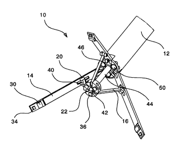
Figure 3 shows the foldable endoscope in a partially open state;
Figure 4 shows the foldable endoscope in the fully open state;
Figure 5 shows another embodiment of the foldable endoscope
that includes a mirror arrangement; and
Figure 6 shows the block diagram of the image processing of the
images acquired by the cameras mounted on the foldable endoscope.
DETAILED DESCRIPTION OF THE INVENTION
Without limitation, the majority of the systems described herein are
directed to folding endoscopes with incorporated optical sensors and light
4
CA 02743187 2011-06-10
source. As required, embodiments of folding endoscopes are disclosed
herein. However, the disclosed embodiments are merely exemplary, and it
should be understood that the disclosure may be embodied in many
various and alternative forms. In certain instances, well-known or
conventional details are not described in order to provide a concise
discussion of embodiments of the present disclosure.
The Figures are not to scale and some features may be
exaggerated or minimized to show details of particular elements while
related elements may have been eliminated to prevent obscuring novel
aspects. Therefore, specific structural and functional details disclosed
herein are not to be interpreted as limiting but merely as a basis for the
claims and as a representative basis for teaching one skilled in the art to
variously employ the present invention. For purposes of teaching and not
limitation, the illustrated embodiments are directed to folding endoscopes
with incorporated optical sensors and light source.
As used herein, the term "about" and "approximately", when used in
conjunction with ranges of dimensions, temperatures or other physical
properties or characteristics is meant to cover slight variations that may
exist in the upper and lower limits of the ranges of dimensions so as to not
exclude embodiments where on average most of the dimensions are
satisfied but where statistically dimensions may exist outside this region.
For example, in embodiments of the present invention dimensions of
components of a folding endoscope are given but it will be understood that
these are not meant to be limiting.
5
CA 02743187 2011-06-10
Referring to Figure 2, herein is disclosed a foldable endoscope 10
that utilizes multiple cameras 30 to create three-dimensional images of the
target. Figure 2 shows the endoscope 10 in collapsed form, and Figure 3
shows the endoscope 10 in half open form. In the collapsed form, the
endoscope 10 assumes a very compact formation and can be easily
introduced into the patient's body through a standard trocar. In the
preferred embodiment, the endoscope 10 contains a slender body 12 that
forms a generally cylindrical housing, a center spoke 40, three connecting
linkages 16, and three folding arms 14 with cameras 30 and light sources
34 integrated into each of the arms. Each folding arm 14 in the preferred
embodiment includes two hinge joints; a first hinge joint 18 with the
endoscope body 12 and a second hinge joint 20 with the connecting
linkage 16. In the preferred embodiment, each connecting linkage 16 also
has a hinge joint 22 with the center spoke 40. The center spoke 40
includes a telescopingly movable hollow drive shaft 44 and may optionally
include a plurality of integrated light sources 36 (light emitting diodes
(LEDs), fiber optic light sources, etc).
The optical sensors or cameras are preferably charge coupled
device (CCD) images sensors, but other types of image sensors may be
used. For example, complementary metal-oxide-semiconductor (CMOS)
image sensors may be preferred in some embodiments due to their low
cost.
The endoscope 10 also includes one or more instrument ports 50
through which various surgical instruments can be introduced to perform
the procedure. Non-limiting examples of such instruments include
6
CA 02743187 2011-06-10
scalpels, incision devices, tweezers, scissors, etc. In the preferred
embodiment, the diameter of slender body 12 is preferably about 10mm
and the diameter of each instrument port 50 is preferably about 2.5mm.
The disclosed invention is particularly suitable for the case of a single port
access surgery where both the visualization and the surgical procedure is
performed through one incision as opposed to the three or four of a typical
laparoscopic procedure. Endoscope 10 may optionally include a fiber optic
illumination port 42 mounted on the center spoke 40 to enhance visibility of
the surgical site. In the preferred embodiment, the diameter of fiber optic
illumination port 42 is preferably about 1.75mm. The fiber optic illumination
port 42 is a hollow shaft that runs concentrically through the center spoke
40 and the hollow drive shaft 44.
Figure 3 shows the preferred embodiment of the disclosed
invention in the half open form. The hollow drive shaft 44 is designed to
translate in and out through the center port 46 of the endoscope body 12.
Here "in" motion is referred to as the motion of the center spoke 40
towards the endoscope body 12 and "out" motion is referred to as the
motion of the center spoke 40 away from the endoscope body 12. Each
hinge joint (18, 20, and 22) is a low friction joint that allows two mating
components to freely rotate with respect to each other about the hinge
axis. A hollow drive shaft 44 is connected on one end to the center spoke
40 and is connected at the other end to the endoscope body 12 to create
the linear "in" and "out" motion of the center spoke 40 with respect to the
endoscope body 12. In a preferred embodiment, this motion is provided by
an actuator (not shown here, preferably located outside the body). Those
7
CA 02743187 2011-06-10
skilled in the art will appreciate that any actuator may be used; some non-
limiting examples include solenoids, motors with rack and pinion gears,
hydraulic actuators, pneumatic actuators, cable actuators, worm gears,
and wheels with tracks.
A fiber optic illumination source may be passed through the hollow
shaft 44 to enhance visibility of the surgical site. Optionally, one of more
of
the illumination sources 36 on the center spoke 40 may be replaced with
one or more cameras 30 that can facilitate easy insertion of the endoscope
into patient's body cavity.
One preferred method of utilizing the disclosed invention in a single
port access surgery can be as following. Initially with the endoscope 10
outside the body, the hollow shaft 44 is actuated such that the center
spoke 40 is at its farthest "out" position and as a result the endoscope is
fully collapsed (as shown in Figure 2) and can be easily introduced into
the patient's body through a standard trocar. Once inside the body, the
hollow shaft 44 is actuated to cause "in" motion of the center spoke 40
towards the endoscope body 12. The umbrella structure of the mechanism
causes it to unfold and gradually take up its open shape as the center
spoke 40 is actuated towards the fully "in" position (as shown in Figure 4).
The tile angle for cameras 30 can be simultaneously controlled by "in" and
"out" motions of the center spoke 40 in the direction of arrow 64. The
actuator is used to control how much the umbrella structure opens up, and
this depends on the user of endoscope and how much overlap is required
between the cameras 30.
8
CA 02743187 2011-06-10
Once fully deployed, endoscope 10 can be firmly held in place
(outside the body) by an assistant, a passive support arm, or a robotic
system. The endoscope 10 can also be rolled through the use of an
optional second actuator about axis 60 in the direction of arrows 62 until
desired visualization of the anatomy is achieved.
Figure 5 shows another embodiment of the foldable endoscope
that includes a mirror arrangement 14 that assists in the insertion of the
endoscope into the body cavity. In its collapsed form (as shown), the
mirror 14 is oriented such that it reflects light rays that are parallel to
the
longitudinal axis of endoscope body 12 onto the image sensor 30 thereby
creating an image that is orthogonal to the endoscope longitudinal axis.
This image is the same image as obtained using the conventional
endoscopes as they are being inserted into the body cavity. Once inside
the body and after the mechanism has been unfolded, mirror 14 has no
function and the endoscope creates 3D images as explained before. This
mirror arrangement obviates the need of another 2D image sensor on the
center spoke 40 that can assist in endoscope insertion through the trocar.
Figure 6 shows the block diagram of the disclosed invention. The video
outputs 122 from various cameras 120 go to an image processor 100 that
performs various image processing algorithms for example stereo
generation, image stitching etc. on these images. The processed images
are provided to the surgeon's in either a two-dimensional or a three-
dimensional format through the use of a display device 104 or 106
(monitor, projector, 3D monitor, 3D goggles, etc). The image processor
100 may also control camera tilt and roll system 110 in order to generate a
9
CA 02743187 2011-06-10
view from a different orientation and perspective. The roll and tilt system is
preferably composed of two actuators that cause the linear motion of
center spoke 40 along arrows 64 and roll of body 12 about axis 60 (as
shown by arrow 62). Depending upon the anatomy, the image processor
may also control the illumination system 102 to adjust for optimal image
quality. The illumination system 102 may automatically adjust camera
parameters based on feedback from image signals received from the
cameras. The illumination system 102 may include any manual input
device such as a physical button, knob, or slider, or it may be a graphical
user interface element displayed on a monitor. Further, when producing
three-dimensional images, the image processor 100 may further rotate,
translate, and scale the produced images either by decisions made from
software control systems or from manual control from the user, or both.
The surgeon may interact with the image processor 100 through a
user interface that includes an input device (computer mouse, keyboard,
microphone, buttons, joystick, touch screen etc) to select various features
and options. The surgeon can optionally switch between two-dimensional
and three-dimensional views or can visualize them side by side on
displays 104 and 106. The surgeon may use the user interface to change
views, to change brightness or contrast parameters, to rotate, scale, or
translate 3D views, or to make other parameter changes that influences
the display shown on monitors.
Those skilled in the art will appreciate that many computer vision
algorithms may be performed by the image processor 100 including but
not limited to: image stitching, 3D reconstruction from multiple views,
CA 02743187 2011-06-10
shape from shading, depth from focus, feature detection, feature matching
for pose estimation, optical flow algorithms, background subtraction,
automatic object classification, and image segmentation. These
techniques may be used to assist the user of the endoscope in performing
operations with the device. Further, those skilled in the art that the image
processor 100 may be a dedicated computer processor such as a CPU,
DSP microchip, or microprocessor, or the image processor 100 may be
integrated in a computer system such as a software program running on a
desktop computer, laptop, mobile device, or mobile phone.
The disclosed invention utilizes an umbrella type mechanism to
mount and control one or more cameras (preferable two or more) that is
not found in conventional two-dimensional and three-dimensional
endoscopes. The increased physical separation between different
cameras of the disclosed invention will lead to an improved 3D depth
perception than that of the close mounted dual cameras in the existing
systems. The increased number of cameras (preferably three or more)
present in the disclosed invention will lead to enhanced visualization of the
anatomy through image stitching. The mechanism disclosed herein is
fairly simple and low cost to produce. The number of folding arms can be
limited to two if reduced cost or functionality is desired.
As used herein, the terms "comprises", "comprising", "includes" and
"including" are to be construed as being inclusive and open ended, and not
exclusive. Specifically, when used in this specification including claims, the
terms "comprises", "comprising", "includes" and "including" and variations
thereof mean the specified features, steps or components are included.
11
CA 02743187 2011-06-10
These terms are not to be interpreted to exclude the presence of other
features, steps or components.
The foregoing description of the preferred embodiments of the
invention has been presented to illustrate the principles of the invention
and not to limit the invention to the particular embodiment illustrated. It is
intended that the scope of the invention be defined by all of the
embodiments encompassed within the following claims and their
equivalents.
12
