Note: Descriptions are shown in the official language in which they were submitted.
CA 02748262 2016-06-30
SILENT EFFUSION REMOVAL
[0001] This application claims the benefit of U.S. Provisional Application No.
61/140,805, filed December 24, 2008.
FIELD OF THE INVENTION
[0002] The invention generally relates to methods and devices for treatment of
the ear,
which may be supplemental to a tympanocentesis procedure.
BACKGROUND OF THE INVENTION
[0003] Middle ear infections are common in young children. Suffering may be
alleviated
by puncturing the tympanic membrane to evacuate the fluid, a treatment known
as
tympanocentesis. The patient may undergo general anesthesia prior to a
tympanocentesis
procedure, but this is not preferred due to cost and health concerns. As a
preferable
alternative, the tympanic membrane can be locally anesthetized with an
iontophoresis
procedure. Thus, the patient may be treated while awake. Devices and methods
for locally
anesthetizing the tympanic membrane are disclosed in co-assigned patent
applications U.S.
11/962,063, U.S. 11/749,729, and U.S. 61/085,360. Figure IA shows a view of an
outer
ear. The outer ear includes a major element known as the Auricle or Pinna 100.
The outer
ear serves as a funnel for directing sounds into the internal portions of the
ear. The major
physical features of the ear include the Lobule 102, Concha 104, Athelix 106,
Helix 108,
Scapha 110, Triangular fossa 112, Externam acoustic meatus 114, Tragus 116,
and
Antitragus 118.
[0004] Figure 1B shows a cross-section of the inner and outer portions of the
ear. The
pinna 100 is shown connected to the External auditory meatus 118, or ear
canal. The ear
canal 118 is shown as a relatively straight passage, but is often a tortuous
passageway. The
ear canal 118 is connected to the middle ear 120, which includes the ear drum
122. The
middle ear 120 in turn is connected to the internal ear 124. When the middle
ear 120
becomes infected, fluid swells inside the ear drum 122. Fluid expansion causes
extreme
pain to one with a middle ear infection.
1
CA 02748262 2011-06-23
WO 2010/075502 PCT/US2009/069388
[0005] Fluid in the middle ear is commonly known as serous otitis media or
"effusion".
Effusion is normally drained through the tympanocentesis procedure. However,
effusion
may thicken and thus be difficult to remove or drain. Thickening of effusion
is common
with patients who suffer from chronic ear infections. Accordingly, a
tympanocentesis
procedure may not be effective in patients with lodged or thickened effusion.
[0006] Tympanocentesis procedures, which implement iontophoresis, often
require
iontophoresis fluid to be evacuated before the tympanic membrane is punctured.
Evacuation of fluid is commonly performed through low pressure suction via a
syringe or
suction cannula. Fluid evacuation is often a painful and uncomfortable process
because
large amounts of noise are created by fluid cavitation. Thus, fluid evacuation
by suction
may cause pain and emotional discomfort which may prevent the completion of
the
tympanocentesis procedure. It should be noted that many patients are young, 5
and under,
and also have endured many hours or days of a painful ear infection, and thus
may be
uncooperative and difficult to treat. Fluid may also be removed by swabbing
the ear with an
absorbent material, however this can be irritating to the patient and
ineffective as well.
Swabbing also requires the patient to vigorously shake their head side to
side, which many
young patients refuse to comply with.
BRIEF SUMMARY OF THE INVENTION
[0007] One embodiment of the invention may include a method for clearing
effusion from
an ear. The method may include applying liquid to an ear canal, which is
proximal to a
perforated tympanic membrane, which is proximal to a middle ear containing
effusion. The
perforated tympanic membrane may have been intentionally perforated in a prior
tympanocentesis procedure. The effusion may not have been removed by the
normal
tympanocentesis procedure. The method may also include applying an ear device
to seal
and pressurize the liquid inside the ear canal, the ear device regulating the
amount of
pressure inside the ear canal. The method may also include inducing a
Eustachian tube,
which is distal to the middle ear, to open, which causes the fluid to displace
the effusion into
the Eustachian tube.
[0008] Another aspect of the invention may include a method for clearing
effusion from
an ear, the method including applying an ear device to seal an ear canal,
which is proximal
to a perforated tympanic membrane, which is proximal to a middle ear
containing effusion.
2
CA 02748262 2011-06-23
WO 2010/075502 PCT/US2009/069388
The method may also include pressurizing the ear canal with air, and inducing
a Eustachian
tube, which is distal to the middle ear, to open, which causes the pressurized
air to displace
the effusion into the Eustachian tube.
[0009] Yet another aspect of the invention may include a device for
pressurizing an ear
canal, the device including a first ear cup which encloses a first external
ear, the first ear cup
having a first sealing member which fluidly seals around the first external
ear, the first ear
cup having a first port which is in fluid communication with the sealed first
external ear.
The device may also include a second ear cup which encloses a second external
ear, the
second ear cup having a second sealing member which fluidly seals around the
second
external ear, the second ear cup having a second port which is in fluid
communication with
the sealed second external ear. A headpiece may connect to each ear cup, the
headpiece
being configured for applying sealing pressure to each sealing member and
retaining each
ear cup on each respective external ear.
[0010] Yet another aspect of the invention may include a method for clearing
liquid from
an ear canal, the method including applying a device including a wicking
tip to a liquid
solution inside an ear canal to wick the liquid from the ear canal. The liquid
may be left
from a iontophoresis procedure. The method may also include applying negative
pressure
to the device to aid in wicking the liquid, wherein the wicking tip regulates
turbulence to
reduce noise caused by wicking the liquid.
[0011] Yet another aspect of the invention may include a device for clearing
liquid from
an ear canal, the device including an elongated cannula including a first end
and a second
end. The device may also include an elongated foam member including a distal
end and a
proximal end, a portion of the elongated foam member compressed within the
cannula, the
distal end uncompressed and exposed past the first end, the proximal end
uncompressed and
exposed past the second end, wherein the proximal end is larger than the
distal end to
provide a wicking action to the distal end, and wherein the proximal end will
enlarge when
fluid is wicked from the distal end into the proximal end.
[0012] Yet another aspect of the invention may include a device for silently
removing
liquid from an ear canal, the device including an elongated multi-lumen
cannula including a
distal end and a proximal end, wherein each lumen includes a cross-sectional
area which
3
CA 02748262 2011-06-23
WO 2010/075502 PCT/US2009/069388
reduces cavitation during suction. A suction apparatus may be coupled to the
proximal end
of the multi-lumen cannula.
[0013] Yet another aspect of the invention may include a method for removing
liquid
from an ear canal, the method including receiving a trigger to apply suction
to a device in an
ear canal filled with liquid, the device including a lumen for removing the
liquid. Suction
may be applied to the device. The method may also include monitoring an
electrical signal
from the device. The method may also include detecting an imminent creation of
noise, or
noise, caused by the suction; and reducing suction until the imminence of
noise, or noise,
subsides.
[0014] Yet another aspect of the invention may include a system for removing
liquid from
an ear canal. The system may include a suction probe, which includes at least
one noise
sensor. A pressure regulator may be coupled to the suction probe, the pressure
regulator
being configured to supply negative pressure to the suction probe. A processor
may also be
electrically coupled to the at least one noise sensor and pressure regulator,
the processor
being configured to detect, based on signals from the at least one noise
sensor, imminent
creation of noise, or noise, caused by the suction probe, the processor being
further
configured to modify pressure supplied by the pressure regulator based on the
signals.
[0015] Yet another aspect of the invention may include a device for silently
removing
liquid from an ear canal. The device may include an elongated cannula.
Filtering material
may be disposed within the cannula. A portion of the filtering material may be
extended out
of an end of the elongated cannula.
[0016] Yet another aspect of the invention may include a method for silently
removing
effusion from a middle ear. A tympanostomy tube including a central lumen may
be
implanted into a tympanic membrane. A device having an Archmides' screw may be
inserted into the central lumen. The Archmides' screw may be actuated to
remove effusion
lodged adjacent to the tympanic membrane.
[0017] Yet another aspect of the invention may include a system for silently
removing
effusion from a middle ear. The system may include a tympanostomy tube
including a
central lumen. An elongated cannula may be configured to be slidably engaged
with the
lumen. An Archmides' screw may be rotatably disposed within the cannula.
4
CA 02748262 2011-06-23
WO 2010/075502 PCT/US2009/069388
[0018] To better understand the nature and advantages of the invention,
reference should
be made to the following description and the accompanying figures. It is to be
understood,
however, that the figures and descriptions of exemplary embodiments are
provided for the
purpose of illustration only and are not intended as a definition of the
limits of the scope of
the present invention.
BRIEF DESCRIPTION OF THE DRAWINGS
[0019] Figure lA shows a direct view of an outer ear.
[0020] Figure 1B shows a cross-sectional view of an outer, middle, and inner
ear, and a
Eustachian tube.
[0021] Figure 2A shows a cross-sectional view of an earplug, according to one
embodiment of the invention.
[0022] Figure 2B shows a perspective view of an earplug in use, according to
one
embodiment of the invention.
[0023] Figure 2C shows a flow chart of a method for removing effusion from a
middle
ear, according to one embodiment of the invention.
[0024] Figure 2D shows a frontal view of a device for sealing both ears of a
patient,
according to one embodiment of the invention.
[0025] Figure 2E shows a frontal view of a device for sealing both ears of a
patient in use,
according to one embodiment of the invention.
[0026] Figure 2F shows a perspective view of a device for sealing both ears of
a patient,
according to one embodiment of the invention.
[0027] Figures 2G and 2H show frontal and side views, respectively, of a
device for
sealing both ears of a patient in use, according to one embodiment of the
invention.
[0028] Figure 3A shows a side view of a device for silently removing liquid
from an ear,
according to one embodiment of the invention.
[0029] Figures 3B and 3C show frontal views of a device for silently removing
liquid
from an ear in use, according to one embodiment of the invention.
CA 02748262 2011-06-23
WO 2010/075502 PCT/US2009/069388
[0030] Figures 4A-4F show frontal views of liquid removal devices, according
to
embodiments of the invention.
[0031] Figures 4G-4I shows frontal views of liquid removal nozzles, according
to
embodiments of the invention.
[0032] Figure 5A shows a side view of a device for silently removing liquid
from an ear,
according to one embodiment of the invention.
[0033] Figure 5B shows a cross-sectional view of a device for silently
removing liquid
from an ear, according to one embodiment of the invention.
[0034] Figure 5C shows a cross-sectional view of a device for silently
removing liquid
from an ear in use, according to one embodiment of the invention.
[0035] Figure 6A shows a perspective view of a device for silently removing
liquid from
an ear, according to one embodiment of the invention.
[0036] Figure 6B shows a cross-sectional view of a prior art device for
removing liquid
from an ear in use.
[0037] Figure 6C shows a cross-sectional view of a device for removing liquid
from an
ear in use, according to one embodiment of the invention.
[0038] Figure 7A shows a system diagram of a system for silently removing
liquid from
an ear, according to one embodiment of the invention.
[0039] Figure 7B shows a flow chart for a method for silently removing liquid
from an
ear, according to one embodiment of the invention.
[0040] Figure 8A shows a cross-sectional view of a device for silently
removing liquid
from an ear, according to one embodiment of the invention.
[0041] Figure 8B shows a side view of the device of Figure 8A coupled to a
suction
catheter, according to one embodiment of the invention.
[0042] Figure 9A shows a cross-sectional view of a device for removing liquid
from an
ear, according to one embodiment of the invention.
6
CA 02748262 2016-06-30
[0043] Figure 9B shows a cross-sectional view of the device of Figure 9A in
use,
according to one embodiment of the invention.
[0044] Figure 9C shows a side view of an alternative embodiment of the device
of Figure
9A, according to one embodiment of the invention.
DETAILED DESCRIPTION OF THE INVENTION
[0045] Effusion Removal:
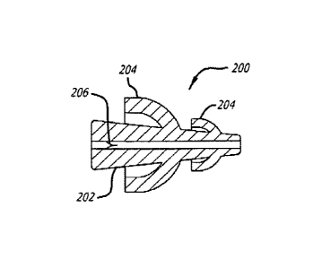
[0046] Figure 2A shows an earplug 200, according to one embodiment of the
invention.
The earplug 200 includes a main lumen 202. One or more sealing members 204
extend
from the main lumen 202. The sealing members 204 are umbrella shaped, and
configured
to partially deform within an ear canal to form a fluid tight seal. The
sealing members 204
are shown to be integral from the main lumen 202, but may also be separately
attached. The
sealing members 204 are preferentially more flexible than the main lumen 202,
as the main
lumen 202 should remain at least partially open in use. A lumen seal 206 is
placed within
the main lumen 202, which prevents fluid and pressure from exiting the lumen.
The lumen
seal 206 is shown configured as a duckbill valve, but may include other
configurations. For
example, the lumen seal 206 may be an elastomeric plug, or wall, with a
compressed lumen,
which may be expanded by a device for inserting fluid, such as a syringe. The
earplug 200
may be constructed from various flexible materials, for example rubber or
silicone. Various
configurations of the earplug 200 are possible, such as shown in U.S.
Provisional Patent
Application No. U.S. 61/085,360.
[0047] Figure 2B shows the earplug 200 in use, according to one embodiment of
the
invention. As shown, a portion of the earplug 200 has been inserted into an
ear canal and
another portion remains exposed adjacent to the outer ear 208. The ear shown
may have
undergone a tympanocentesis procedure, shortly before insertion of the
earplug. A sealing
member 204 is also shown in a partially compressed state. Thus, the earplug
200 is fluidly
sealed within the ear canal. A bulb device 210 or syringe may be coupled with
the earplug
to supply fluidic pressure into the ear canal. The fluid may be a liquid, such
as
iontophoresis fluid, saline, or water, or a gas, such as air. As the ear has
undergone a
tympanocentesis procedure, the tympanic membrane has been punctured, and the
ear canal
118 is in fluidic communication with the middle ear 120. The patient may be
instructed to
swallow, and thus induce the Eustachian tube to open. This action causes a
pressure
7
CA 02748262 2011-06-23
WO 2010/075502 PCT/US2009/069388
differential between the Eustachian tube and the ear canal. Thus, fluid in the
ear canal will
pass through the middle ear, and flush solid or semi-solid effusion inside the
middle ear into
the Eustachian tube. Alternatively, the bulb device 210 may be used without
instructing the
patient to swallow. Creating a large enough pressure differential between the
ear canal and
Eustachian tube will force the Eustachian tube to open and move fluid through
the middle
ear. Care should be taken to avoid damage to the tympanic membrane. In an
alternative
embodiment, a relief valve is included to prevent over-pressurization of the
ear canal. This
procedure may also be performed on both ears simultaneously, and with the
patient sitting
upright.
[0048] Figure 2C shows a flow chart of a method 212 for removing effusion from
a
middle ear, according to one embodiment of the invention. In operation 214, a
liquid is
applied to the ear canal. The fluid may be liquid such as iontophoresis fluid,
saline, or
water. The liquid is preferably at room temperature, or higher, in order to
prevent
discomfort to the patient. In an alternative embodiment, no liquid is provided
for operation
214, and the method begins at operation 216 using only gas as a fluid. At
operation 216 an
ear device is applied to the ear canal of the patient, to form a fluid tight
seal between the ear
canal and the surrounding atmosphere. The ear device may, for example, be
device 200 as
shown in Figures 2A and 2B. At operation 218 the ear device is pressurized
with fluid,
which may be a gas or liquid. The ear device may be pressurized with an
external device
such as a syringe, catheter, or bulb device as shown in Figure 2B. At
operation 220 the
Eustachian tube is induced to open, which may occur from the patient
swallowing or from
the pressure created in operation 218. In operation 222 it is determined
whether more fluid
is required to complete the procedure. If not, then the procedure is complete
and ends at
224. If more fluid is required then the method 212 reverts to operation 218.
[0049] Figure 2D shows a device 228 for sealing both ears of a patient,
according to one
embodiment of the invention. The device includes ear cups 230. Each ear cup
230 includes
sealing members 232, which are configured to fit over and fluidly seal the
outer ear of a
patient. Each ear cup 230 is provided with a fluid chamber 234, which fluidly
communicates with an ear canal. Each fluid chamber 234 in turn is in fluid
communication
with a port 236. The ports 236 include seals 238 for sealing the fluid
chambers from the
external atmosphere. The seals 238 may be constructed from a flexible
material, such as
silicone or rubber. The ports 236 may couple to an external device which
provides fluidic
8
CA 02748262 2011-06-23
WO 2010/075502 PCT/US2009/069388
pressure, for example a syringe, catheter, or bulb device as shown in Figure
2B. In an
alternative embodiment each port 236 is connected to an integral air pump,
which
pressurizes each fluid chamber when manually or electrically activated. In
another
alternative embodiment, a relief valve is included to prevent over-
pressurization of the ear
canal. A band 240 connects each ear cup 230, and provides spring force for
sealing each ear
cup 230 to a patient's head. Figure 2E shows a front view of patient wearing
the device
228.
[0050] Figure 2F shows a device 242 for sealing both ears of a patient,
according to one
embodiment of the invention. The device includes ear cups 230, which may be
constructed
as described regarding Figure 2D. The device 242 includes a wrap-around
headband 242.
The headband 242 wraps around the entire head of a patient, and thus will not
easily be
disturbed during a procedure. The headband may be constructed from an elastic
material,
such as rubber or silicone. Figures 2G and 2H show side and front views,
respectively, of
the device 242 in use on a patient.
[0051] Silent Liquid Removal:
[0052] Figure 3A shows a device 300 for silently removing liquid from a
patient's ear,
according to one embodiment of the invention. Removing liquid in the ear after
a
tympanocentesis procedure may be very disturbing to a patient, as a large
amount of noise is
created in the ear by conventional suction devices. The device 300 includes a
syringe 302, a
nozzle 304, and an absorbent tip 306. The syringe 302 provides negative
pressure for
suctioning and retaining liquid. The nozzle 304 should be flexible to allow
insertion into a
tortuous ear canal without causing patient discomfort. The nozzle 304 should
also be
flexible and long enough to reach the tympanic membrane without buckling or
kinking.
The nozzle 304 may be constructed from a polymer, for example nylon,
polycarbonate,
polypropylene, polyethylene, silicone, or an annealed or super elastic alloy.
The distal
portion of the nozzle 304 may include an outer diameter ranging from 0.5-
3.0mm, which
allows passage through a speculum and visualization past the nozzle to ensure
proper
placement within the ear canal. The proximal portion of the nozzle 304
includes a luer
fitting for coupling to the syringe 302. The absorbent tip 306 is located
within the distal
portion of the nozzle 304. The absorbent tip 306 may be constructed from
absorbent
materials such as porous fibers or foam, which will wick liquids. Suitable
materials include
polyvinyl acetate, rayon, and various blends of the two materials. The
absorbent tip 306
9
CA 02748262 2011-06-23
WO 2010/075502 PCT/US2009/069388
may include pore sizes and interstitial spaces which attract liquid and retain
particles. The
absorbent tip 306 may extend 1-5mm past the distal portion of the nozzle.
[0053] Figure 3B shows device 300 in use, according to one embodiment of the
invention.
The device 300 is shown in use in an ear canal model 308 which is partially
filled with a
liquid solution. The absorbent tip 306 is initially placed in the ear canal
and adjacent to the
tympanic membrane. Contact with the liquid solution causes an immediate
wicking action,
which draws the liquid solution into the device 300. The wicking action is
completely
silent, and thus will not disturb a patient. Figure 3C shows the syringe 302
has been slowly
drawn back to suction the remaining liquid solution, accordingly, the liquid
solution is
silently and quickly removed. This method may be performed implementing a one-
handed
technique by the operator.
[0054] Figures 4A-4F show devices which may be used in lieu of the syringe 302
with
respect to device 300, according to different embodiments of the invention.
Figure 4A
shows a syringe with finger adapters which allows an ergonomic one-handed
suction
motion. Figure 4B shows a spring-loaded syringe, which requires minimal effort
to use.
Figure 4C shows a otology suction device, which may connect to a standard
suction line.
Figure 4D shows a suction bulb, which is compressed before use. Figure 4E
shows a
suction pipette, which is compressed before use. Figure 4F shows a bellows-
type suction
device, which is compressed before use.
[0055] Figures 4G-4I show devices which may be used in lieu of the nozzle 304
with
respect to device 300, according to different embodiments of the invention.
Figure 4G
shows a straight nozzle, which may offer better visibility in use. Figures 4H
and 41 show
shapeable nozzles of different lengths, which may be shaped in the field by
the operator for
better access and visibility.
[0056] Figures 5A and 5B show a device 500 for silently removing liquid from a
patient's
ear, according to one embodiment of the invention. The device 500 includes an
elongated
cannula 502. The elongated cannula 502 may be pre-shaped to include a bend as
shown, or
in a straight configuration. The elongated cannula 502 may constructed from a
malleable
metal, and bent in the field by an operator for better access and visibility.
The elongated
cannula 502 includes an outer diameter which is small enough to reach the
tympanic
membrane, for example 1-3mm. An elongated foam member 504 resides within the
CA 02748262 2011-06-23
WO 2010/075502 PCT/US2009/069388
elongated cannula 502. The elongated foam member 504 includes a distal foam
portion 506
and a proximal foam portion 508. The distal foam portion 506 extends past the
elongated
cannula 502 by a small amount, e.g. 1-3mm, in comparison to the proximal foam
portion
508. A compressed region of foam 510 resides within the elongated cannula, and
connects
the distal and proximal foam portions. The foam may include pore sizes which
can capture
particulates.
[0057] Figure 5C shows the device 500 in use, according to one embodiment of
the
invention. The distal foam portion 506 is shown placed in a liquid solution.
The distal
foam portion 506 expands slightly upon immersion, but is largely restrained by
the
elongated cannula. Liquid is wicked silently from the distal foam portion 506
to the
proximal foam portion 508. The proximal foam portion 508 has a larger volume
than the
distal foam portion 506, and thus acts as a fluid depository. Accordingly,
liquid is wicked
from the distal foam portion 506 to the proximal foam portion 508 in a quick
and silent
manner. The device 500 requires no actuation other than placement in the ear.
The
proximal foam portion 508 may be compressed to remove wicked fluid and reused
during
the procedure or in the other ear.
[0058] Figure 6A shows a device 600 for silently removing liquid from a
patient's ear,
according to one embodiment of the invention. The device 600 is configured as
a multi-
lumen tube. The tube includes an outer diameter which is small enough to reach
the
tympanic membrane, for example 1-3mm. The lumen diameters may range from 0.05-
.5mm. The device 600 may be connected to a suction device, for example a
suction line or
syringe. The device may also be flexible or constructed from a malleable
material. Noise
may be created when air mixes with liquid in a low pressure environment to
cause
cavitation and create a noisy "slurping" sound, as depicted in prior art
device of Figure 68.
Thus, the larger the inner diameter of the suction device, the more likely
noise will be
produced, as any given cross-section of a large lumen may occupy both air and
water.
Device 600 prevents unwanted cavitation by using several smaller diameter
lumens, which
ensures that only air or water occupies a given cross-section of a lumen at a
given time, as
shown in Figure 6C. Accordingly, the device 600 eliminates or greatly reduces
cavitation to
provide a silent liquid evacuation procedure.
11
CA 02748262 2011-06-23
WO 2010/075502 PCT/US2009/069388
[0059] Closed-Loop Control System:
[0060] Figure 7A shows a system 700 for silently removing liquid from a
patient's ear,
according to one embodiment of the invention. The system 700 is configured to
gate the
rate of suction, to a device, using a closed loop control method. The system
700 includes a
suction probe 702, which includes a probe tip 704, and at least one noise
sensor 706. The
suction probe 702 may be configured similarly to any of the devices disclosed
herein, or
may be a standard suction cannula. The sensor 706 may detect noise (e.g.
sound) and/or
pressure and/or flow rate at or about the probe tip 704, or any measureable
artifact which is
related to noise production. For example, as suction noise is caused by
turbulence in a
liquid stream, which is detectable at the fluid/air interface at the probe tip
704, detection of
turbulence (e.g. presence, discontinuity, increase/decrease) may be used a
detectable sensor
artifact. Other measureable artifacts include heat/electrical conductivity
(e.g. between two
points in a probe using the liquid as a conductive medium where conductivity
decreases
with additional turbulence), evaporation, oxygen content, temperature, or some
other micro-
environmental variable. Alternatively, several sensors may monitor conditions
throughout
the entire suction probe 702. The sensor 706 is electronically coupled to a
processor 708.
The processor 708 may be a portion of an embedded computer. A trigger 710
sends user
command signals to the processor 708, for example through a foot or hand
switch. The
suction probe 702 receives suction from a regulator 714 which is further
connected to a
suction source 712. The regulator 714 is electronically coupled to the
processor 708. The
processor 708 controls the regulator 714 to vary the rate and amount of
negative pressure
supplied to the suction probe 702. The sensor 706 may be configured to detect
noise, or the
imminent creation of a predetermined noise level, and indicate the noise
detection to the
processor. The processor 708 may modify, e.g. reduce or eliminate, negative
pressure
supplied to the suction probe 702 based on the sensor 706 signal. In one
example, the
sensor is used to sense a waveform which increases in amplitude. Thus, when
the
waveform increases to a predetermined level in velocity or amplitude, and/or
accelerates at
a predetermined rate, the processor 708 can reduce negative pressure to the
suction probe
702. Accordingly, the imminent increase/creation of noise to a predetermined
level can be
abated, as the processor prevents the waveform from increasing. If no noise
(e.g. no noise
of a significant discomfort level) is sensed by the sensor 706, then the
processor 708 may
increase negative pressure to the suction probe until a predetermined level is
reached. A
test cycle may also be implemented by the processor on start-up or shut-down
by sending a
12
CA 02748262 2011-06-23
WO 2010/075502 PCT/US2009/069388
test pulse of negative suction to create a suction-wave in the system 700 to
check if noise is
initially present, which may occur if the probe tip is only partially
submerged in liquid,
before full negative pressure is enacted by the regulator. Thus, negative
pressure may not
be applied at a full rate and in a continuous mode if the probe is not fully
immersed in
liquid. Accordingly, the system 700 automatically prevents the creation of
noise during a
liquid evacuation procedure, and prevents discomfort to the patient.
[0061] The system 700 may include many of the components of a personal
computer,
such as a data bus, a memory, input and/or output devices (including a touch
screen), and
the like. The system 700 will often include both hardware and software, with
the software
typically comprising machine readable code or programming instructions for
implementing
one, some, or all of the methods described herein. The code may be embodied by
a tangible
media such as a memory, a magnetic recording media, an optical recording
media, or the
like. The system 700 may have (or be coupled to) a recording media reader, or
the code
may be transmitted to the processor 708 by a network connection such as an
interne, an
intranet, an Ethernet, a wireless network, or the like. Along with programming
code, the
system 700 may include stored data for implementing the methods described
herein, and
may generate and/or store data that records parameters reflecting the
treatment of one or
more patients.
[0062] Figure 7B shows a method 716 for silently removing liquid from a
patient's ear,
which may be used with system 700, according to one embodiment of the
invention. A
trigger occurs at input 716 to supply suction to the suction probe 702. At
operation 720 a
processor 708 controls a regulator 714 to supply suction to a suction probe
702. At
operation 722 a sensor 706 monitors noise at a probe tip 704 and sends a
signal to the
processor 708. At operation 724 it is determined whether the signal indicates
noise, or
imminent noise. If no noise, or imminent noise, is detected, then the method
716 loops back
to operation 720. If noise, or imminent noise, is detected, then at operation
726 the
processor 708 instructs the regulator 714 to reduce suction. At operation 728
it is again
determined whether the signal indicates noise, or imminent noise, after
suction reduction. If
no noise, or imminent, noise is detected, then the method 716 loops back to
operation 720.
If noise, or imminent noise, is detected, then at operation 726 the processor
708 instructs the
regulator 714 to reduce suction again. Accordingly, the method 716
automatically prevents
13
CA 02748262 2011-06-23
WO 2010/075502 PCT/US2009/069388
the creation of noise during a liquid evacuation procedure, and prevents
discomfort to the
patient.
[0063] Figure 8A shows a device 800 for silently removing liquid from a
patient's ear,
according to one embodiment. The device 800 includes a cannula 802. In one
embodiment
the cannula 802 is a .075" ID/ .083" OD PTFE tube approximately 3.2 cm in
length, with a
3/32" thick polyolefin material heat shrunk about the PTFE tube surface. The
device 800
includes a filter material 804 within the cannula 802. In one embodiment the
filter material
is 65 thread count cotton gauze strands which are 1.5-1.7 cm long. In one
embodiment, the
filter material may be fibers of the cotton gauze longitudinally arranged
within the cannula
802. Alternatively, the filter material may be constructed from porous foam
strands. A
portion 806 of the filter material 804 extends from the distal end of the
cannula 802. The
portion 806 may be frayed to resemble a mop head. The device 800 can be
coupled to a
commercially available 6 Fr suction catheter 808 as shown in Figure 8B.
[0064] In use, the device 800 is applied to a liquid and/or light effusion
within a patient's
ear and suction is applied to the device 800, for example, by using the
catheter 808. The
filtering material 804 acts as a sound buffer by transferring the suction
noise from the
extreme distal end of the device to a more proximal location within cannula
802. In other
words, the noise of suction does not occur at the extreme distal end, near the
patient's ear
drum, but instead occurs more proximally within cannula 802. Accordingly, the
patient is
protected from excessive noise due to the suction. The portion 806 extending
from the
cannula 802 may also cushion against unintended contact with portions of the
ear canal
and/or be used to physically abrade lodged effusion.
[0065] Figures 9A and 9B show a system for silently removing liquid from a
patient's ear,
according to one embodiment of the invention. The device 900 includes a
cannula 902 and
an Archimedes' screw 904 rotatably disposed within the cannula 902. The
Archimedes'
screw 904 may be coupled to a drive motor (not shown) to rotate at a
relatively slow
revolution, for example at 50-500 RPM, and at a constant torque. The cannula
902 may
include a flared tip 906. The Archimedes' screw 904 may be configured to move
in and out
of the cannula. The cannula 902 may be configured to pass through a lumen 908
of a
tympanostomy tube 910. A suction source may be coupled to the proximal end of
the
device 900.
14
CA 02748262 2016-06-30
[0066] In use, the tympanostomy tube 910 is first implanted within a tympanic
membrane
TM of an ear of a patient, as shown. Devices and methods for locally
anesthetizing the
tympanic membrane for such a tube implant procedure are disclosed in co-
assigned patent
applications U.S. 11/962,063, U.S. 11/749,729, and U.S. 61/085,360. The device
900 can
then be inserted into the lumen 908 of the tympanostomy tube 910 and applied
to a lodged
effusion E. The Archimedes' screw 904 may rotate at a relatively slow RPM, and
accordingly does not generate excessive noise, i.e. sputtering, to disturb the
patient.
Rotation of the Archimedes' screw 904 causes the effusion E to engage
Archimedes' screw
904 and travel out of the ear canal. The Archimedes' screw 904 may rotate at a
constant
torque to prevent jamming with particularly thick effusion. The Archimedes'
screw 904
may also be actuated in and out of the cannula to help disrupt the lodged
effusion. Suction
may be applied to the proximal portion of the device 900 to aid in effusion
removal.
[00671 Figure 9C shows an alternative embodiment of the device 900. A cannula
914
includes a laterally exposed portion 914, which exposes the tip of the
Archimedes' screw
904. The exposed portion 914 may allow the Archimedes' screw 904 to help
initiate
transport of the effusion.
[0068] It should be noted that the silent liquid removal systems and devices
shown and
described herein may also be used to remove effusion. For example, the silent
liquid
systems and devices shown and described herein may be inserted into an ear
canal to
remove effusion. The silent liquid systems and devices shown and described
herein may
also be inserted directly into the middle ear, following a myringotomy or
tympanostomy, to
remove lodged effusion. Accordingly, the systems and devices for silent liquid
removal
described herein are not limited to removing liquid drug solution, and may be
used to
remove any liquid and fluidic particulates within the ear.
[0069] As will be understood by those skilled in the art, the present
invention may be
embodied in other specific forms without departing from the essential
characteristics
thereof. Those skilled in the art will recognize, or be able to ascertain
using no more than
routine experimentation, many equivalents to the specific embodiments of the
invention
described herein. Such equivalents are intended to be encompassed by the
following
claims.