Note: Descriptions are shown in the official language in which they were submitted.
CA 02752054 2014-03-26
FLUID-ASSISTED ELECTROSURGICAL DEVICE
AND METHODS OF USE THEREOF
Field
This invention relates generally to the field of medical devices, systems and
methods
for use upon a human body during surgery. More particularly, the invention
relates to
surgical devices, systems and methods that provide cutting of tissue as well
as coagulation,
hemostasis and sealing of tissue to inhibit blood and other fluid loss during
surgery such as
abdominal, orthopedic, spine and thoracic surgery as well as general surgery
of the body.
Background
Fluid-assisted electrosurgical devices have been developed which, when used in
conjunction with an electrically conductive fluid such as saline, may be moved
along a tissue
surface, without cutting the tissue, to seal tissue to inhibit blood and other
fluid loss during
surgery. However, to cut tissue the surgeon must utilize a second device,
which necessitates
delays associated when switching between devices. What is still needed is an
electrosurgical
device which is capable of cutting of tissue as well as providing fluid-
assisted sealing of
tissue to inhibit blood and other fluid loss during surgery, as well as
inhibit undesirable
effects of tissue desiccation, tissue sticking to the electrode, tissue
perforation, char formation
and smoke generation.
Summary of the Invention
The invention, in one embodiment, may provide an electrosurgical device to
treat
tissue in a presence of a fluid from a fluid source and radio-frequency power
from a radio-
frequency power source, particularly providing a bipolar power output and a
monopolar
power output. The device may comprise a distal portion comprising a first
electrode tip, a
second electrode tip and at least one fluid outlet. The first and second
electrode tips may be
1
CA 02752054 2011-08-09
WO 2010/096809
PCT/US2010/025058
configured as bipolar electrodes, to receive the bipolar power output from the
radio-
frequency power source, and at least one of the electrode tips may be
configured as a
monopolar electrode, to receive the monopolar power output from the radio-
frequency power
source.
In certain embodiments, the at least one electrode tip configured as a
monopolar
electrode may provide an electrosurgical cutting edge, which may be configured
to cut tissue
by moving along a tissue surface in a presence of monopolar power output
provided from the
distal portion.
In certain embodiments, the at least one electrode tip configured as a
monopolar
electrode may comprise a blade portion. The blade portion may comprise
opposing sides and
an electrosurgical cutting edge. The electrosurgical cutting edge may extend
from a proximal
portion of the electrode tip to a distal portion of the electrode tip. The
blade portion may
narrow as the opposing sides approach the cutting edge.
In certain embodiments, at least one of the opposing sides may comprise a
planar
surface, concave surface or convex surface. Furthermore, the opposing sides
may comprise
opposing planer surfaces, concave surfaces or convex surfaces.
In certain embodiments, the first electrode tip and the second electrode tip
may be
configured to treat tissue by moving along a tissue surface in a presence of a
bipolar power
output and a fluid provided simultaneously from the distal portion.
In certain embodiments, the at least one fluid outlet may further comprise at
least one
fluid outlet in fluid communication with the first electrode tip, and at least
one fluid outlet in
fluid communication to the second electrode tip. The at least one fluid outlet
in fluid
communication with the first electrode tip may be proximal to a distal end of
the first
electrode tip, and the at least one fluid outlet in fluid communication with
the second
electrode tip may be proximal to a distal end of the second electrode tip. The
at least one
fluid outlet in fluid communication with the first electrode tip may be at
least partially
defined by the first electrode tip, and the at least one fluid outlet in fluid
communication with
the second electrode tip may be at least partially defined by the second
electrode tip. The at
least one fluid outlet in fluid communication with the first electrode tip may
comprise a
plurality of fluid outlets at least partially defined by the first electrode
tip and the at least one
fluid outlet in fluid communication with the second electrode tip may comprise
a plurality of
fluid outlets at least partially defined by the second electrode tip.
2
CA 02752054 2011-08-09
WO 2010/096809
PCT/US2010/025058
In certain embodiments, the first electrode tip may be laterally spaced from
the second
electrode tip. The first electrode tip may have a blunt distal end, and the
second electrode tip
may have a blunt distal end. The first electrode tip may also have a rounded
distal end, and
the second electrode tip may also have a rounded distal end. The first
electrode tip and
second electrode tip may be at a distal end of a shaft assembly.
In certain embodiments, an electrosurgical device to treat tissue in a
presence of radio
frequency energy and a fluid provided from the device may be provided, with
the device
comprising a distal portion comprising a first electrode tip, a second
electrode tip and at least
one fluid outlet. The first electrode tip may comprise a first electrode
having a distal portion
with an electrically conductive spherical surface, and the second electrode
tip may comprise a
second electrode having a distal portion with an electrically conductive
spherical surface. At
least one of the first electrode and the second electrode may have a blade
portion.
In certain embodiments, the first electrode and the second electrode may be
configured to be electrically coupled to a bipolar power output, and the at
least one electrode
having the blade portion may be configured to be electrically coupled to a
monopolar power
output. The blade portion may extend longitudinally along the electrode, from
a proximal
portion to the distal portion of the electrode. The blade portion may have a
cutting edge, and
more particularly have an electrosurgical cutting edge. The blade portion may
have opposing
sides, and narrow as the opposing sides approach the cutting edge. At least
one of the
opposing sides may comprise a planar surface, a concave surface or a convex
surface.
In certain embodiments, the at least one fluid outlet may further comprise at
least one
fluid outlet in fluid communication with the first electrode and at least one
fluid outlet in fluid
communication with the second electrode. The at least one fluid outlet in
fluid
communication with the first electrode may be proximal to a distal end of the
first electrode
and at least partially defined by the first electrode, and the at least one
fluid outlet in fluid
communication with the second electrode may be proximal to a distal end of the
second
electrode and at least partially defined by the second electrode.
In certain embodiments, the first electrode may be laterally spaced from the
second
electrode. The first electrode may be carried by a first tubing segment at a
distal end thereof,
and the second electrode may be carried by a second tubing segment at a distal
end thereof.
The first electrode may be connected at a distal end of a first tubing
segment, particularly
mechanically joined to the first tubing segment, and the second electrode may
be connected
3
CA 02752054 2014-04-30
at a distal end of the second tubing segment, particularly mechanically joined
to the second
tubing segment. The first electrode also may be welded to the first tubing
segment, and the
second electrode may be welded to the second tubing segment.
In certain embodiments, the first tubing segment may be electrically
conductive and in
electrical contact with the first electrode, and the second tubing segment may
be electrically
conductive and in electrical contact with the second electrode.
In certain embodiments, an electrosurgical device having a distal portion
comprising a
first electrode tip, a second electrode tip and at least one fluid outlet may
be provided, with the
first electrode tip comprising a first electrode having a blade portion and
the second electrode
tip comprising a second electrode having a blade portion. The first and second
electrodes may
be configured to be electrically coupled to a bipolar energy source and at
least one of the
electrodes may be configured to be electrically coupled to a monopolar energy
source. The first
and second electrodes may be electrically coupled to the bipolar energy source
by first and
second bipolar electrical connectors in electrical communication with the
first and second
electrodes, respectively, and at least one of the electrodes may be
electrically coupled to the
monopolar energy source by a monopolar electrical connector in electrical
communication with
at least one of the electrodes.
In one embodiment, an electrosurgical device may be provided to treat tissue
in a
presence of radio frequency energy and a fluid provided from the device, the
device comprising
a distal portion comprising a first electrode tip, a second electrode tip and
at least one fluid
outlet; the first electrode tip comprising a first electrode having a distal
portion with an
electrically conductive, at least substantially spherical surface; the second
electrode tip
comprising a second electrode having a distal portion with an electrically
conductive, at least
substantially spherical surface; at least one of the first electrode and the
second electrode having
a blade portion; the first electrode and the second electrode configured to be
electrically coupled
to a bipolar power output; the at least one of the first electrode and the
second electrode having
the blade portion configured to be electrically coupled to a monopolar power
output; and the
blade portion extending longitudinally along the at least one of the first
electrode and the second
electrode, and wherein the blade portion has a cutting edge.
In one embodiment, an electrosurgical device may be provided comprising a
distal
portion comprising a first electrode tip, a second electrode tip and at least
one fluid outlet; the
first electrode tip comprising a first electrode having a blade portion; the
second electrode tip
4
CA 02752054 2014-04-30
comprising a second electrode having a blade portion; each of the first and
second electrodes
configured to be electrically coupled to a bipolar energy source by first and
second bipolar
electrical connectors in electrical communication with the first and second
electrodes,
respectively; at least one of the first or second electrodes configured to be
electrically coupled
to a monopolar energy source by a monopolar electrical connector in electrical
communication
with at least one of the first or second electrodes; the blade portions
extending longitudinally
along the first and second electrodes, and wherein the blade portions have a
cutting edge.
Brief Description Of The Drawings
FIG. 1 is a front view of one embodiment of a system of the present invention
having an
electrosurgical unit in combination with a fluid source and handheld
electrosurgical device;
FIG. 2 a front perspective view of the electrosurgical unit of FIG. 1;
FIG. 3 is a graph of the bipolar RF power output versus impedance for the
electrosurgical unit of FIG. 1;
FIG. 4 is graph showing a relationship of fluid flow rate Q in units of cubic
centimetres
per minute (cc/min) on the Y-axis, and the RF power setting Ps in units of
watts on the X-axis;
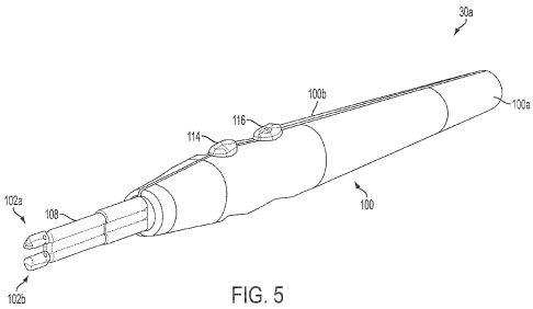
FIG. 5 is a perspective view of an electrosurgical device according to the
present
invention;
FIG. 6A is a plan view showing the various electrical connections and
conductors of the
device of FIG. 5 with the electrosurgical unit of FIG. 1;
4a
CA 02752054 2011-08-09
WO 2010/096809
PCT/US2010/025058
FIG. 6B is a plan view showing the various fluid connections and passages of
the
device of FIG. 5 with the electrosurgical unit and fluid source of FIG. 1;
FIG. 7 is a close-up view of the shaft assembly of the device of FIG. 5;
FIG. 8 is a close-up cross-sectional view of the electrodes of the device of
FIG 5
taken along line 8-8 of FIG. 7;
FIG. 9 is a close-up view of the shape of the electrodes of another embodiment
of the
device of FIG. 5 taken along line 8-8 of FIG. 7;
FIG. 10 is a close-up view of the shape of the electrodes of another
embodiment of
the device of FIG. 5 taken along line 8-8 of FIG. 7;
FIG. 11 is a close-up cross-sectional view of a distal end portion of the
device of FIG.
5 taken perpendicular to line 8-8 of FIG. 7;
FIG. 12 is a close-up view of a distal end portion of the device of FIG. 5
with an
exemplary fluid coupling to a tissue surface of tissue; and
FIG. 13 is a perspective view of the device of FIG. 5 cutting tissue.
Detailed Description
Throughout the description, like reference numerals and letters indicate
corresponding
structure throughout the several views. Also, any particular feature(s) of a
particular
exemplary embodiment may be equally applied to any other exemplary
embodiment(s) of this
specification as suitable. In other words, features between the various
exemplary
embodiments described herein are interchangeable as suitable, and not
exclusive. From the
specification, it should be clear that any use of the terms "distal" and
"proximal" are made in
reference from the user of the device, and not the patient.
The invention provides devices, systems and methods for controlling tissue
temperature at a tissue treatment site during an electrosurgical procedure, as
well as
shrinking, coagulating, cutting and sealing tissue against blood loss, for
example, by
shrinking lumens of blood vessels (e.g., arteries, veins).
The invention will now be discussed with reference to the figures, with FIG. 1
showing a front view of one embodiment of a system of the present invention
having an
exemplary electrosurgical unit 10 in combination with a fluid source 20 and a
handheld
electrosurgical device 30. FIG. 1 shows a movable cart 2 having a support
member 4
5
CA 02752054 2011-08-09
WO 2010/096809
PCT/US2010/025058
comprising a hollow cylindrical post which carries a platform 6 comprising a
pedestal table to
provide a flat, stable surface for location of the electrosurgical unit 10.
As shown, cart 2 further comprises a fluid source carrying pole 8 having a
height
which may be adjusted by sliding the carrying pole 8 up and down within the
support
member 4 and thereafter secured in position with a set screw. On the top of
the fluid source
carrying pole 8 is a cross support provided with loops at the ends thereof to
provide a hook
for carrying fluid source 20.
As shown in FIG. 1, fluid source 20 comprises a bag of fluid from which the
fluid 12
flows through a drip chamber 14 after the bag is penetrated with a spike
located at the end of
the drip chamber 14. Thereafter, fluid 12 flows through flexible delivery
tubing 16 to
handheld electrosurgical device 30. Preferably the fluid delivery tubing 16 is
made from a
polymer material.
As shown in FIG. 1, the fluid delivery tubing 16 passes through pump 22. As
shown
pump 22 comprises a peristaltic pump and, more specifically, a rotary
peristaltic pump. With
a rotary peristaltic pump, a portion of the delivery tubing 16 is loaded into
the pump head by
raising and lower the pump head in a known manner. Fluid 12 is then conveyed
within the
delivery tubing 16 by waves of contraction placed externally on the tubing 16
which are
produced mechanically, typically by rotating pinch rollers which rotate on a
drive shaft and
intermittently compress the tubing 16 against an anvil support. Peristaltic
pumps are
generally preferred, as the electro-mechanical force mechanism, here rollers
driven by
electric motor, does not make contact the fluid 12, thus reducing the
likelihood of inadvertent
contamination.
In the present embodiment the fluid 12 comprises saline solution, and even
more
specifically, normal (physiologic) saline. Although the description herein may
make
reference to saline as the fluid 12, other electrically conductive fluids can
be used in
accordance with the invention.
While an electrically conductive fluid having an electrically conductivity
similar to
normal saline is preferred, as will become more apparent with further reading
of this
specification, fluid 12 may also comprise an electrically non-conductive
fluid. The use of a
non-conductive fluid, while not providing all the advantage of an electrically
conductive
fluid, still provides certain advantages over the use of a dry electrode
including, for example,
reduced occurrence of tissue sticking to the electrode of device 30 and
cooling of the
6
CA 02752054 2011-08-09
WO 2010/096809
PCT/US2010/025058
electrode and/or tissue. Therefore, it is also within the scope of the
invention to include the
use of a non-conducting fluid, such as, for example, deionized water.
Electrosurgical unit 10 is configured to provide both monopolar and bipolar
power
output. However, electrosurgical unit 10 includes a lock out feature which
prevents both
monopolar and bipolar output from being activated simultaneously.
Alternatively, rather than
use a single electrosurgical unit 10, device may be simultaneously connected
to two separate
electrosurgical units. For example, device 30 may be connected to a first
electrosurgical unit
to provide monopolar power output and a second electrosurgical unit to provide
bipolar
power output.
During monopolar operation, a first electrode, often referred to as the active
electrode,
is provided with the monopolar electrosurgical device while a second
electrode, often
referred to as the indifferent or neutral electrode, is provided in the form
of a ground pad
dispersive electrode located on the patient (also known as a patient return
electrode), typically
on the back or other suitable anatomical location. An electrical circuit is
formed between the
active electrode and ground pad dispersive electrode with electrical current
flowing from the
active electrode through the patient to ground pad dispersive electrode in a
manner known in
the art. During bipolar operation, the ground pad electrode located on the
patient is not
required, and a second electrode providing an electrical pole is provided as
part of the device.
An alternating current electrical circuit is then created between the first
and second electrical
poles of the device. Consequently, alternating current no longer flows through
the patient's
body to the ground pad electrode, but rather through a localized portion of
tissue between the
poles of the bipolar device. Monopolar and bipolar power may be provided from
electrosurgical unit 10 as known in the art, or from separate electrosurgical
units.
As shown in FIG. 1, electrosurgical device 30 is connected to electrosurgical
unit 10
via electrical cables 24 and 26. Cable 24 has a plug 34 which connects to
bipolar mode
output receptacle 38 of electrosurgical unit 10. Cable 26 has a plug 42 which
connects to the
monopolar mode output receptacle 46 of electrosurgical unit 10. As shown in
FIG. 6A, when
electrosurgical 10 is used in monopolar mode, an additional cable 28 is
utilized to connect a
ground pad dispersive electrode 48 to the ground pad receptacle 56 of the
electrosurgical unit
10.
FIG. 2 shows the front panel of the exemplary electrosurgical unit 10. A power
switch 58 may be used to turn the electrosurgical unit 10 on and off. After
turning the
7
CA 02752054 2011-08-09
WO 2010/096809
PCT/US2010/025058
electrosurgical unit 10 on, an RF power setting display 60 may be used to
display the RF
power setting numerically in watts. The power setting display 60 may further
comprise a
liquid crystal display (LCD).
Electrosurgical unit 10 may further comprise an RF power selector 62
comprising RF
power setting switches 62a, 62b which may be used to select the RF power
setting.
Pushing the switch 62a may increase the RF power setting, while pushing the
switch
62b may decrease the RF power setting. RF power output may be set in 5 watt
increments in the range of 20 to 100 watts, and 10 watt increments in the
range of 100 to 200
watts. Additionally, electrosurgical unit 10 may include an RF power
activation display 64
comprising an indicator light which may illuminate when RF power is activated,
either via a
handswitch on device 30 or a footswitch. Switches 62a, 62b may comprise
membrane
switches. It should be understood that while only one RF power selector 62 is
shown,
electrosurgical unit 10 will have two such RF power selectors with one each
for monopolar
and bipolar power selection.
In addition to having a RF power setting display 60, electrosurgical unit 10
may
further include a fluid flow rate setting display 66. Flow rate setting
display 66 may
comprise three indicator lights 66a, 66b and 66c with first light 66a
corresponding to
a fluid flow rate setting of low, second light 66b corresponding to a fluid
flow rate
setting of medium (intermediate) and third light 66c corresponding to a flow
rate
setting of high. One of these three indicator lights will illuminate when a
fluid flow
rate setting is selected.
Electrosurgical unit 10 may further include a fluid flow selector 68
comprising flow
rate setting switches 68a, 68b and 68c used to select or switch the flow rate
setting. Three
push switches may be provided with first switch 68a corresponding to the fluid
flow
rate setting of low, second switch 68b corresponding to a fluid flow rate
setting of
medium (intermediate) and third switch 68c corresponding to a flow rate
setting of
high. Pushing one of these three switches may select the corresponding flow
rate
setting of either low, medium (intermediate) or high. The medium, or
intermediate,
flow rate setting may be automatically selected as the default setting if no
setting is
manually selected. Switches 68a, 68b and 68c may comprise membrane switches.
Before starting a surgical procedure, it may be desirable to prime device 30
with fluid
12. Priming may be desirable to inhibit RF power activation without the
presence of fluid 12.
8
CA 02752054 2014-03-26
A priming switch 70 may be used to initiate priming of device 30 with fluid
12. Pushing
switch 70 once may initiate operation of pump 22 for a predetermined time
period to prime
device 30. After the time period is complete, the pump 22 may shut off
automatically. When
priming of device 30 is initiated, a priming display 72 comprising an
indicator light may
illuminate during the priming cycle.
An exemplary bipolar RF power output curve of electrosurgical unit 10 is shown
in
FIG. 3. Impedance Z, shown in units of ohms on the X-axis and output power Po
is shown in
units of watts on the Y-axis. In the illustrated embodiment, the bipolar
electrosurgical power
(RF) is set to 200 watts. As shown in the figure, for an RF power setting Ps
of 200 watts, the
output power Po will remain constant with the set RF power Ps as long as the
impedance Z
stays between the low impedance cut-off of 30 ohms and the high impedance cut-
off of 120
ohms. Below an impedance Z of 30 ohms, the output power Po will decrease as
shown by
the low impedance ramp. Above an impedance Z of 120 ohms, the output power Po
will also
decrease as shown by the high impedance ramp. With respect to monopolar power
output, an
exemplary monopolar RF power output curve would include that of the Valleylab
Force FX.
Electrosurgical unit 10 may be configured such that the speed of pump 22, and
therefore the throughput of fluid 12 expelled by the pump 22, is predetermined
based on two
input variables, the RF power setting and the fluid flow rate setting. In FIG.
4 there is shown
an exemplary functional relationship of fluid flow rate Q in units of cubic
centimetres per
minute (cc/ruin) on the Y-axis, and the RF power setting Ps in units of watts
on the X-axis.
The relationship may be engineered to inhibit undesirable effects such as
tissue desiccation,
electrode sticking, smoke production and char formation, while at the same
time not
providing a fluid flow rate Q at a corresponding RF power setting Ps which is
so great as to
provide too much electrical dispersion and cooling at the electrode/tissue
interface. While
not being bound to a particular theory, a more detailed discussion on how the
fluid flow rate
interacts with the radio frequency power, modes of heat transfer away from the
tissue,
fractional boiling of the fluid and various control strategies may be found in
U.S. Publication
No. 2001/0032002, published October 18, 2001, assigned to the assignee of the
present
invention.
As shown in FIG. 4, electrosurgical unit 10 has been configured to increase
the fluid
flow rate Q linearly with an increasing RF power setting Ps for each of three
fluid flow rate
9
CA 02752054 2014-03-26
settings of low, medium and high corresponding to QL, QM and QH, respectively.
Conversely,
electrosurgical unit 10 has been configured to decrease the fluid flow rate Q
linearly with a
decrease RF power setting Ps for each of three fluid flow rate settings of
low, medium and
high corresponding to QL, QM and QH, respectively.
An electrosurgical unit similar to exemplary electrosurgical unit 10 and
having
detailed schematic drawings, albeit without monopolar output, may be found in
U.S.
Publication No. 2006/0149225, published July 6, 2006, assigned to the assignee
of the
present invention.
While electrosurgical unit 10 as shown above includes an attached pump 22, in
other
embodiments pump 22 may not be integrated with electrosurgical unit 10, but
rather be
separate from electrosurgical unit 10.
In still other embodiments, pump 22 may be eliminated and there may be no
preset
functional relationship of fluid flow rate Q versus RF power setting Ps stored
in the
electrosurgical unit 10. In such an instance, rather than the fluid flow rate
Q being
automatically controlled by the electrosurgical unit 10 based on the RF power
setting Ps, the
fluid flow rate Q may be manually controlled, such as by the user of device 10
or another
member of the surgical team, with a roller (pinch) clamp or other clamp
provided with device
10 and configured to act upon and compress the tubing 16 and control flow in a
manner
known in the art. Exemplary fluid flow control mechanisms may be found in U.S.
Publication No. 2005/0090816, published April 28, 2005, assigned to the
assignee of the
present invention. -
An example of an electrosurgical unit which does not include a pump, but may
be
used in conjunction with a manually operated fluid flow control mechanism on
device 10,
includes an electrosurgical unit such as the Valleylab Force FX.
An exemplary bipolar and/or monopolar electrosurgical device of the present
invention which may be used in conjunction with electrosurgical unit 10 of the
present
invention is shown at reference character 30a in FIG. 5. While various
electrosurgical
devices of the present invention are described herein with reference to use
with
electrosurgical unit 10, it should be understood that the description of the
combination is for
purposes of illustrating the system of the invention. Consequently, it should
be understood
that while the electrosurgical devices disclosed herein may be disclosed for
use with
CA 02752054 2011-08-09
WO 2010/096809
PCT/US2010/025058
electrosurgical unit 10, it may be plausible to use other electrosurgical
devices with
electrosurgical unit 10, or it may be plausible to use the electrosurgical
devices disclosed
herein with another electrosurgical unit.
As shown in FIG. 5, exemplary device 30a comprises an elongated handle 100
comprising mating handle portions 100a, 100b. Handle 100 is slender, along
with the rest of
device 30a, to enable a user of device 30a to hold and manipulate device 30a
between the
thumb and index finger like a pen-type device. Handle 100 may comprise a
sterilizable,
rigid, non-conductive material, such as a polymer (e.g., polycarbonate).
As best shown in FIG. 6A, device 30a also comprises cables 24 and 26 which are
connectable to electrosurgical unit 10 to provide device 30a with bipolar and
monopolar
power output, respectively, from electrosurgical unit 10. As shown, cable 24
of device 30a
comprises three insulated wire conductors 32a, 32b, 32c connectable to bipolar
power output
receptacles 38a, 38b, 38c of electrosurgical unit 10 via three banana (male)
plug connectors
36a, 36b, 36c. The banana plug connectors 36a, 36b, 36c are each assembled
with insulated
wire conductors 32a, 32b, 32c within the housing of plug 34 in a known manner.
On device
30a, insulated wire conductor 32a is connected to a bipolar hand switch
assembly 104, and
insulated wire conductors 32b and 32c are connected to semi-circular barrel
crimp terminals
which snap connect to a proximal portion of shafts 106a, 106b of shaft
assembly 108.
Cable 26 of device 30a comprises two insulated wire conductors 40a, 40b
connectable
to monopolar power output receptacles 46a, 46b of electrosurgical unit 10 via
two banana
(male) plug connectors 44a, 44b. The banana plug connectors 44a, 44b are each
assembled
with insulated wire conductors 40a, 40b within the housing of plug 42 in a
known manner.
On device 30a, insulated wire conductor 40a is connected to a monopolar hand
switch
assembly 110, and insulated wire conductor 40b is connected to a semi-circular
barrel crimp
terminal which snap connects to a proximal portion of shaft 106b of shaft
assembly 108.
When device 30a is used in monopolar mode, an additional cable 28 is utilized
to connect a
ground pad dispersive electrode 48 which is attached to the patient to the
electrosurgical unit
10 comprising wire conductor 50 and plug 52 at the end thereof having plug
connector 54
which connects to the ground pad receptacle 56. As shown wire conductors 32b
and 40b
merge inside handle 100 and share the same attachment location to shaft 106b.
Hand switch assemblies 104 and 110 may comprise push buttons 114 and 116,
respectively, (best shown in FIG. 5) which overlie domed switches on a
platform comprising
11
CA 02752054 2014-03-26
a printed circuit board, with the construction and wiring of the hand switch
assemblies 104
and 110 known in the art. Upon depression of push buttons 114 or 116, a domed
switch
beneath the push button forms a closed circuit which is sensed by
electrosurgical unit 10,
which then provides bipolar or monopolar power, respectively. Exemplary hand
switches
__ may be found in U.S. Publication No. 2006/0149225, published July 6, 2006,
and U.S.
Publication No. 2005/0090816, published April 28, 2005, which are assigned to
the assignee
of the present invention.
As shown FIG. 6B, during use of device 30a, fluid 12 from fluid source 20 is
__ communicated through a tubular fluid passage which provided by various
structures. In the
present embodiment, fluid 12 from the fluid source 20 is first communicated
through lumen
18 of delivery tubing 16. Fluid 12 may also flow through lumen 120 of a
special pump
tubing segment 118 designed to operate specifically with the peristaltic pump
22, which may
be spliced in between portions of delivery tubing 16 and connected thereto
using barbed fluid
__ line connectors 122 at each end thereof.
Within handle 100 of device 30a, fluid delivery tubing 16 is connected to the
inlet
branch of a Y-splitter 124, which thereafter provides two outlet branches
which are connected
to the proximal ends of polymer delivery tubing segments 128a, 128b. The
distal ends of
delivery tubing segments 128a, 128b are thereafter connected to the proximal
ends of shafts
__ 106a, 106b. To connect delivery tubing 128a, 128b to shafts 106a, 106b, the
lumens 130a,
130b are preferably interference fit over the outside diameter of shafts 106a,
106b to provide
an interference fit seal there between. Fluid 12 then may flow through the
lumens 134a, 134b
of shafts 106a, 106b.
Once the semi-circular barrel crimp terminals and delivery tubing segments
128a,
__ 128b are connected to shafts 106a, 106b, a polymer shrink wrap tubing may
then be heat
shrink wrapped around the connections to better electrically insulate the
shafts 106a, 106b
and better secure the connections.
As best shown in FIG. 7, shaft assembly 108 of the present embodiment
comprises
two parallel, self-supporting, electrically conductive hollow shafts 106a,
106b, which
__ comprise metal tubing segments, such as stainless steel tubing segments.
Carried by and
connected to the distal ends of shafts 106a, 106b are two laterally and
spatially separated (by
empty space) contact elements in the form of electrode tips comprising
electrodes 102a, 102b
12
CA 02752054 2014-03-26
which may be configured as mirror images in size and shape, and have a blunt
distal end with
a surface devoid of edges (to provide a uniform current density) to treat
tissue. In the present
embodiment electrodes 102a, 102b comprise an electrically conductive material,
particularly
metal, such as stainless steel. Other suitable materials may include titanium,
gold, silver and
platinum.
In certain embodiments, the tubing segments of one or both shafts 106a, 106b
may be
made of electrically non-conducting material except for the portion at the
distal end that
comes in physical and electrical contact with electrodes 102a, 102b. In these
embodiments, an
insulated wire conductor would extend and be joined to the electrically
conducting portion of
1.0 shaft 106a, 106b. In still other embodiments, shafts 106a, 106b may
completely Comprise
electrically non-conducting material, in which case an insulated wire
conductor would extend
and be joined directly to electrodes 102a, 102b.
As shown in FIG. 7, each electrode 102a, 102b comprises an elongated portion
138a).
138b. With respect to length, in the present embodiment elongated portion
138a, 138b has a
length in the range between and including about 2 mm to 6 mm, and more
specifically have a
length of about 3 mm to 5 mm. With respect to spacing, in the present
embodiment the
spatial gap separation GS between electrodes 102a, 102b in the range between
and including
about 0.1 mm to about 4 mm, and more specifically about 1 mm to 2.5 mm, and
more
specifically about 1.5 mm to 2.3 nun.
As best shown in FIG. 8, opposing sides 140a/142a of elongated portion 138a,
and
opposing sides 140b/142b of elongated portion 138b converge laterally to
provide a wedge
shaped blade portion 144a, 144b which terminates in a lateral cutting edge
146a, 146b which
extends longitudinally along a length of each electrode 102a, 102b. As shown
in FIG. 8,
lateral cutting edge 146a, 146b extends from a proximal to distal portion of
each electrode
102a, 102b, as well as transitions onto the distal end of each electrode 102a,
102b and forms a
portion of the distal end of each electrode 102a, 102b.
Lateral cutting edge 146a, 146b is preferably configured to cut tissue
electrosurgically
in the presence of monopolar radio frequency energy from electrosurgical unit
10 as to
provide an electrosurgical cutting edge, but without any fluid 12 being
provided from fluid
source 20. However, in other embodiments, lateral cutting edge 146a, 146b may
be
configured to cut tissue with fluid 12 being provided simultaneously from
device 30a, or be
configured to cut tissue mechanically without electrosurgical energy.
Furthermore, while two
13
CA 02752054 2011-08-09
WO 2010/096809
PCT/US2010/025058
cutting edges 146a, 146b are shown, only one of the edges 146a or 146b needs
to be
configured to cut tissue electrosurgically or mechanically. In such instance,
the blade portion
of the electrode may be eliminated and the elongated portion may be completely
cylindrical.
As shown in FIG. 8, blade portion 144a, 144b narrows as the opposing sides
140a/142a and 140b/142b approach cutting edge 146a, 146b. More particularly,
as shown in
FIG. 8, the sides 140a/142a and 140b/142b of blade portion 144a, 144b are
concave.
However, in other embodiments, sides 140a/142a and 140b/142b may be planar or
convex as
shown in FIGS. 9 and 10, respectively. Also, in other embodiments, only one of
sides
140a/142a and 140b/142b may be concave, planar or convex.
Returning to FIG. 7, electrodes 102a, 102b and elongated portions 138a, 138b
terminate in distal end portion 148a, 148b. The distal end portion 148a, 148b
of electrodes
102a, 102b are configured to slide across a tissue surface in the presence of
bipolar radio
frequency energy from electrosurgical unit 10 and fluid 12 from the fluid
source 20. As
shown, the distal end portion 148a, 148b of each electrode 102a, 102b has a
blunt, rounded
shape which provides a smooth contour surface which is devoid of points or
edges. More
specifically, as shown, distal end portion 148a, 148b of each electrode 102a,
102b has a
spherical surface provided by spherical portion 150a, 150b. In the present
embodiment,
spherical portion 150a, 150b has a radius in the range between and including
about 0.5 mm to
1.5 mm, and more specifically about 0.75 mm to 1.15 mm.
As best shown in FIGS. 8 and 11, within a cylindrical portion 152a, 152b of
each
electrode 102a, 102b proximal to distal end portion 148a, 148b, each electrode
102a, 102b
includes a longitudinally oriented linear blind bore 158a, 158b and counter
bore 160a, 160b.
As shown in FIG. 11, the outside diameter of a distal end portion of each
shaft 106a, 106b is
configured to extend into counter bore 160a, 160b of electrodes 102a, 102b and
fit with the
diameter of counter bore 160a, 160b, with the distal end of each shaft 106a,
106b in contact
with the bottom of the counter bore. The electrodes 102a, 102b and shafts
106a, 106b may
then be welded together to connect the two components. In alternative
embodiments, the
outside diameter of shafts 106a, 106b may be configured to fit with the
diameter of counter
bore 160a, 160b and mechanically join in the form of a press (interference)
fit to provide a
secure connection. In other alternative embodiments, electrodes 102a, 102b may
be
assembled to shafts 106a, 106b by threaded engagement. In still other
embodiments,
14
CA 02752054 2011-08-09
WO 2010/096809
PCT/US2010/025058
electrodes 102a, 102b may be detachably assembled to shafts 106a, 106b such
that they may
be removed from the shafts 106a, 106b, preferably manually by human hand.
In addition to blind bore 158a, 158b and counterbore 160a, 160b, as shown in
FIG. 8,
electrodes 102a, 102b also include a through bores 162a/164a and 162b/164b
which
perpendicularly intersects bore 158a, 158b and perpendicularly intersect one
another to
provide outlets 166a/168a/170a/172a and 166b/168b/170b/172b (for fluid 12)
which are in
fluid communication with electrodes 102a, 102b. Thus, after fluid 12 flows
through the
lumens 134a, 134b of shafts 106a, 106b, fluid 12 then flows through into the
tubular passage
provided by blind bore 158a, 158b and then into the tubular passage provided
by through
bores 162a/164a and 162b/164b where it thereafter exits device 30a from fluid
outlets
166a/168a/170a/172a and 166b/168b/170b/172b, which are all proximal to distal
end portion
148a, 148b of electrodes 102a, 102b. As shown in FIG. 8, fluid outlets
166a/170a and
166b/170b are at least partially defined by the cylindrical portion 152a, 152b
of electrodes
102a, 102b, while fluid outlets 168a/172a and 168b/172b are at least partially
defined by
sides of 140a/142a and 140b/142b of blade portion 144a, 144b and adjacent
cutting edge
146a, 146b. More particularly, as shown in FIG. 8, fluid outlets 166a/170a and
166b/170b
are fully defined by the cylindrical portion 152a, 152b of electrodes 102a,
102b, while fluid
outlets 168a/172a and 168b/172b are fully defined by sides of 140a/142a and
140b/142b of
blade portion 144a, 144b and adjacent cutting edge 146a, 146b. In certain
embodiments,
each electrode 102a, 102b may have only one fluid outlet in fluid
communication therewith,
such as outlets 168a, 168b. In still other embodiments, only a single one
fluid outlet may be
present.
The relationship between the material for electrodes 102a, 102b and their
surfaces,
and fluid 12 throughout the various embodiments should be such that the fluid
12 wets the
surface of the electrodes 102a, 102b. Contact angle, 0, is a quantitative
measure of the
wetting of a solid by a liquid. It is defined geometrically as the angle
formed by a liquid at
the three phase boundary where a liquid, gas and solid intersect. In terms of
the
thermodynamics of the materials involved, contact angle 0 involves the
interfacial free
energies between the three phases given by the equation
nv cos 0 = ysv - 7st,
CA 02752054 2011-08-09
WO 2010/096809
PCT/US2010/025058
where YIN, 7sv and 7sL refer to the interfacial energies of the liquid/vapor,
solid/vapor and
solid/liquid interfaces, respectively. If the contact angle 0 is less than 90
degrees the liquid is
said to wet the solid. If the contact angle is greater than 90 degrees the
liquid is non-wetting.
A zero contact angle 0 represents complete wetting. Thus, preferably the
contact angle is less
than 90 degrees.
As best shown in FIGS. 7 and 11, a portion of the lengths of shafts 106a, 106b
are
surrounded by and encapsulated in a common outer member 184, which may
comprises a
flexible polymer. Outer member 184 electrically insulates the exposed length
of shafts 106a,
106b.
Outer member 184 may be formed by injection molding. During the injection
molding process, a sub-assembly comprising electrodes 102a, 102b and shafts
106a, 106b is
placed in the injection mold prior to the introduction of polymer. Thereafter,
the mold is
closed and a thermoplastic polymer may be injected into the unoccupied
portions of the mold
cavity to overmold and mold-in place portions of the sub-assembly as shown in
FIG. 7.
During the injection molding process, retainer clips (not shown) may provide
the benefit of
retaining shafts 106a, 106b in position relative to each other to better
ensure that the shafts
106a, 106b are centrally located within the polymer molding.
To be hand shapeable by surgeons and other users of device 30a, so that the
device
30a may be used in a greater multitude of angles and locations, at least a
portion of shafts
106a, 106b of device 30a may be malleable to provide a malleable shaft
assembly 108. Also,
in this manner, a distal portion of shafts 106a, 106b may be bendable at an
angle relative to
the longitudinal axis of the proximal portion of shafts 106a, 106b during
manufacturing of
device 30a so they may be provided to users of device 30a at various angles.
For example,
angle may range from about 5 degrees to 90 degrees, and more preferably, about
15 degrees
to 45 degrees, and even more preferably about 30 degrees. As used herein,
malleable means
able to be shaped, particularly by bending (without a mechanical mechanism,
such as a hinge
or joint). It should be understood that shaft assembly 108 is to independently
maintain the
shape associated with the selected bent shape, and does not require additional
components
(e.g., pull wires, etc.) to maintain the selected bent shape. Furthermore,
shaft assembly 108 is
to maintain the selected shape such that when device 30a is used to treat
tissue, and will not
overtly deflect from the selected shape. Furthermore, shaft assembly 108 is
constructed such
16
CA 02752054 2011-08-09
WO 2010/096809
PCT/US2010/025058
that a user can readily re-shape the shafts back to a straight state and/or
other desired bent
configurations.
Outer member 184, in addition to electrically insulating shafts 106a, 106b
from one
another, has been found to be particularly useful in facilitating the hand
shaping of shafts
106a, 106b of shaft assembly 108 simultaneously and with a similar contour
without
cracking. In this manner, surgeons and other users of device 30a need not bend
the shafts
106a, 106b individually, and the relative spacing and position of the
electrodes 102a, 102b
may be maintained constant.
To provide malleability, shafts 106a, 106b preferably have an outer wall
diameter of
about 0.063 inches and an inner wall diameter of about 0.032 inches. Shafts
106a, 106b also
preferably are made from 304 stainless steel with a temper from about 1/2 to
3/4 hard, 130,000
to 150,000 psi. (pounds per square inch) tensile strength) and an elongation
at break of about
40%. Shafts 106a, 106b with the foregoing properties provide sufficient
stiffness as not to be
too pliable during normal use of device 30a, while at the same time inhibiting
the shafts 106a,
106b from kinking or breaking when shaped for application. When the wall
thickness is too
thin, shafts 106a, 106b may kink, and when the wall thickness is too thick,
the shafts 106a,
106b may be too stiff. Furthermore, a shaft 106a, 106b with a larger diameter
may also kink
more than a shaft of smaller diameter. Shafts 106a, 106b may also be malleable
for a portion
of the length or full length depending on application. For example, the shafts
106a, 106b can
be made with variable stiffness along the length and be malleable only for a
distal portion
thereof. Preferably this is performed by controlled annealing of the shafts
106a, 106b only in
the area where malleability is desired.
As shown in FIG. 12, one way in which device 30a may be used is with the
longitudinal axis of electrodes 102a, 102b vertically orientated, and the
distal end portion
148a, 148b of electrodes 102a, 102b laterally spaced adjacent tissue surface
202 of tissue
200. When device 30a is used in this manner, electrodes 102a, 102b are
connected to
electrosurgical unit 10 and receive bipolar radio frequency energy which forms
an alternating
current electrical field in tissue 200 located between electrodes 102a, 102b.
In the presence
of alternating current, the electrodes 102a, 102b alternate polarity between
positive and
negative charges with current flow from the positive to negative charge.
Without being
bound to a particular theory, heating of the tissue is performed by electrical
resistance
heating.
17
CA 02752054 2011-08-09
WO 2010/096809
PCT/US2010/025058
Fluid 12, in addition to providing an electrical coupling between the device
30a and
tissue 200, lubricates surface 202 of tissue 200 and facilitates the movement
of electrodes
102a, 102b across surface 202 of tissue 200. During movement of electrodes
102a, 102b,
electrodes 102a, 102b typically slide across the surface 202 of tissue 200.
Typically the user
of device 30a slides electrodes 102a, 102b across surface 202 of tissue 200
back and forth
with a painting motion while using fluid 12 as, among other things, a
lubricating coating.
Preferably the thickness of the fluid 12 between the distal end portions 148a,
148b of
electrodes 102a, 102b and surface 202 of tissue 200 at the outer edge of
couplings 204a, 204b
is in the range between and including about 0.05 mm to 1.5 mm. Also, in
certain
embodiments, the distal end portion 148a, 148b of electrodes 102a, 102b may
contact surface
202 of tissue 200 without any fluid 12 in between.
As shown in FIG. 12, fluid 12 expelled from fluid outlets may form into
droplets
208a, 208b which flow distally on electrodes 102a, 102b. As shown in FIG. 12,
droplets
208a, 208b may form at varying times from fluid 12 expelled from any one of
the fluid
outlets. Also, fluid 12 may be expelled in varying quantity from each of the
fluid outlets,
depending on, for example, device orientation, pressure, flow rate and varying
fluid outlet
sizes. With use of device 30a, the size of droplets 208a, 208b may also vary
due to changes in
the surface finish of the electrodes 102a, 102b, for example, as a result of
being contaminated
by blood and tissue.
As shown in FIG. 12, fluid couplings 204a, 204b comprise discrete, localized
webs
and more specifically comprise triangular shaped webs or bead portions
providing a film of
fluid 12 between surface 202 of tissue 200 and electrodes 102a, 102b. When the
user of
electrosurgical device 30a places electrodes 102a, 102b at a tissue treatment
site and moves
electrodes 102a, 102b across the surface 202 of the tissue 200, fluid 12 is
expelled from fluid
outlets 166a/168a/170a/172a and 166b/168b/170b/172b around the surfaces of
electrodes
102a, 102b and onto the surface 202 of the tissue 200 via couplings 204a,
204b. At the same
time, RF electrical energy, shown by electrical field lines 206, is provided
to tissue 200 at
tissue surface 202 and below tissue surface 202 into tissue 200 through fluid
couplings 204a,
204b. As shown in FIG. 13, device 30a may be used to cut tissue by applying
either cutting
edge 146a or 146b to tissue 200, depending which electrode 102a, 102b is
utilized, and
repeatedly moving the electrode 102a or 102b along a desired incision or
resection line in the
tissue to form the depicted crevice.
18
CA 02752054 2014-03-26
Device 30a may be used to perform a solid organ resection such as a liver
resection.
Edge 146a or 146b may be first used to score the outer capsule of the liver
along the planned
line of resection. Thereafter, the distal end portions 148a, 148b of
electrodes 102a, 102b may
be moved back and forth along the line, with radio frequency power and the
flow of fluid on,
resulting in coagulation of the liver parenchyma beneath the scored capsule.
As the tissue is
coagulated under and around the electrode surfaces, the electrodes 102a, 102b
may be used to
separate and blunt dissect the coagulated parenchyma and enter the resulting
crevice. As the
distal end portions 148a, 148b of electrodes 102a, 102b heat the parenchyma,
the treated
parenchyma looses integrity and becomes easier to separate, either alone or in
conjunction
with separation force applied by electrodes 102a, 102b from the user of the
device.
Blunt dissection of the coagulated parenchyma is performed by continuous
abrading
or splitting apart of the parenchyma with substantially the same back and
forth motion as
coagulation and with the device 30a being held substantially in the same
orientation as for
coagulation of the liver parenchyma. However, with blunt dissection, the
surgeon typically
applies more force to the tissue. In various embodiments, once the liver
parenchyma is
coagulated, blunt dissection may be performed with or without the radio
frequency power
(i.e., on or off) and/or with or without the presence of fluid from device
30a. Additionally or
alternatively, the tissue on opposing sides of the line of resection may be
placed into tension
perpendicular to the line of resection to facilitate resection. Furthermore,
resection may also
be accomplished by sharp dissection with edge 146a or 146b of electrodes 102a,
102b. Thus,
with device 30a, a surgeon may perform a resection procedure in a number of
different ways.
As the parenchyma is resected, blood vessels within the parenchyma may be
uncovered which extend across or transverse the line of resection. Device 30a
may be used
to shrink and seal these vessels by heating and shrinking the collagen
contained in the walls
of the vessels thus decreasing the diameter of the lumen of these vessels. For
vessels with a
diameter too large to completely occlude the lumen, the vessels may be tied
with suture on each
side of the line of resection and thereafter severed therebetween. If such
vessels are not first
uncovered by removing the surrounding parenchyma tissue and without being
severed, they
may bleed profusely and require much more time to stop the bleeding.
Consequently, it may
be desirable to avoid separation by sharp dissection in situations where large
vessels are not
first uncovered and exposed.
19
CA 02752054 2014-03-26
This technique can also be used on other parenchymal organs such as the
pancreas,
the kidney, and the lung. In addition, it may also be useful on muscle tissue
and
subcutaneous fat. It's use can also extend to tumors, cysts or other tissue
masses found in the
urological or gynecological areas. It would also enable the removal of highly
vascularized
tumors such as hemangiomas.
'the devices disclosed herein are particularly useful as non-coaptive devices
that
provide cutting of tissue, as well as coagulation, hemostasis and sealing of
tissue to inhibit
blood and other fluid loss during surgery. In other words, grasping of the
tissue is not
necessary to shrink, coagulate, cut and seal tissue against blood loss, for
example, by
shrinking collagen and associated lumens of blood vessels (e.g., arteries,
veins) to provided
the desired hemostasis of the tissue. Furthermore, the control system of the
electrosurgical
unit 10 is not necessarily dependent on tissue feedback such as temperature or
impedance to
operate. Thus, the control system of electrosurgical unit 10 may be open loop
with respect to
the tissue which simplifies use.
Device 30a disclosed herein may be particularly useful to surgeons to achieve
hemostasis after cutting through soft tissue, as part of hip or knee
arthroplasty. The distal end
portions 148a, 148b can be painted over the raw, oozing surface 202 of tissue
200 to seal the
tissue 200 against bleeding, or focused on individual larger bleeding vessels
to stop vessel
bleeding. As part of the same or different procedure, device 30a is also
useful to stop
bleeding from the surface of cut bone, or osseous, tissue as part of any
orthopaedic procedure
that requires bone to be cut. Device 30a may be particularly useful for use
during orthopedic
knee, hip, shoulder and spine procedures. Additional discussion concerning
such procedures
may be found in U.S. Publication No. 2006/0149225, published July 6, 2006, and
U.S.
Publication No. 2005/0090816, published April 28, 2005, which are assigned to
the assignee
of the present invention.
As established above, device 30a of the present invention inhibit such
undesirable
effects of tissue desiccation, electrode sticking, char formation and smoke
generation, and
thus do not suffer from the same drawbacks as prior art dry tip
electrosurgical devices. The
use of the disclosed devices can result in significantly lower blood loss
during surgical
procedures. Such a reduction in blood loss can reduce or eliminate the need
for blood
CA 02752054 2014-03-26
transfusions, and thus the cost and negative clinical consequences associated
with blood
transfusions, such as prolonged hospitalization.
While a preferred embodiment of the present invention has been described, it
should
be understood that various changes, adaptations and modifications can be made
therein
without departing from the spirit of the invention and the scope of the
appended claims. The
scope of the invention should, therefore, be determined not with reference to
the above
description, but instead should be determined with reference to the appended
claims along
with their full scope of equivalents. Furthermore, it should be understood
that the appended
claims do not necessarily comprise the broadest scope of the invention which
the Applicant is
entitled to claim, or the only manner(s) in which the invention may be
claimed, or that all
recited features are necessary.
21