Note: Descriptions are shown in the official language in which they were submitted.
CA 02753853 2016-07-27
THIN FILM VASCULAR STENT AND BIOCOMPATIBLE
SURFACE TREATMENT
[0001]
[0002]
[0003]
NOTICE OF MATERIAL SUBJECT TO COPYRIGHT PROTECTION
[0004] A portion of the material in this patent document is subject to
copyright
protection under the copyright laws of the United States and of other
countries. The owner of the copyright rights has no objection to the facsimile
reproduction by anyone of the patent document or the patent disclosure, as it
appears in the United States Patent and Trademark Office publicly available
file or records, but otherwise reserves all copyright rights whatsoever. The
copyright owner does not hereby waive any of its rights to have this patent
document maintained in secrecy.
-1-
CA 02753853 2011-08-26
WO 2010/102254 PCT/US2010/026430
BACKGROUND OF THE INVENTION
1. Field of the Invention
[0005] This invention pertains generally to implantable devices, and
more
particularly to an implantable medical device, and surface treatments for the
same, for treating diseases and disorders of blood vessels.
2. Description of Related Art
[0006] Aneurysms can occur in the neurovasculature. An aneurysm is a
spherical out-pouching of blood vessels formed from a localized weakness in
the wall of an artery. FIG. 1A illustrates an exemplary cerebral aneurysm 12,
which is a localized dilation of the wall of a blood vessel 10. Aneurysms can
occasionally rupture and cause a life threatening hemorrhage. Postmortem
examinations indicate that 10-12 million people have brain aneurysms in the
United States and 20-50% will potentially rupture. Aneurysm rupture carries a
high rate of morbidity and mortality. Current approaches to prevent
aneurysms from rupturing include both surgical and transcatheter methods.
[0007] A surgical approach to treat aneurysms by "clipping" the
aneurysm neck
has been used for a select group of aneurysms. In the open craniotomy or
surgical clipping approach shown in FIG. 1B, a surgical clip 16 is used to
isolate the aneurysm 14 from the artery 10, and thereby prevent uncontrolled
bleeding upon rupture of the aneurysm 14. However, this procedure requires
a craniotomy (an opening in the skull) and is not always applicable depending
on the aneurysm size, location and complexity.
[0008] More recently, transcatheter procedures to treat vascular
aneurysms
have been developed. In the endovascular coiling or coil embolization
approach shown in FIG. 1C, a wire 18 is introduced through the artery 10 and
made to coil inside and fill the aneurysm 12. The coiled wire induces
formation of a clot in the aneurysm 12, thereby preventing uncontrolled
bleeding upon rupture of the aneurysm.
[0009] Because the coil embolization technique is less invasive and more
cost-
effective than surgery, it has become the standard of care for most
-2-
CA 02753853 2016-07-27
aneurysms. The coils pack the aneurysm sac 12 densely to limit blood flow in
the aneurysm and produce more local thrombosis within the aneurysm.
[0010] While coils are beneficial, they can only be used for aneurysms
with
"necks" narrow enough to hold coils in the aneurysm.
[0011] However, certain aneurysms are difficult to treat with the current
endovascular coiling or coil embolization approach of FIG. IC. For example,
wide neck aneurysms 20 shown in FIGS. 2A and 2B are dangerous and
difficult to treat with endovascular coiling Fig. 2B.
[0012] To address this issue, a stent can be placed across the neck of
a
broad-neck aneurysm and coils placed into the aneurysm through the cells of
the stent. This procedure is complicated (it involves two types of devices - a
stent and multiple coils) and is limited by the physical size of the stent's
delivery system.
[0013] The treatment of many disease processes relies on the ability
to use a
stent that can hold blood vessels open and provides a barrier to the passage
of body fluids. Such a stent can also provide a circumferentially occlusive
boundary between the stent and the vessel. For example, these stents are
useful for re-establishing the integrity of aneurysmal vessels at risk for
rupturing. The potential applications of such covered stents are wide-ranging
and include the treatment of carotid and coronary artery disease, aortic and
central nervous system vascular aneurysms, carotid artery or pulmonary
artery stenoses, carotid artery atheromas, and even treatment of ruptured
vessels or vessels at risk to rupture.
[0014] In the palliation of congenital heart disease, the appropriate
stent would
be useful for stenting the ductus arteriosus, coarctation of the aorta, or
potentially in the treatment of pulmonary artery stenoses and in the stenting
of
pulmonary veins, an intervention often plagued by in-stent stenosis. Various
materials have been used to cover stents, including silicone, polyurethane,
and polytetrafluoroethylene. Examples of commercially available covered
stents include the polytetrafluoroethylene (PTFE) covered JoStente made by
JoMed, the ICASTO stent made by Atrium Medical and the Covered CP Stente
-3-
CA 02753853 2016-07-27
that is available from NuMed, Inc.
[0015] To date, the production of a highly flexible, durable, and
thrombus-
resistant stent material has not been achieved for all applications. Covered
stents generally have a thick covering, making the profile of the stent
unacceptably large for certain applications, such as implantation in small
and/or tortuous blood vessels, such as found in the vasculature supplying the
central nervous system. Accordingly, there are no commercially available
covered stents that are low profile enough and flexible enough for use in the
neurovasculature.
[0016] Thrombotic complications involving indwelling medical devices placed
in the vascular are a challenge and burden to patients and our healthcare
system as a whole. With the development of new devices as well as
concomitant increase in the number of endovascular cases performed, there
exists a need to identify ways to limit thrombotic complications associated
with
vascular devices. The successful treatment of many diseases via
endovascular techniques is particularly limited by clot formation on
indwelling
devices (such as stents). This is especially true in small vessels.
[0017] In the 1950s, it was shown that native blood vessels carry a
net
negative charge. This led to the concept that hydrophilic or electronegative
surfaces can provide thromboresistence. When vessel wall injury occurs, the
native blood vessel charge at the area of injury turns positive,
preferentially
attracting negatively charged platelets to the site of injury. While charge is
important for thromboresistance, it is not the only factor: surface roughness
and binding of other blood products such as fibrinogen or leukocytes have
been shown to activate the clotting cascade. Therefore, the ideal covering for
indwelling devices would be both hydrophilic and very smooth. Because
molecules such as fibrinogen have both hydrophilic and hydrophobic binding
sites, both in vitro and in vivo studies are essential in demonstrating that a
specific super hydrophilic surface treatment indeed provides a thrombotic
advantage to an S-TFN covered stent.
[0018] Other surface treatments have been explored for vascular grafts
to
-4-
CA 02753853 2011-08-26
WO 2010/102254 PCT/US2010/026430
improve hydrophilicity. These include treatments such as polyethylene glycol
and polyethylene oxide which have been shown to prevent platelet adhesion.
However, these polymers bond poorly to grafts and have so far been relegated
to laboratory science. In the case of ePTFE, the surface is electronegative,
but hydrophobic, which has been hypothesized to cause thrombosis in low
flow states.
[0019] It has been demonstrated that the degree of hydrophilicity,
measured
by surface wettability, is important in preventing platelet adhesion. While
there
is evidence that hydrophilic surfaces reduce thrombogenecity, a successful
approach that produces a super hydrophilic surface on metals currently used
in vascular applications has been absent.
[0020] CL. Chu, CY. Chung, and PK. Chu, "Surface oxidation of NiTi
shape
memory alloy in a boiling aqueous solution containing hydrogen peroxide,"
2006, Materials Science and Engineering A, 417, pp.104-109, recently
examined that surface treatment of NiTi with 30% H202 in a boiling aqueous
solution produce approximately 500nm thick TiO2 eliminating most Ni atoms
from the surface in bulk NiTi. This method has been applied to thin film NiTi,
but the results did not provide a superhydrophilic surface, and suggest that a
superhydrohilic surface was not possible based on their methods. Chu et al.
was primarily directed to releasing Ni atoms.
[0021] Accordingly, an object of the present invention is a stent
having both a
low profile and flexibility for use in the neurovasculature.
[0022] Another object is a stent having a material and surface
treatment for
generating a super hydrophilic surface to prevent platelet adhesion. At least
some of these objectives will be met in the description below.
BRIEF SUMMARY OF THE INVENTION
[0023] An aspect of the invention is a vascular implant, comprising: a
sheet
comprising thin film nickel titanium (NiTi), wherein the sheet comprises at
least
one super-hydrophilic surface. In a preferred embodiment, the super-
hydrophilic surface has a water contact angle of less than approximately 5
degrees, and is configured to deter platelet adhesion at a rate of less than 3
-5-
CA 02753853 2016-07-27
parts per mm2 when subjected to platelet rich plasma for 3 or more hours.
In another embodiment, the invention provides a vascular implant,
comprising:
a thin film of nickel titanium (NiTi); and
a non-native titanium monoxide layer on the thin film of nickel
titanium having a super-hydrophilic surface.
[0024] In another embodiment, the hydrophilic surface is fabricated by a
method comprising immersion of the thin film in a hydrogen peroxide solution.
[0025] In another embodiment, the method further includes passivation of
the
thin film in a nitric acid solution prior to immersion of the thin film in a
hydrogen
peroxide solution. Such passivation may follow immersion of the thin film in a
buffered oxide etchant to eliminate the native oxide layer prior to
passivation of
the thin film. Furthermore, the method may include immersion of the thin film
in a cleaning pretreatment dip comprising one or more of the following:
acetone, methanol, and alcohol. Such cleaning pretreatment dip may comprise
sequential dipping of acetone, methanol, and alcohol.
[0026] In another preferred embodiment, the thin film is generated using DC
sputter deposition.
[0027] Preferably, the thin film has a thickness of less than about 30 pm.
More
preferably, the thin film comprises a stent configured to be installed
adjacent a
vascular aneurysm, wherein the thin film has a thickness ranging between
about 4 pm and about 12 pm. In another case, the implant comprises a stent
configured to be installed adjacent a cerebral aneurysm, wherein the thin film
has a thickness ranging between about 6 pm and about 8 pm.
[0028] In another embodiment, the stent comprises a generally rectangular
thin film sheet wrapped into a generally tubular shape having a longitudinal
and radial direction, with two distal edges of the sheet defining two ends of
the
tubular shape, and two longitudinal edges of the sheet overlapping, wherein
the sheet has a compacted form with a first internal diameter and a deployed
form with a second internal diameter larger than the first internal diameter.
[0029] In one mode of the current embodiment, the stent is configured to be
delivered into a blood vessel in the compacted form and expanded to its
deployed form at a treatment location within the blood vessel such that it
expands onto an internal surface of the blood vessel and exerts a radial force
on the internal surface.
[0030] In another embodiment, the treatment location is an aneurysm,
and the
- 6 -
CA 02753853 2011-08-26
WO 2010/102254
PCT/US2010/026430
stent is configured to deploy at the aneurysm to cover at least a portion of
the
aneurysm.
[0031] In yet another embodiment, the stent comprises a truss
comprising one
or more members configured to be disposed in a compressed form when
constrained inside a catheter; wherein the truss is configured to
automatically
expand at the treatment site when not constrained inside said catheter;
wherein the thin film sheet is disposed over the truss covers the truss in the
compacted from; and wherein the thin film sheet is configured to expand with
expansion of said truss.
[0032] Another aspect is a method for generating a super hydrophilic layer
on
the surface of a vascular implant, comprising: fabricating a sheet comprising
thin film nickel titanium (NiTi); and immersing the thin film in a hydrogen
peroxide solution to generate at least one hydrophilic surface on the thin
film.
[0033] In one embodiment, the hydrophilic surface comprises a super-
hydrophilic surface having a water contact angle of less than approximately 5
degrees, and wherein the super-hydrophilic surface is configured to deter
platelet adhesion at a rate of less than 3 parts per mm2 when subjected to
platelet rich plasma for 3 or more hours.
[0034] In another embodiment, the method includes: immersing the thin
film in
a cleaning pretreatment dip comprising one or more of the following: acetone,
methanol, and alcohol, immersing the thin film in a buffered oxide etchant to
eliminate the native oxide layer prior to passivation of the thin film, and
passivating the thin film in a nitric acid solution prior to immersing the
film in
the hydrogen peroxide solution.
[0035] In a preferred embodiment, the method further includes storing the
thin
film in a high-humidity environment to maintain the super-hydrophilic surface.
For example, the environment comprises a container comprising deionized
water.
[0036] Another aspect is a method of forming a hydrophilic thin film
sheet of
nickel titanium, comprising: generating a sheet of thin film nickel titanium;
subjecting the sheet of thin film nickel titanium to a surface treatment to
-7-
CA 02753853 2016-07-27
remove the native titanium dioxide layer; and generating a hydrophilic layer
by
immersion of the thin-film sheet in a concentration of H202. Ideally, the
sheet
is stored in a high-humidity environment prior to delivery within the body.
[0037] Another aspect is a hydrophilic thin film sheet of nickel
titanium
prepared by the process comprising the steps of: generating a sheet of thin
film nickel titanium; subjecting the sheet of thin film nickel titanium to a
surface
treatment to remove the native titanium dioxide layer; and generating a
hydrophilic layer by immersion of the thin-film sheet in a concentration of
H202.
[0038] Another aspect is a system for treating a vascular condition,
comprising: a sheet comprising thin film nickel titanium (NiTi); wherein the
sheet comprises at least one super-hydrophilic surface; and means for storing
the sheet in a high-humidity environment. In one embodiment, the means for
storing the sheet in a high-humidity environment comprises a container
configured to house the thin film and a humidifying element. A further
aspect is a system for treating a vascular condition, comprising:
a vascular implant, comprising:
a thin film of nickel titanium (NiTi); and
a titanium monoxide layer on the thin film of nickel titanium
having a super-hydrophilic surface; and
a storage container for the vascular implant, the storage container
configured to maintain a high-humidity environment for the
vascular implant.
[0039] In another embodiment, the system includes a catheter
configured to
be delivered into a blood vessel, wherein the container is configured to house
the catheter with the stent installed in a compacted form inside said
catheter.
[0040] Another aspect is a vascular implant, comprising: a sheet
comprising
thin film nickel titanium (NiTi); the sheet having a compacted form having a
first internal diameter and a deployed form having a second internal diameter
larger than the first internal diameter; wherein the sheet is configured to be
delivered into a blood vessel in the compacted form; wherein the stent is
- 8 -
CA 02753853 2016-07-27
configured to expanded to its deployed form at a treatment location within the
blood vessel; and wherein the stent is configured to expand onto an internal
surface of the blood vessel and exert a radial force on said internal surface.
[0041] In one embodiment, the thin film comprises a stent configured to be
installed at a treatment site associated with a vascular aneurysm, wherein the
thin film has a thickness ranging between about 4 pm and about 12 pm.
[0042] In another embodiment, the implant comprises a stent configured to
be
installed at a treatment site associated with a cerebral aneurysm, wherein the
- 8a -
CA 02753853 2011-08-26
WO 2010/102254 PCT/US2010/026430
thin film has a thickness ranging between about 6 pm and about 8 pm.
[0043] In another embodiment of the current aspect, sheet comprises at
least
one super-hydrophilic surface having a water contact angle of less than
approximately 5 degrees.
[0044] In another embodiment, the sheet is configured such that the radial
force is larger than a drag force imparted on said sheet from blood flow on
said internal surface.
[0045] Another aspect is a method for treating a vascular condition,
comprising: wrapping a sheet comprising thin film nickel titanium (NiTi) into
a
generally tubular shape having a longitudinal and radial direction;the sheet
having a compacted form having a first internal diameter and a deployed form
having a second internal diameter larger than the first internal diameter
installing the sheet in the compacted form into a catheter; delivering the
catheter to a treatment location inside the blood vessel; wherein the sheet is
configured to be deployed out of the catheter and expanded to its deployed
form at the treatment location; and wherein the sheet is configured to expand
onto an internal surface of the blood vessel and exert a radial force on said
internal surface.
[0046] In one embodiment, the radial force is larger than a drag force
imparted
on said sheet from blood flow on said internal surface. In another embodiment,
the sheet comprises at least one super-hydrophilic surface, wherein the super-
hydrophilic surface has a water contact angle of less than approximately 5
degrees.
[0047] Further aspects of the invention will be brought out in the
following
portions of the specification, wherein the detailed description is for the
purpose
of fully disclosing preferred embodiments of the invention without placing
limitations thereon.
BRIEF DESCRIPTION OF THE SEVERAL VIEWS
OF THE DRAWING(S)
[0048] The invention will be more fully understood by reference to the
following
drawings which are for illustrative purposes only:
-9-
CA 02753853 2016-07-27
[0049] FIG. 1 FIG. 1A illustrates an external view of an artery with
an
aneurysm.
[0050] FIG. 1B illustrates an open craniotomy using surgical clipping
of the
aneurysm of FIG. 1A.
[0051] FIG. 'IC illustrates a coil embolization approach for treating of
the
aneurysm of FIG. 1A.
[0052] FIG. 2A illustrates an external view of a wide neck aneurysm.
[0053] FIG. 2B illustrates an internal view of a wide neck aneurysm.
[0054] FIG. 2C illustrates an exemplary stent in accordance with the
present
invention for a wide-neck aneurysm.
[0055] FIG. 3 illustrates a method in accordance with the present
invention for
preparing and delivering a thin-film microvascular stent to a location in a
blood
vessel/artery associated with an aneurysm.
[0056] FIG. 4 illustrates the stent of FIG. 3 shown deployed in a
vessel/artery.
[0057] FIG. 5 is a graphic representation of a differential scanning
calorimetry
experiment.
[0058] FIG. 6 illustrates a thin film stent coiled by using a
cylindrical
instrument.
[0059] FIG. 7A, shows a thin film stent rolled into a cylinder with no
overlap.
[0060] FIG. 7B shows an oval-shaped thin film stent.
[0061] FIG. 7C shows a spiral shaped thin film stent curled to form a
path
around a central axis.
[0062] FIG. 8 shows a stent having a plurality of joints that allow
the stent to
bend more freely.
[0063] FIG. 9 shows the stent of FIG. 8 deployed in a portion of a blood
vessel
forming an arc.
[0064] FIG. 10A shows a joint in the form of a strip having a series
of holes
separated by undulating wires of metal.
[0065] FIG. 10B shows a photograph of a joint in the form of a strip a
having
holes in a hexagonal array.
[0066] FIG. 10C shows a joint in the form of a strip that includes a
series of
-10-
CA 02753853 2011-08-26
WO 2010/102254 PCT/US2010/026430
holes having the form of slots.
[0067] FIG. 10D illustrates a thin film strip having a plurality of
diamond
shaped fenestrations.
[0068] FIGS. 11A and 11B illustrate the strip of FIG. 10D elongated
over 400%
from a compressed state to an expanded state.
[0069] FIG. 12 comprises a thin film sheet fenestrated with a
plurality of
diamond shaped holes, and wrapped around a collapsible, truss-like stent in
accordance with the present invention.
[0070] FIGS. 13A and 13B illustrate method for attaching a thin film
having a
plurality of fenestrations to a collapsible truss.
[0071] FIG. 14A shows a coil structure of a superelastic nitinol wire
with a
plurality of coils.
[0072] FIG. 14B shows a stent structure having a zigzag structure in
accordance with the present invention.
[0073] FIG. 14C shows a stent structure having dual-zigzag structures in
accordance with the present invention.
[0074] FIG. 15 illustrates a thin-film sheet having a length
sufficient to form two
coils.
[0075] FIG. 16 illustrates an embodiment of a thin-film stent
according to the
present invention having an inside roll tab-and-slot configuration.
[0076] FIG. 17 illustrates an embodiment of a thin-film stent
according to the
present invention having an outside roll tab-and-slot configuration.
[0077] FIG. 18 shows a system including a thin-film stent disposed
around an
inner tube.
[0078] FIG. 19 is a stent formed from a thin film sheet of memory metal
held
into a spiral form via a ring.
[0079] FIG. 20 shows an alternative system for retaining a thin film
sheet of
memory metal wrapped into a spiral in a compacted form using a loop.
[0080] FIG. 21 illustrates a stress-strain curve quantifying the
ductility and
shape memory behavior of the thin film.
[0081] FIG. 22A shows an angiogram of swine cranial vasculature prior
to thin
-11-
CA 02753853 2011-08-26
WO 2010/102254
PCT/US2010/026430
film NiTi neurostent deployment.
[0082] FIG. 22B shows an angiogram of swine cranial vasculature taken
after
deployment of thin film NiTi neurostent of the present invention in the swine
vasculature.
[0083] FIG. 23 shows a plot of the experimental (data points) and
theoretical
results (for radial force of different configurations and thicknesses of
nitinol
stents.
[0084] FIG. 24 provides experimental and theoretical results for the
different
stents studied in a flow loop.
[0085] FIG. 25 is a flow diagram of an exemplary treatment method 400 for
generating a super hydrophilic thin film NiTi stent in accordance with the
present invention.
[0086] FIG. 26 is a flow diagram of a pretreatment dip used in the
method of
FIG. 25.
[0087] FIG. 27A illustrate a plot of DSC for thin film NiTi.
[0088] FIG. 27B shows the XRD pattern of thin film NiTi measured at
room
temperature.
[0089] FIG. 28A shows 3D contour plot along with a line plot (FIG.
28B) of
surface morphology of the Nitinol thin film in the B2 phase.
[0090] FIG. 28B shows more detail of the surface morphology of the thin-
film
sheet of FIG. 28A.
[0091] FIG. 29 is a plot showing the contact angle produced by
hydrogen
peroxide treatment (H PT) as a function immersion time in the H202 solution
treatment step.
[0092] FIGS. 30A and 30B are TEM results between thin film NiTi treated in
accordance with the present invention (FIG. 30A), and untreated NiTi (FIG.
30B)
[0093] FIGS. 31A-C illustrate scanning electron micrograph images
demonstrating increasing platelet adhesion on ePTFE after 30 minutes (FIG.
31A), 60 minutes (FIG. 31B) and 180 minutes (FIG. 31C) of contact with
platelet rich plasma.
-12-
CA 02753853 2016-07-27
[0094] FIGS. 32A-C illustrate scanning electron micrograph images
demonstrating increasing platelet adhesion on Untreated Thin Film Nitinol
after
30 minutes (FIG. 32A), 60 minutes (FIG. 32B) and 180 minutes (FIG. 320) of
contact with platelet rich plasma.
[0095] FIGS. 33A-C are scanning electron micrograph images of super
hydrophilic thin film Nitinol of the present invention, demonstrating minimal
platelet adhesion and no evidence of aggregation at 30 minutes (FIG. 33A), 60
minutes (FIG. 33B), and 180 minutes (FIG. 33C) after contact with platelet
rich
plasma.
[0096] FIG. 34 is a graph of platelet adhesion per mm2 of surface area for
various surfaces after 180 minutes of contact with platelet rich plasma.
Platelet
adhesion and aggregation on DACRON (n=3), ePTFE (n=3), bulk nitinol (n=3),
U-TFN (n=3), and S-TFN (n=5) were quantified using a 180 minute time point
as a marker.
[0097] FIG. 35 is a schematic sectional view of the surface treated thin
film
Nitinol of the present invention.
[0097.1] FIG. 36 is an angiography image of a S-TFN placement site.
[0098] FIG. 37 illustrates results of an S. Aureus adhesion study on
treated
thin film Nitinol of the present invention as compared to ePTFE, Dacron, or
untreated thin film Nitinol.
DETAILED DESCRIPTION OF THE INVENTION
[0099] Referring more specifically to the drawings, for illustrative
purposes the
present invention is embodied in the apparatus generally shown in FIG. 20
through FIG. 37. It will be appreciated that the apparatus may vary as to
configuration and as to details of the parts, and that the method may vary as
to the specific steps and sequence, without departing from the basic concepts
as disclosed herein.
[00100] I. THIN FILM STENT
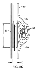
[00101] FIG. 2C illustrates an exemplary stent 30 in accordance with the
present invention for treating an aneurysm, such as a wide-neck aneurysm 20.
As seen in FIG. 2C, the stent 30 is generally a cylindrical body having an
expanded diameter D configured to contact the inner surface of lumen 10.
- 13-
CA 02753853 2011-08-26
WO 2010/102254 PCT/US2010/026430
The stent 30 has a length L configured to block the opening 22 of aneurysm
20 to form clot 32 within the aneurysm sac 20.
[00102] FIGS. 3 and 4 illustrate a method in accordance with the
present
invention for preparing and delivering a thin-film microvascular stent 30 to a
location in a blood vessel/artery 10 associated with an aneurysm 20. The
stent generally comprises a sheet of thin film nitinol that has a thickness h
of
approximately 4pm to 12pm. The sheet 30 has a length L corresponding to
the desired coverage at the aneurysm 20 within the artery 10, and width W
corresponding to the inside diameter of the artery 10. The sheet is initially
tightly rolled and placed into a small diameter catheter 40 (e.g., 0.69 mm
ID).
[00103] The stresses induced in the film cause the material to become
martensitic (i.e. stress induced phase transformation) and more malleable
when compared to the austenitic film. Using an endovascular procedure, the
catheter is guided through the vascular system to the aneurysm location 20
over a 0.014 inch (0.36mm in diameter) guidewire 42. The thin film 30 is
subsequently pushed out of the catheter 40 and deploys conformally with the
artery 10 as shown in FIG. 3. When pushed out into the blood stream, the thin
film 30 reverts to the higher stiffness austenite phase causing it to
conformally
deploy against the inner wall 34 of vascular blood vessel 20.
[00104] The stent 30 is sized to occlude the aneurysm 20 by completely
wrapping around the vascular wall's interior surface 34 without migrating
after
deployment or blocking flow through the vessel 10. FIG. 4 illustrates the
forces present in a deployed thin film stent 30. The forces consist of radial
forces FRachai, frictional forces FF, and hemodynamic shear or wall drag force
Fdrag= The radial forces FRachai induced during stent deployment (from the
stent
reverting to its preformed shape) produce frictional or holding forces FRachai
between the stent 30 and vascular wall 34. The blood flow through the stent
interior introduces hemodynamic shear stress or a drag force on the thin
film. A balance of these forces is calculated to maintain the position of thin
30 film microstent 30 so that it is immobilized and not free to migrate in
the
vascular system.
-14-
CA 02753853 2011-08-26
WO 2010/102254 PCT/US2010/026430
[00105] A basic fluid mechanics model is used to approximate the
hemodynamic shear stress induced on the thin film from blood flow. These
calculations assume that the vessel 10 is straight and the diameter is
constant
which limits the applicability of these results to some areas of the vascular
system. The hemodynamic shear stress (Tvvall) is calculated using Poiseuille's
Law assuming a straight vascular wall. The total hemodynamic drag force
FDrag on the film is:
(4 f]\
FDrag r wall = A = 3 1') 1'=87-1-,u = 1 =
v
Ta^ Eq. (1)
[00106] where ,u is blood viscosity, Q is blood flow rate, r is artery 10
radius, / is
length (i.e., axial) of thin film stent, and v is velocity of blood flow. The
velocity
of fully developed pulsatile blood flow ranges between 0.5-1.0m/s in human
CNS (Central Nervous System) arteries, and the blood viscosity is
approximately 0.004Ns/m2 (4centipoise). The remaining two variables are
functionally dependent upon artery size and thin film stent length. To
immobilize the thin film stent, the frictional forces FF must be larger than
the
drag force FDrag. The frictional force is proportional to the radial force
developed between the thin film and vascular wall.
[00107] Estimating radial force FRachai from the microstent deployment
is based
on the assumption that the nitinol is thin, long and isotropic. Using these
assumptions we argue that the deployment is similar to a long slender beam
subjected to a internal bending moment. The radial force resultant FRacha, is
subsequently due to the bending moment produced by the nitinol unrolling in
the austenite phase due to the shape memory effect. This radial force FRachai
can be approximated by the following equation.
E 11 14!
FRachal _____________________ =¨x h3
= ¨ =
1/2 24 r 1 Eq. (2)
[00108] where w is width, E is Young's modulus of the austenite phase
of nitinol
(83x109Pa), v is Poisson's ratio (v=0.33), and h is thin film thickness. The
frictional force between the vascular wall and the thin film is related to the
radial force and coefficient of friction. A conservative friction coefficient
,u of
-15-
CA 02753853 2011-08-26
WO 2010/102254 PCT/US2010/026430
0.05 was used between thin film nitinol surface 30 and vascular wall 34 to
estimate the frictional force (i.e., FF a 4
[00109] The efficacy of a material used in a stent is subject to a
number of
criteria. Materials must not cause an excessive inflammatory response, must
not be toxic to the body, and must not cause clotting in the blood stream.
Strategies aimed at reducing or eliminating rates of thrombosis have included
research into novel materials and surface treatments that prevent the
adhesion of blood products.
[00110] Nickel-titanium alloys (NiTi or nitinol) are particularly
beneficial for use
in stents and for covering stents. NiTi is ideally suited as biocompatible
material for use in many implantable medical devices, including stents and
atrial septal defect occlusion devices. NiTi is biologically inert in
physiological
solutions; a titanium oxide layer forms on the metal's surface which prevents
corrosion of the bulk material. Furthermore, nitinol is resistant to thrombus
formation and does not calcify. When implanted within blood vessels and
within the heart itself, NiTi has proven to be non-toxic, biocompatible, and
non-
thrombogenic.
[00111] The thin films that may be used to form stents in accordance
with the
present invention may be made from thin films of metal alloys that are phase
transforming and/or exhibit twin boundary motion. For example, NiTi and
other similar metal alloys exhibit a thermally induced crystalline
transformation
between a ductile martensite phase at low temperatures and a rigid austenite
phase at high temperatures. NiTi exhibits both shape-memory and super-
elastic properties. These metal alloys are referred to herein as thin-film
memory or shape memory metal alloys. Upon cooling below the martensite
temperature, unstrained NiTi has a twinned martensite structure. When
placed under stress, the twin orientation is reorganized along the direction
of
stress. When heated above the austenite temperature, the material regains its
rigid highly-ordered austenite phase and recovers the original shape in which
it
was crystallized. In the low temperature martensite phase, nitinol is
exceedingly malleable and can be compressed into catheters. Upon heating
-16-
CA 02753853 2011-08-26
WO 2010/102254 PCT/US2010/026430
(in many cases simply to body temperature), nitinol transforms into its
austenite parent phase and recovers from the deformation induced in the
martensite state. Thus, stents made from thin film NiTi (or other similar
metal
alloy) can make use of these shape memory properties. However, this does
not preclude the use of purely martensite NiTi or possible other acceptable
shape memory or pseudoelastic material.
[00112] In one embodiment, a shape memory alloy used to form a stent
can
have a starting temperature for transition to the austenitic phase of
approximately 20 C. For example, a stent was made of thin film nickel-
titanium alloy that was approximately 50.2 atom% titanium, and exhibited the
following transition temperatures: (start of transition to austenitic phase)
As =
5 C; (finish of transition to austentic phase) Af = 21 C; (start of
transition to
martensitic phase) Ms = 18 C; and (finish of transition to martensitic phase)
Mf
= 1 C. A graphic representation of the differential scanning calorimetry
experiment from which these transition temperatures were determined is
shown in FIG. 5.
[00113] Alternatively, the stent 30 may be configured so that it does
not require
the phase transformation, but rather solely relies on the malleability of the
nitinol. In other words, the stent would produce the restoring deformation.
The nitinol film can be in its martensitic state and rely solely on twin
boundary
motion.
[00114] Exemplary thin-film memory metal alloys useful in any of the
embodiments of the present invention include the nickel-titanium alloys
(NiTi),
as well as alloys having the desired properties selected from the following:
nickel-titanium-copper alloys (NiTiCu) and other copper-based alloys; gold-
cadmium and other cadmium-based alloys (AuCd); nickel-titanium-platinum
(NiTiPt) and other platinum-based alloys; nickel-titanium-palladium (NiTiPd)
and other palladium-based alloys; nickel-titanium-hafnium (NiTiHf) and other
hafnium-based alloys; and nickel-magnesium-gallium alloys (NiMgGa), nickel-
manganese-gallium alloys (NiMnGa) and other gallium-based alloys.
[00115] The thin film metal alloys of the present invention may be
produced with
-17-
CA 02753853 2011-08-26
WO 2010/102254 PCT/US2010/026430
various percentages of the constituent elements. For example, nickel-titanium
alloys, such as nitinol, that contain about 50 atom percent nickel (atom% Ni)
and about 50 atom percent titanium (atom% Ti) can be used. As another
example, NiTi alloys that include from about 45 to about 55 atom percent
nickel and from about 45 to about 55 atom percent titanium can be used. For
example, the shape memory alloy can include from about 46 atom percent
(atom%) to about 53 atom% of titanium. Nickel-titanium alloys with other atom
percentages can also be used.
[00116] Although fabrication of thin film nitinol (about 8 microns in
thickness)
has been attempted using flash and vacuum evaporation, ion beam sputtering,
and laser ablation, most of these fabrication methods have been unsuccessful
in producing high quality uniform film required for medical applications. DC
magnetron sputter deposition under ultra-high vacuum is a preferred method
for the production of medical quality thin film nitinol, as it allows for high
levels
of process controllability and "batch-to-batch" consistency. The sputter
deposition process involves ejecting atoms from a target material and
directing
them to form a thin film on a substrate. Target heating during sputtering
creates films of uniform thickness and composition not achieved with
conventional sputtering. This allows for precise process control of film
composition.
[00117] For example, a film having a compositional variation of no more
than
about 1 atom percent can be produced. The target may be heated to a
temperature of from about 200 C to about 800 C, preferably to a temperature
of from about 400 C to about 700 C, and more preferably from about 550 C
to about 650 C.
[00118] In one embodiment, hot target sputtering was carried out as
follows. A
residual gas analyzer (Stanford Research Systems, Sunnyvale, CA) was used
to monitor residual gas contamination levels prior to sputtering. Residual
gases can deplete the amount of titanium reaching the substrate. The
combined pressure of water, carbon dioxide, and carbon monoxide gases
were maintained below 10-9Torr during sputtering. An argon scrubber further
-18-
CA 02753853 2011-08-26
WO 2010/102254
PCT/US2010/026430
cleaned the argon to 99.999% purity as required for the sputtering process.
Sputtering of thin film nitinol onto a silicon wafer with 500 nm thick wet
thermal
oxide (other layers also used include barrier layers and lift off layers such
as
Cu, which can be lifted off the wafer using a chemical process) was
accomplished with a 3-inch DC magnetron gun (MeiVac. Inc., San Jose, CA).
A target made of bulk nitinol cut from a three inch boule of nitinol
containing
49.5 atom% nickel and 50.5 atom% titanium (SCI Engineering, Columbus,
OH) was used for the sputtering process. All films were deposited at base
pressures below 5x10⁻⁸Torr and at an Ar pressure of 1.5x10⁻³ Torr. The
substrate-to-target distance was 4 cm and a sputtering power of 300 Watts
was used. During deposition the substrate was translated back and forth in
relation to the target at 45 degree arcs with 80 mm length to minimize
compositional variation. The deposited amorphous film is crystallized by
heating to 500 degrees Celsius for 120 minutes. Typically the film is annealed
after removal from the wafer to prevent any diffusion or reactions with the
substrate.
[00119] The thin memory metal alloy films of the present invention
generally
have a thickness of less than 50 microns, and preferably have a thicknesses
ranging from about 0.1 microns to about 30 microns. Preferably, the thin films
may have a thickness ranging from about 0.1, 1, 2, 4, 5, 10, 15, 20, 25, 30 or
50 microns to about 4, 5, 10, 15, 20, 25, or 30 microns. More preferably, the
thin films may have a thickness of from about 4 microns to about 12 microns.
[00120] Thus, covering a stent with the thin memory metal film of the
present
invention (described in further detail below) will result in a minimal and
inconsequential increase in the size of the overall device. For example, thin
film NiTi can be manufactured in films of from about 5 to about 8 pm
thickness, so that covering a stent with thin film NiTi adds very little bulk
to the
devices. For children, for neurointerventional applications, and for coronary
applications, it is highly beneficial that covered stents maintain a very low
profile. Many applications require that stents can be delivered through very
small catheters even after covering them. The stent can have a thickness in
-19-
CA 02753853 2011-08-26
WO 2010/102254
PCT/US2010/026430
the range of, for example, between about 2 pm, 4 pm, 6 pm, 7 pm, 10 pm, 17
pm, 18 pm, or 20 pm.
[00121] Thin memory metal alloy films 30 can be produced in a range of
shapes
and sizes. For example, thin memory metal alloy films can be made square or
rectangular e.g. when laid flat, the sheet can have the appearance of a
rectangle with a longer length dimension and a shorter width dimension. Each
dimension of such a square or rectangle can be selected from a wide range.
For example, the width W of such a square or rectangle may be in the range
of, for example, between about 0.5 mm, 1 mm, 3 mm, 5 mm, 10 mm, 16 mm,
20 mm, 25 mm, 30 mm, or 40 mm. The width, W, is generally a function of the
internal diameter of the lumen, and whether or not the film is wound as a
spiral, single loop, double loop, etc. (described in further detail below).
[00122] Correspondingly, the length L of such a square or rectangle may
be in
the range of, for example, between about 0.5 mm, 2 mm, 5 mm, 15 mm,
20 mm, 30 mm, 50 mm, or 100 mm. Generally, the length Lisa function of the
vessel 10 and size of aneurysm 20 to be occluded.
[00123]
Adjacent sides need not be perpendicular. The sheet 30 can have a
form that is not an endless loop; for example, the sheet can have two distal
edges as ends of the sheet, bounding the length dimension.
[00124] Thin memory metal alloy films may be made in a wide variety of
shapes
other than square or rectangular. For example, thin memory metal alloy films
may be made to resemble other polygons, circles, ovals, crescents, or an
arbitrary shape.
[00125] In one embodiment, photolithography and etching techniques may
be
used to generate precise two-dimensional shapes required to produce thin film
nitinol sheets for covering the stents. For example, a positive photoresist
may
be spin coated onto an 8-micron thin film nitinol coated silicon oxide wafer.
The photoresist (PR) may be exposed through a patterned glass mask
(Computer Circuit Inc, Gardena, CA) and developed, leaving the desired PR
pattern on the nitinol film. The unprotected portions of the thin film nitinol
(areas without PR) may then be etched away in a 1:1:15 HNO3:HF:H20
-20-
CA 02753853 2011-08-26
WO 2010/102254 PCT/US2010/026430
solution, and the remaining PR removed with acetone. The fabricated thin film
nitinol sheets are then mechanically removed from the silicon oxide wafer.
This photolithography approach reduces the number of imperfections on the
edges of the thin film nitinol, thereby reducing/eliminating the incidence of
tearing as compared to mechanical mechanisms.
[00126] After the thin film is formed, thin film nitinol may be removed
from the
substrate (e.g. wafer or silicon wafer) on which it was formed by using a
crack
and peel method to produce a free-standing film. Alternatively, a lift off
method may be used, wherein the thin film nitinol is removed from the wafer
by etching the sacrificial layer, for example, Cu. With the lift-off method,
the
Cu can be deposited onto a layer of silicon dioxide on top of the silicon
wafer
prior to depositing the NiTi thin film.
[00127] The thin film can then be annealed. For example, the thin film
can be
annealed after removal from the substrate for about 2 hours at approximately
500 C. The thin film can also be annealed on the substrate on which it was
deposited through sputtering. However, this can result in diffusion of the
atoms of the material of which the substrate is formed into the thin film,
which
can detrimentally affect the properties of the thin film.
[00128] After being annealed, the thin film 30 may be hot shaped to
form a coil.
For example, the thin film 30 may be hot shaped by heating the film to
approximately 500 C and holding it in a shape to which it is constrained for
about 5 minutes. For example, the film 30 may be rolled into a cylinder having
an outside diameter, that when in its expanded configuration, conforms with
the inside diameter of the lumen and applies a radial force to the inner wall
34
of the lumen 10.
[00129] The thin film 30 may then be compacted into a form for delivery
as
shown in FIG. 3. For example, the thin film can be coiled more tightly into a
compacted form for loading into and delivery through a catheter 40.
[00130] As shown in FIG. 6, the thin film 30 may be coiled by using a
cylindrical
instrument, such as a split cylindrical instrument 50 having slot 52 at its
distal
end for retaining the film 30. An edge of the thin film 30 is be inserted into
the
-21-
CA 02753853 2011-08-26
WO 2010/102254 PCT/US2010/026430
slot 52 of the instrument 50, and the instrument 50 is then rolled to coil the
thin
film 30 into a cylindrical configuration for deploy.
[00131] The rolled thin film into the form of a coiled stent 30 is then
delivered by
a catheter 40 (e.g. size 3 French or less) as shown in FIG. 3. The stent 30 in
its compacted form can be inserted into and deployed from a delivery tube,
such as a catheter, having a diameter of about 1 mm or less, for example,
from a delivery tube, such as a catheter, having a diameter of about 0.5 mm or
less.
[00132] Once deployed, e.g. by being pushed out of the catheter 40, the
stent
30 assumes (or attempts to assume) its shape prior to being compacted to fit
into a catheter 40. As shown in FIG. 7A, the stent may assume the shape of a
cylindrical tube 60 having an outer diameter D larger than the inner diameter
of the catheter 40.
[00133] Because the stent 30 according to the present invention is very
thin, it
assumes a low profile when deployed in a blood vessel. Therefore, when
deployed in a blood vessel, the stent 30 do not substantially impede the flow
of
blood through the vessel 10. The low profile of a stent according to the
present invention is illustrated in FIG. 4 where it is shown in a
configuration of
deployment in a blood vessel 10.
[00134] In the generally tubular shape 60 shown in FIG. 7A, the inside of
the
tube 62 is hollow, so that, for example, a fluid can travel into the shape
through a proximal end 66, through the shape (along the central axis 64), and
out of distal end 68 of the tube 60.
[00135] The generally tubular shape 60 shown in FIG. 7A may also be
varied to
be other than a perfectly cylindrical shape. For example, in FIG. 7B, the tube
may comprise an elliptical shape 80.
[00136] As shown in FIG. 7C, a spiral shape 82 can be formed by curling
a
sheet, so that if the sheet is traced from one end, a path around a central
axis
64 is followed. As the path goes around the central axis 64, the path
generally
moves either continuously inward toward the central axis 64 or continuously
outward away from the central axis 64. The path may also have excursions
-22-
CA 02753853 2011-08-26
WO 2010/102254 PCT/US2010/026430
from such continuously inward or continuously outward movement.
[00137] A broken ring, as shown in FIG. 7A, is similar to a spiral,
except that
there is no overlap, i.e., the winding number is 1 or less. That is, a broken
ring
can have the ends 70 and 72 just touching (as shown in FIG. 7A), or can have
the ends separated (e.g. forming a "c" shape (not shown)).
[00138] Compacted, for purposes of the present invention, means that an
object, for example, a sheet, is temporarily shaped so that at least one
dimension of the object is smaller than in the deployed form of the object.
[00139] For children or adults who require neurointerventional
applications, it is
highly beneficial that the stent maintains a very low profile. Many
applications
require that the stents be delivered through very small catheters. The use of
thin film metal alloy for stents in accordance with the present invention
allows
for the construction of very low profile stents for use in the treatment of,
for
example, aneurysms of the central nervous system vasculature and brain
vessel aneurysms.
[00140] The stent 30 can be used, for example, to support a body
cavity, to
maintain a passage through a body cavity, and/or to seal off a body cavity.
For example, a stent 30 implanted into a blood vessel 10 can act to prevent
the closing of the blood vessel. The stent can be hollow, so that fluid can
travel through it along its central axis. For example, a stent implanted into
blood vessel 10, for example, a blood vessel supplying the peripheral or
central nervous system, can seal off an aneurysm 20 from the blood vessel.
For example, the stent can be implanted in body cavities, such as blood
vessels supplying the central nervous system, in peripheral blood vessels, and
in coronary blood vessels, in order to treat diseases and disorders of the
blood
vessels such as peripheral artery disease (PAD).
[00141] When the stent 30 is deployed or placed inside a body cavity,
for
example, a blood vessel, it can distort somewhat. For example, the stent may
not have a perfectly cylindrical shape, a perfect spiral shape, or another
idealized shape, but may be distorted from this ideal shape, e.g., to conform
to
a support on the walls of the vessel.
-23-
CA 02753853 2011-08-26
WO 2010/102254 PCT/US2010/026430
[00142] The apparatus, e.g. stent 30 formed of the thin film can be
advanced
with a catheter 40, for example, on a 3 French (3 Fr) delivery system. The
stent can be self-expanding. When the apparatus 30 is advanced into position
within the body, the thin film NiTi stent can unravel, so that the stent
expands
to cover the inner surface 34 of the blood vessel 10into which it was
deployed.
The inner surface 34 of the blood vessel 10 is that part of the blood vessel
that can be in contact with fluid moving through the vessel when no stent is
in
place. A large fraction of the outer surface of the stent 30 can be in contact
with the inner surface 34 of the blood vessel 10 into which the stent was
deployed. For example, at least about 80% of the outer surface of the stent
30 can be in contact with the inner surface 34 of the blood vessel into which
the stent is deployed.
[00143] The stent 30 can be deployed in a blood vessel adjacent to an
aneurysm 20, so that in its deployed form, the stent covers at least a portion
of
the aneurysm. See, for example, FIG. 2C. In its deployed form, the stent can
cover at least 40% of the area of a passage from the blood vessel to the
aneurysm.
[00144] The apparatus, e.g. thin film stent 30, can then be advanced
into a
catheter 40. When the apparatus 30 is advanced into position within the body
and released from the catheter 40, the thin film memory metal of the stent
unravels as it is trained to do (as it is heated or as it simply uses twin
boundary
motion forced by the stent) and the stent expands. For example, when the
thin film stent 30 is released from a delivery system 40, the austenitic shape
memory of the thin film can allow the stent to expand and cover the inner
surface 34 of a blood vessel 10 into which it is deployed.
[00145] The thin film 30 may be perforated or non-perforated. For
example, the
thin film can be solid, without holes, pores, or open slots. In this
configuration,
the thin film can be impermeable to body tissue and fluids.
[00146] For certain neurovascular applications, the thin film metal
stent 30 is
ideally configured to be able to bend around curves of small radii. This is
because the blood vessel 10 to be treated can be of small radius and can be
-24-
CA 02753853 2011-08-26
WO 2010/102254
PCT/US2010/026430
tortuous, or the blood vessels through which the stent must pass in order to
reach the blood vessel to be treated are tortuous.
[00147] In order to allow the stent to bend around curves of small
radii, for
example, for the treatment of certain aneurysms, the stent 30 can be made to
have a short length L. The stent can also be made to have a short length L to
improve its ability to track around tortuous vessels, for example, over a
guide
wire 42.
[00148] Referring to FIGS. 8 and 9, a stent 100 is shown having a
plurality of
joints 104 that allow the stent 100 to bend more freely, and thus allow the
stent to bend around curves of small radii, and treat, for example, aneurysms
which extend along a blood vessel. Thus, the thin film sheet 102 may have at
least one joint 104 that allows the structure of the stent to bend about an
axis
in the radial direction.
[00149] As shown in FIG. 9, the stent 100 may be deployed in a portion
of a
blood vessel 10 forming an arc (e.g. 180 degrees) and having a radius r
(e.g.1.5 mm to 3 mm), in order to treat an aneurysm adjacent to or at arced
portion of the blood vessel 10.
[00150] The
stent 100 has a longitudinal direction or axis 106, which extends
along the center of the tube formed by the stent. Thus, if the stent 100 is
bent,
the longitudinal direction 106 has the form of a curve. At a given point along
the longitudinal direction, a radial direction r is perpendicular to the
longitudinal
axis 106. In bending around the curve of a blood vessel 10, the stent 100
bends around an axis in the radial direction.
[00151] The joint 104 may comprise a strip on the rectangular thin film
sheet
102. For example, the strip can have a long direction and a short direction,
with the long direction being parallel to the distal edges of the sheet, and
the
short direction can be perpendicular to the distal edges of the sheet. The
ratio
of the long direction over the short direction can be, for example, at least
2.
Thus, when the thin film is rolled into a spiral to form the stent, a strip
which
forms the joint can have the form of a band around the stent (when the stent
is
formed from a spiral, the strip also has the form of a spiral rather than an
-25-
CA 02753853 2011-08-26
WO 2010/102254 PCT/US2010/026430
endless loop).
[00152] In an embodiment, the thin film sheet has an average thickness.
This
average thickness can be the volume of the material of which the thin film
sheet is formed divided by the area of the thin film sheet. The strip has an
average thickness that is the volume of thin film sheet material in the strip
divided by the area of the strip. The average thickness of the strip is no
greater than about 90% of the average thickness of the thin film sheet.
[00153] In an embodiment, the memory metal of which the strip 104 is
formed is
continuous, that is, has no perforations. The strip 104 may have a different
local thickness. In one embodiment, the local thickness of the strip 104 is no
greater than about 90% of the thickness of the sheet 102, and preferably no
greater than about 50% of the thickness of the sheet 102, and more preferably
no greater than about 25% of the average thickness of the thin film sheet 102.
[00154] The strip 104 and/or the thin film sheet 102 may also be
perforated, for
example, in order to improve the flexibility of the stent formed. In one
embodiment, the thin film sheet or strip is perforated with at least one hole.
For example, the hole can have a profile area of less than about 2000 pm2.
The profile area is the area in the thin film sheet occupied by the hole. For
example, the hole can have a maximum spanning distance of less than about
50 pm. The maximum spanning distance is the maximum distance from one
point on the perimeter of the hole to another point on the perimeter of the
hole.
Thus, for example, the maximum spanning distance of a hole shaped as a
circle is the diameter of the circle, and is less than the maximum spanning
distance of a hole shaped as an ellipse that has the same area as the circle.
[00155] For example, FIG. 10A shows a joint in the form of a strip 104
having a
series of holes separated by undulating wires of metal 110. Thus, the holes
have the form of slots with "fingers" projecting perpendicularly from a long
direction of the slot, the "fingers" of adjacent slots being interdigitated.
[00156] A hole that perforates the thin film sheet can have any one of
a number
of profiles. The profile is the form of the hole when the hole is viewed face
on.
For example, the hole can have a circular, elliptical, or diamond-shaped
-26-
CA 02753853 2011-08-26
WO 2010/102254 PCT/US2010/026430
profile. FIG. 10B shows a photograph of a joint in the form of a strip 104
having holes112 in a hexagonal array, where each hole 112 has an
approximately hexagonal or oval form. Holes can also be positioned in other
types of arrays, for example, periodic, quasiperiodic, and nonperiodic arrays.
Holes can have other forms, such as polygonal or an arbitrary shape.
[00157] FIG. 10C shows a joint in the form of a strip 104 that includes
a series
of holes having the form of slots 114.
[00158] The slots 114 have a long direction Lu and a short direction W.
The
long direction Lu may be parallel to a distal edge 118 of the thin film sheet.
The short direction Wu may be perpendicular to a distal edge 118 of the thin
film sheet and parallel to a short edge 116 of the film 104. The short
direction
Wu may be less than about 50 pm.
[00159] FIG. 10D illustrates a thin film strip 104 having a plurality
of diamond
shaped fenestrations 120 that allow further flexibility, and particularly
expansion and compression in direction DE. As shown in FIGS. 11A and 11B,
the strip 104 may have elongation over 400% from a compressed state 120A
to expanded state 120B.
[00160] As shown in the embodiment of FIG. 8, the thin film sheet 102
may be
continuous, e.g. non-perforated, in a region outside of the joint strip 104,
and
is porous (e.g. fenestrated according to any of the patterns shown in FIGS.
10A-D) within the strip 104.
[00161] Alternative system 150 shown in FIG. 12 comprises a thin film
sheet
160 fenestrated with a plurality of diamond shaped holes 120, and wrapped
around a collapsible, truss-like stent 152 for additional rigidity.
[00162] FIGS. 13A and 13B illustrate method for attaching a thin film 170
having
a plurality of fenestrations 176 to a collapsible truss 152. FIG. 13A shows
the
vessel side (outside surface) of the stent 150 with stitching 172 tied in a
knot
174, and FIG. 13B showing the internal side of the stent 150 with the
stitching
172 looped around the truss 152 and through the fenestrations 176.
[00163] FIGS. 14A -14C illustrate additional truss shapes that may be
implemented along with a thin film 30 according to the present invention.
-27-
CA 02753853 2011-08-26
WO 2010/102254 PCT/US2010/026430
[00164] The coil structure 200 shown in FIG. 14A comprises a
superelastic
nitinol wire 204 with a plurality of coils 202. The wire may be manufactured
by
winding a 0.13mm diameter superelastic nitinol wire 204 around a cylinder (not
shown) approximating the radius of the arterial wall, and heating at 500 C
for
30 minutes to shape set the nitinol wire 204. After hot shaping, the coil 200
may be pulled straight to induce a stress induced phase transformation and
inserted into a delivery catheter 40 containing the rolled microstent 30 (as
described above). The coil 200 may be positioned such that there is
substantially equal length of coil 204 extending out both sides of the rolled
microstent sheet 30. When deployed, one-third of the coil 200 may initially be
pushed out of the catheter 40 prior to deploying the thin film microstent 30.
Once the thin film portion 30 of the stent is delivered, the remainder of the
wire
coil 200 may then be deployed.
[00165] FIG. 14B illustrates a "zigzag" structure stent 210 having a
plurality of
folds 212. Stent 210 may be formed by setting 0.13mm diameter superelastic
nitinol wire on a cylinder approximating the radius of the arterial wall and
heating at 500 C for 30 minutes to shape set. After hot shaping, the
structure may be physically compressed to induce a stress induced phase
transformation and then inserted into the delivery catheter 40 as described
above for coil. Inside the catheter, the wire skeleton 210 preferably has 3-4
mm of overhang length out of both ends of the rolled microstent sheet 30.
This structure is configured to be collapsed into an ultra-low diameter
catheter
(i.e., 0.69mm diameter) with a rolled microstent 30. The supporting nitinol
212
is designed to retain sufficient longitudinal flexibility to permit the
catheter 40
and structural backbone to navigate through a tortuous cerebral vascular
system.
[00166] FIG. 14C shows a stent structure 220 having dual-zigzag
structures 214
and 216. This stent 220 provides radial force at the ends of the thin film
nitinol
stent 30, while preserving flexibility in the body of the stent.
[00167] The thin film sheet 30 may have a winding number that corresponds
to
the number of revolutions of the sheet about an axis, of greater than 1. Thus,
-28-
CA 02753853 2011-08-26
WO 2010/102254 PCT/US2010/026430
with increasing radial distance from the center of the stent (the longitudinal
direction), more than one layer of the thin film can separate the inner region
of
the stent through which fluid can pass from the outer environment of the
stent.
The longitudinal edges of the sheet can overlap each other.
[00168] FIG. 15 illustrates a sheet 180 having a length between end 70 and
opposite end 72 sufficient to form two coils (e.g. winding number of 2+) when
expanded within the lumen 10.
[00169] Alternatively the thin film sheet of the stent can have a
winding number
of less than about 1 (e.g. "c" shape). Thus, the stent can be formed to not
close back on itself, so that in certain radial directions, the inner region
of the
stent through which fluid can pass is open to the outer environment of the
stent.
[00170] A thin film sheet 60 having a winding number of about 1 is
shown in
FIG. 7A. For example, the stent can be formed so that the longitudinal edges
70 and 72 of the thin film 60 touch each other.
[00171] The thin film sheet of the structure of the stent can include a
notch. For
example, a notch can extend from where the strip meets a longitudinal edge of
the sheet along a portion of the strip. For example, the notch can extend from
the longitudinal edge and along the strip half-way across the thin film sheet.
[00172] FIG. 16 illustrates an embodiment of a thin-film stent 250
according to
the present invention having a tab-and-slot configuration. The stent 250 is
formed from a sheet that includes tabs 256 at first end 254, the tabs 256
projecting in the width direction, i.e., perpendicular to the length dimension
of
the sheet. The sheet 250 also comprises a slot 260 at second end 258 of the
sheet. The first end 254 with the tabs 256 may be inserted through the slot
260 to secure the cylindrical shape of the stent 250.
[00173] As shown in FIG. 16, the first end 254 with the tabs 256 is
inserted
through the slot 260 such that the first end 254 extends into the internal
radius
of the stent 250 and wrapped around itself to have a spiral form, i.e. an
inside
roll tab-and-slot design. The sheet 250 generally has a tubular structure,
wherein the spiral formed sheet 250 can be used alone as a stent.
-29-
CA 02753853 2011-08-26
WO 2010/102254 PCT/US2010/026430
[00174] A stent 250 having an inside roll tab-and-slot design can
unroll in a
belt/buckle design. The buckle maintains the sheet's alignment, and prevents
the sheet from unraveling too far. A stent having an inside roll tab-and-slot
design can have its sheet unraveling from the inside out, assuring proper
stent
coverage and alignment.
[00175] In an alternative embodiment shown in FIG. 17, a stent 270
comprises
a sheet formed into a tab-and-slot design the first end 254 with the tabs 256
is
inserted through the slot 260 such that the first end 254 extends outward from
the internal radius of the stent 270 and wrapped around itself to have a
spiral
form with outside roll tab-and-slot design.
[00176] In other words, a stent 270 having an outside roll tab-and-slot
design
can unroll in a belt/buckle design. The buckle maintains the sheet's
alignment,
and prevents the sheet from unraveling too far. A stent having an outside roll
tab-and-slot design can have its sheet unraveling on the outside of the belt,
assuring proper stent coverage and alignment.
[00177] The sheet, whether in the form of a spiral 82 (FIG. 7C), an
inside roll
tab-and-slot design 250 (FIG. 16), an outside roll tab-and-slot design 270
(FIG. 17), or in another form not shown, can have a compacted form to
facilitate its delivery into a body cavity, for example, either inside or on a
catheter 40. By compacted, the sheet is temporarily shaped, so that at least
one dimension of the generally tubular structure formed by the sheet is
smaller
than in the deployed form.
[00178] For example, the sheet can be wrapped tightly, so that it has a
large
winding number, or the sheet can be wrapped loosely. A sheet wrapped
tightly, so that it has a large winding number, can be in a compacted form,
because the diameter of the generally tubular structure formed by the sheet is
smaller than it would be if the sheet were wrapped loosely. In a compacted
form, a structure can have a first internal diameter. For example, when the
sheet is wrapped tightly, the structure formed can have a first internal
diameter
extending from the inside surface of the innermost layer of the sheet, across
the central axis, to an opposite point on the inside surface of the innermost
-30-
CA 02753853 2011-08-26
WO 2010/102254
PCT/US2010/026430
layer of the sheet.
[00179] A
sheet wrapped loosely, so that it has a small winding number, can
be in a deployed form, because the diameter of the generally tubular structure
formed by the sheet is larger than it would be if the sheet were wrapped
tightly. In a deployed form, a structure can have a second internal diameter.
For example, when the sheet is wrapped loosely, the structure formed can
have a second internal diameter extending from the inside surface of the
innermost layer of the sheet, across the central axis, to an opposite point on
the inside surface of the innermost layer of the sheet. The second internal
diameter can be larger than the first internal diameter. The sheet in its
deployed form can be impermeable to body tissue and fluids.
[00180] A sheet formed of a thin film memory metal alloy can be induced
to
transition from its compacted form to its deployed form by a change in
temperature. Alternatively, a sheet which is held under tension in its
compacted form, e.g. because it is inside a catheter, can be induced to
transition to its deployed form by a release of the tension, e.g. after the
sheet
is placed in a blood vessel and the catheter is removed. When the sheet is
induced to transition from its compacted form to its deployed form by a change
in temperature or by a release of tension, no extrinsic force is required to
expand the sheet to its deployed form.
[00181] The expansion of the sheet to its deployed form can be driven
by a
phase change of the shape memory alloy, and can be driven by super-
elasticity of the shape memory alloy.
[00182] In its deployed form, a sheet formed of a thin film memory
metal alloy
can, for example, have the form of a spiral 82, an inner loop tab-and-slot
design 250 (FIG. 16), an outer loop tab-and-slot design 270 (FIG. 17), a ring,
60 (FIG. 7A), a broken ring or another configuration. A broken ring is similar
to a spiral, except that there is no overlap, i.e., the winding number is 1 or
less. A ring can have the ends just touching, and a broken ring can have the
ends separated.
[00183] A stent formed of a sheet of thin film memory metal alloy is
not limited
-31-
CA 02753853 2011-08-26
WO 2010/102254
PCT/US2010/026430
to a spiral, inside roll tab-and-slot, or outside roll tab-and-slot design.
For
example, a sheet of thin film memory metal alloy can be used to form a stent
having a compacted form with any means for packaging the area of the sheet
into a smaller circumference than the deployed circumference, such as folds,
coiling, and layers. These include packaging means like a spiral, but also
having outward protrusions to resemble a star, a twisted star, a keyed wheel,
or other configurations.
[00184] Furthermore, even though the stent may be designed to have a
particular shape for its compacted form, often, when the stent is placed in or
on a catheter, the compacted form may be distorted from the designed shape.
For example, a sheet of thin film memory metal, when placed in or on a
catheter, may not have the ideal shape of a spiral, tab-and-slot, star,
twisted
star, keyed wheel, or other configuration. Rather, the sheet may be distorted
from this ideal shape.
[00185] Referring to FIG. 18, system 300 may include a thin-film stent 30
disposed around an inner tube 302. The inner tube 302 comprises a hollow
bore 304 to allow a fluid (e.g. blood) to travel into the inner tube 302
through
one end, and out of the other end of the inner tube. The walls of the inner
tube 302 may be porous or non-porous. For example, the inner tube 302 may
be a coil or a mesh. The thin film sheet 30 is preferably wrapped around the
inner tube 302. Thus, the inner tube 302 can serve as a support for the thin
film sheet 30.
[00186] The inner tube 302 may be of such structure or material to have
a
compacted form and a deployed form. For example, the compacted form of
the inner tube 302 can have a diameter less than or equal to the first
internal
diameter of the structure of the thin film sheet 30 in its compacted form. The
deployed form of the inner tube 304 can have a diameter greater than the first
internal diameter of the structure of the thin film sheet in its compacted
form.
The inner tube 302 in its compacted form can exert an outward directed radial
force. This outward radial force can cause the inner tube 302 to expand, and
can assist the structure of the thin film sheet 30 in expanding to its
deployed
-32-
CA 02753853 2011-08-26
WO 2010/102254 PCT/US2010/026430
form. The inner tube 302 in its deployed form can exert an outward radial
force. The outward radial force exerted by the inner tube 302 can increase the
pressure applied by an outer surface of the thin film sheet 30 onto a body
cavity, such as the wall of a blood vessel 10. This outward radial force can
help maintain the stent 300 in a given position in a body cavity, for example,
in
a blood vessel 10, without sliding, e.g., without blood flow causing the stent
to
slide.
[00187] To exert the outward radial force, the inner tube 302 may
comprise an
elastic material, e.g. a shape memory alloy. The change of the inner tube 302
from its compacted form to its deployed form can be induced by a change of
temperature or can be caused by twin boundary motion.
[00188] In addition to its shape-memory and super-elastic properties,
thin film
nitinol possesses remarkably high tensile strength. These properties make it
particularly amenable for use in transcatheter devices. Furthermore, thin film
nitinol allows for the construction of extremely low profile stents. When this
material is manufactured as a thin film, there is little room for fluctuations
in
surface texture, resulting in an extremely smooth surface. In contrast to bulk
nitinol, a DC hot target sputtering process in accordance with the present
invention produces thin films of nitinol that are free of contaminants and
uniform in composition, in addition to being more resistant to corrosion in a
biological environment.
[00189] Used alone as a stent, the sheet 30 of thin film memory metal
alloy
may be housed in a catheter 40 or sheath and delivered to the place in the
body, e.g., a part of a blood vessel 10, where it is to be deployed. The stent
30 can then be unsheathed, so that the sheet of thin film memory metal alloy
expands into its deployed form. The expansion can be driven, for example, by
a temperature induced phase change in or by the super-elasticity of the sheet.
[00190] The stent 30 formed from thin film memory metal alloy can be
made
radio-opaque by the addition of radio-opaque markers of a dense metal. The
radio-opaque marker can be formed of a bio-inert metal. For example,
markers of gold, platinum, or palladium, metals which can appear opaque on
-33-
CA 02753853 2011-08-26
WO 2010/102254 PCT/US2010/026430
radiographs and which are substantially bio-inert, can be added to the thin
film
memory metal alloy of a stent. Such markers can be added to thin film shape
memory metal alloy by mechanical attachment, welding, bonding, or sputter
deposition. For example, a sputtering mask with holes can be placed over a
thin film sheet of memory metal alloy. The sputtering mask thus can define
masked and non-masked areas on the thin film sheet. A radioopaque metal,
such as gold, platinum, or palladium, can be sputtered through the holes to
deposit on the non-masked areas of the thin film sheet to form markers. The
sputtering mask can be formed with a photolithography technique. For
example, a photoresist can be spin coated onto the thin film, the photoresist
can be exposed to a pattern of light and developed to form the sputtering
mask. Alternatively, a radio-opaque marker can be mechanically attached to
the thin film.
[00191] In an alternative embodiment, a neurovascular stent 30
according to the
present invention can be delivered to and deployed at a desired position in a
blood vessel 10 or other body cavity without the use of a catheter. The stent
can be maintained in its compacted form by a self-contained constraining
device other than a catheter.
[00192] For example, for system 350 shown in FIG. 19, a stent 30 formed
from
a thin film sheet of memory metal wrapped into a spiral is held in a compacted
form via a ring 352 circumscribing the spiral thin film sheet of the stent 30.
The stent 350 in its compacted form can be delivered to a desired position in
a
blood vessel 10 or other cavity by any of a number of procedures, for
example, pushing by a flexible rod 354 that can have a smaller diameter than
a catheter, pulling by a wire, or, for a stent that includes a ferromagnetic
material, maneuvering by an externally imposed magnetic field. When the
stent is in the desired position, the ring 352 that maintains the stent 30 in
its
compacted form is removed, so that the stent expands 30 to its deployed form.
[00193] The removal of the ring 352 can be effected by any of a number
of
procedures and devices. For example, a wire 356 connected to the ring 352
can pull on the ring in the same direction as a flexible rod 354 maintaining
the
-34-
CA 02753853 2011-08-26
WO 2010/102254 PCT/US2010/026430
stent in the desired position, or a wire connected to the ring can pull on the
ring in the opposite direction as a wire maintaining the stent in the desired
position. The ring 352 may be flexible, like a wire, so that it can be easily
retracted through the blood vessel 10 or other body cavity to an exit point.
Alternatively, the ring can include a ferromagnetic material, and a magnetic
field can be used to displace the ring from the stent, while the stent is
maintained at the desired position with, for example, a flexible rod or a
wire.
Alternatively, the stent can include a ferromagnetic material, the ring can be
maintained at the desired position with, for example, a flexible rod or a
wire,
and a magnetic field can be used to displace the stent to a desired position
from the ring. Alternatively, the ring can be constructed of a biodegradable
material, so that after a predetermined amount of time, the ring degrades and
the stent expands to its deployed form.
[00194] FIG. 20 shows an alternative system 360 for retaining a thin
film sheet
of memory metal 30 wrapped into a spiral in a compacted form with a loop 366
that passes through a first hole 362 in one part of the sheet 30 and a second
hole 364 in another part of the sheet. Thus, a stent 30 in its spiral
compacted
form comprises one or more aligned holes 362, 364 passing through the
layers. The loop 366 passes through the several layers, preventing the layers
from moving with respect to each other and preventing the spiral 30 from
unraveling. The loop 366 can be formed by, for example, the loop passing
toward the center of the stent through one set of aligned holes 362 and then
passing toward the outside of the stent through another set of aligned holes
364, as shown in FIG. 20. Alternatively, the loop can be formed by, for
example, the loop 366 passing toward the center of the stent through one set
of aligned holes 362, out of an end of the tubular shape of the stent, and
around to the outside of the stent, so that an endless loop is formed (not
shown).
[00195] The stent may be delivered to a desired position in a blood
vessel or
body cavity by, for example, a flexible rod 354, wire, or magnetic field. When
the stent is in the desired position, the loop 366 that maintains the stent in
its
-35-
CA 02753853 2011-08-26
WO 2010/102254 PCT/US2010/026430
compacted form can be broken and removed, so that the stent expands to its
deployed form. This breaking and removal of the loop can be effected by any
of a number of procedures and devices. For example, a wire connected to the
loop 366 may pull on the loop in the same direction as a flexible rod
maintaining the stent in the desired position, or a wire connected to the loop
may pull on the loop in the opposite direction as a wire maintaining the stent
in
the desired position. The loop 366 itself may be flexible, like a wire, so
that it
may be easily retracted through the blood vessel or other body cavity to an
exit point. Alternatively, the loop 366 may include a ferromagnetic material,
and a magnetic field may be used to displace the loop from the stent, while
the
stent is maintained at the desired position with, for example, a flexible rod
or a
wire. Alternatively, the stent may include a ferromagnetic material, the loop
may be maintained at the desired position with, for example, a flexible rod or
a
wire, and a magnetic field may be used to displace the stent to a desired
position from the loop. Alternatively, the loop 366 may be constructed of a
biodegradable material, so that after a predetermined amount of time, the loop
degrades and the stent expands to its deployed form.
[00196] Instead of the loop 366 in FIG. 20, a rivet 368 may be used to
maintain
the stent 30 in its compacted form by passing through a set of two or more
aligned holes in two or more layers, 362, 364 of the stent 30. The rivet 368
may be removed by pulling or pushing on the rivet or the stent by, for
example, a flexible rod, wire, or magnetic field, similar to the ways in which
a
loop may be removed. So that the rivet is not prematurely displaced, the rivet
may have a flange 370 at each end of the rivet with a diameter greater than
the holes through which the rivet passes. Alternatively, the rivet may be
constructed of a biodegradable material, so that after a predetermined amount
of time, the rivet degrades and the stent expands to its deployed form.
[00197] For the embodiments of a self-contained constraining device in
which
the stent in its compacted form includes one or more sets of aligned holes in
the layers of the thin film metal sheet that are in a spiral, the stent and
the self-
contained constraining device may be designed, so that after the self-
-36-
CA 02753853 2011-08-26
WO 2010/102254 PCT/US2010/026430
contained constraining device allows the stent to expand to its deployed form,
the holes (e.g. 362, 364) are no longer aligned, so that fluid in the interior
of
the stent is impeded from passing through the holes to the exterior of the
stent.
[00198] In another embodiment, a stent 30 may be maintained in a compacted
form with a clip (not shown) that fastens two portions of the sheet to each
other. For example, a stent may include a first protrusion extending inward
from the longitudinally running edge of the sheet at the innermost layer of
the
spiral and a second protrusion extending inward from a point on the sheet next
to where the sheet becomes covered by the innermost layer of the spiral.
When the stent is in its compacted form, the first protrusion and the second
protrusion may be adjacent to each other and maintained adjacent to each
other with a clip, so that the stent is maintained in its compacted form and
the
spiral of the thin film metal sheet does not unravel.
[00199] Alternatively, the first protrusion can extend outward from the
longitudinally running edge of the sheet at the outermost layer of the spiral,
the
second protrusion can extend outward from a point on the sheet next to where
the sheet becomes covered by the outermost layer of the spiral, and the first
and second protrusions can be held together by a clip.
[00200] Alternatively, a clip can press together the distal edges of the
layers of
the spiral at an end of the tubular shape of the stent, or two clips can press
together the distal edges of the layers of the spiral at each end of the
tubular
shape of the stent.
[00201] The stent may be delivered to a desired position in a blood
vessel or
body cavity by, for example, a flexible rod, wire, or magnetic field. When the
stent is in the desired position, the clip or clips that maintain the stent in
its
compacted form may be removed, so that the stent expands to its deployed
form. This removal of the clip or clips may be effected by any of a number of
procedures and devices. For example, a wire connected to a clip can pull on
the clip in the same direction as a flexible rod maintaining the stent in the
desired position, or a wire connected to the clip can pull on the loop in the
-37-
CA 02753853 2011-08-26
WO 2010/102254 PCT/US2010/026430
opposite direction as a wire maintaining the stent in the desired position.
[00202] Alternatively, the clip may include a ferromagnetic material,
and a
magnetic field can be used to displace the clip from the stent, while the
stent is
maintained at the desired position with, for example, a flexible rod or a
wire.
Alternatively, the stent may include a ferromagnetic material, the clip can be
maintained at the desired position with, for example, a flexible rod or a
wire,
and a magnetic field may be used to displace the stent to a desired position
from the clip. Alternatively, the clip may be constructed of a biodegradable
material, so that after a predetermined amount of time, the clip degrades and
the stent expands to its deployed form.
[00203] In another embodiment, a stent 30 may be maintained in a
compacted
form with glue that fastens two portions of the sheet to each other. When the
stent is in its compacted form, the relative position of the adjacent layers
can
be maintained by the glue, so that the stent 30 is maintained in its compacted
form and the spiral of the thin film metal sheet does not unravel. The glue
can
be constructed of a biodegradable material, so that after a predetermined
amount of time, the glue degrades and the stent expands to its deployed form.
[00204] Thus, for example, a ring, loop, rivet, clip, or glue can be a
self-
contained constraining device. Other devices or features can serve as a self-
contained constraining device for a stent. A self-contained constraining
device
can be a device that is a part of the stent, acts to maintain the stent in a
compacted form, and then allows the stent to expand to its deployed form
upon the passage of a predetermined period of time, upon exposure to a
predetermined environmental condition, such as an environmental condition
that exists within the body, and/or upon triggering by a stimulus having its
origin outside of the body, such as a user controlled imposition or change of
a
magnetic field or application of a mechanical force. Two or more factors can
be necessary for the self-contained constraining device to allow the stent to
expand to its deployed form. For example, the self-contained constraining
device may have the form of a clip made of a biodegradable material. The
biodegradable material can be chosen or designed so that for the clip to allow
-38-
CA 02753853 2011-08-26
WO 2010/102254 PCT/US2010/026430
the stent to expand to its deployed form, the clip must be within a blood
vessel
and exposed to blood and the clip must reside within the blood vessel for a
sufficiently long period of time for the blood to dissolve enough of the clip
material for the clip to release.
[00205] The term "biodegradable" as used herein to describe a material can
mean a material that is chemically broken down by chemical or biochemical
processes in the body. Alternatively, the term "biodegradable" can describe a
material that is solubilized within the body. Alternatively, the term
"biodegradable" can describe a material that, while exhibiting a predetermined
strength at a temperature lower than body temperature, such as at room
temperature, exhibits a lower predetermined strength at body temperature.
Alternatively, the term "biodegradable" can describe a material that exhibits
a
predetermined structural integrity under a condition outside of the body, and
exhibits a lesser predetermined structural integrity under a condition within
the
body.
[00206] In one embodiment, the stent 30 may be formed of a thin film
sheet of
shape memory alloy wrapped into a generally tubular shape. The stent, with
its generally tubular shape, can be bent into an arc of more than 180 degrees
having a radius of less than about 3 mm. When the force inducing the
bending is released, the stent can recover its original shape. For example,
the
curvature, lir, can be defined as the reciprocal of the radius of the arc, r.
A
stent having a generally tubular shape has a configuration initially like that
of a
cylinder. Because the axis of the cylinder is a straight line, its curvature
is
zero. The stent can be bent into an arc of radius rbend, so that the curvature
is
1/rbend = When the force inducing the bending is released, the stent can
recover
most of its original shape, so that its curvature is less than 5% of that when
bent, i.e., is 0.1/rbend. For the purposes of this disclosure, when the
curvature
of the stent upon the release of the bending force is less than 5% of the
curvature of the stent when bent, the stent is referred to as "recoverably
bent".
[00207] For example, a stent 30 of the present invention having a generally
tubular shape may be recoverably bent into an arc (e.g. as shown in FIG. 9 for
-39-
CA 02753853 2011-08-26
WO 2010/102254 PCT/US2010/026430
stent 100) of at least about 180 degrees having a radius of less than about 3
mm. Ideally, the stent can be recoverably bent into an arc of at least about
180 degrees having a radius of less than about 1.5 mm.
[00208] The stent 30 may be passed through a portion of a blood vessel
10
having an arc of more than 180 degrees and a radius of less than about 3 mm
and preferably less than about 1.5 mm.
[00209] The structure of the stent 30 in its compacted form can exert
an
outward directed, radial force. This outward directed, radial force can cause
the stent to expand. For example, the outward directed, radial force of a
15 mm long stent in a 3 mm diameter artery may be configured to be larger
than a drag force of 0.0015 Newtons. For the stent to deploy and anchor itself
at the desired position, the radial force may be configured to range from
about
0.01 Newtons to about 0.03 Newtons. For example, for stents used in studies,
a 7 pm thick stent 15 mm long can produce a 0.016 Newton radial force in a 3
mm diameter artery, while a 15 pm thick stent can produce a 0.03 Newton
radial force.
[00210] The structure of the stent 30 in its deployed form can exert an
outward
directed, radial force. When this outward directed, radial force is divided by
the outer area of the thin film sheet forming the structure, a pressure is
obtained. This outward directed pressure associated with the stent in its
deployed form may be configured so that the stent is maintained at a given
position in a body cavity, for example, in a blood vessel, without sliding,
e.g.,
without blood flow causing the stent to slide. For example, the structure 30
in
its deployed form exerts a pressure at an outer surface of the thin film sheet
onto a body cavity, such as the wall of a blood vessel, of from about 70
Pascals to about 210 Pascals.
[00211] In an embodiment, the stent 30 is preferably configured to be
bent into
an arc without buckling. For example, when the stent is bent into an arc,
blood
can flow through the stent without being restrained from flowing through the
stent. The stent has an internal area of passage. This is the area extending
normal to the longitudinal direction bounded by the inner surface of the
-40-
CA 02753853 2016-07-27
innermost layer of the sheet forming the stent. Thus, each point along the
longitudinal direction has an associated internal area of passage. When the
stent is bent into an arc, the minimum internal area of passage along the
longitudinal direction can be less than about 80% of a circle having the
second
internal diameter associated with the deployed form of the structure of the
stent before it is bent.
[00212] a. Experimental Results
1. Stress-Strain Testing
[00213] To characterize the stress-strain and shape memory properties of
the
thin film nitinol, an MTS Tytron TM (MTS, Eden Prairie, MN) was used. The MTS
Tyron TM has a displacement resolution of 0.1 pm and a minimum force of 0.01
N.
The film tested in this trial was removed from the wafer on which it was
formed using a crack and peel method to produce a free-standing film.
Tensile samples were fabricated using a razor blade and a strips of thin film
nitinol with dimensions of 3 mm by 20 mm. The specimens were arranged in
the grips such that the length of the specimen was 10 mm. All tests were
conducted at room temperature. Prior to testing, a small load (.01 lbs) was
applied to eliminate slack in the test setup. The load on the thin film
nitinol
sample was ramped from 0.22 to 15.5 N (0.05 to 3.5 lbs) at a rate of 0.35
N/sec (0.08 lbs/sec). The load was then returned to 0.22 N and the film was
heated to above the austenite finish temperature in order to record strain
recovery.
[00214] A stress-strain curve quantifying the ductility and shape memory
behavior of the thin film is shown in FIG. 21. The modulus of the film was
calculated to be 17.8 GPa and the stress to induce twin boundary motion in
the material was 136 MPa. The film withstood tensile forces above 425 MPa
and was strained to above 5%. Upon unloading and heating, the thin film
nitinol sample exhibited complete strain recovery showing excellent shape
memory behavior.
[00215] 2. Differential Scanning Calorimetry
[00216] A Shimadzu DSC-50 (Shimadzu, Kyoto, Japan) differential
scanning
-41-
CA 02753853 2011-08-26
WO 2010/102254 PCT/US2010/026430
calorimeter (DSC) was used to determine the transformation temperatures of
the thin film nitinol formed for this study. The thin film nitinol had a
composition of 50.02 atom% Ti. Thin film nitinol was mechanically removed
from the wafer and a sample weighing 19 mg was cut from the freestanding
film. The film was cut into small sections to reduce internal stresses that
may
develop in the film during DSC testing. The specimen was heated to 150 C
and then cooled to ¨20 C at a constant rate of 10 C/min. Transformation
temperatures were determined from the endothermic and exothermic peaks of
the heating and cooling curves. The curves obtained from the differential
scanning calorimetry testing are shown in FIG. 5. The start and finish
transition temperatures of thin film nitinol for the martensite (Ms, Mf) and
austenite (As, Af) phases were determined from the exothermic and
endothermic peaks of the cooling and heating curves.
[00217] 3. Implanted Neurovascular Stent
[00218] A swine animal model was used for the testing of in vivo deployment
of
the thin film nitinol neurovascular stents. FIG. 22A shows an angiogram of
swine cranial vasculature prior to thin film NiTi neurostent deployment. FIG.
22B shows an angiogram of swine cranial vasculature taken after deployment
of thin film NiTi neurostent of the present invention in the swine
vasculature.
[00219] 4. Thin Film Stent Stability Experiments
[00220] FIGS. 23 and 24 illustrate experimental results of the
stability of thin film
stents of varying thicknesses and with or without supporting trusses.
[00221] By using a near equiatomic alloy target, a DC sputtering
technique was
used to produce thin films of NiTi with an austenite finish temperature well
below body temperature. The films were deposited on a 4" silicon wafer
covered with a 500nm thick silicon oxide buffer layer to prevent diffusion of
nickel-titanium atoms into the silicon (i.e., to prevent silicide formation)
and
reduce or eliminate adhesion of the thin film to the silicon substrate.
Depositions were performed at a base pressure below 5x10- 8 Torr and Ar
pressure of 1.5x10⁻³Torr. The deposition rate was 0.1i.im/min and several
different thickness values were fabricated (i.e., t = 6, 8, 10, and 12pm
thick).
-42-
CA 02753853 2016-07-27
During deposition the target was heated and the substrate was translated in
80mm lengths perpendicular to the sputtering direction to minimize
compositional variations. Based on prior measurements, the compositional
variations were expected to be less than 1 atomic% within a 3" diameter zone
of the Si wafer. The deposited film was removed from the substrate and the
stand alone film was crystallized at 500 C for 120minutes in a vacuum less
than 10-7 Tort.
[00222] Following crystallization, the film was machined into
rectangular sheets
(t x 15mm x 15mm). Several approaches were used to roll the film into stents
and to insert the microstent sheets into <3 Fr catheters (<0.69mm inner
diameter). In all cases, stents were rolled on 0.64 mm steel cylinders. After
carefully rolling the film with the cylinder, the rolled thin film is inserted
into the
delivery catheter and the cylinder is removed. In vitro and in vivo studies
utilized Neuroforme delivery catheters (Boston Scientific, Natick, MA 3 Fr).
After loading the thin film stent into the tip of this catheter, it was guided
into
position over an 0.014' guidewire. Deployment is then accomplished by
removing the guidewire and simply pushing the stent out of the catheter with a
0.6mm OD "push wire".
[00223] For deployment in arteries larger than 4-5mm in diameter,
calculations
showed that thin film nitinol microstent (e.g. film 30), without further
support,
may have insufficient forces to prevent migration (radial force is inversely
proportional to the radius). To address this issue, larger diameter thin film
sheets were created similar to sheet 180 shown in FIG. 15. These stents 180
provided a double-wrap design that would allow the film to wrap twice around
the interior of the vessel.
[00224] In addition to the double-wrap design, microstents containing
reinforced
superelastic nitinol wire scaffoldings as shown in FIGS. 14A-C were used.
Although three different skeletal structures were considered, only the
"zigzag"
structure 210 of FIG. 14B was tested in vivo.
[00225] The deployment force of the nitinol sheets was measured in a
specialized test setup (not shown) on a vibration isolation table. A 5 mm
-43-
CA 02753853 2016-07-27
lumen plastic tube was cut in half along its axis to simulate the vascular
geometry. The half plastic tube was placed over a force measurement system
(1.0 x 10⁵N resolution) and a nitinol stent was deployed into the plastic tube
to
measure the stent's deployment force Frachai (see Eq. (2).). Four different
thickness stents (i.e., 611m, 81.1m, 10 m, and 12 m) were evaluated and each
nitinol stent was tested five times.
[00226] A Harvard Apparatus pulsatile pump (Harvard Medical, Holliston,
MA)
was used to approximate flow in small arteries to allow for in vitro testing
of
stent delivery, deployment and stability. The flow loop was constructed with 5
mm ID PVC tubing and the systole/diastole ratio was set to 40:60. The stroke
volume was incrementally increased from 10cc to 30cc per stroke (600 to
2400cc/min) to allow for evaluation of the stent's resistance to migration
with
flow velocities from 0.5-2.0m/s. Prior to animal testing, all stents were
evaluated with multiple deployments in the pulsatile flow loop using the exact
wires and delivery catheters.
[00227] In-vivo tests were conducted in accordance with a protocol
approved by
the UCLA Animal Research Committee. Two 25kg Yorkshire swine were
prepared and draped in the usual sterile fashion. General anesthesia was
administered via an endotracheal tube inserted via the direct laryngoscopy. A
6 French Pinnacle 10 cm introducer sheath (Terumo0 Medical, Tokyo, Japan)
was inserted in the right femoral artery over an 0.035 inch guidewire using
the
Seldinger technique. A 4 Fr Angled Glidecath (Terumo0 Medical) was used to
access and image the target vessel. Initial angiograms were performed by
hand injections of 10 mls of Omnipaque0 contrast (GE Healthcare). After
injections, digitally subtracted roadmaps of the target vessel were
constructed.
An 0.014 inch exchange length (300 cm) Hi-Torque Whisper guidewire
(Guidant, CA) was positioned across the target vessel through the 4 Fr
Angled Gllidecathe. After removing the Glidecath0, the INhisper0 wire was used
to
guide the thin film stent microdelivery catheter to the desired cerebral or
femoral arteries. A 3 French neurocatheter with a 0.60mm diameter push rod
was used to deliver the stent. During the procedure heart rate, respiration
-44-
CA 02753853 2011-08-26
WO 2010/102254 PCT/US2010/026430
rate, blood pressure, and oxygen saturation were monitored. The
catheterization and stent implantation were visualized a continuous x-ray
angiogram system (Philips Medical Systems, Andover, MA). All angiograms
were recorded in single plane at 30 frames per second. After the final
implantation, three dimensional angiograms with endoviews were constructed
of the stents.
[00228] FIG. 23 shows a plot of the experimental (data points) and
theoretical
results (Eq. (2), solid line) for radial force of different thickness nitinol
stents
30, as compared to double-overlapped thin film stent 180 (FIG. 15) and nitinol
stents with skeletal backbones (single-zigzag structure 210 (FIG. 14B) and
dual-zigzag structure 220 (FIG. 14C). By quantifying the radial force of the
various stent designs, these results describe the ability of each stent to
resist
migration. The four thin film nitinol stents 30 (i.e., thickness ranging from
6, 8,
10 and 12 m) exhibit radial forces of 0.0043N, 0.011N, 0.0261N, and
0.0427N, respectively. The theoretical predictions presented calculated from
Equation 2 agrees relatively well with the experimental data. FIG. 23 also
shows the force measurement results for a double-overlap design 180 (with
10i.im thick film) doubles the radial force of its comparable single wrap
counterpart. The single-zigzag 210 and dual-zigzag 220 wire reinforced
segments (i.e., note these measurements are without film 30 attached)
produce radial forces of 0.031 and 0.070N respectively (dashed lines). These
two structures when combined with a 10i.im thick film exhibit 0.0781N and
0.0961N radial force respectively.
[00229] The thin film microstents 30 with a skeletal backbone (210,
220)
produce radial forces 3-3.7 times larger than a 10i.im thin film stent by
itself
(i.e. without skeletal backbone). According to these results, the thin film
stents
should have sufficient force to immobilize the stent in small vessels while
the reinforced stents (with skeletal backbones) certainly have enough force to
seat themselves in larger vessels.
30 [00230] FIG. 24 provides experimental and theoretical results
for the different
stents studied in a flow loop. The ordinate lists the stent while the abscissa
is
-45-
CA 02753853 2011-08-26
WO 2010/102254 PCT/US2010/026430
the flow velocity at which stent migration occurs in tube diameters of 5mm.
The thin film experimental results are represented by circular data points
while
theoretical predictions are diamonds. There is qualitative agreement between
the experimental and theoretical data but quantitative agreement is weak.
This is attributed to the friction coefficient used in the analysis
misrepresents
the actual friction coefficient of the plastic tube.
[00231] The 6 and kim thick films are immobilized at 0.5 and 0.65m/s
velocities
respectively; while the 10 and 12pm thick films show approximately 1.0 and
1.2m/s migration starting velocities. Thus, for typical body flows the 6-8pm
thick films should only be used for lower velocity region (less than 0.5 and
0.65m/s) and 10-12pm thick films can be used for arteries in which flow
velocities are in the range of 0.5-1.0m/s. The double coil stent 180, and dual
zig-zag structure 220, single zig-zag structure 210, coil structure 200 (all
with
loium thick film) showed increasing migration starting velocities from over
1.3m/s to almost 20.0m/s. The maximum velocity of each stent to be
immobilized were calculated from Eq. (1) and presented as 0.15, 0.4, 0.87 and
1.43m/s respectively. This theoretical result shows linear tendency of
frictional
force by increase of flow velocity. However, the slopes from theoretical
calculation and experimental data are most likely different because the
assumed frictional coefficient is different from the actual coefficient.
[00232] In-vivo deployment test was also performed to evaluate the
reinforced
stent design. The swine's right femoral artery was selected as the target
vessel for placement of thin film single-zigzag reinforced stent 210. As the
scaffold was adequate to provide fluoroscopic visualization, the Td marker was
not needed for enhanced visualization. The stents were easily deployed in 6
seconds and thin film and wire structures were well placed in the artery.
Angiograms demonstrated well positioned and deployed stents which were
easily visualized. All stents were well apposed to the vascular wall in the
desired location. No migration and unstable motion were observed during
stent deployment.
[00233] Accordingly, it is contemplated that one embodiment of the
invention
-46-
CA 02753853 2011-08-26
WO 2010/102254 PCT/US2010/026430
comprises a kit of thin film stents 30 ranging in thickness (e.g. from 6 j.im
to
14m) that may be selected for various diameter vessels within the body of a
patient. It is further contemplated that such kit may also include a series of
lengths depending on the type of treatment or anatomy desired for treatment
(e.g. wide neck aneurysm, etc.).
[00234] II. BIOCOMPATIBLE SURFACE TREATMENT
[00235] For many stents, thrombosis is an issue, especially in small
diameter
vessels. Thrombosis in either NiTi or ePTFE is primarily related to
platelets/fibrinogens/proteins adsorbed on the surface. Platelet-induced
thrombosis is a major factor dictating success of vascular device
implantation.
[00236] In general, the surface characteristics of bulk and thin film
NiTi are
substantially different. Bulk NiTi has grain size approximately five times
larger
than thin film NiTi and surface roughness values approximately 60-100 times
higher. These will significantly influence the hemocompatibility of the system
in an unpredictable manner. While both bulk and thin film NiTi's surface have
a titanium oxide layer, it is not fully understood if and what differences
exist
between the titanium dioxide (Ti02) formed on them. The oxide layer is
important for two reasons. First, it is relatively hydrophilic (i.e. wetting
angle of
50-100 degrees) reducing platelet adhesion on the surface minimizing
thorombogenic behavior. Second, the TiO2 layer acts as a barrier preventing
the release of toxic Ni into the blood stream.
[00237] Untreated, sputter deposited thin film NiTi has a surface
roughness that
is two orders of magnitude smoother than bulk nitinol (5nm vs. 500nm), there
is an interest in examining the thrombogenicity of thin film NiTi compared to
both bulk nitinol and other commercially available graft materials.
[00238] The present invention details a surface treatment for
generating a
hydroxylated surface layer that confers surface electronegativity as well as
super hydrophilicity. This surface treatment mimics the properties of
endothelial cells lining blood vessels, which have a net negative charge and
are also hydrophilic, two properties that decrease the attachment of blood
products (specifically platelets, which carry a net negative charge).
Therefore,
-47-
CA 02753853 2011-08-26
WO 2010/102254 PCT/US2010/026430
the super hydrophilic treated thin film NiTi of the present invention
represents
a non-thrombogenic material that is vastly superior to existing material
systems used in the treatment of vascular diseases. This finding has been
confirmed in platelet rich plasma studies, whole blood flow loops, and in vivo
animal studies as described in this patent. In addition to reducing thrombosis
the super hydrophilic treated thin film NiTi (S-TFN) of the present invention
reduces the adhesion of bacteria to the surface of the structure.
[00239] The potential applications of S-TFN are numerous. Because TFN
can
be fabricated to 5 micron thicknesses or less, delivery catheters can be
miniaturized because of its ultra low profile. This low profile is also
important
for the delivery of covered stents into smaller arteries. For example, small
diameter arteries may be able to be treated with S-TFN covered stents without
thrombotic complications. Unlike heparin, whose administration is associated
with known morbidity and mortality, the S-TFN stent of the present invention
is
non-pharmacologic, with nitinol based medical devices having a safe history of
human implantation. Furthermore, S-TFN covered stents of the present
invention avoid the need for mandatory and in most cases lifelong antiplatelet
therapy after device implantation.
[00240] FIG. 25 illustrates an exemplary treatment method 400 for
generating a
super hydrophilic thin film NiTi stent in accordance with the present
invention.
A thin film NiTi sheet (e.g. sheet 30 generated using the DC sputter
deposition
technique described above) is first pretreated according to steps 402, 404,
and
406. In one embodiment shown in FIG. 26, a cleaning pretreatment dip 402
comprises sequentially dipping the film in acetone at 414, methanol at 416,
and finally alcohol at 418 for 5 minutes. At step 404, the film is
subsequently
placed in a buffered oxide etchant (BOE: aqueous NH4-HF etchant) to
eliminate the native oxide layer. Next, the film undergoes passivation in a
nitric acid (HNO3) solution (e.g. 30%) for 40min at step 406. It is
appreciated
that while the above steps may be optimal for pretreating the film, one or
more
of the above pretreatment steps may be modified or omitted. For example,
the total pretreatment process may simply comprise the passivation step 406.
-48-
CA 02753853 2011-08-26
WO 2010/102254
PCT/US2010/026430
[00241] At step 408, the thin film NiTi is then surface treated using a
hydrogen
peroxide treatment which comprises placing the film in a concentration of
hydrogen peroxide (H202) solution mixed with deionized water at a specified
temperature for a specified period of time. It is appreciated that the ideal
treatment (e.g. for creating a super hydrophilic surface) is a function of the
concentration of H202, time, and temperature (e.g. HPTg, where d = H202
concentration, e.g. 3 -30%, f = temperature, e.g. 25 C or 110 C, and g = time,
e.g. .5-15hrs). For example, a super hydrophilic surface may be achieved by
immersion of thin film NiTi in a H202 concentration of 30% at 25 C for 15
hours. It is appreciated that in an increased temperature, e.g. boiling at
110 C, and/or concentration percentage, may result in a super hydrophilic
surface being achieved in less time.
[00242] At step 410, the film is then removed from the H202 solution,
and then
stored in a high humidity environment at step 412. Step 412 is configured to
maintain the surface condition of the super hydrophilic surface generated from
the treatment step 408 without decaying of hydrophilicity. In one embodiment,
step 412 comprises fully immersing the film in a deionized water (DI)
solution.
Alternatively, the film may be contained in high humidity air (e.g. >90%
humidity) via a humidifying element, humidor, or the like.
[00243] a. Surface Treatment Experiment
[00244] Thin film NiTi was fabricated by a DC sputter deposition
technique
using a near equiatomic NiTi alloy target under UHV (ultra-high vacuum)
atmosphere. The base pressure of the sputter chamber was below
5x10 8Torr and the Ar pressure was 1.5x10⁻³Torr. A 4- silicon wafer was used
as a substrate with a 5000A thick silicon dioxide layer. To minimize
compositional variations, the wafer was translated in 80mm lengths
perpendicular to the heated NiTi target. Films (i.e., 6pm thick) were
fabricated
with a deposition rate of 0.1pm/min. Following deposition, the film was
mechanically removed from the wafer and crystallized at 500 C for
120minutes in a vacuum less than 10-7Torr. Following this, the film was cut
into 10x1Omm square specimens.
-49-
CA 02753853 2016-07-27
[00245] The
fundamental properties of the film were also measured. The
transformation temperature of the NiTi film was measured in a Shimazdu
DSC-50 using approximately 10mg of the film. Specimens were heated to
100 C and then cooled to -60 C at a constant rate of 7 C/min. To study
crystallinity of the films x-ray diffraction spectra of the films are obtained
using
x-ray diffractometer with Cu Ka (1.54 A) radiation. Surface morphology was
characterized with a 3D optical profiling system (Wyko NT3300, VEECO). The
PSI (Phase Shifting Interferometry) mode was used to produce the
interference light on the thin film NiTi surface. Resolution was set to full
mode
and the objective was set to 20 times magnification on a 231.5pm x 304.2 pm
area.
[00246] Following
fabrication, the film was dipped sequentially in acetone,
methanol, and finally alcohol for 5 minutes. The films were subsequently
placed in a buffered oxide etchant (BOE: aqueous NH4-HF etchant) to
eliminate the native oxide layer followed by passivation in a 30% nitric acid
(HNO3) for 40min. Following the above, the thin film NiTi was surface treated
using one of the following processes: UV irradiation, thermal treatments, and
hydrogen peroxide treatments (H202). For this patent we focus on the results
of the hydrogen peroxide treatments which consist of placing the film in
different concentrations of hydrogen peroxide (H202) solution mixed with
deionized water at either room or boiling temperatures for different time
periods. (HPTg, where d = H202 concentration, 3 or 30%, f = temperature,
or 110 C, and g = time, 0.5-15hrs).
[00247] The hydrophilicity was determined by measuring the wetting
angles
25 produced by a water droplet on the film surface (i.e., surface facing
the target
and not the surface in contact with the Si02 wafer). Contact angle
measurements were performed in ambient air condition (i.e., humidity 65%
and temperature 22.5 C) with a First Ten Angstroms system (Edmund
Industrial Optics). A deionized water droplet of 1 ml was used and a total of
three measurements were made for each reported data. Average values
along with maximum and minimum values are reported. For all tests
-50-
CA 02753853 2011-08-26
WO 2010/102254 PCT/US2010/026430
performed, the samples were stored in a high humidity environment prior to
measurements.
[00248] An energy dispersive spectroscopy (EDX: CAMBRIDGE
STEREOSCAN 250, Cambridge Instrument) was used for surface element
analysis (20kV, 138 picoA, 200sec incident, 25mm working distance). Also
TEM studies were performed to evaluate the oxide layer produces before and
following treatment.
[00249] To evaluate thrombogenicity platelet adhesion assays, whole
blood
studies and bacterial studies were performed. All of these demonstrated the
lack of adhesion of blood products on the thin film Nitinol material. To
further
demonstrate the lack of thrombosis, in vivo swine tests were conducted. A
description of these tests are provided below.
[00250] Expanded polytetrafluoroethylene (ePTFE) and Dacron samples
were
taken from unused commercially available endografts using sterile technique,
and cut into 1cm x 1cm pieces. Bulk, electro-polished, medical grade nitinol
(gift of Johnson Matthey, Inc., London, England) was also cut into 1cm x 1cm
pieces for platelet studies. These samples were used as controls to compare
the results of the treated thin film Nitinol of the present invention.
[00251] Platelet rich plasma (PRP) was prepared by centrifuging 45cc of
fresh
whole blood from a healthy adult donor with 5cc of a 3.8wt% citrate solution
at
400g for 15 min. The prepared PRP contained 3-3.5 x 10⁷ platelets/ml. 1cm x
1cm samples (ePTFE, Dacron, bulk Nitinol, untreated U-TFN, or surface
treated S-TFN) were placed in 24-well microplates, and incubated with 1.0 ml
of PRP. After contacting the substrate for 30, 60, or 180 min, the PRP was
removed and the samples were gently rinsed three times with phosphate
buffered saline (PBS) to remove platelets that were non-specifically adsorbed
to the surface. Samples were fixed with 2% glutaraldehyde and 1% osmic acid
at 4 C for 1 hr before undergoing serial dehydration with increasing
concentrations of ethanol (50/ 50, 60/40, 75/25, 90/10, 95/5, 100/0) twice for
10 min each. After dehydration, the substrates were critical point dried
overnight.
-51-
CA 02753853 2016-07-27
[00252] Samples were evaluated using scanning electron microscopy (SEM).
The number of platelets per unit area was quantified by randomly selecting ten
images (1mm x lmm) on the 1cmx1cm surface. The results were quantified
as the mean sem. The significance of the difference among these mean
values was statistically evaluated by the Student's t-test and compared
against
that of super hydrophilic S-TFN.
[00253] An example device was also evaluated for thrombosis both in vivo
and
in vitro. A self expanding stent 20mm long and 4mm in diameter, self-
expanding nitinol Neuroform stent (Boston Scientific, Natick, MA) was covered
with the superhydrophillic thin film nitinol. This covered stent was collapsed
and inserted into a 4 French Terumo glide catheter (Terumo Medical).
[00254] For in vitro tests, two coverings were evaluated, i.e. neuroform
type
stents covered with either ePTFE or STFN. All stent coverings were of equal
surface area and were weighed prior to deployment. The stents were exposed
to whole human blood for 3 hours at a WSR of 2200 s-1. Coverings were
subsequently weighed again and analyzed by scanning electron microscopy
(SEM). Coverings not used for SEM were placed in a thrombolytic buffer at
37 C for 30 minutes.
[00255] In-vivo testing was conducted in accordance with a protocol
approved
by the UCLA Animal Research Committee. An 18kg Yorkshire swine was
prepared, placed under general anesthesia and percutaneous vascular access
was obtained via the Seldinger technique and fifty units per kilogram of
heparin was administered before stent deployment. The prepared S-TFN
covered stent was percutaneously implanted into the external iliac artery (3.5
mm in diameter) and the swine was survived for two weeks. Prior to and
following deployment, angiograms were performed with machine injections of
contrast solution via marker calibrated pigtail catheters. The pig was not
anticoagulated post procedure. Repeat angiography was performed two
weeks following the procedure under general anesthesia. The swine was then
euthanized and the stent specimen was collected for scanning electron
microscopy (SEM) and histopathologic analysis.
-52-
CA 02753853 2011-08-26
WO 2010/102254 PCT/US2010/026430
[00256] FIGS. 27A and 27B illustrate plots of DSC (FIG. 27A) and XRD
(FIG.
27B) for the thin film NiTi used in this experiment. FIG. 27A shows one
endothermic peak during heating corresponding to the transformation from
martensite to austenite (austenite start temperature, As = 22.09 C and the
austenite finish temperature, Af = 35.44 C). The Af temperature is slightly
below human body temperature. During cooling, two exothermic peaks are
observed corresponding to the transformation from austenitic to intermediate
rhombohedral phase, R-phase, and subsequently to the martensitic phase (Rs
= 16.37 C, Ms = Rf = - 3.62 C and Mf = - 17.58 C). FIG. 27A clearly
demonstrates that both the Af and As temperatures are in the range needed for
heat activation of thin film NiTi devices in the vascular system as desired
for a
self-expanding configuration in accordance with the present invention.
[00257] FIG. 27B shows the XRD pattern of thin film NiTi measured at
room
temperature after it was heated above Af. A strong peak corresponding to 20
= 42.5 is observed between 20 and 60 corresponding to the (110) peak of
the B2 phase. There is an absence of any martensite peaks indicating a
material fully austenite. All subsequent data reported on wetting angle
measurements were made on films fully in the B2 phase.
[00258] FIG. 28A shows 3D contour plot along with a line plot (FIG.
28B) of
surface morphology of the Nitinol thin film in the B2 phase. FIG. 28A shows
an average surface roughness Ra of approximately 5nm for the surface
contour. FIG. 28B provides more detail on the surface morphology showing
peaks and valleys of 14nm and -19nm, respectively. This ultra flat surface
reduces platelet adhesion and fibrinogen adsorption which are known to
generate thromobosis during contact with blood.
[00259] FIG. 29 is a plot showing the contact angle produced by
hydrogen
peroxide treatment (H PT) as a function immersion time in the H202 solution
treatment step. FIG. 29 shows that a super hydrophilic surface is achieved
using a treatment of 30% H202 solution at room temperature (25 C) for 15
hours. A super hydrophilic surface is herein defined as a surface having zero
or near zero (e.g. less than 5 degrees) degree wetting angle.
-53-
CA 02753853 2011-08-26
WO 2010/102254 PCT/US2010/026430
[00260] It is important to note that these large reductions in wetting
angle are
not observed in bulk NiTi and this is an unexpected result. The surface
treatment of NiTi with 30% H202 in a boiling aqueous solution in Chu et al.
did
not result in a superhydrophilic surface It should also be noted that the
super
hydrophilic response of the thin film NiTi surface treatment of the present
invention does not result in any increased brittleness as is associated with
thermal treatments.
[00261] Tests were also performed to study the effect of increase
temperature
of the solution, and decrease concentration of the solution. For example,
treatment with a solution of 30% H202 at boiling temperature (110 C) showed
reduced contact angles at about 10 ¨ 15 , which became saturated at 3 hours,
and did not improve with increase of time (up to 6 hours). Thus HPT in boiling
solution is not believed to provide desired super hydrophilic surface,
possibly
due to titanium oxide (TiO) removal at higher temperatures. Furthermore, it
was found that treatment with a solution of 3% H202 at room temperature did
not achieve lower than 10% contact angle after 3 hours.
[00262] Pretreatment processes among three pretreatments (i.e.,
cleaning
pretreatment, native oxide layer removal, and passivation) showed that the
passivating process using HNO3 solution after removing native oxide layer on
thin film NiTi showed the best results (zero or near zero contact angle).
[00263] FIGS. 30A and 30B are TEM results between thin film NiTi
treated in
accordance with the present invention (FIG. 30A), and untreated NiTi (FIG.
30B), which illustrate a clear difference in the oxide layer. As seen in FIG.
30A, the treated thin film Nitinol 320 has an oxide layer 322 of approximately
100 nm. As shown in FIG. 30B, the untreated NiTi 330 has an oxide layer
approximately 5-10 nm (note that different scales are presented with a higher
magnification of the untreated film). The PT layer's 324, 336 are present
merely for generating the TEM image, and are not generally associated with
the treatment process of the present invention.
[00264] Thus, the super hydrophilic response is an unexpected result of the
process of the present invention as applied to thin-film nitinol 470 (shown in
-54-
CA 02753853 2011-08-26
WO 2010/102254 PCT/US2010/026430
FIG. 35). The thin film nitinol 470 results in a significantly increased oxide
layer 474 covering the NiTi film 472. Attached to the surface are an
approximately 1 atomic thick layer of hydroxyl groups 476 attached to the
oxide layer 474.
[00265] The wetting angle observed on the film NiTi material after being
surface
treated with the method of the present invention is substantially different
than
reported previously on any NiTi material. This large reduction in wetting
angle
is attributed to one or more of: the relative smoothness of the thin film NiTi
472, the significantly larger oxide layer 474 (and possible presence of TiO),
and/or the presence of hydroxyl groups 476 on the surface of the film.
[00266] It is important to point out that the oxide surface at least
for bulk nitinol
is presumed to be Ti02. However, SEM images of the thin film nitinol treated
in accordance with the present invention, revealed a lattice structure
consistent with an oxide layer that comprises primarily, if not entirely TiO.
This
is significant because TiO has more bonds available for bonding the OH
groups when compared to Ti02. It is believed that that the forming of the TiO
layer in the thin film Nitinol of the present invention, as opposed to the
TiO2
layer present in bulk film NiTi processed according to Chu et al., is a result
of
the thin film Nitinol, the pre-treatment removal of the native TiO2 layer
and/or
passivation, the lower temperature of the hydrogen peroxide treatment dip
(e.g. room temperature vs. boiling temperature), or a combination of one or
more of the above.
[00267] It is thus contemplated that the room-temperature or non-
boiling
temperature hydrogen peroxide treatment process, along with native oxide
removal and passivation process of the present invention, may be used to
generate a super hydrophilic surface on bulk nitinol or other material with a
nitinol layer.
[00268] Storage of the film in a high humidity environment, as detailed
above in
treatment method 400, aids in preventing the release of the hydroxyl groups.
While reducing the wetting angle, the hydroxyl groups bound to the surface
are unstable and are easily be decomposed in ambient air environment. By
-55-
CA 02753853 2011-08-26
WO 2010/102254 PCT/US2010/026430
storing the surface treated thin film in a high humidity environment (e.g.
step
412), decay of super hydrophilicity is prevented. In one embodiment, step 412
comprises placing a fully saturated deionized (DI) water cloth in a vacuum
bagged container along with the treated thin film. The thin film may be coiled
inside a catheter for ready installation for a desired procedure. While the
above preservation approach may be the most practical, it is contemplated
that other preservation/hydration processes may also be employed.
[00269] Platelet adhesion studies were done to compare three specific
samples,
ePTFE, U-TFN, and the S-TFN of the present invention, to assess the time
dependent differences measured after 30, 60, and 180 minutes of platelet
contact.
[00270] FIGS. 31A-C illustrate scanning electron micrograph images
demonstrating increasing platelet adhesion on ePTFE after 30 minutes (FIG.
31A), 60 minutes (FIG. 31B) and 180 minutes (FIG. 31C) of contact with
platelet rich plasma.
[00271] FIGS. 32A-C illustrate scanning electron micrograph images
demonstrating increasing platelet adhesion on Untreated Thin Film Nitinol
after
30 minutes (FIG. 32A), 60 minutes (FIG. 32B) and 180 minutes (FIG. 32C) of
contact with platelet rich plasma.
[00272] FIGS. 33A-C are scanning electron micrograph images of super
hydrophilic thin film Nitinol of the present invention, demonstrating minimal
platelet adhesion and no evidence of aggregation at 30 minutes (FIG. 33A), 60
minutes (FIG. 33B), and 180 minutes (FIG. 33C) after contact with platelet
rich
plasma.
[00273] While platelets adhered in a non-uniform manner, the SEM images
demonstrated clear differences between these three materials with increasing
platelet adhesion and aggregation over time noted in the ePTFE group (FIGS.
31A-C) and the U-TFN group (FIGS. 32 A-C). However, there were very few
adherent platelets in the S-TFN group of the present invention and there was
evidence that platelet aggregation did not occur at any time point for the S-
TFN (FIGS. 33 A-C).
-56-
CA 02753853 2011-08-26
WO 2010/102254 PCT/US2010/026430
[00274] FIG. 34 is a graph of platelet adhesion per mm2 of surface area
for
various surfaces after 180 minutes of contact with platelet rich plasma.
Platelet
adhesion and aggregation on Dacron (n=3), ePTFE (n=3), bulk nitinol (n=3),
U-TFN (n=3), and S-TFN (n=5) were quantified using a 180 minute time point
as a marker. The S-TFN of the present invention demonstrated a significant
decrease in the number of adherent platelets (range 0-3 in all images
evaluated) with a mean value of 1 0.5 platelets per mm2. In contrast, the
mean platelet adhesion on ePTFE (71 21 platelets per mm2), Dacron (36
18 platelets per mm2), bulk nitinol (41 17 platelets per mm2) and U-TFN (34
17 platelets per mm2) were all significantly higher compared to S-TFN
(p<.05). For ePTFE, there were many instances where the aggregated
platelets were very dense with >100 platelets per mm2.
[00275] Bacterial studies have also been conducted on the similar
representative samples. It is known that bacterial adherence is most
prominent in surfaces which are hydrophobic, positively charged, and
relatively rough. FIG. 37 illustrates results of an S. Aureus adhesion study
on
treated thin film Nitinol as compared to ePTFE, Dacron, or untreated thin film
Nitinol. It was shown that treated thin film Nitinol of the present invention
is
the least prevalent as compared to ePTFE, Dacron, or untreated thin film
Nitinol.
[00276] Following the platelet rich plasma and bacterial adhesion
studies, in
vitro flow loop studies using covered stents and whole blood was evaluated for
ePTFE and S-TFN. Average weight measuring the degree of thrombosis was
measured for each sample. The net change in weight for S-TFN before and
after exposure to the flow loop was 1.85 mg/cm2 0.63, compared to ePTFE
with 7.15 mg/cm2 1.23 (n = 4, p < 0.01) . ELISA assay for fibrin showed an
average of 51.9 pg/cm2 6.7 for S-TFN compared to 466.9 pg/cm2 73.6 for
ePTFE (n = 5, p <0.001). Qualitative and quantitative measurements of S-
TFN covered stents exposed to an in vitro flow loop at a WSR designed to
simulate a moderate vascular stenosis show significantly less thrombosis
compared to ePTFE covered stents.
-57-
CA 02753853 2011-08-26
WO 2010/102254 PCT/US2010/026430
[00277] To better quantify these results, in vivo studies were
conducted. It
should be noted that the in vivo studies conducted on untreated TFN all
yielded thrombotic complications within the first hour following deployment
and
these results are not repeated here. A swine model was used to obtain
histopathology on S-TFN in vivo. An S-TFN covered stent was placed in a
3.5mm in diameter left external iliac artery with angiography confirming
proper
placement and patency at the time of the initial procedure. Repeat
angiography was performed two weeks after placement. The S-TFN covered
stent remained completely patent with good flow demonstrated on
angiography of FIG. 36. Histopathology demonstrated endothelialization of the
S-TFN without thrombus formation and without excessive neointimal
hyperplasia. The vessel wall was also pristine without damage from the S-
TFN covered stent.
[00278] From the above results, it is clear that the super hydrophilic
surface
treated TFN in accordance with the present invention significantly reduces
platelet adhesion and aggregation. In our in vitro platelet assays, S-TFN
demonstrated neither platelet adhesion nor aggregation over time, suggesting
that as long as the super hydrophilic surface layer is intact, platelet
adhesion
will be prevented. Furthermore, when placed in a small diameter artery
(3.5mm), an S-TFN covered stent remained patent and rapidly endothelialized
which is in sharp contrast to untreated TFN materials that typically thrombus
within an hour of placement in this small of diameter artery.
[00279] As can be seen, therefore, the present invention includes the
following
inventive embodiments among others:
[00280] 1. A vascular implant, comprising a sheet comprising thin film
nickel
titanium (NiTi), wherein the sheet comprises at least one super-hydrophilic
surface.
[00281] 2. An implant according to embodiment 1, wherein the super-
hydrophilic surface has a water contact angle of less than approximately 5
degrees.
[00282] 3. An implant according to embodiment 1, wherein the super-
-58-
CA 02753853 2011-08-26
WO 2010/102254 PCT/US2010/026430
hydrophilic surface is configured to deter platelet adhesion at a rate of less
than 3 parts per mm2 when subjected to platelet rich plasma for 3 or more
hours.
[00283] 4. An implant according to embodiment 1, wherein the
hydrophilic
surface is fabricated by a method comprising the following: immersion of the
thin film in a hydrogen peroxide solution.
[00284] 5. An implant according to embodiment 4, wherein the method
further
comprises: passivation of the thin film in a nitric acid solution prior to
immersion of the thin film in a hydrogen peroxide solution.
[00285] 6. An implant according to embodiment 5, wherein the method further
comprises: immersion of the thin film in a buffered oxide etchant to eliminate
the native oxide layer prior to passivation of the thin film.
[00286] 7. An implant according to embodiment 6, wherein the method
further
comprises: immersion of the thin film in a cleaning pretreatment dip
comprising
one or more of the following: acetone, methanol, and alcohol.
[00287] 8. An implant according to embodiment 6, wherein the thin film
is
generated using DC sputter deposition.
[00288] 9. An implant according to embodiment 1, wherein the thin film
has a
thickness of less than about 30 pm.
[00289] 10. An implant according to embodiment 9: wherein the thin film
comprises a stent configured to be installed adjacent a vascular aneurysm;
and wherein the thin film has a thickness ranging between about 4 pm and
about 12 pm.
[00290] 11. An implant according to embodiment 10: wherein the implant
comprises a stent configured to be installed adjacent a cerebral aneurysm;
and wherein the thin film has a thickness ranging between about 6 pm and
about 8 pm.
[00291] 12. An implant according to embodiment 10, wherein the stent
comprises:a generally rectangular thin film sheet wrapped into a generally
tubular shape having a longitudinal and radial direction; wherein two distal
edges of the sheet define two ends of the tubular shape; wherein two
-59-
CA 02753853 2011-08-26
WO 2010/102254 PCT/US2010/026430
longitudinal edges of the sheet overlap; and wherein the sheet has a
compacted form with a first internal diameter and a deployed form with a
second internal diameter larger than the first internal diameter.
[00292] 13. An implant according to embodiment 12: wherein the stent is
configured to be delivered into a blood vessel in the compacted form; wherein
the stent is configured to be expanded to its deployed form at a treatment
location within the blood vessel; and wherein the stent is configured to
expand
onto an internal surface of the blood vessel and exert a radial force on said
internal surface.
[00293] 14. An implant according to embodiment 13: wherein the treatment
location is an aneurysm; and wherein the stent is configured to deploy at the
aneurysm to cover at least a portion of the aneurysm.
[00294] 15. An implant according to embodiment 12: wherein the stent
comprises a truss comprising one or more members configured to be disposed
in a compressed form when constrained inside a catheter; wherein the truss is
configured to automatically expand at the treatment site when not constrained
inside said catheter; wherein the thin film sheet is disposed over the truss
covers the truss in the compacted from; and wherein the thin film sheet is
configured to expand with expansion of said truss.
[00295] 16. A method for generating a super hydrophilic layer on the
surface of
a vascular implant, comprising: fabricating a sheet comprising thin film
nickel
titanium (NiTi); and immersing the thin film in a hydrogen peroxide solution
to
generate at least one hydrophilic surface on the thin film.
[00296] 17. A method according to embodiment 16, wherein the
hydrophilic
surface comprises a super-hydrophilic surface having a water contact angle of
less than approximately 5 degrees.
[00297] 18. A method according to embodiment 17, wherein the super-
hydrophilic surface is configured to deter platelet adhesion at a rate of less
than 3 parts per mm2 when subjected to platelet rich plasma for 3 or more
hours.
[00298] 19. A method according to embodiment 16, further comprising:
-60-
CA 02753853 2011-08-26
WO 2010/102254 PCT/US2010/026430
passivating the thin film in a nitric acid solution prior to immersing the
film in
the hydrogen peroxide solution.
[00299] 20. A method according to embodiment 19, further comprising:
immersing the thin film in a buffered oxide etchant to eliminate the native
oxide
layer prior to passivation of the thin film.
[00300] 21. A method according to embodiment 19, further comprising:
immersing the thin film in a cleaning pretreatment dip comprising one or more
of the following: acetone, methanol, and alcohol.
[00301] 22. A method according to embodiment 16, wherein the thin film
is
fabricated using DC sputter deposition.
[00302] 23. A method according to embodiment 16, wherein the thin film
has a
thickness of less than about 30 pm.
[00303] 24. A method according to embodiment 23: wherein the thin film
has a
thickness ranging between about 4 pm and about 12 pm.
[00304] 25. A method according to embodiment 24: wherein the thin film has
a
thickness ranging between about 6 pm and about 8 pm.
[00305] 26. A method according to embodiment 16, further comprising:
storing
the thin film in a high-humidity environment to maintain the super-hydrophilic
surface.
[00306] 27. A method according to embodiment 26, wherein the environment
comprises a container comprising deionized water.
[00307] 28. A method of forming a hydrophilic thin film sheet of nickel
titanium,
comprising: generating a sheet of thin film nickel titanium; subjecting the
sheet
of thin film nickel titanium to a surface treatment to remove the native
titanium
dioxide layer; and generating a hydrophilic layer by immersion of the thin-
film
sheet in a concentration of H202.
[00308] 30. A method according to embodiment 29, wherein the sheet is
stored
in a high-humidity environment prior delivery within the body.
[00309] 31. A hydrophilic thin film sheet of nickel titanium prepared
by the
process comprising the steps of: generating a sheet of thin film nickel
titanium;
subjecting the sheet of thin film nickel titanium to a surface treatment to
-61-
CA 02753853 2011-08-26
WO 2010/102254 PCT/US2010/026430
remove the native titanium dioxide layer; and generating a hydrophilic layer
by
immersion of the thin-film sheet in a concentration of H202.
[00310] 32. A system for treating a vascular condition, comprising: a
sheet
comprising thin film nickel titanium (NiTi); wherein the sheet comprises at
least
one super-hydrophilic surface; and means for storing the sheet in a high-
humidity environment.
[00311] 33. A system according to embodiment 32, wherein the super-
hydrophilic surface has a water contact angle of less than approximately 5
degrees.
[00312] 34. An implant according to embodiment 33, wherein the super-
hydrophilic surface is configured to deter platelet adhesion at a rate of less
than 3 parts per mm2 when subjected to platelet rich plasma for 3 or more
hours.
[00313] 35. A system according to embodiment 33, wherein the super-
hydrophilic surface is fabricated by a method comprising: immersion of the
thin
film in a hydrogen peroxide solution.
[00314] 36. A system according to embodiment 33, wherein the thin film
has a
thickness of less than about 30 pm.
[00315] 37. A system according to embodiment 36, wherein the thin film
comprises a stent configured to be installed adjacent a vascular aneurysm;
and wherein the thin film has a thickness ranging between about 4 pm and
about 12 pm.
[00316] 38. A system according to embodiment 37: wherein the implant
comprises a stent configured to be installed adjacent a cerebral aneurysm;
and wherein the thin film has a thickness ranging between about 6 pm and
about 8 pm.
[00317] 39. A system according to embodiment 37, wherein the stent
comprises:a generally rectangular thin film sheet wrapped into a generally
tubular shape having a longitudinal and radial direction; wherein two distal
edges of the sheet define two ends of the tubular shape; wherein two
longitudinal edges of the sheet overlap; and wherein the sheet has a
-62-
CA 02753853 2011-08-26
WO 2010/102254 PCT/US2010/026430
compacted form with a first internal diameter and a deployed form with a
second internal diameter larger than the first internal diameter.
[00318] 40. A system according to embodiment 39, further comprising: a
catheter configured to be delivered into a blood vessel; wherein the stent is
configured to be delivered in the compacted form inside the catheter; wherein
the stent is configured to be deployed out the catheter and expanded to its
deployed form at a treatment location associated with the aneurysm; and
wherein the stent is configured to expand onto an internal surface of the
blood
vessel and exert a radial force on said internal surface.
[00319] 41. A system according to embodiment 32: wherein the means for
storing the sheet in a high-humidity environment comprises a container
configured to house the thin film and a humidifying element;
[00320] 42. A system according to embodiment 41, further comprising:
a
catheter configured to be delivered into a blood vessel; wherein the container
is configured to house the catheter with the stent installed in a compacted
form inside said catheter.
[00321] 43. A vascular implant, comprising: a sheet comprising thin
film
nickel titanium (NiTi); the sheet having a compacted form having a first
internal
diameter and a deployed form having a second internal diameter larger than
the first internal diameter; wherein the sheet is configured to be delivered
into
a blood vessel in the compacted form:wherein the stent is configured to
expanded to its deployed form at a treatment location within the blood vessel;
and wherein the stent is configured to expand onto an internal surface of the
blood vessel and exert a radial force on said internal surface.
[00322] 44. An implant according to embodiment 43: wherein the sheet
comprises a generally rectangular thin film sheet wrapped into a generally
tubular shape having a longitudinal and radial direction; wherein two distal
edges of the sheet define two ends of the tubular shape; and wherein two
longitudinal edges of the sheet overlap in the compacted form; and wherein
the sheet comprises at least one super-hydrophilic surface.
[00323] 45. An implant according to embodiment 43, wherein the thin
film is
-63-
CA 02753853 2011-08-26
WO 2010/102254 PCT/US2010/026430
generated using DC sputter deposition.
[00324] 46. An implant according to embodiment 43, wherein the thin
film has a
thickness of less than about 30 pm.
[00325] 47. An implant according to embodiment 46: wherein the thin
film
comprises a stent configured to be installed at a treatment site associated
with
a vascular aneurysm; and wherein the thin film has a thickness ranging
between about 4 pm and about 12 pm.
[00326] 48. An implant according to embodiment 47: wherein the implant
comprises a stent configured to be installed at a treatment site associated
with
a cerebral aneurysm; and wherein the thin film has a thickness ranging
between about 6 pm and about 8 pm.
[00327] 49. An implant according to embodiment 47: wherein the stent
comprises a truss comprising one or more members configured to be disposed
in a compressed form when constrained inside a catheter; wherein the truss is
configured to automatically expand at the treatment site when not constrained
inside said catheter; wherein the thin film sheet is disposed over the truss
covers the truss in the compacted from; and wherein the thin film sheet is
configured to expand with expansion of said truss.
[00328] 50. An implant according to embodiment 48, wherein the sheet
comprises at least one super-hydrophilic surface having a water contact angle
of less than approximately 5 degrees.
[00329] 51. An implant according to embodiment 50, wherein the super-
hydrophilic surface is configured to deter platelet adhesion at a rate of less
than 3 parts per mm2 when subjected to platelet rich plasma for 3 or more
hours.
[00330] 52. An implant according to embodiment 50, wherein the
hydrophilic
surface is fabricated by a method comprising: immersion of the thin film in a
hydrogen peroxide solution.
[00331] 53. An implant according to embodiment 43, wherein the sheet is
configured such that the radial force is larger than a drag force imparted on
said sheet from blood flow on said internal surface.
-64-
CA 02753853 2011-08-26
WO 2010/102254
PCT/US2010/026430
[00332] 54. A method for treating a vascular condition, comprising:
wrapping a
sheet comprising thin film nickel titanium (NiTi) into a generally tubular
shape
having a longitudinal and radial direction; the sheet having a compacted form
having a first internal diameter and a deployed form having a second internal
diameter larger than the first internal diameter; installing the sheet in the
compacted form into a catheter; and delivering the catheter to a treatment
location inside the blood vessel; wherein the sheet is configured to be
deployed out of the catheter and expanded to its deployed form at the
treatment location; and wherein the sheet is configured to expand onto an
internal surface of the blood vessel and exert a radial force on said internal
surface.
[00333] 55. A method according to embodiment 54, wherein the radial
force is
larger than a drag force imparted on said sheet from blood flow on said
internal surface.
[00334] 56. A method according to embodiment 55: wherein the thin film
comprises a stent configured to be installed adjacent a vascular aneurysm;
and wherein the thin film has a thickness ranging between about 4 pm and
about 12 pm.
[00335] 57. A system according to embodiment 56: wherein the implant
comprises a stent configured to be installed adjacent a cerebral aneurysm;
and wherein the thin film has a thickness ranging between about 6 pm and
about 8 pm.
[00336] 58. A system according to embodiment 56: wherein the sheet
comprises at least one super-hydrophilic surface; and wherein the super-
hydrophilic surface has a water contact angle of less than approximately 5
degrees.
[00337] 59. An implant according to embodiment 4, wherein immersion of
the
thin film in a hydrogen peroxide solution is performed at a temperature below
boiling temperature.
[00338] 60. An implant according to embodiment 59, wherein immersion of the
thin film in a hydrogen peroxide solution is performed at a temperature below
-65-
CA 02753853 2016-07-27
boiling temperature.
[00339] 61. A method according to embodiment 16, wherein immersion of
the
thin film in a hydrogen peroxide solution is performed at a temperature below
boiling temperature.
[00340] 62. A system according to embodiment 35, wherein immersion of the
thin film in a hydrogen peroxide solution is performed at a temperature below
boiling temperature
[00341] Although the description above contains many details, these
should not
be construed as limiting the scope of the invention but as merely providing
illustrations of some of the presently preferred embodiments of this
invention.
Therefore, it will be appreciated that the scope of the present invention
fully
encompasses other embodiments which may become obvious to those skilled
in the art, and that the scope of the present invention is accordingly to be
limited by nothing other than the appended claims, in which reference to an
element in the singular is not intended to mean "one and only one" unless
explicitly so stated, but rather "one or more." All structural, chemical, and
functional equivalents to the elements of the above-described preferred
embodiment that are known to those of ordinary skill in the art are expressly
incorporated herein by reference and are intended to be encompassed by the
present claims. Moreover, it is not necessary for a device or method to
address each and every problem sought to be solved by the present invention,
for it to be encompassed by the present claims. Furthermore, no element,
component, or method step in the present disclosure is intended to be
dedicated to the public regardless of whether the element, component, or
method step is explicitly recited in the claims.
-66-