Note: Descriptions are shown in the official language in which they were submitted.
CA 02757493 2016-08-16
52923-32
NUCLEIC ACID PREPARATION COMPOSITIONS AND METHODS
Related Patent Aoolication(s1
This application claims the benefit of U.S. provisional patent application no.
61/166,671 filed on
April 3, 2009, entitled NUCLEIC ACID PREPARATION COMPOSITIONS AND METHODS,
naming
Michele Elizabeth Wisniewski, William Hang Kwong, Firouz Mohsenian, and Jian-
Hua Ding as
inventors.
Field of the Technology'
The technology relates in part to compositions and methods for nucleic acid
preparation and
.. enrichment.
Background
The isolation and subsequent amplification of nucleic acids play a central
role in molecular biology.
Isolated, purified nucleic acids may be used, inter alia, as a starting
material for diagnosis and
prognosis of diseases or disorders. Therefore, the isolation of nucleic acids,
particularly by non-
invasive means, is of particular importance for use in genetic analyses.
Current methods for the extraction of nucleic acids include the use of organic-
based methods (e.g.,
phenol/chloroform/isoamyl alcohol), or capitalize upon ion interaction of
nucleic acids in an
aqueous solution (e.g., salting out in combination with alcohol, solution pH
and temperature) alone
or in combination with anion exchange chromatography or cation exchange
chromatography.
Organic-based methods employ the use of phenol/chloroform/isoamyl alcohol or
variations thereof
for isolating DNA, but have serious disadvantages, namely the processes are
very time-
consuming, require considerable experimental effort, and are associated with
an acute risk of
exposure to toxic substances to those carrying out the isolation.
Chromatography-based methods
increase flexibility and automation since these methods can be used in
combination with multiple
matrices (e.g., membranes, latex, magnetic beads, micro-titer plate, etc.) and
in the presence or
1
CA C27574932011-&9-30
WO 2010/115016 PCT/US2010/029653
absence of ligands (e.g., DEAE, silica, acrylamide, etc.). However, these
methods are better
suited to extract larger strands of nucleic acids to ensure greater success in
downstream analysis.
Previously, the recovery of smaller, fragmented nucleic acids from biological
samples was
considered unimportant, and extraction methods were designed to isolate large,
undegraded
nucleic acid molecules. Recently, however, it is shorter base pair nucleic
acids (e.g., highly
degraded RNA or mRNA and apoptotic DNA) that have been shown to be highly
informative for a
wide range of applications, including prenatal diagnostics and the study of
apoptotic DNA from host
or non-host sources.
Summary
The present technology provides improved nucleic acid preparation compositions
and methods
suitable for enrichment, isolation and analysis of relatively short nucleic
acid species targets,
sometimes found in cell free or substantially cell free biological
compositions containing mixed
compositions (e.g., viral nucleic acid in host background, fetal nucleic acid
in maternal background,
mixed nucleic acid populations from environmental samples, and the like), and
often associated
with various disease conditions or apoptotic cellular events (e.g., cancers
and cell proliferative
disorders, prenatal or neonatal diseases, genetic abnormalities, and
programmed cell death
events). The relatively short nucleic acid species targets, which can
represent degraded or
fractionated nucleic acids, can also be used for haplotyping and genotyping
analysis, such as fetal
genotyping for example.
Methods and compositions described herein are useful for size selection of
nucleic acids, in a
simple, cost effective manner that also can be compatible with automated and
high throughput
processes and apparatus. Methods and compositions provided herein are useful
for enriching or
extracting a target nucleic acid from a cell free or substantially cell free
biological composition
containing a mixture of non-target nucleic acids, based on the size of the
nucleic acid, where the
target nucleic acid is of a different size, and often is smaller, than the non-
target nucleic acid.
Thus provided in some embodiments is a method for enriching relatively short
nucleic acid from a
nucleic acid composition, which comprises, (a) contacting nucleic acid of a
nucleic acid
composition with a solid phase under association conditions, wherein: (i) the
nucleic acid of the
nucleic acid composition comprises relatively short nucleic acid and
relatively long nucleic acid, (ii)
2
CA C27574932011-&9-30
WO 2010/115016 PCT/US2010/029653
the relatively short nucleic acid is about 300 base pairs or less, and (iii)
the relatively long nucleic
acid is larger than about 300 base pairs; whereby the relatively short nucleic
acid and the relatively
long nucleic acid are associated with the solid phase; (b) introducing the
solid phase after (a) to
dissociation conditions that comprise a volume exclusion agent and a salt,
wherein: (i) the salt is
not a chaotropic salt, and (ii) the relatively short nucleic acid
preferentially dissociates from the
solid phase under the dissociation conditions as compared to the relatively
long nucleic, thereby
yielding dissociated nucleic acid; and (c) separating the dissociated nucleic
acid from the solid
phase, whereby the relatively short nucleic acid is enriched in the
dissociated nucleic acid relative
to the relatively long nucleic acid in the nucleic acid composition. In some
embodiments, the
dissociated nucleic acid comprises ribonucleic acid (RNA), and in certain
embodiments consists
essentially of RNA. In some embodiments, the dissociated nucleic acid
comprises
deoxyribonucleic acid (DNA), and in certain embodiments consists essentially
of DNA.
In some embodiments, about 30% to about 90% of the nucleic acid of the nucleic
acid composition
associates with the solid phase. In certain embodiments, about 60% of the
nucleic acid of the
nucleic acid composition associates with the solid phase. In some embodiments,
the method
further comprises washing the solid phase after (a). In certain embodiments,
the solid phase is
washed under conditions that remove material of the nucleic acid composition
not associated with
the solid phase from the solid phase. In some embodiments, the solid phase is
washed under
conditions that dissociate any non-nucleic acid material of the nucleic acid
composition from the
solid phase. In certain embodiments, the wash comprises an alcohol solution.
In some embodiments, the association conditions comprise a C1-06 alkyl
alcohol, and in certain
embodiments the association conditions consist essentially of a C1-C6 alkyl
alcohol. In certain
embodiments, the association conditions do not comprise a C1-C6 alkyl alcohol.
In some
embodiments, the alcohol comprises ethanol. In some embodiments, the
association conditions
comprise a salt. In certain embodiments, the salt comprises a chaotropic salt,
an ionic salt or
combination thereof. In some embodiments using ionic salts or a combination of
salts, the ionic
salt is sodium chloride. In certain embodiments, the association conditions
consist essentially of a
salt. In some embodiments, the association conditions do not comprise a salt.
In some
embodiments, the association conditions do not comprise a chaotropic agent
(e.g., no chaotropic
salt).
3
CA C27574932011-&9-30
WO 2010/115016 PCT/US2010/029653
In certain embodiments, the association conditions comprise a volume exclusion
agent. In some
embodiments, the association conditions consist essentially of a volume
exclusion agent. In
certain embodiments, the volume exclusion agent comprises a polyalkyl alcohol
(e.g., polyalkyl
glycol or polyethylene glycol), dextran, Ficoll, polyvinyl pyrollidone or
combination thereof. In some
embodiments, the polyalkyl alcohol is polyethylene glycol (PEG), and in
certain embodiments the
PEG is PEG 8000. In some embodiments, the association conditions do not
comprise a volume
exclusion agent. In some embodiments, the association conditions do not
comprise polyethylene
glycol.
In some embodiments, the dissociation conditions comprise about 0.25M to about
0.5M of the ionic
salt. In certain embodiments, the dissociation conditions comprise about 10%
PEG. In some
embodiments, the salt and the volume exclusion agent are present in the
dissociation conditions at
concentrations according to Table 1 (presented below in Example 3). In some
embodiments, the
dissociation conditions do not comprise a chaotropic agent (e.g., no
chaotropic salt). In certain
embodiments, the relatively short nucleic acid preferentially dissociates from
the solid phase under
the association conditions as compared to the relatively long nucleic acid at
a ratio of about 1.05 to
about 5 relatively short nucleic acid to relatively long nucleic acid. In
certain embodiments, the
relatively short nucleic acid is enriched about 10% to about 45% in the
dissociated nucleic acid
relative to in the nucleic acid composition.
In some embodiments, the solid phase is paramagnetic and the dissociated
nucleic acid is
separated from the solid phase by a magnet or magnetic field. In certain
embodiments, the solid
phase is separated from the dissociated nucleic acid by centrifugation. In
certain embodiments,
the solid phase is not paramagnetic. In some embodiments, the solid phase is
separated from the
dissociated nucleic acid by transferring the dissociated nucleic acid to an
environment that does
not contain the solid phase used in (a) of the method described above. In
certain embodiments,
the solid phase is separated from the dissociated nucleic acid by transferring
the solid phase to an
environment that does not contain the dissociated nucleic acid. In certain
embodiments, the
environment is a vessel. The term "vessel" as used herein, refers to any
container, plate (e.g.,
multiwell plate), tube, and the like, suitable for carrying out the methods
described herein.
In some embodiments, the method further comprises associating the dissociated
nucleic acid to a
second solid phase. In certain embodiments, the method further comprises
dissociating the
dissociated nucleic acid from the second solid phase, thereby releasing the
dissociated nucleic
4
CA C27574932011-&9-30
WO 2010/115016 PCT/US2010/029653
acid from the second solid phase. In some embodiments, the method further
comprises analyzing
the dissociated nucleic acid and/or nucleic acid associated with the solid
phase after (c) by mass
spectrometry. In certain embodiments, the method further comprises contacting
the dissociated
nucleic acid and/or nucleic acid associated with the solid phase after (c)
with an oligonucleotide
that hybridizes to the dissociated nucleic acid and is extended under
extension conditions, thereby
yielding extended oligonucleotide.
In some embodiments, the method further comprises amplifying the dissociated
nucleic acid and/or
the nucleic acid associated with the solid phase after (c), thereby yielding
amplified product. In
certain embodiments, the method further comprises contacting the amplified
product with an
oligonucleotide that hybridizes to the amplified product and is extended under
extension
conditions, thereby yielding extended oligonucleotide. In some embodiments,
the method further
comprises analyzing the extended oligonucleotide or the amplified product. In
certain
embodiments, the extended oligonucleotide or the amplified product is analyzed
by mass
spectrometry.
In some embodiments, the nucleic acid composition is a biological composition.
In certain
embodiments, the biological composition is a substantially cell-free
biological composition. In
certain embodiments, the nucleic acid is cell free nucleic acid. In some
embodiments, the
substantially cell free biological composition is from a pregnant female. In
certain embodiments,
the pregnant female is in the first trimester of pregnancy. In some
embodiments, the substantially
cell-free biological composition is blood plasma and in certain embodiments
the substantially cell-
free biological composition is blood serum. In certain embodiments, the
substantially cell-free
biological composition is urine.
In some embodiments, the method further comprises detecting the presence or
absence of fetal
nucleic acid, and in some embodiments comprises detecting the presence or
absence of a fetal-
specific nucleotide sequence. In certain embodiments, the fetal-specific
nucleotide sequence is a
Y-chromosome sequence. In some embodiments, the fetal-specific nucleotide
sequence is a
mRNA sequence. In some embodiments, the fetal-specific nucleotide sequence is
labeled. In
certain embodiments, the method further comprises quantifying the labeled
fetal-specific nucleotide
sequence. In certain embodiments, the method further comprises detecting the
presence or
absence of a prenatal disorder. In some embodiments, the prenatal disorder is
a chromosome
abnormality. In certain embodiments, the chromosome abnormality is a trisomy.
In some
5
81625137
embodiments, the trisomy is trisomy 21, trisomy 18, trisomy 1301 combination
thereof. In certain embodiments, the method further comprises detecting the
presence or absence of a cell proliferation disorder. In some embodiments, the
cell
proliferation disorder is a cancer.
In an embodiment, there is provided a method for enriching relatively short
nucleic
acids from a cell-free or substantially cell-free biological nucleic acid
composition,
which comprises: (a) contacting nucleic acid of a nucleic acid composition
with a
solid phase under association conditions, wherein: (i) the nucleic acid of the
nucleic
acid composition comprises relatively short nucleic acid and relatively long
nucleic
acid, (ii) the relatively short nucleic acid is 300 base pairs or less, (iii)
the relatively
long nucleic acid is larger than 300 base pairs; and (iv) the association
conditions do
not comprise a volume exclusion agent, whereby the relatively short nucleic
acid and
the relatively long nucleic acid are associated with the solid phase; (b)
introducing the
solid phase after (a) to dissociation conditions that comprise a volume
exclusion
agent and a salt, wherein: (i) the salt is comprises an ionic salt and not a
chaotropic
salt, (ii) the ionic salt is present at a concentration of 0.05M to 2M, and
(ii) the
relatively short nucleic acid preferentially dissociates from the solid phase
under the
dissociation conditions as compared to the relatively long nucleic acid,
thereby
yielding dissociated nucleic acid; and (c) separating the dissociated nucleic
acid from
the solid phase, whereby the relatively short nucleic acid is enriched in the
dissociated nucleic acid relative to in the cell-free or substantially cell-
free biological
nucleic acid composition.
According to one aspect of the present invention, there is provided a method
for
detecting the presence or absence of fetal nucleic acid in a substantially
cell-free
biological composition or a cell-free biological composition, which comprises:
(a)
contacting cell-free nucleic acid from the biological composition with a solid
phase
under association conditions, wherein: (i) the nucleic acid of the biological
composition comprises relatively short nucleic acid and relatively long
nucleic acid,
(ii) the relatively short nucleic acid may comprise fetal nucleic acid, (iii)
the relatively
short nucleic acid is 300 base pairs or less, (iv) the relatively long nucleic
acid is
6
CA 2757493 2018-09-19
81625137
larger than 300 base pairs, and (v) the association conditions include a Cl-
C6 alkyl
alcohol and do not include a volume exclusion agent, whereby the relatively
short
nucleic acid and the relatively long nucleic acid are associated with the
solid phase;
(b) introducing the solid phase after (a) to dissociation conditions that
comprise a
volume exclusion agent and a salt, wherein: (i) the salt is an ionic salt and
not a
chaotropic salt, and (ii) the relatively short nucleic acid preferentially
dissociates from
the solid phase under the dissociation conditions as compared to the
relatively long
nucleic acid, thereby yielding dissociated nucleic acid; (c) separating the
dissociated
nucleic acid from the solid phase, whereby the fetal nucleic acid, when
present, is
enriched in the dissociated nucleic acid relative to in the cell-free nucleic
acid from
the biological composition; and (d) detecting the presence or absence of fetal
nucleic
acid in the biological composition, wherein "substantially cell-free" refers
to up to 50
cells per milliliter.
Certain embodiments are described further in the following description,
examples,
claims and drawings.
Brief Description of the Drawings
The drawings illustrate embodiments of the technology and are not limiting.
For
clarity and ease of illustration, the drawings are not made to scale and, in
some
instances, various aspects may be shown exaggerated or enlarged to facilitate
an
understanding of particular embodiments.
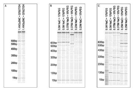
FIG. 1 illustrates the results of gel electrophoresis of nucleic acids, in a 1-
kilobase
size ladder, extracted or enriched after association and size selective
dissociation of
nucleic acids. FIG. 1A show the results of sample nucleic acid association to
solid
support followed by dissociation to illustrate the size distribution of
fragments in the
sample. FIG. 1B show the results of elution of nucleic acids from the solid
support
after an initial size selection was performed and the initially eluted
fragments were
separated from the material still bound to the solid support. The initial size
selection
dissociates the smaller fragments (shown in FIG. 1C), leaving behind larger
6a
CA 2757493 2018-09-19
81625137
fragments, according to the salt concentration used. FIG. 1C illustrates the
size
distribution of the nucleic acids initially dissociated, according to the
dissociation
conditions given above each gel lane.
FIG. 2 shows the percent male fetus DNA relative to total DNA isolate from the
serum
of a pregnant female, and the recovery of small fragments at various salt
concentrations as compared to the recovery of large fragments at the same salt
concentrations. The percent fold enrichment (e.g., approximately 30%) can be
calculated from the data presented in FIG. 2, as described in Example 2.
Enrichment
was performed using three different salt titrations 0.375M NaCI / 10%PEG, 0.5M
NaCI / 10% PEG, and 1 M NaCI / 10%PEG, which selects for less than 500 base
pairs, less than 400 base pairs, and less than 300 base pairs, respectively.
In
Figure 2, "short" fragment refers to DNA fragments less than the designated
cutoff
size provided, for example, less than 500 base pairs at 0.375M NaCI / 10%PEG,
less
than 400 base pairs at 0.5M NaCI / 10%PEG, and less than 300 base pairs at 1 M
NaCI / 10%PEG.
6b
CA 2757493 2018-09-19
CA C27574932011-&9-30
WO 2010/115016 PCT/US2010/029653
Detailed Description
The presence of cell-free nucleic acid in peripheral blood is a well
established phenomenon. While
cell-free nucleic acid may originate from several sources, it has been
demonstrated that one
source of circulating extracellular nucleic acid originates from programmed
cell death, also known
as apoptosis. The source of nucleic acid that arise as a result of apoptosis
may be found in many
body fluids and originate from several sources, including, but not limited to,
normal programmed
cell death in the host, induced programmed cell death in the case of an
autoimmune disease,
septic shock, neoplasms (malignant or non-malignant), or non-host sources such
as an allograft
(transplanted tissue), or the fetus or placenta of a pregnant woman. The
applications for the
detection, extraction and relative enrichment of extracellular nucleic acid
from peripheral blood or
other body fluids are widespread and may include inter alia, non-invasive
prenatal diagnosis,
cancer diagnostics, pathogen detection, auto-immune response and allograft
rejection.
In some embodiments, methods and compositions are provided that enable
enrichment and/or
extraction of relatively short target nucleic acid fragments, of specific size
ranges (e.g., 50-500
nucleotides or base pairs, and more specifically 50 to 200 nucleotides or base
pairs, for example,
and herein referred to as "target" or "sample" nucleic acid), contained within
a nucleic acid
composition of mixed fragment sizes (e.g., 1 to 100,000 nucleotides or base
pairs (bp), or more).
The enrichment and/or extraction of the target nucleic acid can be
accomplished by a partial, or
complete, physical separation of the target nucleic acid from the rest of the
nucleic acid in the
nucleic acid composition. More specifically, the methods and compositions
described herein, are
useful for the selective extraction and relative enrichment, based on size
discrimination, of nucleic
acids of in the range of about 50 to about 500 nucleotides or base pairs, and
more specifically
about 50 to about 200 nucleotides or base pairs, in a high background of
genomic nucleic acids
(herein referred to as "non-target" nucleic acid). The methods and
compositions described herein
lead to a relatively enriched fraction of nucleic acids that has a higher
concentration of smaller
nucleic acids, where the smaller nucleic acids sometimes contain target
nucleic acids. In some
embodiments, further enrichment of the specific target nucleic acids can be
accomplished by
amplification of the specific size selected target nucleic acid sequences
using amplification
procedures known in the art or described below.
7
CA C27574932011-&9-30
WO 2010/115016 PCT/US2010/029653
Disorders
Nucleic acid prepared using methods and compositions described herein can be
utilized to detect
the presence or absence of one or more prenatal or neonatal disorders. Non-
limiting examples of
.. prenatal and neonatal disorders include achondroplasia, Angelman syndrome,
Cockayne
syndrome, cystic fibrosis (autosomal recessive), congenital adrenal
hyperplasia (autosomal
recessive), DiGeorge syndrome, Duchenne's muscular dystrophy, (X-linked
recessive), hemophilia
A (X-linked recessive), alpha- and beta-thalassemia (autosomal recessive),
fragile X syndrome (X-
linked dominant), polycystic kidney disease (adult type; autosomal dominant),
sickle cell anemia
(autosomal recessive), Marfan syndrome, Prader-Wlli syndrome, Waardenburg
syndrome, Tay-
Sachs disease (autosomal) and the like.
A prenatal or neonatal disorder in some embodiments is a chromosome
abnormality. In certain
embodiments chromosome abnormalities include, without limitation, a gain or
loss of an entire
.. chromosome or a region of a chromosome comprising one or more genes.
Chromosome
abnormalities include monosomies, trisomies, polysomies, loss of
heterozygosity, deletions and/or
duplications of one or more nucleotide sequences (e.g., one or more genes),
including deletions
and duplications caused by unbalanced translocations in some embodiments. The
terms
"aneuploidy" and "aneuploid" as used herein refer to an abnormal number of
chromosomes in cells
.. of an organism. As different organisms have widely varying chromosome
complements, the term
"aneuploidy" does not refer to a particular number of chromosomes, but rather
to the situation in
which the chromosome content within a given cell or cells of an organism is
abnormal.
The term "monosomy" as used herein refers to lack of one chromosome of the
normal
complement. Partial monosomy can occur in unbalanced translocations or
deletions, in which only
a portion of the chromosome is present in a single copy (see deletion
(genetics)). Monosomy of
sex chromosomes (45, X) causes Turner syndrome.
The term "disomy" refers to the presence of two copies of a chromosome. For
organisms such as
humans that have two copies of each chromosome (those that are diploid or
"euploid"), it is the
normal condition. For organisms that normally have three or more copies of
each chromosome
(those that are triploid or above), disomy is an aneuploid chromosome
complement. In uniparental
disomy, both copies of a chromosome come from the same parent (with no
contribution from the
other parent).
8
CA C27574932011-&9-30
WO 2010/115016 PCT/US2010/029653
The term "trisomy" refers to the presence of three copies, instead of the
normal two, of a particular
chromosome. The presence of an extra chromosome 21, which is found in Down
syndrome, is
called trisomy 21. Trisomy 18 and Trisomy 13 are the two other autosomal
trisomies recognized in
live-born humans. Trisomy of sex chromosomes can be seen in females (47, XXX)
or males (47,
XXY which is found in Klinefelter's syndrome; or 47,XYY).
The terms "tetrasomy" and "pentasomy" as used herein refer to the presence of
four or five copies
of a chromosome, respectively. Although rarely seen with autosomes, sex
chromosome tetrasomy
and pentasomy have been reported in humans, including XXXX, XXXY, XXYY, XYYY,
XXXXX,
XXXXY, XXXYY, XXYYY and XYYYY.
Chromosome abnormalities can be caused by a variety of mechanisms. Mechanisms
include, but
are not limited to (i) nondisjunction occurring as the result of a weakened
mitotic checkpoint, (ii)
inactive mitotic checkpoints causing non-disjunction at multiple chromosomes,
(iii) merotelic
attachment occurring when one kinetochore is attached to both mitotic spindle
poles, (iv) a
multipolar spindle forming when more than two spindle poles form, (v) a
monopolar spindle forming
when only a single spindle pole forms, and (vi) a tetraploid intermediate
occurring as an end result
of the monopolar spindle mechanism.
The terms "partial monosomy" and "partial trisomy" as used herein refer to an
imbalance of genetic
material caused by loss or gain of part of a chromosome. A partial monosomy or
partial trisomy
can result from an unbalanced translocation, where an individual carries a
derivative chromosome
formed through the breakage and fusion of two different chromosomes. In this
situation, the
individual would have three copies of part of one chromosome (two normal
copies and the portion
that exists on the derivative chromosome) and only one copy of part of the
other chromosome
involved in the derivative chromosome.
The term "mosaicism" as used herein refers to aneuploidy in some cells, but
not all cells, of an
organism. Certain chromosome abnormalities can exist as mosaic and non-mosaic
chromosome
abnormalities. For example, certain trisomy 21 individals have mosaic Down
syndrome and some
have non-mosaic Down syndrome. Different mechanisms can lead to mosaicism. For
example, (i)
an initial zygote may have three 21st chromosomes, which normally would result
in simple trisomy
21, but during the course of cell division one or more cell lines lost one of
the 21st chromosomes;
and (ii) an initial zygote may have two 21st chromosomes, but during the
course of cell division one
9
CA C27574932011-&9-30
WO 2010/115016 PCT/US2010/029653
of the 21st chromosomes were duplicated. Somatic mosaicism most likely occurs
through
mechanisms distinct from those typically associated with genetic syndromes
involving complete or
mosaic aneuploidy. Somatic mosaicism has been identified in certain types of
cancers and in
neurons, for example. In certain instances, trisomy 12 has been identified in
chronic lymphocytic
leukemia (CLL) and trisomy 8 has been identified in acute myeloid leukemia
(AML). Also, genetic
syndromes in which an individual is predisposed to breakage of chromosomes
(chromosome
instability syndromes) are frequently associated with increased risk for
various types of cancer,
thus highlighting the role of somatic aneuploidy in carcinogenesis. Methods
and kits described
herein can identify presence or absence of non-mosaic and mosaic chromosome
abnormalities.
Following is a non-limiting list of chromosome abnormalities that can be
potentially identified by
methods and kits described herein.
Chromosome Abnormality Disease Association
X XO Turner's Syndrome
Y XXY Klinefelter syndrome
Y XYY Double Y syndrome
Y XXX Trisomy X syndrome
Y XXXX Four X syndrome
Y Xp21 deletion Duchenne's/Becker syndrome, congenital
adrenal
hypoplasia, chronic granulomatus disease
Y Xp22 deletion steroid sulfatase deficiency
Y Xq26 deletion X-linked lymph proliferative disease
1 1p (somatic) neuroblastoma
monosomy trisomy
2 monosomy trisomy growth retardation, developmental and
mental delay, and
2q minor physical abnormalities
3 monosomy trisomy Non-Hodgkin's lymphoma
(somatic)
4 monosomy trisomy Acute non lymphocytic leukemia (ANLL)
(somatic)
5 5p Cri du chat; Lejeune syndrome
5 5q myelodysplastic syndrome
(somatic) monosomy
trisomy
6 monosomy trisomy clear-cell sarcoma
(somatic)
7 7q11.23 deletion William's syndrome
7 monosomy trisomy monosomy 7 syndrome of childhood;
somatic: renal cortical
adenomas; myelodysplastic syndrome
8 8q24.1 deletion Langer-Giedon syndrome
8 monosomy trisomy myelodysplastic syndrome; Warkany
syndrome; somatic:
chronic myelogenous leukemia
9 monosomy 9p Alfi's syndrome
9 monosomy 9p partial Rethore syndrome
trisomy
9 trisomy complete trisomy 9 syndrome; mosaic trisomy
9 syndrome
CA C27574932011-&9-30
WO 2010/115016 PCT/US2010/029653
Monosomy trisomy ALL or ANLL
(somatic)
11 11p- Aniridia; Wilms tumor
11 11q- Jacobson Syndrome
11 monosomy (somatic) myeloid lineages affected (ANLL, MDS)
trisomy
12 monosomy trisomy CLL, Juvenile granulosa cell tumor
(JGCT)
(somatic)
13 13q- 13q-syndrome; Orbeli syndrome
13 13q14 deletion retinoblastoma
13 monosomy trisomy Patau's syndrome
14 monosomy trisomy myeloid disorders (MDS, ANLL, atypical
CML)
(somatic)
15q11-q13 deletion Prader-Willi, Angelman's syndrome
monosomy
15 trisomy (somatic) myeloid and lymphoid lineages affected,
e.g., MDS, ANLL,
ALL, CLL)
16 16q13.3 deletion Rubenstein-Taybi
monosomy trisomy papillary renal cell carcinomas (malignant)
(somatic)
17 17p-(somatic) 17p syndrome in myeloid malignancies
17 17q11.2 deletion Smith-Magenis
17 17q13.3 Miller-Dieker
17 monosomy trisomy renal cortical adenomas
(somatic)
17 17p11.2-12 trisomy Charcot-Marie Tooth Syndrome type 1;
HNPP
18 18p- 18p partial monosomy syndrome or Grouchy
Lamy Thieffry
syndrome
18 18q- Grouchy Lamy Salmon Landry Syndrome
18 monosomy trisomy Edwards Syndrome
19 monosomy trisomy
20p- trisomy 20p syndrome
20 20p11.2-12 deletion Alagille
20 20q- somatic: MDS, ANLL, polycythemia vera,
chronic
neutrophilic leukemia
20 monosomy trisomy papillary renal cell carcinomas
(malignant)
(somatic)
21 monosomy trisomy Down's syndrome
22 22q11.2 deletion DiGeorge's syndrome, velocardiofacial
syndrome,
conotruncal anomaly face syndrome, autosomal dominant
Opitz G/BBB syndrome, Caylor card iofacial syndrome
22 monosomy trisomy complete trisomy 22 syndrome
In certain embodiments, presence or absence of a fetal chromosome abnormality
is identified (e.g.,
trisomy 21, trisomy 18 and/or trisomy 13). In some embodiments, presence or
absence of a
5 chromosome abnormality related to a cell proliferation condition or
cancer is identified. Presence
or absence of one or more of the chromosome abnormalities described in the
table above may be
identified in some embodiments.
11
CA C27574932011-&9-30
WO 2010/115016 PCT/US2010/029653
In some embodiments, a prenatal or neonatal condition is a cell proliferation
condition. Cell
proliferation conditions include, without limitation, cancers of the
colorectum, breast, lung, liver,
pancreas, lymph node, colon, prostate, brain, head and neck, skin, liver,
kidney, and heart.
Examples of cancers include hematopoietic neoplastic disorders, which are
diseases involving
hyperplastic/neoplastic cells of hematopoietic origin (e.g., arising from
myeloid, lymphoid or
erythroid lineages, or precursor cells thereof). The diseases can arise from
poorly differentiated
acute leukemias, e.g., erythroblastic leukemia and acute megakaryoblastic
leukemia. Additional
myeloid disorders include, but are not limited to, acute promyeloid leukemia
(APML), acute
myelogenous leukemia (AML) and chronic myelogenous leukemia (CML) (reviewed in
Vaickus,
Crit. Rev. in Oncol./Hemotol. 11:267-297 (1991)); lymphoid malignancies
include, but are not
limited to acute lymphoblastic leukemia (ALL), which includes B-lineage ALL
and T-lineage ALL,
chronic lymphocytic leukemia (CLL), prolymphocytic leukemia (PLL), hairy cell
leukemia (HLL) and
Waldenstrom's macroglobulinemia (WM). Additional forms of malignant lymphomas
include, but
are not limited to non-Hodgkin lymphoma and variants thereof, peripheral T
cell lymphomas, adult
T cell leukemia/lymphoma (ATL), cutaneous T-cell lymphoma (CTCL), large
granular lymphocytic
leukemia (LGF), Hodgkin's disease and Reed-Sternberg disease. In a particular
embodiment, a
cell proliferative disorder is non-endocrine tumor or endocrine tumors.
Illustrative examples of non-
endocrine tumors include but are not limited to adenocarcinomas, acinar cell
carcinomas,
adenosquamous carcinomas, giant cell tumors, intraductal papillary mucinous
neoplasms,
mucinous cystadenocarcinomas, pancreatoblastomas, serous cystadenomas, solid
and
pseudopapillary tumors. An endocrine tumor may be an islet cell tumor.
Cell proliferative conditions also include inflammatory conditions, such as
inflammation conditions
of the skin, including, for example, eczema, discoid lupus erythematosus,
lichen planus, lichen
sclerosus, mycosis fungoides, photodermatoses, pityriasis rosea, psoriasis.
Also included are cell
proliferative conditions related to obesity, such as proliferation of
adipocytes, for example.
Cell proliferative conditions also include viral diseases, including for
example, Acquired
Immunodeficiency Syndrome, Adenoviridae Infections, Alphavirus Infections,
Arbovirus Infections,
Borna Disease, Bunyaviridae Infections, Caliciviridae Infections, Chickenpox,
Coronaviridae
Infections, Coxsackievirus Infections, Cytomegalovirus Infections, Dengue, DNA
Virus Infections,
Ecthyma, Contagious, Encephalitis, Arbovirus, Epstein-Barr Virus Infections,
Erythema
Infectiosum, Hantavirus Infections, Hemorrhagic Fevers, Viral, Hepatitis,
Viral, Human, Herpes
Simplex, Herpes Zoster, Herpes Zoster Oticus, Herpesviridae Infections,
Infectious Mononucleosis,
12
CA C27574932011-&9-30
WO 2010/115016 PCT/US2010/029653
Influenza in Birds, Influenza, Human, Lassa Fever, Measles, Molluscum
Contagiosum, Mumps,
Paramyxoviridae Infections, Phlebotomus Fever, Polyomavirus Infections,
Rabies, Respiratory
Syncytial Virus Infections, Rift Valley Fever, RNA Virus Infections, Rubella,
Slow Virus Diseases,
Smallpox, Subacute Sclerosing Panencephalitis, Tumor Virus Infections, Warts,
West Nile Fever,
.. Virus Diseases and Yellow Fever. For example, Large T antigen of the SV40
transforming virus
acts on UBF, activates it and recruits other viral proteins to Pol I complex,
and thereby stimulates
cell proliferation to promote virus propagation.
Cell proliferative conditions also include cardiac conditions resulting from
cardiac stress, such as
hypertension, balloon angioplasty, valvular disease and myocardial infarction.
For example,
cardiomyocytes are differentiated muscle cells in the heart that constitute
the bulk of the ventricle
wall, and vascular smooth muscle cells line blood vessels. Although both are
muscle cell types,
cardiomyocytes and vascular smooth muscle cells vary in their mechanisms of
contraction, growth
and differentiation. Cardiomyocytes become terminally differentiated shortly
after heart formation
and thus lose the capacity to divide, whereas vascular smooth muscle cells are
continually
undergoing modulation from the contractile to proliferative phenotype. Under
various
pathophysiological stresses such as hypertension, balloon angioplasty,
valvular disease and
myocardial infarction, for example, the heart and vessels undergo morphologic
growth-related
alterations that can reduce cardiac function and eventually manifest in heart
failure. Cell
proliferative conditions also include conditions related to angiogenesis
(e.g., cancers) and obesity
caused by proliferation of adipocytes and other fat cells.
In some embodiments, methods and compositions described herein can be used to
extract cell-
free nucleic acids from biological samples, from animals or humans for
example, for the purpose of
detecting or diagnosing a disease condition (e.g., cancer, genetic
abnormality, and the like). In
certain embodiments, the biological sample is from a human, who also may be a
cancer patient in
certain embodiments. Methods and compositions described herein may be used in
conjunction
with any method known to elevate nucleic acids (e.g., nucleotide sequences)
associated with
cancer conditions, from sample nucleic acid compositions (e.g., patient
samples). Alternatively,
methods and compositions described herein may be used in conjunction with any
method known to
decrease nucleic acid sequences associated with cancer conditions, from in
sample nucleotide
compositions.
13
CA C27574932011-&9-30
WO 2010/115016 PCT/US2010/029653
Nucleic acids
Target or sample nucleic acid may be derived from one or more samples or
sources. "Sample
nucleic acid" as used herein refers to a nucleic acid from a sample. "Target
nucleic acid" and
.. "template nucleic acid" are used interchangeably throughout the document
and refer to a nucleic
acid of interest. The terms "total nucleic acid" or "nucleic acid composition"
as used herein, refer to
the entire population of nucleic acid species from or in a sample or source.
Non-limiting examples
of nucleic acid compositions containing "total nucleic acids" include, host
and non-host nucleic
acid, maternal and fetal nucleic acid, genomic and acellular nucleic acid, or
mixed-population
nucleic acids isolated from environmental sources. As used herein, "nucleic
acid" refers to
polynucleotides such as deoxyribonucleic acid (DNA) and ribonucleic acid
(RNA), and refers to
derivatives, variants and analogs of RNA or DNA made from nucleotide analogs,
single (sense or
antisense) and double-stranded polynucleotides. The term "nucleic acid" does
not refer to or infer
a specific length of the polynucleotide chain, thus nucleotides,
polynucleotides, and
oligonucleotides are also included within "nucleic acid."
In some embodiments, target nucleic acid is relatively short and may comprise
fragments in the of
about 5 to about 500 nucleotides or base pairs, for example. In certain
embodiments, the target
nucleic acid can be in the range of about 5 to about 300 nucleotides or base
pairs. In certain
.. embodiments, the relatively short target nucleic acid can be in the range
of about 5 to about 200
nucleotides or base pairs. That is, target nucleic acids can be about 10, 15,
20, 25, 30, 35, 40, 45,
50, 55, 60, 65, 70, 75, 80, 85, 90, 95, 100, 110, 120, 130, 140, 150, 160,
170, 180, 190, 200, 210,
220, 230 250, 300, 350, 400, 450, or up to about 500 nucleotides or base pairs
in length. In certain
embodiments, the relatively long nucleic acid can be greater than about 200
nucleotides or base
pairs. The term "nucleotides", as used herein, in reference to the length of
nucleic acid chain,
refers to a single stranded nucleic acid chain. The term "base pairs", as used
herein, in reference
to the length of nucleic acid chain, refers to a double stranded nucleic acid
chain.
Deoxyribonucleotides include deoxyadenosine, deoxycytidine, deoxyguanosine and
deoxythymidine. For RNA, the uracil base is uridine. A source or sample
containing sample
nucleic acid(s) may contain one or a plurality of sample nucleic acids. A
plurality of sample nucleic
acids as described herein refers to at least 2 sample nucleic acids and
includes nucleic acid
sequences that may be identical or different. That is, the sample nucleic
acids may all be
representative of the same nucleic acid sequence, or may be representative of
two or more
14
CA C27574932011-&9-30
WO 2010/115016 PCT/US2010/029653
different nucleic acid sequences (e.g., from 1, 2, 3, 4, 5, 6, 7, 8, 9, 10,
11, 12, 13, 14, 15, 16, 17,
18, 19, 20, 50, 100, 1000 or more sequences).
A sample containing nucleic acids may be collected from an organism, mineral
or geological site
.. (e.g., soil, rock, mineral deposit, combat theater), forensic site (e.g.,
crime scene, contraband or
suspected contraband), or a paleontological or archeological site (e.g.,
fossil, or bone) for example.
A sample may be a "biological sample," which refers to any material obtained
from a living source
or formerly-living source, for example, an animal such as a human or other
mammal, a plant, a
bacterium, a fungus, a protist or a virus. The biological sample can be in any
form, including
without limitation a solid material such as a tissue, cells, a cell pellet, a
cell extract, or a biopsy, or a
biological fluid such as urine, blood, saliva, amniotic fluid, exudate from a
region of infection or
inflammation, or a mouth wash containing buccal cells, urine, cerebral spinal
fluid and synovial fluid
and organs.
The biological sample can be maternal blood, including maternal plasma or
serum. In some
circumstances, the biological sample is acellular. In other circumstances, the
biological sample
does contain cellular elements or cellular remnants in maternal blood. Other
biological samples
include amniotic fluid, chorionic villus sample, biopsy material from a pre-
implantation embryo,
maternal urine, maternal saliva, a celocentesis sample, fetal nucleated cells
or fetal cellular
.. remnants, or the sample obtained from washings of the female reproductive
tract. In some
embodiments, a biological sample may be blood.
As used herein, the term "blood" encompasses whole blood or any fractions of
blood, such as
serum and plasma as conventionally defined. Blood plasma refers to the
fraction of whole blood
.. resulting from centrifugation of blood treated with anticoagulants. Blood
serum refers to the watery
portion of fluid remaining after a blood sample has coagulated. Fluid or
tissue samples often are
collected in accordance with standard protocols hospitals or clinics generally
follow. For blood, an
appropriate amount of peripheral blood (e.g., between 3-40 milliliters) often
is collected and can be
stored according to standard procedures prior to further preparation in such
embodiments. A fluid
or tissue sample from which template nucleic acid is extracted may be
acellular. In some
embodiments, a fluid or tissue sample may contain cellular elements or
cellular remnants. In some
embodiments, the nucleic acid composition containing the target nucleic acid
or nucleic acids may
be collected from a cell free or substantially cell free biological
composition, blood plasma, blood
serum or urine for example.
CA C27574932011-&9-30
WO 2010/115016 PCT/US2010/029653
The term "substantially cell free" as used herein, refers to biologically
derived preparations or
compositions that contain a substantially small number of cells, or no cells.
A preparation intended
to be completely cell free, but containing cells or cell debris can be
considered substantially cell
free. That is, substantially cell free biological preparations can include up
to about 50 cells or
fewer per milliliter of preparation (e.g., up to about 50 cells per milliliter
or less, 45 cells per milliliter
or less, 40 cells per milliliter or less, 35 cells per milliliter or less, 30
cells per milliliter or less, 25
cells per milliliter or less, 20 cells per milliliter or less, 15 cells per
milliliter or less, 10 cells per
milliliter or less, 5 cells per milliliter or less, or up to about 1 cell per
milliliter or less).
For prenatal applications of technology described herein, fluid or tissue
sample may be collected
from a female at a gestational age suitable for testing, or from a female who
is being tested for
possible pregnancy. Suitable gestational age may vary depending on the
chromosome
abnormality tested. In certain embodiments, a pregnant female subject
sometimes is in the first
trimester of pregnancy, at times in the second trimester of pregnancy, or
sometimes in the third
trimester of pregnancy. In certain embodiments, a fluid or tissue is collected
from a pregnant
woman at 1-4, 4-8, 8-12, 12-16, 16-20, 20-24, 24-28, 28-32, 32-36, 36-40, or
40-44 weeks of fetal
gestation, and sometimes between 5-28 weeks of fetal gestation.
Target and/or total nucleic acid can be extracellular nucleic acid in certain
embodiments. The term
"extracellular nucleic acid" as used herein refers to nucleic acid isolated
from a source having
substantially no cells (e.g., no detectable cells, or fewer than 50 cells per
milliliter or less as
described above, or may contain cellular elements or cellular remnants).
Examples of acellular
sources for extracellular nucleic acid are blood plasma, blood serum and
urine. Without being
limited by theory, extracellular nucleic acid may be a product of cell
apoptosis and cell breakdown,
which provides basis for extracellular nucleic acid often having a series of
lengths across a large
spectrum (e.g., a "ladder"). In some embodiments, the nucleic acids can be
cell free nucleic acid.
Extracellular template nucleic acid can include different nucleic acid
species. For example, blood
serum or plasma from a person having cancer can include nucleic acid from
cancer cells and
nucleic acid from non-cancer cells. In another example, blood serum or plasma
from a pregnant
female can include maternal nucleic acid and fetal nucleic acid. In some
instances, fetal nucleic
acid sometimes is about 5% to about 40% of the overall template nucleic acid
(e.g., about 6, 7, 8,
9, 10, 11, 12, 13, 14, 15, 16, 17, 18, 19, 20, 21, 22, 23, 24, 25, 26, 27, 28,
29, 30, 31, 32, 33, 34,
35, 36, 37, 38 or 39% of the template nucleic acid is fetal nucleic acid). In
some embodiments, the
16
CA 02757493 2016-08-16
52923-32
majority of fetal nucleic acid in template nucleic acid is of a length of
about 500 base pairs or less
(e.g., about 80, 85, 90, 91, 92, 93, 94, 95, 96, 97, 98, 99 or 100% of fetal
nucleic acid is of a length
of about 500 base pairs or less).
The amount of fetal nucleic acid (e.g., concentration) in template nucleic
acid sometimes is
determined. In certain embodiments, the amount of fetal nucleic acid is
determined according to
markers specific to a male fetus (e.g., Y-chromosome STR markers (e.g., DYS
19, DYS 385, DYS
392 markers); RhD marker in RhD-negative females), or according to one or more
markers specific
to fetal nucleic acid and not maternal nucleic acid (e.g., fetal RNA markers
in maternal blood
.. plasma; Lo, 2005, Journal of Histochemistry and Cytochemistry 53 (3): 293-
296). The amount of
fetal nucleic acid in extracellular template nucleic acid can be quantified
and utilized for the
identification of the presence or absence of a chromosome abnormality in
certain embodiments.
In some embodiments, extracellular nucleic acid can be enriched or relatively
enriched for fetal
nucleic acid, using methods described herein alone, or in conjunction with
other methods known in
the art. Non-limiting examples of additional methods known in the art for
enriching a sample for a
particular species of nucleic acid are described in; PCT Patent Application
Number
PCT/US07/69991, filed May 30, 2007, PCT Patent Application Number
PCT/US2007/071232, filed
June 15, 2007, US Provisional Application Numbers 60/968,876 and 60/968,878,
and PCT Patent
Application Number PCT/EP05/012707, filed November 28, 2005.
In certain embodiments, maternal nucleic acid can be selectively removed
(partially, substantially, almost completely or completely) from the sample.
A sample also may be isolated at a different time point as compared to another
sample, where
each of the samples may be from the same or a different source. A sample
nucleic acid may be
from a nucleic acid library, such as a cDNA or RNA library, for example. A
sample nucleic acid
may be a result of nucleic acid purification or isolation and/or amplification
of nucleic acid
molecules from the sample. Sample nucleic acid provided for sequence analysis
processes
described herein may contain nucleic acid from one sample or from two or more
samples (e.g.,
from 1, 2, 3, 4, 5, 6, 7, 8, 9, 10, 11, 12, 13, 14, 15, 16, 17, 18, 19 or 20
samples).
Sample nucleic acid may comprise or consist essentially of any type of nucleic
acid suitable for use
with processes of the technology, such as sample nucleic acid that can
hybridize to solid phase
nucleic acid (described hereafter), for example. A sample nucleic in certain
embodiments can
17
CA C27574932011-&9-30
WO 2010/115016 PCT/US2010/029653
comprise or consist essentially of DNA (e.g., complementary DNA (cDNA),
genomic DNA (gDNA)
and the like), RNA (e.g., message RNA (mRNA), short inhibitory RNA (siRNA),
microRNA,
ribosomal RNA (rRNA), tRNA and the like), and/or DNA or RNA analogs (e.g.,
containing base
analogs, sugar analogs and/or a non-native backbone and the like). A nucleic
acid can be in any
.. form useful for conducting processes herein (e.g., linear, circular,
supercoiled, single-stranded,
double-stranded and the like). A nucleic acid may be, or may be from, a
plasmid, phage,
autonomously replicating sequence (ARS), centromere, artificial chromosome,
chromosome, a cell,
a cell nucleus or cytoplasm of a cell in certain embodiments. A sample nucleic
acid in some
embodiments is from a single chromosome (e.g., a nucleic acid sample may be
from one
.. chromosome of a sample obtained from a diploid organism).
Sample nucleic acid may be provided for conducting methods described herein
without processing
of the sample(s) containing the nucleic acid in certain embodiments. In some
embodiments,
sample nucleic acid is provided for conducting methods described herein after
processing of the
sample(s) containing the nucleic acid. For example, a sample nucleic acid may
be extracted,
isolated, purified or amplified from the sample(s). The term "isolated" as
used herein refers to
nucleic acid removed from its original environment (e.g., the natural
environment if it is naturally
occurring, or a host cell if expressed exogenously), and thus is altered "by
the hand of man" from
its original environment. An isolated nucleic acid generally is provided with
fewer non-nucleic acid
.. components (e.g., protein, lipid) than the amount of components present in
a source sample. A
composition comprising isolated sample nucleic acid can be substantially
isolated (e.g., about
90%, 91%, 92%, 93%, 94%, 95%, 96%, 97%, 98%, 99% or greater than 99% free of
non-nucleic
acid components). The term "purified" as used herein refers to sample nucleic
acid provided that
contains fewer nucleic acid species than in the sample source from which the
sample nucleic acid
is derived. A composition comprising sample nucleic acid may be substantially
purified (e.g., about
90%, 91%, 92%, 93%, 94%, 95%, 96%, 97%, 98%, 99% or greater than 99% free of
other nucleic
acid species). The term "amplified" as used herein refers to subjecting
nucleic acid of a sample to
a process that linearly or exponentially generates amplicon nucleic acids
having the same or
substantially the same nucleotide sequence as the nucleotide sequence of the
nucleic acid in the
sample, or portion thereof.
Sample nucleic acid also may be processed by subjecting nucleic acid to a
method that generates
nucleic acid fragments, in certain embodiments, before providing sample
nucleic acid for a process
described herein. In some embodiments, sample nucleic acid subjected to
fragmentation or
18
CA C27574932011-&9-30
WO 2010/115016 PCT/US2010/029653
cleavage may have a nominal, average or mean length of about 5 to about 10,000
base pairs,
about 100 to about 1,000 base pairs, about 100 to about 500 base pairs, or
about 10, 15, 20, 25,
30, 35, 40, 45, 50, 55, 60, 65, 70, 75, 80, 85, 90, 95, 100, 200, 300, 400,
500, 600, 700, 800, 900,
1000, 2000, 3000, 4000, 5000, 6000, 7000, 8000, 9000 or 10000 base pairs.
Fragments can be
generated by any suitable method known in the art, and the average, mean or
nominal length of
nucleic acid fragments can be controlled by selecting an appropriate fragment-
generating
procedure by the person of ordinary skill. In certain embodiments, sample
nucleic acid of a
relatively shorter length can be utilized to analyze sequences that contain
little sequence variation
and/or contain relatively large amounts of known nucleotide sequence
information. In some
embodiments, sample nucleic acid of a relatively longer length can be utilized
to analyze
sequences that contain greater sequence variation and/or contain relatively
small amounts of
unknown nucleotide sequence information.
Sample nucleic acid fragments often contain overlapping nucleotide sequences,
and such
overlapping sequences can facilitate construction of a nucleotide sequence of
the previously non-
fragmented sample nucleic acid, or a portion thereof. For example, one
fragment may have
subsequences x and y and another fragment may have subsequences y and z, where
x, y and z
are nucleotide sequences that can be 5 nucleotides in length or greater.
Overlap sequence y can
be utilized to facilitate construction of the x-y-z nucleotide sequence in
nucleic acid from a sample.
Sample nucleic acid may be partially fragmented (e.g., from an incomplete or
terminated specific
cleavage reaction) or fully fragmented in certain embodiments.
Sample nucleic acid can be fragmented by various methods known to the person
of ordinary skill,
which include without limitation, physical, chemical and enzymic processes.
Examples of such
processes are described in U.S. Patent Application Publication No. 20050112590
(published on
May 26, 2005, entitled "Fragmentation-based methods and systems for sequence
variation
detection and discovery," naming Van Den Boom et al.). Certain processes can
be selected by the
person of ordinary skill to generate non-specifically cleaved fragments or
specifically cleaved
fragments. Examples of processes that can generate non-specifically cleaved
fragment sample
nucleic acid include, without limitation, contacting sample nucleic acid with
apparatus that expose
nucleic acid to shearing force (e.g., passing nucleic acid through a syringe
needle; use of a French
press); exposing sample nucleic acid to irradiation (e.g., gamma, x-ray, UV
irradiation; fragment
sizes can be controlled by irradiation intensity); boiling nucleic acid in
water (e.g., yields about 500
base pair fragments) and exposing nucleic acid to an acid and base hydrolysis
process.
19
CA C27574932011-&9-30
WO 2010/115016 PCT/US2010/029653
Sample nucleic acid may be specifically cleaved by contacting the nucleic acid
with one or more
specific cleavage agents. The term "specific cleavage agent" as used herein
refers to an agent,
sometimes a chemical or an enzyme that can cleave a nucleic acid at one or
more specific sites.
Specific cleavage agents often will cleave specifically according to a
particular nucleotide
sequence at a particular site.
Examples of enzymic specific cleavage agents include without limitation
endonucleases (e.g.,
DNase (e.g., DNase I, II); RNase (e.g., RNase E, F, H, P); CleavaseTM enzyme;
Taq DNA
polymerase; E. coli DNA polymerase I and eukaryotic structure-specific
endonucleases; murine
FEN-1 endonucleases; type I, ll or III restriction endonucleases such as Acc
I, Afl III, Alu I, Alw44 I,
Apa I, Asn I, Ava I, Ava II, BamH I, Ban II, Bc1 I, Bgl I. Bgl II, Bin I, Bsm
I, BssH II, BstE II, Cfo I, Cla
I, Dde I, Dpn I, Dra I, EcIX I, EcoR I, EcoR I, EcoR II, EcoR V, Hae II, Hae
II, Hind II, Hind III, Hpa I,
Hpa II, Kpn I, Ksp I, Mlu I, MluN I, Msp I, Nci I, Nco I, Nde I, Nde II, Nhe
I, Not I, Nru I, Nsi I, Pst I,
Pvu I, Pvu II, Rsa I, Sac I, Sal I, Sau3A I, Sca I, ScrF I, Sfi I, Sma I, Spe
I, Sph I, Ssp I, Stu I, Sty I,
Swa I, Taq I, Xba I, Xho I.); glycosylases (e.g., uracil-DNA glycolsylase
(UDG), 3-methyladenine
DNA glycosylase, 3-methyladenine DNA glycosylase II, pyrimidine hydrate-DNA
glycosylase,
FaPy-DNA glycosylase, thymine mismatch-DNA glycosylase, hypoxanthine-DNA
glycosylase, 5-
Hydroxymethyluracil DNA glycosylase (HmUDG), 5-Hydroxymethylcytosine DNA
glycosylase, or
1,N6-etheno-adenine DNA glycosylase); exonucleases (e.g., exonuclease III);
ribozymes, and
DNAzymes. Sample nucleic acid may be treated with a chemical agent, or
synthesized using
modified nucleotides, and the modified nucleic acid may be cleaved. In non-
limiting examples,
sample nucleic acid may be treated with (i) alkylating agents such as
methylnitrosourea that
generate several alkylated bases, including N3-methyladenine and N3-
methylguanine, which are
recognized and cleaved by alkyl purine DNA-glycosylase; (ii) sodium bisulfite,
which causes
deamination of cytosine residues in DNA to form uracil residues that can be
cleaved by uracil N-
glycosylase; and (iii) a chemical agent that converts guanine to its oxidized
form, 8-
hydroxyguanine, which can be cleaved by formamidopyrimidine DNA N-glycosylase.
Examples of
chemical cleavage processes include without limitation alkylation, (e.g.,
alkylation of
phosphorothioate-modified nucleic acid); cleavage of acid lability of P3'-N5'-
phosphoroamidate-
containing nucleic acid; and osmium tetroxide and piperidine treatment of
nucleic acid.
As used herein, the term "complementary cleavage reactions" refers to cleavage
reactions that are
carried out on the same sample nucleic acid using different cleavage reagents
or by altering the
cleavage specificity of the same cleavage reagent such that alternate cleavage
patterns of the
CA C27574932011-&9-30
WO 2010/115016 PCT/US2010/029653
same target or reference nucleic acid or protein are generated. In certain
embodiments, sample
nucleic acid may be treated with one or more specific cleavage agents (e.g.,
1, 2, 3, 4, 5, 6, 7, 8, 9,
or more specific cleavage agents) in one or more reaction vessels (e.g.,
sample nucleic acid is
treated with each specific cleavage agent in a separate vessel).
5
Sample nucleic acid also may be exposed to a process that modifies certain
nucleotides in the
nucleic acid before providing sample nucleic acid for a method described
herein. A process that
selectively modifies nucleic acid based upon the methylation state of
nucleotides therein can be
applied to sample nucleic acid. The term "methylation state" as used herein
refers to whether a
10 particular nucleotide in a polynucleotide sequence is methylated or not
methylated. Methods for
modifying a target nucleic acid molecule in a manner that reflects the
methylation pattern of the
target nucleic acid molecule are known in the art, as exemplified in U.S. Pat.
No. 5,786,146 and
U.S. patent publications 20030180779 and 20030082600. For example, non-
methylated cytosine
nucleotides in a nucleic acid can be converted to uracil by bisulfite
treatment, which does not
modify methylated cytosine. Non-limiting examples of agents that can modify a
nucleotide
sequence of a nucleic acid include methylmethane sulfonate, ethylmethane
sulfonate,
diethylsulfate, nitrosoguanidine (N-methyl-N'-nitro-N-nitrosoguanidine),
nitrous acid, di-(2-
chloroethyl)sulfide, di-(2-chloroethyl)methylamine, 2-aminopurine, t-
bromouracil, hydroxylamine,
sodium bisulfite, hydrazine, formic acid, sodium nitrite, and 5-methylcytosine
DNA glycosylase. In
addition, conditions such as high temperature, ultraviolet radiation, x-
radiation, can induce changes
in the sequence of a nucleic acid molecule.
Sample nucleic acid may be provided in any form useful for conducting a
sequence analysis or
manufacture process described herein, such as solid or liquid form, for
example. In certain
embodiments, sample nucleic acid may be provided in a liquid form optionally
comprising one or
more other components, including without limitation one or more buffers or
salts selected by the
person of ordinary skill.
Solid supports
The term "solid support" or "solid phase" as used herein refers to an
insoluble material with which
nucleic acid can be associated, and the terms can be used interchangeably.
Examples of solid
supports for use with processes described herein include, without limitation,
chips, flat surfaces
filters, one or more capillaries and/or fibers, arrays, filters, beads, beads
(e.g., paramagnetic
21
CA C27574932011-&9-30
WO 2010/115016 PCT/US2010/029653
beads, magnetic beads, microbeads, nanobeads) and particles (e.g.,
microparticles,
nanoparticles). Beads and/or particles may be free or in connection with one
another (e.g.,
sintered). In some embodiments, the solid phase can be a collection of
particles. In certain
embodiments, the particles can comprise silica, and the silica may comprise
silica dioxide. In
some embodiments the silica can be porous, and in certain embodiments the
silica can be non-
porous. In some embodiments, the particles further comprise an agent that
confers a
paramagnetic property to the particles. In certain embodiments, the agent
comprises a metal, and
in certain embodiments the agent is a metal oxide, (e.g., iron or iron oxides,
where the iron oxide
contains a mixture of Fe2+ and Fe3+). Magnetically responsive silica dioxide
beads can be
obtained commercially. Non-limiting examples of magnetically responsive silica
beads are;
DynaL0 beads (Invitrogen, Carlsbad, California), SiMage beads (Chemicell,
Berlin, Germany),
MagAttractO beads (Qiagen, Hilden, Germany), Magnesil0 beads (Promega,
Madison, Wisconsin),
and functional magnetic silica beads (MoBiTec, Gottingen, Germany;
Microspheres-
Nanospheres.com (a division of Corpuscular, Inc) Lincolndale, New York;
G.Kisker Biotech,
Steinfurt, Germany).
In some embodiments, the solid phase does not comprise a functional group that
interacts with the
nucleic acid. In certain embodiments, the solid phase does not comprise a
carboxy functional
group. In some embodiments, the solid phase has a net charge. In certain
embodiments, the net
charge is positive, and sometimes the net charge is negative.
Nucleic acids may reversibly associate with a solid support (e.g., magnetic
silica dioxide particles)
under association conditions. The association may be reversed under
dissociation conditions, and
all or a subset of nucleic acid associated with the solid phase may dissociate
from the solid phase
.. under the dissociation conditions. The term "associate" as used herein
refers to an interaction
between a nucleic acid and a solid phase, which interaction often is non-
covalent, often is
adsorption, sometimes is absorption, often is binding, and generally is
reversible. The term
"association conditions" as used herein, refers to conditions under which
nucleic acid from a
nucleic acid composition is associated with a solid support. In some
embodiments, nucleic acid of
substantially all sizes in the composition associates with a solid support
under the association
conditions. Sometimes, substantially all of the nucleic acid in a composition
associates with a solid
support, and sometimes about 30 percent to about 100 percent of the nucleic
acid, from the total
nucleic acid in a sample, associates or binds to the solid support (e.g., 30%
or greater, 35% or
greater, 40% or greater, 45% or greater, 50% or greater, 55% or greater, 60%
or greater, 65% or
22
CA C27574932011-&9-30
WO 2010/115016 PCT/US2010/029653
greater, 70% or greater, 75% or greater, 80% or greater, 85% or greater, 90%
or greater, 95% or
greater, or 99% or greater of the total nucleic acid present in a sample
associates with the solid
phase).
In some embodiments, association conditions can include one or more of the
following: salts,
alcohols, volume excluding agents (e.g., sometimes also referred to as
crowding agents), or
combinations thereof. Salts may comprise chaotropic salts, ionic salts or a
combination of such
salts. Non-limiting examples of chaotropic salts include guanidine salt,
guanidinium salt, sodium
iodide, potassium iodide, sodium thiocyanate and urea. Non-limiting examples
of ionic salts
include sodium chloride, magnesium chloride, calcium chloride, potassium
chloride, lithium
chloride, barium chloride, cesium chloride, ammonium acetate, sodium acetate,
ammonium
perchlorate and sodium perchlorate. In some embodiments, a chaotropic salt can
be a guanidine
salt (e.g., guanidine (iso)thiocyanate, for example). In certain embodiments,
an ionic salt can be a
sodium salt (e.g., sodium chloride, for example).
In certain embodiments, a salt may be introduced at a concentration sufficient
to associate the
nucleic acid to a solid support (e.g., substantially all of the nucleic acid),
and the salt may be the
only component that associates the nucleic acid to the solid phase or more be
utilized in
combination with other components to perform the same function. Salt
concentrations for binding
nucleic acids may be dependent on length of nucleic acid, base sequence,
combinations thereof
and the like, and can be determined. In some embodiments a salt is utilized in
an amount that
yields a final salt concentration in the range of about 0.25M to about 5M of
the salt (e.g., 0.5M, 1M,
1.5M, 2M, 2.5M, 3M, 4M, or 5M). Salt concentrations also can be expressed as
percent weight
per volume and salt concentration ranges expressed as ranges of percent weight
per volume (e.g.,
40 to 60% weight per volume), and can be used interchangeably with Molar
concentrations. In
some embodiments, the salt concentration chosen may be sufficient to bind
substantially all non-
target nucleic acid to a solid support, while minimizing the binding of target
nucleic acid. In certain
embodiments, the salt concentration chosen may be sufficient to associate all
or substantially all
the nucleic acid in solution. In some embodiments a salt can be added to yield
a solution with a
concentration in the range of about 5% to about 60% weight per volume that may
be sufficient to
associate target or total nucleic acid to a solid support. In some
embodiments, additional solid
phase also may be used to ensure capture of all nucleic acid from a sample.
23
CA C27574932011-&9-30
WO 2010/115016 PCT/US2010/029653
Alcohols suitable for use in association conditions with the methods described
herein are the C1-
06 alkyl alcohols, and their branched chain derivatives or isoforms. Non-
limiting examples of the
C1-C6 alcohols are methanol (Cl), ethanol (02), propanol (03), butanol (04),
pentanol (05), and
hexanol (C6), and linear and branched variants thereof. In some embodiments
the alkyl alcohol is
included in a final amount (percent volume of alcohol in water or aqueous
buffered solution) in the
range of about 25% or more, 30% or more, 35% or more, 40% or more, 45% or
more, 50% or
more, 55% or more, 60% or more, 65% or more, 70% or more, 75% or more, 80% or
more, 85% or
more, 90% or more, 95 % or more, or up to 99% or more. In some embodiments,
ethanol is used
for associating nucleic acids with the solid phase (e.g., magnetically
responsive silica dioxide
beads). In certain embodiments, the final concentration of ethanol is about
33%. In some
embodiments, an alcohol is used as a wash solution to remove impurities. In
embodiments using
an alcohol as a wash solution, the alcohol often is between about 75% to about
95% alcohol (e.g.,
ethanol).
Volume excluding agents sometimes may be included in association conditions,
in some
embodiments. In certain embodiments, volume excluding agents can be used in
size selection
(e.g., dissociation) buffers or solutions. Volume excluding agents can be
suitable for use in (i)
association conditions, and/or (ii) dissociation conditions that allow for
preferential dissociation of
nucleic acid of a particular size (e.g., size selection). Volume excluding
agents include, without
limitation, polyalkyl glycol (e.g., polyethylene glycol (PEG), for example),
dextran, Ficoll, polyvinyl
pyrollidone or combinations thereof. In some embodiments, volume exclusion
agents (also
referred to as "crowding agents") can be added to yield a solution containing
between about 5% to
about 30% volume exclusion agent, and more specifically between about 8% to
about 20% volume
exclusion agent. That is, a volume excluding agent may be added to size
selection or dissociation
conditions to yield solutions containing up to about 5% volume excluding
agent, up to about 6%, up
to about 7%, up to about 8%, up to about 9%, up to about 10%, up to about 11%,
up to about 12%,
up to about 13%, up to about 14%, up to about 15%, up to about 16%, up to
about 17%, up to
about 18%, up to about 19%, up to about 20%, up to about 25%, up to about, and
up to about 30%
volume excluding agent.
The term "dissociation conditions" as used herein refers to conditions under
which (i) a subset of
nucleic acid associated with the solid phase, or (ii) substantially all of the
nucleic acid associated
with a solid phase, is removed from the solid phase. For example, target
nucleic acid may exist in
a population that is smaller than 300 nucleotides or base pairs, and
dissociation conditions may be
24
CA C27574932011-&9-30
WO 2010/115016 PCT/US2010/029653
selected to selectively dissociate nucleic acids smaller than 300 nucleotides
or base pairs. The
terms "preferential dissociation", "preferentially dissociates" and
grammatical variants thereof, as
used herein, refers to conditions under which target nucleic acids within a
specific size range (e.g.,
between 5 and 300 nucleotides or base pairs, for example) are substantially or
completely eluted
from the solid support, while the larger, non-target nucleic acid remains
substantially bound. That
is, a specifically selected size range of nucleic acids (e.g., relatively
short nucleic acids) may be
preferentially removed from the solid support under the appropriate
dissociation conditions, while
leaving behind the larger, unwanted or non-target nucleic acids.
The term "eluate" as used herein refers to the solution portion in a
composition that comprises a
solid phase and a solution. An eluate under dissociation conditions can
include relatively short
nucleic acid and relatively long nucleic acid dissociated from the solid
phase, where the relatively
short nucleic acid is preferentially dissociated from the solid phase as
compared to the relatively
long nucleic acid under the dissociation conditions, in some embodiments.
Thus, in certain
embodiments, an eluate can include about 1.5-fold to about 5-fold more
relatively short nucleic
acid as compared to relatively long nucleic acid, where the relatively short
nucleic acid is about 300
base pairs or less (e.g., 10, 20, 30, 40, 50, 60, 70, 80, 90, 100, 110, 120,
130, 140, 150, 160, 170,
180, 190, 200, 210, 220, 230, 340, 250, 260, 270, 280, 290 base pairs), after
the nucleic acid and
solid phase have been exposed to dissociation conditions to completion.
Certain dissociation conditions for eluting specific size ranges of nucleic
acids are presented below
in Table 1 (presented below in Example 3). In some embodiments, dissociation
conditions contain
one or more salts (e.g., ionic salt, chaotropic salt) and one or more volume
exclusion agents (e.g.,
polyalkyl glycol, Ficoll, dextran, polyvinyl pyrollidone (PVP) and the like).
In some embodiments, a
dissociation condition may include C1-C6 alkyl alcohols. The components
utilized in the
dissociation conditions can be utilized in any suitable amount that allow for
preferential dissociation
of a relatively short nucleic acid, in some embodiments. In some embodiments,
an ionic salt may
be utilized in dissociation conditions in an amount between about 0.05M to
about 2.0M, and
sometimes between about 0.05M to about 1.0M (e.g., about 0.1, 0.2, 0.3, 0.4,
0.5, 0.6, 0.7, 0.8, 0.9
and 1.0 M). In some embodiments, the ionic salt used for dissociation of
specific size fractions of
nucleic acids is sodium chloride (NaCI). In some embodiments the concentration
of sodium
chloride is in the range of about 0.25M to about 1.0M NaCI, and specific
concentrations useful for
isolating specific sized fractions may be found in Table 1.
CA C27574932011-&9-30
WO 2010/115016 PCT/US2010/029653
In certain embodiments, a volume exclusion agent may be utilized in
dissociation conditions in an
amount between about 5% to about 30% (e.g., weight to volume). Where the
volume exclusion
agent is a polyalkyl glycol, the polyalkyl glycol sometimes is utilized within
a range of about 5% to
about 25% in certain embodiments (e.g., about 6, 7, 8, 9, 10, 11, 12, 13, 14,
15, 16, 17, 18, 19 or
20%). The polyalkyl glycol can have an average, mean or nominal molecular
weight of about 10,
20, 30, 40, 50, 60, 70, 80, 90, 100, 200, 300, 400, 500, 600, 700, 800, 900,
1000, 2000, 3000,
4000, 5000, 6000, 7000, 8000, 9000, 10000, 20000, 30000, 40000, 50000, 60000,
70000, 80000
or 90000 grams per mole. The polyalkyl glycol sometimes is branched or linear,
and sometimes is
a polyethylene glycol (PEG).
Where one volume exclusion agent is utilized at an optimum amount, the amount
of a different
volume exclusion agent for alternative dissociation conditions can be
optimized based in part on
the molecular weight of the different volume exclusion agent. For example, if
a volume exclusion
agent that has been included in optimized dissociation conditions has a
particular molecular
weight, a different volume exclusion agent having a higher molecular weight
sometimes will be
utilized at a lower amount, and a different volume exclusion agent having a
lower molecular weight
sometimes will be utilized at a higher amount. Where PEG8000 is utilized at a
particular
percentage for optimized dissociation conditions, for example, a person of
ordinary skill in the art
often use a lower amount of a different volume exclusion agent having a higher
molecular weight
(e.g., PEG16000, Ficoll, dextran or polyvinyl pyrollidone), and often will use
a higher amount of a
different volume exclusion agent having a lower molecular weight (e.g.,
PEG4000). In some
embodiments, dissociation conditions can include about 5% to about 8% Ficoll,
about 2% to about
4% dextran, or about 8% to about 10% polyvinyl pyrollidone (PVP). The PVP
sometimes is PVP40
and often is not PVP10.
Particles or beads having a nominal, average or mean diameter of about 1
nanometer to about 500
micrometers can be utilized, such as those having a nominal, mean or average
diameter, for
example, of about 10 nanometers to about 100 micrometers; about 100 nanometers
to about 100
micrometers; about 1 micrometer to about 100 micrometers; about 10 micrometers
to about 50
micrometers; about 1, 5, 10, 15, 20, 25, 30, 35, 40, 45, 50, 55, 60, 65, 70,
75, 80, 85, 90, 95, 100,
200, 300, 400, 500, 600, 700, 800 or 900 nanometers; or about 1,5, 10, 15, 20,
25, 30, 35, 40, 45,
50, 55, 60, 65, 70, 75, 80, 85, 90, 95, 100, 200, 300, 400, 500 micrometers.
26
CA C27574932011-&9-30
WO 2010/115016 PCT/US2010/029653
A solid support can comprise virtually any insoluble or solid material, and
often a solid support
composition is selected that is insoluble in water. For example, a solid
support can comprise or
consist essentially of silica gel, glass (e.g. controlled-pore glass (CPG)),
nylon, Sephadex ,
Sepharose , cellulose, a metal surface (e.g. steel, gold, silver, aluminum,
silicon and copper), a
magnetic material, a plastic material (e.g., polyethylene, polypropylene,
polyamide, polyester,
polyvinylidenedifluoride (PVDF)) and the like. Beads or particles may be
swellable (e.g., polymeric
beads such as Wang resin) or non-swellable (e.g., CPG). Commercially available
examples of
beads include without limitation Wang resin, Merrifield resin and Dynabeads
and SoluLink.
A solid support may be provided in a collection of solid supports. A solid
support collection
comprises two or more different solid support species. The term "solid support
species" as used
herein refers to a solid support in association with one particular solid
phase nucleic acid species
or a particular combination of different solid phase nucleic acid species. In
certain embodiments, a
solid support collection comprises 2 to 10,000 solid support species, 10 to
1,000 solid support
species or about 2, 3, 4, 5, 6, 7, 8, 9, 10, 15, 20, 25, 30, 35, 40, 45, 50,
55, 60, 65, 70, 75, 80, 85,
90, 95, 100, 200, 300, 400, 500, 600, 700, 800, 900, 1000, 2000, 3000, 4000,
5000, 6000, 7000,
8000, 9000 or 10000 unique solid support species. The solid supports (e.g.,
beads) in the
collection of solid supports may be homogeneous (e.g., all are Wang resin
beads) or
heterogeneous (e.g., some are Wang resin beads and some are magnetic beads).
Each solid
.. support species in a collection of solid supports sometimes is labeled with
a specific identification
tag. An identification tag for a particular solid support species sometimes is
a nucleic acid (e.g.,
"solid phase nucleic acid") having a unique sequence in certain embodiments.
An identification
tag can be any molecule that is detectable and distinguishable from
identification tags on other
solid support species.
In certain embodiments, a biological sample can be contacted with a solid
support in the presence
of a lysing/binding reagent to bind all nucleic acids to a solid support, the
inhibitors are washed
away, then size selection is performed by adding different concentrations of
salts and crowding
agents to the solid support selectively removing smaller fragments and leaving
larger fragments on
the solid support. Additional size selected elutions can be performed if a
particular range of
fragments is required that is not eluted in the first size elution. Larger
fragments or non-target
fragments also can be enriched by eluting the larger fragments from the solid
support under
appropriate conditions, or by eluting the smaller fragments and removing the
supernatant to a new
tube, where the larger fragments remain on the solid support and are thereby
enriched. In some
27
CA C27574932011-&9-30
WO 2010/115016 PCT/US2010/029653
embodiments, the eluted small or target fragments can be further concentrated,
enriched and/or
purified by binding to new beads in the presence of the appropriate
concentration of salt and
precipitating agent, washing to remove non-nucleic acid impurities, and
eluting in an appropriate
aqueous buffer or water.
Enrichment
Target nucleic acid (e.g., relatively short nucleic acids from about 50 to
about 200 nucleotides or
base pairs in length), can be enriched relative to the target nucleic acid
concentration in a total
nucleic acid composition, or with respect to larger non-target nucleic acid
fractions, using methods
and compositions described herein. In some embodiments, relatively short
nucleic acids may be
enriched relative to the total population of nucleic acids from a sample.
Total nucleic acid from a
sample may be bound to solid support under appropriate association conditions
(see Example 1 for
a non-limiting example of appropriate association conditions). Relatively
short, target nucleic acids
may be purified by collecting the solid support (e.g., by centrifugation or
use of a magnetic field for
paramagnetic particles, for example), and optionally removing the supernatant
to a new tube, after
incubating under dissociation conditions for a sufficient period of time that
preferentially release the
relatively short nucleic acid from the solid phase. The relatively short
nucleic acid is thereby
enriched, relative to total nucleic acid by virtue of preferential
dissociation from the solid phase
relative to the relatively large non-target nucleic acid.
In certain embodiments, enrichment is a measure of the percent increase in the
amount of
relatively short nucleic acid in the disassociated nucleic acid as compared to
in the nucleic acid
composition subjected to the enrichment process (e.g., percent increase in the
relatively small
nucleic acid). In certain embodiments, this measure of enrichment is about 10%
to about 45%
(e.g., about 15, 20, 25, 26, 27, 28, 29, 30, 31, 32, 33, 34, 35, 36, 37, 38,
39, 40, 41, 42, 43 or 44%
enrichment).
In some embodiments, enrichment is a ratio of relatively small nucleic acid to
relatively large
nucleic acid in all of the nucleic acid eluted from the solid support under
dissociation conditions. In
certain embodiments, the ratio is about 1.05 to about 5 (e.g., ratio of about
1.2, 1.4, 1.6, 1.8, 2.0,
2.2, 2.4, 2.6, 2.8, 3.0, 3.2, 3.4, 3.6, 3.8, 4.0, 4.2, 4.4, 4.6, 4.8
relatively short nucleic acid to
relatively long nucleic acid).
28
CA C27574932011-&9-30
WO 2010/115016 PCT/US2010/029653
Further enrichment of subspecies of relatively short target nucleic acids also
may be performed
using similar procedures and the appropriate association and dissociation
conditions, using the
size selected nucleic acids described above, in certain embodiments. Longer
target, or non-target
nucleic acids also may be enriched using similar methods. The total nucleic
acid can be subjected
to association conditions sufficient to bind only larger nucleic acids, while
leaving smaller nucleic
acids in solution. The solid support is removed from the non-bound nucleic
acids in the
supernatant, thereby enriching larger nucleic acids.
Enriched nucleic acids of any size also may be further concentrated using the
methods and
compositions described herein. Concentration of nucleic acids may be performed
by binding the
size selected fraction of nucleic acids, under appropriate association
conditions, washing, one or
more times, to remove impurities, followed by dissociation (elution) in a
smaller volume, of an
appropriate buffer or solution, than the original starting volume, thereby
concentrating the
previously size selected fraction. In some embodiments, concentration and
additional size
selection can be performed concurrently using the appropriate elution or
dissociation buffer, as
shown in Table 1 (see Example 3). Concentration also may be achieved by
precipitating
dissociated target nucleic, for example.
FIG. 2 (see Example 2) illustrates the successful enrichment of relatively
short nucleic acids in
relation to the relatively long nucleic acids. The fold enrichment is
calculated to be approximately
30% enrichment (e.g., 100% - (9%/13%)) of the male fetal DNA, and is achieved
by selecting for
nucleic acids 300 nucleotides or base pairs and lower.
Amplification
In some embodiments, it may be desirable to amplify the target sequence using
any of several
nucleic acid amplification procedures (described in greater detail below).
Nucleic acid amplification
may be particularly beneficial when target sequences exist at low copy number,
or the target
sequences are non-host sequences and represent a small portion of the total
nucleic acid in the
sample (e.g., fetal nucleic acid in a maternal nucleic acid background). In
some embodiments,
amplification of target sequences may aid in detection of gene dosage
imbalances, as might be
seen in genetic disorders involving chromosomal aneuploidy, for example. In
some embodiments
it may be desirable to amplify target nucleic acids that have been size
selected using methods and
compositions described herein. In certain embodiments, total nucleic acid
isolated from
29
CA C27574932011-&9-30
WO 2010/115016 PCT/US2010/029653
substantially cell free samples may be amplified prior to using size selection
methods and
compositions described herein. In some embodiments, size selection of nucleic
acid species of a
particular size range (e.g., between about 50 to about 300 nucleotides of base
pairs, or between
about 50 to about 200 nucleotides or base pairs, for example) can be performed
prior to
amplification, to allow amplification and further enrichment of only target
nucleic acid species.
Nucleic acid amplification often involves enzymatic synthesis of nucleic acid
amplicons (copies),
which contain a sequence complementary to a nucleotide sequence species being
amplified. An
amplification product (amplicon) of a particular nucleotide sequence species
(e.g., target
sequence) is referred to herein as an "amplified nucleic acid species."
Amplifying target
sequences and detecting the amplicon synthesized, can improve the sensitivity
of an assay, since
fewer target sequences are needed at the beginning of the assay, and can
improve detection of
target sequences.
The terms "amplify", "amplification", "amplification reaction", or
"amplifying" refers to any in vitro
processes for multiplying the copies of a target sequence of nucleic acid.
Amplification sometimes
refers to an "exponential" increase in target nucleic acid. However,
"amplifying" as used herein can
also refer to linear increases in the numbers of a select target sequence of
nucleic acid, but is
different than a one-time, single primer extension step. In some embodiments,
a one-time, single
oligonucleotide extension step can be used to generate a double stranded
nucleic acid feature
(e.g., synthesize the complement of a restriction endonuclease cleavage site
contained in a single
stranded oligonucleotide species, thereby creating a restriction site).
In some embodiments, a limited amplification reaction, also known as pre-
amplification, can be
performed. Pre-amplification is a method in which a limited amount of
amplification occurs due to a
small number of cycles, for example 10 cycles, being performed. Pre-
amplification can allow some
amplification, but stops amplification prior to the exponential phase, and
typically produces about
500 copies of the desired nucleotide sequence(s). Use of pre-amplification may
also limit
inaccuracies associated with depleted reactants in standard PCR reactions, and
also may reduce
amplification biases due to nucleotide sequence or species abundance of the
target. In some
embodiments, a one-time primer extension may be used may be performed as a
prelude to linear
or exponential amplification. In some embodiments, amplification of the target
nucleic acid may
not be required, due to the use of ultra sensitive detections methods (e.g.,
single nucleotide
sequencing, sequencing by synthesis and the like).
CA 02757493 2016-08-16
52923-32
Where amplification may be desired, any suitable amplification technique can
be utilized. Non-
limiting examples of methods for amplification of polynucleotides include,
polymerase chain
reaction (PCR); ligation amplification (or ligase chain reaction (LCR));
amplification methods based
on the use of Q-beta replicase or template-dependent polymerase (see US Patent
Publication
Number US20050287592); helicase-dependant isothermal amplification (Vincent et
al., "Helicase-
dependent isothermal DNA amplification". EMBO reports 5 (8); 795-800 (2004));
strand
displacement amplification (SDA); thermophilic SDA nucleic acid sequence based
amplification
(3SR or NASBA) and transcription-associated amplification (TAA). Non-limiting
examples of PCR
amplification methods include standard PCR, AFLP-PCR, Allele-specific PCR, Alu-
PCR,
Asymmetric PCR, Colony PCR, Hot start PCR, Inverse PCR (IPCR), In situ PCR
(ISH),
Intersequence-specific PCR (ISSR-PCR), Long PCR, Multiplex PCR, Nested PCR,
Quantitative
PCR, Reverse Transcriptase PCR (RT-PCR), Real Time PCR, Single cell PCR, Solid
phase PCR,
combinations thereof, and the like. Reagents and hardware for conducting PCR
are commercially
available.
In some embodiments, amplification target nucleic acid may be accomplished by
any suitable
method available to one of skill in the art or selected from the listing above
(e.g., ligase chain
reaction (LCR), transcription-mediated amplification, and self-sustained
sequence replication or
nucleic acid sequence-based amplification (NASBA)). More recently developed
branched-DNA
technology also may be used to amplify the signal of target nucleic acids. For
a review of
branched-DNA (bDNA) signal amplification for direct quantification of nucleic
acid sequences in
clinical samples, see Nolte, Adv. Clin. Chem. 33:201-235, 1998.
Amplification also can be accomplished using digital PCR, in certain
embodiments (e.g., Kalinina
and colleagues (Kalinina et al., "Nanoliter scale PCR with TaqMan detection."
Nucleic Acids
Research. 25; 1999-2004, (1997); Vogelstein and Kinzler (Digital PCR. Proc
Natl Acad Sci U S A.
96; 9236-41, (1999); PCT Patent Publication No. W005023091A2;
US Patent Publication No. 20070202525). Digital
PCR takes advantage of nucleic acid (DNA, cDNA or RNA) amplification on a
single molecule
level, and offers a highly sensitive method for quantifying low copy number
nucleic acid. Systems
for digital amplification and analysis of nucleic acids are available (e.g.,
Fluidigm0 Corporation).
In some embodiments, where RNA nucleic acid species may be used for detection
of fetal
sequences, a DNA copy (cDNA) of the RNA transcripts of interest can be
synthesized prior to the
31
CA C27574932011-&9-30
WO 2010/115016 PCT/US2010/029653
amplification step. The cDNA copy can be synthesized by reverse transcription,
which may be
carried out as a separate step, or in a homogeneous reverse transcription-
polymerase chain
reaction (RT-PCR), a modification of the polymerase chain reaction for
amplifying RNA. Methods
suitable for PCR amplification of ribonucleic acids are described by Romero
and Rotbart in
Diagnostic Molecular Biology: Principles and Applications pp. 401-406; Persing
et al., eds., Mayo
Foundation, Rochester, Minn., 1993; Egger et al., J. Clin. Microbiol. 33:1442-
1447, 1995; and U.S.
Pat. No. 5,075,212.
Use of a primer extension reaction also can be applied in methods of the
technology. A primer
extension reaction operates, for example, by discriminating nucleic acid
sequences, SNP alleles
for example, at a single nucleotide mismatch (e.g., a mismatch between
paralogous sequences, or
SNP alleles). The terms "paralogous sequence" or "paralogous sequences" refer
to sequences
that have a common evolutionary origin but which may be duplicated over time
in the genome of
interest. Paralogous sequences may conserve gene structure (e.g., number and
relative position
.. of introns and exons and preferably transcript length), as well as
sequence. Therefore, the
methods described herein can be used to detect sequence mismatches in SNP-
alleles or in
evolutionarily conserved regions that differ by one or more point mutations,
insertions or deletions
(both will hereinafter be referred to as "mismatch site" or "sequence
mismatch").
The mismatch may be detected by the incorporation of one or more
deoxynucleotides and/or
dideoxynucleotides to a primer extension primer or oligonucleotide species,
which hybridizes to a
region adjacent to the SNP site (e.g., mismatch site). The extension
oligonucleotide generally is
extended with a polymerase. In some embodiments, a detectable tag, detectable
moiety or
detectable moiety is incorporated into the extension oligonucleotide or into
the nucleotides added
.. on to the extension oligonucleotide (e.g., biotin or streptavidin). The
extended oligonucleotide can
be detected by any known suitable detection process (e.g., mass spectrometry;
sequencing
processes). In some embodiments, the mismatch site is extended only by one or
two
complementary deoxynucleotides or dideoxynucleotides that are tagged by a
specific label or
generate a primer extension product with a specific mass, and the mismatch can
be discriminated
and quantified.
For embodiments using primer extension methods to amplify a target sequence,
the extension of
the oligonucleotide species is not limited to a single round of extension, and
is therefore
distinguished from "one-time primer extension" described above. Non-limiting
examples of primer
32
CA C27574932011-&9-30
WO 2010/115016 PCT/US2010/029653
extension or oligonucleotide extension methods suitable for use with
embodiments described
herein are described in U.S. Patent Nos. 4,656,127; 4,851,331; 5,679,524;
5,834,189; 5,876,934;
5,908,755; 5,912,118; 5,976,802; 5,981,186; 6,004,744; 6,013,431; 6,017,702;
6,046,005;
6,087,095; 6,210,891; and WO 01/20039, for example.
A generalized description of an amplification process is presented herein.
Oligonucleotide species
compositions described herein and target nucleic acid are contacted, and
complementary
sequences anneal to one another, for example. Oligonucleotides can anneal to a
nucleic acid, at
or near (e.g., adjacent to, abutting, and the like) a target sequence of
interest. A reaction mixture,
containing all components necessary for full enzymatic functionality, is added
to the oligonucleotide
species ¨ target nucleic acid hybrid, and amplification can occur under
suitable conditions.
Components of an amplification reaction may include, but are not limited to,
e.g., oligonucleotide
species compositions (e.g., individual oligonucleotides, oligonucleotide
pairs, oligonucleotide sets
and the like) a polynucleotide template (e.g., nucleic acid containing a
target sequence),
polymerase, nucleotides, dNTPs, an appropriate endonuclease and the like.
Extension conditions
are sometimes a subset of, or substantially similar to amplification
conditions.
In some embodiments, non-naturally occurring nucleotides or nucleotide
analogs, such as analogs
containing a detectable moiety or feature (e.g., fluorescent or colorimetric
label) may be used, for
example. Polymerases can be selected by a person of ordinary skill and include
polymerases for
thermocycle amplification (e.g., Taq DNA Polymerase; Q-Bio TM Taq DNA
Polymerase
(recombinant truncated form of Taq DNA Polymerase lacking 5'-3'exo activity);
SurePrimeTM
Polymerase (chemically modified Taq DNA polymerase for "hot start" PCR);
ArrowTM Taq DNA
Polymerase (high sensitivity and long template amplification)) and polymerases
for thermostable
amplification (e.g., RNA polymerase for transcription-mediated amplification
(TMA) described at
World Wide Web URL "gen-probe.com/pdfs/tma_whiteppr.pdf"). Other enzyme
components can
be added, such as reverse transcriptase for transcription mediated
amplification (TMA) reactions,
for example.
The terms "near" or "adjacent to" when referring to a nucleotide target
sequence refers to a
distance or region between the end of the primer and the nucleotide or
nucleotides of interest. As
used herein adjacent is in the range of about 5 nucleotides to about 500
nucleotides (e.g., about 5
nucleotides away from nucleotide of interest, about 10, about 20, about 30,
about 40, about 50,
33
CA C27574932011-&9-30
WO 2010/115016 PCT/US2010/029653
about 60, about 70, about 80, about 90, about 100, about 150, about 200, about
250, about 300,
abut 350, about 400, about 450 or about 500 nucleotides from a nucleotide of
interest).
Each amplified nucleic acid species independently can be about 10 to about
1000 base pairs in
length in some embodiments. In certain embodiments, an amplified nucleic acid
species is about
20 to about 250 base pairs in length, sometimes is about 50 to about 150 base
pairs in length and
sometimes is about 100 base pairs in length. Thus, in some embodiments, the
length of each of
the amplified nucleic acid species products independently is about 10, 15, 20,
25, 30, 35, 40, 45,
50, 55, 60, 65, 70, 75, 80, 82, 84, 86, 88, 90, 92, 94, 96, 98, 100, 102, 104,
106, 108, 110, 112,
114, 116, 118, 120, 125, 130, 135, 140, 145, 150, 175, 200, 250, 300, 350,
400, 450, 500, 550,
600, 650, 700, 750, 800, 850, 900, 950 or 1000 base pairs (bp) in length.
An amplification product may include naturally occurring nucleotides, non-
naturally occurring
nucleotides, nucleotide analogs and the like and combinations of the
foregoing. An amplification
product often has a nucleotide sequence that is identical to or substantially
identical to a target
sequence or complement thereof. A "substantially identical" nucleotide
sequence in an
amplification product will generally have a high degree of sequence identity
to the nucleotide
sequence species being amplified or complement thereof (e.g., about 75%, 76%,
77%, 78%, 79%,
80%, 81%, 82%, 83%, 84%, 85%, 86%, 87%, 88%, 89%, 90%, 91%, 92%, 93%, 94%,
95%, 96%,
97%, 98%, 99% or greater than 99% sequence identity), and variations sometimes
are a result of
infidelity of the polymerase used for extension and/or amplification, or
additional nucleotide
sequence(s) added to the primers used for amplification.
PCR conditions can be dependent upon primer sequences, target abundance, and
the desired
amount of amplification, and therefore, one of skill in the art may choose
from a number of PCR
protocols available (see, e.g., U.S. Pat. Nos. 4,683,195 and 4,683,202; and
PCR Protocols: A
Guide to Methods and Applications, Innis et al., eds, 1990. PCR often is
carried out as an
automated process with a thermostable enzyme. In this process, the temperature
of the reaction
mixture is cycled through a denaturing region, a primer-annealing region, and
an extension
reaction region automatically. Machines specifically adapted for this purpose
are commercially
available. A non-limiting example of a PCR protocol that may be suitable for
embodiments
described herein is, treating the sample at 95 C for 5 minutes; repeating
forty-five cycles of 95 C
for 1 minute, 59 C for 1 minute, 10 seconds, and 72 C for 1 minute 30 seconds;
and then treating
the sample at 72 C for 5 minutes. Additional PCR protocols are described in
the example section.
34
CA C27574932011-&9-30
WO 2010/115016 PCT/US2010/029653
Multiple cycles frequently are performed using a commercially available
thermal cycler. Suitable
isothermal amplification processes known and selected by the person of
ordinary skill in the art
also may be applied, in certain embodiments.
In some embodiments, multiplex amplification processes may be used to amplify
target sequences,
such that multiple amplicons are simultaneously amplified in a single,
homogenous reaction. As
used herein "multiplex amplification" refers to a variant of PCR where
simultaneous amplification of
many target sequences in one reaction vessel may be accomplished by using more
than one pair
of primers (e.g., more than one primer set). Multiplex amplification may be
useful for analysis of
deletions, mutations, and polymorphisms, or quantitative assays, in some
embodiments. In certain
embodiments multiplex amplification may be used for detecting paralog sequence
imbalance,
genotyping applications where simultaneous analysis of multiple markers is
required, detection of
pathogens or genetically modified organisms, or for microsatellite analyses.
In some embodiments
multiplex amplification may be combined with another amplification (e.g., PCR)
method (e.g.,
nested PCR or hot start PCR, for example) to increase amplification
specificity and reproducibility.
In some embodiments, multiplex amplification processes may be used to amplify
the Y-
chromosome loci described herein.
In certain embodiments, nucleic acid amplification can generate additional
nucleic acid species of
different or substantially similar nucleic acid sequence. In certain
embodiments described herein,
contaminating or additional nucleic acid species, which may contain sequences
substantially
complementary to, or may be substantially identical to, the target sequence,
can be useful for
sequence quantification, with the proviso that the level of contaminating or
additional sequences
remains constant and therefore can be a reliable marker whose level can be
substantially
reproduced. Additional considerations that may affect sequence amplification
reproducibility are;
PCR conditions (number of cycles, volume of reactions, melting temperature
difference between
primers pairs, and the like), concentration of target nucleic acid in sample
(e.g. fetal nucleic acid in
maternal nucleic acid background, viral nucleic acid in host background), the
number of
chromosomes on which the nucleotide species of interest resides (e.g.,
paralogous sequences or
SNP-alleles), variations in quality of prepared sample, and the like. The
terms "substantially
reproduced" or "substantially reproducible" as used herein refer to a result
(e.g., quantifiable
amount of nucleic acid) that under substantially similar conditions would
occur in substantially the
same way about 75% of the time or greater, about 80%, about 85%, about 90%,
about 95%, or
about 99% of the time or greater.
CA C27574932011-&9-30
WO 2010/115016 PCT/US2010/029653
In some embodiments, amplification may be performed on a solid support. In
some embodiments,
primers may be associated with a solid support. In certain embodiments, target
nucleic acid (e.g.,
template nucleic acid or target sequences) may be associated with a solid
support. A nucleic acid
(primer or target) in association with a solid support often is referred to as
a solid phase nucleic
acid.
In some embodiments, nucleic acid molecules provided for amplification are in
a "microreactor".
As used herein, the term "microreactor" refers to a partitioned space in which
a nucleic acid
molecule can hybridize to a solid support nucleic acid molecule. Examples of
microreactors
include, without limitation, an emulsion globule (described hereafter) and a
void in a substrate. A
void in a substrate can be a pit, a pore or a well (e.g., microwell, nanowell,
picowell, micropore, or
nanopore) in a substrate constructed from a solid material useful for
containing fluids (e.g., plastic
(e.g., polypropylene, polyethylene, polystyrene) or silicon) in certain
embodiments. Emulsion
globules are partitioned by an immiscible phase as described in greater detail
hereafter. In some
embodiments, the microreactor volume is large enough to accommodate one solid
support (e.g.,
bead) in the microreactor and small enough to exclude the presence of two or
more solid supports
in the microreactor.
The term "emulsion" as used herein refers to a mixture of two immiscible and
unblendable
substances, in which one substance (the dispersed phase) often is dispersed in
the other
substance (the continuous phase). The dispersed phase can be an aqueous
solution (i.e., a
solution comprising water) in certain embodiments. In some embodiments, the
dispersed phase is
composed predominantly of water (e.g., greater than 70%, greater than 75%,
greater than 80%,
greater than 85%, greater than 90%, greater than 95%, greater than 97%,
greater than 98% and
greater than 99% water (by weight)). Each discrete portion of a dispersed
phase, such as an
aqueous dispersed phase, is referred to herein as a "globule" or
"microreactor." A globule
sometimes may be spheroidal, substantially spheroidal or semi-spheroidal in
shape, in certain
embodiments.
The terms "emulsion apparatus" and "emulsion component(s)" as used herein
refer to apparatus
and components that can be used to prepare an emulsion. Non-limiting examples
of emulsion
apparatus include without limitation counter-flow, cross-current, rotating
drum and membrane
apparatus suitable for use by a person of ordinary skill to prepare an
emulsion. An emulsion
component forms the continuous phase of an emulsion in certain embodiments,
and includes
36
CA C27574932011-&9-30
WO 2010/115016 PCT/US2010/029653
without limitation a substance immiscible with water, such as a component
comprising or consisting
essentially of an oil (e.g., a heat-stable, biocompatible oil (e.g., light
mineral oil)). A biocompatible
emulsion stabilizer can be utilized as an emulsion component. Emulsion
stabilizers include without
limitation Atlox 4912, Span 80 and other biocompatible surfactants.
In some embodiments, components useful for biological reactions can be
included in the dispersed
phase. Globules of the emulsion can include (i) a solid support unit (e.g.,
one bead or one
particle); (ii) sample nucleic acid molecule; and (iii) a sufficient amount of
extension agents to
elongate solid phase nucleic acid and amplify the elongated solid phase
nucleic acid (e.g.,
extension nucleotides, polymerase, primer). In some embodiments, endonucleases
and
components necessary for endonuclease function may be included in the
components useful for
biological reactions as described below in the example section. Inactive
globules in the emulsion
may include a subset of these components (e.g., solid support and extension
reagents and no
sample nucleic acid) and some can be empty (i.e., some globules will include
no solid support, no
sample nucleic acid and no extension agents).
Emulsions may be prepared using known suitable methods (e.g., Nakano et al.
"Single-molecule
PCR using water-in-oil emulsion;" Journal of Biotechnology 102 (2003) 117-
124). Emulsification
methods include without limitation adjuvant methods, counter-flow methods,
cross-current
methods, rotating drum methods, membrane methods, and the like. In certain
embodiments, an
aqueous reaction mixture containing a solid support (hereafter the "reaction
mixture") is prepared
and then added to a biocompatible oil. In certain embodiments, the reaction
mixture may be added
dropwise into a spinning mixture of biocompatible oil (e.g., light mineral oil
(Sigma)) and allowed to
emulsify. In some embodiments, the reaction mixture may be added dropwise into
a cross-flow of
biocompatible oil. The size of aqueous globules in the emulsion can be
adjusted, such as by
varying the flow rate and speed at which the components are added to one
another, for example.
The size of emulsion globules can be selected by the person of ordinary skill
in certain
embodiments based on two competing factors: (i) globules are sufficiently
large to encompass one
solid support molecule, one sample nucleic acid molecule, and sufficient
extension agents for the
degree of elongation and amplification required; and (ii) globules are
sufficiently small so that a
population of globules can be amplified by conventional laboratory equipment
(e.g., thermocycling
equipment, test tubes, incubators and the like). Globules in the emulsion can
have a nominal,
mean or average diameter of about 5 microns to about 500 microns, about 10
microns to about
37
CA C27574932011-&9-30
WO 2010/115016 PCT/US2010/029653
350 microns, about 50 to 250 microns, about 100 microns to about 200 microns,
or about 5, 10, 15,
20, 25, 30, 35, 40, 45, 50, 55, 60, 65, 70, 75, 80, 85, 90, 95, 100, 200, 300,
400 or 500 microns in
certain embodiments.
In certain embodiments, amplified nucleic acid species in a set are of
identical length, and
sometimes the amplified nucleic acid species in a set are of a different
length. For example, one
amplified nucleic acid species may be longer than one or more other amplified
nucleic acid species
in the set by about 1 to about 100 nucleotides (e.g., about 2, 3,4, 5, 6, 7,
8, 9, 10, 11, 12, 13, 14,
15, 16, 17, 18, 19, 20, 30, 40, 50, 60, 70, 80 or 90 nucleotides longer).
In some embodiments, a ratio can be determined for the amount of one amplified
nucleic acid
species in a set to the amount of another amplified nucleic acid species in
the set (hereafter a "set
ratio"). In some embodiments, the amount of one amplified nucleic acid species
in a set is about
equal to the amount of another amplified nucleic acid species in the set
(i.e., amounts of amplified
nucleic acid species in a set are about 1:1), which generally is the case when
the number of
chromosomes or the amount of DNA representative of nucleic acid species in a
sample bearing
each nucleotide sequence species amplified is about equal. The term "amount"
as used herein
with respect to amplified nucleic acid species refers to any suitable
measurement, including, but
not limited to, copy number, weight (e.g., grams) and concentration (e.g.,
grams per unit volume
(e.g., milliliter); molar units). In some embodiments, the ratio of fetal
nucleic acid to maternal
nucleic acid (or conversely maternal nucleic acid to fetal nucleic acid) can
be used in conjunction
with measurements of the ratios of mismatch sequences for determination of
chromosomal
abnormalities possibly associated with sex chromosomes. That is, the
percentage of fetal nucleic
acid detected in a maternal nucleic acid background or the ratio of fetal to
maternal nucleic acid in
a sample, can be used to detect chromosomal aneuploidies.
In certain embodiments, the amount of one amplified nucleic acid species in a
set can differ from
the amount of another amplified nucleic acid species in a set, even when the
number of
chromosomes in a sample bearing each nucleotide sequence species amplified is
about equal. In
some embodiments, amounts of amplified nucleic acid species within a set may
vary up to a
threshold level at which a chromosome abnormality can be detected with a
confidence level of
about 95% (e.g., about 90, 91, 92, 93, 94, 95, 96, 97, 98, 99, or greater than
99%). In certain
embodiments, the amounts of the amplified nucleic acid species in a set vary
by about 50% or less
(e.g., about 45, 40, 35, 30, 25, 20, 15, 10, 5, 4, 3, 2 or 1%, or less than
1%). Thus, in certain
38
CA C27574932011-&9-30
WO 2010/115016 PCT/US2010/029653
embodiments amounts of amplified nucleic acid species in a set may vary from
about 1:1 to about
1:1.5. Without being limited by theory, certain factors can lead to the
observation that the amount
of one amplified nucleic acid species in a set can differ from the amount of
another amplified
nucleic acid species in a set, even when the number of chromosomes in a sample
bearing each
nucleotide sequence species amplified is about equal. Such factors may include
different
amplification efficiency rates and/or amplification from a chromosome not
intended in the assay
design.
Each amplified nucleic acid species in a set generally is amplified under
conditions that amplify that
species at a substantially reproducible level. The term "substantially
reproducible level" as used
herein refers to consistency of amplification levels for a particular
amplified nucleic acid species per
unit template nucleic acid (e.g., per unit template nucleic acid that contains
the particular
nucleotide sequence species amplified). A substantially reproducible level
varies by about 1% or
less in certain embodiments, after factoring the amount of template nucleic
acid giving rise to a
particular amplification nucleic acid species (e.g., normalized for the amount
of template nucleic
acid). In some embodiments, a substantially reproducible level varies by 5%,
4%, 3%, 2%, 1.5%,
1%, 0.5%, 0.1%, 0.05%, 0.01%, 0.005% 01 0.001% after factoring the amount of
template nucleic
acid giving rise to a particular amplification nucleic acid species.
In some embodiments amplification nucleic acid species (e.g., amplified target
sequences) of
oligonucleotide species composition sets described herein may be generated in
one reaction
vessel. In some embodiments amplification of mismatch sequences may be
performed in a single
reaction vessel. In certain embodiments, mismatch sequences (on the same or
different
chromosomes) may be amplified by a single oligonucleotide species pair or set.
In some
embodiments target sequences may be amplified by a single oligonucleotide
species pair or set.
In some embodiments target sequences in a set may be amplified with two or
more oligonucleotide
species pairs. In some embodiments a subsequence of a target nucleic acid may
be amplified
using a single oligonucleotide species pair or set. In some embodiments a
subsequence of a
target nucleic acid may be amplified using two or more oligonucleotide species
pairs.
Polymerase extendable oligonucleotide species compositions
In certain embodiments, relatively short nucleic acid of a nucleic acid
composition is enriched using
methods and compositions described herein, and the all, or a subset of, the
enriched relatively
39
CA C27574932011-&9-30
WO 2010/115016 PCT/US2010/029653
short nucleic acid is analyzed. An oligonucleotide species that hybridizes to
one or more nucleic
acids (e.g., target nucleic acids) in the enriched nucleic acid sometimes are
utilized.
Oligonucleotide species can be useful for amplification, detection,
quantification and sequencing of
target nucleic acids. In some embodiments the oligonucleotide species
compositions may be
complementary to, and hybridize or anneal specifically to or near (e.g.,
adjacent to) sequences that
flank a target region therein. In some embodiments the oligonucleotide species
compositions
described herein are used in sets, where a set contains at least a pair. In
some embodiments a
set of oligonucleotide species may include a third or a fourth nucleic acid
(e.g., two pairs of
oligonucleotide species or nested sets of oligonucleotide species, for
example). A plurality of
oligonucleotide species pairs may constitute a primer set in certain
embodiments (e.g., about 2, 3,
4, 5, 6, 7, 8, 9, 10, 15, 20, 25, 30, 35, 40, 45, 50, 55, 60, 65, 70, 75, 80,
85, 90, 95 or 100 pairs). In
some embodiments a plurality of oligonucleotide species sets, each set
comprising pair(s) of
primers, may be used.
The term "oligonucleotide species" as used herein refers to a nucleic acid
that comprises a
nucleotide sequence capable of hybridizing or annealing to a target nucleic
acid, at or near (e.g.,
adjacent to) a specific region of interest. As used herein, the term "PCR
oligonucleotide species
composition(s)" refers to oligonucleotides that can be used in a polymerase
chain reaction (PCR)
to amplify a target nucleotide sequence, for example. In certain embodiments,
at least one of the
PCR oligonucleotide species for amplification of a nucleotide sequence
encoding a target nucleic
acid can be a sequence-specific oligonucleotide species composition. In some
embodiments,
oligonucleotide species compositions described herein may be modified (e.g.,
addition of a
universal primer sequence) to improve multiplexing.
Oligonucleotide species compositions described herein can allow for specific
determination of a
target nucleic acid nucleotide sequence or detection of the target nucleic
acid sequence (e.g.,
presence or absence of a sequence or copy number of a sequence), or feature
thereof, for
example. Oligonucleotide species compositions described herein may also be
used to detect
amplification products or extension products, in certain embodiments. The
oligonucleotide
compositions and methods of use described herein are useful for minimizing or
eliminating
extension and/or amplification artifacts (e.g., "primer-dimers" and artifacts
caused by annealing and
extension during temperature transitions in a PCR thermocycling profile, for
example) that can
sometimes occur in nucleic acid extension or amplification based assays. The
oligonucleotide
species compositions described herein include endonuclease cleavage sites for
thermostable
CA C27574932011-&9-30
WO 2010/115016 PCT/US2010/029653
endonucleases that can be used in methods (single tube assays, multiplexed
assays and the like),
also described herein, that combine hybridization, cleavage and extension or
amplification
conditions to allow specific target identification and/or amplification.
The oligonucleotide species compositions described herein are often synthetic,
but naturally
occurring nucleic acid sequences with similar structure and/or function may be
used, in some
embodiments. The term "specific", "specifically" or "specificity", as used
herein with respect to
nucleic acids, refers to the binding or hybridization of one molecule to
another molecule, such as a
primer for a target polynucleotide sequence. That is, "specific",
"specifically" or "specificity" refers
to the recognition, contact, and formation of a stable complex between two
molecules, as
compared to substantially less recognition, contact, or complex formation of
either of those two
molecules with other molecules. As used herein, the term "anneal" refers to
the formation of a
stable complex between two molecules. The terms "oligonucleotide species",
"oligonucleotide
species composition", "oligonucleotide composition", "primer", "oligo", or
"oligonucleotide" may be
.. used interchangeably throughout the document, when referring to primers.
The oligonucleotide species compositions described herein can be designed and
synthesized
using suitable processes, and may be of any length suitable for hybridizing to
a nucleotide
sequence of interest (e.g., where the nucleic acid is in liquid phase or bound
to a solid support) and
performing analysis processes described herein. Oligonucleotide species
compositions described
herein may be designed based upon a target nucleotide sequence. An
oligonucleotide species
composition in some embodiments may be about 10 to about 100 nucleotides,
about 10 to about
70 nucleotides, about 10 to about 50 nucleotides, about 15 to about 30
nucleotides, or about 5, 6,
7, 8, 9, 10, 11, 12, 13, 14, 15, 16, 17, 18, 19, 20, 25, 30, 35, 40, 45, 50,
55, 60, 65, 70, 75, 80, 85,
90, 95 or 100 nucleotides in length. An oligonucleotide species composition
may be composed of
naturally occurring and/or non-naturally occurring nucleotides (e.g., labeled
nucleotides), or a
mixture thereof. Oligonucleotide species composition embodiments suitable for
use with method
embodiments described herein may be synthesized and labeled using known
techniques.
Oligonucleotides (e.g., primers) may be chemically synthesized according to
the solid phase
phosphoramidite triester method first described by Beaucage and Caruthers,
Tetrahedron Letts.,
22:1859-1862, 1981, using an automated synthesizer, as described in Needham-
VanDevanter et
al., Nucleic Acids Res. 12:6159-6168, 1984. Purification of oligonucleotides
can be effected by
native acrylamide gel electrophoresis or by anion-exchange high-performance
liquid
41
CA C27574932011-&9-30
WO 2010/115016 PCT/US2010/029653
chromatography (HPLC), for example, as described in Pearson and Regnier, J.
Chrom., 255:137-
149, 1983.
All or a portion of an oligonucleotide species composition nucleic acid
sequence (naturally
occurring or synthetic) may be substantially complementary to a target nucleic
acid sequence, in
some embodiments. As referred to herein, "substantially complementary" with
respect to
sequences refers to nucleotide sequences that will hybridize with each other.
The stringency of
the hybridization conditions can be altered to tolerate varying amounts of
sequence mismatch.
Included are regions of counterpart, target and capture nucleotide sequences
55% or more, 56%
or more, 57% or more, 58% or more, 59% or more, 60% or more, 61% or more, 62%
or more, 63%
or more, 64% or more, 65% or more, 66% or more, 67% or more, 68% or more, 69%
or more, 70%
or more, 71% or more, 72% or more, 73% or more, 74% or more, 75% or more, 76%
or more, 77%
or more, 78% or more, 79% or more, 80% or more, 81% or more, 82% or more, 83%
or more, 84%
or more, 85% or more, 86% or more, 87% or more, 88% or more, 89% or more, 90%
or more, 91%
or more, 92% or more, 93% or more, 94% or more, 95% or more, 96% or more, 97%
or more, 98%
or more or 99% or more complementary to each other.
Oligonucleotide compositions that contain subsequences that are substantially
complimentary to a
target nucleic acid sequence are also substantially identical to the
compliment of the target nucleic
acid sequence. That is, primers can be substantially identical to the anti-
sense strand of the
nucleic acid. As referred to herein, "substantially identical" with respect to
sequences refers to
nucleotide sequences that are 55% or more, 56% or more, 57% or more, 58% or
more, 59% or
more, 60% or more, 61% or more, 62% or more, 63% or more, 64% or more, 65% or
more, 66% or
more, 67% or more, 68% or more, 69% or more, 70% or more, 71% or more, 72% or
more, 73% or
more, 74% or more, 75% or more, 76% or more, 77% or more, 78% or more, 79% or
more, 80% or
more, 81% or more, 82% or more, 83% or more, 84% or more, 85% or more, 86% or
more, 87% or
more, 88% or more, 89% or more, 90% or more, 91% or more, 92% or more, 93% or
more, 94% or
more, 95% or more, 96% or more, 97% or more, 98% or more or 99% or more
identical to each
other. One test for determining whether two nucleotide sequences are
substantially identical is to
determine the percent of identical nucleotide sequences shared.
Oligonucleotide species sequences and length may affect hybridization to
target nucleic acid
sequences. Depending on the degree of mismatch between the oligonucleotide
species and target
nucleic acid, low, medium or high stringency conditions may be used to effect
42
CA C27574932011-&9-30
WO 2010/115016 PCT/US2010/029653
oligonucleotide/target annealing. As used herein, the term "stringent
conditions" refers to
conditions for hybridization and washing. Methods for hybridization reaction
temperature condition
optimization are known to those of skill in the art, and may be found in
Current Protocols in
Molecular Biology, John Wiley & Sons, N.Y., 6.3.1-6.3.6 (1989). Aqueous and
non-aqueous
methods are described in that reference and either can be used. Non-limiting
examples of
stringent hybridization conditions are hybridization in 6X sodium
chloride/sodium citrate (SSC) at
about 45 C, followed by one or more washes in 0.2X SSC, 0.1% SDS at 50 C.
Another example
of stringent hybridization conditions are hybridization in 6X sodium
chloride/sodium citrate (SSC) at
about 45 C, followed by one or more washes in 0.2X SSC, 0.1% SDS at 55 C. A
further example
of stringent hybridization conditions is hybridization in 6X sodium
chloride/sodium citrate (SSC) at
about 45 C, followed by one or more washes in 0.2X SSC, 0.1% SDS at 60 C.
Often, stringent
hybridization conditions are hybridization in 6X sodium chloride/sodium
citrate (SSC) at about
45 C, followed by one or more washes in 0.2X SSC, 0.1% SDS at 65 C. More
often, stringency
conditions are 0.5M sodium phosphate, 7% SDS at 65 C, followed by one or more
washes at 0.2X
SSC, 1% SDS at 65 C. Stringent hybridization temperatures can also be altered
(i.e. lowered) with
the addition of certain organic solvents, formamide for example. Organic
solvents, like formamide,
reduce the thermal stability of double-stranded polynucleotides, so that
hybridization can be
performed at lower temperatures, while still maintaining stringent conditions
and extending the
useful life of nucleic acids that may be heat labile.
In embodiments using extension or amplification methods described herein,
"stringent conditions"
can also refer to conditions under which an intact oligonucleotide species
composition can anneal
to a target nucleic acid, but where one or more cleaved fragments of the
oligonucleotide species
composition cannot anneal to the target nucleic acid (e.g., intact
oligonucleotide anneals at 65 C
and one or more fragments anneals at 50 C). In some embodiments, the
"stringent conditions" for
extension and/or amplification methods described herein are; substantially
similar to, a subset of,
or include as a subset, hybridization conditions, cleavage conditions,
extension conditions,
amplification conditions or combinations thereof.
As used herein, the phrase "hybridizing" or grammatical variations thereof,
refers to binding of a
first nucleic acid molecule to a second nucleic acid molecule under low,
medium or high stringency
conditions, or under nucleic acid synthesis conditions. Hybridizing can
include instances where a
first nucleic acid molecule binds to a second nucleic acid molecule, where the
first and second
nucleic acid molecules are complementary. As used herein, "specifically
hybridizes" refers to
43
CA C27574932011-&9-30
WO 2010/115016 PCT/US2010/029653
preferential hybridization under nucleic acid synthesis conditions of an
oligonucleotide species, to a
nucleic acid molecule having a sequence complementary to the oligonucleotide
species compared
to hybridization to a nucleic acid molecule not having a complementary
sequence. For example,
specific hybridization includes the hybridization of an oligonucleotide
species composition to a
target nucleic acid sequence that is complementary to at least a portion of
the oligonucleotide
species composition.
In some embodiments oligonucleotide species compositions can include a
nucleotide subsequence
that may be complementary to a solid phase nucleic acid oligonucleotide
hybridization sequence or
substantially complementary to a solid phase nucleic acid primer hybridization
sequence (e.g.,
about 75%, 76%, 77%, 78%, 79%, 80%, 81%, 82%, 83%, 84%, 85%, 86%, 87%, 88%,
89%, 90%,
91%, 92%, 93%, 94%, 95%, 96%, 97%, 98%, 99% or greater than 99% identical to
the primer
hybridization sequence complement when aligned). An oligonucleotide species
composition may
contain a nucleotide subsequence not complementary to or not substantially
complementary to a
solid phase nucleic acid oligonucleotide hybridization sequence (e.g., at the
3' or 5' end of the
nucleotide subsequence in the oligonucleotide species composition
complementary to or
substantially complementary to the solid phase oligonucleotide hybridization
sequence).
An oligonucleotide species composition, in certain embodiments, may contain a
detectable feature,
moiety, molecule or entity (e.g., a fluorophore, radioisotope, colorimetric
agent, particle, enzyme
and the like). In some embodiments, a detectable feature may be a capture
agent or a blocking
agent. In some embodiments each oligonucleotide species may contain a blocking
moiety. In
some embodiments the blocking moiety of a first oligonucleotide species is
different than the
blocking moiety of a second oligonucleotide species. Non-limiting examples of
blocking agents
include; phosphate group, thiol group, phosphorothioate group, amino modifier,
biotin, biotin-TEG,
cholestery1-TEG, digoxigenin NHS ester, thiol modifier C3 S-S (Disulfide),
inverted dT, C3 spacer
and the like. In some embodiments more than one blocking group can be
incorporated into an
oligonucleotide species at, or near, one more endonuclease cleavage sites to
allow the
oligonucleotide species composition to be sequentially deblocked to allow
multiple rounds of
extension.
When desired, the nucleic acid can be modified to include a detectable feature
or blocking moiety
using any method known to one of skill in the art. The feature may be
incorporated as part of the
synthesis, or added on prior to using the oligonucleotide species composition
in any of the
44
CA C27574932011-&9-30
WO 2010/115016 PCT/US2010/029653
processes described herein. Incorporation of a detectable feature may be
performed either in
liquid phase or on solid phase. In some embodiments the detectable feature may
be useful for
detection of targets. In some embodiments the detectable feature may be useful
for the
quantification target nucleic acids (e.g., determining copy number of a
particular sequence or
species of nucleic acid). Any detectable feature suitable for detection of an
interaction or biological
activity in a system can be appropriately selected and utilized by the
artisan. Examples of
detectable features are fluorescent labels such as fluorescein, rhodamine, and
others (e.g.,
Anantha, et al., Biochemistry (1998) 37:2709 2714; and Qu & Chaires, Methods
Enzymol. (2000)
321:353 369); radioactive isotopes (e.g., 1251, 1311, 35S, 31P, 32P, 33P, 14C,
3H, 7Be, 28Mg,
57Co, 65Zn, 67Cu, 68Ge, 82Sr, 83Rb, 95Tc, 96Tc, 103Pd, 109Cd, and 127Xe);
light scattering
labels (e.g., U.S. Patent No. 6,214,560, and commercially available from
Genicon Sciences
Corporation, CA); chemiluminescent labels and enzyme substrates (e.g.,
dioxetanes and
acridinium esters), enzymic or protein labels (e.g., green fluorescence
protein (GFP) or color
variant thereof, luciferase, peroxidase); other chromogenic labels or dyes
(e.g., cyanine), and other
cofactors or biomolecules such as digoxigenin, strepdavidin, biotin (e.g.,
members of a binding pair
such as biotin and avidin for example), affinity capture moieties, 3' blocking
agents (e.g.,
phosphate group, thiol group, phosphorothioate, amino modifier, biotin, biotin-
TEG, cholestery1-
TEG, digoxigenin NHS ester, thiol modifier C3 S-S (Disulfide), inverted dT, C3
spacer) and the like.
In some embodiments an oligonucleotide species composition may be labeled with
an affinity
capture moiety. Also included in detectable features are those labels useful
for mass modification
for detection with mass spectrometry (e.g., matrix-assisted laser desorption
ionization (MALDI)
mass spectrometry and electrospray (ES) mass spectrometry).
An oligonucleotide species composition also may refer to a polynucleotide
sequence that
hybridizes to a subsequence of a target nucleic acid or another
oligonucleotide species and
facilitates the detection of an oligonucleotide, a target nucleic acid or
both, and amplification
products or extension products, as with molecular beacons, for example. The
term "molecular
beacon" as used herein refers to detectable molecule, wherein the detectable
feature, or property,
of the molecule is detectable only under certain specific conditions, thereby
enabling it to function
as a specific and informative signal. Non-limiting examples of detectable
properties are optical
properties, electrical properties, magnetic properties, chemical properties
and time or speed
through an opening of known size.
CA C27574932011-&9-30
WO 2010/115016 PCT/US2010/029653
In some embodiments a molecular beacon can be a single-stranded
oligonucleotide capable of
forming a stem-loop structure, where the loop sequence may be complementary to
a target nucleic
acid sequence of interest and is flanked by short complementary arms that can
form a stem. The
oligonucleotide may be labeled at one end with a fluorophore and at the other
end with a quencher
molecule. In the stem-loop conformation, energy from the excited fluorophore
is transferred to the
quencher, through long-range dipole-dipole coupling similar to that seen in
fluorescence resonance
energy transfer, or FRET, and released as heat instead of light. When the loop
sequence is
hybridized to a specific target sequence, the two ends of the molecule are
separated and the
energy from the excited fluorophore is emitted as light, generating a
detectable signal. Molecular
beacons offer the added advantage that removal of excess probe is unnecessary
due to the self-
quenching nature of the unhybridized probe. In some embodiments molecular
beacon probes can
be designed to either discriminate or tolerate mismatches between the loop and
target sequences
by modulating the relative strengths of the loop-target hybridization and stem
formation. As
referred to herein, the term "mismatched nucleotide" or a "mismatch" refers to
a nucleotide that is
not complementary to the target sequence at that position or positions. A
probe may have at least
one mismatch, but can also have 2, 3, 4, 5, 6 or 7 or more mismatched
nucleotides.
In some embodiments the oligonucleotide species compositions described herein
can contain
internal subsequences that may form stem-loop structures, where the stem-loop
sequences are
not complementary to any sequence in the template DNA. The Tm of the internal
structure is too
low for it to form a stem-loop structure, unless the two sides are brought
together by the annealing
of the 5' and 3' ends to the template (e.g., the reverse of a molecular
beacon).
Detection
Relatively short nucleic acid enriched by the methods and compositions
described herein can be
analyzed, in certain embodiments. For example, the presence, absence or amount
of a particular
nucleic acid (e.g., target nucleic acid) or subsequence thereof (e.g.,
polymorphism) may be
detected in some embodiments. Thus, polymorphisms, polynucleotide sequences
generated,
amplified nucleic acid species (e.g. amplicons or amplification products) or
detectable products
(e.g., extension products), prepared from the foregoing, can be detected by a
suitable detection
process in some embodiments. Non-limiting examples of methods of detection,
quantification,
sequencing and the like are; mass detection of mass modified amplicons (e.g.,
matrix-assisted
laser desorption ionization (MALDI) mass spectrometry and electrospray (ES)
mass spectrometry),
46
CA C27574932011-&9-30
WO 2010/115016 PCT/US2010/029653
a primer extension method (e.g., 1PLEXTM; Sequenom, Inc.), microsequencing
methods (e.g., a
modification of primer extension methodology), ligase sequence determination
methods (e.g., U.S.
Pat. Nos. 5,679,524 and 5,952,174, and WO 01/27326), mismatch sequence
determination
methods (e.g., U.S. Pat. Nos. 5,851,770; 5,958,692; 6,110,684; and 6,183,958),
direct DNA
sequencing, restriction fragment length polymorphism (RFLP analysis), allele
specific
oligonucleotide (ASO) analysis, methylation-specific PCR (MSPCR),
pyrosequencing analysis,
acycloprime analysis, Reverse dot blot, GeneChip microarrays, Dynamic allele-
specific
hybridization (DASH), Peptide nucleic acid (PNA) and locked nucleic acids
(LNA) probes, TaqMan,
Molecular Beacons, Intercalating dye, FRET primers, AlphaScreen, SNPstream,
genetic bit
analysis (GBA), Multiplex minisequencing, SNaPshot, GOOD assay, Microarray
miniseq, arrayed
primer extension (APEX), Microarray primer extension (e.g., microarray
sequence determination
methods), Tag arrays, Coded microspheres, Template-directed incorporation
(TDI), fluorescence
polarization, Colorimetric oligonucleotide ligation assay (0 LA), Sequence-
coded OLA, Microarray
ligation, Ligase chain reaction, Padlock probes, Invader assay, hybridization
methods (e.g.,
hybridization using at least one probe, hybridization using at least one
fluorescently labeled probe,
and the like), conventional dot blot analyses, single strand conformational
polymorphism analysis
(SSCP, e.g., U.S. Patent Nos. 5,891,625 and 6,013,499; Orita et al., Proc.
Natl. Acad. Sci. U.S.A
86: 27776-2770 (1989)), denaturing gradient gel electrophoresis (DGGE),
heteroduplex analysis,
mismatch cleavage detection, and techniques described in Sheffield et al.,
Proc. Natl. Acad. Sci.
USA 49: 699-706 (1991), White et al., Genomics 12: 301-306 (1992), Grompe et
al., Proc. Natl.
Acad. Sci. USA 86: 5855-5892 (1989), and Grompe, Nature Genetics 5: 111-117
(1993), cloning
and sequencing, electrophoresis, the use of hybridization probes and
quantitative real time
polymerase chain reaction (QRT-PCR), digital PCR, nanopore sequencing, chips
and
combinations thereof. The detection and quantification of alleles or paralogs
can be carried out
using the "closed-tube" methods described in U.S. Patent Application
11/950,395, which was filed
December 4, 2007. In some embodiments the amount of each amplified nucleic
acid species is
determined by mass spectrometry, primer extension, sequencing (e.g., any
suitable method, for
example nanopore or pyrosequencing), Quantitative PCR (Q-PCR or QRT-PCR),
digital PCR,
combinations thereof, and the like.
In addition to the methods of detection listed above, the following detection
methods may also be
used to detect amplified nucleic acid species (e.g., target sequences). In
some embodiments, the
amplified nucleic acid species can be sequenced directly using any suitable
nucleic acid
sequencing method. Non-limiting examples of nucleic acid sequencing methods
useful for process
47
CA C27574932011-&9-30
WO 2010/115016 PCT/US2010/029653
described herein are; pyrosequencing, nanopore based sequencing methods (e.g.,
sequencing by
synthesis), sequencing by ligation, sequencing by hybridization,
microsequencing (primer
extension based polymorphism detection), and conventional nucleotide
sequencing (e.g., dideoxy
sequencing using conventional methods).
In some embodiments, the amplified sequence(s) may be cloned prior to sequence
analysis. That
is, the amplified nucleic acid species may be ligated into a nucleic acid
cloning vector by any
process known to one of skill in the art. Cloning of the amplified nucleic
acid species may be
performed by including unique restriction sites in oligonucleotide species
subsequences, which can
be used to generate a fragment flanked by restriction sites useful for cloning
into an appropriately
prepared vector, in some embodiments. In certain embodiments blunt-ended
cloning can be used
to clone amplified nucleic acid species into an appropriately prepared cloning
vector. Cloning of
the amplified nucleic acid species may be useful for further manipulation,
modification, storage,
and analysis of the target sequence of interest. In some embodiments,
oligonucleotide species
compositions may be designed to overlap an SNP site to allow analysis by
allele-specific PCR.
Allele-specific PCR may be used to discriminate between nucleic acids in a
nucleic acid
composition (e.g., fetal target in nucleic acid isolated from maternal sample,
for example), because
only the correctly hybridized primers will be amplified. In some embodiments,
the amplified nucleic
acid species may be further analyzed by hybridization (e.g., liquid or solid
phase hybridization
using sequence specific probes, for example).
Amplified nucleic acids (including amplified nucleic acids that result from
reverse transcription) may
be modified nucleic acids. Reverse transcribed nucleic acids also may be
modified nucleic acids.
Modified nucleic acids can include nucleotide analogs, and in certain
embodiments include a
detectable feature and/or a capture agent (e.g., biomolecules or members of a
binding pair, as
listed below). In some embodiments the detectable feature and the capture
agent can be the
same moiety. Modified nucleic acids can be detected by detecting a detectable
feature or "signal-
generating moiety" in some embodiments. The term "signal-generating" as used
herein refers to
any atom or molecule that can provide a detectable or quantifiable effect and
that can be attached
to a nucleic acid. In certain embodiments, a detectable feature generates a
unique light signal, a
fluorescent signal, a luminescent signal, an electrical property, a chemical
property, a magnetic
property and the like.
48
CA C27574932011-&9-30
WO 2010/115016 PCT/US2010/029653
Detectable features include, but are not limited to, nucleotides (labeled or
unlabelled), compomers,
sugars, peptides, proteins, antibodies, chemical compounds, conducting
polymers, binding
moieties such as biotin, mass tags, colorimetric agents, light emitting
agents, chemiluminescent
agents, light scattering agents, fluorescent tags, radioactive tags, charge
tags (electrical or
magnetic charge), volatile tags and hydrophobic tags, biomolecules (e.g.,
members of a binding
pair antibody/antigen, antibody/antibody, antibody/antibody fragment,
antibody/antibody receptor,
antibody/protein A or protein G, hapten/anti-hapten, biotin/avidin,
biotin/streptavidin, folic
acid/folate binding protein, vitamin B12/intrinsic factor, chemical reactive
group/complementary
chemical reactive group (e.g., sulfhydryl/maleimide, sulfhydryl/haloacetyl
derivative,
amine/isotriocyanate, amine/succinimidyl ester, and amine/sulfonyl halides)
and the like, some of
which are further described below. In some embodiments a probe or
oligonucleotide species may
contain a signal-generating moiety that hybridizes to a target and alters the
passage of the target
nucleic acid through a nanopore, and can generate a signal when released from
the target nucleic
acid when it passes through the nanopore (e.g., alters the speed or time
through a pore of known
size).
A solution containing amplicons produced by an amplification process, or a
solution containing
extension products produced by an extension process, can be subjected to
further processing. For
example, a solution can be contacted with an agent that removes phosphate
moieties from free
nucleotides that have not been incorporated into an amplicon or extension
product. An example of
such an agent is a phosphatase (e.g., alkaline phosphatase). Amplicons and
extension products
also may be associated with a solid phase, may be washed, may be contacted
with an agent that
removes a terminal phosphate (e.g., exposure to a phosphatase), may be
contacted with an agent
that removes a terminal nucleotide (e.g., exonuclease), may be contacted with
an agent that
cleaves (e.g., endonuclease, ribonuclease), and the like.
Mass spectrometry is a particularly effective method for the detection of
nucleic acids (e.g., PCR
amplicon, primer extension product, detector probe cleaved from a target
nucleic acid). Presence
of a target nucleic acid is verified by comparing the mass of the detected
signal with the expected
mass of the target nucleic acid. The relative signal strength, e.g., mass peak
on a spectra, for a
particular target nucleic acid indicates the relative population of the target
nucleic acid amongst
other nucleic acids, thus enabling calculation of a ratio of target to other
nucleic acid or sequence
copy number directly from the data. For a review of genotyping methods using
Sequenom0
standard iPLEXTM assay and MassARRAY technology, see Jurinke, C., Oeth, P.,
van den Boom,
49
CA 02757493 2016-08-16
52923-32
D. "MALDI-TOF mass spectrometry: a versatile tool for high-performance DNA
analysis." Mol.
Biotechnol. 26, 147-164 (2004); and Oeth, P. et al., IPLEXTM Assay: Increased
Plexing Efficiency
and Flexibility for MassARRAY System through single base primer extension
with mass-modified
Terminators." SEQUENOM Application Note (2005). For a review of detecting and
quantifying
target nucleic using cleavable detector probes (e.g., oligonucleotide
compositions described
herein) that are cleaved during the amplification process and detected by mass
spectrometry, see
US Patent Application Number 11/950,395, which was filed December 4, 2007,
Such approaches may be adapted to detection of chromosome
abnormalities using oligonucleotide species compositions and methods described
herein.
In some embodiments, amplified nucleic acid species may be detected by (a)
contacting the
amplified nucleic acid species (e.g., amplicons) with extension
oligonucleotide species
compositions (e.g., detection or detector oligonucleotides or primers), (b)
preparing extended
extension oligonucleotide species compositions, and (c) determining the
relative amount of the one
or more mismatch nucleotides (e.g., SNP that exist between SNP-alleles or
paralogous
sequences) by analyzing the extended detection oligonucleotide species
compositions (e.g.,
extension oligonucleotides, or detection of extension products). In certain
embodiments one or
more mismatch nucleotides may be analyzed by mass spectrometry. In some
embodiments
amplification, using methods described herein, may generate between about Ito
about 100
amplicon sets, about 2 to about 80 amplicon sets, about 4 to about 60 amplicon
sets, about 6 to
= about 40 amplicon sets, and about 8 to about 20 amplicon sets (e.g.,
about 1, 2, 3, 4, 5, 6, 7, 8, 9,
10, 15, 20, 25, 30, 35, 40, 45, 50, 55, 60, 65, 70, 75, 80, 85, 90, 95, or
about 100 amplicon sets).
An example using mass spectrometry for detection of amplicon sets (e.g., sets
of amplification
products) is presented herein. Amplicons may be contacted (in solution or on
solid phase) with a
set of oligonucleotides (the same oligonucleotide species compositions used
for amplification or
different oligonucleotides representative of subsequences in the oligo or
target nucleic acid) under
= hybridization conditions, where: (1) each oligonucleotide in the set
comprises a hybridization
sequence capable of specifically hybridizing to one amplicon under the
hybridization conditions
when the amplicon is present in the solution, (2) each oligonucleotide in the
set comprises a
distinguishable tag located 5' of the hybridization sequence, (3) a feature of
the distinguishable tag
of one oligonucleotide detectably differs from the features of distinguishable
tags of other
oligonucleotides in the set; and (4) each distinguishable tag specifically
corresponds to a specific
amplicon and thereby specifically corresponds to a specific target nucleic
acid. The hybridized
CA C27574932011-&9-30
WO 2010/115016 PCT/US2010/029653
amplicon and "detection" oligonucleotide species are subjected to nucleotide
synthesis conditions
that allow extension of the detection oligonucleotide by one or more
nucleotides (labeled with a
detectable entity or moiety, or unlabeled), where one of the one or more
nucleotides can be a
terminating nucleotide. In some embodiments one or more of the nucleotides
added to the
oligonucleotide species may comprises a capture agent. In embodiments where
hybridization
occurred in solution, capture of the oligo/amplicon to solid support may be
desirable. The
detectable moieties or entities can be released from the extended detection
oligonucleotide
species composition, and detection of the moiety determines the presence,
absence, copy number
of the nucleotide sequence of interest, or in some embodiments can provide
information regarding
the status of a reaction. In certain embodiments, the extension may be
performed once yielding
one extended oligonucleotide. In some embodiments, the extension may be
performed multiple
times (e.g., under amplification conditions) yielding multiple copies of the
extended oligonucleotide.
In some embodiments performing the extension multiple times can produce a
sufficient number of
copies such that interpretation of signals, representing copy number of a
particular sequence, can
be made with a confidence level of 95% or more (e.g., confidence level of 95%
or more, 96% or
more, 97% or more, 98% or more, 99% or more, or a confidence level of 99.5% or
more). In some
embodiments, the method for detecting amplicon sets can be used to detect
extension products.
Methods provided herein allow for high-throughput detection of nucleic acid
species in a plurality of
nucleic acids (e.g., nucleotide sequence species, amplified nucleic acid
species and detectable
products generated from the foregoing). Multiplexing refers to the
simultaneous detection of more
than one nucleic acid species. General methods for performing multiplexed
reactions in
conjunction with mass spectrometry are known (see, e.g., U.S. Pat. Nos.
6,043,031, 5,547,835 and
International PCT application No. WO 97/37041). Multiplexing provides an
advantage that a
plurality of nucleic acid species (e.g., some having different sequence
variations) can be identified
in as few as a single mass spectrum, as compared to having to perform a
separate mass
spectrometry analysis for each individual target nucleic acid species. Methods
provided herein
lend themselves to high-throughput, highly automated processes for analyzing
sequence variations
with high speed and accuracy, in some embodiments. In certain embodiments,
methods herein
may be multiplexed at high levels in a single reaction.
Microarrays may be adapted for use with oligonucleotide species compositions
and method
embodiments described herein. A microarray can be utilized for determining
whether a
polymorphic variant is present or absent in a nucleic acid sample. A
microarray may include any
51
CA C27574932011-&9-30
WO 2010/115016 PCT/US2010/029653
oligonucleotides species compositions described herein, and methods for making
and using
oligonucleotide microarrays suitable for prognostic use are disclosed in U.S.
Pat. Nos. 5,492,806;
5,525,464; 5,589,330; 5,695,940; 5,849,483; 6,018,041; 6,045,996; 6,136,541;
6,142,681;
6,156,501; 6,197,506; 6,223,127; 6,225,625; 6,229,911; 6,239,273; WO 00/52625;
WO 01/25485;
and WO 01/29259. The microarray typically comprises a solid support and the
oligonucleotides
may be linked to this solid support by covalent bonds or by non-covalent
interactions. The
oligonucleotides may also be linked to the solid support directly or by a
spacer molecule. A
microarray may comprise one or more oligonucleotides complementary to a
polymorphic target
nucleic acid site. Microarrays may be used with multiplexed protocols
described herein.
In certain embodiments, the number of nucleic acid species multiplexed
include, without limitation,
about Ito about 500 (e.g., about 1-3, 3-5, 5-7, 7-9, 9-11, 11-13, 13-15, 15-
17, 17-19, 19-21, 21-23,
23-25, 25-27, 27-29, 29-31, 31-33, 33-35, 35-37, 37-39, 39-41, 41-43, 43-45,
45-47, 47-49, 49-51,
51-53, 53-55, 55-57, 57-59, 59-61, 61-63, 63-65, 65-67, 67-69, 69-71, 71-73,
73-75, 75-77, 77-79,
79-81, 81-83, 83-85, 85-87, 87-89, 89-91, 91-93, 93-95, 95-97, 97-101, 101-
103, 103-105, 105-
107, 107-109, 109-111, 111-113, 113-115, 115-117, 117-119, 121-123, 123-125,
125-127, 127-
129, 129-131, 131-133, 133-135, 135-137, 137-139, 139-141, 141-143, 143-145,
145-147, 147-
149, 149-151, 151-153, 153-155, 155-157, 157-159, 159-161, 161-163, 163-165,
165-167, 167-
169, 169-171, 171-173, 173-175, 175-177, 177-179, 179-181, 181-183, 183-185,
185-187, 187-
189, 189-191, 191-193, 193-195, 195-197, 197-199, 199-201, 201-203, 203-205,
205-207, 207-
209, 209-211, 211-213, 213-215, 215-217, 217-219, 219-221, 221-223, 223-225,
225-227, 227-
229, 229-231, 231-233, 233-235, 235-237, 237-239, 239-241, 241-243, 243-245,
245-247, 247-
249, 249-251, 251-253, 253-255, 255-257, 257-259, 259-261, 261-263, 263-265,
265-267, 267-
269, 269-271, 271-273, 273-275, 275-277, 277-279, 279-281, 281-283, 283-285,
285-287, 287-
289, 289-291, 291-293, 293-295, 295-297, 297-299, 299-301, 301- 303, 303- 305,
305- 307, 307-
309, 309- 311, 311- 313, 313- 315, 315- 317, 317- 319, 319-321, 321-323, 323-
325, 325-327, 327-
329, 329-331, 331-333, 333- 335, 335-337, 337-339, 339-341, 341-343, 343-345,
345-347, 347-
349, 349-351, 351-353, 353-355, 355-357, 357-359, 359-361, 361-363, 363-365,
365-367, 367-
369, 369-371, 371-373, 373-375, 375-377, 377-379, 379-381, 381-383, 383-385,
385-387, 387-
389, 389-391, 391-393, 393-395, 395-397, 397-401, 401- 403, 403- 405, 405-
407, 407- 409, 409-
411, 411- 413, 413- 415, 415- 417, 417- 419, 419-421, 421-423, 423-425, 425-
427, 427-429, 429-
431, 431-433, 433- 435, 435-437, 437-439, 439-441, 441-443, 443-445, 445-447,
447-449, 449-
451, 451-453, 453-455, 455-457, 457-459, 459-461, 461-463, 463-465, 465-467,
467-469, 469-
52
CA C27574932011-&9-30
WO 2010/115016 PCT/US2010/029653
471, 471-473, 473-475, 475-477, 477-479, 479-481, 481-483, 483-485, 485-487,
487-489, 489-
491, 491-493, 493-495, 495-497, 497-501).
Design methods for achieving resolved mass spectra with multiplexed assays
often include primer
and oligonucleotide species composition design methods and reaction design
methods. For primer
and oligonucleotide species composition design in multiplexed assays, the same
general
guidelines for oligonucleotide species composition design apply for uniplexed
reactions. The
oligonucleotide species compositions described herein are designed to minimize
or eliminate
artifacts, thus avoiding false priming and primer dimers, the only difference
being more
oligonucleotides species are involved for multiplex reactions. For mass
spectrometry applications,
analyte peaks in the mass spectra for one assay are sufficiently resolved from
a product of any
assay with which that assay is multiplexed, including pausing peaks and any
other by-product
peaks. Also, analyte peaks optimally fall within a user-specified mass window,
for example, within
a range of 5,000-8,500 Da. In some embodiments multiplex analysis may be
adapted to mass
spectrometric detection of chromosome abnormalities, for example. In certain
embodiments
multiplex analysis may be adapted to various single nucleotide or nanopore
based sequencing
methods described herein. Commercially produced micro-reaction chambers or
devices or arrays
or chips may be used to facilitate multiplex analysis, and are commercially
available.
Nucleotide sequence species, amplified nucleic acid species, or detectable
products generated
from the foregoing may be subject to sequence analysis. The term "sequence
analysis" as used
herein refers to determining a nucleotide sequence of an extension or
amplification product. The
entire sequence or a partial sequence of an extension or amplification product
can be determined,
and the determined nucleotide sequence is referred to herein as a "read." For
example, one-time
"primer extension" products or linear amplification products may be analyzed
directly without
further amplification in some embodiments (e.g., by using single-molecule
sequencing
methodology (described in greater detail hereafter)). In certain embodiments,
linear amplification
products may be subject to further amplification and then analyzed (e.g.,
using sequencing by
ligation or pyrosequencing methodology (described in greater detail
hereafter)). Reads may be
subject to different types of sequence analysis. Any suitable sequencing
method can be utilized to
detect, and determine the amount of, nucleotide sequence species, amplified
nucleic acid species,
or detectable products generated from the foregoing. Examples of certain
sequencing methods
are described hereafter.
53
CA C27574932011-&9-30
WO 2010/115016 PCT/US2010/029653
The terms "sequence analysis apparatus" and "sequence analysis component(s)"
used herein refer
to apparatus, and one or more components used in conjunction with such
apparatus, that can be
used by a person of ordinary skill to determine a nucleotide sequence from
amplification products
resulting from processes described herein (e.g., linear and/or exponential
amplification products).
Examples of sequencing platforms include, without limitation, the 454 platform
(Roche) (Margulies,
M. et al. 2005 Nature 437, 376-380), Illumina Genomic Analyzer (or Solexa
platform) or SOLID
System (Applied Bios stems) or the Helicos True Single Molecule DNA sequencing
technology
(Harris TD et al. 2008 Science, 320, 106-109), the single molecule, real-time
(SMRTTM)
technology of Pacific Biosciences, and nanopore sequencing (Soni GV and Meller
A. 2007 Clin
Chem 53: 1996-2001). Such platforms allow sequencing of many nucleic acid
molecules isolated
from a specimen at high orders of multiplexing in a parallel manner (Dear
Brief Funct Genomic
Proteomic 2003; 1: 397-416). Each of these platforms allows sequencing of
clonally expanded or
non-amplified single molecules of nucleic acid fragments. Certain platforms
involve, for example,
(i) sequencing by ligation of dye-modified probes (including cyclic ligation
and cleavage), (ii)
pyrosequencing, and (iii) single-molecule sequencing. Nucleotide sequence
species, amplification
nucleic acid species and detectable products generated there from can be
considered a "study
nucleic acid" for purposes of analyzing a nucleotide sequence by such sequence
analysis
platforms.
Sequencing by ligation is a nucleic acid sequencing method that relies on the
sensitivity of DNA
ligase to base-pairing mismatch. DNA ligase joins together ends of DNA that
are correctly base
paired. Combining the ability of DNA ligase to join together only correctly
base paired DNA ends,
with mixed pools of fluorescently labeled oligonucleotides or primers, enables
sequence
determination by fluorescence detection. Longer sequence reads may be obtained
by including
primers containing cleavable linkages that can be cleaved after label
identification. Cleavage at
the linker removes the label and regenerates the 5' phosphate on the end of
the ligated
oligonucleotide species, preparing the oligonucleotide for another round of
ligation. In some
embodiments oligonucleotide species compositions may be labeled with more than
one fluorescent
label (e.g., 1 fluorescent label, 2, 3, or 4 fluorescent labels).
An example of a system that can be used by a person of ordinary skill based on
sequencing by
ligation generally involves the following steps. Clonal bead populations can
be prepared in
emulsion microreactors containing target nucleic acid sequences ("template"),
amplification
reaction components (e.g., including cleavage reaction components where
applicable), beads and
54
CA C27574932011-&9-30
WO 2010/115016 PCT/US2010/029653
oligonucleotide species compositions described herein. After amplification,
templates are
denatured and bead enrichment is performed to separate beads with extended
templates from
undesired beads (e.g., beads with no extended templates). The template on the
selected beads
undergoes a 3' modification to allow covalent bonding to the slide, and
modified beads can be
deposited onto a glass slide. Deposition chambers offer the ability to segment
a slide into one, four
or eight chambers during the bead loading process. For sequence analysis,
primers hybridize to
the adapter sequence. A set of four-color dye-labeled probes competes for
ligation to the
sequencing oligonucleotide species. Specificity of probe ligation is achieved
by interrogating every
4th and 5th base during the ligation series. Five to seven rounds of ligation,
detection and cleavage
record the color at every 5th position with the number of rounds determined by
the type of library
used. Following each round of ligation, a new complimentary primer offset by
one base in the 5'
direction is laid down for another series of ligations. Oligonucleotide
species reset and ligation
rounds (5-7 ligation cycles per round) are repeated sequentially five times to
generate 25-35 base
pairs of sequence for a single tag. With mate-paired sequencing, this process
is repeated for a
second tag. Such a system can be used to exponentially amplify amplification
products generated
by a process described herein, e.g., by ligating a heterologous nucleic acid
to the first amplification
product generated by a process described herein and performing emulsion
amplification using the
same or a different solid support originally used to generate the first
amplification product. Such a
system also may be used to analyze amplification products directly generated
by a process
described herein by bypassing an exponential amplification process and
directly sorting the solid
supports described herein on the glass slide.
Pyrosequencing is a nucleic acid sequencing method based on sequencing by
synthesis, which
relies on detection of a pyrophosphate released on nucleotide incorporation.
Generally,
sequencing by synthesis involves synthesizing, one nucleotide at a time, a DNA
strand
complimentary to the strand whose sequence is being sought. Target nucleic
acids may be
immobilized to a solid support, hybridized with a sequencing oligonucleotide
species (e.g.,
oligonucleotide species compositions described herein, for example), incubated
with DNA
polymerase, an appropriate endonuclease, ATP sulfurylase, luciferase, apyrase,
adenosine 5'
phosphsulfate and luciferin. Nucleotide solutions are sequentially added and
removed. Correct
incorporation of a nucleotide releases a pyrophosphate, which interacts with
ATP sulfurylase and
produces ATP in the presence of adenosine 5' phosphsulfate, fueling the
luciferin reaction, which
produces a chemiluminescent signal allowing sequence determination. The amount
of light
CA C27574932011-&9-30
WO 2010/115016 PCT/US2010/029653
generated is proportional to the number of bases added. Accordingly, the
sequence downstream
of the sequencing oligonucleotide species can be determined.
An example of a system that can be used by a person of ordinary skill based on
pyrosequencing
generally involves the following steps: ligating an adaptor nucleic acid to a
study nucleic acid and
hybridizing the study nucleic acid to a bead; amplifying a nucleotide sequence
in the study nucleic
acid in an emulsion; sorting beads using a picoliter multiwell solid support;
and sequencing
amplified nucleotide sequences by pyrosequencing methodology (e.g., Nakano et
al., "Single-
molecule PCR using water-in-oil emulsion;" Journal of Biotechnology 102: 117-
124 (2003)). Such
a system can be used to exponentially amplify amplification products generated
by a process
described herein, e.g., by ligating a heterologous nucleic acid to the first
amplification product
generated by a process described herein.
Certain single-molecule sequencing embodiments are based on the principal of
sequencing by
synthesis, and utilize single-pair Fluorescence Resonance Energy Transfer
(single pair FRET) as a
mechanism by which photons are emitted as a result of successful nucleotide
incorporation. The
emitted photons often are detected using intensified or high sensitivity
cooled charge-couple-
devices in conjunction with total internal reflection microscopy (TIRM).
Photons are only emitted
when the introduced reaction solution contains the correct nucleotide for
incorporation into the
growing nucleic acid chain that is synthesized as a result of the sequencing
process. In FRET
based single-molecule sequencing, energy is transferred between two
fluorescent dyes,
sometimes polymethine cyanine dyes Cy3 and Cy5, through long-range dipole
interactions. The
donor is excited at its specific excitation wavelength and the excited state
energy is transferred,
non-radiatively to the acceptor dye, which in turn becomes excited. The
acceptor dye eventually
returns to the ground state by radiative emission of a photon. The two dyes
used in the energy
transfer process represent the "single pair", in single pair FRET. Cy3 often
is used as the donor
fluorophore and often is incorporated as the first labeled nucleotide. Cy5
often is used as the
acceptor fluorophore and is used as the nucleotide label for successive
nucleotide additions after
incorporation of a first Cy3 labeled nucleotide. The fluorophores generally
are within 10
nanometers of each for energy transfer to occur successfully.
An example of a system that can be used based on single-molecule sequencing
generally involves
hybridizing an oligonucleotide species to a target nucleic acid sequence to
generate a complex;
associating the complex with a solid phase; iteratively extending the
oligonucleotide species by a
56
CA C27574932011-&9-30
WO 2010/115016 PCT/US2010/029653
nucleotide tagged with a fluorescent molecule; and capturing an image of
fluorescence resonance
energy transfer signals after each iteration (e.g., U.S. Patent No. 7,169,314;
Braslaysky et al.,
PNAS 100(7): 3960-3964 (2003)). Such a system can be used to directly sequence
amplification
products (linearly or exponentially amplified products) generated by processes
described herein.
In some embodiments the amplification products can be hybridized to an
oligonucleotide that
contains sequences complementary to immobilized capture sequences present on a
solid support,
a bead or glass slide for example. Hybridization of the oligonucleotide
species -amplification
product complexes with the immobilized capture sequences, immobilizes
amplification products to
solid supports for single pair FRET based sequencing by synthesis. The
oligonucleotide species
often is fluorescent, so that an initial reference image of the surface of the
slide with immobilized
nucleic acids can be generated. The initial reference image is useful for
determining locations at
which true nucleotide incorporation is occurring. Fluorescence signals
detected in array locations
not initially identified in the "primer only" reference image are discarded as
non-specific
fluorescence. Following immobilization of the oligonucleotide species -
amplification product
complexes, the bound nucleic acids often are sequenced in parallel by the
iterative steps of, a)
polymerase extension in the presence of one fluorescently labeled nucleotide,
b) detection of
fluorescence using appropriate microscopy, TIRM for example, c) removal of
fluorescent
nucleotide, and d) return to step a with a different fluorescently labeled
nucleotide.
In some embodiments, nucleotide sequencing may be by solid phase single
nucleotide sequencing
methods and processes. Solid phase single nucleotide sequencing methods
involve contacting
target nucleic acid and solid support under conditions in which a single
molecule of sample nucleic
acid hybridizes to a single molecule of a solid support. Such conditions can
include providing the
solid support molecules and a single molecule of target nucleic acid in a
"microreactor." Such
conditions also can include providing a mixture in which the target nucleic
acid molecule can
hybridize to solid phase nucleic acid on the solid support. Single nucleotide
sequencing methods
useful in the embodiments described herein are described in United States
Provisional Patent
Application Serial Number 61/021,871 filed January 17, 2008.
In certain embodiments, nanopore sequencing detection methods include (a)
contacting a target
nucleic acid for sequencing ("base nucleic acid," e.g., linked probe molecule)
with sequence-
specific detectors (e.g., oligonucleotide species compositions described
herein), under conditions
in which the detectors specifically hybridize to substantially complementary
subsequences of the
base nucleic acid; (b) detecting signals from the detectors and (c)
determining the sequence of the
57
CA C27574932011-&9-30
WO 2010/115016 PCT/US2010/029653
base nucleic acid according to the signals detected. In certain embodiments,
the detectors
hybridized to the base nucleic acid are disassociated from the base nucleic
acid (e.g., sequentially
dissociated) when the detectors interfere with a nanopore structure as the
base nucleic acid
passes through a pore, and the detectors disassociated from the base sequence
are detected. In
some embodiments, a detector disassociated from a base nucleic acid emits a
detectable signal,
and the detector hybridized to the base nucleic acid emits a different
detectable signal or no
detectable signal. In certain embodiments, nucleotides in a nucleic acid
(e.g., linked probe
molecule) are substituted with specific nucleotide sequences corresponding to
specific nucleotides
("nucleotide representatives"), thereby giving rise to an expanded nucleic
acid (e.g., U.S. Patent
No. 6,723,513), and the detectors hybridize to the nucleotide representatives
in the expanded
nucleic acid, which serves as a base nucleic acid. In such embodiments,
nucleotide
representatives may be arranged in a binary or higher order arrangement (e.g.,
Soni and Meller,
Clinical Chemistry 53(11): 1996-2001(2007)). In some embodiments, a nucleic
acid is not
expanded, does not give rise to an expanded nucleic acid, and directly serves
a base nucleic acid
(e.g., a linked probe molecule serves as a non-expanded base nucleic acid),
and detectors are
directly contacted with the base nucleic acid. For example, a first detector
may hybridize to a first
subsequence and a second detector may hybridize to a second subsequence, where
the first
detector and second detector each have detectable labels that can be
distinguished from one
another, and where the signals from the first detector and second detector can
be distinguished
from one another when the detectors are disassociated from the base nucleic
acid. In certain
embodiments, detectors include a region that hybridizes to the base nucleic
acid (e.g., two
regions), which can be about 3 to about 100 nucleotides in length (e.g., about
4, 5, 6, 7, 8, 9, 10,
11, 12, 13, 14, 15, 16, 17, 18, 19, 20, 25, 30, 35, 40, 50, 55, 60, 65, 70,
75, 80, 85, 90, or 95
nucleotides in length). A detector also may include one or more regions of
nucleotides that do not
hybridize to the base nucleic acid. In some embodiments, a detector is a
molecular beacon. In
some embodiments a detector can be an oligonucleotide species composition
having an internal
stem-loop that can function as a detectable feature when cleaved from the
intact oligonucleotide
species composition, as described herein. A detector often comprises one or
more detectable
features independently selected from those described herein. Each detectable
feature or label can
.. be detected by any convenient detection process capable of detecting a
signal generated by each
label (e.g., magnetic, electric, chemical, optical and the like). For example,
a CD camera can be
used to detect signals from one or more distinguishable quantum dots linked to
a detector.
58
CA C27574932011-&9-30
WO 2010/115016 PCT/US2010/029653
In certain sequence analysis embodiments, reads may be used to construct a
larger nucleotide
sequence, which can be facilitated by identifying overlapping sequences in
different reads and by
using identification sequences in the reads. Such sequence analysis methods
and software for
constructing larger sequences from reads are known to the person of ordinary
skill (e.g., Venter et
al., Science 291: 1304-1351 (2001)). Specific reads, partial nucleotide
sequence constructs, and
full nucleotide sequence constructs may be compared between nucleotide
sequences within a
sample nucleic acid (i.e., internal comparison) or may be compared with a
reference sequence
(i.e., reference comparison) in certain sequence analysis embodiments.
Internal comparisons
sometimes are performed in situations where a sample nucleic acid is prepared
from multiple
samples or from a single sample source that contains sequence variations.
Reference
comparisons sometimes are performed when a reference nucleotide sequence is
known and an
objective is to determine whether a sample nucleic acid contains a nucleotide
sequence that is
substantially similar or the same, or different, than a reference nucleotide
sequence. Sequence
analysis can be facilitated by the use of sequence analysis apparatus and
components described
above.
Target nucleic acid sequences also can be detected using standard
electrophoretic techniques.
Although the detection step can sometimes be preceded by an amplification
step, amplification is
not required in the embodiments described herein. Examples of methods for
detection and
quantification of target nucleic acid sequences using electrophoretic
techniques can be found in
the art. A non-limiting example is presented herein. After running a sample
(e.g., mixed nucleic
acid sample isolated from maternal serum, or amplification nucleic acid
species, for example) in an
agarose or polyacrylamide gel, the gel may be labeled (e.g., stained) with
ethidium bromide (see,
Sambrook and Russell, Molecular Cloning: A Laboratory Manual 3d ed., 2001).
The presence of a
band of the same size as the standard control is an indication of the presence
of a target nucleic
acid sequence, the amount of which may then be compared to the control based
on the intensity of
the band, thus detecting and quantifying the target sequence of interest. In
some embodiments,
restriction enzymes capable of distinguishing between maternal and paternal
alleles may be used
to detect and quantify target nucleic acid species. In certain embodiments,
oligonucleotide species
compositions specific to target nucleic acids (e.g., a specific allele, for
example) can be used to
detect the presence of the target sequence of interest. The oligonucleotides
can also be used to
indicate the amount of the target nucleic acid molecules in comparison to the
standard control,
based on the intensity of signal imparted by the oligonucleotide species.
59
CA C27574932011-&9-30
WO 2010/115016 PCT/US2010/029653
Sequence-specific oligonucleotide species hybridization can be used to detect
a particular nucleic
acid in a mixture or mixed population comprising other species of nucleic
acids. Under sufficiently
stringent hybridization conditions, the oligonucleotide species (e.g., probes)
hybridize specifically
only to substantially complementary sequences. The stringency of the
hybridization conditions can
be relaxed to tolerate varying amounts of sequence mismatch. A number of
hybridization formats
are known in the art, which include but are not limited to, solution phase,
solid phase, or mixed
phase hybridization assays. The following documents provide an overview of the
various
hybridization assay formats: Singer et al., Biotechniques 4:230, 1986; Haase
et al., Methods in
Virology, pp. 189-226, 1984; Wilkinson, In situ Hybridization, Wilkinson ed.,
IRL Press, Oxford
University Press, Oxford; and Flames and Higgins eds., Nucleic Acid
Hybridization: A Practical
Approach, IRL Press, 1987.
Hybridization complexes can be detected by techniques known in the art.
Nucleic acid probes
(e.g., oligonucleotide species) capable of specifically hybridizing to a
target nucleic acid (e.g.,
mRNA or amplified DNA) can be labeled by any suitable method, and the labeled
probe used to
detect the presence of hybridized nucleic acids. One commonly used method of
detection is
autoradiography, using probes labeled with 3H, 1251, 35S, 140, 32P, or the
like. The choice of
radioactive isotope depends on research preferences due to ease of synthesis,
stability, and half-
lives of the selected isotopes. Other labels include compounds (e.g., biotin
and digoxigenin),
which bind to antiligands or antibodies labeled with fluorophores,
chemiluminescent agents, and
enzymes. In some embodiments, probes can be conjugated directly with labels
such as
fluorophores, chemiluminescent agents or enzymes. The choice of label depends
on sensitivity
required, ease of conjugation with the probe, stability requirements, and
available instrumentation.
"Primer extension" polymorphism detection methods also referred to herein as
"microsequencing"
methods, typically are carried out by hybridizing a complementary
oligonucleotide species to a
nucleic acid carrying the polymorphic site. In these methods, the
oligonucleotide typically
hybridizes adjacent to the polymorphic site. The term "adjacent" as used in
reference to
"microsequencing" methods, refers to the 3' end of the extension
oligonucleotide being sometimes
1 nucleotide from the 5' end of the polymorphic site, often 2 or 3, and at
times 4, 5, 6, 7, 8, 9, or 10
nucleotides from the 5' end of the polymorphic site, in the nucleic acid when
the extension
oligonucleotide is hybridized to the nucleic acid. The extension
oligonucleotide then is extended by
one or more nucleotides, often 1, 2, or 3 nucleotides, and the number and/or
type of nucleotides
that are added to the extension oligonucleotide determine which polymorphic
variant or variants
CA C27574932011-&9-30
WO 2010/115016 PCT/US2010/029653
are present. Oligonucleotide extension methods are disclosed, for example, in
U.S. Patent Nos.
4,656,127; 4,851,331; 5,679,524; 5,834,189; 5,876,934; 5,908,755; 5,912,118;
5,976,802;
5,981,186; 6,004,744; 6,013,431; 6,017,702; 6,046,005; 6,087,095; 6,210,891;
and WO 01/20039.
The extension products can be detected in any manner, such as by fluorescence
methods (see,
e.g., Chen & Kwok, Nucleic Acids Research 25: 347-353 (1997) and Chen et al.,
Proc. Natl. Acad.
Sci. USA 94/20: 10756-10761 (1997)) or by mass spectrometric methods (e.g.,
MALDI-TOF mass
spectrometry) and other methods described herein. Oligonucleotide extension
methods using
mass spectrometry are described, for example, in U.S. Patent Nos. 5,547,835;
5,605,798;
5,691,141; 5,849,542; 5,869,242; 5,928,906; 6,043,031; 6,194,144; and
6,258,538.
Microsequencing detection methods often incorporate an amplification process
that precedes the
extension step. The amplification process typically amplifies a region from a
nucleic acid sample
that comprises the polymorphic site. Amplification can be carried out
utilizing methods described
above, below in the example section or for example using a pair of
oligonucleotide species
compositions described herein, in a polymerase chain reaction (PCR), in which
one oligonucleotide
species typically is complementary to a region 3' of the polymorphism and the
other typically is
complementary to a region 5' of the polymorphism. A PCR oligonucleotide
species pair may be
used in methods disclosed in U.S. Patent Nos. 4,683,195; 4,683,202, 4,965,188;
5,656,493;
5,998,143; 6,140,054; WO 01/27327; and WO 01/27329 for example. PCR
oligonucleotide
species pairs may also be used in any commercially available machines that
perform PCR, such as
any of the GeneAmp Systems available from Applied Biosystems.
Whole genome sequencing may also be utilized for discriminating alleles of
target nucleic acids
(e.g., RNA transcripts or DNA), in some embodiments. Examples of whole genome
sequencing
methods include, but are not limited to, nanopore-based sequencing methods,
sequencing by
synthesis and sequencing by ligation, as described above.
Data Processing
After conducting an enrichment process described herein, enriched nucleic acid
or a subset of the
enriched nucleic acid or target nucleic acid thereof (collectively enriched
nucleic acid"), may be
detected and/or analyzed by any suitable method and any suitable detection
device, such as a
method or detection device described herein. In some embodiments, one or more
target nucleic
acids in the enriched nucleic acid may be detected and/or analyzed.
61
CA C27574932011-&9-30
WO 2010/115016 PCT/US2010/029653
Presence or absence of an outcome can be determined from the detection and/or
analysis results.
The term "outcome" as used herein refers to a phenotype indicated by the
presence, absence or
amount of one or more nucleic acids in the enriched nucleic acid. Non-limiting
examples of
outcomes include presence or absence of a fetus (e.g., pregnancy test),
prenatal or neonatal
.. disorder, chromosome abnormality, chromosome aneuploidy (e.g., trisomy 21,
trisomy 18, trisomy
13), cell proliferation condition, other disease condition and the like. As
described herein,
algorithms, software, processors and/or machines, for example, can be utilized
to (i) process
detection data pertaining to enriched nucleic acid, and/or (ii) identify the
presence or absence of an
outcome.
Presence or absence of an outcome may be determined for all samples tested,
and in some
embodiments, presence or absence of a outcome is determined in a subset of the
samples (e.g.,
samples from individual pregnant females). In certain embodiments, an outcome
is determined for
about 60, 65, 70, 75, 76, 77, 78, 79, 80, 81, 82, 83, 84, 85, 86, 87, 88, 89,
90, 91, 92, 93, 94, 95,
96, 97, 98, 99%, or greater than 99%, of samples analyzed in a set. A set of
samples can include
any suitable number of samples, and in some embodiments, a set has about 10,
15, 20, 25, 30, 35,
40, 45, 50, 55, 60, 65, 70, 75, 80, 90, 100, 200, 300, 400, 500, 600, 700,
800, 900 or 1000
samples, or more than 1000 samples. The set may be considered with respect to
samples tested
in a particular period of time, and/or at a particular location. The set may
be otherwise defined by,
.. for example, gestational age and/or ethnicity. The set may be comprised of
a sample which is
subdivided into subsamples or replicates all or some of which may be tested.
The set may
comprise a sample from the same subject collected at two different times. In
certain embodiments,
an outcome is determined about 60% or more of the time for a given sample
analyzed (e.g., about
65, 70, 75, 76, 77, 78, 79, 80, 81, 82, 83, 84, 85, 86, 87, 88, 89, 90, 91,
92, 93, 94, 95, 96, 97, 98,
99%, or more than 99% of the time for a given sample). In certain embodiments,
analyzing a
higher number of characteristics (e.g., sequence variations) that discriminate
alleles can increase
the percentage of outcomes determined for the samples (e.g., discriminated in
a multiplex
analysis). In some embodiments, one or more tissue or fluid samples (e.g., one
or more blood
samples) are provided by a subject (e.g., pregnant female). In certain
embodiments, one or more
RNA or DNA samples, or two or more replicate RNA or DNA samples, are isolated
from a single
tissue or fluid sample, and analyzed by methods described herein.
Presence or absence of an outcome can be expressed in any suitable form, and
in conjunction
with any suitable variable, collectively including, without limitation, ratio,
deviation in ratio,
62
CA C27574932011-&9-30
WO 2010/115016 PCT/US2010/029653
frequency, distribution, probability (e.g., odds ratio, p-value), likelihood,
percentage, value over a
threshold, or risk factor, associated with the presence of a outcome for a
subject or sample.
An outcome may be provided with one or more variables, including, but not
limited to, sensitivity,
specificity, standard deviation, probability, ratio, coefficient of variation
(CV), threshold, score,
probability, confidence level, or combination of the foregoing, in certain
embodiments.
In certain embodiments, one or more of ratio, sensitivity, specificity and/or
confidence level are
expressed as a percentage. In some embodiments, the percentage, independently
for each
variable, is greater than about 90% (e.g., about 90, 91, 92, 93, 94, 95, 96,
97, 98 or 99%, or
greater than 99% (e.g., about 99.5%, or greater, about 99.9% or greater, about
99.95% or greater,
about 99.99% or greater)). Coefficient of variation (CV) in some embodiments
is expressed as a
percentage, and sometimes the percentage is about 10% or less (e.g., about 10,
9, 8, 7, 6, 5, 4, 3,
2 or 1%, or less than 1% (e.g., about 0.5% or less, about 0.1% or less, about
0.05% or less, about
0.01% or less)). A probability (e.g., that a particular outcome determined by
an algorithm is not
due to chance) in certain embodiments is expressed as a p-value, and sometimes
the p-value is
about 0.05 or less (e.g., about 0.05, 0.04, 0.03, 0.02 or 0.01, or less than
0.01 (e.g., about 0.001 or
less, about 0.0001 or less, about 0.00001 or less, about 0.000001 or less)).
For example, scoring or a score may refer to calculating the probability that
a particular outcome is
actually present or absent in a subject/sample. The value of a score may be
used to determine for
example the variation, difference, or ratio of amplified nucleic detectable
product that may
correspond to the actual outcome. For example, calculating a positive score
from detectable
products can lead to an identification of an outcome, which is particularly
relevant to analysis of
single samples.
In certain embodiments, simulated (or simulation) data can aid data processing
for example by
training an algorithm or testing an algorithm. Simulated data may for instance
involve hypothetical
various samples of different concentrations of fetal and maternal nucleic acid
in serum, plasma and
the like. Simulated data may be based on what might be expected from a real
population or may
be skewed to test an algorithm and/or to assign a correct classification based
on a simulated data
set. Simulated data also is referred to herein as "virtual" data.
Fetal/maternal contributions within
a sample can be simulated as a table or array of numbers (for example, as a
list of peaks
corresponding to the mass signals of enriched nucleic acids of a reference
biomolecule or
amplified nucleic acid sequence), as a mass spectrum, as a pattern of bands on
a gel, label
63
CA C27574932011-&9-30
WO 2010/115016 PCT/US2010/029653
intensity, or as a representation of any technique that measures mass
distribution. Simulations can
be performed in most instances by a computer program. One possible step in
using a simulated
data set is to evaluate the confidence of the identified results, i.e. how
well the selected
positives/negatives match the sample and whether there are additional
variations. A common
approach is to calculate the probability value (p-value) which estimates the
probability of a random
sample having better score than the selected one. As p-value calculations can
be prohibitive in
certain circumstances, an empirical model may be assessed, in which it is
assumed that at least
one sample matches a reference sample (with or without resolved variations).
Alternatively other
distributions such as Poisson distribution can be used to describe the
probability distribution.
In certain embodiments, an algorithm can assign a confidence value to the true
positives, true
negatives, false positives and false negatives calculated. The assignment of a
likelihood of the
occurrence of a outcome can also be based on a certain probability model.
.. Simulated data often is generated in an in silico process. As used herein,
the term "in silico" refers
to research and experiments performed using a computer. In silico methods
include, but are not
limited to, molecular modeling studies, karyotyping, genetic calculations,
biomolecular docking
experiments, and virtual representations of molecular structures and/or
processes, such as
molecular interactions.
As used herein, a "data processing routine" refers to a process that can be
embodied in software
that determines the biological significance of acquired data (i.e., the
ultimate results of an assay).
For example, a data processing routine can determine the amount of each
nucleotide sequence
species based upon the data collected. A data processing routine also may
control an instrument
and/or a data collection routine based upon results determined. A data
processing routine and a
data collection routine often are integrated and provide feedback to operate
data acquisition by the
instrument, and hence provide assay-based judging methods provided herein.
As used herein, software refers to computer readable program instructions
that, when executed by
a computer, perform computer operations. Typically, software is provided on a
program product
containing program instructions recorded on a computer readable medium,
including, but not
limited to, magnetic media including floppy disks, hard disks, and magnetic
tape; and optical media
including CD-ROM discs, DVD discs, magneto-optical discs, and other such media
on which the
program instructions can be recorded.
64
CA C27574932011-&9-30
WO 2010/115016 PCT/US2010/029653
Different methods of predicting abnormality or normality can produce different
types of results. For
any given prediction, there are four possible types of outcomes: true
positive, true negative, false
positive or false negative. The term "true positive" as used herein refers to
a subject correctly
diagnosed as having a outcome. The term "false positive" as used herein refers
to a subject
wrongly identified as having a outcome. The term "true negative" as used
herein refers to a
subject correctly identified as not having a outcome. The term "false
negative" as used herein
refers to a subject wrongly identified as not having a outcome. Two measures
of performance for
any given method can be calculated based on the ratios of these occurrences:
(i) a sensitivity
value, the fraction of predicted positives that are correctly identified as
being positives (e.g., the
fraction of nucleotide sequence sets correctly identified by level comparison
detection/determination as indicative of outcome, relative to all nucleotide
sequence sets identified
as such, correctly or incorrectly), thereby reflecting the accuracy of the
results in detecting the
outcome; and (ii) a specificity value, the fraction of predicted negatives
correctly identified as being
negative (the fraction of nucleotide sequence sets correctly identified by
level comparison
detection/determination as indicative of chromosomal normality, relative to
all nucleotide sequence
sets identified as such, correctly or incorrectly), thereby reflecting
accuracy of the results in
detecting the outcome.
The term "sensitivity" as used herein refers to the number of true positives
divided by the number
of true positives plus the number of false negatives, where sensitivity (sens)
may be within the
range of 0 sens 1. Ideally, method embodiments herein have the number of false
negatives
equaling zero or close to equaling zero, so that no subject is wrongly
identified as not having at
least one outcome when they indeed have at least one outcome. Conversely, an
assessment
often is made of the ability of a prediction algorithm to classify negatives
correctly, a
complementary measurement to sensitivity. The term "specificity" as used
herein refers to the
number of true negatives divided by the number of true negatives plus the
number of false
positives, where sensitivity (spec) may be within the range of 0 spec 1.
Ideally, methods
embodiments herein have the number of false positives equaling zero or close
to equaling zero, so
that no subject wrongly identified as having at least one outcome when they do
not have the
.. outcome being assessed. Hence, a method that has sensitivity and
specificity equaling one, or
100%, sometimes is selected.
One or more prediction algorithms may be used to determine significance or
give meaning to the
detection data collected under variable conditions that may be weighed
independently of or
CA C27574932011-&9-30
WO 2010/115016 PCT/US2010/029653
dependently on each other. The term "variable" as used herein refers to a
factor, quantity, or
function of an algorithm that has a value or set of values. For example, a
variable may be the
design of a set of amplified nucleic acid species, the number of sets of
amplified nucleic acid
species, percent fetal genetic contribution tested, percent maternal genetic
contribution tested,
type of outcome assayed, type of sex-linked abnormalities assayed, the age of
the mother and the
like. The term "independent" as used herein refers to not being influenced or
not being controlled
by another. The term "dependent" as used herein refers to being influenced or
controlled by
another. For example, a particular chromosome and a trisomy event occurring
for the particular
chromosome that results in a viable being are variables that are dependent
upon each other.
Any suitable type of method or prediction algorithm may be utilized to give
significance to the data
of the present technology within an acceptable sensitivity and/or specificity.
For example,
prediction algorithms such as Mann-Whitney U Test, binomial test, log odds
ratio, Chi-squared test,
z-test, t-test, ANOVA (analysis of variance), regression analysis, neural
nets, fuzzy logic, Hidden
Markov Models, multiple model state estimation, and the like may be used. One
or more methods
or prediction algorithms may be determined to give significance to the data
having different
independent and/or dependent variables of the present technology. And one or
more methods or
prediction algorithms may be determined not to give significance to the data
having different
independent and/or dependent variables of the present technology. One may
design or change
parameters of the different variables of methods described herein based on
results of one or more
prediction algorithms (e.g., number of sets analyzed, types of nucleotide
species in each set). For
example, applying the Chi-squared test to detection data may suggest that
specific ranges of
maternal age are correlated to a higher likelihood of having an offspring with
a specific outcome,
hence the variable of maternal age may be weighed differently verses being
weighed the same as
other variables.
In certain embodiments, several algorithms may be chosen to be tested. These
algorithms then
can be trained with raw data. For each new raw data sample, the trained
algorithms will assign a
classification to that sample (e.g., trisomy or normal). Based on the
classifications of the new raw
data samples, the trained algorithms' performance may be assessed based on
sensitivity and
specificity. Finally, an algorithm with the highest sensitivity and/or
specificity or combination
thereof may be identified.
66
CA C27574932011-&9-30
WO 2010/115016 PCT/US2010/029653
For a chromosome abnormality, such as aneuploidy for example, chromosome ratio
of about 1:1 is
expected for a normal, euploid fetus. In some embodiments a ratio of
nucleotide sequence
species in a set is expected to be about 1.0:1.0, which can indicate the
nucleotide sequence
species in the set are in different chromosomes present in the same number in
the subject. When
nucleotide sequence species in a set are on chromosomes present in different
numbers in the
subject (for example, in trisomy 21) the set ratio which is detected is lower
or higher than about
1.0:1Ø Where extracellular nucleic acid is utilized as template nucleic
acid, the measured set
ratio often is not 1.0:1.0 (euploid) or 1.0:1.5 (e.g., trisomy 21) , due to a
variety of factors. The
expected measured ratio can vary, so long as such variation is substantially
reproducible and
detectable. For example, a particular set might provide a reproducible
measured ratio (for example
of peaks in a mass spectrograph) of 1.0:1.2 in a euploid measurement. The
aneuploid
measurement for such a set might then be, for example, 1.0:1.3. The, for
example, 1.3 versus 1.2
measurement is the result of measuring the fetal nucleic acid against a
background of maternal
nucleic acid, which decreases the signal that would otherwise be provided by a
"pure" fetal sample,
such as from amniotic fluid or from a fetal cell.
In certain embodiments, provided are methods for identifying the presence or
absence of an
outcome that comprise: (a) providing a system, wherein the system comprises
distinct software
modules, and wherein the distinct software modules comprise a signal detection
module, a logic
processing module, and a data display organization module; (b) detecting
signal information
indicating the presence, absence or amount of enriched nucleic acid; (c)
receiving, by the logic
processing module, the signal information; (d) calling the presence or absence
of an outcome by
the logic processing module; and (e) organizing, by the data display
organization model in
response to being called by the logic processing module, a data display
indicating the presence or
absence of the outcome.
Provided also are methods for identifying the presence or absence of an
outcome, which comprise
providing signal information indicating the presence, absence or amount of
enriched nucleic acid;
providing a system, wherein the system comprises distinct software modules,
and wherein the
distinct software modules comprise a signal detection module, a logic
processing module, and a
data display organization module; receiving, by the logic processing module,
the signal information;
calling the presence or absence of an outcome by the logic processing module;
and, organizing, by
the data display organization model in response to being called by the logic
processing module, a
data display indicating the presence or absence of the outcome.
67
CA C27574932011-&9-30
WO 2010/115016 PCT/US2010/029653
Provided also are methods for identifying the presence or absence of an
outcome, which comprise
providing a system, wherein the system comprises distinct software modules,
and wherein the
distinct software modules comprise a signal detection module, a logic
processing module, and a
data display organization module; receiving, by the logic processing module,
signal information
.. indicating the presence, absence or amount of enriched nucleic acid;
calling the presence or
absence of an outcome by the logic processing module; and, organizing, by the
data display
organization model in response to being called by the logic processing module,
a data display
indicating the presence or absence of the outcome.
By "providing signal information" is meant any manner of providing the
information, including, for
example, computer communication means from a local, or remote site, human data
entry, or any
other method of transmitting signal information. The signal information may be
generated in one
location and provided to another location.
By "obtaining" or "receiving" signal information is meant receiving the signal
information by
computer communication means from a local, or remote site, human data entry,
or any other
method of receiving signal information. The signal information may be
generated in the same
location at which it is received, or it may be generated in a different
location and transmitted to the
receiving location.
By "indicating" or "representing" the amount is meant that the signal
information is related to, or
correlates with, for example, the amount of enriched nucleic acid or presence
or absence of
enriched nucleic acid. The information may be, for example, the calculated
data associated with
the presence or absence of enriched nucleic acid as obtained, for example,
after converting raw
data obtained by mass spectrometry.
Also provided are computer program products, such as, for example, a computer
program products
comprising a computer usable medium having a computer readable program code
embodied
therein, the computer readable program code adapted to be executed to
implement a method for
.. identifying the presence or absence of an outcome, which comprises (a)
providing a system,
wherein the system comprises distinct software modules, and wherein the
distinct software
modules comprise a signal detection module, a logic processing module, and a
data display
organization module; (b) detecting signal information indicating the presence,
absence or amount
of enriched nucleic acid; (c) receiving, by the logic processing module, the
signal information; (d)
68
CA C27574932011-&9-30
WO 2010/115016 PCT/US2010/029653
calling the presence or absence of an outcome by the logic processing module;
and, organizing, by
the data display organization model in response to being called by the logic
processing module, a
data display indicating the presence or absence of the outcome.
Also provided are computer program products, such as, for example, computer
program products
comprising a computer usable medium having a computer readable program code
embodied
therein, the computer readable program code adapted to be executed to
implement a method for
identifying the presence or absence of an outcome, which comprises providing a
system, wherein
the system comprises distinct software modules, and wherein the distinct
software modules
comprise a signal detection module, a logic processing module, and a data
display organization
module; receiving signal information indicating the presence, absence or
amount of enriched
nucleic acid; calling the presence or absence of an outcome by the logic
processing module; and,
organizing, by the data display organization model in response to being called
by the logic
processing module, a data display indicating the presence or absence of the
outcome.
Signal information may be, for example, mass spectrometry data obtained from
mass spectrometry
of a enriched nucleic acid, or of amplified nucleic acid. As the enriched
nucleic acid may be
amplified into a nucleic acid that is detected, the signal information may be
detection information,
such as mass spectrometry data, obtained from enriched nucleic acid or
stoichiometrically
amplified nucleic acid from the enriched nucleic acid, for example. The mass
spectrometry data
may be raw data, such as, for example, a set of numbers, or, for example, a
two dimensional
display of the mass spectrum. The signal information may be converted or
transformed to any
form of data that may be provided to, or received by, a computer system. The
signal information
may also, for example, be converted, or transformed to identification data or
information
representing an outcome. An outcome may be, for example, a fetal allelic
ratio, or a particular
chromosome number in fetal cells. Where the chromosome number is greater or
less than in
euploid cells, or where, for example, the chromosome number for one or more of
the
chromosomes, for example, 21, 18, or 13, is greater than the number of other
chromosomes, the
presence of a chromosomal disorder may be identified.
Also provided is a machine for identifying the presence or absence of an
outcome wherein the
machine comprises a computer system having distinct software modules, and
wherein the distinct
software modules comprise a signal detection module, a logic processing
module, and a data
display organization module, wherein the software modules are adapted to be
executed to
69
CA C27574932011-&9-30
WO 2010/115016 PCT/US2010/029653
implement a method for identifying the presence or absence of an outcome,
which comprises (a)
detecting signal information indicating the presence, absence or amount of
enriched nucleic acid;
(b) receiving, by the logic processing module, the signal information; (c)
calling the presence or
absence of an outcome by the logic processing module, wherein a ratio of
alleles different than a
normal ratio is indicative of a chromosomal disorder; and (d) organizing, by
the data display
organization model in response to being called by the logic processing module,
a data display
indicating the presence or absence of the outcome. The machine may further
comprise a memory
module for storing signal information or data indicating the presence or
absence of a chromosomal
disorder. Also provided are methods for identifying the presence or absence of
an outcome,
wherein the methods comprise the use of a machine for identifying the presence
or absence of an
outcome.
Also provided are methods identifying the presence or absence of an outcome
that comprises: (a)
detecting signal information, wherein the signal information indicates
presence, absence or amount
of enriched nucleic acid; (b) transforming the signal information into
identification data, wherein the
identification data represents the presence or absence of the outcome, whereby
the presence or
absence of the outcome is identified based on the signal information; and (c)
displaying the
identification data.
Also provided are methods for identifying the presence or absence of an
outcome that comprises:
(a) providing signal information indicating the presence, absence or amount of
enriched nucleic
acid; (b) transforming the signal information representing into identification
data, wherein the
identification data represents the presence or absence of the outcome, whereby
the presence or
absence of the outcome is identified based on the signal information; and (c)
displaying the
identification data.
Also provided are methods for identifying the presence or absence of an
outcome that comprises:
(a) receiving signal information indicating the presence, absence or amount of
enriched nucleic
acid; (b) transforming the signal information into identification data,
wherein the identification data
.. represents the presence or absence of the outcome, whereby the presence or
absence of the
outcome is identified based on the signal information; and (c) displaying the
identification data.
For purposes of these, and similar embodiments, the term "signal information"
indicates information
readable by any electronic media, including, for example, computers that
represent data derived
CA C27574932011-&9-30
WO 2010/115016 PCT/US2010/029653
using the present methods. For example, "signal information" can represent the
amount of a
enriched nucleic acid or amplified nucleic acid. Signal information, such as
in these examples, that
represents physical substances may be transformed into identification data,
such as a visual
display that represents other physical substances, such as, for example, a
chromosome disorder,
or a chromosome number. Identification data may be displayed in any
appropriate manner,
including, but not limited to, in a computer visual display, by encoding the
identification data into
computer readable media that may, for example, be transferred to another
electronic device (e.g.,
electronic record), or by creating a hard copy of the display, such as a print
out or physical record
of information. The information may also be displayed by auditory signal or
any other means of
information communication. In some embodiments, the signal information may be
detection data
obtained using methods to detect a enriched nucleic acid.
Once the signal information is detected, it may be forwarded to the logic-
processing module. The
logic-processing module may "call" or "identify" the presence or absence of an
outcome.
Provided also are methods for transmitting genetic information to a subject,
which comprise
identifying the presence or absence of an outcome wherein the presence or
absence of the
outcome has been determined from determining the presence, absence or amount
of enriched
nucleic acid from a sample from the subject; and transmitting the presence or
absence of the
outcome to the subject. A method may include transmitting prenatal genetic
information to a
human pregnant female subject, and the outcome may be presence or absence of a
chromosome
abnormality or aneuploidy, in certain embodiments.
The term "identifying the presence or absence of an outcome" or "an increased
risk of an
outcome," as used herein refers to any method for obtaining such information,
including, without
limitation, obtaining the information from a laboratory file. A laboratory
file can be generated by a
laboratory that carried out an assay to determine the presence or absence of
an outcome. The
laboratory may be in the same location or different location (e.g., in another
country) as the
personnel identifying the presence or absence of the outcome from the
laboratory file. For
example, the laboratory file can be generated in one location and transmitted
to another location in
which the information therein will be transmitted to the subject. The
laboratory file may be in
tangible form or electronic form (e.g., computer readable form), in certain
embodiments.
71
CA C27574932011-&9-30
WO 2010/115016 PCT/US2010/029653
The term "transmitting the presence or absence of the outcome to the subject"
or any other
information transmitted as used herein refers to communicating the information
to the subject, or
family member, guardian or designee thereof, in a suitable medium, including,
without limitation, in
verbal, document, or file form.
Also provided are methods for providing to a subject a medical prescription
based on genetic
information, which comprise identifying the presence or absence of an outcome,
wherein the
presence or absence of the outcome has been determined from the presence,
absence or amount
of enriched nucleic acid from a sample from the subject; and providing a
medical prescription
based on the presence or absence of the outcome to the subject.
The term "providing a medical prescription based on prenatal genetic
information" refers to
communicating the prescription to the subject, or family member, guardian or
designee thereof, in
a suitable medium, including, without limitation, in verbal, document or file
form.
The medical prescription may be for any course of action determined by, for
example, a medical
professional upon reviewing the prenatal genetic information. For example, the
prescription may
be for a pregnant female subject to undergo an amniocentesis procedure. Or, in
another example,
the medical prescription may be for the subject to undergo another genetic
test. In yet another
example, the medical prescription may be medical advice to not undergo further
genetic testing.
Also provided are files, such as, for example, a file comprising the presence
or absence of a
chromosomal disorder in the fetus of the pregnant female subject, wherein the
presence or
absence of the outcome has been determined from the presence, absence or
amount of enriched
nucleic acid in a sample from the subject.
Also provided are files, such as, for example, a file comprising the presence
or absence of
outcome for a subject, wherein the presence or absence of the outcome has been
determined from
the presence, absence or amount of enriched nucleic acid in a sample from the
subject. The file
may be, for example, but not limited to, a computer readable file, a paper
file, or a medical record
file.
Computer program products include, for example, any electronic storage medium
that may be
used to provide instructions to a computer, such as, for example, a removable
storage device, CD-
72
CA C27574932011-&9-30
WO 2010/115016 PCT/US2010/029653
ROMS, a hard disk installed in hard disk drive, signals, magnetic tape, DVDs,
optical disks, flash
drives, RAM or floppy disk, and the like.
The systems discussed herein may further comprise general components of
computer systems,
such as, for example, network servers, laptop systems, desktop systems,
handheld systems,
personal digital assistants, computing kiosks, and the like. The computer
system may comprise
one or more input means such as a keyboard, touch screen, mouse, voice
recognition or other
means to allow the user to enter data into the system. The system may further
comprise one or
more output means such as a CRT or LCD display screen, speaker, FAX machine,
impact printer,
inkjet printer, black and white or color laser printer or other means of
providing visual, auditory or
hardcopy output of information.
The input and output means may be connected to a central processing unit which
may comprise
among other components, a microprocessor for executing program instructions
and memory for
storing program code and data. In some embodiments the methods may be
implemented as a
single user system located in a single geographical site. In other embodiments
methods may be
implemented as a multi-user system. In the case of a multi-user
implementation, multiple central
processing units may be connected by means of a network. The network may be
local,
encompassing a single department in one portion of a building, an entire
building, span multiple
buildings, span a region, span an entire country or be worldwide. The network
may be private,
being owned and controlled by the provider or it may be implemented as an
Internet based service
where the user accesses a web page to enter and retrieve information.
The various software modules associated with the implementation of the present
products and
methods can be suitably loaded into the a computer system as desired, or the
software code can
be stored on a computer-readable medium such as a floppy disk, magnetic tape,
or an optical disk,
or the like. In an online implementation, a server and web site maintained by
an organization can
be configured to provide software downloads to remote users. As used herein,
"module," including
grammatical variations thereof, means, a self-contained functional unit which
is used with a larger
system. For example, a software module is a part of a program that performs a
particular task.
Thus, provided herein is a machine comprising one or more software modules
described herein,
where the machine can be, but is not limited to, a computer (e.g., server)
having a storage device
such as floppy disk, magnetic tape, optical disk, random access memory and/or
hard disk drive, for
example.
73
CA C27574932011-&9-30
WO 2010/115016 PCT/US2010/029653
The present methods may be implemented using hardware, software or a
combination thereof and
may be implemented in a computer system or other processing system. An example
computer
system may include one or more processors. A processor can be connected to a
communication
bus. The computer system may include a main memory, sometimes random access
memory
(RAM), and can also include a secondary memory. The secondary memory can
include, for
example, a hard disk drive and/or a removable storage drive, representing a
floppy disk drive, a
magnetic tape drive, an optical disk drive, memory card etc. The removable
storage drive reads
from and/or writes to a removable storage unit in a well-known manner. A
removable storage unit
includes, but is not limited to, a floppy disk, magnetic tape, optical disk,
etc. which is read by and
written to by, for example, a removable storage drive. As will be appreciated,
the removable
storage unit includes a computer usable storage medium having stored therein
computer software
and/or data.
In alternative embodiments, secondary memory may include other similar means
for allowing
computer programs or other instructions to be loaded into a computer system.
Such means can
include, for example, a removable storage unit and an interface device.
Examples of such can
include a program cartridge and cartridge interface (such as that found in
video game devices), a
removable memory chip (such as an EPROM, or PROM) and associated socket, and
other
removable storage units and interfaces which allow software and data to be
transferred from the
removable storage unit to a computer system.
The computer system may also include a communications interface. A
communications interface
allows software and data to be transferred between the computer system and
external devices.
Examples of communications interface can include a modem, a network interface
(such as an
Ethernet card), a communications port, a PCMCIA slot and card, etc. Software
and data
transferred via communications interface are in the form of signals, which can
be electronic,
electromagnetic, optical or other signals capable of being received by
communications interface.
These signals are provided to communications interface via a channel. This
channel carries signals
and can be implemented using wire or cable, fiber optics, a phone line, a
cellular phone link, an RF
link and other communications channels. Thus, in one example, a communications
interface may
be used to receive signal information to be detected by the signal detection
module.
In a related aspect, the signal information may be input by a variety of
means, including but not
limited to, manual input devices or direct data entry devices (DDEs). For
example, manual devices
74
CA C27574932011-&9-30
WO 2010/115016 PCT/US2010/029653
may include, keyboards, concept keyboards, touch sensitive screens, light
pens, mouse, tracker
balls, joysticks, graphic tablets, scanners, digital cameras, video digitizers
and voice recognition
devices. DDEs may include, for example, bar code readers, magnetic strip
codes, smart cards,
magnetic ink character recognition, optical character recognition, optical
mark recognition, and
.. turnaround documents. In one embodiment, an output from a gene or chip
reader my serve as an
input signal.
Examples
The examples set forth below illustrate certain embodiments and do not limit
the technology.
Example 1: Size selection based DNA extraction
Figure 1 illustrates the results of size selection based DNA extraction using
a 1-kilobase (kb) ladder
and various salt concentrations. The DNA is first bound to a silica dioxide
solid support at a high
salt concentration of guanidine thiocyanate to bind all nucleic acids in a
nucleic acid composition
(Figure 1A). Different salt concentrations were used to select for different
sizes of nucleic acids
from the beads. Nucleic acids within specific size ranges (e.g., 100 to 200
bp, 200 to 300 bp and
the like) can be enriched or extracted based on the use of particular salt
concentrations, as
described below and presented in Table 1 (see Example 3).
To identify salt concentrations useful for eluting nucleic acids of varying
sizes from the solid
support beads, a titration of different salt concentrations was used to remove
the smaller nucleic
acid fragments of a 1-kb ladder from the beads and determine the salt
concentrations at which
different sized, bound, nucleic acids were eluted from the beads (figure 1B).
This was
accomplished by first binding substantially all of the nucleic acid
composition (e.g., the 1-kb ladder)
to the beads, then applying different salt concentrations (0.25M Figure 1B
lanes 1-2, 0.375M
Figure 1B lanes 3-4, and 0.5M NaCI Figure 1B lanes 5-6) to the same beads to
extract (e.g., elute)
the smaller fragments, leaving behind the larger fragments. The larger
fragments were eluted off
the beads and analyzed in an Agilent BioAnalyzer. For selection of smaller
size DNA fragments,
the titration of different salts was performed as above ( e.g., 0.25M Figure
1C lanes 1-2, 0.375M
Figure 1C lanes 3-4, and 0.5M NaCI Figure 1C lanes 5-6) to remove the smaller
fragments from
the beads. The supernatant containing the eluted fragments was removed to a
new tube,
contacted with new beads in the presence of a high concentration of guanidine
thiocyanate
CA C27574932011-&9-30
WO 2010/115016 PCT/US2010/029653
chaotropic salt (e.g., binding or adsorbing), followed by extracting and
concentrating the small
fragments (figure 1C).
In some embodiments, the method can be employed to facilitate size selective
separation of
nucleic acid ranges useful for further analysis using many different methods,
such as enrichment of
fetal or disease nucleic acids from a background of maternal or healthy
nucleic acids or library
preparations for sequencing, for example. Figure specific methods are further
described below.
Figure 1A
1. 1 tig of 1-kb ladder was added to a final concentration of 1.4M GuSCN, 33%
ETOH and
sufficient silica dioxide magnetic beads to bind all of the DNA. The mixture
was shaken at
800 revolutions per minute (RPM) at room temperature for 10 minutes.
2. The mixture was placed in a magnetic field and the supernatant was removed.
3. 400 I of a 90% ETOH solution was added to the beads, placed in a magnetic
field and
removed twice to wash out the inhibitors.
4. The beads were allowed to air dry 5 minutes to remove the ETOH wash.
5. 10 I of DEPC H20 was added the beads to elute the DNA.
6. 1.5 I of the sample was run on an Agilent BioAnalyzer DNA 1000 chip.
Figure 1B
1. 1 lig of 1-kb ladder was added to a final concentration of 1.4M GuSCN, 33%
ETOH and
sufficient silica dioxide magnetic beads to bind all of the DNA. The mixture
was shaken at
800 revolutions per minute (RPM) at room temperature for 10 minutes.
2. The mixture was placed in a magnetic field and the supernatant was removed.
3. 400 [LI of a 90% ETOH solution was added to the beads, placed in a magnetic
field and
removed twice to wash out the inhibitors.
4. The beads were allowed to air dry 5 minutes to remove the ETOH wash.
5. The beads are added to a solution containing 10% Crowding Agent and salt at
a final
concentration of 0.5M, 0.375M or 0.25M NaCI, and incubated at 45 C for 10
minutes.
6. The mixture was placed in a magnetic field and the supernatant was removed.
7. 400 I of a 90% ETOH solution was added to the beads, placed in a magnetic
field and
removed twice to wash out the inhibitors.
8. The beads were allowed to air dry 5 minutes to remove the ETOH wash.
9. 10 I of DEPC H20 was added the beads to elute the DNA.
76
CA C27574932011-&9-30
WO 2010/115016 PCT/US2010/029653
10. 1.5 p1 of the sample was run on an Agilent BioAnalyzer DNA 1000 chip.
Figure 1C
1. 1 lag of 1-kb ladder was added to a final concentration of 1.4M GuSCN, 33%
ETOH and
sufficient silica dioxide magnetic beads to bind all of the DNA. The mixture
was shaken at
800 revolutions per minute (RPM) at room temperature for 10 minutes.
2. The mixture was placed in a magnetic field and the supernatant was removed.
3. 400 pl of a 90% ETOH solution was added to the beads, placed in a magnetic
field and
removed twice to wash out the inhibitors.
4. The beads were allowed to air dry 5 minutes to remove the ETOH wash.
5. The beads are added to a solution containing 10% crowding agent and salt at
a final
concentration of 0.5M, 0.375M or 0.25M NaCI, and incubated at 45 C for 10
minutes.
6. The mixture was placed in a magnetic field and the supernatant was removed.
7. The supernatant was added to a solution containing a final concentration of
1.4M
GuSCN and 33% ETOH and sufficient silica dioxide magnetic beads to bind all of
the DNA.
8. 400 jil of a 90% ETOH solution was added to the beads, placed in a magnetic
field and
removed twice to wash out the inhibitors.
9. The beads were allowed to air dry 5 minutes to remove the ETOH wash.
10. 10 jil of DEPC H20 was added the beads to elute the DNA.
11. 1.5 I of the sample was run on an Agilent BioAnalyzer DNA 1000 chip.
Table A shows ratios of relatively small to relatively large nucleic acid
eluted from the solid
supports (200 bp or less and the 300 bp or less fractions), and ratios of
relatively large to relatively
small nucleic acid associated with solid supports (greater than 200 bp and
greater than 300 bp
fractions) for various dissociation conditions.
77
TABLE A
0.5M NaCl/ 0.375M NaCl/0.25M NaCl/ 0.5M NaCl/ 0.375M NaCl/0.25M NaCl/
Base Pairs for a 1kb 10% 10% 10% 18%
18% 18%
7:1
Ladder PEG8000 PEG8000 PEG8000 PEG8000 PEG8000
PEG8000
200bp or less 2.3 1.6 0.23 0.7
0.9 0.25
>200bp 16 68 109 1.4
1.2 3.8
300bp or less 3.6 3.48 0.53 0.9 1
0.3
>300bp 6.4 28 62 0.7 1
3.48
2
"0
;=-1-
ci)
Cd4
CA C27574932011-&9-30
WO 2010/115016 PCT/US2010/029653
Example 2: Isolation of DNA in a plasma sample
The methods and compositions provided herein can be used to selectively enrich
and/ or extract
DNA based on its size in a maternal plasma sample (see figure 2). Whole blood
was collected
from a pregnant female and centrifuged to obtain the plasma fraction
containing cell free DNA from
both mother and fetus. Extracellular maternal nucleic acid ranges in size
distribution from about
50bp to about 800bp, while fetal DNA ranges from about 50bp to about 300bp
(Chan et al (2004)
and Li et al (2004). The difference in size ranges at the upper end of the
nucleic acid fragment
size range, as seen between maternal and fetal nucleic acids, allows for size
specific enrichment of
fetal nucleic acid. The results illustrated in Fig. 2 show a 30% enrichment of
a male fetal DNA from
maternal DNA by selecting for sizes 300bp, with the greatest enrichment seen
200bp. Without
enrichment, fetal DNA is 9% of the total nucleic acid isolated from maternal
plasma. Enrichment
was performed by using three different salt titrations 0.375M NaCI / 10%PEG,
0.5M NaCI / 10%
PEG, and 1M NaCI / 10%PEG, which selects for less than 500 base pairs, less
than 400 base
pairs, and less than 300 base pairs, respectively. In FIG. 2, 30% enrichment
(e.g., 100% -
(9%/13%)) of the male fetal DNA is achieved by selecting for 300bp and lower.
1. Protein denaturation and protein digestion
An aqueous buffer with a pH in the range of about 5 to about 8 and containing;
a low concentration
of chaotropic salt (e.g., less than 30% solution (weight per volume, or w/v),
for example), a
detergent at 5-20% w/v, 1-50 mM EDTA, and protease or proteinase k at 5-
100mg/mlwas added
to a blood plasma sample to denature proteins and inactivate nucleases. The
solution was mixed
thoroughly, and incubated according to the requirements for the chosen protein
degrading enzyme
(e.g., 30 minutes at 55 C, for example).
2. Binding of nucleic acids
The sample was contacted with a solid support (e.g., beads, for example) and
adjusted to a final
concentration of 1.4M GuSCN and 33% ETOH. The mixture was incubated with
rotation at room
temperature for 20 minutes.
3. Separation of the solid support from the solution
The beads were separated from the supernatant by centrifugation or magnetic
field.
79
CA C27574932011-&9-30
WO 2010/115016 PCT/US2010/029653
4. Washing the beads to remove inhibitors
The beads were resuspended in 800 p.l of 90% ETOH, placed in a magnetic field
and the wash
solution removed. Washes were typically performed twice to ensure removal of
substantially all
PCR inhibitors.
5. Size Selection of nucleic acids isolated from plasma samples
The washed beads, to which total serum DNA was bound, were resuspended in a
solution
containing 10% Crowding Agent and a specific salt concentration chosen
according to the specific
size range of nucleic acids desired. The beads were mixed thoroughly, and
incubated for 10
minutes at 45 C.
6. Separation of eluted nucleic acids fragments and binding to new beads
The beads are placed in a magnetic field and the supernatant was collected and
transferred to a
new tube. The concentration of the solution was adjusted to 1.4M GuSCN and 33%
ETOH to bind
the eluted fragments (e.g., small fragments) contained in the supernatant
solution.
7. Washing
The beads were resuspended in 800 l of 90% ETOH, placed in a magnetic field
and the wash
solution removed. Washes were typically performed twice to ensure removal of
substantially all
PCR inhibitors. The beads with the larger fragments were also washed using the
same conditions,
to allow a comparison of the size ranges eluted under the various salt
conditions. The larger
fragments can also be analyzed further. All beads were subjected to air drying
to remove any
remaining alcohol that might inhibit further analysis.
8. Elution
The target nucleic acids were eluted from the solid support by addition of a
sufficient quantity of
sterile water or aqueous buffered solution (e.g., lx TE pH 7-8.5). The elution
can be performed at
ambient temperature or by exposing to heat. The eluate was collected and
prepared for further
analysis
CA C27574932011-&9-30
WO 2010/115016 PCT/US2010/029653
9. Analysis of the target DNA
An aliquot of the eluted DNA is subjected to PCR using multiplexed primers and
a reference target
of known copy number and sequence, as listed in Table 2 (presented below in
Example 4). The
PCR conditions used to amplify the target nucleic acid were; 50 C for 3
minutes, 93 C for 10
minutes, 45 cycles of 93 C for 5 seconds, 60 C for 30 seconds and 72 C for 1
minute and 15
seconds, followed by a hold at 72 C for 10 minutes. The PCR products were
analyzed with
MassArray spectrometry, using qGE protocols to determine copy number ratio of
male fetus Y
chromosome DNA to total DNA using PLAC 4 genes and RhD loci. The data
illustrates successful
enrichment and extraction of small fragments containing fetal nucleic acids.
81
Table 1 illustrates the percent recovery of specific size fractions of nucleic
acid, using specific dissociation conditions, from the total
nucleic acid isolated from a substantially cell free sample.
2
I Large Fragments j
Small Fragments
.
_ =
,
..,
..,
ul
0.5M NaCl/ 0.375M 0.25M 0.5M NaCl/ 0.375M 0.25M
0.5M NaCl/ 0.375M 0.25M 0.5M NaCl/ 0.375M 0.25M
--
Base 10% NaCl/ 10% NaCl/ 10% 18% NaCl/ 18% NaCl/ 18% 10%
NaCl/ 10% NaCl/ 10% 18% NaCl/ 18% NaCl/ 18%
Pairs for PEG8000 PEG8000 PEG8000 PEG8000 PEG8000 PEG8000 PEG8000 PEG8000
PEG8000 PEG8000 PEG8000 PEG8000
a lkb Large Large Large Large Large Large Small
Small Small Small Small Small
Ladder Fragments Fragments Fragments Fragments Fragments Fragments Fragments
Fragments Fragments Fragments Fragments Fragments
-
-
100bp ' 0% -0% -0% ' 0% ' 0% ' 0% 100% 100%
100% 100% 100% 100%
200bp 0% 0% 0% 51% 0% 0% 100% 100% 100%
49% 100% 100%
300bp 0% 13% 0% 60% -11% -0% 100% 87% 100%
40% 89% 100%
.7,
ot 400bp 39% 50% 0%
84% 27% 12% 61% 50% 100% 16% 73% 88% -2
l,1
500bp 78% 80% 0% 100% 34% 17% 22% 20% 100% 0%
66% 83% E
600bp 100% 97% 0% 100% 34% 27% 0% 3%
100% 0% 66% 73%
700bp 100% 100% 0% 100% 51% 44% 0% 0%
100% 0% 49% 56%
800bp 100% 100% 0% 100% 61% 52% 0% 0%
100% 0% 39% 48%
900bp 100% 100% 21% 100% 72% 59% 0% 0%
79% 0% ________ 28% 41%
1000bp 100% 100% 35% 100% 80% 65% 0% 0%
65% 0% 20% 35%
1200bp 100% 100% 100% 100% 100% 114% 0% 0%
0% 0% 0% 0%
1-o
1300bp 100% 100% 77% 100% 22% 18% 0% 0%
23% 0% 78% 82% n
-i
ci)
t.,
=
-,
-o--
t,..,
=,
C4J
Table 2 provides the sequences of oligonucleotides, and probes used to analyze
the nucleic acids recovered using the methods and
compositions described herein.
0
t.)
SNP_ID Forward Primers Reverse
Primers Probe
cTGCAGACAGACTACCAC
RhD Ex4 ACGTTGGATGGACTATCAGGGCTTGCCCCG
ACGTTGGATGTGCGAACACGTAGATGTGCA ATGAAC
CTTGCTGGGTCTGCTTGG
RhD Ex7-D1 ACGTTGGATGAGCTCCATCATGGGCTACAAC
ACGTTGGATGTTGCCGGCTCCGACGGTATC AGAGATCA
SRY ACGTTGGATGAGATGGCTCTAGAGAATCCC
ACGTTGGATGGCATTTTCCACTGGTATCCC CCAGAATGCGAAACTC
TTTGGGTTAAATACAAGTT
P4_rs8130833NE ACGTTGGATGTATAGAACCATGTTTAGG
ACGTTGGATGACCATTTGGGTTAAATAC AGA
GGGATGGCTTGCGCAGT
P4_rs4818219Cur ACGTTGGATGTCTGGGACTAGTACCCAAAG
ACGTTGGATGAAAGCCACTGACAAGCAGAC G
P4 rs8130833
GCATGTTTAGGCCAGATA 2
Ot
c,4
Competitor ACGTTGGATGACCATTTGGGTTAAATAC
ACGTTGGATGTATAGAACCATGTTTAGG TATTCG
P4_rs4818219
-CCAAAGCACCTAGCTCTC
Competitor ACGTTGGATGTCTGGGACTAGTACCCAAAG
ACGTTGGATGAAAGCCACTGACAAGCAGAC C
Control ACGTTGGATGAGTGGACTCCAGGTAAGATG
ACGTTGGATGGATGGCAGCCTGAATATGTC TCGATTCCTAGAACTGTT
CCCGTGTTCAACACCTAC
RhD Ex5-D2 ACGTTGGATGAATCGAAAGGAAGAATGCCG
ACGTTGGATGCTGAGATGGCTGTCACCACG TATGCT
TCGACCCGGAGCACGTTGGAGCTGGTAGGG CCCAGCAGCCAAACCTCC
X/Y TCGACCCGGAGCACGTTGGAACACTCCATGACTCCAACCC CTGCTGGGC
CTC
GATCAGAGGCGCAAGATGGCTCTAGAGAATCCCAGAATGCGAAAC
SRY Competitor TCTGAGATCAG CAAG CAG CT GG GATACCAGTGGAAAATG CTTACT
ci)
Template GAAGCCGA
t=.)
RHD-Ex5 Competitor AAGGATGACCCTGAGATGGCTGTCACCACGCTGACTGCTATAGCA
=-==
Template TAGTAGGTGTTGAACACGGCATTCTTCCTTTCGATTGGACTTCTCA
r.)
C4J
RHD-Ex4 Competitor TTCTCCAAGGACTATCAGGGCTTGCCCCGGACGACACTCACTGCT
SNP_ID Forward Primers Reverse Primers
Probe
Tern plate CTTACTGGGTTTTATTGCAGACAGACTACCACATGAACGTGATGCA
CATCTACGTGTTCGCAG
ATTCCCCACAGCTCCATCATGGGCTACAACTAGCTTGCTGGGICT
RHD-Ex7 Competitor GCTTGGAGAGATCAACTTTGTGCTGCTGGTGCTTGATACCGTCGG
Tern plate AGCCGGCAATGGCATGTG
rs8130833 Competitor AACACCATTTGGGTTAAATACACAAGTCTTGTCGAATATATCTGGC
Tern plate CTAAACATGGTTCTATATACT
rs4818219 Competitor AGAAAAGCCACTGACAAGCAGACAGAATACTACTGTCAATATAGGA
Tern plate GAGCTAGGTGCTTTGGGTACTAGTCCCAGAGCT
-0
ci)
Cd.)
CA C27574932011-&9-30
WO 2010/115016 PCT/US2010/029653
Example 3: Embodiments
Provided hereafter are certain non-limiting embodiments. Not all embodiments
are sequentially
labeled.
Al. A method for enriching relatively short nucleic acid from a nucleic acid
composition, which
comprises:
(a) contacting nucleic acid of a nucleic acid composition with a solid phase
under
association conditions, wherein:
(i) the nucleic acid of the nucleic acid composition comprises relatively
short nucleic
acid and relatively long nucleic acid,
(ii) the relatively short nucleic acid is about 300 base pairs or less, and
(iii) the relatively long nucleic acid is larger than about 300 base pairs;
whereby the relatively short nucleic acid and the relatively long nucleic acid
are
associated with the solid phase;
(b) introducing the solid phase after (a) to dissociation conditions that
comprise a volume
exclusion agent and a salt, wherein:
(i) the salt is not a chaotropic salt, and
(ii) the relatively short nucleic acid preferentially dissociates from the
solid phase
under the dissociation conditions as compared to the relatively long nucleic,
thereby
yielding dissociated nucleic acid; and
(c) separating the dissociated nucleic acid from the solid phase, whereby the
relatively short
nucleic acid is enriched in the dissociated nucleic acid relative to in the
nucleic acid composition.
A1.1 The method of embodiment Al, wherein the nucleic acid composition is a
biological
composition.
A1.2 The method of embodiment A1.1, wherein the biological composition is a
substantially cell-
free biological composition.
A1.3 The method of embodiment A1.1, wherein the nucleic acid is cell-free
nucleic acid.
A2. The method of embodiment A1.2, wherein the substantially cell-free
biological composition is
from a pregnant female.
CA C27574932011-&9-30
WO 2010/115016 PCT/US2010/029653
A3. The method of embodiment A2, wherein the pregnant female is in the first
trimester of
pregnancy.
A4. The method of any one of embodiments A1.2-A3, wherein the substantially
cell-free biological
composition is blood serum.
A5. The method of any one of embodiments A1.2-A3, wherein the substantially
cell-free biological
composition is blood plasma.
A6. The method of any one of embodiments A1.2-A3, wherein the substantially
cell-free biological
composition is urine.
A6.5. The method of any one of embodiments Al-A6, wherein the solid phase is a
collection of
particles.
A7. The method of embodiment A6.5, wherein the particles comprise silica.
A7.1. The method of embodiment A7, wherein the silica comprises silica
dioxide.
A8. The method of embodiment A7 or A7.1, wherein the particles further
comprise an agent that
confers a paramagnetic property to the particles.
A9. The method of embodiment A8, wherein the agent comprises a metal.
A9.1. The method of embodiment A9, wherein the agent is a metal oxide.
A10. The method of any one of embodiments A1-A9.1, wherein the solid phase
does not comprise
a functional group that interacts with the nucleic acid.
A11. The method of embodiment A10, wherein the solid phase does not comprise a
carboxy
functional group.
A11.1. The method of any one of embodiments Al-All, wherein the solid phase
has a net charge.
86
CA C27574932011-&9-30
WO 2010/115016 PCT/US2010/029653
A11.2. The method of embodiment A11.1, wherein the net charge is positive.
A11.3. The method of embodiment A11.1, wherein the net charge is negative.
Al2. The method of any one of embodiments Al -A11.3, wherein the dissociated
nucleic acid
comprises deoxyribonucleic acid (DNA).
A13. The method of any one of embodiments Al -Al2, wherein the dissociated
nucleic acid
comprises ribonucleic acid (RNA).
A14. The method of any one of one of embodiments Al-Al 2, wherein the
dissociated nucleic acid
consists essentially of DNA.
A15. The method of any one of embodiments Al -A11.3, wherein the dissociated
nucleic acid
consists essentially of RNA.
BI. The method of any one of embodiments Al-A15, wherein the association
conditions comprise
a C1-C6 alkyl alcohol.
B2. The method of any one of embodiments Al-A15, wherein the association
conditions consist
essentially of a C1-06 alkyl alcohol.
B3. The method of any one of embodiments Al-Al 5, wherein the association
conditions do not
comprise a Cl-C6 alkyl alcohol.
B4. The method of any one of embodiments B1-133, wherein the alcohol comprises
ethanol.
B5. The method of any one of embodiments Al-A15, wherein the association
conditions comprise
a salt.
B6. The method of any one of embodiments Al-A15, wherein the association
conditions consist
essentially of a salt.
87
CA C27574932011-&9-30
WO 2010/115016 PCT/US2010/029653
B7. The method of any one of embodiments A1-A15, wherein the association
conditions do not
comprise a salt.
B8. The method of any one of embodiments B5-137, wherein the salt comprises a
chaotropic salt,
an ionic salt or combination thereof.
B9. The method of any one of embodiments Al-A15, wherein the association
conditions comprise
a volume exclusion agent.
B10. The method of any one of embodiments Al -Al 5, wherein the association
conditions consist
essentially of a volume exclusion agent.
B11. The method of any one of embodiments Al -A15, wherein the association
conditions do not
comprise a volume exclusion agent.
B12. The method of any one of embodiments B9-B11, wherein the volume exclusion
agent
comprises a polyalkyl glycol, dextran, Ficoll, polyvinyl pyrollidone or
combination thereof.
B13. The method of any one of embodiments Al -A15 and B1-1312, wherein the
relatively short
.. nucleic acid is about 200 base pairs or less and the relatively long
nucleic acid is larger than about
200 base pairs.
B14. The method of embodiment B13, wherein the relatively short nucleic acid
is about 50 to
about 180 base pairs.
B15. The method of any one of embodiments Al -A15 and B1-1314, wherein about
30% to about
90% of the nucleic acid of the nucleic acid composition associates with the
solid phase.
B16. The method of embodiment B15, wherein about 60% of the nucleic acid of
the nucleic acid
.. composition associates with the solid phase.
B17. The method of any one of embodiments Al-A15 and B1-B16, which further
comprises
washing the solid phase after (a).
88
CA C27574932011-&9-30
WO 2010/115016 PCT/US2010/029653
B17.1. The method of embodiment B17, wherein the solid phase is washed under
conditions that
remove material of the nucleic acid composition not associated with the solid
phase from the solid
phase.
B17.2. The method of embodiment B17, wherein the solid phase is washed under
conditions that
dissociate any non-nucleic acid material of the nucleic acid composition from
the solid phase.
Cl. The method of any one of embodiments A1-A15 and B1-1317, wherein the salt
comprises an
ionic salt.
02. The method of embodiment Cl, wherein the ionic salt is sodium chloride.
C3. The method of embodiment Cl or C2, wherein the dissociation conditions
comprise about
0.25M to about 0.5M of the ionic salt.
C4. The method of any one of embodiments A1-A15, B1-617 and C1-C3, wherein the
volume
exclusion agent comprises a polyalkyl alcohol, dextran, Ficoll, polyvinyl
pyrollidone or combination
thereof.
05. The method of embodiment 04, wherein the polyalkyl alcohol is polyethylene
glycol (PEG).
06. The method of embodiment 05, wherein the PEG is PEG 8000
07. The method of embodiment C5 or 06, wherein the dissociation conditions
comprise about
10% PEG.
08. The method of any one of embodiments A1-A15, B1-1317 and 01-07, wherein
the salt and the
volume exclusion agent are present in the dissociation conditions at
concentrations according to
Table 1.
09. The method of any one of embodiments A1-A15, B1-1317 and 01-08, wherein
the relatively
short nucleic acid preferentially dissociates from the solid phase under the
dissociation conditions
as compared to the relatively long nucleic acid at a ratio of about 1.05 to
about 5 relatively short
nucleic acid to relatively long nucleic acid.
89
CA C27574932011-&9-30
WO 2010/115016 PCT/US2010/029653
C11. The method of any one of embodiments Al-A15, Bl-B17 and Cl-C10, wherein
the relatively
short nucleic acid is enriched about 10% to about 45% in the dissociated
nucleic acid relative to in
the nucleic acid composition.
012. The method of any one of embodiments Al-A15, BI-BI 7 and CI-Oil, wherein
the solid
phase is paramagnetic and the dissociated nucleic acid is separated from the
solid phase by a
magnet.
013. The method of any one of embodiments A1-A15, B1-617 and Cl-C11, wherein
the solid
phase is separated from the dissociated nucleic acid by centrifugation.
014. The method of any one of embodiments Al-A15, B1-617 and C1-013, wherein
the solid
phase is separated from the dissociated nucleic acid by transferring the
dissociated nucleic acid to
an environment that does not contain the solid phase used in (a) of embodiment
Al.
015. The method of any one of embodiments Al-A15, B1-617 and Cl-C13, wherein
the solid
phase is separated from the dissociated nucleic acid by transferring the solid
phase to an
environment that does not contain the dissociated nucleic acid.
016. The method of embodiment 014 or 015, wherein the environment is a vessel.
017. The method of any one of embodiments Al-A15, B1-617 and C1-016, which
further
comprises associating the dissociated nucleic acid to a second solid phase.
018. The method of embodiment 017, which further comprises dissociating the
dissociated
nucleic acid from the second solid phase, thereby releasing the dissociated
nucleic acid from the
second solid phase.
Dl. The method of any one of embodiments Al-Al 5, Bl-B17 and Cl-C18, which
further
comprises analyzing the dissociated nucleic acid and/or nucleic acid
associated with the solid
phase after (c) by mass spectrometry.
CA C27574932011-&9-30
WO 2010/115016 PCT/US2010/029653
D2. The method of any one of embodiments Al-Al 5, B1-B17 and C1-C18, which
further
comprises contacting the dissociated nucleic acid and/or nucleic acid
associated with the solid
phase after (c) with an oligonucleotide that hybridizes to the dissociated
nucleic acid and is
extended under extension conditions, thereby yielding extended
oligonucleotide.
D3. The method of any one of embodiments Al-Al 5, B1-617 and C1-C18, which
further
comprises amplifying the dissociated nucleic acid and/or the nucleic acid
associated with the solid
phase after (c), thereby yielding amplified product.
D4. The method of embodiment D3, which further comprises contacting the
amplified product with
an oligonucleotide that hybridizes to the amplified product and is extended
under extension
conditions, thereby yielding extended oligonucleotide.
D5. The method of any one of embodiments D2-D4, which further comprises
analyzing the
extended oligonucleotide or the amplified product.
06. The method of embodiment D5, wherein the extended oligonucleotide or the
amplified product
is analyzed by mass spectrometry.
D7. The method of any one of embodiments Al-Al 5, B1-617, C1-C18 and Dl-D6,
which further
comprises detecting the presence or absence of fetal nucleic acid.
D8. The method of embodiment D7, which comprises detecting the presence or
absence of a
fetal-specific nucleotide sequence.
D9. The method of embodiment D8, wherein the fetal-specific nucleotide
sequence is a Y-
chromosome sequence.
010. The method of embodiment 08, wherein the fetal-specific nucleotide
sequence is a mRNA
sequence.
D11. The method of any one of embodiments A1-A15, B1-B17, C1-C18 and D1-D10,
which further
comprises detecting the presence or absence of a prenatal disorder.
91
CA 02757493 2016-08-16
52923-32
D12. The method of embodiment D11, wherein the prenatal disorder is a
chromosome
abnormality.
D13. The method of embodiment D12, wherein the chromosome abnormality is a
trisomy.
D14. The method of embodiment D13, wherein the trisomy is trisomy 21, trisomy
18, trisomy 13 or
combination thereof.
D15. The method of any one of embodiments A1-A15, B1-617, C1-C18 and D1-D14,
which further
comprises detecting the presence or absence of a cell proliferation disorder.
D16. The method of embodiment D15, wherein the cell proliferation disorder is
a cancer.
Citation of patents, patent applications, publications and documents herein is
not an
admission that any of the foregoing is pertinent prior art, nor does it
constitute any
admission as to the contents or date of these publications or documents.
Modifications may be made to the foregoing without departing from the basic
aspects of the
technology. Although the technology has been described in substantial detail
with reference to one
or more specific embodiments, those of ordinary skill in the art will
recognize that changes may be
made to the embodiments specifically disclosed in this application, yet these
modifications and
improvements are within the scope and spirit of the technology.
The technology illustratively described herein suitably may be practiced in
the absence of any
element(s) not specifically disclosed herein. Thus, for example, in each
instance herein any of the
terms "comprising," "consisting essentially of," and "consisting of' may be
replaced with either of
the other two terms. The terms and expressions which have been employed are
used as terms of
description and not of limitation, and use of such terms and expressions do
not exclude any
equivalents of the features shown and described or portions thereof, and
various modifications are
possible within the scope of the claimed technology. The term "a" or "an" can
refer to one of or a
plurality of the elements it modifies (e.g., "a reagent" can mean one or more
reagents) unless it is
92
CA C27574932011-&9-30
WO 2010/115016 PCT/US2010/029653
contextually clear either one of the elements or more than one of the elements
is described. The
term "about" as used herein refers to a value within 10% of the underlying
parameter (i.e., plus or
minus 10%), and use of the term "about" at the beginning of a string of values
modifies each of the
values (i.e., "about 1, 2 and 3" refers to about 1, about 2 and about 3). For
example, a weight of
"about 100 grams" can include weights between 90 grams and 110 grams. Further,
when a listing
of values is described herein (e.g., about 50%, 60%, 70%, 80%, 85% or 86%) the
listing includes
all intermediate and fractional values thereof (e.g., 54%, 85.4%). Thus, it
should be understood
that although the present technology has been specifically disclosed by
representative
embodiments and optional features, modification and variation of the concepts
herein disclosed
may be resorted to by those skilled in the art, and such modifications and
variations are considered
within the scope of this technology.
Certain embodiments of the technology are set forth in the claim(s) that
follow(s).
93