Note: Descriptions are shown in the official language in which they were submitted.
WO 2011/011372 PCT/US2010/042539
NEEDLELESS INJECTION DEVICE COMPONENTS SYSTEMS AND
METHODS
PRIORITY CLAIM
The present patent application claims priority to provisional application
serial no. 61/226,864, filed July 20, 2009, by Rykhus, Jr., entitled HIGH-
PRESSURE INJECTION SYSTEM HAVING DIRECTIONAL APPOSITION
DEVICE; provisional application serial no. 61/226,840, filed July 20, 2009, by
Ogdahl, entitled INJECTION CATHETER AND OPTICAL DEVICE MOUNTING
SYSTEM; and provisional application serial no. 61/226,844, filed July 20,
2009, by
Rykhus, Jr., entitled MULTI-CHANNEL HIGH-PRESSURE INJECTION
SYSTEM AND METHOD, each of these applications being incorporated herein by
reference in their entireties.
FIELD OF THE INVENTION
The present invention relates generally to jet injection devices for the
delivery of therapeutic fluids to a treatment site. Described device and
method
embodiments involve a fitting such as an elastic adapter or other removable or
permanent fitting to attach to a distal end of a shaft. Exemplary elastic
adapters can
be elastically stretched to fit over a distal end of a flexible scope or other
medical
device shaft. Other exemplary adapters can involve non-elastic mechanisms.
Optionally and preferably an injection shaft such as a non-metal reinforced
polymeric injection tube can be mounted to the fitting (e.g., elastic adapter)
so as to
be aligned parallel to the flexible scope. In other embodiments, an adapter
can be
attached to an injection shaft that is movably disposed within a lumen of a
flexible
scope or other medical device shaft.
BACKGROUND
Lower urinary tract health is an increasingly important health issue, e.g.,
based on an aging population. Treatment of lower urinary tract conditions is
an area
of much investigation. Prostate disease, for example, is a significant health
risk for
males. Diseases of the prostate include prostatitis, benign prostatic
hyperplasia
(BPH, also known as benign prostatic hypertrophy), and prostatic carcinoma.
Prostatitis is an inflammation of the prostate gland. Types include acute and
chronic bacterial forms of prostatitis, and a non-bacterial form. Symptoms can
include difficult urination, burning or painful urination, perineal or lower
back pain,
1
WO 2011/011372 PCT/US2010/042539
joint or muscle pain, tender or swollen prostate, blood in the urine, or
painful
ejaculation. Prostatitis is caused by bacterial infection in many instances,
in which
case treatment generally includes antimicrobial medication. Noninfectious
forms of
prostatitis are treated by other means such as administration of an alpha-I-
adrenoreceptor antagonist drug to relax the muscle tissue in the prostate and
reduce
the difficulty in urination.
Benign prostatic hypertrophy (BPH) is a very common disorder affecting an
estimated 12 million men in the United States alone. BPH is a chronic
condition and
is strongly age-related; approximately 50% of men over the age of fifty, 75%
of men
beyond the age of seventy, and 90% of men over the age of eighty are afflicted
with
BPH. BPH is a non-cancerous condition characterized by enlargement of the
prostate, obstruction of the urethra, and gradual loss of bladder function.
Symptoms
include difficult urination, frequent urination, incomplete emptying of the
bladder,
and urgency.
BPH may be treated with a number of therapeutic modalities including
surgical and medical methods, depending on severity of symptoms. Treatments
range from "watchful waiting" for men with mild symptoms, to medications, to
surgical procedures. Examples of useful medications include 5-alpha reductase
inhibitors such as AvodartTM and Proscar .
Transurethral resection of the prostate (TURP) is a preferred surgical method
of treating BPH. A typical TURP procedure requires general anesthesia and the
placement of a resectoscope in the urethra for removal of multiple small chips
of
hyperplastic prostatic tissue to relieve the obstruction. Complications from
TURF
include bleeding, incontinence, retrograde ejaculation, and impotence.
An alternate surgical method for treating BPH is transurethral incision of the
prostate (TUIP). In the TUIP procedure, incisions are made in the prostate to
relieve
pressure and improve flow rate. Incisions are made where the prostate meets
the
bladder. No tissue is removed in the TUIP procedure. Cutting muscle in this
area
relaxes the opening to the bladder, which decreases resistance to urine flow
from the
bladder. A variant of the TUIP procedure in which a laser is used to make the
incision is known as transurethral laser incision of the prostate (TULIP).
2
WO 2011/011372 PCT/US2010/042539
Other surgical methods used to relieve the symptoms of BPH include
methods of promoting necrosis of tissue that blocks the urethra. Hyperthermic
methods, for example, use the application of heat to "cook" tissue and kill
the cells.
The necrosed tissue is gradually absorbed by the body. Several methods of
applying
heat or causing necrosis have been demonstrated, including direct heat
(transurethral
needle ablation, or TUNA), microwave (transurethral microwave treatment, or
TUMT), ultrasound (high-intensity focused ultrasound, or HIFU), electrical
vaporization (transurethral electrical vaporization of the prostate, or TUEVP)
and
laser ablation (visual laser ablation of the prostate, or VLAP), among others.
Chemical ablation (chemoablation) techniques for promoting prostate tissue
necrosis have also been considered. In one chemical ablation technique,
absolute
ethanol is injected transurethrally into the prostate tissue. This technique
is known
as transurethral ethanol ablation of the prostate (TEAP). The injected ethanol
causes
cells of the prostate to burst, killing the cells. The prostate shrinks as the
necrosed
cells are absorbed.
In addition to prostate conditions, other tissue of the urinary tract can be
affected by medical conditions that can be treated by delivery of various
therapeutic
materials in the form of fluids. Tissues of the bladder (which includes the
bladder
neck), ureter, kidneys, urethra, as well as the prostate, can be treated by
delivery of
drugs or other therapeutic agents, such as botox. Therapeutic agents should be
delivered with minimized discomfort and procedure time, and with the best
degree
of accuracy of delivery location and delivery volume as possible. As such,
there
exists continuing need to provide improved devices for delivering therapeutic
fluids
to the lower urinary tract, kidneys, ureters, etc. A wide variety of medical
treatments are at least partially performed through the delivery and
introduction of
therapeutic compositions to a treatment location. In home or outpatient
settings,
typical delivery methods can comprise oral delivery, via liquid or solid
forms, as
well as a variety of inhalant style devices. In clinical or hospital settings,
therapeutic
fluids can be injected using needle based or in some minimally invasive
procedures.
The therapeutic fluid can be delivered through a tubular device such as a
catheter or
endoscope based systems.
3
WO 2011/011372 PCT/US2010/042539
One way in which therapeutic fluids can be delivered internally is through
the use a tube-like device configured to provide a jet-injection of the
therapeutic
fluid at a desired treatment site. Generally, a remote injector is utilized to
deliver the
therapeutic fluid from an external reservoir located at a proximal end of the
tube-like
device so such administration can occur at a distal end of the tube-like
device. Due
to the relatively long travel length of the therapeutic fluid through the tube-
like
device, the remote injector must generally be capable of pressurizing the
therapeutic
fluid to pressures exceeding about 200 pounds per square inch, e.g., a
pressure of
2,000 psi. In order to accommodate these pressures, the tube-like devices have
been
fabricated of alloys such as NiTi or stainless steel or with metal-reinforced
polymers
such as the braided tubes typically found in catheters.
Currently a number of manufacturers make a variety of flexible scopes to
navigate the tortuous paths often found in the human body. Scopes such as
cytoscopes, endoscopes, ureteroscopes, choledoscopes, and hysteroscopes vary
slightly in size and shape by brand. There is advantage to using existing
scopes for
directing an injection device to a treatment site. Furthermore, there is
advantage to
controlling the overall size of the injection system and scope so as minimize
the
invasiveness of the procedure.
SUMMARY
The invention relates generally to needleless or high-pressure injection
devices useful for injecting fluid to tissue, such as tissue of the lower
urinary tract
including the prostate or bladder. The devices inject a therapeutic fluid or
"injectate" at high-pressure using an orifice at the end of an elongate shaft
inserted
into a body lumen such as the urethra. To treat the prostate, injectate fluid
can be
passed through the urethra and dispersed in the prostate as a cloud of
particles.
Devices of the present description can be useful to treat tissue of the
urinary tract in
females or males. For example, devices as described may be useful to inject
the
bladder, bladder neck, the urethral tissue itself or the external sphincter,
or for
transurethral injection of the prostate in a male. In other embodiments, a
fluid may
be injected into tissue of the urinary tract (e.g., bladder, urethra, kidneys,
ureters,
prostate, etc.) such as individual or combination treatments using drugs or
other
4
WO 2011/011372 PCT/US2010/042539
therapeutic agents, e.g., botulism toxin ("botox"), an antiandrogen, among
others as
will be understood.
The needleless systems can overcome undesired or disadvantageous features
of systems and methods that use a needle, e.g., for transurethral injections
of fluid
into the prostate or the bladder. A needleless mode of injecting a fluid into
the
prostate or other tissue of the lower urinary tract requires that certain
technical
challenges be overcome to accommodate the specific technical and medical needs
of
injecting a therapeutic fluid to internal tissue, optionally transurethrally,
without a
needle. For instance, to inject the prostate, a needleless injector must be of
a size
and shape that may be placed within the urethra while also providing an
injectate at
the injection orifice in the prostatic urethra at a pressure sufficient to
penetrate
urethral and prostate tissues. The injectate must penetrate urethral and
prostate
tissues in a predictable and desired fashion to become dispersed throughout
the
tissue.
Features of described needleless injector devices are included as part of the
present disclosure and may be included in a needleless injector device
individually
or in any desired combination. For example, embodiments of the invention
include
needleless injector devices that include positioning features that facilitate
proper
positioning of an injection orifice in the urethra. Positioning features are
various in
nature and may include one or more of. a balloon or multiple balloons located
at the
distal end of the device for placement and fixing the distal end; multiple
orifices;
moveable orifices; demarcation of distances to distal end features, at the
proximal
end; and an optical feature such as an endoscope or optical fiber. Other
embodiments of needleless injector devices include the above features along
with
one or more tissue tensioners that contact and optionally place pressure on
tissue at a
desired location relative to an injection orifice, and optionally can also
place a strain
or tension on the tissue as desired for delivery of an injection at the
surface of the
tissue. Examples of tissue tensioners include inflatable or extendable
features such
as balloons or mechanically extendable features such as paddles, metal cages,
other
mechanically extendable protrusions, vacuum, etc.
Needleless injector devices as described can be used with various delivery
methods such as methods that allow for direct vision of an injection wherein
an
5
WO 2011/011372 PCT/US2010/042539
internal location of an injection orifice is determined visually, and methods
referred
to as blind delivery methods wherein location of an injection orifice is
determined
indirectly. Direct vision methods can involve the use of an optical feature to
view
an injection site directly, such as by use of an endoscope or optical fiber
that is
included in an injector device, e.g., as a component of the shaft. A device
that
allows for blind delivery can instead include one or more non-optical features
that
allow a surgeon to identify the position of a device, and in particular an
injection
orifice, e.g., within the urethra, so that an injection can be performed at a
desired
location. Blind delivery techniques can identify a delivery location based on
features of the device such as a length-measuring feature such as demarcations
at the
proximal end of the device that reference locations and provide visualization
of
features at the distal end, by using demarcations in combination with known
dimensions of a device and of anatomy. Demarcations may be used also in
combination with measurement of anatomical features such as the length of the
prostate, e.g., by known techniques including those that use ultrasound or x-
ray
position measuring equipment. Blind delivery techniques can also involve other
features of devices as described herein such as positioning features (e.g.,
balloons at
the distal end of the device) and moveable injection orifices.
Devices described herein allow for localized delivery of therapeutic fluids
that include biologically active species and agents such as chemical and
biochemical
agents at desired anatomical tissue locations, e.g., at tissue of or near a
body lumen,
including but not limited to locations in the male or female urinary tract,
e.g.,
urethra, prostate, bladder, bladder neck, etc. Exemplary devices can be
designed to
deliver fluid at various tissue locations, optionally also multiple different
therapeutic
fluids or multiple different tissue locations.
Embodiments of exemplary devices include a tissue tensioner attached
(removably or otherwise, such as through a removable or non-removable fitting)
to a
distal end of a shaft, which may be a working shaft or an injection shaft.
Other embodiments of exemplary devices include a fitting at a distal end of a
shaft, e.g., a removable fitting or a non-removable fitting, to attach one
distal end
structure to another distal end structure. A fitting may be used, for example,
to
attach one distal end of a shaft (such as an injection shaft distal end) to
another distal
6
WO 2011/011372 PCT/US2010/042539
end of a shaft (such as a working shaft distal end). A distal end of a shaft
may also
optionally attach or be attached to a tissue tensioner optionally through the
fitting or
otherwise; the optional tissue tensioner may be associated with (e.g.,
integrally
connected to or removably attached to) the fitting, or may be associated with
the
injection shaft or the working shaft apart from the fitting.
Still other exemplary embodiments include a tissue tensioner and a fitting in
the form of a tissue tensioner assembly. The fitting may be a fitting that
attaches to
a distal end of a shaft (e.g., working shaft or injection shaft), removably or
non-
removably.
In slightly more detail, certain exemplary devices include a tissue tensioner
assembly comprising a tissue tensioner and a fitting, wherein the fitting can
be
attached to a distal end of a shaft. The fitting can be attached to a shaft,
such as an
injection shaft or a working shaft, in a removable or a non-removable, e.g.,
semi-
permanent or permanent, fashion. As used herein, a fitting is considered
"removable" if the fitting can be attached to a shaft in a manner sufficiently
secure
to allow the fitting to remain securely attached to the shaft during an
injection
procedure without the fitting becoming undone, and the fitting can be removed
from
the shaft without permanently damaging the shaft or the fitting so at least
one of the
fitting or the shaft can be re-used.
In certain embodiments a tissue tensioner (e.g., as part of a tissue tensioner
assembly) can be attached (removably or non-removably) to a distal end of an
injection shaft, and the injection shaft can be inserted into a working lumen
of a
working shaft. Optionally a proximal end of the injection shaft can be
inserted into
a distal end of the working lumen (alternately a distal end of the injection
shaft can
be inserted into a proximal end of the working lumen) and the injection shaft
can be
placed within the length of the working lumen. A tissue tensioner assembly can
be
attached to the distal end of the injection shaft, before or after inserting
the injection
shaft into the working shaft. The tissue tensioner assembly may include an
elongate
actuating shaft, lumen, or mechanism that extends to a proximal end; a
proximal end
of this elongate shaft, lumen, or actuating mechanism can also be inserted
into a
distal end of the working lumen.
7
WO 2011/011372 PCT/US2010/042539
In alternate embodiment a tissue tensioner (e.g., in the form of a tissue
tensioner assembly) can be attached to a distal end of a working shaft, such
as by use
of a fitting and in a removable or non-removable fashion. An injection shaft
can be
associated with the working shaft; for example an injection shaft can be
secured
adjacent to the working shaft, length-wise along an external surface of the
working
shaft, optionally by attachment to the same fitting that attaches to the
working shaft
and to the tissue tensioner. Alternately an injection shaft may be placed
permanently, removably, integrally, securely, or movably, within a working
shaft,
such as but not necessarily within a working lumen.
Exemplary embodiments of described devices can include a non-metal,
polymeric tube-like device (e.g., an "injection lumen") for delivering a
therapeutic
fluid to a treatment site within a patient, attached (removably or non-
removably) at a
distal end to an elastic adapter (or other type of removable "fitting,"
included but not
limited to elastic adapters) sized to fit over a flexible scope (or "working
shaft")
distal end. An exemplary fitting can be an elastic adapter in the form of a
sleeve-
like device disposed about a distal end of the flexible scope. The exemplary
elastic
adapter may be manufactured from compliant or semi-compliant material. The
elastic adapter has a diameter less than the outer diameter of the scope
associated
with the injection treatment. The needle-less injection lumen (or "injection
shaft")
may be attached to the outer diameter of the elastic adapter or to an inner
diameter
with the injection port (or "injection orifice") disposed adjacent to an
aperture (in the
adapter). The elastic adapter may also include an upper rim to prevent the
elastic
adapter from axially sliding from the distal end of the scope.
In one embodiment, an elastic adapter may be a two layer device so as to
include an inflation element (or "inflatable balloon" that can function as a
"tissue
tensioner"). An inner elastic sleeve comprises a first layer. The first layer
is
elastically mounted about the distal end of a flexible scope (e.g., working
shaft). As
the flexible scope is stiffer than the elastic adapter, the elastic tension
created by the
stretched elastic adapter does not impinge upon the scope. The second layer is
attached around the outer diameter of the first layer to create a balloon. A
balloon
inflation lumen is disposed axially along a central aperture (of the working
shaft)
with a first end in communication with a media source such as compressed air
or a
8
WO 2011/011372 PCT/US2010/042539
fluid. A second end of the balloon inflation lumen is in communication with
the
space between the first and second layer. It is envisioned that the second
layer may
radially overlap the axial ends of the first layer. In this embodiment, the
injection
lumen may be attached to the second layer.
It is further envisioned that in some embodiments the second layer may only
partially surround the first layer. For example, the second layer maybe
disposed
eccentrically around the first layer leaving an axial section of the first
layer exposed.
The injection lumen would thus be attached to the first layer along the
exposed
section. As the apposition balloon inflates the injection lumen can thus be
positioned. The eccentric geometry allows the apposition balloon to force the
injection lumen against the tissue chosen for treatment.
A non-metal, polymeric tube-like injection device (e.g., injection shaft) can
be fabricated using suitable high strength polymers including, for example,
polyimide, polyetherimide available from General Electric under the trade name
Ultem and linear aromatic polymers such as PEEKTM available from Victrex plc.
In some embodiments, a non-metal, polymeric tube-like device can be reinforced
through the inclusion of materials including nano-particles, clays and/or
glass. In
some presently contemplated embodiments, the non-metal, polymeric tube-like
device can be reinforced with one or more polymers such as, for example, tubes
braided with Kevlar or other high-strength polymers. The non-metal, polymeric
tube-like device can be fabricated so as to have a burst strength exceeding at
least
about 200 pounds per square inch, e.g., exceeding 1,000 or 2,000 psi, and in
some
embodiments, having a burst strength within a range of about 2,000 psi to
about
5,000 psi. The non-metal, polymeric tube-like device can be fabricated so as
to have
distention properties, wherein an orifice or jet port located at a distal end
of the
polymeric tube-like device retains its shape and/or size without suffering
swelling
that can have a detrimental impact on a fluid jet used to deliver the
therapeutic fluid
at the treatment site.
In one aspect the invention relates to a tissue tensioner assembly capable of
being connected to an elongate shaft. The tissue tensioner assembly includes:
a
tissue tensioner comprising an expandable surface capable of exhibiting an
9
WO 2011/011372 PCT/US2010/042539
expanded state and a non-expanded state, and a fitting connected to the tissue
tensioner, the fitting capable of attaching the tissue tensioner to a shaft.
In another aspect the invention relates to an elongate shaft capable of
injecting fluid into tissue. The shaft includes: a working shaft comprising a
working
shaft proximal end, a working shaft distal end, and a working lumen extending
between the working shaft proximal end and the working shaft distal end; an
injection shaft comprising an injection shaft proximal end and an injection
shaft
distal end, the injection shaft moveably disposed within the working lumen;
and a
tissue tensioner located at the injection shaft distal end, the tissue
tensioner
comprising an expandable surface capable of exhibiting an expanded state and a
non-expanded state.
In yet another aspect the invention relates to an elongate shaft capable of
injecting fluid into tissue. The shaft includes: a working shaft comprising a
working
shaft proximal end and a working shaft distal end, and an injection shaft
comprising
an injection shaft proximal end and an injection shaft distal end. The
injection shaft
distal end is attached to the working shaft distal end by a removable fitting.
In yet another aspect the invention relates to a method of connecting a
working shaft distal end and an injection shaft distal end. The method
includes:
providing a fitting assembly comprising an injection shaft distal end and a
removable fitting capable of being attached to a working shaft distal end, and
attaching the removable fitting to the working shaft distal end.
In yet another aspect the invention relates to a method of assembling a shaft
and tissue tensioner. The method includes: providing a tissue tensioner
assembly
comprising a tissue tensioner comprising an expandable surface capable of
exhibiting an expanded state and a non-expanded state, and a fitting connected
to the
tissue tensioner; and attaching the fitting to an elongate shaft.
In another aspect the invention relates to a method of assembling a shaft and
tissue tensioner. The method includes: providing an injection shaft comprising
an
injection shaft proximal end, an injection shaft distal end, and a tissue
tensioner at
the injection shaft distal end, the tissue tensioner comprising an expandable
surface
capable of exhibiting an expanded state and a non-expanded state; providing a
working shaft comprising a working shaft distal end, a working shaft proximal
end,
WO 2011/011372 PCT/US2010/042539
and a working lumen extending between the working shaft distal end and the
working shaft proximal end; and inserting the injection shaft proximal end
into a
distal end of the working lumen.
In another aspect the invention relates to a combination of two or more
components of a needleless injection system selected from: a console, a
removable
pressure chamber, an injection shaft, a tissue tensioner, a fitting, and a
working
shaft.
In yet another aspect the invention relates to tissue tensioner assembly
comprising a tissue tensioner, a fitting, and an adapter. The tissue tensioner
includes
an inflatable balloon, the fitting is connected to the tissue tensioner and is
capable of
attaching to a shaft, and the adapter is capable of connecting to a lumen
assembly
that includes an inflation lumen and an injection lumen. The adapter is in
fluid
communication with an interior of the expandable balloon.
In another aspect the invention relates to a needleless injection device
capable of injecting fluid into tissue. The device includes: a working shaft
comprising a working shaft proximal end and a working shaft distal end; a
lumen
assembly comprising a proximal end, a distal end, a tubular inner shaft
extending
from the proximal end to the distal end, and a tubular outer shaft extending
from the
proximal end to the distal end, an injection lumen being located at an
interior of the
tubular inner shaft, and an inflation lumen being located at an annular space
between
the tubular inner shaft and the tubular outer shaft, and a tissue tensioner
engaged
with the working shaft, the tissue tensioner comprising an inflatable balloon
in fluid
communication with the proximal end of the inflation lumen.
The above summary of the various representative embodiments of the
invention is not intended to describe each illustrated embodiment or every
implementation of the invention. Rather, the embodiments are chosen and
described
so that others skilled in the art may appreciate and understand the principles
and
practices of the invention. The figures in the detailed description that
follows more
particularly exemplify these embodiments.
11
WO 2011/011372 PCT/US2010/042539
BRIEF DESCRIPTION OF THE DRAWINGS
The invention may be more completely understood in consideration of the
following detailed description of various embodiments of the invention in
connection with the accompanying drawings, in which:
Figure 1 is a perspective view of an embodiment of an elastic adapter
(fitting) with a therapeutic fluid delivery system for delivering a
therapeutic fluid to
a treatment location according to the present disclosure.
Figure 2 is a perspective view of an embodiment of an elastic adapter with a
therapeutic fluid delivery system disposed about a flexible scope according to
the
present disclosure.
Figure 3 is an alternate two layer embodiment of an elastic adapter with a
therapeutic fluid delivery system for delivering a therapeutic fluid disposed
about a
flexible scope according to the present disclosure.
Figure 4 is a sectional view of the alternate embodiment of Figure 3.
Figure 5 is another alternate cross sectional view of the present invention.
Figure 6 is another alternate cross sectional view of the present invention.
Figures 7A and 7B are side views of distal end components of shafts and
assemblies as described.
Figure 8 is an illustration of an exemplary needleless injection system as
described.
Figure 9 illustrates options of combinations of systems as described.
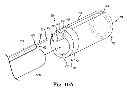
Figure I OA is a perspective view of an embodiment of a tissue tensioner
assembly according to the present disclosure.
Figure I OB is a perspective view of an embodiment of an inner piece of a
multi-piece tissue tensioner assembly according to the present disclosure.
Figure 1 OC is a perspective view of an embodiment of an outer piece of a
multi-piece tissue tensioner assembly according to the present disclosure.
Figure 11 is a perspective view of an embodiment of a lumen assembly
according to the present disclosure.
Figure 12A is a top perspective view of an embodiment of an inner piece of a
tissue tensioner assembly according to the present disclosure.
Figure 12B is a side cut-away view of the piece of figure 12A.
12
WO 2011/011372 PCT/US2010/042539
Figure 12C is an end view of the piece of figure 12A.
Figure 12D is a detail end view of the piece of figure 12A.
Figure 13A is a bottom perspective view of an embodiment of an outer piece
of a tissue tensioner assembly according to the present disclosure.
Figure 13B is a top perspective view of the piece of figure 13A.
Figure 13C is a side cut-away view of the piece of figure 13A.
Figure 13D is an end view of the piece of figure 13A.
Figure 14 is a side cut-away view of an embodiment of a tissue tensioner
assembly and working shaft.
While the invention is amenable to various modifications.and alternative
forms, specifics thereof have been shown by way of example in the drawings and
will be described in detail. It should be understood, however, that the
intention is
not to limit the invention to the particular embodiments described. On the
contrary,
the intention is to cover all modifications, equivalents, and alternatives.
DETAILED DESCRIPTION
In the following detailed description of the present invention, numerous
specific details are set forth in order to provide a thorough understanding of
the
present invention. However, it will be obvious to one skilled in the art that
the
present invention may be practiced without these specific details. In other
instances,
well-known methods, procedures, and components have not been described in
detail
so as to not unnecessarily obscure aspects of the present invention.
The invention relates to devices comprising a shaft for injecting a fluid into
tissue, such as a needleless injection device. Needleless devices as described
generally include a distal end and a proximal end. As used herein, the "distal
end"
refers to a portion of the device that is located internally within a
patient's body
during a treatment procedure, generally including the distal end of an
elongate shaft;
i.e., a distal end or distal portion of a device or a component is the end or
portion
that is toward the patient, and the "proximal" end or portion of the device or
component is the end or portion toward the surgeon or operator of the device.
A shaft distal end may include functional features that operate on fluid or
tissue during use, such as one or more injection orifice, optional delivery
head (end,
effector, nozzle, etc.) to house one or more injection orifices, optionally a
tissue
13
WO 2011/011372 PCT/US2010/042539
tensioner (as described), optionally a fitting to attach one component of a
shaft distal
end to one or more other component, optionally one or more of a light, optical
feature, steering feature, etc. A "proximal end". of an exemplary needleless
device
can include an injector body or "console" that remains external to the patient
during
use. An exemplary console can include a housing that connects to or is
otherwise
(directly or indirectly) in fluid communication with the shaft. The console
can
include fluid that can be pressurized by a pressure source to cause the fluid
to flow
through the shaft for injection into tissue at the distal end. The term
"distal end tip"
refers to a terminus of a distal end; for example, a distal end tip of a shaft
is the
location that defines the shaft end itself, as opposed to a portion of a
length of a shaft
that is referred to as a "distal end."
A device can eject fluid from at least one injection orifice located at the
distal end of the shaft. Optionally, multiple injection orifices may be
located at one
or more locations along a length of or about a circumference of a shaft distal
end.
Devices, systems, and methods as described can be used to inject fluid
(sometimes
referred to as an "injectate" or "injection fluid," which may be any type of
fluid such
as a therapeutic fluid) into tissue in a needleless manner whereby the
injectate passes
as a pressurized fluid stream (or "jet") through a surface of a tissue,
penetrating
without the use of a needle through the tissue surface and into the bulk of
the tissue,
and dispersing as particles or droplets within the tissue below the tissue
surface.
This contrasts with injections performed using a needle, whereby a hollow
needle
structure is used to penetrate tissue to locate a hollow end of the needle
within a
tissue mass, below the tissue surface, after which the needle carries fluid
into the
bulk of the tissue and delivers the fluid at a relatively low pressure to the
tissue in
the form of a body or pool of fluid known as a bolus.
A fluid stream or jet ejected for injection into tissue by a needleless
injection
system can be of a size (e.g., diameter), velocity, pressure, and volume to
allow the
fluid stream to penetrate directly through a tissue surface, then disperse
within the
tissue. The stream can be considered to be a relatively high velocity, high
pressure,
small diameter jet that after entry through a tissue surface disperses within
the tissue,
preferably as a multi-directional collection of particles (e.g., a "cloud") or
droplets
within the bulk of the tissue. Exemplary pressures of a fluid at a pressure
chamber
14
WO 2011/011372 PCT/US2010/042539
can be at least 200 pounds per square inch (psi), e.g., from 300 to 5000
pounds per
square inch. Without limiting the scope of the present description: when
injecting
bladder tissue a pressure of from 250 to 1000 psi can be effective, measured
at the
pressure chamber; when injecting prostate tissue a pressure of from 3500 to
5000 psi
can be effective, measured at the pressure chamber.
Exemplary needleless devices may be used for treating various physical
ailments or conditions at any bodily tissue, for example to treat tissue that
contains
or is within reach of injection through a body cavity or body lumen, e.g., by
accessing tissue through a body lumen, vessel, or cavity, and injecting tissue
by
placing an injection orifice within the lumen, vessel, or cavity. The type of
tissue
injected for treatment can be any amenable tissue, especially tissue
accessible
through a body lumen such as prostate tissue accessible through a urethra.
Exemplary needleless fluid delivery devices or systems can include a
proximal end that includes a console, and an elongate shaft extending from a
proximal end in communication with the console to a distal end. The elongate
shaft
can include an injection shaft and an injection lumen, optionally disposed
permanently, semi-permanently, or loosely and movably within or adjacent to a
working lumen. A distal end of the injection shaft can include one or more
injection
orifice in fluid communication with the console, through an injection lumen.
A console generally can include a housing, a pressure chamber, and a
pressure source. A console can be of any configuration, size, or design,
ranging
from a small, hand-held design to a relatively larger floor or table-mounted
console.
Optionally a console can include separate or separable components such as a
pressure chamber (e.g. "connector member") that can be attached between a
housing
and a proximal shaft end, used for an injection procedure, and detached and
optionally discarded. A shaft (e.g., an injection shaft or a working shaft, or
a shaft
assembly containing one or more of an injection shaft or an inflation shaft)
can also
be attached to a console, pressure chamber, or connector member, in a manner
to
allow separation and optional re-attachment or disposal after one or more use.
With
separable components, a shaft or pressure chamber can be attached to a console
housing and used to inject a first patient or a first injectate; the shaft or
pressure
chamber (e.g. "connector member") can then be discarded or sterilized. A
second
WO 2011/011372 PCT/US2010/042539
shaft or pressure chamber can be attached to the console to treat a second
patient or
the first patient with second injectate or another amount of the first
injectate. The
second patient or injectate can involve injection and treatment of the same
type of
tissue as the first patient or injectate, or of a new type of tissue (e.g.,
prostate or
bladder). In this manner, separable and optionally disposable shaft or
pressure
chamber components of a needleless injection system can allow a console
housing to
be used multiple times to inject the same or different injectates, to the same
or
different patients, and to the same or different types of body tissue.
A console can include actuating features to control distal end features, e.g.,
for steering a steerable distal end of a steerable shaft, to actuate ejection
of fluid, to
move a moveable or extendable injection shaft or one or more injection orifice
relative to another shaft component such as a working shaft, optional ports to
connect a console housing to auxiliary devices, electronics such as controls,
optic
features such as a lens, fiber optic, or electronic viewing mechanism to allow
viewing through an optical feature (to view a location of delivery), and an
actuating
mechanism or pressure source for a tissue tensioner in the form of a
mechanical
tissue tensioner or an inflatable balloon. One or more attachment ports can
optionally attach a console to an external and optionally remote component
such as
an external or remote pressure source, vacuum source, or an external or remote
fluid
reservoir to supply injectate or other fluid, such as to inflate a balloon.
For example,
a console (e.g., console housing or connector member) may have a fluid port
that
attaches to a source of a fluid to supply the fluid to the console, such as to
a
permanent or detachable pressure chamber. Embodiments of consoles can include
a
permanent or removable pressure chamber and a pressure source capable of
pressurizing a fluid contained in the pressure chamber to cause the fluid to
flow
from the console, through a lumen in the shaft, and then through an injection
orifice.
A fluid chamber can be a space (volume) at a proximal end of a device such
as at a console housing, useful to contain pressurized or non-pressurized
fluid, such
as injectate or a gaseous or liquid fluid to inflate a balloon (e.g., tissue
tensioner).
Examples of specific types of fluid chambers include fluid reservoirs and
pressure
chambers. Optionally a proximal end of a device may include one or multiple
fluid
reservoirs and pressure chambers.
16
WO 2011/011372 PCT/US2010/042539
A fluid reservoir is generally a type of fluid chamber that can contain a
fluid
for a purpose of containing, transferring, holding, or storing a fluid, such
as a fixed
volume fluid chamber, and may be included as a permanent or removable
(attachable and detachable) component of a console.
A pressure chamber can be a type of fluid chamber for containing fluid (e.g.,
injectate) for a purpose of placing the fluid under pressure to deliver the
fluid
through a lumen to a distal end of a shaft for ejection from an ejection
orifice.
Examples of pressure chambers include a syringe chamber and other variable
volume spaces that can be used to contain and pressurize a fluid. Examples of
variable volume pressure chambers include spaces that can exhibit a variable
volume
based, e.g., on a plunger, piston, bellows, or other mechanism for increasing
or
decreasing the volume (and correspondingly decreasing or increasing pressure)
within the variable volume chamber space. A pressure chamber can be
pressurized
by a pressure source attached to the plunger, bellows, or piston, etc., such
that fluid
contained in the pressure chamber is ejected under pressure, e.g., for priming
a
device, or for ejecting fluid from an ejection orifice for injection or to
produce a
control force. A pressure source may be any source of energy (e.g.,
mechanical,
electrical, hydraulically derived, pneumatically derived, etc.) such as a
spring,
solenoid, compressed air, manual syringe, electric power, hydraulic, pneumatic
pressure sources, etc. A pressure chamber may be a permanent or removable
(attachable and detachable) component of a console.
Examples of consoles, console features and combinations of console features
that can be useful according to the present description are identified at U.S.
Pat.
Publ. Nos. 2006-0129125 and 2009-0312696, and in Assignee's copending patent
applications PCT/US2009/006383, filed December 4, 2009, entitled METHOD
AND APPARATUS FOR COMPENSATING FOR INJECTION MEDIA
VISCOSITY IN A PRESSURIZED DRUG INJECTION SYSTEM, by Crank; WO
2010/065126 A2; WO 2010/065127 A2; Attorney Docket No. AMS0180/WO
entitled NEEDLELESS INJECTION DEVICE COMPONENTS, SYSTEMS, AND
METHODS filed on even date herewith; AMSO181/WO entitled DEVICES,
SYSTEMS, AND RELATED METHODS FOR DELIVERY OF FLUID TO
TISSUE filed on even date herewith; AMSOI82/WO entitled DEVICES,
17
WO 2011/011372 PCT/US2010/042539
SYSTEMS, AND METHODS FOR DELIVERING FLUID TO TISSUE filed on
even date herewith; AMSO183/WO entitled HIGH PRESSURE INJECTION
CATHETER SYSTEMS filed on even date herewith; and AMS0184/WO entitled
NEEDLELESS INJECTION DEVICE COMPONENTS, SYSTEMS, AND
METHODS filed on even date herewith, the entireties of these documents being
incorporated herein by reference.
In communication with a proximal end of a device is an elongate shaft that
extends from the proximal end (i.e., from a proximal shaft end), that is
optionally
removably connected to the console (or a component of the console such as a
removable pressure chamber), to a distal end that can be placed in a patient
during
an injection procedure. A shaft can be of various designs, minimally including
an
injection lumen to carry injectate from a proximal end of the device to a
distal end of
the injection shaft. Shafts for needleless devices as described are also
described in
Assignee's copending patent application WO 2010/065133 A2.
An injection shaft minimally includes an injection lumen in communication
with an injection orifice. The injection shaft can include structure such as
sidewalls
that define the injection lumen, the sidewalls being of sufficient strength to
withstand operating pressures sufficient to deliver injectate from the
injection orifice
at an elevated pressure sufficient to cause the injectate to be ejected from
the
injection orifice to penetrate a tissue surface and become injected and into
and
dispersed below the tissue surface, as described herein. Exemplary elevated
pressures ("injection pressures") may be at least 200, e.g. 1,000, or 2,000
pounds per
square inch or greater as measured at the distal end of the injection lumen,
or at the
pressure chamber. An injection shaft may be of a flexible material (e.g., a
metal or
polymeric tube) that can withstand such injection pressure, and may be
prepared
from exemplary materials capable of withstanding pressure of an injection,
e.g.,
nitinol, stainless steel, reinforced (e.g., braided) polymer, as also
described
elsewhere herein.
A basic version of a useful shaft as described can be an "injection shaft"
that
includes a proximal end, a distal end, a sidewall that defines an internal
lumen
("injection lumen"), and at least one injection orifice at the distal end in
connection
with the injection lumen.
18
WO 2011/011372 PCT/US2010/042539
An injection shaft can be any elongate structure capable of delivering fluid
to
a distal end of the injection shaft at a pressure suitable to inject tissue,
as described.
Exemplary injection shaft structures include relatively flexible hollow bodies
having
a distal end, a proximal end, sidewalls extending between the ends, an
internal
lumen defined by interior surfaces of the sidewall. The injection lumen is in
communication with one or more injection orifice at the distal end; the
injection
orifice may be as described herein, such as an aperture or bore in an
injection shaft
sidewall, an aperture or bore in a nozzle, end effector, injection head, or
other
structure in communication with the injection lumen.
An exemplary injection shaft can be in the form of a non-metal, polymeric
tube-like device and can be fabricated using suitable high strength polymers
including, for example, polyimide, polyetherimide available from General
Electric
under the trade name Ultem and linear aromatic polymers such as PEEKTM
available from Victrex plc for transporting the treatment fluid to the
treatment area.
In some embodiments, the non-metal, polymeric tube-like device can be
reinforced
through the inclusion of materials including nano-particles, clays and/or
glass. In
some presently contemplated embodiments, the non-metal, polymeric tube-like
device can be reinforced with one or more polymers such as, for example, tubes
braided with Kevlar or other high-strength polymers. The non-metal, polymeric
tube-like device can be fabricated so as to have a burst strength exceeding at
least
about 200, e.g., 1,000 or 2,000 psi and in some embodiments, having a burst
strength within a range of about 2,000 psi to about 5,000 psi. The non-metal,
polymeric tube-like device can be fabricated so as to have distention
properties,
wherein one or more orifices or jet ports located at a distal end of the
polymeric
tube-like device retains its shape and/or size without suffering swelling that
can have
a detrimental impact on a fluid jet used to deliver the therapeutic fluid at
the
treatment site. See, e.g., U.S. Pat. Publ. No. 2008/0119823.
An exemplary injection shaft can include a sidewall that defines an outer
shaft surface and an inner injector lumen, these being of continuous and
relatively
uniform dimensions of inner diameter, outer diameter, and wall thickness,
along an
entire length of the injection shaft. Alternately, an injection shaft,
injector lumen, or
sidewall, may change dimensions (e.g., wall thickness) along the length of the
19
WO 2011/011372 PCT/US2010/042539
injection shaft, with a larger wall thickness (e.g., greater outer diameter)
at a
proximal end and a thinner wall thickness (e.g., reduced outer diameter) at
the distal
end. An example of an inner diameter of an injection shaft (i.e., a diameter
of an
injection lumen) can be greater than 0.020 inches, e.g., from 0.022 to 0.030
inches
(for a lumen made of polyetheretherketone, or "PEEK"); exemplary outer
diameters
for the same exemplary injection shaft may be at least 0.032 inches e.g., from
0.034
to 0.045 inches. (An inner dimension of a fitting for placement on such an
injection
shaft may be, e.g., in the range from about 0.03 to about 0.05 inches.) A
length of
an injection shaft can be any length that functions to place a proximal end at
a
console and a distal end at a desired tissue location; exemplary lengths can
be from
as little as 15 inches if the console is a hand-held console, to as long as
100 inches if
the console is floor based or table based.
An injection shaft can be a component of a shaft of a useful needleless
injection device or system. Other shaft components may include additional
elongate
shaft structures with desired functionality, a single example being a device
referred
to herein as "medical device shaft" or a "working shaft," which can be used to
securely or moveably support or house an injection shaft. For instance, an
injection
shaft can be incorporated permanently or movably (e.g., removably) against
(alongside) or within (e.g., in a "working lumen" of) a working shaft. In
exemplary
embodiments an injection shaft can be loosely contained in a working lumen of
a
working shaft to allow movement of the injection shaft length-wise and
rotationally
relative to the working shaft; an injection shaft may be capable of moving
longitudinally within a working lumen to allow the injection lumen to be
extended
distally from an open end of a working lumen at a distal end of the working
shaft.
An example of a "working shaft" or "medical device shaft" can be a shaft
that is useful in conjunction with an injection shaft, to manipulate and place
the
injection orifice of an injection shaft at a desired location for treatment of
tissue. A
"working shaft" or "medical device shaft" can function to support the
injection shaft
and can optionally and preferably include any of a variety of optional
functionalities
such as steerability, an optical function, a tissue tensioner, or combinations
of these,
in addition to supporting the injection shaft.
WO 2011/011372 PCT/US2010/042539
An example of a particularly preferred working shaft can include features of
a typical cystoscope, endoscope, ureteroscope, choledoscope, hysteroscope,
catheter
(e.g., urinary catheter), or the like, or other similar type of medical device
shaft,
including one or more feature of flexibility, an optical function, a steerable
distal
shaft end, and a working lumen. A working lumen can be sized to loosely house
or
contain the injection shaft, preferably in a manner to allow the injection
shaft to be
moved lengthwise and rotationally within the working lumen, relative to the
working lumen, such as to allow the injection lumen (and optionally an
attached
tissue tensioner) to be extended from an opening at a distal end of the
working
lumen, at a distal end of the working shaft. A typical diameter (or other
dimension)
of a working lumen extending along a length of a distal end of a working shaft
can
be in the range from about 1 to about 3 millimeters. A typical length of
working
shaft for placement of a distal end at a location of the urinary tract can be,
e.g., from
to 25 centimeters. A typical outside diameter of a working shaft may be, for
15 example, from about 4 to about 10 millimeters.
As used herein, the term. "flexible shaft" refers to a shaft (e.g., an
injection
shaft or a working shaft) that is sufficiently pliable to allow bending and
flexing that
allow the shaft to be inserted through the meatus or an external incision,
into the
urethra or another body lumen, and to allow a portion of a distal end of the
shaft to
be guided into a body lumen or body cavity such as a urethra and optionally
the
bladder neck or bladder, as can be done with a Foley catheter. A flexible
shaft can
be sufficiently soft and pliable to conform or partially conform to a
patient's
anatomy, such as would a Foley-type catheter. A "steerable" shaft is a type of
a
flexible shaft having a distal end that can be maneuvered directionally (e.g.,
bent or
curved) from a proximal end; steerable shaft distal ends are sometimes
features of
endoscopes and other medical device shafts.
Optionally, a shaft of a device as described may also be malleable, or
"shapeable," meaning that a shaft distal end, or portion thereof, can be of a
material
capable of being shaped to a form, and to remain in that form during use, such
as for
insertion into a body lumen, until re-formed. A shaft or a shaft component,
such as a
working shaft or an injection shaft, can include a malleable component such as
a
bendable metal wire, coil, ribbon, tube, or the like, capable of being shaped,
used
21
WO 2011/011372 PCT/US2010/042539
without substantial deformation, and re-shaped. A malleable distal end can
allow a
distal end to be shaped by a user to assist in placement of the distal end
through a
body lumen such as a urinary tract, at a desired location. In some methods of
treatment, there may be difficulties or challenges in passing a shaft distal
end
through a body lumen, or to place the distal end in contact with tissue for
injection.
A malleable shaft distal end, e.g., of an injection shaft in particular, e.g.,
used in
conjunction with a working shaft within which the malleable injection shaft
distal
end is moveably disposed, or in conjunction with a working shaft adjacent to
which
the malleable injection shaft distal end is disposed, may assist in overcoming
such
potential difficulties. The malleable distal end tip may be formed by a user
to a
desired curve or bend, before or after placement in a working channel or
adjacent to
a working shaft; the working shaft may be inserted into a body lumen such as a
urethra, and the formed, malleable injection shaft distal end may be extended
from
or placed adjacent to the working shaft with a shape that improves the ability
to
position the injection shaft or an injection orifice thereof, at tissue for
injection. A
shapeable portion may vary in stiffness, length, resilience, material,
radiopacity, etc.,
and may be of any malleable material such as a polymer, metal, or polymer-
metal
composite.
A distal end of an injection shaft includes one or multiple injection orifices
for ejecting fluid within a body of a patient. An injection orifice can be any
form of
opening, aperture, or orifice, such as an aperture or bore in an injection
shaft
sidewall, or an aperture or bore in a nozzle, end effector, injection head, or
other
structure in communication with an injection lumen. Injection orifices can be
located at relative locations and orientations along a length or circumference
of an
injection shaft distal end to result in ejection and distribution of ejected
fluid in
different directions (e.g., circumferentially relative to the shaft),
optionally or
alternately at different distances along the length of the injection shaft. An
injection
orifice can be directed at any angle relative to a longitudinal axis of a
shaft, such as
perpendicular, angled toward a distal end, or angled toward a proximal end.
An injection orifice may have any useful size (e.g., length and diameter) to
produce a fluid stream of ejected fluid that can penetrate a tissue surface to
become
injected into tissue. Examples of a useful range of diameter of an injection
orifice
22
WO 2011/011372 PCT/US2010/042539
may be from about 0.001 to 0.05 inches, e.g., from 0.001 to 0.010 inches,
depending
on factors such as desired injection parameters (injection depth, volume,
pressure,
exit velocity, etc.) and the type and size (e.g., depth) of tissue being
injected. An
injection orifice may be larger or smaller than an injection lumen leading to
the
injection orifice, if desired, to affect the exit velocity of the jet of inj
ectate from the
injection orifice. Examples of useful orifice shapes may include features such
as a
venturi, a continuous uniform diameter along the length of an orifice, a
funnel-
shape, etc.
According to exemplary injection methods and devices, an injection orifice
may be located on a proximal side of a distal end tip of an injection shaft,
at a
location that allows the injection orifice and adjacent injection shaft
sidewall to
contact a tissue surface as a longitudinal axis of a shaft that contains the
injection
orifice is positioned in an orientation that is parallel to the tissue
surface. These
device embodiments are sometimes referred to as "side-fire" devices, herein.
As
used herein, a "distal end tip" can be considered a location of a distal end
of an
injection shaft that is the farthest (most distal) feature of the injection
shaft distal
end.
In certain embodiments of "side-fire" devices an injection orifice can be
located a distance away from a distal end tip on a proximal side of the distal
end tip
so the injection orifice is located to contact tissue for injection by placing
the shaft
sidewall in contact with tissue. Examples of injection orifice locations for
these
embodiments can be locations along a distal end of a shaft that are in the
range from
about 1 to about 40 millimeters from the distal end tip, on a proximal side of
the
distal end tip, e.g., such as a distance in the range from about 1 to about 25
millimeters from the distal end tip.
According to certain exemplary devices, a distal end of a shaft (injection
shaft, working shaft, or the like) can include a tissue tensioner, the tissue
tensioner
optionally being attached to a shaft such as a working shaft, e.g., attached
to the
distal end of the shaft, by a fitting that is attached to the tissue
tensioner, such as part
of a tissue tensioner assembly. A tissue tensioner can be attached to or
located at a
distal end of a shaft, somewhat near to an injection orifice, e.g., to be
within a body
lumen such as a urethra, e.g., a prostatic urethra, and near the injection
orifice when
23
WO 2011/011372 PCT/US2010/042539
the distal end of the shaft is installed in a patient for injection. For
example a tissue
tensioner can be located at a length-wise location along an injection shaft,
working
shaft, or generally a shaft of a needleless injection device, that is the same
length-
wise location as the length-wise location of an injection orifice.
A tissue tensioner can comprise an expandable surface, e.g., a continuous
expandable surface such as an inflatable balloon, or a non-continuous
expandable
surface such as an expandable metal (or plastic) cage or the like. The
expandable
surface can exhibit an expanded state and a non-expanded state. According to
exemplary methods, a tissue tensioner can be placed in a body lumen in a non-
expanded state and expanded within the lumen to the expanded state. In the
expanded state, the tissue tensioner contacts an internal surface of the lumen
to hold
the distal end of the shaft and an associated injection orifice in place
relative to
desired tissue for injection. The tissue tensioner can optionally produce
tension or
strain on the tissue in a manner that can affect the manner in which an
injected fluid
stream penetrates the tissue surface and becomes distributed in the tissue
upon
injection. A tissue tensioner can facilitate a good result upon injection of
fluid
through luminal tissue by ensuring that the luminal tissue is fixed and
includes a
desired amount of tension for receiving an injection.
Depending on the configuration of an injection orifice at a shaft of a device,
or at an injector head, a tissue tensioner can be used to place a desired
portion of
tissue in (e.g., direct) contact with an injection orifice, i.e., a surface
that contains an
injection orifice. Alternately, a tissue tensioner can place a desired portion
of tissue
at a desired distance away from an injection orifice, e.g., in the instance of
an
injector head that includes two surfaces with a recessed surface including an
injection orifice. The distance, if any, between an injection orifice and
tissue, at
injection, can be selected to affect properties of the injection, e.g., to
affect the
distance an injectate penetrates into tissue, the size of droplets formed
beneath the
tissue surface, and the pattern over which droplets of injectate are dispersed
throughout tissue when injected. Other factors can also be adjusted to affect
properties of the injection such as pressure and volume of injectate, size and
shape
of the injection orifice, etc.
24
WO 2011/011372 PCT/US2010/042539
Examples of tissue tensioners include inflatable balloons located at a shaft
distal end near an injection orifice (e.g., at the same length-wise location
as the
injection orifice), and mechanically extendable structures such as paddles,
protrusions, levers, metal or plastic cages, metal or plastic springs or
spirals, and the
like, any of which can be include a surface that can be extended (e.g.,
mechanically)
from a distal end of a working shaft or injection shaft to place pressure on
internal
tissue, e.g., on urethral tissue within the prostatic urethra or other luminal
tissue.
Tissue tensioners, device shafts, and related mechanisms and methods are
described
in Applicants' copending U.S. Patent Publ. Nos. 2006-0129125 and 2009-0312696,
the entireties of both of these being incorporated herein by reference.
A balloon or a mechanically extendable tissue tensioner can be inflated or
extended at a location that is approximately at a length along a distal end of
a shaft
that is near an injection orifice, e.g., at a length-wise location that is the
same as the
length-wise location of the injection orifice. When used within a lumen such
as a
urethra, the tissue tensioner can push luminal tissue (e.g., urethral tissue)
away from
the distal end of the shaft in a manner that causes the luminal tissue and an
injection
orifice to contact each other. This can be done, for example, by a balloon
expanding
from an opposite side of a shaft relative to an injection orifice to place
pressure on
luminal tissue located opposite from an injection orifice and to cause the
injection
orifice to contact adjacent luminal tissue, optionally to produce pressure,
strain, or
tension on the luminal tissue opposite of the balloon. A mechanical tensioner
may
be extended from a distal end of a shaft by use of an actuating mechanism such
as a
mechanical connection between the tissue tensioner and the proximal end of a
device, such as at a working shaft proximal end. An inflatable balloon may be
extended from a distal end of a shaft by inflating the balloon with
pressurized fluid
such as air or another gaseous or liquid fluid.
A distal end of a device as described may optionally include a fitting that
functions to attach together two or more components of a distal end. Exemplary
fittings can be any device or structure that engages and attaches to a distal
end of an
injection shaft or a working shaft. A fitting can be a component of or
attached to
another feature as described herein, such as a tissue tensioner, an injection
shaft, or a
working shaft.
WO 2011/011372 PCT/US2010/042539
Optionally, a fitting can be attached to an outer surface of an injection
shaft
or a working shaft; such a fitting can be in the form of a complete or partial
ring or
cylindrical surface that includes an interior dimension that fits around an
outer
surface (or portion thereof) of the injection shaft or working shaft. A
different
exemplary fitting can be in the form of or may include as a component of the
fitting,
a solid, small diameter rod (diameter approximately that of a working lumen of
a
working shaft) that extends longitudinally between two components of a distal
end,
or that extends from one component into another component, e.g., to connect a
working shaft to a tissue tensioner assembly. A small diameter rod may be a
permanent or removable structure of a tissue tensioner assembly, can extend
longitudinally in a proximal direction from the tissue tensioner assembly
toward a
working shaft, and an enter a distal end (e.g., from a distal end tip) of the
working
shaft to fit into the working lumen of a working shaft.
Optionally, a surface of an injection shaft or a working shaft can include an
opposing or complementary shape, form, or surface, that engages a shape or
form of
the fitting; examples of complementary or opposing surfaces can include
opposing
threaded surfaces; opposing snap-fit engagement elements; opposing elements of
a
mechanical detent engagement, a mechanical spring-engagement; a mechanical key-
fit engagement, and the like. Other examples of fittings include opposing
press-fit
surfaces, and elastic band surfaces. These and like types of fittings can be
prepared
from plastic or metal materials. Elastic band fittings can be prepared from
one or
more elastic materials such as rubber (natural or synthetic), elastic polymer,
silicone,
latex, and the like.
Certain preferred embodiments of fittings can be orientation specific to allow
an engagement at only a single orientation, e.g., a fitting may be "keyed. As
a single
example, a fitting in the form of a cylindrical or partially cylindrical
receiver (or
receptor) sized to engage a shaft may be keyed (opposing surface structures of
the
fitting and the shaft may allow engagement in only a single rotational
orientation).
A keyed fitting can be used to allow an engagement between two attached shaft
elements to occur only at a desired orientation between elements of the
shafts, e.g.,:
a fastener that attaches an injection shaft to a working shaft may be keyed to
require
desired orientation between an injection orifice of the injection shaft and
the
26
WO 2011/011372 PCT/US2010/042539
working shaft, for example to allow viewing of the injection shaft or
injection orifice
or to require desired positioning of the injection orifice relative to a
tissue tensioner
associated with the working shaft; alternately a fitting of a tissue tensioner
assembly
maybe keyed to require placement of the tissue tensioner assembly at a desired
orientation relative to a working shaft or an injection lumen (and injection
orifice).
A fitting can be part of an assembly (e.g., a "fitting assembly") that
includes
the fitting removably or non-removably attached to another component such as a
tissue tensioner, an injection shaft, or a working shaft. An example of a
fitting
assembly can be a fitting assembly that includes a fitting attached to an
injection
shaft distal end, wherein the fitting removably attaches to a working shaft.
See
figure 1. The fitting assembly can include one of any of the described
fittings
attached securely to the injection shaft, and situated to allow the fitting to
be
attached to a working shaft. Exemplary fittings include an elongate receptor
that
includes one or more of: threads; a snap-fit engagement; a mechanical detent
engagement; a spring; a keyed engagement surface; or an elastic band, capable
of
being placed on a distal end of a working shaft. In use, the fitting assembly
including the injection shaft distal end securely attached to the fitting
assembly, can
be removably attached to the distal end of the working shaft by attaching the
fitting
to the working shaft distal end. If desired, the fitting can be keyed to
require a
determined orientation between the working shaft and the injection shaft. If
the
fitting is an elastic band, the elastic band can be stretched over a working
shaft distal
end. Alternately, if the fitting is of a different type, such as a mechanical
(threaded,
etc.) fitting, the fitting can be attached mechanically. In injection methods,
the
fitting assembly can be removably attached to a distal end of a working shaft,
the
working shaft can be placed within a tissue lumen, an optional tissue
tensioner can
be expanded, fluid can be ejected from the injection shaft to inject tissue,
the distal
end of the working shaft can be removed from the patient, and the fitting
assembly
can be removed from the distal end of the working shaft. The working shaft can
be
re-used in later procedures, and the fitting assembly including the injection
shaft
may be disposed of or re-used. This embodiment of a fitting assembly can
optionally include a tissue tensioner that becomes located about the working
shaft
27
WO 2011/011372 PCT/US2010/042539
distal end when the fitting assembly is placed on the working shaft distal
end. See
figures 1 through 6.
Another example of a fitting assembly can be a fitting assembly that includes
a tissue tensioner (i.e., a tissue tensioner assembly), and attached to a
fitting,
wherein the fitting can be removably or non-removably attached to an injection
shaft
distal end. The tissue tensioner assembly can include one of any of the
described
fittings attached securely to a tissue tensioner, and situated to allow the
fitting to be
attached to a distal end of a shaft such as a working shaft or an injection
shaft.
Exemplary fittings include an elongate receptor that includes one or more of:
threads; a snap-fit engagement; a mechanical detent engagement; a spring; a
keyed
engagement surface; or an elastic band; capable of being placed on a distal
end of a
working shaft or injection shaft. If desired, the fitting can be keyed to
require a pre-
determined rotational orientation between the tissue tensioner and the working
shaft
or injection shaft. In use, the fitting of the tissue tensioner assembly can
be
removably (or non-removably) attached to the distal end of an injection shaft
or a
working shaft. If the fitting is an elastic band, for example, the elastic
band can be
placed (e.g., stretched) around the injection shaft distal end. See figures 7A
and 7B,
showing such a tissue tensioner assembly removably attached to a distal end of
an
injection lumen.
A tissue tensioner assembly that includes a fitting that can be removably
attached to a distal end of a working shaft can, in use, be used according to
steps that
include: removably attaching the tissue tensioner assembly to a distal end of
a
working shaft, placing the working shaft (the distal end of the shaft also
being
associated with an injection shaft) within a tissue lumen, expanding the
tissue
tensioner, ejecting fluid from an injection shaft associated with the working
shaft to
inject tissue, and removing the distal end of the working shaft from the
patient. The
tissue tensioner assembly can be removed from the distal end of the working
shaft.
The working shaft can be re-used in later procedures, and the tissue tensioner
assembly may be disposed of or re-used. In this embodiment, the tissue
tensioner
assembly may optionally be securely attached to a distal end of an injection
shaft
and in use the injection shaft becomes disposed adjacent to an exterior
surface, and
along a length of, the working shaft.
28
WO 2011/011372 PCT/US2010/042539
A tissue tensioner assembly that includes a fitting that can be attached
(removably or non-removably, such as by adhesive or by integral construction)
to a
distal end of an injection lumen can, in use, be used according to steps that
include:
placing the injection shaft within a working lumen of a working shaft such as
by
loading the proximal end of the injection shaft into the distal end of the
working
lumen or alternately by loading the distal end of the injection shaft into the
proximal
end of the working lumen, attaching the tissue tensioner assembly to a distal
end of
an injection shaft (optionally with the injection shaft already being loaded
into the
working lumen), placing the working shaft distal end (and injection shaft and
tissue
tensioner) within a tissue lumen, expanding the tissue tensioner, ejecting
fluid from
the injection shaft to inject tissue, and removing the distal end of the
working shaft
(and injection shaft and tissue tensioner assembly) from the patient. The
tissue
tensioner assembly can be removed from the distal end of the injection shaft;
alternately, the entire injection shaft and tissue tensioner assembly can be
removed
from the working shaft. The working shaft can be re-used in later procedures,
and
the tissue tensioner assembly, working shaft, or both, may be disposed of or
re-used.
Figures 7A and 7B illustrate an embodiment of a tissue tensioner assembly.
Assembly 110 includes fastener 111 that includes an elongate receptor sized to
receive a distal end of injection shaft 120 (including injection orifices 122
and distal
end tip 124). Rings or bands 112 can be elastic or non-elastic, plastic,
metal, rubber,
etc., bands to removably secure assembly 110 to the distal end of injection
shaft 120.
Tissue tensioner 114, illustrated as an inflatable balloon in a non-expanded
state, is
securely attached or optionally integral with fastener 111. Inflation lumen
116 is in
fluid communication with tissue tensioner (balloon) 114 in a manner to allow
gas or
liquid fluid (e.g., air) to be inserted into tissue tensioner 114 to inflate
and expand
tissue tensioner 114. Optionally a proximal end of inflation lumen 116 can be
accessible at the proximal end of a working lumen that can be associated with
injection lumen 120 and tissue tensioner assembly 110.
Referring to figure 7B, assembly 110 is shown attached to a distal end of
injection shaft 120, which is in turn disposed within working lumen 132 of
working
shaft 130. This distal end configuration comprising injection shaft 120,
working
shaft 130, and tissue tensioner assembly 110, is an example of a useful side-
fire
29
WO 2011/011372 PCT/US2010/042539
injection shaft configuration movably disposed within a working lumen. Side-
firing
injection orifices 122 are apposed by tissue tensioner (balloon) 114; when
balloon
114 is expanded within a body lumen, side-firing injection orifices 122 are
pressured
against internal luminal tissue.
.5 Still referring to figures 7A and 7B, tissue tensioner 114 is an inflatable
balloon but the tissue tensioner may alternately be of other types, such as an
expandable cage. Also, fitting 111 is illustrated to be removable from
injection shaft
120, but could alternately be permanent, semi-permanent, or non-removable, or
could even be absent in that tissue tensioner 114 could optionally be integral
with or
otherwise attached to the distal end of injection shaft 120. In still
alternate
embodiments, inflation lumen 116 could be incorporated into injection shaft
120.
In injection methods, a distal end as shown in figures 7A and 7B can be
prepared by attaching the tissue tensioner assembly 110 (removably or
permanently)
to the distal end of injection shaft 120, as illustrated. The tissue tensioner
assembly
110 and injection shaft 120 can be inserted into a distal end of working lumen
132
and passed through working lumen 132 to extend from the distal end of working
lumen 132 to a proximal end (not shown) of working lumen 132. Working shaft
130
can be placed within a tissue lumen (e.g., urethra). Tissue tensioner 114 can
be
expanded to secure placement of injection orifices 122 against internal
luminal
tissue. Fluid can be ejected from injection orifices 122 to inject tissue. The
distal
end of working shaft 130 and injection shaft 120 can be removed from the
patient.
In embodiments wherein fitting 111 is removable, tissue tensioner assembly 110
can
be removed from the distal end of injection shaft 120. Working shaft 130 can
be re-
used in later injection procedures. Injection shaft 120 may be removed from
working lumen 132 and may optionally be re-used or discarded.
A needleless fluid delivery system 100 is illustrated generally in Figure 1 as
attached to elastic adapter (i.e., a fitting in the form of an elastic band)
10. The
elastic adapter 10 is comprised of compliant or semi-complaint elastic
material. The
elastic adapter 10 defines a central aperture 12 through which a flexible
scope (e.g.,
a working lumen) is inserted. The elastic adapter 10 has an inner face 14 and
an
outer face 16 separated by material thickness 18. It is envisioned that the,
elastic
adapter 10 could be disposed about the distal end of a cystoscope, uretero
scope,
WO 2011/011372 PCT/US2010/042539
choledoscope, endoscope or hysteroscope (e.g., any type of working shaft). The
amount of elastic tension about the flexible scope may be varied by selecting
the
thickness and/or type of the elastic material (and the size, e.g., inner
diameter, of the
elastic adapter). Furthermore, the axial length 20 of elastic adapter 10 may
include
designated bending areas or areas of greater elastic tension so as not to
interfere with
the efficiency of the flexible scope. The needless fluid delivery system 100
is
attached to the elastic adapter 10 axially at connection region 22. In
alternative
embodiments it is envisioned that the needless fluid delivery system 100 may
be
connected by radial bands attached to the outer face 16 of the elastic adapter
10 or is
disposed within central aperture 12 of the elastic adapter 10.
Needleless fluid delivery system 100 can comprise an injector (e.g., at a
proximal end, not shown), an applicator lumen ("injection lumen") 104, and an
injection orifice 102. The injector (e.g., including a console as described
herein) can
be as simple as manually activated syringe, or can comprise an automated
injector
including a user interface and a connector member. A connector member at a
proximal end or other fluid chamber can include a therapeutic fluid supply and
the
user interface can comprise an input means for selectively delivering a
pressurized
fluid through the connector member. Representative input means can include
foot
pedal, switches, buttons or a touch-screen capable of receiving touch commands
as
well as displaying system information including a mode of operation as well as
operating parameters. The applicator lumen 104 can comprise a non-metal,
polymeric tube like device having a proximal attachment end 106 and a distal
treatment end (or injection shaft distal end) 108. A non-metal, polymeric tube
like
device can have a tube length that corresponds to a type of treatment to be
performed within a patient's body. For example, when a non-metal, polymeric
tube
like device is configured to perform a cystoscope or endoscopic procedure, the
tube
length can range from about 18 to about 72 inches in length. Once the distal
treatment end 108, and more specifically, the administration orifice 102 is
positioned
with respect to the treatment location, the injector can be actuated so as to
begin
delivery of a therapeutic fluid. In positioning the needless fluid delivery
system 100
at treatment location, it will be understood that a medical professional
frequently
employs a medical imaging system such as, for example, computer axial
31
WO 2011/011372 PCT/US2010/042539
tomography (CAT), magnetic resonance imaging (MRI), or in the case of
treatment
of a prostate gland, an exemplary imaging means is transrectal ultrasound
(TRUS)
so as to achieve the desired position of administration orifice 102. Another
imaging
means is by direct vision of the distal end of the inserted device, optionally
the
injection shaft or injection orifice, through direct vision by use of an
endoscope.
As illustrated in Figure 2, elastic adapter 10 is disposed about a flexible
scope (e.g., working shaft) 30 such as a cystoscope to deliver therapeutic
fluid to a
treatment location, such as, for example, the urinary bladder, urethra,
prostate, etc.
Cystoscope 30 can include a working channel (working lumen) 36, a fiber optic
light
source 32 and lens 38 such that a medical professional can verify the distal
treatment
end 34 is positioned proximate the treatment location. It is envisioned that
elastic
adapter 10 could include an upper face that caps a portion of the distal
treatment end
34 of the cystoscope 30. However, any cap portion must be positioned so as not
to
interfere with the cystoscope operation.
An alternate two-layer embodiment of an elastic adapter (or fitting) 200, is
illustrated in Figures 3-6. A cystoscope 30 (or other working shaft) is
positioned
within elastic adapter 200 to deliver therapeutic fluid to a treatment
location, such
as, for example, the urinary bladder, urethra, prostate, etc. Cystoscope 30
can
include a working channel (working lumen) 36, a fiber optic light source 32,
and
lens 38 such that a medical professional can verify the distal treatment end
34 is
positioned proximate the treatment location. Needleless fluid delivery system
100
can comprise an injector (not shown), an applicator lumen ("injection lumen")
104,
and an injection orifice 102.
The elastic adapter 200 may be a two layer device so as to include an
inflation element 202. An inner elastic sleeve comprises a first layer 204.
The first
layer 204 is elastically mounted about the distal end 34 of the flexible scope
30. As
the flexible scope 30 is stiffer than the elastic adapter 200, the elastic
tension created
by the stretched elastic adapter does not impinge upon the scope. The second
layer
206 is attached around the outer diameter of the first layer 204 to create a
balloon
210. A balloon inflation lumen 208 is disposed axially along a central
aperture 212
with a first end (proximal end) in communication with a media source such as
compressed air or a fluid. A second end 212 of the balloon inflation lumen 208
is in
32
WO 2011/011372 PCT/US2010/042539
communication with the space between the first layer 204 and second layer 206.
It
is envisioned that the second layer 206 may radially overlap the axial ends of
the
first layer 204. The apposition balloon 210 is thus defined by the second
layer 206
overlap of the first layer 204.
It is further envisioned that in some embodiments the second layer 206 may
only partially surround the first layer 204 as illustrated in Figure 4. For
example, the
second layer 206 maybe disposed eccentrically around the first layer 204
leaving an
axial section of the first layer 204 exposed. The injection lumen (injection
shaft)
104 would thus be attached to the first layer 204 along the exposed section.
As the
apposition balloon 210 inflates the injection lumen 104 can thus be positioned
(within a body lumen). The eccentric geometry allows the apposition balloon
210 to
force the injection lumen 104 against the tissue chosen for treatment.
In an additional embodiment, the injection lumen 104 would be attached
between the first layer 204 and second layer 206, as illustrated in Figure 5
or be
attached to the second layer 206 as illustrated in Figure 6.
In operation, the elastic adapter 200 would be placed about the distal end 34
of the flexible scope 30 by stretching the first layer 204. Elastic tension of
the first
layer 204 will maintain the position of the needleless injection system 100
relative to
the flexible scope 30. The flexible scope 30 would be advanced to a treatment
location at which time the balloon 210 would be filled through inflation lumen
212.
Selectively inflating balloon 210 will assist in proper positioning of the
injection
orifice 102. Treatment may include providing a jet-injection of the
therapeutic fluid
through the injection orifice 102 at a desired treatment site. Generally, a
remote
injector is utilized to deliver the therapeutic fluid from an external
reservoir located
at a proximal end of the tube-like device 100. After treatment is complete,
the
balloon 210 is deflated and the flexible scope 30 withdrawn.
Another exemplary embodiment of a needleless injection system according
to the present description is illustrated at figure 8. Device 500 includes a
handle 502
and distal shaft end 504 of working shaft 503, which includes injection shaft
508
disposed within working lumen 518. The proximal end of the devices includes
handle 502 of a scope that connects to working shaft 503 (e.g., of a
cystoscope,
endoscope, catheter, or other medical device shaft), including features useful
for
33
WO 2011/011372 PCT/US2010/042539
manipulating or operating features at distal end 504. Handle 502 includes:
fiber
optic light source 516; steering actuator 514, which can be manipulated to
cause the
steerable distal end of device 500 to move in at two or more dimensions);
viewing
lens 520 that allows viewing through fiber optic cable 510; and port 524,
which
allows for connection of a fluid source to handle 502. Articulation for
steering of
distal end 504 is indicated in dashed lines.
Still referring to figure 8, body 512 connects to working shaft 503, which
includes lumens and mechanisms that connect features of proximal end handle
502
to distal end 504. Working lumen 518 is a hollow lumen or channel that extends
within working shaft 503 and supports and contains injection shaft 508 in a
manner
that allows injection shaft 508 to move longitudinally along the length of
working
shaft 503, to allow the distal end of injection shaft 508 to extend from end
opening
522 of working lumen 518. Working shaft 503 also includes fiber optic 510 and
a
steering mechanism (not shown) that allows steering (deflecting) of distal end
504
by movement of actuator 514. Light source 516 transmits light to distal end
504 by
fiber optic 510.
Distal end 504 includes end opening 522 of working lumen 518 from which
can be extended injection shaft 508, which includes at least one injection
orifice.
Also distal end 504 can be steered to allow movement of the tip of working
shaft
distal end 504, in coordination with extension of injection shaft 508, based
on
viewing through fiber optic 510, to deliver a fluid with accurate placement at
a
desired tissue location. The distal end of injection shaft 508 can be any
design as
described herein, e.g.: can include multiple injection orifices at different
length-wise
or circumferential locations; can include a tissue tensioner for apposition of
an
injection orifice against tissue; etc. As illustrated, fluid stream 509 is
shown being
ejected from an injection orifice (not shown); tissue tensioner (balloon) 511
is
located on an opposite side of injection lumen 508 from the injection orifice.
While figure 8 illustrates an embodiment of a needleless injection system
having an elongate shaft that includes an injection shaft disposed within a
working
lumen of a working shaft, other embodiments are alternately useful, such as
embodiments of distal shaft ends of figures I. through 6, including an
injection shaft
34
WO 2011/011372 PCT/US2010/042539
dispose on an exterior of a working shaft, and an optional tissue tensioner
disposed
about a distal end of the working shaft.
Also illustrated at figure 8 is shaft 546 extending between port 524 of handle
502 and console 542. Console 542 includes pressure chamber 540 and pressure
source 544.
Certain embodiments of tissue tensioner assemblies can include a tissue
tensioner, a fitting useful to engage an end of a shaft such as a working
shaft, and a
fitting ("adapter") useful to engage an end of a lumen assembly. The tissue
tensioner can include an inflatable balloon; the fitting can be connected to
the tissue
tensioner and can also be capable of removably or securely (non-removably)
attaching to a shaft such as a working shaft. The adapter can be designed to
connect
to a second shaft, such as a shaft of a lumen assembly or an injection lumen.
The
injection lumen can include one or more shaft and one or more lumen, in any
configuration, including an inflation shaft that defines an inflation lumen
and an
injection shaft that defines an injection lumen. The adapter is in fluid
communication with an interior space of the inflatable balloon so the lumen
assembly can engage the adapter and connect the balloon, through the adapter,
through the inflation lumen, to a proximal end of a needleless injection
device.
Figures 10 through 13D illustrate embodiments of tissue tensioning
assemblies and lumen assemblies that can be used with needleless injection
devices
and systems according to the present description.
Referring to figure 10A, tissue tensioner assembly 700 includes an inflatable
balloon tissue tensioner that is (optionally and as illustrated) constructed
from two
pieces that include an inner balloon piece 704 and an outer balloon piece 730.
Opening 712 at proximal end 713 is adapted to removably engage and fit over a
distal end of a shaft, such as a distal end of a cylindrical working shaft
(716 of figure
1 OA). Distal end 715 can also include an opening (717 shown in dashed lines)
and
the space between the proximal end opening and the distal end opening is
configured
to engage a distal end of a shaft. Opening 714 at proximal end 713 can be
adapted
to (removably or securely) fit over a distal end of a lumen assembly (such as
a distal
end of a lumen assembly 724 at figure 1 OA).
WO 2011/011372 PCT/US2010/042539
Referring to figures 1OA, 1OB, and 10C, inner balloon piece 704 and outer
balloon piece 730 can be assembled into two-piece tissue tensioner assembly
700, by
placing inner balloon piece 704 within an interior space of outer balloon
piece 730.
Adhesive or another type of securement mechanism can be used to maintain the
relative positioning of the pieces, for example adhesive can be placed between
opposing surfaces at the proximal end, and between opposing surfaces at the
distal
end of the pieces, to secure the pieces around the circumferences of each end
to
allow expansion and inflation of the outer balloon piece. Consequently, an
inflation
fluid (e.g., gas) can be pumped under pressure through an inflation lumen
(e.g., held
in place at tissue tensioner assembly 700 by adapter 706) and into an interior
space
(not numbered) located between outer surface 720 of inner balloon piece 704
and
inner surface 722 of outer balloon piece 730, to cause expansion of outer
balloon
piece 730, e.g., for apposition.
A tissue tensioner assembly as described may be prepared by any useful
method and from any useful material or materials, specifically including metal
or
plastic (polymeric) materials, with preferred materials including surgical
grade
polymeric materials. The materials can be elastomeric and sufficiently
stretchable to
be deformed and stretched to be adapted to and to fit over an end of a shaft
such as a
working shaft or a shaft of a lumen assembly or an injection lumen. Specific
example of useful biocompatible, materials include polyolefins (e.g.,
polyethylene,
polypropylene) polyacrylates, polyurethane, silicone polymers, natural and
synthetic
rubbers, latex, vinyl, polyamide ether (e.g., commercially available from
Autochem
under the trade name PebaxTM), and any other relatively inert elastomeric
materials.
Optionally, with an assembly having a two-piece design, two different
materials can
be used to construct the assembly, a different material being selected for
each of the
inner piece and the outer piece, if desired.
According to certain embodiments of tissue tensioning assemblies, an
assembly may be constructed of two pieces, one piece that defines an inner
portion
of a balloon and an adapter, and a second piece that defines an expandable
outer
portion of a balloon. For example, figure 10B illustrates one piece of an
exemplary
multi-piece construction of a tissue tensioner assembly, inner balloon piece
704.
Inner balloon piece 704 includes adapter 706 generally located at proximal end
713
36
WO 2011/011372 PCT/US2010/042539
of assembly 700, designed to removably or securely, and in an air-tight
fashion,
engage a lumen assembly (e.g., distal end of lumen assembly 724).
Adapter 706 is illustrated as a flexible, elastomeric, cylindrical extension
on
a proximal side of assembly 700 that defines opening (714) sized to engage a
distal
end of a lumen assembly, such as a tube or tubular shaft. Adapter 706, a
deformable, elastomeric cylinder, includes an inner surface that frictionally
or non-
frictionally engages an outer surface of a lumen assembly that is also
optionally and
preferably cylindrical. Other types of adapters will also be useful, including
any
adapter that can maintain engagement between a tissue tensioner assembly and a
lumen assembly. Examples of useful engagement structures that may allow
engagement between a surface of a tissue tensioner assembly and a surface of a
lumen assembly include opposing threaded surfaces, opposing snap-fit
engagement
elements; opposing elements of a mechanical detent engagement, a mechanical
spring-engagement, a mechanical key-fit engagement, and the like.
An adapter (e.g., 706, but also optionally of any other useful design) can
engage a lumen assembly and maintain a position of the lumen assembly at a
location relative to a tissue tensioner assembly (e.g., 700) such that one or
more
lumen or lumens of the lumen assembly is in fluid communication with the
balloon,
or that otherwise places and maintains a position of a lumen or shaft of a
lumen
assembly at a desired location relative to a tissue tensioner assembly. For
example,
an adapter can hold a lumen assembly in a position to place an inflation lumen
or an
opening leading to an inflation lumen at a location of a tissue tensioner
assembly
that results in fluid communication between the inflation lumen and a balloon
of the
tissue tensioner assembly to allow inflation of the balloon using pressurized
inflation
fluid passed through the inflation lumen. Alternately or in addition, an
adapter can
hold a lumen assembly in a position to place an injection lumen at a position
relative
to a tissue tensioner assembly that places an injection orifice at a location
to eject
fluid for injection into tissue, such as opposite a tissue tensioner of the
tissue
tensioner assembly or opposite a direction of expansion of a tissue tensioner.
For
example, tissue tensioner assembly 700 includes channel 718 that includes a
length-
wise depression at an exterior surface of outer balloon piece 730; adapter 706
can
hold an injection lumen in place at channel 718 in a manner to position an
injection
37
WO 2011/011372 PCT/US2010/042539
orifice of the injection lumen at a desired location for injecting fluid into
tissue, such
as opposite of a direction in which a balloon surface of assembly 700 will
expand
upon inflation.
Generally, a tissue tensioner assembly can include one or more passage,
channel, or the like that extends from an adapter useful to engage a lumen
assembly,
to other locations on the assembly. A passage can function to engage an
element of
a lumen assembly such as an injection shaft, or a passage can create fluid
communication between a space defined by or adjacent to the adapter and an
interior
space of a balloon. Examples of useful passages of a tissue tensioner assembly
are
shown, e.g., at figures 1OA and I OB.
Referring to figure 10A, adapter 706 includes backwall surface 732.
Aperture 734 (in dashed lines) (injection lumen aperture) is an aperture that
connects to a channel sized to contain an injection shaft. Injection lumen
aperture
734 connects to a channel (injection lumen channel) situated generally
parallel to a
longitudinal axis of tissue tensioner assembly 700 and parallel with
(optionally but
not necessarily aligned with) a longitudinal axis of a lumen assembly engaged
with
adapter 706. Accordingly, an injection shaft can extend from a lumen assembly
engaged with adapter 706, through aperture 734, and to a location along a
length of
tissue tensioner assembly 700.
More specifically, as illustrated lumen assembly 724 can be inserted into
adapter 706 so that distal end tip 725 of lumen assembly 724 may approach or
may
abut backwall surface 732. Backwall surface 732 defines two apertures:
injection
lumen aperture 734 and inflation fluid aperture 736. Injection lumen aperture
734
defines a proximal end of injection lumen channel 740, extending to a distal
end at
aperture 741 (see figure 10B). Injection lumen channel 740 is a channel that
can
receive and contain injection lumen 727 (having injection orifice 729) of
lumen
assembly 724 when lumen assembly 724 is engaged with adapter 706. In use, when
lumen assembly 724 engages tissue tensioner assembly 700 at adapter 706,
injection
lumen 727 can be placed within injection lumen channel 740 to locate and
secure
injection lumen 727 at a position that places injection orifice 729 in a
position to
inject tissue (i.e., within channel 718) when tissue tensioning assembly 700
is placed
38
WO 2011/011372 PCT/US2010/042539
in a body lumen of a patient and the tissue tensioning balloon of assembly 700
is
inflated.
Inflation fluid aperture 736 defines a proximal end of inflation fluid channel
742 (see figure 10B). Inflation fluid channel 742 is channel that aligns with
or is at
least in fluid communication with an inflation lumen (annular space 721 as
illustrated) of lumen assembly 724 when lumen assembly 724 is engaged with
adapter 706. Inflation fluid channel 742 extends between proximal aperture 736
and
distal aperture 743. In use, when lumen assembly 724 engages tissue tensioner
assembly 700 at adapter 706, inflation lumen 721 can be placed adjacent to and
in
fluid communication with inflation fluid aperture 736 and inflation fluid
channel 742
to allow inflation fluid to pass from inflation lumen 721 of lumen assembly
724,
through inflation fluid aperture 736, further through inflation fluid channel
742, and
into an interior space of an inflatable balloon of assembly 700.
Referring to figure 10C, outer balloon piece 730 is shown to include interior
surface 722, which defines an expandable outer piece (expandable surface) of a
balloon. Interior surface 722 includes ridges or ribs 726 extending around
surface
722 in a substantially circumferential manner, which can direct and control a
direction expansion of the balloon upon inflation, or prevent adhesion between
surfaces (720 and 722) of the balloon.
Figure I OC also shows optional channel 718, which defines a surface
structure useful to maintain a position of a distal end of an injection lumen,
especially to locate the injection lumen at a position to place an injection
orifice of
the injection lumen at a location to inject tissue. Channel 718 is located at
a distal
end of injection lumen channel 740 so that an injection lumen located to pass
through injection lumen channel 740 will become located at channel 718.
Channel
718 is located on a side of assembly 700 that does not expand upon inflation
of the
balloon surface of outer balloon piece 730. Specifically, channel 718 is
located at a
surface of assembly 700, extends in a length-wise direction parallel to a
length-wise
axis of assembly 700, on a side of assembly 700 that is opposite the side of
assembly
700 that includes a balloon surface that expands when inflated.
As also illustrated at figure 1 OC, a wall con- figuration (e.g., thickness)
of an
outer balloon piece may be nonsymmetrical. In specific, upper wall 760 is of
greater
39
WO 2011/011372 PCT/US2010/042539
thickness than lower wall 762. A nonsymmetrical configuration (thickness) can
allow different portions of the balloon, around a circumference of the
balloon, to
expand or to fail to expand upon inflation of the balloon, or to expand at
different
rates or to different extents. A nonsymmetrical configuration can be
accomplished
by separating or isolating portions of the balloon wall (e.g., grooves,
structures,
breaks in the wall, etc.), or by using different wall thickness or different
wall
materials. As such, the inflation of one side of the respective balloon wall
will not
undesirably distort the opposing or adjacent side of the balloon. A balloon
may
expand in a non-symmetrical or asymmetrical manner, either on a length-wise
basis
(when viewed from a side) or on a radial basis (when viewed from an end, as in
figure 13D). That is, a balloon may expand a greater distance in one direction
(when
viewed from an end or a side) compared to the extent of expansion in another
direction. In certain embodiments, a balloon can include a one-sided balloon
inflation configuration created by bonding the inner balloon piece to the
outer
balloon piece along a length of the assembly (e.g., on the side where
inflation is not
desired). It is envisioned that various structure, materials, and techniques
can be
implemented to achieve the nonsymmetrical and selectively expandable
characteristics of the balloon assembly.
A lumen assembly as described can be in the form of a shaft that contains or
defines one or more lumen or lumens, preferably at least two lumens that may
be
arranged in any fashion such as in a concentric configuration, an annular
configuration, a side-by-side configuration, or any other useful arrangement.
The
lumen assembly can include an injection lumen for carrying an injectate from a
proximal end to distal end, as well as an injection orifice at the distal end
through
which the injectate can be ejected at pressure sufficient to penetrate tissue.
The
lumen assembly can also include an inflation lumen for carrying an inflation
fluid
from a proximal end to a distal end. The inflation lumen can include an
opening at a
distal end that can connect to an adapter in a manner that results in fluid
communication (through the adapter) between the balloon interior space and the
inflation lumen.
A lumen assembly or a constituent inner shaft or outer shaft can be prepared
of any useful material. An injection lumen can be constructed from material as
WO 2011/011372 PCT/US2010/042539
described herein, such as a polymeric material capable of withstanding
pressure
necessary for injection of tissue. An inflation lumen may be made of the same
materials, or of materials that are not necessarily capable of withstanding
the same
pressure. An inflation lumen may be made of a material that may be metal or
plastic
(polymeric), with preferred materials including surgical grade polymeric
materials.
The materials can be elastomeric but not necessarily stretchable, and can
preferably
bend without kinking. Specific example of useful materials include
biocompatible
polymers such as polyolefins (e.g., polyethylene, polypropylene), polyesters,
Nylon,
polyester, polyacrylates, silicone polymers, polyurethane, natural and
synthetic
rubbers, latex, vinyl, polyamide ether (e.g., commercially available from
Autochem
under the trade name PebaxTM), and any other relatively inert elastomeric-
materials.
Referring to figure 11, an exemplary lumen assembly can be in the form of
two shafts arranged in a "concentric," "coaxial," or "annular configuration.
As
illustrated to embody this configuration, lumen assembly 776 includes proximal
end
772 and distal end 774. Inner shaft 778 functions as an injection lumen and is
located within outer shaft 770. Injection lumen 780 is defined as the interior
space
of inject shaft 778. Inflation lumen 782 extends length-wise along the annular
space
defined between the inner surface of outer shaft 770 and the outer surface of
inner
shaft 778. Optionally inner shaft 778 can be fixed to prevent lengthwise
movement
of inner shaft 778 relative to outer shaft 770, or alternately inner shaft 778
can be
moveable to allow lengthwise movement of inner shaft 778 relative to outer
shaft
770. Distal end 774 can be configured (e.g., sized) to engage an adapter of a
tissue
tensioner assembly.
According to an alternate embodiment (see lumen assembly 724 at figure
1OA), inner lumen (e.g., injection lumen) 778 can extend a distance beyond the
length of outer lumen 770. Preferably in this embodiment, inner shaft 778 can
be
fixed to prevent lengthwise movement of inner shaft 778 relative to outer
shaft 770.
A length of an inner shaft that is greater than a length of an outer shaft may
be
desired, e.g., to allow the inner shaft and outer shaft to fit an adapter and
engage a
tissue tensioner in a desired configuration. For example, an outer shaft may
be
located at and may contact an adapter at a proximal end or proximal side of a
tissue
tensioner assembly, and an inner (injection) lumen may be designed to extend
41
WO 2011/011372 PCT/US2010/042539
beyond the adapter to place an injection orifice at a mid or middle (based on
length)
location or at a distal end or distal side of a tissue tensioner assembly. See
figure
1 OA and related text.
Figures 12A, 12B, 12C, and 12D illustrate another version of an inner
balloon piece 704, slightly modified relative to piece 704 of figure 10B.
Piece 704
includes features of piece 704 of figure 10B, including adapter 706 (to be
generally
located at proximal end 713 of an assembly 700), designed to removably or
securely,
and in an air-tight fashion, engage a lumen assembly (e.g., distal end of
lumen
assembly 724). Adapter 706 is illustrated as a flexible, elastomeric,
cylindrical
extension on a proximal side of assembly 700 that defines opening (714) sized
to
engage a distal end of a lumen assembly, such as a tube or tubular shaft.
Backwall
surface 732 defines two apertures: injection lumen aperture 734 and inflation
fluid
aperture 736. Injection lumen aperture 734 defines a proximal end of injection
lumen channel 740, extending to a distal end at aperture 741. Injection lumen
channel 740 is a channel that can receive and contain injection lumen (not
shown) of
lumen assembly 724 when lumen assembly 724 is engaged with adapter 706.
Inflation fluid aperture 73 6 defines a proximal end of inflation fluid
channel 742.
Inflation fluid channel 742 extends between proximal aperture 736 and distal
aperture 743. Modifications include abutment 745 extending into distal
aperture
717, against which a distal end tip of a working lumen will rest when inserted
through proximal opening 712 and fitted longitudinally into piece 704; and
circumferential ridges 742 to assist engagement with an outer balloon piece,
and
define an interior space of a balloon for inflation.
Figures 13A, 13B, 13C, and 13D illustrate another version of an outer
balloon piece 730, slightly modified relative to piece 730 of figure 1 OC.
Referring
to figures 13A, 13B, 13C, and 13D, outer balloon piece 730 includes interior
surface
722, which defines an expandable outer piece (expandable surface) of a
balloon.
Optional channel 718 defines a surface structured to maintain a position of a
distal
end of an injection lumen, especially to locate the injection lumen at a
position to
place an injection orifice of the injection lumen at a location to inject
tissue.
Channel 718 is located at a distal end of injection lumen channel 740 (at
aperture
741) so that an injection lumen located to pass through injection lumen
channel 740
42
WO 2011/011372 PCT/US2010/042539
will become located at channel 718. Channel 718 is located on a side of
assembly
700 that does not expand upon inflation of the balloon surface of outer
balloon piece
73 0. As indicated by the arrows, balloon lower wall 762 is of a smaller
thickness
compared to upper wall 760. Lower wall 762 expands in a non-symmetrical or
asymmetrical manner on a radial basis, expanding a greater distance in a
direction
away from channel 718, compared to the extent of expansion in other
directions.
An alternate tissue tensioner assembly can include a tissue tensioner (e.g., a
balloon), an adapter for a lumen assembly, and an alternate fitting or adapter
that
allows secure or removable attachment of the assembly to a shaft, such as a
working
shaft or a smaller shaft (e.g., having a diameter of from 0.5 to 2
millimeters) that can
extend from the tissue tensioner assembly in a proximal direction and engage a
working lumen of a working shaft. For example, a fitting can include or engage
with an intermediate mechanical attachment (e.g., a solid, relatively rigid
post) that
is engaged with or is secured to a working shaft, removably or non-removably.
The
fitting may be, for example, a post or shaft that engages an aperture of the
tissue
tensioner assembly at one portion, side, or end, and that engages or is
attached to the
working shaft (e.g., a working lumen of a working shaft) at a second portion,
side, or
end.
As an example, figure 14 shows a tissue tensioner assembly that includes a
tissue tensioner (e.g., a balloon), an adapter for a lumen assembly (e.g., as
described
herein), and a fitting that includes a post having a first end secured
directly or
indirectly to the tissue tensioner, and a second end removably engage to a
working
shaft, e.g., that can be placed within and engaged with a working lumen of the
working shaft. The engagement between an end of the post and the working lumen
may be any mechanical engagement useful to removably secure the post to the
working lumen for use of the needleless injection device, including a
frictional
engagement, opposing threaded surfaces; opposing snap-fit engagement elements;
opposing elements of a mechanical detent engagement, a mechanical spring-
engagement, a mechanical key-fit engagement, and the like.
Figure 14 illustrates an embodiment of a needleless injection device that
includes working shaft (e.g., cystoscope) 724 and tissue tensioner assembly
780.
Working shaft 724 includes working lumen 750. Tissue tensioner assembly 780 is
43
WO 2011/011372 PCT/US2010/042539
illustrated schematically as a generic block structure that can take on any
form,
shape, or size, etc., useful to support the structures presently described.
For
example, assembly 780 may include an open cylindrical ring or partial ring
structure, or other open structure, that allows placement of an injection
orifice at one
location of the ring and a tissue tensioner (e.g., balloon) that can expand in
the
opposite direction, while still allowing other optional functionality of a
working
shaft (e.g., cystoscope) such as viewing of the injection lumen. A distal end
of post
770 engages assembly 780 in a permanent or a removable manner, e.g., by
engaging
an elastic aperture (not shown) in assembly 780. The elastic lumen can have a
diameter suitable to securely or removably engage the outer surface of post
780, and
an depend on the diameter of post 780; examples of suitable diameters of a
post and
an elastic lumen can be from 0.5 to 2 millimeters.
Exemplary tissue tensioner assembly 780 engages a post 770, permanently or
removably. In one embodiment, assembly 780 can includes an adapter (e.g.,
comprising an elastic aperture, not specifically shown) as described herein,
capable
of engaging a distal end of lumen assembly 724, which includes an injection
lumen
and an inflation lumen. Tissue tensioner assembly 780 also includes inflatable
balloon 758, inflation fluid channel 756, orifice 760, and channel 754.
Channel 754
can be adapted to contain an injection lumen to allow the injection lumen to
extend
from an lumen assembly 724 to aperture 760, where an injection orifice of the
injection lumen can be placed for tissue injection (similar to the
configurations of
figures 10A, IOB, and 12A, 12B). Alternately (and as illustrated) channel 754
can
constitute a pressure resistant extension of an injection lumen connected to
an
injection lumen of lumen assembly 724, and orifice 760 be an injection
orifice.
Post 770 extends from tissue tensioner assembly 780 and engages working
lumen 750, preferably in a removable configuration. Accordingly, post 770 and
the
assembly of post 770 with tissue tensioner assembly 780 are selectively
attachable to
the working shaft (e.g., endoscope device) 716 rather than integrally formed.
The
structure by which assembly 780 is attached to shaft 716 is post (rod or tube)
770, or
another like attachment mechanism, configured for selective attachment into
working lumen 750. Various size and dimensional configurations are available
for
the post 770 and working lumen 750 interconnection. For instance, the post 770
can
44
WO 2011/011372 PCT/US2010/042539
include geometrical features, e.g., axial slots, tapered profile, or a soft
outer material
to facilitate press fitting of the post 770 within working lumen 750. In
various
embodiments, the post 770 and lumen 750 interconnection can be configured such
that fluid is still permitted to flow through the lumen 750. The embodiment of
figure 14 enables use of the working shaft 716 with the high-pressure delivery
device without attaching the delivery device or to the outside of the scope,
and
without running the delivery lumen along the entire length of the working
channel of
the endoscope.
With any of the above features of fluid delivery devices, a device could
include an electronic process control system that can be programmed to make
fluid
deliveries having various locations, volumes, and other injection properties
such as
depth and degree (e.g., shape and distance) of dispersion and size of
particles of
fluid.
A needleless injection system can be use to perform treatment methods by
steps that include one or more of the following: providing a needleless
injection
device substantially as described herein; inserting a distal end of a shaft of
the fluid
delivery device into a patient, e.g., through the meatus and into the urethra;
navigating the distal end until an injection orifice at the distal end of the
shaft is
positioned at a desired delivery site. An injection shaft distal end can be
positioned
with a sidewall in contact with tissue, with a longitudinal axis of the shaft
in line
with (e.g., parallel to) tissue; an optional tissue tensioner can be used to
cause a
sidewall of the injection shaft distal end to contact and be pressed against
the tissue
surface to cause an injection orifice to contact the tissue surface for
injection.
By any of the described methods, multiple injection orifices can provide the
ability to place one or more different fluids at multiple locations of the
urethra,
prostate, bladder, or bladder neck, or other tissue, etc. Other treatment
locations can
include a rectal treatment location, a gastrointestinal treatment location, a
nasal
treatment location, a bronchial treatment location, or an esophageal treatment
location. Features of devices described herein, such as optical features,
steerable
shafts, tissue tensioners, and the ability to deliver multiple different types
of fluid,
allow for improved control over the location of injection or instillation of a
fluid.
WO 2011/011372 PCT/US2010/042539
According to certain exemplary fluid delivery procedures of the invention,
fluid such as ethanol or a biologically active agent can be delivered to the
bladder,
urethra, prostate, or bladder neck, etc., in a manner that causes the fluid to
be
injected into the tissue using a needleless delivery orifice.
Devices of the present description can be useful to treat of various tissues,
including of the urinary tract, in females or males. For example, devices as
described may be useful to inject the bladder, bladder neck, the urethral
tissue itself
or the external sphincter, or for transurethral injection of the prostate in a
male. In
other embodiments, a fluid may be injected into tissue of the urinary tract
(e.g.,
bladder, urethra, kidneys, ureters, prostate, etc.) such as individual or
combination
treatments using drugs or other therapeutic agents, e.g., botulinum toxin
("botox"),
an antiandrogen, a neurotoxin,, among others as will be understood. One
advantage
of injection of an active pharmaceutical agent at a location of use is the
placement of
the agent to avoid systemic side effects. Specific examples of active
pharmaceutical
agents that may be injected include botulinum toxin types A through G; 5-alpha
reductase inhibitors such as dutasteride and finasteride; alpha blockers such
as
alfuzosin, doxazosin, prazosin, tamsulosin hydrochloride, trazosin, ethanol,
to treat
BPH; or any of various antibiotics (e.g., to treat prostatitis) and
analgesics.
Figure 9 illustrates components of combination 620 of the invention. Any
different combination of components can be included in a system or set. The
components include console 600, optional "connector member" or external,
removable pressure chamber 602, multiple varieties of injection shaft
attachments (i)
through (iv) that can be separately attached to console 600 or removable
pressure
chamber 602, and a single working shaft 610 including handle 612. Console or
console housing 600 can be as described, and includes at least a pressure
source.
Port 601 allows connection to optional removable pressure chamber 602, which
can
be connected at a proximal end to port 601, and has distal end 605 that can be
connected to a proximal end of an injection shaft attachment. Optional port
603 of
pressure chamber 602 can be used to insert fluid into pressure chamber 602.
Each of
injection shaft attachments (i), (ii), (iii), (iv), (v), and (vi), is
exemplary and for
purposes of illustration of exemplary combinations. Each includes a proximal
end
(611) that can removably attach to console or console housing 600, optionally
by
46
WO 2011/011372 PCT/US2010/042539
removably attaching to connector member 602 at distal end 605. Each injection
shaft attachment also includes one or more injection orifice 606 at a distal
end 604,
connected through an inflation lumen (not shown, or included at lumen assembly
724 of attachment (v)) to the proximal end. Each injection orifice as
illustrated is on
a proximal side of a distal end tip 607.
An optional component of combination 620 is working shaft 610, which may
be as described herein, e.g., including handle 612, port 622 suitable to
introduce an
injection shaft into working lumen 616 of working shaft 614, optional
steerable
distal end 618, and an optional optical feature (not shown).
Another optional component of a combination 620 can be a fastener
assembly 620 having fastener 624 (e.g., an elastic fastener or other form of
elongate
receptor, optionally keyed) capable of attaching to a distal end of working
shaft 624,
and another fastener 626 (e.g., an elastic fastener or other form of elongate
receptor,
optionally keyed) capable of attaching to a distal end of an injection shaft.
A combination can include any one or combination of injection shaft
attachments as shown or otherwise described herein. An exemplary injection
shaft
attachment can include any one or more of. a side-fire distal end with an
elongate
receptor 609 that may be an elongate elastic receptor or a non-elastic
elongate
receptor capable of attaching to an outside surface of working shaft distal
end 618,
and that is also removably attached to distal end 604 (i); a side-fire distal
end with
an optional malleable distal end feature (not shown) and multiple injection
orifices
along a length of the distal end (ii); a distal end with a single injection
orifice near
distal end tip 607, including tissue tensioner (e.g., inflatable balloon) 613
attached
(e.g., securely) to the injection shaft distal end on the side opposite the
injection
orifice, and inflation lumen (or mechanical actuator, if the tissue tensioner
is
mechanically actuated) 615 extending alongside the injection shaft to a
proximal end
(iii); and, a distal end with a single injection orifice near distal end tip
607, including
combined fitting and tissue tensioner 615 attached (e.g., securely) to the
injection
shaft distal end on the side opposite the injection orifice, an inflation
lumen (not
shown) extending alongside or within the injection shaft to a proximal end,
and the
combined fitting and tissue tensioner being an elastic or non-elastic fitting
sized to
fit at the distal end 618 of working shaft 614.
47
WO 2011/011372 PCT/US2010/042539
While the invention is amenable to various modifications and alternative
forms, specifics thereof have been shown by way of example in the drawings and
will be described in detail. It should be understood, however, that the
intention is
not to limit the invention to the particular embodiments described. On the
contrary,
the intention is to cover all modifications, equivalents, and alternatives.
48