Note: Descriptions are shown in the official language in which they were submitted.
CA 02761312 2011-11-07
WO 2010/129641 PCT/US2010/033673
SURGICAL PATCH COVER AND METHOD OF USE
BACKGROUND OF THE INVENTION
Field of the Invention
[0001] The present invention, in general, is directed to a mesh patch for
surgical
procedures and, more particularly, to a cover for such mesh patches and
methods of
performing surgeries with such mesh patches.
Description of Related Art
[0002] The groin is one of the natural weak areas in the abdominal wall and is
the most
common site for abdominal herniation. In particular, inguinal hernias are
defined as those
occurring above the abdominocrural crease. Inguinal hernias are usually
classified as direct or
indirect. The sac of an indirect inguinal hernia passes obliquely or
indirectly toward and
ultimately into the scrotum. The sac of a direct inguinal hernia protrudes
directly outward and
forward. Clinically distinguishing an indirect from a direct inguinal hernia
is often impossible
and is of little importance since the operation to repair them is
substantially the same.
[0003] Traditionally, inguinal hernias have been repaired via one of two types
of operative
procedure. A first technique is a laparoscopic approach which requires the
patient to be under
a general anesthetic. However, this approach is less preferred because it has
a high early
recurrence rate and a steep learning curve for the surgeon. A more preferred
approach is
called an "open technique" in which a small two (2) to three (3) inch incision
is made in the
inguinal area after the patient is under local, regional, or general
anesthesia and the hernia is
repaired.
[0004] In order to enhance the "open technique", surgically implantable mesh
patches for
the repair of inguinal and other abdominal wall hernias have been provided.
Tension free
surgical repairs of hernias have been developed using-synthetic mesh materials
to bridge and
to patch hernia defects. Repairs utilizing such surgically implantable mesh
patches resulted in
both a decrease in the recurrence rate as well as a decrease in the amount of
a patient's post
operative discomfort. Patients undergoing these more advanced procedures were
able and are
able to resume their normal activities sooner.
[0005] With reference to FIGS. 1 through 3, an example of such a mesh patch,
denoted
generally as reference numeral 1, is the Paritex ProGri pTM Mesh manufactured
by Covidien
AG, 150 Glover Avenue, Norwalk, CT 06856. Mesh patch 1 has, viewed from above,
the
general shape of an ellipse. This ellipse includes a lower edge 3a and an
upper edge 3b with a
-1-
CA 02761312 2011-11-07
WO 2010/129641 PCT/US2010/033673
large radius of curvature and two lateral edges 3c, 3d with a small radius of
curvature. The
shape of upper edge 3b is specifically adapted to the anterior inguinal region
and more
precisely to the space formed after opening of the aponeurosis of the external
oblique muscle,
access to the conjoined tendon and the aponeurosis of the rectus muscle, the
latter being fixed
between the insertion of the aponeurosis of the external oblique muscle and
that of the rectus
muscle. This anatomical asymmetry combined with the presence of a flap 5 means
mesh
patch 1 can provide either a right reinforcement or a left reinforcement. Mesh
patch 1 shown
in FIG. 1 is a right reinforcement (relative to the patient). The large
curvature of lower edge
3a allows a perfect match to the crural arch as far as the pubis.
[0006] Mesh patch 1 also includes a slit 7 positioned perpendicular to upper
edge 3b and
extending substantially over half of the width of mesh patch 1. The inner end
of slit 7 opens
into an orifice 9 which is cylindrical and which, for example, has a diameter
of 3 to 7
millimeters. Flap 5 has a shape of a sector of a circular annulus and is
connected via one of its
radial edges to one of the edges of mesh patch 1 which delimits the slit 7.
Flap 5 is joined to
mesh patch 1 in such a way that the concavity of its inner and outer arched
edges is directed
towards orifice 9.
[0007] Mesh patch 1 is made of a sheet of low-weight monofilament polyester
knitted
fabric 11 having a top surface 13 and a bottom surface 15. The polyester
knitted fabric 11 has
a low weight isoelastic structure with large pores as shown in FIG. 2. In
addition, polyester
knitted fabric 11 incorporates resorbable polylactic acid (PLA) micro-hooks 17
on bottom
surface 15 thereof. Resorbable PLA micro-hooks 17 provide self-gripping
properties to mesh
patch 1 during a procedure and the first month post-implantation. Mesh patch 1
is configured
to be secured around a spermatic cord of the patient using flap 5. Flap 5 is
made of the same
fabric as mesh patch 1 (i.e., polyester with polylactic acid micro-hooks
knitted fabric),
thereby providing it with self-gripping properties. After complete tissue
ingrowth and
complete resorption of PLA micro-hooks 17, mesh patch 1 ensures long term
abdominal wall
reinforcement.
[0008] However, the implantation of such a patch can be challenging. More
particularly,
during implantation using the recommended procedure, PLA micro-hooks 17 of
mesh patch 1
can stick to tissue surrounding the implantation site and may need to be
removed- from such
tissue. As mesh patch 1 is removed, the fragile PLA micro-hooks can be torn
from bottom
surface 15 of mesh patch 1. Accordingly, once mesh patch 1 arrives at the
desired location,
most, if not all, of the PLA micro-hooks 17 may be disengaged from bottom
surface 15 of
mesh patch 1, and the surgeon may be required to stitch mesh patch 1 in
position. In addition,
-2-
CA 02761312 2011-11-07
WO 2010/129641 PCT/US2010/033673
if flap 5 is positioned too loose or tight around the patient's spermatic cord
various problems
can arise. For instance, positioning flap 5 too tightly around the spermatic
cord can lead to
testicular ischemia whereas positioning flap 5 too loosely around the
spermatic cord can lead
to a recurrent hernia.
[0009] Accordingly, a need exists for a removable cover for mesh patch 1 that
prevents
PLA micro-hooks 17 from securing themselves to surrounding tissue during
implantation of
mesh patch 1. In addition, a need exists for a surgical method of implanting
mesh patch 1
using such a cover.
SUMMARY OF THE INVENTION
[0010] In some embodiments the present invention provides a surgical
combination having
a removable cover for a mesh patch that prevents the micro-hooks of the mesh
patch from
securing themselves to surrounding tissue during implantation of the patch.
Another aspect of
the present invention is to provide a surgical method of implanting a mesh
patch using such a
cover.
[0011] The present invention provides a device for performing surgery on a
patient. The
device comprises: a mesh patch comprising a top surface and a bottom surface;
and a
removable cover positioned adjacent to and in facing engagement with the
bottom surface of
the mesh patch. The bottom surface has a plurality of hooks positioned
thereon. The cover is
removed from the mesh patch as the mesh patch is positioned at a surgical site
such that the
hooks on the bottom of the mesh patch grip surrounding tissue of a patient and
secure the
mesh patch to surrounding tissue of the surgical site.
[0012] The present invention also provides a surgical combination that
comprises a mesh
patch having a top surface and a bottom surface; and a removable cover
positioned adjacent
to and in facing engagement with the bottom surface of the mesh patch. The
bottom surface
has a plurality of hooks positioned thereon. The cover is removed from the
mesh patch as the
mesh patch is positioned at a surgical site such that the hooks on the bottom
of the mesh
patch grip surrounding tissue of a patient and secure the mesh patch to
surrounding tissue of
the surgical site.
[0013] The present invention is further directed to a kit for surgical repair.
The kit
comprises a mesh patch comprising a top surface and a bottom surface; and a
removable
cover configured to be positioned adjacent to and in facing engagement with
the bottom
surface of the mesh patch. The bottom surface has a plurality of hooks
positioned thereon.
The cover is removed from the mesh patch as the mesh patch is positioned at a
surgical site
-3-
CA 02761312 2011-11-07
WO 2010/129641 PCT/US2010/033673
such that the hooks on the bottom of the mesh patch grip surrounding tissue of
a patient and
secure the mesh patch to surrounding tissue of the surgical site. The kit may
further comprise
an additional sheet of polymeric material configured to be cut into pieces by
a surgeon to line
the surgical site prior to positioning the cover and patch.
[0014] The present invention also provides a method for repairing an abdominal
wall
hernia. The method includes the steps of. a) cutting a transverse oblique
incision in a fold of a
groin of a patient; b) widely dissecting a superficial inguinal space of the
patient, thereby
freeing fascial surfaces and creating a surgical site; c) mobilizing a
spermatic cord of the
patient using a latex band; d) repairing the abdominal wall hernia and making
a relaxing
incision in an internal oblique fascia of the patient; e) providing a device
that includes a mesh
patch with a top surface and a bottom surface having a plurality of hooks
positioned thereon;
and a removable cover having a first side positioned on the bottom surface of
the mesh patch;
f) positioning the device over the surgical site such that a second side of
the cover is adjacent
to the surgical site; g) removing the cover as the mesh patch is positioned at
the surgical site
such that the hooks on the bottom of the mesh patch grip the surrounding
tissue and secure
the mesh patch to the surgical site; and h) closing the incision with
stitches. The method may
further comprise the step of lining the surgical site with pieces of a sheet
of polymeric
material prior to step e).
[0015] These and other features and characteristics of the present invention,
as well as the
methods of operation and functions of the related elements of structures and
the combination
of parts and economies of manufacture, will become more apparent upon
consideration of the
following description and the appended claims with reference to the
accompanying drawings,
all of which form a part of this specification, wherein like reference
numerals designate
corresponding parts in the various figures. As used in the specification and
the claims, the
singular form of "a", "an", and "the" include plural referents unless the
context clearly
dictates otherwise.
BRIEF DESCRIPTION OF THE DRAWINGS
[0016] FIG. 1 is a perspective view of a conventional mesh patch for use in
repairing
abdominal wall hernias;
[0017] FIG. 2 is a magnified photographic view of the bottom surface of the
mesh patch of
FIG. 1;
[0018] FIG. 3 is a cross-sectional view of the mesh patch of FIG. 1 taken
along line 3--3;
-4-
CA 02761312 2011-11-07
WO 2010/129641 PCT/US2010/033673
[0019] FIG. 4 is a perspective view of a device for performing surgery on a
patient in
accordance with the present invention;
[0020] FIG. 5 is a top plan view of a kit for use in performing a surgical
repair in
accordance with the present invention;
[0021] FIG. 6 is a schematic front view of a patient's body indicating, in
respect to the
surgical repair of an inguinal hernia, where an incision is made in a fold of
a groin of the
patient; and
[0022] FIGS. 7 through 14 illustrate the various steps of a method for
repairing an inguinal
hernia using the device of the present invention.
DETAILED DESCRIPTION OF THE PRESENT INVENTION
[0023] For purposes of the description hereinafter, the terms "upper",
"lower", "right",
"left", "vertical", "horizontal", "top", "bottom", "lateral", "longitudinal",
and derivatives
thereof shall relate to the invention as it is oriented in the drawing
figures. However, it is to
be understood that the invention may assume various alternative variations,
except where
expressly specified to the contrary. It is also to be understood that the
specific devices
illustrated in the attached drawings, and described in the following
specification, are simply
exemplary embodiments of the invention. Hence, specific dimensions and other
physical
characteristics related to the embodiments disclosed herein are not to be
considered as
limiting.
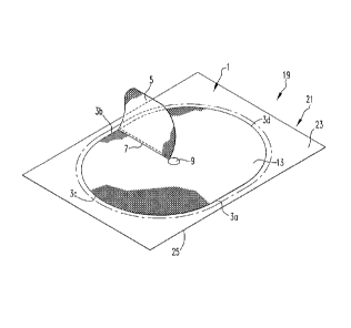
[0024] With reference to FIG. 4, a device, denoted generally as reference
numeral 19, for
performing surgery comprises a mesh patch 1 as described hereinabove; and a
removable
cover, denoted generally as reference numeral 21.
[0025] While the mesh patch configuration is discussed with respect to a patch
for hernia
repair, one skilled in the art will understand that the shape of the patch can
vary, depending
upon the surgical procedure to be performed, and may or may not include a
flap. The mesh
patch 1 comprises a top surface 13 and a bottom surface 15 opposite the top
surface 13. In
some non-limiting embodiments, mesh patch 1 can have a generally ellipse-like
shape that
includes a lower edge 3a and an upper edge 3b with a large radius of curvature
and two
lateral edges 3c, 3d with a small radius of curvature. A slit 7 is positioned
perpendicular to
upper edge 3b and extending substantially over half of the width of mesh patch
1. The inner
end of slit 7 opens into an orifice 9. A flap 5 having a shape of a sector of
a circular annulus
is also provided and is connected via one of its radial edges to one of the
edges of mesh patch
1 which delimits the slit 7.
-5-
CA 02761312 2011-11-07
WO 2010/129641 PCT/US2010/033673
[0026] Mesh patch 1 can be made of a sheet of low-weight monofilament
polyester knitted
fabric 11 having a top surface 13 and a bottom surface 15. However, this is
not to be
construed as limiting the present invention as any suitable material, for
example, a plastic
material such as polypropylene, may be used. Mesh patch 1 also comprises a
plurality of
hooks 17. In addition, polyester knitted fabric 11 incorporates bioresorbable
polylactic acid
(PLA) micro-hooks 17 on bottom surface 15 thereof.
[0027] Removable cover 21 has a top side 23 and a bottom side 25. Top side 23
of cover
21 is configured to be positioned adjacent to and in facing engagement with
bottom surface
15 of mesh patch 1. Bottom side 25 is configured to be positioned adjacent to
a surgical site
as will be discussed in greater detail hereinafter. Removable cover 21 may be
of any suitable
size such that it completely covers bottom surface 15 of mesh patch 1. For
example, if mesh
patch 1 has an elliptical shape that is 12 cm by 8 cm, the removable cover 21
may be of a
rectangular or elliptical shape that is 15 cm by 9 cm. In addition, removable
cover 21 may
have a thickness in the range of 0.005 nun to 1 mm.
[0028] Removable cover 21 is provided as a layer of sterile, smooth polymeric
material
such as a polymer film manufactured from polyolefins, such as polypropylene.
Desirably,
removable cover 21 is prepared from the same material as the inner, sterile
liner of esteem
SMT non-porous, powder-free, latex-free surgical gloves distributed by
Cardinal Health,
McGaw Park, IL 60085. As shown in FIG. 4, device 19 may be manufactured and
sold as a
combination. Alternatively, mesh patch 1 and removable cover 21 may be sold as
individual
components and used in combination.
[0029] In addition, and as shown in FIG. 5, mesh patch 1 and removable cover
21 may be
sold in a kit, denoted generally as reference numeral 27. Kit 27 includes mesh
patch 1 and
removable cover 21 provided in a package 29. Package 29 may also include one
or more
additional sheet(s) 31 of sterile, polymeric material configured to be cut
into pieces by a
surgeon to line the surgical site prior to positioning the combination of
removable cover 21
and mesh patch 1. Sheet 31 can be prepared from any sterile surgical polymer
material, and
can be prepared from the same polymeric material as removable cover 21, if
desired. While
an exemplary embodiment of kit 27 is illustrated in FIG. 5, this is not to be
construed as
limiting the present invention as any number of configurations for kit 27 has
been envisioned.
[0030] With reference to FIGS. 6-14, and with continuing reference to FIGS. 4
and 5, the
method for repairing an inguinal hernia using device 19 and/or kit 27 of the
present invention
is described hereinafter. While the following description is provided for the
repair of an
inguinal hernia, the device of the present invention is not limited to use in
repairing inguinal
-6-
CA 02761312 2011-11-07
WO 2010/129641 PCT/US2010/033673
hernias. Different shapes and sizes of mesh patch 1 and removable cover 21 may
be provided
to perform various types of surgery where mesh patches are used, such as
laparoscopic
ventral hernia repair, diaphragmatic hernia repair, and soft tissue
reconstruction.
[0031] The repair of an inguinal hernia in accordance with the procedure of
the present
invention begins with the cutting of a transverse oblique incision 33 in a
fold of a groin of a
patient 35. Thereafter, a superficial inguinal space of patient 35 is widely
dissected, thereby
freeing fascial surfaces 37 and creating a surgical site 39 (see FIG. 7).
Next, the spermatic
cord 41 of patient 35 is mobilized using a latex band 43 (see FIG. 8). In some
instances, latex
band 43 may be covered with a piece of polymeric material cut from additional
sheet 31.
[0032] The surgeon then identifies a hernia sac (indirect hernia) or a hernia
bulge (direct
hernia), and repairs the hernia. Thereafter, a relaxing incision 51 in the
internal oblique fascia
is made by the surgeon to reduce tension in this area. For an indirect hernia,
the surgeon
tightens the internal inguinal ring with an absorbable stitch. For a direct
hernia, the hernia sac
45 is imbricated to temporarily reduce the hernia bulge and then the superior
and medial
portion of the transverse layer is drawn downward and sutured to the superior
pubic ligament
as denoted by reference numeral 47 and to the anterior femoral sheath as
denoted by
reference numeral 49 (see FIG. 9).
[0033] As shown in FIG. 10, the surgeon then increases the size of surgical
site 39 using a
flag retractor 53 and a Gelpi retractor 55. In addition, at this point in the
surgical procedure,
the surgeon also may line surgical site 39 with pieces cut from additional
sheet 31 of
polymeric material. Then, device 19 of the present invention is positioned
over surgical site
39 such that bottom side 25 of removable cover 21 is adjacent to surgical site
39 (see FIGS.
11 and 12). As discussed hereinabove, device 19 may be provided as a
prepackaged unit, as a
kit, or as separate cover 21 and mesh patch 1 components.
[0034] Removable cover 21 covers micro-hooks 17 until the surgeon maneuvers
mesh
patch 1 into a proper position, thereby avoiding accidental exposure of micro-
hooks 17 to
prevent premature fixation to structures that destroy the fragile nature of
micro-hooks 17. A
slit 56 may be cut into cover 21 to correspond with slit 7 provided in mesh
patch 1. Flap 5 is
folded on itself and held temporarily open with an Alice clamp 57. Alice clamp
57 looks like
a hemostat but has "c"-shaped tips that only contact a very small surface area
so as to not
crush too many micro-hooks 17.
[0035] Next, the surgeon directs mesh patch 1 medially over the pubic tubercle
59 such
that slit 7 of mesh patch 1 is toward the patient's head and flap 5 is open.
Once mesh patch 1
is in position, removable cover 21 is slid laterally in the direction of arrow
A to expose
-7-
CA 02761312 2011-11-07
WO 2010/129641 PCT/US2010/033673
bottom surface 15 and micro-hooks 17 of mesh patch 1 to surgical site 39 (see
FIG. 13). The
surgeon then gently presses mesh patch 1 into pubic tubercle 59 overlapping
the inguinal
ligament, and extends mesh patch 1 to cover relaxing incision 51. Desirably,
an assistant
exerts an upward pressure on the surrounding tissue with a Richardson (blunt)
retractor (not
shown). Thereafter, the surgeon continues to slide cover 21 laterally along
the inguinal
ligament in the direction of arrow A while moving the retractor to the lateral
border of
incision 33. Once edges 3a and 3d of mesh patch 1 have been fixed, the
remainder of mesh
patch 1 is properly positioned around spermatic cord 41. In some instances,
the surgeon may
need to place stitches along the border of the inguinal ligament to assure
mesh patch 1 does
not move.
[0036] The final step in positioning mesh patch 1 is to remove Alice clamp 57
and to close
flap 5 around spermatic cord 41, thus creating a new internal ring that is
completely tension
fee and custom made (see FIG. 14). Once mesh patch 1 is in place, all pieces
of additional
sheet 31 that may have been placed in incision 33 are removed along with any
latex bands
(around the spermatic cord). The external fascial surfaces 37 are then closed
with an
absorbable stitch, and the skin is also closed with an absorbable stitch.
[0037] Although the invention has been described in detail for the purpose of
illustration
based on what is currently considered to be the most practical embodiments, it
is to be
understood that such detail is solely for that purpose and that the invention
is not limited to
the disclosed embodiments, but, on the contrary, is intended to cover
modifications and
equivalent arrangements. For example, it is to be understood that the present
invention
contemplates that, to the extent possible, one or more features of any
embodiment can be
combined with one or more features of any other embodiment.
-8-