Note: Descriptions are shown in the official language in which they were submitted.
CA 02765419 2011-12-13
WO 2009/151645 PCT/US2009/003567
-1-
WOUND TREATMENT APPARATUS AND METHOD
Background and Summary of the Invention
[0001] The present invention relates to a wound treatment apparatus and
method. More
particularly the present invention relates to a wound treatment apparatus and
method using
negative pressure therapy to treat a wound.
[0002] The use of negative pressure for treating wounds is well known.
The apparatus of
the present invention provides improvements over conventional negative
pressure wound
treatment devices.
[0003] For illustrative purposes, the wound treatment apparatus and method
of the
present invention may be used to treat the following exemplary conditions:
1. Acute surface wounds, chronic surface wounds and wounds that reopen
after
initial closure.
2. Sinus tract, tunnel or fistula located on the surface of the body.
3. Removing edema from wounds/periwound tissue after surgery.
4. Treating skin and deep tissue injury secondary to burns (after patient
stabilization and wound debridenient).
5. Positional stabilization of skin flaps and grafts.
[0004] In an illustrated embodiment of the present disclosure, a wound
treatment
apparatus includes a negative pressure source; a bandage configured to cover a
wound and
provide a sealed region around the wound; a drainage tube coupled to the
bandage and the
negative pressure source to drain fluid from the wound; and a controller
coupled to the
negative pressure source. The apparatus also includes at least one of an
oxygen sensor, a
carbon dioxide sensor, a pH sensor, and a temperature sensor in fluid
communication with the
drainage tube to provide at least one of an oxygen saturation level of fluid
drained from the
wound, a carbon dioxide level fluid drained from the wound, a pH level of
fluid drained from
the wound, and a temperature of fluid drained from the wound, respectively.
[0005] In an illustrated embodiment, the at least one sensor is coupled
to the controller so
that the controller controls the negative pressure source based at least one
output signal from
the at least one sensor. The controller may be configured to adjust the
negative pressure
CA 02765419 2011-12-13
WO 2009/151645 PCT/US2009/003567
-2-
source based the at least one output signal from the at least one sensor to
maintain at least one
of the oxygen saturation level, the carbon dioxide level, the pH level, and
the temperature of
the wound at substantially a desired level. In one embodiment, the oxygen
sensor, the carbon
dioxide sensor, the pH sensor, and the temperature sensor are all
simultaneously in fluid
communication with the drainage tube, the oxygen sensor, the carbon dioxide
sensor, the pH
sensor, and the temperature sensor being coupled to the controller so that the
controller
controls the negative pressure source based output signals from the sensors.
[0006] In another illustrated embodiment of the present disclosure, a
method of treating a
wound includes providing a wound treatment apparatus having a negative
pressure source, a
bandage configured to cover the wound and provide a sealed region around a
perimeter of the
wound, at least one drainage tube coupled to the bandage and the negative
pressure source, a
controller coupled to the negative pressure source, and a data collector
coupled to the at least
one drainage tube and the controller. The method also includes transmitting
data collected by
the data collector from the controller of the wound treatment apparatus to a
caregiver's
computer and/or an insurance company's computer at a remote location via a
communication
network. In one embodiment, the method includes receiving control instructions
with the
controller of the wound treatment apparatus from the caregiver's computer at
the remote
location via the communication network, the control instructions being used by
the controller
to adjust a therapy applied to the wound by the wound treatment apparatus. In
another
embodiment, the method includes receiving a reimbursement authorization from
the
insurance company's computer at the controller of the wound treatment
apparatus via the
communication network, and beginning a treatment therapy using the controller
of the wound
treatment apparatus after receipt of the authorization from the insurance
company's computer.
[0007] Additional features of the present invention will become apparent
to those skilled
in the art upon consideration of the following detailed description of
illustrative embodiments
exemplifying the best mode of carrying out the invention as presently
perceived.
Brief Description of the Drawings
[0008] The above-mentioned and other features of this invention, and the
manner of
attaining them, will become more apparent and the invention itself will be
better understood
CA 02765419 2011-12-13
WO 2009/151645
PCT/US2009/003567
-3-
by reference to the following description of illustrated embodiments of the
invention taken in
conjunction with the accompanying drawings, wherein:
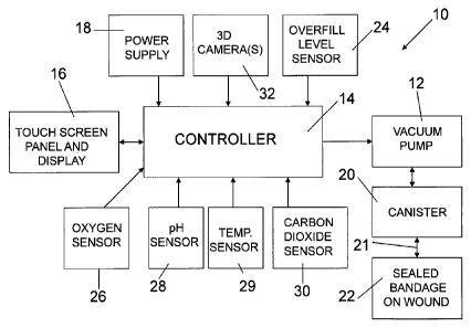
[0009] Fig. 1 is a block diagram illustrating components of the wound
therapy apparatus
of the present invention;
[0010] Fig. 2 is a block diagram illustrating communication between the
wound therapy
apparatus of Fig. 1, a caregiver's computer and an insurance company's
computer;
[0011] Fig. 3 is a block diagram illustrating operation of the wound
treatment apparatus
of the present invention; and
[0012] Figs. 4-8 are illustrative screen displays on a touch screen
control panel of the
wound treatment apparatus.
Detailed Description of the Drawings
[0013] For the purposes of promoting an understanding of the principles
of the invention,
reference will now be made to certain illustrated embodiments and specific
language will be
used to describe the same. It will nevertheless be understood that no
limitation of the scope
of the invention is thereby intended. Alterations and modifications of the
invention and such
further applications of the principles of the invention as described herein as
would normally
occur to one skilled in the art to which the invention pertains, are
contemplated, and desired
to be protected.
[0014] Referring now to the drawings, Fig. 1 is a block diagram of
components of a
wound treatment apparatus 10 of the present invention. The wound treatment
apparatus
includes a vacuum pump 12 or other suitable negative pressure source
controlled by a
controller 14 which illustratively includes a microprocessor. The controller
14 is
illustratively coupled to a touch screen panel and display 16. Therefore, the
controller 14
may display a plurality of menu options for the user on the touch screen
display. The user
may enter input commands into the controller using the touch screen panel 16.
The touch
screen panel 16 and display facilitates operation of the wound treatment
apparatus 10.
CA 02765419 2011-12-13
WO 2009/151645
PCT/US2009/003567
-4-
[0015] A power supply 18 supplies power to the controller 14 and vacuum
pump 12. The
power supply is illustratively a 12V DC power supply supplied either from a
wall outlet or a
backup battery.
[0016] Vacuum pump 12 is coupled to a canister 20 and to a sealed bandage
22 on a
wound. In one illustrated embodiment, a Chariker-Jeter bandage is used to
provide a sealed
bandage 22 on the wound. Drainage from the wound under the sealed bandage 22
is
collected in the canister 20. Bandage 22 illustratively includes antimicrobial
gauze
surrounded by a transparent adhesive dressing. A suction or drainage tube 21
includes a first
end portion located below the transparent adhesive dressing and within the
gauze and a
second end portion connected to the canister 20. It is understood that
multiple bandages 22
may be coupled to the same pump 12, if desired.
[0017] In an illustrated embodiment, a housing of vacuum pump includes an
integral
overfill level sensor 24. Illustratively, the overfill level sensor 24 is a
capacitive sensor
having an output coupled to controller 14. In the illustrated embodiment,
canister 20 is
mounted directly to a housing of the vacuum pump 12 adjacent the overfill
level sensor 24.
Therefore, as fluid fills the canister 20, overfill level sensor 24 detects a
full condition and
communicates with controller 14 to shut off the vacuum pump 12. An illustrated
embodiment of the canister 20 is generally opaque except for sight glasses on
opposite sides
of the canister 20 to permit a visual inspection of the level of fluid in the
canister 20. The
canister 20 is easily attached via a "slide-on" connection in which the
plurality of mounting
pegs fixed to the housing of the vacuum pump 12 engage slots formed in the
canister 20 to
secure the canister 20 to the pump 12. Other coupling techniques for the
canister 20 may also
be used.
[0018] In an illustrated embodiment, the wound treatment apparatus 10
includes an
oxygen sensor 26, a pH sensor 28, a temperature sensor 29, and a carbon
dioxide sensor 30
coupled to the controller 14. In the illustrative embodiment, the oxygen
sensor 26 and carbon
dioxide sensor 30 measure an oxygen saturation level and a carbon dioxide
saturation level
from the drainage tube 21 coupled to the bandage 22. Sensors 26, 28, 29 and 30
may also be
located in the canister 20. In an illustrated embodiment, the pH sensor 28
also measures the
pH of fluids drained from the wound through drainage tube 21. Temperature
sensor 29 also
CA 02765419 2011-12-13
WO 2009/151645
PCT/US2009/003567
-5-
measures the temperature of drainage fluid. Therefore, readings from oxygen
sensor 26, pH
sensor 28, temperature sensor 29, and carbon dioxide sensor 30 provide actual
sensor
readings from the wound cavity. Controller 14 monitors the oxygen, pH,
temperature and
carbon dioxide levels readings and may adjust or regulate the pump pressure
automatically
based on these levels to improve treatment of the wound.
[0019] In an illustrated embodiment, the caregiver may set desired
levels for the oxygen
saturation level from oxygen sensor 26, the pH level from pH sensor 28, the
temperature
from temperature sensor 29, and the carbon dioxide level from carbon dioxide
sensor 30.
Based on the preset levels and the output signals from the sensors 26, 28, 29,
30, the
controller may adjust the negative pressure applied by the vacuum pump 12 to
the bandage
22 to maintain the oxygen saturation level, the pH level, the temperature,
and/or the carbon
dioxide level substantially at the preset levels or within predetermined
ranges. Threshold
levels for each of the sensors 26, 28, 29, 30 may also be set. Therefore, an
alarm may be
issued by the controller 14 to alert the caregiver if the oxygen saturation
level, pH level,
temperature, and/or carbon dioxide level cross the preset threshold levels. In
one
embodiment, the alarm is transmitted to the caregiver's computer 36 through
the
communication network 34 as discussed below so that the caregiver is alerted
to check
operation of the wound treatment apparatus 10. In certain embodiments, the
controller may
not automatically adjust the pressure as discussed above based on the sensor
readings.
Instead, the sensor readings are provided to the caregiver either at the wound
treatment
apparatus 10 location or a remote location, and the caregiver then manually
makes
adjustments to the settings of the wound treatment apparatus 10 to adjust
therapy applied to
the wound.
[0020] In an illustrated embodiment, a 3-D camera 32 is provided to take
images of a
wound to help with wound assessment. Camera 32 provides images to controller
14. The 3-
D camera 32 illustratively takes pictures of a wound to read a depth, a
length, a width and a
color of the wound. In the illustrative embodiment, two cameras 32 may be used
to generate
a 3-D model of the wound including any undermining or tunnels which may be
formed
beneath the surface of the wound. In certain embodiments, a laptop computer
may be
coupled to the controller 14 to provide additional processing power to
generate the 3-D
CA 02765419 2011-12-13
WO 2009/151645
PCT/US2009/003567
-6-
models. In another embodiment, image data from 3-D cameras 32 may be
transmitted to a
remote location for processing as discussed below.
[0021] Fig. 2 illustrates the wound treatment apparatus 10 coupled to a
communication
network 34. Communication network 34 may be any suitable wired or wireless
network,
such as, for example, cell phone networks, local area networks, wide area
networks such as
the Internet, or other suitable network for transmitting data from one
location to another.
Therefore, the controller 14 of wound treatment apparatus 10 can send data and
receive data
from a caregiver's computer 36 and an insurance company's computer 38 located
at remote
locations. Illustratively, the controller 14 of wound treatment apparatus 10
may send the data
from 3-D camera 32 over communication network 34 to a caregiver's computer 36
or the
insurance company's computer 38. The 3-D camera data may be processed to
determine the
depth, length, width and color of the wound. The insurance company can review
the images
and make a determination regarding insurance company reimbursement for the
therapy based
on the images. The controller 14 may receive a reimbursement authorization
from the
insurance company's computer via the communication network and begin a
treatment therapy
after receipt of the authorization from the insurance company's computer.
[0022] In another embodiment, data such as oxygen saturation level from
sensor 26, pH
level from sensor 28, temperature from temperature sensor 29, carbon dioxide
data from
sensor 30, wound images from 3-D camera 32, or other data may be sent to a
caregiver's
computer 36 at remote a location. The caregiver may then review the data
related to the
particular wound being treated and adjust the therapy from the remote
caregiver's computer
36. In other words, instructions can be sent from caregiver's computer 36 over
the
communication network 34 to the wound treatment apparatus 10 to adjust the
therapy
provided to the particular patient. This enables caregivers at a remote
location to monitor a
plurality of different patients at remote settings or in the patient's home
and adjust the therapy
applied by wound treatment apparatus 10, if needed.
[0023] The end of a particular wound treatment cycle or therapy session
also provides a
time to change the bandage 22. The wound treatment apparatus 10 stores the
elapsed therapy
time including any time that the apparatus was shut off during a therapy
session in a memory.
Wound treatment apparatus 10 also stores the actual pressure applied to the
wound during the
CA 02765419 2011-12-13
WO 2009/151645
PCT/US2009/003567
-7-
therapy session and the readings from sensors 26, 28, 29 and 30 during the
therapy session.
At the end of the therapy session, the data from the therapy session may be
automatically sent
to the caregiver's computer 36 or the insurance company's computer 38 via an e-
mail or other
transmission if the wound treatment apparatus 10 is coupled to the
communication network
34. Therefore, the caregiver and insurance company can review the therapy
session data and
make an assessment regarding treatment of the wound. The data also provides an
accurate
historical database of the therapy sessions. A database may be stored
including data for
treating wounds, as well as image data from the 3-D camera 32 showing results
of the
treatment, from a plurality of therapy sessions from a plurality of different
patients. By
analyzing the database, caregivers and insurance companies may determine the
most effective
treatment times, pressure levels, or the like for particular types of wounds
by analyzing actual
results received from a plurality of patients stored in the database.
Therefore, new treatment
protocols may be developed based on the stored data in the database.
[0024] In one embodiment, a size of the wound may be determined using the
images
obtained by the 3-D camera 32. Controller 14 may calculate a surface area of
the wound
using the size as determined by the 3-D camera images. Controller 14 may then
adjust the
negative pressure applied by the vacuum pump 12 based on the size of the
wound. For
example, as a wound heals and becomes smaller, the initial pressure setting
may actually
apply a greater pressure to the wound than desired causing discomfort to the
patient.
Therefore, the controller 14 may automatically reduce the pressure applied by
the vacuum
pump 12 as the wound size decreases to maintain the effective pressure applied
to the wound
surface area substantially constant as the wound gets smaller. If the wound
gets bigger or
otherwise changes, the controller 14 may automatically increase the pressure
applied by the
vacuum pump 12 to maintain the effective pressure applied to the wound surface
area
substantially constant as the surface area of the wound gets larger.
[0025] Fig. 3 illustrates a software flow chart for control of the wound
treatment
apparatus of the present invention.
[0026] Controller 14 monitors operation of the vacuum pump 12 and the
drainage tube 21
from the bandage 22 to determine if a leak has occurred in the drainage tube
21 or the
bandage 22. If a leak is detected, a visual and/or audio alarm is activated
CA 02765419 2011-12-13
WO 2009/151645
PCT/US2009/003567
-8-
[0027] The wound treatment apparatus 10 of the present invention is
illustratively
controlled by a microprocessor of controller 14 with fully operational touch
screen interface.
The apparatus 10 adapts easily for use in a general hospital, clinic,
outpatient or homecare
environment. One illustrated embodiment includes a piston vacuum pump 12
utilizing a
microprocessor controller 14 with a custom vacuum feedback device. The vacuum
pump
motor is illustratively controlled via a proportional duty cycle DC drive. The
motor speed is
mathematically calculated based on current vacuum pressure (mmHg) and the rate
of change
of the vacuum until the desired vacuum set point is achieved. This algorithm
of the present
invention allows the desired negative pressure set point to be maintained
within a very close
tolerance (such as, for example, +/- 2 mmHg pressure variance) without the
need for
mechanical regulators. It also allows the pump 12 to implement an auto clamp
feature on the
vacuum so that the vacuum value does not exceed 200 mmHg. This auto clamp
feature
reduces the likelihood that the patient will receive unwanted sudden
alterations in vacuum
pressures, and is based on the desired vacuum pressure set point, not a
mechanical safety with
only one setting.
[0028] In an illustrative embodiment, a pressure sensor monitors pressure
within the
canister 20 at a very fast sampling rate so that the negative pressure level
applied to the
wound by the vacuum pump 12 is generally continuously monitored by the
controller 14. If
the actual negative pressure level measured by the pressure sensor within the
canister varies
from the desired preset level, controller 14 adjusts the vacuum pump 12 to
change the
negative pressure accordingly in order to maintain the very close tolerance to
the actual preset
pressure as discussed above. In one illustrated embodiment, the tolerance is
maintained at
about a +/- 2 mmHg pressure variance as discussed above. In another
illustrated
embodiment, the tolerance is maintained at about a +/- 1 mmHg pressure
variance. In yet
another illustrated embodiment, the tolerance is maintained at about a +/- 'A
mmHg pressure
variance. Such real time feedback control of the pressure within the canister
20 reduces
pulsation applied by the vacuum pump 12 to the wound which may occur in
conventional
wound treatment devices.
[0029] The apparatus 10 of the present invention is easy to operate due
to the fully
operational touch screen 16. Important operational information, such as
negative pressure
therapy set point, alarms due to canister full, blocked filter, and low
battery are in an easy to
CA 02765419 2011-12-13
WO 2009/151645
PCT/US2009/003567
-9-
understand format. The therapy terminates automatically when certain alarm is
triggered to
protect the unit. Items like Run and Stop switches are configured as easy to
use touch cells,
not mechanical switches with contacts that are prone to failure. While other
vacuum pumps
must run at full speed continuously to maintain their negative pressure set
point, the pump 12
of the present apparatus 10 runs at the minimum speed required to maintain the
vacuum level
setting. This makes these units environmentally friendly by reducing the
amount of energy
needed to provide therapy. Equally important is that the units are lighter and
more portable
since less energy needs allows for a smaller, lighter battery and thus
smaller, lighter device.
The battery of the apparatus may illustratively be charged in about 15 minutes
and can
maintain its charge for about 6 to 8 hours.
[0030] Figs. 4-8 illustrate screen displays on touch screen panel 16
which permit a user to
control operation of the wound treatment apparatus 10. The apparatus 10 may be
password
protected. After entering a password and patient's identification number using
a keyboard
screen (not shown) on the touch screen panel, the user enters the total
therapy time for the
wound treatment apparatus as illustrated in Fig. 4. The total therapy time is
the total number
of hours for one treatment. A user presses the SET HOURS button 50 shown in
Fig. 4 to
enter the number of hours for treatment. Illustratively, a numerical keypad
screen (not
shown) will be displayed on touch screen 16 when the SET HOURS button 50 is
pressed.
User uses the keypad to enter the total therapy time. Illustratively, the
total therapy time is
between 1 and 255 hours, although any number of hours may be used. The total
therapy time
entered by the user is illustratively displayed at location 52 on screen 16.
When the total
elapsed therapy time reaches the entered total therapy time, the apparatus 10
automatically
stops the therapy. In an illustrated embodiment, the apparatus will
automatically set the total
therapy time to 255 hours as a default if the total therapy time is zero. The
user can press the
arrow 54 to advance to the next screen.
[0031] Next, a user inputs the desired negative pressure setting using
the screen 16 shown
in Fig. 5. The user may press the SET VACUUM button 56 to enter a set point
for the
negative pressure. If the wound treatment apparatus 10 provides variable
therapy, the SET
VACUUM button 56 sets the high negative pressure value. The SET LOW VACUUM
button 58 is then pressed to input the low vacuum level for variable therapy
using the keypad
screen as discussed above. Illustratively, the vacuum set point may be set
between 20 mmHg
CA 02765419 2011-12-13
WO 2009/151645 PCT/US2009/003567
-10-
and 185 mmHg. Once the user saves the vacuum set points, the set points are
displayed at
locations 60 and 62 shown in Fig. 5.
[0032] Fig. 6 illustrates the touch screen 16 for selecting between a
continuous therapy
mode and a variable therapy mode. Illustratively, user presses button 64 for
the continuous
therapy mode and button 66 for the variable therapy mode. If button 66 is
pressed, screen 16
shown in Fig. 7 appears on the touch screen display. The user then uses
sliders 68 and 70 to
adjust the time for the high negative pressure settings and the low negative
pressure settings.
Other suitable means such as the keypad screen may also be used for data
entry.
Illustratively, the high and low settings are between one and ten minutes. The
selected values
for the high and low intervals are displayed at locations 72 and 74,
respectively, in Fig. 7.
[0033] An illustrative operation screen for the wound treatment
apparatus 10 is shown in
Fig. 8. The actual negative pressure is displayed at location 76. The high and
low settings
are displayed at locations 78 and 80, respectively. An oxygen percentage is
displayed at
section 82. In order to reduce the likelihood that a patient may change the
settings, there is a
hidden button illustratively located in the right bottom corner and shown by
dotted lines 84.
Pressing the hidden button 84 will make all the controller buttons disappear.
Pressing hidden
button 84 again will make the controls reappear. When the system is running, a
STOP button=
86 is displayed. When the system is stopped, a RUN button is displayed at
location 86. The
apparatus 10 illustratively displays the current therapy status (either on or
off) and whether or
not the apparatus 10 is in continuous or variable mode. The apparatus 10 also
displays the
Elapsed Therapy Time (ETT) on display screen 16. The controller 14 keeps track
of elapsed
therapy time during normal operation of the pump 12. If the patent stops the
therapy, the
timer stops. In other words, if therapy is stopped by pressing the STOP button
86, the
elapsed therapy time is paused until the user presses the RUN button to
continue the therapy.
Therefore, the caregiver can determine whether or not the patient followed the
prescribed
treatment regimen since the caregiver's last visit based on the timer.
Illustratively, a message
such as "Therapy is completed" is displayed on screen 16 when the elapsed
therapy time
reaches the entered total therapy time. The user may press the menu button 90
to stop
therapy and return to the patent identification screen or other location.
CA 02765419 2011-12-13
WO 2009/151645
PCT/US2009/003567
-11-
[0034] If any alarm is triggered, the related alarm message is displayed
at location 88. In
an illustrated embodiment, the alarm message is provided on the screen under
the following
conditions:
ALARM TYPE NOTIFICATION
REMEDY
Visual message "High Vacuum" with audible
alarm when the actual negative pressure is 15
mmHg higher than the set point. Device will Push stop button and
restart.
High Vacuum automatically stop pumping when the set If alarm
persists, discontinue
point is reached. Device resumes pumping use and contact
manufacturer.
automatically when the vacuum goes back
within the proper set point range.
Visual message "Canister Full" with audible
Change canister and restart
Canister Full alarm when canister is full; NPWT stops
NPWT .
automatically.
Visual message "Blocked Filter" with audible
Replace the filter and restart
Blocked Filter alarm when filter is blocked; NPWT stops
NPWT.
automatically.
Visual message "Air Leak" with audible Check dressing,
canister,
Air Leak
alarm after leakage is detected. filter and tubing
for leaks.
Visual message "Low Battery" and audible Connect device to AC
power
Low Battery alarm (beeps once per minute when low source and turn
on device to
battery detected). recharge the
battery.
Visual message "No Activity - Turn Therapy
On" with audible alarm after 15 minutes
No Activity Turn therapy ON.
without NPWT. (Only occurs when device
turned ON for the first time)
[0035] Illustrative Features of the Wound Treatment Apparatus Include:
> Pressure Controls: Solid state
> Pressure Setting: Adjustable
> Pressure Monitoring: Display actual negative pressure
> Filter: External back up filter with silicon tubing
> Oxygen Sensor: Monitoring the oxygen saturation of the wound as the
treatment parameters are changed (such as changing the intensity of the
negative
pressure, changing the wound contact layer, etc.) improves the wound's
response to
treatment.
> Carbon Dioxide Sensor: Monitoring a carbon dioxide saturation of the
wound
as the treatment parameters are changed (such as changing the intensity of the
CA 02765419 2011-12-13
WO 2009/151645
PCT/US2009/003567
-12-
negative pressure, changing the wound contact layer, etc.) improves the
wound's
response to treatment.
> pH: Monitoring the pH level of sludge coming out of the wound cavity.
> Alarms: Visual and audio alarms (High Vacuum, Blocked Filter, Air Leak,
Canister Full, No Activity and Low Battery)
> Therapy Mode: Continuous or Variable
> Battery Backup: Up to 6 hours
> Safety features: electronic and manual
> Overflow sensor: A capacitive sensor that monitors the fluid level in the
suction
canister. If the fluid level reaches the maximum, the unit will stop
automatically
to prevent the back-up fluid into the pump and patient.
> Overflow valve: A manual safety feature positioned in the cap of the
canister. If
fluid reaches the maximum level in the canister, the filter or float in the
overflow
valve will block the inlet port, which will stop the suction automatically.
The
"Blocked Filter" alarm is then activated and the unit is stopped.
Accessories:
Illustrative accessories which may accompany the apparatus of the present
invention
include:
> A Fluid Collection Bottle/Canister: Lg. 800 ml, Med. 500 ml, Sm. 250m1
> Filter
> Connection Tube with Adaptor Connector
> 10 mm Flat Wound Drain
> Transparent Adhesive Dressing (Large, Medium and Small)
> Antimicrobial Gauze (Large, Medium and Small)
> Tubing Clamps (Large and Small)
> Tubing Caps
Wound Drainage Kit Instructions
[0036] A medical team and doctor should make an individual assessment of
how to apply
the wound drainage kit.
These general instructions should be followed:
1. Irrigate the wound thoroughly with normal saline using a syringe.
CA 02765419 2016-09-08
=
- 13 -
2. Wipe off any excess saline that might have spilled onto the outer skin;
for dressing prep.
3. Measure wound for depth, tunneling and undermining, use the clock
method, when
assessment of wound is be made.
4. Measure deepest area of wound, shorten length of 10 mm silicone drain
tube (21)
accordingly, 1 cm less than deepest area in wound bed, tunneling or
undermining.
5. Apply antimicrobial sponge to entire wound bed, cover tip of lOmm
silicone drain with
antimicrobial gauze, place drain in wound bed. The drain should not be placed
in the fistula tract.
6. Select proper size dressing, allow at least one inch of intact skin
beyond wound edges.
Place dressing over packed wound, crimp dressing around tubing and seal.
7. Set Negative Pressure Wound Therapy Unit, (NPWT) to recommended
settings;
(approximately 60-80mmHg), at continuous suction, observe the wound site. The
dressing should
contract immediately.
8. Check for leaks; listen for sounds, sounds indicate a leak in the
dressing. Pat down lightly
with your hand around the area where there is sound, until the sound is gone.
Again observe the
wound dressing, it should be completely contracted.
If the wound is irregular and there is a chance of leakage, check for dressing
integrity, each
shift change by occluding suction tubing. If dressing balloons, than contracts
after releasing the
tubing, check dressing for leaks.
100371 In an illustrated embodiment, the wound treatment apparatus may include
a security system.
Each apparatus 10 is illustratively assigned a serial number. Before
operation, the user or caregiver
must access a manufacturer's computer 40 (such as via a web site) and enter
the serial number to
activate the apparatus 10. Manufacturer's computer 40 illustratively sends an
access or activation
code to the apparatus 10 which must be entered via the touch screen 14 or
other input device to
begin operation. This permits the manufacturer to monitor operation of the
apparatus for billing,
servicing or other purposes from a remote location.
[0038] The scope of the claims should not be limited by the preferred
embodiments set forth in the
examples, but should be given the broadest interpretation consistent with the
description as a whole.