Note: Descriptions are shown in the official language in which they were submitted.
CA 02766192 2016-11-22
OCULAR IMPLANTS FOR RESIDING PARTIALLY IN
SCHLEMM'S CANAL
FIELD OF THE INVENTION
15 [0003] The present invention relates generally to devices that are
implanted within the eye.
More particularly, the present invention relates to systems, devices and
methods for delivering
ocular implants into the eye.
BACKGROUND OF THE INVENTION
20 [00041 According to a draft report by The National Eye Institute
(NEI) at The United States
National Institutes of Health (NIH), glaucoma is now the leading cause of
irreversible blindness
worldwide and the second leading cause of blindness, behind cataract, in the
world. Thus, the
NEI draft report concludes, "it is critical that significant emphasis and
resources continue to be
devoted to determining the pathophysiology and management of this disease."
Glaucoma
25 researchers have found a strong correlation between high intraocular
pressure and glaucoma. For
this reason, eye care professionals routinely screen patients for glaucoma by
measuring
intraocular pressure using a device known as a tonometer. Many modem
tonometers make this
measurement by blowing a sudden puff of air against the outer surface of the
eye.
[00051 The eye can be conceptualized as a ball filled with fluid. There
are two types of fluid
30 inside the eye. The cavity behind the lens is filled with a viscous
fluid known as vitreous humor.
The cavities in front of the lens are filled with a fluid know as aqueous
humor. Whenever a
person views an object, he or she is viewing that object through both the
vitreous humor and the
aqueous humor.
- 1 -
CA 02766192 2011-12-20
WO 2011/006113
PCT/US2010/041589
[0006] Whenever a person views an object, he or she is also viewing that
object through the
cornea and the lens of the eye. In order to be transparent, the cornea and the
lens can include no
blood vessels. Accordingly, no blood flows through the cornea and the lens to
provide nutrition
to these tissues and to remove wastes from these tissues. Instead, these
functions are performed
by the aqueous humor. A continuous flow of aqueous humor through the eye
provides nutrition
to portions of the eye (e.g., the cornea and the lens) that have no blood
vessels. This flow of
aqueous humor also removes waste from these tissues.
[0007] Aqueous humor is produced by an organ known as the ciliary body.
The ciliary body
includes epithelial cells that continuously secrete aqueous humor. In a
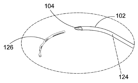
healthy eye, a stream of
aqueous humor flows out of the anterior chamber of the eye through the
trabecular meshwork
and into Schlemm's canal as new aqueous humor is secreted by the epithelial
cells of the ciliary
body. This excess aqueous humor enters the venous blood stream from Schlemm's
canal and is
carried along with the venous blood leaving the eye.
[0008] When the natural drainage mechanisms of the eye stop functioning
properly, the
pressure inside the eye begins to rise. Researchers have theorized prolonged
exposure to high
intraocular pressure causes damage to the optic nerve that transmits sensory
information from the
eye to the brain. This damage to the optic nerve results in loss of peripheral
vision. As
glaucoma progresses, more and more of the visual field is lost until the
patient is completely
blind.
[0009] In addition to drug treatments, a variety of surgical treatments for
glaucoma have
been performed. For example, shunts were implanted to direct aqueous humor
from the anterior
chamber to the extraocular vein (Lee and Scheppens, "Aqueous-venous shunt and
intraocular
pressure," Investigative Ophthalmology (Feb. 1966)). Other early glaucoma
treatment implants
led from the anterior chamber to a sub-conjunctival bleb (e.g., US 4,968,296
and US 5,180,362).
Still others were shunts leading from the anterior chamber to a point just
inside Schlemm's canal
(Spiegel et al., "Schlemm's canal implant: a new method to lower intraocular
pressure in patients
with POAG?" Ophthalmic Surgery and Lasers (June 1999); US 6,450,984; US
6,450,984).
SUMMARY OF THE DISCLOSURE
[00010] The invention pertains to aspects of ocular implants, ocular implant
delivery systems,
and methods for delivering ocular implants. One aspect of the invention an
ocular implant
adapted to reside at least partially in a portion of Schlemm's canal of an
eye. In some
embodiments, the ocular implant includes a body having a first major surface
and a second major
surface, the body being curved about a longitudinal central axis so that the
first major surface
comprises a concave surface and the second major surface comprises a convex
surface, a distal
- 2 -
CA 02766192 2011-12-20
WO 2011/006113
PCT/US2010/041589
portion of the body defining a longitudinal channel including a channel
opening, the channel
opening being disposed diametrically opposite a central portion of the concave
surface, and the
body being adapted and configured such that the ocular implant assumes an
orientation in which
the channel opening is adjacent a major side of Schlemm's canal when the
ocular implant is
disposed in Schlemm's canal. The channel may opens away from the pupil of the
when the
channel opening is adjacent an outer major side of Schlemm's canal.
[00011] In some embodiments, the channel has a width and a depth and an aspect
ratio of the
width to the depth is such that the ocular implant assumes an orientation in
which the channel
opening is adjacent a major side of Schlemm's canal when the ocular implant is
disposed in
Schlemm's canal. In some useful embodiments, the aspect ratio of channel width
WD to channel
depth DP is greater than about one. In some particularly useful embodiments,
the aspect ratio of
channel width WD to channel depth DP is about two. In some useful embodiments,
the aspect
ratio of channel width WD to channel depth DP is greater than about two.
[00012] In some embodiments, the body has a first lateral extent, a second
lateral extent, and a
longitudinal length and an aspect ratio of the first lateral extent to the
second lateral extent is
such that the ocular implant assumes an orientation in which the channel
opening is adjacent a
major side of Schlemm's canal when the ocular implant is disposed in Schlemm's
canal. In some
useful embodiments, an aspect ratio of first lateral extent EF to second
lateral extent ES is
greater than about one. In some particularly useful embodiments, the aspect
ratio of first lateral
extent EF to second lateral extent ES is about two. In some useful
embodiments, the aspect ratio
of first lateral extent EF to second lateral extent ES is greater than about
two.
[00013] In some embodiments, a distal portion of the body of the ocular
implant extends
across an angular span of less than 180 degrees as the body curves about the
longitudinal central
axis. In some embodiments, the body defines additional openings fluidly
communicating with
the channel and the body of the implant is more than 50% open due to the
openings defined by
the body. In some embodiments, the body of the ocular implant has a diameter
of between about
0.005 inches and about 0.04 inches.
[00014] In some embodiments, the ocular implant comprises a therapeutic agent
deposited on
the body. In some of these embodiments, a therapeutic agent comprises an anti-
glaucoma drug.
The anti-glaucoma drug comprises a prostaglandin analog in some embodiments.
The
prostaglandin analog comprises latanprost in some embodiments.
[00015] In some embodiments, the body of the ocular implant has a thickness
extending
between the concave surface and the convex surface. The thickness of the body
is substantially
uniform along a length of the body in some embodiments. In some embodiments,
the thickness
of the body is substantially uniform along a circumference of the body.
- 3 -
CA 02766192 2011-12-20
WO 2011/006113
PCT/US2010/041589
[00016] In some embodiments, the body is curved about a lateral central axis
so that a
longitudinal axis of the body defines a plane. When this is the case, the body
has a lateral radius
of curvature extending between the lateral central axis and an outer extent of
the body. The
lateral radius of curvature is substantially constant in some embodiments. In
other embodiments,
the lateral radius of curvature varies along a length of the body.
[00017] Another aspect of the invention provides an ocular implant system for
treating an eye.
In some embodiments, the ocular implant system comprises a delivery cannula
comprising a
tubular member defining a distal opening, a proximal opening, and a passageway
extending
between the proximal opening and the distal opening. In some embodiments, the
delivery
cannula includes a curved portion disposed between the distal opening and the
proximal opening,
the delivery cannula being adapted and configured such that the distal opening
can be placed in
fluid communication with Schlemm's canal when the cannula is extending through
the cornea of
the eye and the curved portion of the cannula is at least partially disposed
in the anterior chamber
of the eye. In some embodiments, the implant system includes an ocular implant
disposed in the
passageway defined by the delivery cannula, the ocular implant comprising a
body having a first
major surface and a second major surface, the body being curved about a
longitudinal central
axis so that the first major surface comprises a concave surface and the
second major surface
comprises a convex surface, a distal portion of the body defining a
longitudinal channel
including a channel opening. In some useful embodiments, the ocular implant is
oriented
relative to the delivery cannula such that the channel of the ocular implant
opens in a radially
outward direction when the ocular implant passes through the curved portion of
the delivery
cannula.
[00018] An additional aspect of the invention provides another ocular implant
system for
treating an eye. In some embodiments, the ocular implant system comprises an
ocular implant
defining a plurality of openings and a sheath disposed about the body of the
ocular implant. In
some embodiments, the sheath covers at least some of the openings and the
sheath is adapted and
configured such that the sheath can be selectively removed from the body for
uncovering the
openings.
[00019] In some embodiments, the sheath comprises a proximal portion defining
a lumen and
a distal portion defining a distal aperture, the lumen having a lumen width
and the distal aperture
having an aperture width. The aperture width is smaller than the lumen width
in some
embodiments. The distal portion provides a transition from the lumen width to
the aperture
width in some embodiments. In some embodiments, the lumen width is equal to or
greater than
a width of the implant and the aperture width is smaller than the width of the
implant.
- 4 -
CA 02766192 2011-12-20
WO 2011/006113
PCT/US2010/041589
[00020] In some embodiments, the distal portion of the sheath comprises a
first region, a
second region, and a slit disposed between the first region and the second
region. The sheath
includes a frangible connection between the first region and the second region
in some
embodiments. In some embodiments, the frangible connection comprises a bridge
extending
across the slit. The aperture width of the distal aperture may become larger
when the frangible
connection is broken.
[00021] In some embodiments, the distal portion of the sheath has a first hoop
strength, the
proximal portion of the sheath has a second hoop strength, and the second hoop
strength is
greater than the first hoop strength. The hoop strength of the distal portion
is limited by the
frangible connection in some embodiments.
[00022] In some embodiments, the distal portion of the sheath extends beyond a
distal end of
the implant. The frangible connection breaks when the sheath is moved in a
proximal direction
relative to the implant in some embodiments. The distal portion of the sheath
has a tapered
shape in some embodiments. In other embodiments, the distal portion of the
sheath has a blunt
shape.
[00023] In some embodiments, the ocular implant system may include a core
resting in the
longitudinal channel of the implant and a push tube contacting a proximal end
of the implant.
The core, the push tube, and the sheath extend into a lumen defined by a
cannula in some
embodiments. The implant may be disposed in a lumen defined by the cannula.
[00024] Yet another aspect of the invention provides a method of deploying an
ocular implant
into Schlemm's canal of a human eye. In some embodiments, the method includes
providing an
ocular implant comprising a body having a first major surface and a second
major surface, the
body being curved about a longitudinal central axis so that the first major
surface comprises a
concave surface and the second major surface comprises a convex surface, a
distal portion of the
body defining a longitudinal channel including a channel opening, the body
defining additional
openings fluidly communicating with the channel. The method may include the
following steps:
covering at least some of the openings; advancing at least a distal portion of
the implant into
Schlemm's canal while at least some of the openings are covered; and
uncovering at least some
of the openings while the distal portion of the implant is disposed in
Schlemm's canal. In some
embodiments, the method includes orienting the ocular implant so that the
channel opening is
adjacent an outer major side of Schlemm's canal.
[00025] In some embodiments, covering at least some of the apertures comprises
positioning a
sheath over at least a portion of the implant and uncovering at least some of
the apertures
comprises moving the sheath in a proximal direction relative to the implant.
Uncovering at least
some of the apertures comprises breaking a frangible portion of the sheath in
some embodiments.
- 5 -
CA 02766192 2011-12-20
WO 2011/006113
PCT/US2010/041589
The frangible portion of the sheath may be broken, for example, when the
sheath is moved in a
proximal direction relative to the implant. Moving the sheath in a proximal
direction relative to
the implant may be accomplished by, for example, applying a proximal directed
force to the
sheath while applying a distally directed reaction force on the implant.
Applying a distally
directed reaction force on the implant may be accomplished by, for example,
pushing on a
proximal end of the implant with a push tube.
BRIEF DESCRIPTION OF THE DRAWINGS
[00026] Figure 1 is a stylized representation of a medical procedure in
accordance with this
detailed description.
[00027] Figure 2A is a perspective view further illustrating a delivery system
100 used in the
medical procedure shown in the previous Figure. Figure 2B is an enlarged
detail view further
illustrating a cannula of the delivery system shown in the previous Figure.
[00028] Figure 3 is a stylized perspective view illustrating the anatomy of an
eye.
[00029] Figure 4 is a stylized perspective view showing Schlemm's canal and an
iris of the
eye shown in the previous Figure.
[00030] Figure 5 is an enlarged cross-sectional view further illustrating
Schlemm's canal SC
shown in the previous Figure.
[00031] Figure 6 is a perspective view showing an ocular implant in accordance
with this
detailed description.
[00032] Figure 7A and Figure 7B are section views showing an ocular implant
disposed in
Schlemm's canal of an eye.
[00033] Figure 8A, Figure 8B and Figure 8C are multiple plan views
illustrating an implant in
accordance with the present detailed description.
[00034] Figure 9 is a lateral cross-sectional view of an ocular implant taken
along section line
A-A shown in the previous Figure.
[00035] Figure 10A is a perspective view of an ocular implant and Figure 10B
is a stylized
perspective view showing Schlemm's canal SC encircling an iris.
[00036] Figure 11A is a perspective view showing a delivery system 100 that
may be used to
advance an ocular implant into Schlemm's canal of an eye. Figure 11B is an
enlarged detail view
illustrating a cannula portion of the delivery system.
[00037] Figure 12 is an enlarged perspective view of an assembly including a
cannula, an
ocular implant, and a sheath.
- 6 -
CA 02766192 2011-12-20
WO 2011/006113
PCT/US2010/041589
[00038] Figure 13 is an additional perspective view of the assembly shown in
the previous
Figure.
[00039] Figure 14 is another perspective view of an assembly including a
cannula, an ocular
implant, and a sheath.
[00040] Figure 15 is an additional perspective view of the assembly shown in
the previous
Figure.
[00041] Figure 16A and Figure 16B are perspective views showing a sheath in
accordance
with the present detailed description.
[00042] Figure 17 is a perspective view of an assembly including the sheath
shown in the
previous Figure.
[00043] Figure 18A and Figure 18B are simplified plan views showing a sheath
in accordance
with the present detailed description.
[00044] Figure 19A, Figure 19B and Figure 19C are plan views showing an
implant in
accordance with the present detailed description.
[00045] Figure 20 is a lateral cross-sectional view of an ocular implant taken
along section
line A-A shown in the previous Figure.
[00046] Figure 21 is a plan view showing an implant in accordance with the
present detailed
description.
[00047] Figure 22A, Figure 22B and Figure 22C are plan views showing an
additional
implant in accordance with the present detailed description.
[00048] Figure 23 is a lateral cross-sectional view of an ocular implant taken
along section
line B-B shown in the previous Figure.
[00049] Figure 24 is a plan view showing an implant in accordance with the
present detailed
description.
[00050] Figure 25A through Figure 25D are a series of plan views illustrating
a method in
accordance with the present detailed description.
[00051] Figure 26A through Figure 26D are a series of section views
illustrating a method in
accordance with the present detailed description.
[00052] Figure 27A and Figure 27B are simplified plan views showing a sheath
in accordance
with the present detailed description.
DETAILED DESCRIPTION OF THE INVENTION
[00053] The following detailed description should be read with reference to
the drawings in
which similar elements in different drawings are numbered the same. The
drawings, which are
- 7 -
CA 02766192 2011-12-20
WO 2011/006113
PCT/US2010/041589
not necessarily to scale, depict illustrative embodiments and are not intended
to limit the scope
of the invention.
[00054] Figure 1 is a stylized representation of a medical procedure in
accordance with this
detailed description. In the procedure of Figure 1, a physician is treating an
eye 20 of a patient
__ P. In the procedure of Figure 1, the physician is holding a delivery system
100 in his or her right
hand RH. The physician's left hand (not shown) may be used to hold the handle
H of a gonio
lens 23. It will be appreciated that some physician's may prefer holding the
delivery system
handle in the left hand and the gonio lens handle H in the right hand RH.
[00055] During the procedure illustrated in Figure 1, the physician may view
the interior of
__ the anterior chamber using gonio lens 23 and a microscope 25. Detail A of
Figure 1 is a stylized
simulation of the image viewed by the physician. A distal portion of a cannula
102 is visible in
Detail A. A shadow-like line indicates the location of Schlemm's canal SC
which is lying under
various tissue (e.g., the trabecular meshwork) that surround the anterior
chamber. A distal
opening 104 of cannula 102 is positioned near Schlemm's canal SC of eye 20. In
some methods
__ in accordance with this detailed description, distal opening 104 of cannula
102 is placed in fluid
communication with Schlemm's canal Sc. When this is the case, an ocular
implant may be
advanced through distal opening 104 and into Schlemm's canal SC.
[00056] Figure 2A is a perspective view further illustrating delivery system
100 and eye 20
shown in the previous Figure. In Figure 2A, cannula 102 of delivery system 100
is shown
__ extending through a cornea 40 of eye 20. A distal portion of cannula 102 is
disposed inside the
anterior chamber defined by cornea 40 of eye 20. In the embodiment of Figure
2A, cannula 102
is configured so that a distal opening 104 of cannula 102 can be placed in
fluid communication
with Schlemm's canal.
[00057] In the embodiment of Figure 2A, an ocular implant is disposed in a
lumen defined by
__ cannula 102. Delivery system 100 includes a mechanism that is capable of
advancing and
retracting the ocular implant along the length of cannula 102. The ocular
implant may be placed
in Schlemm's canal of eye 20 by advancing the ocular implant through distal
opening 104 of
cannula 102 while distal opening 104 is in fluid communication with Schlemm's
canal.
[00058] Figure 2B is an enlarged detail view further illustrating cannula 102
of delivery
__ system 100. In the illustrative embodiment of Figure 2B, an ocular implant
126 has been
advanced through distal opening 104 of cannula 102. cannula 102 of Figure 2B
defines a
passageway 124 that fluidly communicates with distal opening 104. Ocular
implant 126 may be
moved along passageway 124 and through distal opening by delivery system 100.
Delivery
system 100 includes a mechanism capable of performing this function.
- 8 -
CA 02766192 2011-12-20
WO 2011/006113
PCT/US2010/041589
[00059] Figure 3 is a stylized perspective view illustrating a portion of eye
20 discussed
above. Eye 20 includes an iris 30 defining a pupil 32. In Figure 3, eye 20 is
shown as a cross-
sectional view created by a cutting plane passing through the center of pupil
32. Eye 20 can be
conceptualized as a fluid filled ball having two chambers. Sclera 34 of eye 20
surrounds a
posterior chamber PC filled with a viscous fluid known as vitreous humor.
Cornea 36 of eye 20
encloses an anterior chamber AC that is filled with a fluid known as aqueous
humor. The cornea
36 meets the sclera 34 at a limbus 38 of eye 20. A lens 40 of eye 20 is
located between anterior
chamber AC and posterior chamber PC. Lens 40 is held in place by a number of
ciliary zonules
42.
[00060] Whenever a person views an object, he or she is viewing that object
through the
cornea, the aqueous humor, and the lens of the eye. In order to be
transparent, the cornea and the
lens can include no blood vessels. Accordingly, no blood flows through the
cornea and the lens
to provide nutrition to these tissues and to remove wastes from these tissues.
Instead, these
functions are performed by the aqueous humor. A continuous flow of aqueous
humor through
the eye provides nutrition to portions of the eye (e.g., the cornea and the
lens) that have no blood
vessels. This flow of aqueous humor also removes waste from these tissues.
[00061] Aqueous humor is produced by an organ known as the ciliary body. The
ciliary body
includes epithelial cells that continuously secrete aqueous humor. In a
healthy eye, a stream of
aqueous humor flows out of the eye as new aqueous humor is secreted by the
epithelial cells of
the ciliary body. This excess aqueous humor enters the blood stream and is
carried away by
venous blood leaving the eye.
[00062] Schlemm's canal SC is a tube-like structure that encircles iris 30.
Two laterally cut
ends of Schlemm's canal SC are visible in the cross-sectional view of Figure
3. In a healthy eye,
aqueous humor flows out of anterior chamber AC and into Schlemm's canal SC.
Aqueous
humor exits Schlemm's canal SC and flows into a number of collector channels.
After leaving
Schlemm's canal SC, aqueous humor is absorbed into the venous blood stream and
carried out of
the eye.
[00063] Figure 4 is a stylized perspective view showing Schlemm's canal SC and
iris 30 of
eye 20 shown in the previous Figure. In Figure 4, Schlemm's canal SC is shown
encircling iris
30. With reference to Figure 4, it will be appreciated that Schlemm's canal SC
may overhang iris
30 slightly. Iris 30 defines a pupil 32. In the embodiment of Figure 4,
Schlemm's canal SC and
iris 30 are shown in cross-section, with a cutting plane passing through the
center of pupil 32.
[00064] The shape of Schlemm's canal SC is somewhat irregular, and can vary
from patient to
patient. The shape of Schlemm's canal SC may be conceptualized as a
cylindrical-tube that has
been partially flattened. With reference to Figure 4, it will be appreciated
that Schlemm's canal
- 9 -
CA 02766192 2011-12-20
WO 2011/006113
PCT/US2010/041589
SC has a first major side 50, a second major side 52, a first minor side 54,
and a second minor
side 56.
[00065] Schlemm's canal SC forms a ring around iris 30 with pupil 32 disposed
in the center
of that ring. With reference to Figure 4, it will be appreciated that first
major side 50 is on the
outside of the ring formed by Schlemm's canal SC and second major side 52 is
on the inside of
the ring formed by Schlemm's canal SC. Accordingly, first major side 50 may be
referred to as
an outer major side of Schlemm's canal SC and second major side 52 may be
referred to as an
inner major side of Schlemm's canal SC. With reference to Figure 4, it will be
appreciated that
first major side 50 is further from pupil 32 than second major side 52.
[00066] Figure 5 is an enlarged cross-sectional view further illustrating
Schlemm's canal SC
shown in the previous Figure. With reference to Figure 5, it will be
appreciated that Schlemm's
canal SC comprises a wall W defining a lumen 58. The shape of Schlemm's canal
SC is
somewhat irregular, and can vary from patient to patient. The shape of
Schlemm's canal SC may
be conceptualized as a cylindrical-tube that has been partially flattened. The
cross-sectional
shape of lumen 58 may be compared to the shape of an ellipse. A major axis 60
and a minor axis
62 of lumen 58 are illustrated with dashed lines in Figure 5.
[00067] The length of major axis 60 and minor axis 62 can vary from patient to
patient. The
length of minor axis 62 is between one and thirty micrometers in most
patients. The length of
major axis 60 is between one hundred and fifty micrometers and three hundred
and fifty
micrometers in most patients.
[00068] With reference to Figure 5, it will be appreciated that Schlemm's
canal SC comprises
a first major side 50, a second major side 52, a first minor side 54, and a
second minor side 56.
In the embodiment of Figure 5, first major side 50 is longer than both first
minor side 54 and
second minor side 56. Also in the embodiment of Figure 5, second major side 52
is longer than
both first minor side 54 and second minor side 56.
[00069] Figure 6 is a perspective view showing an ocular implant in accordance
with this
detailed description. Ocular implant 126 of Figure 6 comprises a body 128 that
extends along a
generally curved longitudinal central axis 148. In the embodiment of Figure 6,
body 128 has a
radius of curvature R that is represented with an arrow extending between a
lateral central axis
176 and body 128.
[00070] Body 128 of ocular implant 126 has a first major surface 130 and a
second major
surface 132. With reference to Figure 6, it will be appreciated that body 128
is curved about
longitudinal central axis 148 so that first major surface 130 comprises a
concave surface 136 and
second major surface 132 comprises a convex surface 134. The curvature of body
128 can be
pre-sized and configured to align with the curvature of Schlemm's canal in a
patient's eye.
- 10 -
CA 02766192 2011-12-20
WO 2011/006113
PCT/US2010/041589
[00071] A distal portion of body 128 defines a longitudinal channel 138
including a channel
opening 139. Channel opening 139 is disposed diametrically opposite a central
portion 135 of
concave surface 136. Because of the curvature of the body 128, an outer
diameter of the implant
defined by the channel opening 139 will be greater than an inner diameter of
the implant defined
by surface 132. In some embodiments, the body is pre-biased to assume a
configuration in
which the channel opening 139 is disposed along an outer diameter of the body,
ensuring that the
channel opening can be positioned adjacent to the first major side 50 of
Schlemm's canal.
[00072] In the embodiment of Figure 6, central portion 135 of concave surface
136 defines a
plurality of apertures 137. Each aperture 137 fluidly communicates with
channel 138. In some
useful embodiments, body 128 is adapted and configured such that ocular
implant 126 assumes
an orientation in which channel opening 139 is adjacent a major side of
Schlemm's canal when
ocular implant 126 is disposed in Schlemm's canal. Ocular implant 126 can be
made, for
example, by laser cutting body 128 from a length of metal or a shape memory
material (e.g.,
nitinol or stainless steel) tubing.
[00073] Figure 7A and Figure 7B are section views showing an ocular implant
126 disposed
in Schlemm's canal SC of an eye. Figure 7A and Figure 7B may be collectively
referred to as
Figure 7. The eye of Figure 7 includes an iris 30. A central portion of iris
30 defines a pupil 32.
Schlemm's canal SC is disposed near an outer edge of iris 30. The trabecular
meshwork TM
extends up from the iris of overlays Schlemm's canal SC. The picture plane of
Figure 7 extends
laterally across Schlemm's canal SC and the trabecular meshwork TM.
[00074] Schlemm's canal SC forms a ring around iris 30 with pupil 32 disposed
in the center
of that ring. Schlemm's canal SC has a first major side 50, a second major
side 52, a first minor
side 54, and a second minor side 56. With reference to Figure 7, it will be
appreciated that first
major side 50 is further from pupil 32 than second major side 52. In the
embodiment of Figure
7, first major side 50 is an outer major side of Schlemm's canal SC and second
major side 52 is
an inner major side of Schlemm's canal SC.
[00075] In the embodiment of Figure 7A, a distal portion of ocular implant 126
is shown
resting in Schlemm's canal SC. A proximal portion of ocular implant 126 is
shown extending
out of Schlemm's canal SC, through trebecular meshwork TM and into anterior
chamber AC.
Ocular implant 126 of Figure 7 comprises a body having a first major surface
130 and a second
major surface 132. With reference to Figure 6, it will be appreciated that the
body of ocular
implant 126 is curved about a longitudinal central axis so that first major
surface 130 comprises
a concave surface and second major surface 132 comprises a convex surface.
[00076] A distal portion of ocular implant 126 defines a longitudinal channel
138 including a
channel opening 139. Channel opening 139 is disposed diametrically opposite a
central portion
- 11 -
CA 02766192 2011-12-20
WO 2011/006113
PCT/US2010/041589
135 of first major surface 130. In the embodiment of Figure 7A, ocular implant
126 is assuming
an orientation in which channel opening 139 is adjacent and open to first
major side 50 of
Schlemm's canal. In the embodiment of Figure 7B, ocular implant 126 is
assuming an
orientation in which channel opening 139 is adjacent and open to second major
side 52 of
Schlemm's canal.
[00077] Figure 8A, Figure 8B and Figure 8C illustrate multiple plan views of
an implant 126
in accordance with the present detailed description. Figure 8A, Figure 8B and
Figure 8C may be
referred to collectively as Figure 8. It is customary to refer to multi-view
projections using terms
such as front view, top view, and side view. In accordance with this
convention, Figure 8A may
be referred to as a top view of implant 126, Figure 8B may be referred to as a
side view of
implant 126, and Figure 8C may be referred to as a bottom view of implant 126.
The terms top
view, side view, and bottom view are used herein as a convenient method for
differentiating
between the views shown in Figure 8. It will be appreciated that the implant
shown in Figure 8
may assume various orientations without deviating from the spirit and scope of
this detailed
description. Accordingly, the terms top view, side view, and bottom view
should not be
interpreted to limit the scope of the invention recited in the attached
claims.
[00078] Ocular implant 126 of Figure 8 comprises a body 128 that extends along
a
longitudinal central axis 148. Body 128 of ocular implant 126 has a first
major surface 130 and a
second major surface 132. In the embodiment of Figure 8, body 128 is curved
about longitudinal
central axis 148 so that first major surface 130 comprises a concave surface
136 and second
major surface 132 comprises a convex surface 134.
[00079] A distal portion of body 128 defines a longitudinal channel 138
including a channel
opening 139. Channel opening 139 is disposed diametrically opposite a central
portion 135 of
concave surface 136. In the embodiment of Figure 8, central portion 135 of
concave surface 136
defines a plurality of apertures 137. Each aperture 137 fluidly communicates
with channel 138.
In some useful embodiments, body 128 is adapted and configured such that
ocular implant 126
assumes an orientation in which channel opening 139 is adjacent a major side
of Schlemm's
canal when ocular implant 126 is disposed in Schlemm's canal.
[00080] Figure 9 is a lateral cross-sectional view of ocular implant 126 taken
along section
line A-A shown in the previous Figure. Ocular implant 126 comprises a body 128
having a first
major surface 130 and a second major surface 132. With reference to Figure 9,
it will be
appreciated that body 128 curves around a longitudinal central axis 148 so
that first major
surface 130 comprises a concave surface 136 and second major surface 132
comprises a convex
surface 134. The concave surface 136 of body 128 defines a longitudinal
channel 138 having a
channel opening 139.
- 12 -
CA 02766192 2011-12-20
WO 2011/006113
PCT/US2010/041589
[00081] As shown in Figure 9, channel 138 has a width WD and a depth DP. Body
128 of
ocular implant 126 has a first lateral extent EF and a second lateral extent
ES. In some cases,
body 128 is adapted and configured such that ocular implant 126 automatically
assumes an
orientation in which the channel opening is adjacent a major side of Schlemm's
canal when
ocular implant 126 is disposed in Schlemm's canal. In some useful embodiments,
an aspect ratio
of first lateral extent EF to second lateral extent ES is greater than about
one. In some
particularly useful embodiments, the aspect ratio of first lateral extent EF
to second lateral extent
ES is about two. In some useful embodiments, the aspect ratio of first lateral
extent EF to
second lateral extent ES is greater than about two. In some useful
embodiments, an aspect ratio
of channel width WD to channel depth DP is greater than about one. In some
particularly useful
embodiments, the aspect ratio of channel width WD to channel depth DP is about
two. In some
useful embodiments, the aspect ratio of channel width WD to channel depth DP
is greater than
about two.
[00082] Figure 10A is a perspective view of an ocular implant 126 and Figure
10B is a
stylized perspective view showing Schlemm's canal SC encircling an iris 30.
Figure 10A and
Figure 10B may be collectively referred to as Figure 10. With reference to
Figure 10B, it will be
appreciated that Schlemm's canal SC may overhang iris 30 slightly. Iris 30
defines a pupil 32.
Schlemm's canal SC forms a ring around iris 30 with pupil 32 disposed in the
center of that ring.
With reference to Figure 10B, it will be appreciated that Schlemm's canal SC
has a first major
side 50, a second major side 52, a first minor side 54, and a second minor
side 56. With
reference to Figure 10B, it will be appreciated that first major side 50 is
further from pupil 32
than second major side 52. In the embodiment of Figure 10B, first major side
50 is an outer
major side of Schlemm's canal SC and second major side 52 is an inner major
side of Schlemm's
canal SC.
[00083] For purposes of illustration, a window 70 is cut through first major
side 50 of
Schlemm's canal SC in Figure 10B. Through window 70, an ocular implant 126 can
be seen
residing in a lumen defined by Schlemm's canal. Ocular implant 126 of Figure
10 comprises a
body 128 having a first major surface 130. First major surface 130 of body 128
comprises a
concave surface 136. Body 128 defines a longitudinal channel 138 including a
channel opening
139. Channel opening 139 is disposed diametrically opposite a central portion
135 of concave
surface 136. In the embodiment of Figure 10B, ocular implant 126 is assuming
an orientation in
which channel opening 139 is adjacent first major side 50 of Schlemm's canal.
[00084] Figure 11A is a perspective view showing a delivery system 100 that
may be used to
advance an ocular implant 126 into Schlemm's canal of an eye. Delivery system
100 includes a
cannula 102 that is coupled to a handle H. Cannula 102 defines a distal
opening 104. The distal
- 13 -
CA 02766192 2011-12-20
WO 2011/006113
PCT/US2010/041589
portion of cannula 102 of delivery system 100 is configured and adapted to be
inserted into the
anterior chamber of a human subject's eye so that distal opening 104 is
positioned near
Schlemm's canal of the eye. Carmula 102 is sized and configured so that the
distal end of
cannula 102 can be advanced through the trabecular meshwork of the eye and
into Schlemm's
canal. Positioning cannula 102 in this way places distal opening 104 in fluid
communication
with Schlemm's canal.
[00085] In the embodiment of Figure 11A, an ocular implant is disposed in a
passageway
defined by cannula 102. Delivery system 100 includes a mechanism that is
capable of advancing
and retracting the ocular implant along the length of cannula 102. The ocular
implant may be
placed in Schlemm's canal of eye 20 by advancing the ocular implant through
distal opening 104
of cannula 102 while distal opening 104 is in fluid communication with
Schlemm's canal.
[00086] Figure 11B is an enlarged detail view further illustrating cannula 102
of delivery
system 100. With reference to Figure 11B, it will be appreciated that cannula
102 comprises a
tubular member defining a distal opening 104, a proximal opening 105, and a
passageway 124
extending between proximal opening 105 and distal opening 104. With reference
to Figure 11B,
it will be appreciated that cannula 102 includes a curved portion 107 disposed
between distal
opening 104 and proximal opening 105.
[00087] In the embodiment of Figure 11B, an ocular implant 126 is disposed in
passageway
124 defined by cannula 102. Ocular implant 126 of Figure 11B comprises a body
128 that
extends along a generally curved longitudinal central axis 148. Body 128 of
ocular implant 126
has a first major surface 130 and a second major surface 132. With reference
to Figure 11B, it
will be appreciated that body 128 is curved about longitudinal central axis
148 so that first major
surface 130 defines a longitudinal channel 138 and second major surface 132
comprises a convex
surface 134. Longitudinal channel 138 includes a channel opening 139. Ocular
implant 126 is
orient relative to delivery cannula 102 such that longitudinal channel 138 of
ocular implant 126
opens in a radially outward direction RD when ocular implant 126 is disposed
in curved portion
107. Radially outward direction RD is illustrated using an arrow in Figure
11B. Distal opening
104 of cannula 102 may be placed in fluid communication with Schlemm's canal
of an eye.
Implant 126 may be advanced through distal opening 104 and into Schlemm's
canal while
assuming the orientation shown in Figure 11B. When this is the case, ocular
implant 126 may be
oriented such that channel opening 139 is adjacent an outer major side of
Schlemm's canal when
ocular implant 126 is disposed in Schlemm's canal.
[00088] Figure 12 is an enlarged perspective view of an assembly 106 including
an ocular
implant 126, a sheath 120, and a cannula 102. For purposes of illustration,
cannula 102 is cross-
sectionally illustrated in Figure 12. In the embodiment of Figure 12, a sheath
120 is shown
- 14 -
CA 02766192 2011-12-20
WO 2011/006113
PCT/US2010/041589
extending into a passageway 124 defined by cannula 102. In Figure 12, sheath
120 is illustrated
in a transparent manner with a pattern of dots indicating the presence of
sheath 120.
[00089] With reference to Figure 12, it will be appreciated that an implant
126 is disposed in a
lumen 122 defined by sheath 120. Implant 126 comprises a body 128 having a
first major
surface 130 and a second major surface 132. In the embodiment of Figure 12,
body 128 curves
around a longitudinal central axis so that first major surface 130 comprises a
concave surface and
second major surface 132 comprises a convex surface 134. The concave surface
of body 128
defines a longitudinal channel 138. In Figure 12, a core 166 is shown
extending through
longitudinal channel 138.
[00090] Body 128 of ocular implant 126 defines a plurality of openings 140. In
the
embodiment of Figure 12, sheath 120 is covering openings 140. With reference
to Figure 12, it
will be appreciated that sheath 120 comprises a proximal portion 150 defining
a lumen 122 and a
distal portion 152 defining a distal aperture 154. Core 166 is shown extending
through distal
aperture 154 in Figure 12. In the embodiment of Figure 12, distal portion 152
of sheath 120 has
a generally tapered shape.
[00091] Figure 13 is an additional perspective view of assembly 106 shown in
the previous
Figure. In Figure 13, core 166, sheath 120, and implant 126 are shown
extending through a
distal port 104 of cannula 102. Core 166, sheath 120, and implant 126 have
been moved in a
distal direction relative to the position of those elements shown in the
previous Figure.
[00092] A push tube 180 is visible in Figure 13. In Figure 13, a distal end of
push tube 180 is
shown contacting a proximal end of implant 126. In the embodiment of Figure
13, push tube
180 is disposed in a lumen 122 defined by sheath 120. Sheath 120 comprises a
proximal portion
150 defining a passageway 124 and a distal portion 152 defining a distal
aperture 154. Implant
126 is disposed in lumen 122 defined by sheath 120. In Figure 13, core 166 is
shown extending
through a channel 138 defined by implant 126 and a distal aperture 154 defined
by distal portion
152 of sheath 120.
[00093] Figure 14 is an additional perspective view showing assembly 106 shown
in the
previous Figure. With reference to Figure 14, it will be appreciated that
implant 126 is disposed
outside of cannula 102. In the embodiment of Figure 14, core 166, sheath 120,
and push tube
180 have been advanced further so that implant 126 is in a position outside of
cannula 102.
[00094] Methods in accordance with the present invention can be used to
deliver an implant
into Schlemm's canal of an eye. In these methods, a distal portion of core 166
and sheath 120
may be advanced out of the distal port of cannula 102 and into Schlemm's
canal. Ocular implant
126 may be disposed inside sheath 120 while the distal portion of the sheath
120 is advanced into
- 15 -
CA 02766192 2011-12-20
WO 2011/006113
PCT/US2010/041589
Schlemm's canal. Sheath 120 and core 166 may then be retracted while push tube
180 prevents
implant 126 from being pulled proximally.
[00095] Figure 15 is an additional perspective view showing the assembly 106
shown in the
previous Figure. In the embodiment of Figure 15, core 166 and sheath 120 have
been moved in
a proximal direction relative to implant 126. With reference to Figure 15, it
will be appreciated
that implant 126 is now disposed outside of sheath 120. Some methods in
accordance with the
present detailed description include the step of applying a proximally
directed force to sheath
120 and core 166 while providing a distally directed reactionary force on
implant 126 to prevent
implant 126 from moving proximally. When this is the case, implant 126 may
pass through
distal aperture 154 of sheath 120 as sheath 120 is retracted over implant 126.
[00096] In the embodiment of Figure 15, distal portion 152 of sheath 120
comprises a first
region 156 and a second region 158. The frangible connection between first
region 156 and
second region 158 has been broken in the embodiment of Figure 15. This
frangible connection
may be selectively broken, for example, when sheath 120 is moved in a proximal
direction
relative to implant 126 due to the larger diameter of implant 126 with respect
to the diameters of
distal portion 152 and opening 154 of sheath 120. With reference to Figure 15,
it will be
appreciated that the width of distal aperture 154 becomes larger when the
frangible connection is
broken.
[00097] With reference to the Figures described above, it will be appreciated
that methods in
accordance with the present detailed description may be used to position a
distal portion of an
implant in Schlemm's canal of an eye. A method in accordance with the present
detailed
description may include the step of advancing a distal end of a cannula
through a cornea of the
eye so that a distal portion of the cannula is disposed in the anterior
chamber of the eye. The
cannula may be used to access Schlemm's canal, for example, by piercing the
wall of Schlemm's
canal with a distal portion of the cannula. A distal portion of a sheath may
be advanced out of a
distal port of the cannula and into Schlemm's canal. An ocular implant may be
disposed inside
the sheath while the distal portion of the sheath is advanced into Schlemm's
canal.
[00098] In some useful methods, the ocular implant comprises a body defining a
plurality of
apertures and the method includes the step of covering the apertures with a
sheath. When this is
the case, the distal portion of the implant may be advanced into Schlemm's
canal while the
apertures are covered by the sheath. Covering the apertures as the implant is
advanced into
Schlemm's canal may reduce the trauma inflicted on Schlemm's canal by the
procedure. The
apertures may be uncovered, for example, after the implant has reached a
desired location (e.g.,
inside Schlemm's canal).
- 16 -
CA 02766192 2011-12-20
WO 2011/006113
PCT/US2010/041589
[00099] The apertures of the implant may be uncovered, for example, by moving
the sheath in
a proximal direction relative to the implant. In some applications, this may
be accomplished by
applying a proximal directed force to the sheath while holding the implant
stationary. The
implant may be held stationary, for example, by applying a distally directed
reaction force on the
implant. In one embodiment, a distally directed reaction force is provided by
pushing on a
proximal end of the implant with a push tube.
[000100] Some methods include the step of ceasing advancement of the sheath
into Schlemm's
canal when a proximal portion of the implant remains in an anterior chamber of
the eye and a
distal portion of the implant lies in Schlemm's canal. When this is the case,
only a distal portion
of the implant is advanced into Schlemm's canal. The portion of the implant
extending out of
Schlemm's canal and into the anterior chamber may provide a path for fluid
flow between the
anterior chamber and Schlemm's canal.
[000101] An assembly may be created by placing a core in a channel defined by
the ocular
implant. A sheath may be placed around the implant and the core. For example,
the core and
the implant may then be inserted into the lumen of a sheath. By way of another
example, the
sheath may be slipped over the implant and the core. The core may be withdrawn
from the
channel defined by the ocular implant, for example, after the implant has been
delivered to a
desired location.
[000102] The core may be withdrawn from the channel, for example, by moving
the core in a
proximal direction relative to the implant. In some applications, this may be
accomplished by
applying a proximal directed force to the core while holding the implant
stationary. The implant
may be held stationary, for example, by applying a distally directed reaction
force on the
implant. In one embodiment, a distally directed reaction force is provided by
pushing on a
proximal end of the implant with a push tube.
[000103] The core, the implant, and the sheath may be advanced into Schlemm's
canal
together. Once the implant is in a desired location, the core and the sheath
may be withdrawn
from the Schlemm's canal leaving the implant in the desired location. In some
methods, the core
and the sheath are withdrawn from Schlemm's canal simultaneously.
[000104] Figure 16A and Figure 16B are perspective views showing a sheath 120
in
accordance with the present detailed description. Figure 16A and Figure 16B
may be referred to
collectively as Figure 16. Sheath 120 of Figure 16 comprises a proximal
portion 150 defining a
lumen 122 and a distal portion 152 defining a distal aperture 154. With
reference to Figure 16, it
will be appreciated that lumen 122 is generally larger than distal aperture
154.
[000105] In the embodiment of Figure 16A, distal portion 152 of sheath 120
comprises a first
region 156, a second region 158, and a frangible connection 160 between first
region 156 and
- 17-
CA 02766192 2011-12-20
WO 2011/006113
PCT/US2010/041589
second region 158. In Figure 16A, a slit 164 defined by distal portion 152 is
shown disposed
between first region 156 and second region 158. In the embodiment of Figure
16A, frangible
connection 160 comprises a bridge 162 extending across slit 164.
[000106] In the embodiment of Figure 16B, frangible connection 160 has been
broken.
Frangible connection 160 may be selectively broken, for example, by moving
sheath 120 in a
proximal direction relative to an implant disposed in lumen 122 having a
diameter larger than the
diameters of distal opening 154 and distal portion 152 of sheath 120. With
reference to Figure
16, it will be appreciated that distal aperture 154 becomes larger when
frangible connection 160
is broken.
[000107] In the embodiment of Figure 16, the presence of slit 164 creates a
localized line of
weakness in distal portion 152 of sheath 120. This localized line of weakness
causes distal
portion 152 to selectively tear in the manner shown in Figure 16. It is to be
appreciated that
distal portion 152 may comprise various elements that create a localized line
of weakness
without deviating from the spirit and scope of the present detailed
description. Examples of
possible elements include: a skive cut extending partially through the wall of
distal portion 120,
a series of holes extending through the wall of distal portion 120, a perf
cut, a crease, and a
score cut.
[000108] Figure 17 is a perspective view of an assembly including sheath 120
shown in the
previous Figure. In the embodiment of Figure 17, an implant 126 is shown
extending through
distal aperture 154 defined by distal portion 152 of sheath 120. Implant 126
defines a channel
138. In Figure 17, a core 166 can be seen resting in channel 138. Implant 126
and core 166
extend proximally into lumen 122 defined by sheath 120. Distal portion 152 of
sheath 120
comprises a first region 156 and a second region 158.
[000109] Figure 18A and Figure 18B are simplified plan views showing a sheath
120 in
accordance with the present detailed description. Sheath 120 comprises a
distal portion 152
including a first region 156, a second region 158 and a frangible connection
between first region
156 and second region 158. In the embodiment of Figure 18A, frangible
connection 160 is
intact. In the embodiment of Figure 18B, frangible connection 160 is broken.
Figure 18A and
Figure 18B may be referred to collectively as Figure 18.
[000110] Sheath 120 of Figure 18 comprises a proximal portion 150 defining a
lumen 122. In
the embodiment of Figure 18, an implant 126 is disposed in lumen 122. Lumen
122 fluidly
communicates with a distal aperture 154 defined by distal portion 152 of
sheath 120. Distal
portion 152 includes a slit 164 disposed between first region 156 and second
region 158. In
Figure 18A, a bridge 162 can be seen spanning slit 164. In some useful
embodiments, distal
portion 152 of sheath 120 has a first hoop strength and proximal portion 150
sheath 120 has a
- 18-
CA 02766192 2011-12-20
WO 2011/006113
PCT/US2010/041589
second hoop strength. The first hoop strength may be limited by the frangible
connection in the
embodiment of Figure 18A. When this is the case, the second hoop strength is
greater than the
first hoop strength.
[000111] Sheath 120 of Figure 18 comprises a proximal portion 150 defining a
lumen 122 and
a distal portion 152 defining a distal aperture 154. Lumen 122 has a lumen
width LW. Distal
aperture has an aperture width AW when frangible connection 160 is intact.
With reference to
Figure 18B, it will be appreciated that the distal aperture 154 is free to
open further when
frangible connection 160 is broken.
[000112] In some useful embodiments, lumen width LW of lumen 122 is equal to
or greater
than the width of an implant 126 disposed in lumen 122. In some of these
useful embodiments,
aperture width AW is smaller than the width of the implant 126. When this is
the case, frangible
connection 160 can be selectively broken by moving sheath 120 in a proximal
direction relative
to the implant 126.
[000113] Figure 19A, Figure 19B and Figure 19C are multiple plan views of an
implant 326 in
accordance with the present detailed description. Figure 19A, Figure 19B and
Figure 19C may
be referred to collectively as Figure 19. Figure 19A may be referred to as a
top view of implant
326, Figure 19B may be referred to as a side view of implant 326, and Figure
19C may be
referred to as a bottom view of implant 326. The terms top view, side view,
and bottom view are
used herein as a convenient method for differentiating between the views shown
in Figure 19. It
will be appreciated that the implant shown in Figure 19 may assume various
orientations without
deviating from the spirit and scope of this detailed description. Accordingly,
the terms top view,
side view, and bottom view should not be interpreted to limit the scope of the
invention recited
in the attached claims.
[000114] Ocular implant 326 of Figure 19 comprises a body 328 that extends
along a
longitudinal central axis 348. Body 328 of ocular implant 326 has a first
major surface 330 and a
second major surface 332. In the embodiment of Figure 19, body 328 is curved
about
longitudinal central axis 348 so that first major surface 330 comprises a
concave surface 336 and
second major surface 332 comprises a convex surface 334.
[000115] A distal portion of body 328 defines a longitudinal channel 338
including a channel
opening 339. Channel opening 339 is disposed diametrically opposite a central
portion 335 of
concave surface 336. In the embodiment of Figure 19, central portion 335 of
concave surface
336 defines a plurality of apertures 337. Each aperture 337 fluidly
communicates with channel
338.
[000116] Figure 20 is a lateral cross-sectional view of ocular implant 326
taken along section
line B-B shown in the previous Figure. Ocular implant 326 comprises a body 328
having a first
- 19 -
CA 02766192 2011-12-20
WO 2011/006113
PCT/US2010/041589
major surface 330 and a second major surface 332. With reference to Figure 20,
it will be
appreciated that body 328 curves around a longitudinal central axis 348 so
that first major
surface 330 comprises a concave surface 336 and second major surface 332
comprises a convex
surface 334. The concave surface 336 of body 328 defines a longitudinal
channel 338 having a
channel opening 339. As shown in Figure 20, body 328 has a circumferential
extent that spans
an angle W. In the embodiment of Figure 20, angle W has a magnitude that is
greater than one
hundred eighty degrees.
[000117] Figure 21 is a cross-sectional view showing an implant 326 in
accordance with the
present detailed description. Ocular implant 326 of Figure 21 comprises a body
328 that extends
along a generally curved longitudinal central axis 348. In the embodiment of
Figure 21, body
328 has a distal radius of curvature RD and a proximal radius of curvature RP.
Each radius of
curvature is represented with an arrow in Figure 21. Distal radius of
curvature RD is represented
by an arrow extending between a first lateral central axis 376 and a distal
portion of longitudinal
central axis 348. Proximal radius of curvature RP is represented by an arrow
extending between
a second lateral central axis 378 and a proximal portion of longitudinal
central axis 348. In the
embodiment of Figure 21, body 328 of ocular implant 326 has an at rest shape
that is generally
curved. This at rest shape can be established, for example, using a heat-
setting process. The rest
shape of the implant can be generally aligned with the radius of curvature of
Schlemm's canal in
a human eye.
[000118] Figure 22A, Figure 22B and Figure 22C are multiple plan views of an
implant 526 in
accordance with the present detailed description. Figure 22A, Figure 22B and
Figure 22C may
be referred to collectively as Figure 22. Figure 22A may be referred to as a
top view of implant
526, Figure 22B may be referred to as a side view of implant 526, and Figure
22C may be
referred to as a bottom view of implant 526. The terms top view, side view,
and bottom view are
used herein as a convenient method for differentiating between the views shown
in Figure 22. It
will be appreciated that the implant shown in Figure 22 may assume various
orientations without
deviating from the spirit and scope of this detailed description.
[000119] Accordingly, the terms top view, side view, and bottom view should
not be
interpreted to limit the scope of the invention recited in the attached
claims.
[000120] Ocular implant 526 of Figure 22 comprises a body 528 that extends
along a
longitudinal central axis 548. Body 528 of ocular implant 526 has a first
major surface 530 and a
second major surface 532. In the embodiment of Figure 22, body 528 is curved
about
longitudinal central axis 548 so that first major surface 530 comprises a
concave surface 536 and
second major surface 532 comprises a convex surface 534.
- 20 -
CA 02766192 2011-12-20
WO 2011/006113
PCT/US2010/041589
[000121] A distal portion of body 528 defines a longitudinal channel 538
including a channel
opening 539. Channel opening 539 is disposed diametrically opposite a central
portion 535 of
concave surface 536. In the embodiment of Figure 22, central portion 535 of
concave surface
536 defines a plurality of apertures 537. Each aperture 537 fluidly
communicates with channel
538.
[000122] Figure 23 is a lateral cross-sectional view of ocular implant 526
taken along section
line C-C shown in the previous Figure. Ocular implant 526 comprises a body
having a first
major side 530 and a second major side 532. With reference to Figure 23, it
will be appreciated
that body 528 curves around a longitudinal central axis 548 so that first
major side 530
comprises a concave surface 536 and second major side 532 comprises a convex
surface 534.
The concave surface 536 of body 528 defines a longitudinal channel 538 having
a channel
opening 539. As shown in Figure 23, body 528 has a circumferential extent that
spans an angle
C. In the embodiment of Figure 23, angle C has a magnitude that is about one
hundred eighty
degrees. Some useful implants in accordance with the present detailed
description comprise a
body having a circumferential extend that spans an angle that is about one
hundred eighty
degrees. Some particularly useful implants in accordance with the present
detailed description
comprise a body having a circumferential extend that spans an angle that is
equal to or less than
one hundred eighty degrees.
[000123] Figure 24 is a plan view showing an implant 526 in accordance with
the present
detailed description. Ocular implant 526 of Figure 24 comprises a body 528
that extends along a
generally curved longitudinal central axis 548. In the embodiment of Figure
24, body 528 has a
distal radius of curvature RD and a proximal radius of curvature RP. Each
radius of curvature is
represented with an arrow in Figure 24. Distal radius of curvature RD is
represented by an arrow
extending between a first lateral central axis 576 and a distal portion of
longitudinal central axis
548. Proximal radius of curvature RP is represented by an arrow extending
between a second
lateral central axis 578 and a proximal portion of longitudinal central axis
548. In the
embodiment of Figure 24, body 528 of ocular implant 526 has an at rest shape
that is generally
curved. This at rest shape can be established, for example, using a heat-
setting process.
[000124] Figure 25A through Figure 25D are a series of plan views illustrating
a method in
accordance with the present detailed description. Figure 25A is a plan view
showing an implant
426. Implant 426 comprises a body 428 defining a plurality of openings 440.
Openings 440
include a first opening 442 and a second opening 444.
[000125] Figure 25B is a plan view showing an assembly 408 including implant
426.
Assembly 408 of Figure 25B may be created by placing a core 406 in a channel
438 defined by
implant 426. A sheath 420 may be placed around implant 426 and core 406. For
example, core
- 21 -
CA 02766192 2011-12-20
WO 2011/006113
PCT/US2010/041589
406 and implant 426 may be inserted into a lumen defined by sheath 420. By way
of another
example, sheath 420 may be slipped over implant 426 and core 406.
[000126] Figure 25C is a plan view showing assembly 408 disposed in Schlemm's
canal Sc.
The wall W of Schlemm's canal SC comprises a plurality of cells 90. With
reference to Figure
25C, it will be appreciated that sheath 420 is disposed between implant 426
and cells 90. A
method in accordance with the present detailed description may include the
step of advancing a
distal end of a cannula through a cornea of the eye so that a distal portion
of the cannula is
disposed in the anterior chamber of the eye. The cannula may be used to access
Schlemm's
canal, for example, by piercing the wall of Schlemm's canal with a distal
portion of the cannula.
A distal portion of sheath 420 may be advanced out of a distal port of the
cannula and into
Schlemm's canal SC. Ocular implant 426 may be disposed inside sheath 420 while
the distal
portion of sheath 420 is advance into Schlemm's canal Sc.
[000127] In the embodiment of Figure 25C, ocular implant 426 comprises a body
defining a
plurality of openings 440. With reference to Figure 25C, it will be
appreciated that openings 440
are covered by sheath 420 and that a distal portion of implant 426 may be
advanced into
Schlemm's canal while openings 440 are covered by sheath 420. Covering
openings 440 as
implant 426 is advanced into Schlemm's canal SC may reduce the trauma
inflicted on cells 90 by
the procedure.
[000128] In some useful embodiments, sheath 420 comprises a coating disposed
on an outer
surface thereof. The properties of the coating may be selected to further
reduce the trauma
inflicted on cells 90 by the procedure. The coating may comprise, for example,
a hydrophilic
material. The coating may also comprise, for example, a lubricious polymer.
Examples of
hydrophilic materials that may be suitable in some applications include:
polyalkylene glycols,
alkoxy polyalkylene glycols, copolymers of methylvinyl ether and maleic acid
poly(vinylpyrrolidone), poly(N-alkylacrylamide), poly(acrylic acid),
poly(vinyl alcohol),
poly(ethyleneimine), methyl cellulose, carboxymethyl cellulose, polyvinyl
sulfonic acid, heparin,
dextran, modified dextran and chondroitin sulphate.
[000129] In Figure 25C, the distal portion of sheath 420 is shown extending
between a smaller,
distal diameter and a larger, proximal diameter. In the embodiment of Figure
25C, the distal
portion of sheath 420 has a generally tapered shape. The tapered transition of
the distal portion
of sheath 420 may create a non traumatic transition that dilates Schlemm's
canal SC as sheath
420 is advanced into Schlemm's canal S. This arrangement may reduce the
likelihood that
skiving of wall W occurs as sheath 420 is advanced into Schlemm's canal Sc.
[000130] Figure 25D is a plan view showing implant 426 disposed in Schlemm's
canal SC. In
the embodiment of Figure 25/D, openings 440 defined by body 428 have been
uncovered.
- 22 -
CA 02766192 2011-12-20
WO 2011/006113
PCT/US2010/041589
Openings 440 may be uncovered, for example, by moving sheath 420 in a proximal
direction
relative to implant 426. In some applications, this may be accomplished by
applying a proximal
directed force to sheath 420 while holding implant 426 stationary. Implant 426
may be held
stationary, for example, by applying a distally directed reaction force on
implant 426. In the
embodiment of Figure 25, a distally directed reaction force may be provided by
pushing on a
proximal end of implant 426 with a push tube.
[000131] In the embodiment of Figure 25D, core 406 has been removed channel
438 defined
by implant 426. Core 406 may be withdrawn from channel 438, for example, by
moving core
406 in a proximal direction relative to implant 426. In some applications,
this may be
accomplished by applying a proximal directed force to core 406 while holding
implant 426
stationary. Implant 426 may be held stationary, for example, by applying a
distally directed
reaction force on implant 426.
[000132] Figure 26A through Figure 26D are a series of section views
illustrating a method in
accordance with the present detailed description. The picture plane of Figure
26A extends
laterally across Schlemm's canal SC and the trabecular meshwork 596 overlaying
Schlemm's
canal SC. In the embodiment of Figure 26A, the distal end of a cannula 502 has
been positioned
proximate Schlemm's canal SC. A method in accordance with the present detailed
description
may include the step of advancing the distal end of cannula 502 through the
cornea of an eye so
that a distal portion of cannula 502 is disposed in the anterior chamber 594
of the eye.
[000133] Figure 26B is an additional section view showing Schlemm's canal SC
shown in the
previous Figure. In Figure 26, a distal portion of cannula 502 is shown
extending through a wall
W of Schlemm's canal SC and trabecular meshwork 596. A distal port 504 of
cannula 502
fluidly communicates with Schlemm's canal in the embodiment of Figure 26B.
[000134] Figure 26C is an additional section view showing Schlemm's canal SC
shown in the
previous Figure. In the embodiment of Figure 26C, a distal portion of a sheath
520 is shown
extending through distal port 504 of cannula 502 and into Schlemm's canal SC.
Methods in
accordance with the present invention can be used to deliver an implant 526
into Schlemm's
canal SC. In these methods, a distal portion of sheath 520 and a core 506 may
be advanced out
of distal port 504 of cannula 502 and into Schlemm's canal SC. Ocular implant
526 may be
disposed inside sheath 520 while the distal portion of sheath 520 is advanced
into Schlemm's
canal SC.
[000135] Figure 26D is an additional section view showing implant 526 shown in
the previous
Figure. In the embodiment of Figure 26, sheath 520, core 506, and cannula 502
have all been
withdrawn from the eye. Implant 526 is shown resting in Schlemm's canal SC in
Figure 26.
- 23 -
CA 02766192 2011-12-20
WO 2011/006113
PCT/US2010/041589
[000136] Figure 26 is section view illustrating an additional embodiment in
accordance with
the present detailed description. The picture plane of Figure 26 extends
laterally across
Schlemm's canal SC and the trabecular meshwork 596 overlaying Schlemm's canal
Sc. In the
embodiment of Figure 26, an implant 626 is disposed in Schlemm's canal.
[000137] Figure 27A and Figure 27B are simplified plan views showing a sheath
720 in
accordance with the present detailed description. Figure 27A and Figure 27B
may be referred to
collectively as Figure 27. Sheath 720 of Figure 27 comprises a proximal
portion 750 defining a
lumen 722 and a distal portion 752 defining a distal aperture 754. With
reference to Figure 27, it
will be appreciated that lumen 722 is generally larger than distal aperture
754.
[000138] In the embodiment of Figure 27A, distal portion 752 of sheath 720
comprises a first
region 756, a second region 758, and a frangible connection 760 between first
region 756 and
second region 758. In Figure 27A, a first slit 764 defined by distal portion
752 is shown
disposed between first region 756 and second region 758. In the embodiment of
Figure 27A,
frangible connection 760 comprises a bridge 762 extending across first slit
764. With reference
to Figure 27A, it will be appreciated that distal portion 752 defines a number
of slits in addition
to first slit 764.
[000139] In the embodiment of Figure 27B, frangible connection 760 has been
broken.
Frangible connection 760 may be selectively broken, for example, by moving
sheath 720 in a
proximal direction relative to an implant disposed in lumen 722 having a
diameter larger than the
diameters of distal opening 754 and distal portion 752 of sheath 720. With
reference to Figure
27, it will be appreciated that distal aperture 754 becomes larger when
frangible connection 760
is broken.
[000140] In the embodiment of Figure 27, the presence of slit 764 creates a
localized line of
weakness in distal portion 752 of sheath 720. This localized line of weakness
causes distal
portion 752 to selectively tear in the manner shown in Figure 27. It is to be
appreciated that
distal portion 752 may comprise various elements that create a localized line
of weakness
without deviating from the spirit and scope of the present detailed
description. Examples of
possible elements include: a skive cut extending partially through the wall of
distal portion 720,
a series of holes extending through the wall of distal portion 720, a perf
cut, a crease, and a
score cut.
[000141] In Figure 27, distal portion 752 of sheath 720 is shown extending
between distal
opening 754 and lumen 722. In the embodiment of Figure 27, distal portion 752
of sheath 720
has a blunt shape. The blunt shape of distal portion 752 of sheath 720 may
create a non
traumatic transition that dilates Schlemm's canal as sheath 720 is advanced
into Schlemm's canal.
- 24 -
CA 02766192 2011-12-20
WO 2011/006113
PCT/US2010/041589
This arrangement may reduce the likelihood that skiving of the canal wall
occurs as sheath 720 is
advanced into Schlemm's canal.
[000142] While embodiments of the present invention have been shown and
described,
modifications may be made, and it is therefore intended in the appended claims
to cover all such
changes and modifications which fall within the true spirit and scope of the
invention.
-25 -