Note: Descriptions are shown in the official language in which they were submitted.
CA 02766693 2011-12-21
WO 2011/119898 PCT/US2011/029884
MEDICAL DEVICE INSERTERS AND PROCESSES OF INSERTING AND
USING MEDICAL DEVICES
RELATED APPLICATIONS
[0001] The present application claims the benefit of U.S. Provisional
Application
Nos. 61/317,243, filed March 24, 2010; 61/361,374, filed May 17, 2010;
61/359,774,
filed June 29, 2010; 61/411,262, filed July 2, 2010; and 61/411,774, filed
November 8,
2010, the disclosures of which are incorporated herein by reference for all
purposes.
INCORPORATION BY REFERENCE
[0002] Patents, applications and/or publications described herein, including
the
following patents, applications and/or publications are incorporated herein by
reference
for all purposes: U.S. Patent Nos. 4,545,382; 4,711,245; 5,262,035; 5,262,305;
5,264,104; 5,320,715; 5,356,786; 5,509,410; 5,543,326; 5,593,852; 5,601,435;
5,628,890;
5,820,551; 5,822,715; 5,899,855; 5,918,603; 6,071,391; 6,103,033; 6,120,676;
6,121,009;
6,134,461; 6,143,164; 6,144,837; 6,161,095; 6,175,752; 6,270,455; 6,284,478;
6,299,757;
6,338,790; 6,377,894; 6,461,496; 6,503,381; 6,514,460; 6,514,718; 6,540,891;
6,560,471;
6,579,690; 6,591,125; 6,592,745; 6,600,997; 6,605,200; 6,605,201; 6,616,819;
6,618,934;
6,650,471; 6,654,625; 6,676,816; 6,730,200; 6,736,957; 6,746,582; . 6,749,740;
6,764,581; 6,773,671; 6,881,551; 6,893,545; 6,932,892; 6,932,894; 6,942,518;
7,041,468;
7,167,818; and 7,299,082; 7,381,184; 7,740,581; 7,811,231 U.S. Published
Application
Nos. 2005/0182306; 2006/0091006; 2007/0056858; 2007/0068807; 2007/0095661;
2007/0108048; 2007/0149873; 2007/0149875; 2007/0199818; 2007/0227911;
2007/0233013; 2008/0058625; 2008/0064937; 2008/0066305; 2008/0071157;
2008/0071158; 2008/0081977; 2008/0102441; 2008/0148873; 2008/0161666;
2008/0179187; 2008/0267823; 2008/0319295; 2008/0319296; 2009/0018425;
2009/0247857; 2009/0257911, 2009/0281406; 2009/0294277; 2009/0054748;
2009/0054749; 2010/0030052; 2010/0065441; 2010/0081905; 2010/0081909;
2010/0213057; 2010/0325868; 2010/0326842; 2010/0326843; 2010/0331643;
2011/0046466; U.S. Patent Application Serial Nos. 12/624,767; 12/625,185;
12/625,208;
12/625,524; 12/625,525; 12/625,528; 12/628,177; 12/628,198; 12/628,201;
12/628,203;
1
CA 02766693 2011-12-21
WO 2011/119898 PCT/US2011/029884
12/628,210; 12/698,124; 12/698,129; 12/699,653; 12/699,844; 12/714,439;
12/730,193;
12/794,721; 12/807,278; 12/842,013; 12/870,818; 12/871,901; 12/873,301;
12/873,302;
13/011,897; and U.S. Provisional Application Nos. 61/238,646; 61/246,825;
61/247,516;
61/249,535; 61/317,243; 61/325,155; 61/345,562; and 61/359,265.
BACKGROUND OF THE INVENTION
[0003] The detection and/or monitoring of glucose levels or other analytes,
such as
lactate, oxygen, Al C, or the like, in certain individuals is vitally
important to their health.
For example, the monitoring of glucose is particularly important to
individuals with
diabetes. Diabetics generally monitor glucose levels to determine if their
glucose levels
are being maintained within a clinically safe range, and may also use this
information to
determine if and/or when insulin is needed to reduce glucose levels in their
bodies or
when additional glucose is needed to raise the level of glucose in their
bodies.
[0004] Growing clinical data demonstrates a strong correlation between the
frequency of glucose monitoring and glycemic control. Despite such
correlation, many
individuals diagnosed with a diabetic condition do not monitor their glucose
levels as
frequently as they should due to a combination of factors including
convenience, testing
discretion, pain associated with glucose testing, and cost.
[0005] Devices have been developed for the automatic monitoring of analyte(s),
such
as glucose, in bodily fluid such as in the blood stream or in interstitial
fluid ("ISF"), or
other biological fluid. Some of these analyte measuring devices are configured
so that at
least a portion of the devices are positioned below a skin surface of a user,
e.g., in a blood
vessel or in the subcutaneous tissue of a user, so that the monitoring is
accomplished in
vivo.
[0006] With the continued development of analyte monitoring devices and
systems,
there is a need for such analyte monitoring devices, systems, and methods, as
well as for
processes for manufacturing analyte monitoring devices and systems that are
cost
effective, convenient, and with reduced pain, provide discreet monitoring to
encourage
frequent analyte monitoring to improve glycemic control.
2
CA 02766693 2011-12-21
WO 2011/119898 PCT/US2011/029884
SUMMARY
[0007] An apparatus for inserting a medical device through the skin of a
subject is
provided, which includes a housing defining a longitudinal cavity therein and
a
interference member extending into the cavity; a biasing member; a driver
member
coupled to the biasing member for movement from a proximal position to a
distal
position and further configured for movement between a misaligned
configuration in
which the driver member is impeded from distal movement by the interference
member
and an aligned configuration in which the driver member is not impeded from
distal
movement by the interference member; and an actuator having an alignment
surface for
moving the driver member from the misaligned configuration to the aligned
configuration.
[0008] In some embodiments the actuator is movable from a proximal position to
a
distal position. In some embodiments distal movement of the actuator
compresses the
first biasing member. In some embodiments the first position of the driver
member is at
an oblique angle with respect to the longitudinal cavity. In some embodiments
the
second position of the driver member includes a configuration substantially
aligned with
the longitudinal cavity.
[0009] An apparatus for inserting a medical device through the skin of a
subject is
provided, which includes a housing defining a cantilever member; a sharp
movable
within the housing from a retracted position to a partially exposed position;
and an
electrochemical sensor releasably coupled to the sharp for movement with the
sharp, and
for subsequent insertion in the skin of a subject; wherein the cantilever
member
resiliently contacts at least one of the sharp and the sensor.
[0010] In some embodiments the housing includes a distal opening for release
of the
electrochemical sensor therefrom. In some embodiments the housing defines a
longitudinal notch for reception of a drive member of an inserter. In some
embodiments
the housing contains a desiccant. In some embodiments the housing defines one
or more
longitudinal ridges for aligning one of the sharp and the sensor.
3
CA 02766693 2011-12-21
WO 2011/119898 PCT/US2011/029884
[0011] These and other features, objects, and advantages of the disclosed
subject
matter will become apparent to those persons skilled in the art upon reading
the detailed
description as more fully described below.
BRIEF DESCRIPTION OF THE DRAWINGS
[0012] A detailed description of various aspects, features, and embodiments of
the
subject matter described herein is provided with reference to the accompanying
drawings,
which are briefly described below. The drawings are illustrative and are not
necessarily
drawn to scale, with some components and features being exaggerated for
clarity. The
drawings illustrate various aspects and features of the present subject matter
and may
illustrate one or more embodiment(s) or example(s) of the present subject
matter in whole
or in part.
[0013] FIGURE 1 illustrates analyte monitoring system for real time analyte
(e.g.,
glucose) measurement, data acquisition and/or processing in certain
embodiments;
[0014] FIGURES 2-3 are views of an electrochemical sensor in accordance with a
further embodiment of the disclosed subject matter;
[0015] FIGURES 4-5 are schematic views of a needle hub in accordance with one
embodiment of the disclosed subject matter;
[0016] FIGURE 6 is a distal end view of a sharp in accordance with one
embodiment
of the disclosed subject matter;
[0017] FIGURE 7 is a side view of a sharp in accordance with one embodiment of
the disclosed subject matter;
[0018] FIGURE 8 is a side view of a sharp in accordance with one embodiment of
the disclosed subject matter;
[0019] FIGURE 9 is a perspective view of an inserter in accordance with one
embodiment of the disclosed subject matter;
4
CA 02766693 2011-12-21
WO 2011/119898 PCT/US2011/029884
[0020] FIGURE 10 is a schematic view of an alternate embodiment for forming a
sharp to be used in an inserter in accordance with one embodiment of the
disclosed
subject matter;
[0021] FIGURE 11 is a perspective view of an inserter in accordance with one
embodiment of the disclosed subject matter;
[0022] FIGURE 12 is a perspective view with parts separated of an inserter in
accordance with one embodiment of the disclosed subject matter;
[0023] FIGURE 13 is an enlarged sectional view with parts separated of an
inserter
in accordance with one embodiment of the disclosed subject matter;
[0024] FIGURE 14 is a side view of another inserter in accordance with the
disclosed subject matter;
[0025] FIGURES 15-17 are sectional, perspective views of the inserter of
FIGURE 14 in
accordance with another embodiment of the disclosed subject matter;
[0026] FIGURE 18 is a perspective view of another inserter in accordance with
the
disclosed subject matter;
[0027] FIGURES 19-21 are side views with transparency of the inserter of
FIGURE 18
in accordance with another embodiment of the disclosed subject matter;
[0028] FIGURE 22 is a perspective view of another inserter in accordance with
the
disclosed subject matter;
[0029] FIGURES 23-25 are sectional, perspective views of the inserter of
FIGURE 22 in
accordance with another embodiment of the disclosed subject matter;
[0030] FIGURE 26 is a sectional view in section of another embodiment of an
inserter in
accordance with the disclosed subject matter;
[0031 ] FIGURE 27 is a perspective view of the inserter of FIGURE 26 in
accordance
with the disclosed subject matter;
5
CA 02766693 2011-12-21
WO 2011/119898 PCT/US2011/029884
[0032] FIGURES 28-31 are perspective views of components of the inserter of
FIGURE
26 in accordance with the disclosed subject matter;
[0033] FIGURE 32 illustrates an exploded view of the inserter of FIGURE 26 in
accordance with the disclosed subject matter;
[0034] FIGURE 33 is a side view of the inserter of FIGURE 26 in accordance
with the
disclosed subject matter;
[0035] FIGURES 34-36 are side views with transparency of the inserter of
FIGURE 26
in accordance with the disclosed subject matter;
[0036] FIGURE 37 is an inserter in accordance with another embodiment in
accordance
with the disclosed subject matter;
[0037] FIGURE 38 is a perspective view in section of the inserter of FIGURE 37
in
accordance with the disclosed subject matter;
[0038] FIGURE 39 is a cross-sectional view of the inserter of FIGURE 37 in
accordance
with the disclosed subject matter;
[0039] FIGURE 40 is a side view of the inserter of FIGURE 37 in accordance
with the
disclosed subject matter;
[0040] FIGURES 41-44 are perspective views of components of the inserter of
FIGURE
37 in accordance with the disclosed subject matter;
[0041] FIGURE 45 is an exploded view of the inserter of FIGURE 37 in
accordance
with the disclosed subject matter;
[0042] FIGURE 46 is a side view with transparency of the inserter of FIGURE 37
in
accordance with the disclosed subject matter;
[0043] FIGURES 47-51 are perspective views of another inserter in accordance
with the
disclosed subject matter;
6
CA 02766693 2011-12-21
WO 2011/119898 PCT/US2011/029884
[0044] FIGURE 52 is an exploded perspective view of another embodiment of the
disclosed subject matter;
[0045] FIGURE 53 is a sectional view of an inserter in accordance with the
disclosed
subject matter;
[0046] FIGURES 54-55 are perspective views from below of the inserter of
FIGURE
53;
[0047] FIGURE 56 is a perspective view of another inserter in accordance with
the
disclosed subject matter;
[0048] FIGURES 57-60 are perspective views of various components of the
inserter of
FIGURE 56;
[0049] FIGURE 61-62 are perspective views of various components of the
inserter of
FIGURE 56;
[0050] FIGURES 63-65 depict different subassemblies of the components of the
inserter
of FIGURE 56;
[0051] FIGURES 66-67 are cross-sectional views of a subassembly of the
components
of the inserter of FIGURE 56;
[0052] FIGURES 68-70 depict the steps used to actuate the inserter of FIGURE
56;
[0053] FIGURE 71 is a perspective view of an inserter assembly in accordance
with the
disclosed subject matter;
[0054] FIGURE 72 is a perspective view of a component of an inserter assembly
of
FIGURE 71 in accordance with the disclosed subject matter;
[0055] FIGURE 73 is a perspective view of a component of an analyte
measurement
system in accordance with the disclosed subject matter;
7
CA 02766693 2011-12-21
WO 2011/119898 PCT/US2011/029884
[0056] FIGURE 74 is a perspective view with parts separated of the inserter
assembly of
FIGURE 71 in accordance with the disclosed subject matter;
[0057] FIGURE 75 is a cross-sectional view of the inserter assembly of FIGURE
71 in
accordance with the disclosed subject matter;
[0058] FIGURE 76 is a side view of a portion of an inserter assembly in
accordance
with the disclosed subject matter;
[0059] FIGURE 77 is a view of an analyte sensor in accordance with the
disclosed
subject matter;
[0060] FIGURES 78-79 are perspective views of a portion of an inserter
assembly in
accordance with the disclosed subject matter;
[0061] FIGURES 80-90 are views of analyte sensors in accordance with the
disclosed
subject matter;
[0062] FIGURES 91-92 are cross-sectional views of an inserter assembly in
accordance
with the disclosed subject matter;
[0063] FIGURES 93-94 are cross-sectional views of another inserter assembly in
accordance with the disclosed subject matter;
[0064] FIGURES 95-96 are cross-sectional views of a further inserter assembly
in
accordance with the disclosed subject matter;
[0065] FIGURES 97-98 are views of a portion of an inserter assembly in
accordance
with the disclosed subject matter;
[0066] FIGURE 99 is a cross-sectional view of a portion of an inserter
assembly in
accordance with the disclosed subject matter;
[0067] FIGURES 100-101 are perspective views of a portion of an inserter
assembly in
accordance with the disclosed subject matter;
8
CA 02766693 2011-12-21
WO 2011/119898 PCT/US2011/029884
[0068] FIGURES 102-105 are side views of various inserter assemblies in
accordance
with the disclosed subject matter;
[0069] FIGURES 106-112 are views of a sharp and sharp carrier in accordance
with the
disclosed subject matter;
[0070] FIGURE 113 is a perspective view of a portion of an inserter assembly
in
accordance with the disclosed subject matter;
[0071] FIGURES 114-117 are perspective views illustrating the operation of an
inserter
assembly in accordance with the disclosed subject matter;
[0072] FIGURE 118 is a side view of an inserter assembly in accordance with
the
disclosed subject matter;
[0073] FIGURES 119-121 are cross-sectional views of an inserter assembly in
accordance with the disclosed subject matter;
[0074] FIGURE 122 is a perspective view with parts separated of an embodiment
of a
sharp/sensor cartridge in accordance with the disclosed subject matter;
[0075] FIGURE 123 is a perspective view of the cartridge of FIGURE 122 in a
first
stage of deployment in accordance with the disclosed subject matter;
[0076] FIGURE 124 is a cross-sectional view of the cartridge of FIGURE 122 in
a
neutral state in accordance with the disclosed subject matter;
[0077] FIGURE 125 is a perspective view of the cartridge of FIGURE 122 in a
neutral
state in accordance with the disclosed subject matter;
[0078] FIGURE 126 is a cross-sectional view of the cartridge of FIGURE 122 in
a first
stage of deployment in accordance with the disclosed subject matter;
[0079] FIGURE 127 is a perspective view of the cartridge of FIGURE 122 in a
second
stage of deployment in accordance with the disclosed subject matter;
9
CA 02766693 2011-12-21
WO 2011/119898 PCT/US2011/029884
[0080] FIGURE 128 is a cross-sectional view of the cartridge of FIGURE 122 in
a
second stage of deployment in accordance with the disclosed subject matter;
[0081] FIGURE 129 is a perspective view of the cartridge of FIGURE 122 in a
third
stage of deployment in accordance with the disclosed subject matter;
[0082] FIGURE 130 is a cross-sectional view of the cartridge of FIGURE 122 in
a third
stage of deployment in accordance with the disclosed subject matter;
[0083] FIGURE 131 is a perspective view with parts separated of another
embodiment
of a component of an inserter in accordance with the disclosed subject matter;
[0084] FIGURE 132 is a perspective view of the cartridge of FIGURE 131 in a
neutral
state in accordance with the disclosed subject matter;
[0085] FIGURE 133 is a cross-sectional view of the cartridge of FIGURE 131 in
a
neutral state in accordance with the disclosed subject matter;
[0086] FIGURE 134 is a perspective view of the cartridge of FIGURE 131 in a
second
stage of deployment in accordance with the disclosed subject matter;
[0087] FIGURE 135 is a cross-sectional view of the cartridge of FIGURE 131 in
a
second stage of deployment in accordance with the disclosed subject matter;
[0088] FIGURE 136 is a perspective view of the cartridge of FIGURE 131 in a
third
stage of deployment in accordance with the disclosed subject matter; and
[0089] FIGURE 137 is a cross-sectional view of the cartridge of FIGURE 131 in
a third
stage of deployment in accordance with the disclosed subject matter.
DETAILED DESCRIPTION OF THE EMBODIMENTS
[0090] A detailed description of the disclosure is provided herein. It should
be
understood, in connection with the following description, that the subject
matter is not
limited to particular embodiments described, as the particular embodiments of
the subject
matter may, of course, vary. It is also to be understood that the terminology
used herein
CA 02766693 2011-12-21
WO 2011/119898 PCT/US2011/029884
is for the purpose of describing particular embodiments only, and is not
intended to be
limiting, since the scope of the disclosed subject matter will be limited only
by the
appended claims.
[0091] Where a range of values is provided, it is understood that each
intervening
value between the upper and lower limit of that range, and any other stated or
intervening
value in that stated range, is encompassed within the disclosed subject
matter. Every
range stated is also intended to specifically disclose each and every
"subrange" of the
stated range. That is, each and every range smaller than the outside range
specified by
the outside upper and outside lower limits given for a range, whose upper and
lower
limits are within the range from said outside lower limit to said outside
upper limit
(unless the context clearly dictates otherwise), is also to be understood as
encompassed
within the disclosed subject matter, subject to any specifically excluded
range or limit
within the stated range. Where a range is stated by specifying one or both of
an upper
and lower limit, ranges excluding either or both of those stated limits, or
including one or
both of them, are also encompassed within the disclosed subject matter,
regardless of
whether or not words such as "from," "to," "through," or "including" are or
are not used
in describing the range.
[0092] Unless defined otherwise, all technical and scientific terms used
herein have
the same meaning as commonly understood by one of ordinary skill in the art to
which
this disclosed subject matter belongs. Although any methods and materials
similar or
equivalent to those described herein can also be used in the practice or
testing of the
present disclosed subject matter, this disclosure may specifically mention
certain
exemplary methods and materials.
[0093] All publications mentioned in this disclosure are, unless otherwise
specified,
incorporated by reference herein for all purposes, including without
limitation to disclose
and describe the methods and/or materials in connection with which the
publications are
cited.
[0094] The publications discussed herein are provided solely for their
disclosure prior
to the filing date of the present application. Nothing herein is to be
construed as an
11
CA 02766693 2011-12-21
WO 2011/119898 PCT/US2011/029884
admission that the present disclosed subject matter is not entitled to
antedate such
publication by virtue of prior invention. Further, the dates of publication
provided may
be different from the actual publication dates, which may need to be
independently
confirmed.
[0095] As used herein and in the appended claims, the singular forms "a,"
"an," and
"the" include plural referents unless the context clearly dictates otherwise.
[0096] Nothing contained in the Abstract or the Summary should be understood
as
limiting the scope of the disclosure. The Abstract and the Summary are
provided for
bibliographic and convenience purposes and due to their formats and purposes
should not
be considered comprehensive.
[0097] As will be apparent to those of skill in the art upon reading this
disclosure,
each of the individual embodiments described and illustrated herein has
discrete
components and features which may be readily separated from or combined with
the
features of any of the other several embodiments without departing from the
scope or
spirit of the present disclosed subject matter. Any recited method can be
carried out in
the order of events recited, or in any other order which is logically
possible.
[0098] Reference to a singular item includes the possibility that there are
plural of the
same item present. When two or more items (for example, elements or processes)
are
referenced by an alternative "or," this indicates that either could be present
separately or
any combination of them could be present together except where the presence of
one
necessarily excludes the other or others.
[0099] Generally, embodiments of the present disclosure relate to apparatus
for
inserting a medical device at least partially into the skin of the patient.
Some
embodiments relate to in vivo methods and devices for detecting at least one
analyte such
as glucose in body fluid. Accordingly, embodiments include in vivo analyte
sensors
configured so that at least a portion of the sensor is positioned in the body
of a user (e.g.,
within the ISF), to obtain information about at least one analyte of the body,
e.g.,
transcutaneously positioned in user's body. In certain embodiments, an in vivo
analyte
12
CA 02766693 2011-12-21
WO 2011/119898 PCT/US2011/029884
sensor is coupled to an electronics unit that is maintained on the body of the
user to
process information obtained from the sensor.
[00100] In certain embodiments, analyte information is communicated from a
first
device such as an on body electronics unit to a second device which may
include user
interface features, including a display, and/or the like. Information may be
communicated from the first device to the second device automatically and/or
continuously when the analyte information is available, or may not be
communicated
automatically and/or continuously, but rather stored or logged in a memory of
the first
device. Accordingly, in many embodiments of the system, analyte information
derived
by the sensor/on body electronics (for example, on body electronics) is made
available in
a user-usable or viewable form only when queried by the user such that the
timing of data
communication is selected by the user. In some embodiments, the display of
information
is selected by the user, while the timing of data communication is not.
[00101] In this manner, analyte information is only provided or evident to a
user
(provided at a user interface device) in some embodiments when desired by the
user even
though an in vivo analyte sensor automatically and/or continuously monitors
the analyte
level in vivo, i.e., the sensor automatically monitors analyte such as glucose
on a pre-
defined time interval over its usage life. For example, an analyte sensor may
be
positioned in vivo and coupled to on body electronics for a given sensing
period, e.g.,
about 14 days. In certain embodiments, the sensor-derived analyte information
is
automatically communicated from the sensor electronics assembly to a remote
monitor
device or display device for output to a user throughout the 14 day period
according to a
schedule programmed at the on body electronics (e.g., about every 1 minute or
about
every 5 minutes or about every 10 minutes, or the like). In certain
embodiments, sensor-
derived analyte information is only communicated from the sensor electronics
assembly
to a remote monitor device or display device at user-determined times, e.g.,
whenever a
user decides to check analyte information. At such times, a communications
system is
activated and sensor-derived information is then sent from the on body
electronics to the
remote device or display device.
13
CA 02766693 2011-12-21
WO 2011/119898 PCT/US2011/029884
[00102] In still other embodiments, the information may be communicated from
the
first device to the second device automatically and/or continuously when the
analyte
information is available, and the second device stores or logs the received
information
without presenting or outputting the information to the user. In such
embodiments, the
information is received by the second device from the first device when the
information
becomes available (e.g., when the sensor detects the analyte level according
to a time
schedule). However, the received information is initially stored in the second
device and
only output to a user interface or an output component of the second device
(e.g., display)
upon detection of a request for the information on the second device.
[00103] Accordingly, in certain embodiments an inserter as described herein is
used to
place a sensor electronics assembly on the body so that at least a portion of
the in vivo
sensor is in contact with bodily fluid such as ISF. Once the sensor is
electrically coupled
to the electronics unit, sensor derived analyte information may be
communicated from
the on body electronics to a display device on-demand by powering on the
display device
(or it may be continually powered), and executing a software algorithm stored
in and
accessed from a memory of the display device, to generate one or more request
commands, control signal or data packet to send to the on body electronics.
The software
algorithm executed under, for example, the control of the microprocessor or
application
specific integrated circuit (ASIC) of the display device may include routines
to detect the
position of the on body electronics relative to the display device to initiate
the
transmission of the generated request command, control signal and/or data
packet.
[00104] Display devices may also include programming stored in memory for
execution by one or more microprocessors and/or ASICs to generate and transmit
the one
or more request command, control signal or data packet to send to the on body
electronics
in response to a user activation of an input mechanism on the display device
such as
depressing a button on the display device, triggering a soft button associated
with the data
communication function, and so on. The input mechanism may be alternatively or
additionally provided on or in the on body electronics which may be configured
for user
activation. In certain embodiments, voice commands or audible signals may be
used to
prompt or instruct the microprocessor or ASIC to execute the software
routine(s) stored
14
CA 02766693 2011-12-21
WO 2011/119898 PCT/US2011/029884
in the memory to generate and transmit the one or more request command,
control signal
or data packet to the on body device. In the embodiments that are voice
activated or
responsive to voice commands or audible signals, on body electronics and/or
display
device includes a microphone, a speaker, and processing routines stored in the
respective
memories of the on body electronics and/or the display device to process the
voice
commands and/or audible signals. In certain embodiments, positioning the on
body
electronics and the display device within a predetermined distance (e.g.,
close proximity)
relative to each other initiates one or more software routines stored in the
memory of the
display device to generate and transmit a request command, control signal or
data packet.
[00105] Different types and/or forms and/or amounts of information may be sent
for
each on demand reading, including but not limited to one or more of current
analyte level
information (i.e., real time or the most recently obtained analyte level
information
temporally corresponding to the time the reading is initiated), rate of change
of an analyte
over a predetermined time period, rate of the rate of change of an analyte
(acceleration in
the rate of change), historical analyte information corresponding to analyte
information
obtained prior to a given reading and stored in memory of the assembly. Some
or all of
real time, historical, rate of change, rate of rate of change (such as
acceleration or
deceleration) information may be sent to a display device for a given reading.
In certain
embodiments, the type and/or form and/or amount of information sent to a
display device
may be preprogrammed and/or unchangeable (e.g., preset at manufacturing), or
may not
be preprogrammed and/or unchangeable so that it may be selectable and/or
changeable in
the field one or more times (e.g., by activating a switch of the system, etc).
Accordingly,
in certain embodiments, for each on demand reading, a display device will
output a
current (real time) sensor-derived analyte value (e.g., in numerical format),
a current rate
of analyte change (e.g., in the form of an analyte rate indicator such as a
arrow pointing
in a direction to indicate the current rate), and analyte trend history data
based on sensor
readings acquired by and stored in memory of on body electronics (e.g., in the
form of a
graphical trace). Additionally, the on skin or sensor temperature reading or
measurement
associated with each on demand reading may be communicated from the on body
electronics to the display device. The temperature reading or measurement,
however,
CA 02766693 2011-12-21
WO 2011/119898 PCT/US2011/029884
may not be output or displayed on the display device, but rather, used in
conjunction with
a software routine executed by the display device to correct or compensate the
analyte
measurement output to the user on the display device.
[00106] As described, embodiments include inserters for in vivo analyte
sensors and
on body electronics that together provide body wearable sensor electronics
assemblies. In
certain embodiments, in vivo analyte sensors are fully integrated with on body
electronics
(fixedly connected during manufacture), while in other embodiments they are
separate
but connectable post manufacture (e.g., before, during or after sensor
insertion into a
body). On body electronics may include an in vivo glucose sensor, electronics,
battery,
and antenna encased (except for the sensor portion that is for in vivo
positioning) in a
waterproof housing that includes or is attachable to an adhesive pad. In
certain
embodiments, the housing withstands immersion in about one meter of water for
up to at
least 30 minutes. In certain embodiments, the housing withstands continuous
underwater
contact, e.g., for longer than about 30 minutes, and continues to function
properly
according to its intended use, e.g., without water damage to the housing
electronics where
the housing is suitable for water submersion.
[00107] Embodiments include sensor insertion devices, which also may be
referred to
herein as sensor delivery units, or the like. Insertion devices may retain on
body
electronics assemblies completely in an interior compartment, i.e., an
insertion device
may be "pre-loaded" with on body electronics assemblies during the
manufacturing
process (e.g., on body electronics may be packaged in a sterile interior
compartment of an
insertion device). In such embodiments, insertion devices may form sensor
assembly
packages (including sterile packages) for pre-use or new on body electronics
assemblies,
and insertion devices configured to apply on body electronics assemblies to
recipient
bodies.
[00108] Embodiments include portable handheld display devices, as separate
devices
and spaced apart from an on body electronics assembly, that collects
information from
the assemblies and provide sensor derived analyte readings to users. Such
devices may
also be referred to as meters, readers, monitors, receivers, human interface
devices,
16
CA 02766693 2011-12-21
WO 2011/119898 PCT/US2011/029884
companions, or the like. Certain embodiments may include an integrated in
vitro analyte
meter. In certain embodiments, display devices include one or more wired or
wireless
communications ports such as USB, serial, parallel, or the like, configured to
establish
communication between a display device and another unit (e.g., on body
electronics,
power unit to recharge a battery, a PC, etc). For example, a display device
communication port may enable charging a display device battery with a
respective
charging cable and/or data exchange between a display device and its
compatible
informatics software.
[00109] Compatible informatics software in certain embodiments include, for
example, but not limited to stand alone or network connection enabled data
management
software program, resident or running on a display device, personal computer,
a server
terminal, for example, to perform data analysis, charting, data storage, data
archiving and
data communication as well as data synchronization. Informatics software in
certain
embodiments may also include software for executing field upgradable functions
to
upgrade firmware of a display device and/or on body electronics unit to
upgrade the
resident software on the display device and/or the on body electronics unit,
e.g., with
versions of firmware that include additional features and/or include software
bugs or
errors fixed, etc. Embodiments may include a haptic feedback feature such as a
vibration
motor or the like, configured so that corresponding notifications (e.g., a
successful on-
demand reading received at a display device), may be delivered in the form of
haptic
feedback.
[00110] Embodiments include programming embedded on a computer readable
medium, i.e., computer-based application software (may also be referred to
herein as
informatics software or programming or the like) that processes analyte
information
obtained from the system and/or user self-reported data. Application software
may be
installed on a host computer such as a mobile telephone, PC, an Internet-
enabled human
interface device such as an Internet-enabled phone, personal digital
assistant, or the like,
by a display device or an on body electronics unit. Informatics programming
may
transform data acquired and stored on a display device or on body unit for use
by a user.
17
CA 02766693 2011-12-21
WO 2011/119898 PCT/US2011/029884
[00111] Embodiments of the subject disclosure are described primarily with
respect to
glucose monitoring devices and systems, and methods of glucose monitoring, for
convenience only and such description is in no way intended to limit the scope
of the
disclosure. It is to be understood that the analyte monitoring system may be
configured to
monitor a variety of analytes at the same time or at different times.
[00112] As described in detail below, embodiments include devices, systems,
kits
and/or methods to monitor one or more physiological parameters such as, for
example,
but not limited to, analyte levels, temperature levels, heart rate, user
activity level, over a
predetermined monitoring time period. Also provided are methods of
manufacturing.
Predetermined monitoring time periods may be less than about 1 hour, or may
include
about 1 hour or more, e.g., about a few hours or more, e.g., about a few days
of more,
e.g., about 3 or more days, e.g., about 5 days or more, e.g., about 7 days or
more, e.g.,
about 10 days or more, e.g., about 14 days or more, e.g., about several weeks,
e.g., about
1 month or more. In certain embodiments, after the expiration of the
predetermined
monitoring time period, one or more features of the system may be
automatically
deactivated or disabled at the on body electronics assembly and/or display
device.
[00113] For example, a predetermined monitoring time period may begin with
positioning the sensor in vivo and in contact with a body fluid such as ISF,
and/or with
the initiation (or powering on to full operational mode) of the on body
electronics.
Initialization of on body electronics may be implemented with a command
generated and
transmitted by a display device in response to the activation of a switch
and/or by placing
the display device within a predetermined distance (e.g., close proximity) to
the on body
electronics, or by user manual activation of a switch on the on body
electronics unit, e.g.,
depressing a button, or such activation may be caused by the insertion device,
e.g., as
described in U.S. Patent Application No. 12/698,129 filed on February 1, 2010
and U.S.
Provisional Application Nos. 61/238,646, 61/246,825, 61/247,516, 61/249,535,
61/317,243, 61/345,562, and 61/361,374, the disclosures of each of which are
incorporated herein by reference for all purposes.
18
CA 02766693 2011-12-21
WO 2011/119898 PCT/US2011/029884
[00114] When initialized in response to a received command from a display
device,
the on body electronics retrieves and executes from its memory software
routine to fully
power on the components of the on body electronics, effectively placing the on
body
electronics in full operational mode in response to receiving the activation
command
from the display device. For example, prior to the receipt of the command from
the
display device, a portion of the components in the on body electronics may be
powered
by its internal power supply such as a battery while another portion of the
components in
the on body electronics may be in powered down or maintained in a low power
state
including no power state, inactive mode, or all components may be in an
inactive mode,
powered down mode. Upon receipt of the command, the remaining portion (or all)
of the
components of the on body electronics is switched to active, fully operational
mode.
[00115] Embodiments of on body electronics may include one or more printed
circuit
boards with electronics including control logic implemented in ASIC,
microprocessors,
memory, and the like, and transcutaneously positionable analyte sensors
forming a single
assembly. On body electronics may be configured to provide one or more signals
or data
packets associated with a monitored analyte level upon detection of a display
device of
the analyte monitoring system within a predetermined proximity for a period of
time (for
example, about 2 minutes, e.g., 1 minute or less, e.g., about 30 seconds or
less, e.g., about
10 seconds or less, e.g., about 5 seconds or less, e.g., about 2 seconds or
less) and/or until
a confirmation, such as an audible and/or visual and/or tactile (e.g.,
vibratory)
notification, is output on the display device indicating successful
acquisition of the
analyte related signal from the on body electronics. A distinguishing
notification may
also be output for unsuccessful acquisition in certain embodiments.
[00116] In certain embodiments, the monitored analyte level may be correlated
and/or
converted to glucose levels in blood or other fluids such as ISF. Such
conversion may be
accomplished with the on body electronics, but in many embodiments will be
accomplished with display device electronics. In certain embodiments, glucose
level is
derived from the monitored analyte level in the ISF.
19
CA 02766693 2011-12-21
WO 2011/119898 PCT/US2011/029884
[00117] Analyte sensors may be insertable into a vein, artery, or other
portion of the
body containing analyte. In certain embodiments, analyte sensors may be
positioned in
contact with ISF to detect the level of analyte, where the detected analyte
level may be
used to infer the user's glucose level in blood or interstitial tissue.
[00118] Embodiments include transcutaneous sensors and also wholly implantable
sensors and wholly implantable assemblies in which a single assembly including
the
analyte sensor and electronics are provided in a sealed housing (e.g.,
hermetically sealed
biocompatible housing) for implantation in a user's body for monitoring one or
more
physiological parameters.
[00119] Embodiments include analyte monitors that are provided in small,
lightweight,
battery-powered and electronically-controlled systems. Such systems may be
configured
to detect physical parameters of subjects, such as signals indicative of in
vivo analyte
levels using an electrochemical sensor, and collect such signals, with or
without
processing. Any suitable measurement technique may be used to obtain signals
from the
sensors, e.g., may detect current, may employ potentiometry, etc. Techniques
may
include, but are not limited to amperometry, coulometry, and voltammetry. In
some
embodiments, sensing systems may be optical, colorimetric, and the like. In
some
embodiments, the portion of the system that performs this initial processing
may be
configured to provide the raw or at least initially processed data to another
unit for further
collection and/or processing. Such provision of data may be effected, for
example, by a
wired connection, such as an electrical, or by a wireless connection, such as
an IR or RF
connection.
[00120] In certain systems, the analyte sensor is in communication with on
body
electronics. The on-body unit may include a housing in which the on body
electronics
and at least a portion of the sensor are received.
[00121] Certain embodiments are modular. The on-body unit may be separately
provided as a physically distinct assembly from a monitor unit, e.g., which
displays or
otherwise indicates analyte levels to a user. The on-body unit may be
configured to
provide the analyte levels detected by the sensor and/or other information
(such as
CA 02766693 2011-12-21
WO 2011/119898 PCT/US2011/029884
temperature, sensor life, etc.) over a communication link to the monitor unit.
The
monitor unit, in some embodiments, may include, e.g., a mobile telephone
device, an in
vitro glucose meter, a personal digital assistant, or other consumer
electronics such as
MP3 device, camera, radio, personal computer, etc., or other communication-
enabled
data-processing device.
[00122] The display unit may perform a variety of functions such as but not
limited to
data storage and/or processing and/or analysis and/or communication, etc., on
the
received analyte data to generate information pertaining to the monitored
analyte levels
and/or process the other information. The monitor unit may incorporate a
display screen,
which can be used, for example, to display measured analyte levels, and/or an
audio
component such as a speaker to audibly provide information to a user, and/or a
vibration
device to provide tactile feedback to a user. It is also useful for a user of
an analyte-
monitoring system to be able to see trend indications (including the magnitude
and
direction of any ongoing trend, e.g., the rate of change of an analyte or
other parameter,
and the amount of time a subject is above and/or below a threshold, such as a
hypoglycemic and/or hyperglycemic threshold, etc.); such data may be displayed
either
numerically, or by a visual indicator such as an arrow that may vary in visual
attributes,
like size, shape, color, animation, or direction. The monitor unit may further
be adapted
to receive information from or about an in vitro analyte test strip, which may
be manually
or automatically entered into the monitor unit. In some embodiments a monitor
unit may
incorporate an in vitro analyte test strip port and related electronics in
order to be able to
make discrete (e.g., blood glucose) measurements using an in vitro test strip
(see, e.g.,
6,175,752, the disclosure of which is incorporated by reference herein for all
purposes).
[00123] The modularity of these systems may vary where one or more components
may be constructed to be single use and one or more may be constructed to be
re-useable.
In some embodiments the sensor is designed to be attachable and detachable
from the on
body electronics (and the on-body unit may be reusable), e.g., so that one or
more of the
components may be reused one or more times, while in other embodiments, the
sensor
and on body electronics may be provided as an integrated, undetachable
package, which
may be designed to be disposable after use, i.e., not re-used.
21
CA 02766693 2011-12-21
WO 2011/119898 PCT/US2011/029884
Embodiments of In Vivo Monitoring Systems
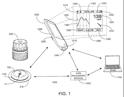
[00124] For purpose of illustration, and not limitation, the inserters
described herein
may be used in connection with an exemplary analyte monitoring system is
depicted in
FIGURE 1. It is understood that the inserters described herein may be used
with any
medical device on its own or in connection with a system. FIGURE 1 shows an
exemplary in vivo-based analyte monitoring system 100 in accordance with
embodiments
of the present disclosure. As shown, in certain embodiments, analyte
monitoring system
100 includes on body electronics 1100 electrically coupled to in vivo analyte
sensor 14 (a
proximal portion of which is shown in FIG. 1, and attached to adhesive layer
218 for
attachment on a skin surface on the body of a user. On body electronics 1100
includes on
body housing 122 that defines an interior compartment.
[00125] Also shown in FIGURE 1 is insertion device 200 (or insertion devices
300,
400, 2400, 2500, 2700, 3700 described herein) that, when operated,
transcutaneously
positions a portion of analyte sensor 14 through a skin surface and in fluid
contact with
ISF, and positions on body electronics 1100 and adhesive layer 218 on a skin
surface, as
will be described in greater detail herein. In certain embodiments, on body
electronics
1100, analyte sensor 14 and adhesive layer 218 are sealed within the housing
of insertion
device 200 before use, and in certain embodiments, adhesive layer 218 is also
sealed
within the housing or the adhesive layer can provide a seal for preserving the
sterility of
the apparatus. Additional details regarding insertion devices are discussed,
e.g., in U.S.
Patent Application No. 12/698,129 and U.S. Provisional Application Nos.
61/238,646,
61/246,825, 61/247,516, 61/249,535, and 61/345,562, the disclosures of each of
which
are incorporated herein by reference for all purposes.
[00126] Referring back to the FIGURE 1, analyte monitoring system 100 includes
display device 1200 which includes a display 1220 to output information to the
user, an
input component 1210 such as a button, actuator, a touch sensitive switch, a
capacitive
switch, pressure sensitive switch, jog wheel or the like, to input data or
command to
display device 1200 or otherwise control the operation of display device 1200.
It is noted
that some embodiments may include display-less devices or devices without any
user
22
CA 02766693 2011-12-21
WO 2011/119898 PCT/US2011/029884
interface components. These devices may be functionalized to store data as a
data logger
and/or provide a conduit to transfer data from on body electronics and/or a
display-less
device to another device and/or location. Embodiments will be described herein
as
display devices for exemplary purposes which are in no way intended to limit
the
embodiments of the present disclosure. It will be apparent that display-less
devices may
also be used in certain embodiments.
[00127] In certain embodiments, on body electronics 1100 may be configured to
store
some or all of the monitored analyte related data received from analyte sensor
14 in a
memory during the monitoring time period, and maintain it in memory until the
usage
period ends. In such embodiments, stored data is retrieved from on body
electronics
1100 at the conclusion of the monitoring time period, for example, after
removing analyte
sensor 14 from the user by detaching on body electronics 1100 from the skin
surface
where it was positioned during the monitoring time period. In such data
logging
configurations, real time monitored analyte level is not communicated to
display device
1200 during the monitoring period or otherwise transmitted from on body
electronics
1100, but rather, retrieved from on body electronics 1100 after the monitoring
time
period.
[00128] In certain embodiments, input component 1210 of display device 1200
may
include a microphone and display device 1200 may include software configured
to
analyze audio input received from the microphone, such that functions and
operation of
the display device 1200 may be controlled by voice commands. In certain
embodiments,
an output component of display device 1200 includes a speaker for outputting
information as audible signals. Similar voice responsive components such as a
speaker,
microphone and software routines to generate, process and store voice driven
signals may
be provided to on body electronics 1100.
[00129] In certain embodiments, display 1220 and input component 1210 may be
integrated into a single component, for example a display that can detect the
presence and
location of a physical contact touch upon the display such as a touch screen
user
interface. In such embodiments, the user may control the operation of display
device
23
CA 02766693 2011-12-21
WO 2011/119898 PCT/US2011/029884
1200 by utilizing a set of pre-programmed motion commands, including, but not
limited
to, single or double tapping the display, dragging a finger or instrument
across the
display, motioning multiple fingers or instruments toward one another,
motioning
multiple fingers or instruments away from one another, etc. In certain
embodiments, a
display includes a touch screen having areas of pixels with single or dual
function
capacitive elements that serve as LCD elements and touch sensors.
[00130] Display device 1200 also includes data communication port 1230 for
wired
data communication with external devices such as remote terminal (personal
computer)
1700, for example. Example embodiments of the data communication port 1230
include
USB port, mini USB port, RS-232 port, Ethernet port, Firewire port, or other
similar data
communication ports configured to connect to the compatible data cables.
Display
device 1200 may also include an integrated in vitro glucose meter, including
in vitro test
strip port 1240 to receive an in vitro glucose test strip for performing in
vitro blood
glucose measurements.
[00131] Referring still to FIGURE 1, display 1220 in certain embodiments is
configured to display a variety of information - some or all of which may be
displayed at
the same or different time on display 1220. In certain embodiments the
displayed
information is user-selectable so that a user can customize the information
shown on a
given display screen. Display 1220 may include but is not limited to graphical
display
1380, for example, providing a graphical output of glucose values over a
monitored time
period (which may show important markers such as meals, exercise, sleep, heart
rate,
blood pressure, etc, numerical display 1320, for example, providing monitored
glucose
values (acquired or received in response to the request for the information),
and trend or
directional arrow display 1310 that indicates a rate of analyte change and/or
a rate of the
rate of analyte change, e.g., by moving locations on display 1220.
[00132] As further shown in FIGURE 1, display 1220 may also include date
display
1350 providing for example, date information for the user, time of day
information
display 1390 providing time of day information to the user, battery level
indicator display
1330 which graphically shows the condition of the battery (rechargeable or
disposable) of
24
CA 02766693 2011-12-21
WO 2011/119898 PCT/US2011/029884
the display device 1200, sensor calibration status icon display 1340 for
example, in
monitoring systems that require periodic, routine or a predetermined number of
user
calibration events, notifying the user that the analyte sensor calibration is
necessary,
audio/vibratory settings icon display 1360 for displaying the status of the
audio/vibratory
output or alarm state, and wireless connectivity status icon display 1370 that
provides
indication of wireless communication connection with other devices such as on
body
electronics, data processing module 1600, and/or remote terminal 1700. As
additionally
shown in FIGURE 1, display 1220 may further include simulated touch screen
button
1250, 1260 for accessing menus, changing display graph output configurations
or
otherwise for controlling the operation of display device 1200.
[00133] Referring back to FIGURE 1, in certain embodiments, display 1220 of
display
device 1200 may be additionally, or instead of visual display, configured to
output alarms
notifications such as alarm and/or alert notifications, glucose values etc,
which may be
audible, tactile, or any combination thereof. In one aspect, the display
device 1200 may
include other output components such as a speaker, vibratory output component
and the
like to provide audible and/or vibratory output indication to the user in
addition to the
visual output indication provided on display 1220. Further details and other
display
embodiments can be found in, e.g., U.S. Patent Application No. 12/871,901,
U.S.
provisional application nos. 61/238,672, 61/247,541, 61/297,625, the
disclosures of each
of which are incorporated herein by reference for all purposes.
[00134] After the positioning of on body electronics 1100 on the skin surface
and
analyte sensor 14 in vivo to establish fluid contact with ISF (or other
appropriate body
fluid), on body electronics 1100 in certain embodiments is configured to
wirelessly
communicate analyte related data (such as, for example, data corresponding to
monitored
analyte level and/or monitored temperature data, and/or stored historical
analyte related
data) when on body electronics 1100 receives a command or request signal from
display
device 1200. In certain embodiments, on body electronics 1100 may be
configured to at
least periodically broadcast real time data associated with monitored analyte
level which
is received by display device 1200 when display device 1200 is within
communication
CA 02766693 2011-12-21
WO 2011/119898 PCT/US2011/029884
range of the data broadcast from on body electronics 1100, i.e., it does not
need a
command or request from a display device to send information.
[00135] For example, display device 1200 may be configured to transmit one or
more
commands to on body electronics 1100 to initiate data transfer, and in
response, on body
electronics 1100 may be configured to wirelessly transmit stored analyte
related data
collected during the monitoring time period to display device 1200. Display
device 1200
may in turn be connected to a remote terminal 1700 such as a personal computer
and
functions as a data conduit to transfer the stored analyte level information
from the on
body electronics 1100 to remote terminal 1700. In certain embodiments, the
received
data from the on body electronics 1100 may be stored (permanently or
temporarily) in
one or more memory of the display device 1200. In certain other embodiments,
display
device 1200 is configured as a data conduit to pass the data received from on
body
electronics 1100 to remote terminal 1700 that is connected to display device
1200.
[00136] Referring still to FIGURE 1, also shown in analyte monitoring system
1000
are data processing module 1600 and remote terminal 1700. Remote terminal 1700
may
include a personal computer, a server terminal a laptop computer or other
suitable data
processing devices including software for data management and analysis and
communication with the components in the analyte monitoring system 1000. For
example, remote terminal 1700 may be connected to a local area network (LAN),
a wide
area network (WAN), or other data network for uni-directional or bi-
directional data
communication between remote terminal 1700 and display device 1200 and/or data
processing module 1600.
[00137] Remote terminal 1700 in certain embodiments may include one or more
computer terminals located at a physician's office or a hospital. For example,
remote
terminal 1700 may be located at a location other than the location of display
device 1200.
Remote terminal 1700 and display device 1200 could be in different rooms or
different
buildings. Remote terminal 1700 and display device 1200 could be at least
about one
mile apart, e.g., at least about 100 miles apart, e.g., at least about 1000
miles apart. For
example, remote terminal 1700 could be in the same city as display device
1200, remote
26
CA 02766693 2011-12-21
WO 2011/119898 PCT/US2011/029884
terminal 1700 could be in a different city than display device 1200, remote
terminal 1700
could be in the same state as display device 1200, remote terminal 1700 could
be in a
different state than display device 1200, remote terminal 1700 could be in the
same
country as display device 1200, or remote terminal 1700 could be in a
different country
than display device 1200, for example.
[00138] In certain embodiments, a separate, optional data
communication/processing
device such as data processing module 1600 may be provided in analyte
monitoring
system 1000. Data processing module 1600 may include components to communicate
using one or more wireless communication protocols such as, for example, but
not
limited to, infrared (IR) protocol, Bluetooth protocol, Zigbee protocol, and
802.11
wireless LAN protocol. Additional description of communication protocols
including
those based on Bluetooth protocol and/or Zigbee protocol can be found in U.S.
Patent
Publication No. 2006/0193375 incorporated herein by reference for all
purposes. Data
processing module 1600 may further include communication ports, drivers or
connectors
to establish wired communication with one or more of display device 1200, on
body
electronics 1100, or remote terminal 1700 including, for example, but not
limited to USB
connector and/or USB port, Ethernet connector and/or port, FireWire connector
and/or
port, or RS-232 port and/or connector.
[00139] In certain embodiments, data processing module 1600 is programmed to
transmit a polling or query signal to on body electronics 1100 at a
predetermined time
interval (e.g., once every minute, once every five minutes, or the like), and
in response,
receive the monitored analyte level information from on body electronics 1100.
Data
processing module 1600 stores in its memory the received analyte level
information,
and/or relays or retransmits the received information to another device such
as display
device 1200. More specifically in certain embodiments, data processing module
1600
may be configured as a data relay device to retransmit or pass through the
received
analyte level data from on body electronics 1100 to display device 1200 or a
remote
terminal (for example, over a data network such as a cellular or WiFi data
network) or
both.
27
CA 02766693 2011-12-21
WO 2011/119898 PCT/US2011/029884
[00140] In certain embodiments, on body electronics 1100 and data processing
module
1600 may be positioned on the skin surface of the user within a predetermined
distance of
each other (for example, about 1-12 inches, or about 1-10 inches, or about 1-7
inches, or
about 1-5 inches) such that periodic communication between on body electronics
1100
and data processing module 1600 is maintained. Alternatively, data processing
module
1600 may be worn on a belt or clothing item of the user, such that the desired
distance for
communication between the on body electronics 1100 and data processing module
1600
for data communication is maintained. In a further aspect, the housing of data
processing
module 1600 may be configured to couple to or engage with on body electronics
1100
such that the two devices are combined or integrated as a single assembly and
positioned
on the skin surface. In further embodiments, data processing module 1600 is
detachably
engaged or connected to on body electronics 1100 providing additional
modularity such
that data processing module 1600 may be optionally removed or reattached as
desired.
[00141] Referring again to FIGURE 1, in certain embodiments, data processing
module 1600 is programmed to transmit a command or signal to on body
electronics
1100 at a predetermined time interval such as once every minute, or once every
5 minutes
or once every 30 minutes or any other suitable or desired programmable time
interval to
request analyte related data from on body electronics 1100. When data
processing
module 1600 receives the requested analyte related data, it stores the
received data. In
this manner, analyte monitoring system 1000 may be configured to receive the
continuously monitored analyte related information at the programmed or
programmable
time interval, which is stored and/or displayed to the user. The stored data
in data
processing module 1600 may be subsequently provided or transmitted to display
device
1200, remote terminal 1700 or the like for subsequent data analysis such as
identifying
frequency of periods of glycemic level excursions over the monitored time
period, or the
frequency of the alarm event occurrence during the monitored time period, for
example,
to improve therapy related decisions. Using this information, the doctor,
healthcare
provider or the user may adjust or recommend modification to the diet, daily
habits and
routines such as exercise, and the like.
28
CA 02766693 2011-12-21
WO 2011/119898 PCT/US2011/029884
[00142] In another embodiment, data processing module 1600 transmits a command
or
signal to on body electronics 1100 to receive the analyte related data in
response to a user
activation of a switch provided on data processing module 1600 or a user
initiated
command received from display device 1200. In further embodiments, data
processing
module 1600 is configured to transmit a command or signal to on body
electronics 1100
in response to receiving a user initiated command only after a predetermined
time
interval has elapsed. For example, in certain embodiments, if the user does
not initiate
communication within a programmed time period, such as, for example about 5
hours
from last communication (or 10 hours from the last communication, or 24 hours
from the
last communication), the data processing module 1600 may be programmed to
automatically transmit a request command or signal to on body electronics
1100.
Alternatively, data processing module 1600 may be programmed to activate an
alarm to
notify the user that a predetermined time period of time has elapsed since the
last
communication between the data processing module 1600 and on body electronics
1100.
In this manner, users or healthcare providers may program or configure data
processing
module 1600 to provide certain compliance with analyte monitoring regimen, so
that
frequent determination of analyte levels is maintained or performed by the
user.
[00143] In certain embodiments, when a programmed or programmable alarm
condition is detected (for example, a detected glucose level monitored by
analyte sensor
14 that is outside a predetermined acceptable range indicating a physiological
condition
which requires attention or intervention for medical treatment or analysis
(for example, a
hypoglycemic condition, a hyperglycemic condition, an impending hyperglycemic
condition or an impending hypoglycemic condition), the one or more output
indications
may be generated by the control logic or processor of the on body electronics
1100 and
output to the user on a user interface of on body electronics 1100 so that
corrective action
may be timely taken. In addition to or alternatively, if display device 1200
is within
communication range, the output indications or alarm data may be communicated
to
display device 1200 whose processor, upon detection of the alarm data
reception, controls
the display 1220 to output one or more notification.
29
CA 02766693 2011-12-21
WO 2011/119898 PCT/US2011/029884
[00144] In certain embodiments, control logic or microprocessors of on body
electronics 1100 include software programs to determine future or anticipated
analyte
levels based on information obtained from analyte sensor 14, e.g., the current
analyte
level, the rate of change of the analyte level, the acceleration of the
analyte level change,
and/or analyte trend information determined based on stored monitored analyte
data
providing a historical trend or direction of analyte level fluctuation as
function time
during monitored time period. Predictive alarm parameters may be programmed or
programmable in display device 1200, or the on body electronics 1100, or both,
and
output to the user in advance of anticipating the user's analyte level
reaching the future
level. This provides the user an opportunity to take timely corrective action.
[00145] Information, such as variation or fluctuation of the monitored analyte
level as
a function of time over the monitored time period providing analyte trend
information,
for example, may be determined by one or more control logic or microprocessors
of
display device 1200, data processing module 1600, and/or remote terminal 1700,
and/or
on body electronics 1100. Such information may be displayed as, for example, a
graph
(such as a line graph) to indicate to the user the current and/or historical
and/or and
predicted future analyte levels as measured and predicted by the analyte
monitoring
system 1000. Such information may also be displayed as directional arrows (for
example, see trend or directional arrow display 1310) or other icon(s), e.g.,
the position
of which on the screen relative to a reference point indicated whether the
analyte level is
increasing or decreasing as well as the acceleration or deceleration of the
increase or
decrease in analyte level. This information may be utilized by the user to
determine any
necessary corrective actions to ensure the analyte level remains within an
acceptable
and/or clinically safe range. Other visual indicators, including colors,
flashing, fading,
etc., as well as audio indicators including a change in pitch, volume, or tone
of an audio
output and/or vibratory or other tactile indicators may also be incorporated
into the
display of trend data as means of notifying the user of the current level
and/or direction
and/or rate of change of the monitored analyte level. For example, based on a
determined
rate of glucose change, programmed clinically significant glucose threshold
levels (e.g.,
hyperglycemic and/or hypoglycemic levels), and current analyte level derived
by an in
CA 02766693 2011-12-21
WO 2011/119898 PCT/US2011/029884
vivo analyte sensor, the system 1000 may include an algorithm stored on
computer
readable medium to determine the time it will take to reach a clinically
significant level
and will output notification in advance of reaching the clinically significant
level, e.g., 30
minutes before a clinically significant level is anticipated, and/or 20
minutes, and/or 10
minutes, and/or 5 minutes, and/or 3 minutes, and/or 1 minute, and so on, with
outputs
increasing in intensity or the like.
[00146] Referring again back to FIGURE 1, in certain embodiments, software
algorithm(s) for execution by data processing module 1600 may be stored in an
external
memory device such as an SD card, microSD card, compact flash card, XD card,
Memory Stick card, Memory Stick Duo card, or USB memory stick/device including
executable programs stored in such devices for execution upon connection to
the
respective one or more of the on body electronics 1100, remote terminal 1700
or display
device 1200. In a further aspect, software algorithms for execution by data
processing
module 1600 may be provided to a communication device such as a mobile
telephone
including, for example, WiFi or Internet enabled smart phones or personal
digital
assistants (PDAs) as a downloadable application for execution by the
downloading
communication device.
[00147] Examples of smart phones include Windows , AndroidTM, iPhone
operating
system, Palm WebOSTM, Blackberry operating system, or Symbian operating
system
based mobile telephones with data network connectivity functionality for data
communication over an internet connection and/or a local area network (LAN).
PDAs as
described above include, for example, portable electronic devices including
one or more
microprocessors and data communication capability with a user interface (e.g.,
display/output unit and/or input unit, and configured for performing data
processing, data
upload/download over the internet, for example. In such embodiments, remote
terminal
1700 may be configured to provide the executable application software to the
one or
more of the communication devices described above when communication between
the
remote terminal 1700 and the devices are established.
31
CA 02766693 2011-12-21
WO 2011/119898 PCT/US2011/029884
[00148] In still further embodiments, executable software applications may be
provided over-the-air (OTA) as an OTA download such that wired connection to
remote
terminal 1700 is not necessary. For example, executable applications may be
automatically downloaded as software download to the communication device, and
depending upon the configuration of the communication device, installed on the
device
for use automatically, or based on user confirmation or acknowledgement on the
communication device to execute the installation of the application. The OTA
download
and installation of software may include software applications and/or routines
that are
updates or upgrades to the existing functions or features of data processing
module 1600
and/or display device 1200.
[00149] Referring back to remote terminal 1700 of FIGURE 1, in certain
embodiments, new software and/or software updates such as software patches or
fixes,
firmware updates or software driver upgrades, among others, for display device
1200
and/or on body electronics 1100 and/or data processing module 1600 may be
provided by
remote terminal 1700 when communication between the remote terminal 1700 and
display device 1200 and/or data processing module 1600 is established. For
example,
software upgrades, executable programming changes or modification for on body
electronics 1100 may be received from remote terminal 1700 by one or more of
display
device 1200 or data processing module 1600, and thereafter, provided to on
body
electronics 1100 to update its software or programmable functions. For
example, in
certain embodiments, software received and installed in on body electronics
1100 may
include software bug fixes, modification to the previously stalled software
parameters
(modification to analyte related data storage time interval, resetting or
adjusting time base
or information of on body electronics 1100, modification to the transmitted
data type,
data transmission sequence, or data storage time period, among others).
Additional
details describing field upgradability of software of portable electronic
devices, and data
processing are provided in U.S. Application Nos. 12/698,124, 12/794,721,
12/699,653,
and 12/699,844, and U.S. Provisional Application Nos. 61,359,265, and
61/325,155 the
disclosure of which is incorporated by reference herein for all purposes.
32
CA 02766693 2011-12-21
WO 2011/119898 PCT/US2011/029884
The Sensor
[00150] The analyte sensor 14 of the analyte measurement system 100 may be
used to
monitor levels of a wide variety of analytes. Analytes that may be monitored
include, for
example, acetylcholine, amylase, bilirubin, cholesterol, chorionic
gonadotropin, creatine
kinase (e.g., CK-MB), creatine, DNA, fructosamine, glucose, glutamine, growth
hormones, hormones, ketones, lactate, peroxide, prostate-specific antigen,
prothrombin,
RNA, thyroid-stimulating hormone, and troponin. The concentration of drugs,
such as,
for example, antibiotics (e.g., gentamicin, vancomycin, and the like),
digitoxin, digoxin,
drugs of abuse, theophylline, and warfarin, may also be monitored. One or more
analyte
may be monitored by a given sensor. In those embodiments that monitor more
than one
analyte, the analytes may be monitored at the same or different times, which
may use the
same on body electronics (e.g., simultaneously) or with different on body
electronics.
[00151] In one embodiment of the present disclosure, sensor 14 is physically
positioned in or on the body of a user whose analyte level is being monitored.
Sensor 14
may be configured to continuously sample the analyte level of the user and
convert the
sampled analyte level, e.g., glucose concentration into a corresponding data
signal, e.g., a
current or voltage, for input into on body electronics. Alternatively, sensor
14 may be
configured to sample analyte levels on demand. The on body electronics may
amplify,
filter, average, and/or otherwise process signal provided by the sensor.
[00152] An embodiment of the sensor 14 is illustrated in FIGURE 2. It is
understood
that the inserters described herein can be used with other medical devices.
The shape(s)
described herein are exemplary only. Other sensor shapes are contemplated. In
some
embodiments, sensor 14 includes a substrate which is a dielectric, e.g., a
polymer or
plastic material, such as polyester or polyamide. In this embodiment, the
sensor is
constructed so that a portion is positionable beneath skin and a portion is
above skin.
Accordingly, sensor 14 includes an insertion or internal portion 30 and an
external or
electrical contact portion 32. In some embodiments, the contact portion 32
includes
several conductive contacts 36, 38, and 40 (herein shown as three contacts)
for
connection to other electronics, e.g., at the on body electronics 1100. (See
Figure 1.)
33
CA 02766693 2011-12-21
WO 2011/119898 PCT/US2011/029884
The contacts provided in this embodiment are for a working electrode, a
reference
electrode, and a counter electrode. In some embodiments, two or more working
electrodes are provided. The operative portions of these electrodes, that is,
working
electrode, reference electrode, and counter electrode (not individually
shown), are
provided at the insertion portion, e.g., at the distal end of insertion
portion 30, e.g.,
portion 34. In some embodiments, one or more electrodes may be external to the
body,
e.g., an external counter electrode. The contact and operative portions of the
electrodes
are connected by circuit traces 42, 44, and 46 running on the surface of the
substrate. In
some embodiments, the traces are provided in channels, or may be embedded
within the
substrate, or may traverse different sides of the substrate. The conductive
contacts,
conductive traces, and electrodes are fabricated from conductive material,
such as
platinum, palladium, gold, carbon, or the like. More than one material may be
used for a
given sensor. Further details of sensors are described, e.g., in U.S. Patent
Nos. 6,175,572
and 6,103,033, which are incorporated by reference herein for all purposes.
[00153] Sensor 14 may include a proximal retention portion 48. The insertion
portion
30 and the proximal retention portion 48 are sized and configured to be
positioned with a
sharp for installation into the skin of a subject, as described herein. In
use, the sensor 14
may be configured to bend (e.g., along the line B) and therefore be positioned
in two
substantially perpendicular, intersecting planes. Such bending may occur prior
to or
during coupling to the on body electronics as described below. (See FIGURE
17).
[00154] Portions 48 and 52 which provide a path for electrical connections,
e.g., the
conductive traces, between the proximal and distal portions of the sensor.
Sensor 14 is
further provided with a notch or cut-out 54. Such configuration facilitates
the sensor 14
to bend (e.g., along the line indicated by line B) such that retention portion
48 remains
upright and therefore be positioned in two substantially perpendicular,
intersecting
planes, as illustrated in FIGURE 3. As will be described below, the sensor tab
50 can be
encased in the on body housing 122 to aid in securing and positioning the
sensor 14.
Proximal retention portion 48 maintains its longitudinal alignment with
insertion portion
for positioning within an insertion sharp.
34
CA 02766693 2011-12-21
WO 2011/119898 PCT/US2011/029884
[00155] Embodiments of analyte sensors have been described herein to operate
electrochemically, through an arrangement of electrodes having chemical
sensing layers
applied thereto, by generating an electrical current proportional to the
volume of a redox
reaction of the analyte (and indicative of analyte concentration), catalyzed
by an analyte-
specific oxidizing enzyme. Embodiments exist in which the number of electrodes
provided to bring about and detect the level of these reactions is two, three,
or a greater
number. However, other types of sensors may be employed as described herein.
[00156] A portion of sensor 14 may be situated above the surface of the skin,
with a
distal portion 30 penetrating through the skin and into the subcutaneous space
in contact
with the user's biofluid, such as ISF. Further details regarding the
electrochemistry of
sensor 14 is provided in U.S. Patent Nos. 5,264,104; 5,356,786; 5,262,035;
5,320,725;
and 6,990,366, each of which is incorporated by reference herein for all
purposes.
[00157] In some embodiments, the sensor is implantable into a subject's body
for a
usage period (e.g., a minute or more, at least one day or more, about one to
about 30 days
or even longer, about three to about fourteen days, about three to about seven
days, or in
some embodiments, longer periods of up to several weeks) to contact and
monitor an
analyte present in a biological fluid. In this regard, the sensor can be
disposed in a
subject at a variety of sites (e.g., abdomen, upper arm, thigh, etc.),
including
intramuscularly, transcutaneously, intravascularly, or in a body cavity.
[00158] In some embodiments, sensor 14 is employed by insertion and/or
implantation
into a user's body for some usage period. In such embodiments, the substrate
may be
formed from a relatively flexible material.
[00159] While the embodiments illustrated in FIGURES 2-3 have three
electrodes,
other embodiments can include a fewer or greater number of electrodes. For
example, a
two-electrode sensor can be utilized. The sensor 14 may be externally-powered
and
allow a current to pass which current is proportional to the amount of analyte
present.
Alternatively, the sensor 14 itself may act as a current source in some
embodiments. In
some two-electrode embodiments, the sensor may be self-biasing and there may
be no
need for a reference electrode. An exemplary self-powered, two-electrode
sensor is
CA 02766693 2011-12-21
WO 2011/119898 PCT/US2011/029884
described in U.S. Patent Application Serial No. 12/393,921, filed February 26,
2009, and
entitled "Self-Powered Analyte Sensor," which is hereby incorporated by
reference
herein for all purposes. The level of current provided by a self-powered
sensor may be
low, for example, on the order of nanoamperes, in certain embodiments.
Insertion Assembly
[00160] Insertion assemblies are provided which are used to install a medical
device to
the subject. In some embodiments, an insertion assembly includes an inserter
and the
medical device itself. The inserter can be configured to insert various
medical devices
into the subject, such as, for example, an analyte sensor, an infusion set, or
a cannula. In
some embodiments, the inserter can be configured to install a combination of
such
devices, e.g., a combined sensor/infusion set, etc., at the same or different
times or
locations. For example, in certain embodiments, a given inserter can be
configured to
install a first device and a second device at different times. In this regard,
the inserter can
be reusable. For example, an inserter may be modifiable to be used with more
than one
medical device, to include more than one type of medical device, e.g., by
attaching an
adapter and/or removing detaching a portion of an inserter. The inserter can
install the
medical device in, under, or through the skin of the subject, or place the
medical device
on the surface of the skin. The medical device can include features or
structures, e.g.,
barbs, tabs, adhesive, etc., to maintain the device in position with respect
to the skin after
insertion. The inserter device may also be used as a lancet, e.g., to pierce
the skin
without inserting or installing a medical device.
[00161] In some embodiments, an insertion assembly includes an inserter, a
medical
device, such as an analyte sensor, and a mount for supporting the medical
device at least
partially in or on the skin of the subject. In some embodiments, the mount is
a support
structure, plate and/or member which is attached, adhered, or otherwise
secured to the
skin of the subject. The mount may be inserted simultaneously with the medical
device
by the inserter. In other embodiments, the mount is installed after or before
installation
of the medical device. A mount may be applied by the inserter or separately.
The mount
may include features or structures (e.g., adhesive, guides, barbs, tabs, etc.)
to maintain the
36
CA 02766693 2011-12-21
WO 2011/119898 PCT/US2011/029884
sensor in position with respect to the skin after insertion and/or maintain
the sensor in
relative position with respect to the sensor control unit. In some
embodiments, an
adhesive pad or strip is used to secure the medical device, e.g., the sensor
and/or the
sensor control unit, and no mount is used.
[00162] In some embodiments, an insertion assembly includes an inserter, an
analyte
sensor, a mount, and a power supply. The mount and power supply may be
inserted
simultaneously with the analyte sensor by the inserter. In other embodiments,
the mount
and battery are installed after or before installation of the analyte sensor.
In such case,
the mount and/or power supply may be applied by the inserter or separately.
The power
supply may be used to provide a current or a potential to the sensor and/or to
provide
power for communication of one or more signals to the monitor unit.
[00163] In some embodiments, an insertion assembly includes an inserter, a
medical
device such as an analyte sensor, a mount, and a sensor control unit. The
mount and
sensor control unit may be deployed and/or installed simultaneously with the
analyte
sensor by the inserter. In other embodiments, the mount and sensor control
unit are
installed after or before installation of the analyte sensor. For example, the
mount and
the analyte sensor may be installed by the inserter, and the sensor control
unit may be
subsequently installed. In other embodiments, the mount is installed, followed
by
insertion of the analyte sensor by the inserter, and further followed by
installation of the
sensor control unit. In other embodiments, the mount and sensor control unit
are installed
first, and the analyte sensor is subsequently installed.
[00164] In some embodiments, electronics of the sensor control unit provide a
voltage
or current to the analyte sensor. In some embodiments, the electronics
processes signals
provided by the analyte sensor. In further embodiments, the electronics may
include
communication functionality for providing signal relating to signal provided
by the
analyte sensor to a further component, such as, e.g., a monitor unit, a
computer, or other
component. In some embodiments, communications circuitry, such as RFID antenna
or
other communications circuitry, is provided. The power supply may be used to
power
37
CA 02766693 2011-12-21
WO 2011/119898 PCT/US2011/029884
some or all of these functions. In some embodiments, power is provided from
the
monitor unit, e.g., via inductive coupling.
[00165] An inserter may include a plurality of different components. For
example, an
inserter may include one or more components for advancing a sharp towards the
skin of
the subject. The sensor and sensor control unit and/or mounting structure may
be
supported by a support structure, such as a carriage. A driver may be provided
for
advancing the sharp and/or the analyte sensor/support structure. In some
embodiments,
the actuator is directly or indirectly coupled to the sharp and/or support
structure, such
that manual force and speed applied by the subject to the actuator is
transferred to the
sharp and/or support structure. In some embodiments, the applied force drives
the sharp
and/or support structure between a retracted position and an advanced
position. In some
embodiments, the sensor and sensor control unit and/or mounting structure is
maintained
in a retracted position prior to installation by contacting projections
extending inwardly
from a sheath. In accordance with this embodiment, the sensor and sensor
control unit
and/or mounting structure are temporarily maintained operatively between the
support
structure and the projections disposed on the interior wall of the sheath.
[00166] An inserter can also include one or more components for retracting the
sharp,
while allowing the analyte sensor and optional mount and/or electronics to
remain on the
subject. The components for retracting the sharp can include a retractor. It
is understood
that the retractor and the actuator may be the same structure or include some
common
components. In some embodiments, the retractor is directly or indirectly
coupled to the
sharp such that the manual force applied by the user is transferred from the
retractor to
the sharp to retract the sharp from the skin. In other embodiments, a drive
assembly may
be provided to retract the sharp. For example, the drive assembly may include
a spring,
motor, hydraulic piston, etc., to retract the sharp away from the skin of the
subject. The
drive assembly may also include a linear drive component.
[00167] In some embodiments, the retractor withdraws the sharp upon actuation
by the
user. In such cases, the user actuates the retractor when it is desired to
withdraw the
sharp. For example, the retractor may include a release switch. Upon
activation of the
38
CA 02766693 2011-12-21
WO 2011/119898 PCT/US2011/029884
release switch, the drive assembly, e.g., the spring or other driver, retracts
the sharp from
the skin. In other embodiments, the retractor and the actuator include common
components. After activating the actuator to advance the sharp and the analyte
sensor,
the user releases the actuator, which allows the drive assembly to withdraw
the sharp
from the skin.
[00168] In some embodiments, the retractor withdraws the sharp without further
user
interaction after actuation of insertion. For example, the inserter may
include features or
components which automatically retract the sharp upon advancement of the sharp
and
support structure by a predetermined amount. Inserter devices, in which no
further action
by the user is required to initiate withdrawal of the sharp after insertion,
are referred to
herein as having "automatic" withdrawal of the sharp.
Inserter Devices
[00169] One embodiment of a needle hub for an inserter is illustrated in
FIGURES 4-
5. Needle hub 136 supports sharp 124, having a sharpened distal portion 160.
In some
embodiments, as discussed herein, a longitudinal wall opening or gap 162 is
provided in
at least a portion of the wall of the sharp 124. The length N of the gap 162
is selected to
be commensurate with the length of the insertion portion 30 through to the
proximal
retention portion 48 of the sensor, and in certain embodiments may be about 3
mm to
about 50 mm, e.g., about 5 mm, or about 10 mm, or about 15 mm, or about 20 mm.
The
length L of the sharp 124 may be about 3 mm to about 50 mm, e.g., 5mm or more,
or
about 10 mm, or about 20 mm, or about 30 mm, or about 50 mm, and is selected
based
upon the length of the insertion portion 30 of a sensor and the desired depth
of the
insertion portion 30 of the sensor 14. In some embodiments, the distance or
spacing
between the two edges of the gap is about 0.2mm to about 0.5mm, e.g., about
0.22mm,
about 0.25mm, etc.
[00170] The distal portion 160 of sharp 124 is illustrated in greater detail
in FIGURES
6-8. As illustrated in FIGURE 6, sharp 124 has a substantially "C"- or "U"-
shaped
profile in this embodiment, but may have other configurations, e.g.,
substantially "V"-
shaped. A longitudinal gap 162 is provided in the wall of the sharp 124.
FIGURE 7
39
CA 02766693 2011-12-21
WO 2011/119898 PCT/US2011/029884
illustrates distal portion 160 is provided with an angled tip. In some
embodiments, the
angled tip may be provided with a first angled tip portion 164 and a second
steep-angled
tip portion 166. The exemplary configuration, which includes multiple edges
and faces,
provides a sharp point to reduce penetration force, trauma, and bleeding for
the subject.
The distal section of the sensor body has a width sized to fit within the
notch 162 of the
insertion sharp 124 having a diameter less than about 20 to about 26 gauge,
e.g., 21 gauge
to about 25 gauge, where in certain embodiments the sharp is 21 gauge or 23
gauge or 25
gauge. Such sharp may be used with a sensor having a width or diameter - - at
least the
portion that is carried by the sharp - - of about .20 mm to about .80 mm,
e.g., about .25
mm to about .60 mm, where in some embodiments the width or diameter of at
least a
portion of a sensor is .27 mm or .33 mm or .58 mm. In some embodiments, sharp
124 is
fabricated from a sheet of metal and folded into a substantially "V," "U" or
"Cl
configuration in cross-section. Various technologies can be used to
manufacture a folded
sheet of metal to form sharp 124. For example, etched-sheet metal technology
can be
used to form the sharp 124. In this manner, the sharp can be formed having a
very sharp
edge so that penetration through the skin during insertion is less painful. In
other
embodiments, a progressive die technology may be utilized to form a complex
sheet-
metal shape that has a sharp edge as depicted in FIGURE 9. In some
embodiments, the
sharp 124 can be molded with a plastic cap so that the sharp can be handled
during the
inserter assembly process. Further, the die cut sharp may be molded with
plastic to
reinforce the "V," "U" or "C" shaped sheet metal configuration. In other
embodiments, a
laser-cut sharp can be formed. In this manner, the laser can be used to form
the wall
opening or gap 162 and first-angled tip portion 164 and a second, steep-angled
tip portion
166.
[00171] In another embodiment, a sharp 124 may be formed from a standard
hypodermic needle utilizing the method depicted in FIGURE 10. First, the
hypodermic
needle (having a circular cross-section) is cut to the desired length for
sharp 124. Next,
the hypodermic needle is compressed so that its cross-section is permanently
deformed
from a circular shape to an oval shape. The tip of the hypodermic needle is
then ground
to a bevel to produce a sharp point to reduce the required penetration force,
as previously
CA 02766693 2011-12-21
WO 2011/119898 PCT/US2011/029884
discussed. Finally, the top section of the needle is removed by appropriate
techniques
(e.g., grinding, electropolishing, etc.). The resulting sharp 124 has a "U"-
shaped
configuration and provides ample space for the insertion of sensor 14. In some
embodiments, the tip-grinding step and the compression step may be carried out
in
reversed order.
[00172] Due to the compression step, a user may initially start with a larger
diameter
hypodermic needle so that the finished sharp 124 will have similar dimensions
to the
previously described sharps.
[00173] FIGURES 11-12 illustrate the position of on body housing 122 with
respect to
the needle hub 136 and sharp 124. The on body housing 122 can be configured to
hold at
least a portion of sensor 14 and sensor control unit 12. As illustrated in
FIGURE 11, the
sharp 124 extends through an aperture 168 in the on body housing 122. Thus, in
some
embodiments, the sharp 124 is uncoupled to on body housing 122. The distal
portion of
sensor 14 is positioned with the sharp 124. As further illustrated in FIGURE
12,
electronics 80 of the sensor control unit 12 (e.g., a printed circuit board
containing
electronics components of the on-body unit 16) and sensor hub 123 are
positioned within
on body housing 122. Sensor 14 may include a positioning structure, or slit
127, which
receives a positioning member, such as tab 129 of sensor hub 123. A power
supply 82,
such as a battery, e.g., a single use disposable battery, or rechargeable
battery, is
provided. The power supply 82 is used to provide potential or current to the
sensor in
some embodiments. In embodiments where a passive communications protocol such
as
passive RFID is used, no power supply is provided for the communications. Such
power
is provided by the monitor unit 18. In some embodiments, where the sensor
control unit
is used to transmit one or more signals, one or more power supplies may be
used to
provide power for such communications circuitry. In some embodiments, the
active
operational life of the battery may exceed the active operational life of the
sensor 14.
[00174] FIGURE 13 illustrates in cross-section the orientation of the on body
housing
122 with respect to the sharp 124 of an inserter, such as inserter 1500. As
discussed
herein, sensor 14 is disposed in a substantially bent configuration in some
embodiments,
41
CA 02766693 2011-12-21
WO 2011/119898 PCT/US2011/029884
such that a portion of the sensor, e.g., the insertion portion 30 and the
proximal retention
portion 48, are substantially vertical (e.g., substantially aligned with the
longitudinal axis
of an inserter and substantially perpendicular to the skin surface) and the
contact portion
32 (shown in profile) is oriented in a substantially horizontal configuration,
and in
electrical contact with the data-processing unit electronics, such as circuit
80. The sensor
tab 50 can be encased in the plastic of the on body housing 122 (e.g.,
"overmolded") and
secured in place. The notch 56 provides further stability to the sensor 14,
e.g., by
allowing the sensor tab 50 to be encased by the material of the on body
housing 122, and
further provides a means for vertically orienting the sensor 14 during
mounting, e.g., by
allowing vertical positioning of the notch 56 with respect to a vertical
landmark of the on
body housing 122.
[00175] The sensor 14, mounted with the on body housing 122, can be disposed
within
a recess of the carriage 130 such as a concave recess in the carriage 130.
Alternatively,
the sensor 14, mounted with the on body housing 122, can be disposed between
the
support structure and one or more projections extending from the wall of the
sheath. In
yet another alternative, the sensor 14, mounted with the on body housing 122,
can be held
in position by a releasable friction fit coupling to the sharp 124. In this
manner, the
carriage need not have a recess within which the sensor mounted with the on
body
housing is disposed. In the initial configuration of the inserter, the sharp
124 extends
through a longitudinal aperture 168 formed in a carriage 130. In some
embodiments, the
aperture 168 is appropriately sized, such that neither the sharp 124 nor
needle hub 136 is
in contact with the carriage 130. Accordingly, the needle hub 136 (and sharp
124) on the
one hand, and the carriage 130 (FIGURE 13) and the on body housing 122, on the
other
hand, move simultaneously, but independently from one another. In other
embodiments,
a friction fit may be provided between the aperture and the sharp.
[00176] The insertion portion 30 and proximal retention portion 48 of the
sensor 14
are disposed within a longitudinal bore 162 within the sharp 124 (See, e.g.,
FIGURE 6).
The proximal retention portion 48 is disposed within the longitudinal bore of
the sharp
124 and provides additional stability to the mounting of the sensor 14 within
the sharp
124. The longitudinal wall gap or opening 162 of sharp 124 is aligned with the
sensor
42
CA 02766693 2011-12-21
WO 2011/119898 PCT/US2011/029884
14, such that the tab 50 and the contact portion 32 extend laterally outward
from the
sharp 124.
[00177] An embodiment of an inserter is illustrated in FIGURES 14-17. An
inserter
900 includes a handle 902 and may include a removable distal cap or seal (not
shown) for
maintaining a sterile environment for the medical device and sharp housed
therein. As
illustrated in FIGURE 14, distal cap or seal is removed from handle 902.
Inserter 900
includes a base 942 that defines a distal surface 912 for placement on the
skin of a
subject. Inserter 900 may be utilized to advance a medical device into the
skin of the
subject. In some embodiments, handle 902 is advanced relative to base 942 in
order to
advance the sensor into the skin of the patient.
[00178] As illustrated in FIGURE 15, the inserter 900 includes an initial
configuration
in which the handle 902 is disposed in a proximal position with respect to the
base 942.
In such configuration, the sharp 924 is disposed in a configuration spaced
apart from the
aperture 920 of the adhesive layer 918. Extending distally from the upper
surface of
handle 902 are side walls 974, which terminate in a shoulder portion 975.
[00179] Needle carrier 934 is axially slidable within handle 902 and base 942
in the
direction of arrow N. Needle carrier 934 supports needle hub 936, from which
sharp 924
extends longitudinally within the inserter 900. In some embodiments, the sharp
is
supported at an oblique angle, e.g., between 0 and 90 with respect to the
skin surface.
Initially, needle carrier 934 is coupled to inner rail 928 through inter-
engagement of
finger 986 of needle carrier 934 with shoulder 987 of inner rail.
[00180] Inserter 900 includes an advancement spring 947. Inner rail 928
includes a
spring retention portion 984 for advancement spring 947. The inner surface of
handle
902 serves as an upper engagement surface for advancement spring 947. In some
embodiments, spring retention portion 984 is fabricated having several
projections that
extend through an aperture 982 in the wall of base 942. Spring retention
portion 984 is
resiliently movable. As downward force is applied to handle 902, the ledge 937
of
handle 902 contacts spring retention portion 984, driving such projections
inward. Such
inward motion releases the inner rail 928, and allows advancement spring 947
to advance
43
CA 02766693 2011-12-21
WO 2011/119898 PCT/US2011/029884
the inner rail 928 distally, thereby advancing the sharp 924 into the skin of
the subject, as
illustrated in FIGURE 16.
[00181] Retraction spring 946 is disposed between spring retention portion 948
of base
942 and shoulder portion 949 of needle carrier 934. Initially, retraction
spring 946 is in a
compressed state. As carriage 930 reaches a distal position, the distal
surface of the on
body housing 122 engages the upper surface of adhesive pad 918, thereby
becoming
adhered to the skin surface S of the subject. At the same time, flanges 970 of
base 942
engage fingers 986 disposed on the needle carrier 934. Fingers 986 are pivoted
outwards
by flanges 970. Such pivoting of fingers 974 causes fingers 986 to become
disengaged
from shoulder 984 of inner rail 928. Shuttle 934 is thereby disengaged from
inner rail
928.
[00182] As illustrated in FIGURE 17, disengagement of the shuttle 934 from the
inner
rail 928 permits the spring 946 to expand, thereby advancing the needle
carrier 934 to a
proximal position, thereby withdrawing the sharp 934 from the skin S of the
subject,
while leaving the sensor 14 in the skin.
[00183] A further embodiment of an inserter is illustrated in FIGURES 18-21
and
designated inserter 1000. Inserter 1000 is substantially identical to inserter
900
described herein, with the substantial differences noted herein and
illustrated in the
accompanying figures.
[00184] An inserter 1000 includes a housing 1002, and may include a removable
distal
cap or seal (not shown) for maintaining a sterile environment tor the medical
device and
sharp housed therein. As illustrated in FIGURE 18, distal cap or seal is
removed from
housing 1002. Inserter 1000 includes a base 1042 which defines a distal
surface 1012 for
placement on the skin of a subject. Inserter 1000 may be utilized to advance a
medical
device into the skin of the subject. In some embodiments, side buttons 1084
are
depressed relative to base 1042 in order to advance the medical device into
the skin of the
patient.
44
CA 02766693 2011-12-21
WO 2011/119898 PCT/US2011/029884
[00185] As illustrated in FIGURE 19, the inserter 1000 includes an initial
configuration in which the sharp 1024 is disposed in a configuration spaced
apart from
distal portion 1012 of the base 1042. It is understood that inserter 1000 may
incorporate
an adhesive layer (not shown) positioned across the distal portion 1012 of
base 1042.
[00186] On-body unit inserter 1028 includes carriage 1030 for supporting on
body
housing 122. On-body unit inserter 1028 also includes flange 1029 which
normally rests
on shoulder portions 1086 (as illustrated in FIGURES 19-21). When side buttons
1084
are depressed inwardly, latch portions on flange 1029 are deflected from
engagement
with shoulder portions 1086. As illustrated in FIGURE 20, advancement spring
1047 is
released from compression, thereby advancing on-body unit inserter 1028 as
elsewhere,
along with needle carrier 1034 into the skin of the subject.
[00187] Retraction spring 1046 is disposed between needle carrier 1034 and
housing
1002. Initially, retraction spring 1046 is in a compressed state. As carriage
1028 reaches
a distal position, on-body unit inserter 1028 is disengaged from needle
carrier 1034. The
distal surface of the on body housing 122 engages the skin or the upper
surface of
adhesive pad (not shown), thereby becoming adhered to the skin surface S of
the subject.
Latches on needle carrier 1034 attach it to on-body unit inserter 1028. Upon
firing the
inserter by pressing tabs 1084, the on-body unit inserter 1028 is pushed by
spring 1047
towards the skin surface, bringing along needle carrier 1034 and loading
extension spring
1046. As on-body unit inserter 1028 reaches the distal position (towards the
skin of the
subject), protrusions on housing 1002 disengage latches on needle carrier
1034,
connecting it to on-body unit inserter 1028. Extension spring 1046 now pulls
needle
carrier 1034 back into the proximal position.
[00188] As illustrated in FIGURE 21, disengagement of the needle carrier 1034
from
the inner rail 1028 permits the spring 1046 to contract with the bias, thereby
advancing
the needle carrier 1034 to a proximal position and withdrawing the sharp 1024
from the
skin S of the subject, while leaving the sensor 14 in the skin.
CA 02766693 2011-12-21
WO 2011/119898 PCT/US2011/029884
[00189] A further embodiment of an inserter is illustrated in FIGURES 22-25.
Inserter
1100 is substantially identical to inserters 900 and 1000 described herein,
with the
substantial differences noted herein and illustrated in the accompanying
figures.
[00190] An inserter 1100 includes a housing 1102 and may include a removable
distal
cap or seal (not shown) for maintaining a sterile environment for the medical
device
and/or sharp housed therein. As illustrated in FIGURE 22, distal cap or seal
is removed
from housing 1102. Inserter 1100 includes a base 1142 which defines a distal
surface
1112 for placement on the skin of a subject. Inserter 1100 may be utilized to
advance a
medical device into the skin of the subject. In some embodiments, a manual
actuator bar
1114 is depressed relative to base 1142 in order to advance the medical device
into the
skin of the patient. It is understood that inserter 1100 may incorporate an
adhesive layer
(not shown) which is positioned across the distal portion 1112 of base 1142.
[00191] As illustrated in FIGURE 23, the inserter 1100 includes an initial
configuration in which the sharp 1124 is disposed in a configuration spaced
apart from
distal portion 1112 of the base 1142. Manual advancement of the actuator may
be
performed by distal advancement of actuator bar 1114.
[00192] On-body unit inserter 1128 includes carriage 1130 for supporting on
body
housing 122. Retraction spring 1146 is disposed between needle carrier 1134
and base
1102. Initially, retraction spring 1146 is in a compressed state. As actuator
bar 1114 is
moved distally, carriage 1128 is moved distally and reaches a distal position,
such that
flanges 1188 of base 1142 engage fingers 1186 disposed on the shuttle 1134.
Fingers
1186 are pivoted outwards by flanges 1188. Such pivoting of fingers 1186
causes fingers
1186 to become disengaged from shoulder 1187 of inner rail 1128. Shuttle 1134
is
thereby disengaged from inner rail 1128. The distal surface of the on body
housing 122
engages the skin or the upper surface of adhesive pad (not shown), thereby
becoming
adhered to the skin surface of the subject (FIGURE 24).
[00193] As illustrated in FIGURE 25, disengagement of the shuttle 1136 from
the
inner rail 1028 permits the spring 1146 to expand with the bias, thereby
advancing the
needle carrier 1136 to a proximal position and withdrawing the sharp 1124 from
the skin
46
CA 02766693 2011-12-21
WO 2011/119898 PCT/US2011/029884
S of the subject, while leaving the sensor 14 in the skin. Once the sharp 1124
has been
withdrawn from the subject, it is no longer accessible from the distal portion
of the
inserter 1100, and unable to make contact by accident with the subject's skin.
[00194] A further embodiment of an inserter is illustrated in FIGURES 26-36.
Inserter
1200 is substantially identical to inserters 900, 1000, and 1100 described
herein, with the
substantial differences noted herein and illustrated in the accompanying
figures.
[00195] Inserter 1200 includes a housing 1202, an on-body unit inserter 1228,
a needle
retractor 1234, and a rotor 1208 with a torsion spring (not shown), all of
which are
depicted separately in FIGURES 28-3 1. As illustrated in FIGURES 28-3 1,
housing 1202
includes slots 1254; on-body unit inserter 1228 includes slots 1252, tabs
1253, and
opening 1262; needle retractor 1234 includes tabs 1250 and opening 1260; and
rotor
1208 includes rotor follower 1264.
[00196] FIGURE 32 illustrates an exploded view of inserter 1200. As
illustrated, in
some embodiments, inserter 1200 includes housing 1202, on-body unit inserter
1228,
needle retractor 1234, and rotor 1208. Tabs 1250 can be disposed on needle
retractor
1234 and configured to engage slots 1252 disposed on on-body unit inserter
1228 when
assembled as shown in FIGURE 33. Tabs 1253 can be disposed on on-body unit
inserter
1228 and configured to engage slots 1254 disposed on housing 1202. Needle
retractor
1234 and on-body unit inserter 1228 can each contain openings (1260, 1262) to
receive
rotor follower 1264 disposed on rotor 1208 after rotor 1208 has been inserted
into
housing 1202. Additionally, on-body unit inserter 1228 can provide support for
on body
housing 122 (not shown) for installation to the skin of a subject.
[00197] As illustrated in FIGURE 34, in some embodiments manually depressing
the
on-body unit inserter 1228 can cause the torsion spring to wind as the rotor
1208 rotates,
guided by the cam path. As illustrated in FIGURE 35, continued distal
advancement of
the on-body unit inserter 1228 causes the rotor follower 1264 to go "over
center" within
cam 1264. At such point, the torsion spring (not shown) is allowed to unwind.
(FIGURE
93). With reference to FIGURE 36, further unwinding of the torsion spring
pulls up the
47
CA 02766693 2011-12-21
WO 2011/119898 PCT/US2011/029884
needle retractor 1234 from the insertion site, while leaving the on-body unit
inserter in
place.
[00198] A further embodiment of an inserter is illustrated in FIGURES 37-46.
Inserter
1300 is substantially identical to inserters 900, 1000, 1100, and 1200
described herein,
with the substantial differences noted herein and illustrated in the
accompanying figures.
[00199] Inserter 1300 includes a housing 1302, an on-body unit inserter 1334,
a needle
retractor 1334, and a rotor 1308 with torsion spring 1367. As illustrated in
FIGURES 37,
38, 39, 45 and 46, the on-body unit inserter 1334 and needle retractor 1328
are disposed
within the housing 1302. Rotor 1308 includes a rotor follower 1364 which
engages a slot
1362 provided in the needle retractor 1328 and a slot 1360 in the on-body unit
inserter
1334. The on-body unit inserter 1334 provides a support portion for supporting
on body
housing 1302 (not shown).
[00200] In operation, torsion spring 1365 is loaded. A release button (not
shown) is
depressed in order to release torsion spring 1365. Rotor 1308 drives on-body
unit
inserter 1334 and needle retractor 1328 downwardly simultaneously. A
compression
spring 1367 holds down on-body unit inserter 1334. As rotor 1308 continues to
rotate,
slot 1360 allows on-body unit inserter 1334 to travel down and then be held
down, while
slot 1362 allows needle retractor 1328 to travel down and then back up to lift
the needle
from insertion site.
[00201] A further embodiment of an inserter is illustrated in FIGURES 47-5 1.
Inserter
1400 is substantially identical to inserters 900, 1000, 1100, 1200, and 1300
described
herein, with the substantial differences noted herein and illustrated in the
accompanying
figures.
[00202] Inserter 1400 may be used for installation of a medical device, e.g.,
on body
housing 122, in connection with a cartridge or carrier 1406. In some
embodiments,
inserter 1400 includes a body 1402 and an actuator 1404. The carrier 1406 may
include a
needle hub 1436 including a sharp (not shown) as well as an on body housing
122. In
use, the body 1402 is placed on a mount 1408 attached to subject by use of an
adhesive
48
CA 02766693 2011-12-21
WO 2011/119898 PCT/US2011/029884
pad 1418. As shown in FIGURE 47, carrier 1406 is loaded into inserter 1400
laterally as
shown.
[00203] With reference to FIGURE 48, a first disposable portion of inserter
1400
includes adhesive pad 1418 and mount 1408. The second disposable portion of
inserter
1400 includes carrier 1406 which removably contains needle hub 1436 and sensor
hub
1437 (not shown) which substantially identical to on body housing 122
described herein.
Inserter 1400 further includes body 1402, actuator 1404, and return spring
1409 (not
shown). Mount 1408 is removably attached to body 1402. Carrier 1406 can be
slid on to
body 1402 to transfer needle hub 1436 and carrier 1406 to inserter 1400. As
shown in
FIGURE 48, carrier 1406 can then be removed from inserter 1400. Needle hub
1436 and
sensor hub 1437 remain in inserter 1400. Adhesive pad 1418 is then adhesively
applied
to the skin of a subject. Alternatively, adhesive pad 1418 may be applied to
the skin of a
subject before attaching body 1402 and mount 1408.
[00204] In FIGURE 49, actuator 1404 is pressed down, performing a manual
insertion
of needle hub 1436 and sensor hub 1437, with the needle located in needle hub
1436 and
a distal sensor portion penetrating the skin of the subject. When pressure is
released from
actuator 1404, return spring 1409 (not shown) retracts the needle from the
skin of the
subject and out of sensor hub 1437. As shown in FIGURE 50, body 1402 can be
removed from mount 1408, leaving sensor hub 1437 in mounting base 1408. In
FIGURE
51, in some exemplary embodiments, carrier 1406 is slid on to housing 1402,
transferring
needle 1436 to carrier 1406. Carrier 1406, with needle 1436, is then removed
from
housing 1402 and may be disposed of properly, and inserter 1400 is ready to be
used
again. Communication electronics (not shown) are then attached to mounting
base 1408,
making electrical contact with sensor hub 1437.
[00205] A further embodiment of an inserter is illustrated in FIGURE 52.
Inserter
1500 is substantially identical to inserters 900, 1000, 1100, 1200, 1300, and
1400
described herein, with the substantial differences noted herein and
illustrated in the
accompanying figures.
49
CA 02766693 2011-12-21
WO 2011/119898 PCT/US2011/029884
[00206] Inserter 1500 includes a housing 1502, which may be fabricated as a
molded
plastic tube and may further include a living hinge cap 1504. In some
embodiments, a
living hinge is a thin flexible plastic junction that connects two mating
parts. The hinge
keeps the parts attached and may be folded completely without failure to allow
the two
parts to fully mate.
[00207] Cap 1504 may be further provided with a cutout portion to provide
access to
the index finger and restrict the insertion motion along a desired axis. Cap
1504 is closed
up and bonded to the housing through chemical or ultrasonic bonding after the
bellow-
button assembly (described herein) is provided.
[00208] Inserter further includes bellows 1506, which in some embodiments is
constructed from molded thin wall plastic with an integrated compression
spring which
collapses during the insertion action, and which provides the force to retract
sharp 1524
after insertion. Bellows 1506 also hides sharp 1524 from exposure. The top
portion
1520 of the bellows 1506 is secured to button 1514, and bottom portion 1521 is
secured
to housing 1502.
[00209] A button 1514 provides the capability of the user to insert on body
housing
122 into the skin of the subject. Button 1514 may be fabricated from plastic
and may
further include anti-rotation features, e.g., recess 1515 which glides along
longitudinal
ridge 1517, restricting motion to the desired longitudinal axis. Needle hub
1536 is glued
or directly molded onto button 1514. Sharp 1524 is securely held on to on body
housing
122, as described herein. (See, e.g., FIGURES 11-13.)
[00210] An adhesive patch 1518 secures on body housing 122 to the skin of the
subject. In some embodiments, the adhesive patch 1518 includes a high-tack
(i.e., high
strength adhesive) region for contact with the skin. A protective liner is
provided for
removal prior to insertion. On the side of the adhesive patch facing inserter
1500, low-
tack region 1530 is provided on the periphery of the patch to allow easy
removal of
housing 1502 after insertion is completed. A high-tack region 1532 is provided
in the
center portion of the patch to secure on body housing 122 in place.
CA 02766693 2011-12-21
WO 2011/119898 PCT/US2011/029884
[00211] Referring to FIGURES 53-55, inserter kit 1600 is illustrated with
inserter
2310 and an adhesive mount 2312 removably attached to the bottom thereof.
Inserter
1600 is further described in U.S. Patent No. 7,381,184, which is incorporated
by
reference herein for all purposes.
[00212] After preparing an insertion site on the skin, typically in the
abdominal region,
the user removes a liner (not shown) from adhesive mount 2312 to expose the
bottom
surface and a portion of the top surface of an adhesive tape 2320 located
beneath mount
2312. Mount 2312, with inserter 2310 attached, is then applied to the
subject's skin at the
insertion site. Actuator button 2324 is depressed, causing inserter 2310 to
fire, thereby
inserting sensor 2314 into the subject's skin with a predetermined velocity
and force.
Once sensor 2314 has been inserted into the skin, the patient removes inserter
2310 from
mount 2312 by pressing release tabs (not shown) on opposite sides of inserter
2310 and
lifting inserter 2310 away from mount 2312.
[00213] Once inserter 2310 is removed from mount 2312, communications
electronics
(not shown) can be slid into place. The circuitry of the communications
electronics
device makes electrical contact with the contact pads on sensor 2314 after the
communications electronics is fully seated on mount 2312. Once initialization
and
synchronization procedures are completed, electrochemical measurements from
sensor
2314 can be sent wirelessly from the communications electronics to a receiver
unit
[00214] In some embodiments, inserter kit 1600 is assembled from the following
components: housing 2334, actuator button 2324, drive spring 2336, shuttle
2338,
introducer sharp 2340, sensor 2314, retraction spring 2342, inserter base
2344, upper
liner (not shown), adhesive mount 2312, adhesive tape 2320, and lower liner
(not shown).
[00215] Shuttle 2338 is slidably and non-rotabably constrained on base 2344 by
arcuate guides 2360. Shuttle 2338 is generally formed by an outer ring 2362
and an inner
cup-shaped post 2364 connected by two bridges 2366. Bridges 2366 slide between
the
two slots (not shown) formed between guides 2360 and allow shuttle 2338 to
travel along
guides 2360 without rotating. Retraction spring 2342 is captivated at its
outer
circumference by guides 2360, at its bottom by the floor 2370 of base 2344, at
its top by
51
CA 02766693 2011-12-21
WO 2011/119898 PCT/US2011/029884
bridges 2366, and at its inner circumference by the outer surface of shuttle
post 2364.
Drive spring 2336 is captivated at its bottom and outer circumference by the
inside
surface of shuttle post 2364, at its top by the ceiling 2372 inside actuator
button 2324,
and at its inner circumference by stem 2374 depending from ceiling 2372. When
drive
spring 2336 is compressed between actuator button 2324 and shuttle 2338 it
urges shuttle
2338 towards base 2344. When retraction spring 2342 is compressed between
shuttle
2338 and base 2344, it urges shuttle 338 towards actuator button 324.
[00216] Retraction spring 2342 will return shuttle 2338 to the neutral
position as
shown after firing, but without sensor 2314 which remains inserted in
subject's skin.
Drive spring 2336 is preferably designed to be stiffer than retraction spring
2342 so that
shuttle 2338 oscillations are quickly dampened out, and so introducer sharp
2340 does
not return to sensor 2314 or the patient to cause injury. With sensor 2314
inserted in the
subject's skin, inserter 2310 can be removed from mount 2312 and then lifting
inserter
2310 away from mount 2312. Introducer sharp 2340 remains protected inside
housing
2334 during disposal of inserter 2310. Communications electronics can now be
slid into
place on mount 2312 as previously described.
[00217] In some embodiments, a proximal portion of stem 2374 is enlarged to
create
an interference fit with the coils of spring 2336. For example, one or more
laterally
extending ribs 2610 are added to stem 2374. The interference fit between ribs
2610 and
spring 2336 is typically provided at the proximal (top) coils of spring 2336.
[00218] The diameter of the coils of spring 2336 increases slightly when
compressed
and decreases slightly when extended. Ribs 2610 on stem 2374 provide greatest
interference when spring 2336 is extended. The interference fit provides
increased
stability of shuttle 2338 during insertion. In some embodiments, the
interference fit
prevents or minimizes distal movement of spring 2336, which may cause impact
of
spring 2336 against shuttle 2338 and the end of the distal travel of these
components.
[00219] Referring to FIGURES 56-70, in another exemplary embodiment, inserter
2600 generally includes: actuator button 2602 (FIGURE 57), shuttle 2604
(FIGURE 58),
inserter sharp 2610 with skin piercing edge 2612 (FIGURE 58), retraction
spring 2608
52
CA 02766693 2011-12-21
WO 2011/119898 PCT/US2011/029884
(FIGURE 59), sensor 2614 (FIGURE 60), housing 2616 (FIGURE 61), and base 2618
(FIGURE 62). It is understood that inserter 2600 may include additional or
fewer
components than described herein. Inserter 2600 is substantially identical to
inserter
1600 described herein, with the substantial differences noted herein and
illustrated in the
accompanying figures. The assembly of the different components of inserter
2600 is
depicted in FIGURES 63-65. As shown in FIGURE 63, actuator button is adapted
to be
slidably inserted through an opening in shuttle 2604 where it locks into
position. Further,
as shown in FIGURE 64, retraction spring 2608 is adapted to be inserted into a
second
opening on the opposite side of shuttle 2604. Additionally, in this view,
sensor 2614 has
been attached to inserter sharp 2610. As shown in FIGURE 60, sensor 2614
incorporates
a biasing arm 2615 that positively seats the sensor 2614 into the inserter
sharp 2610.
This allows the inserter sharp 2610 and sensor insertion portion 30 to be
smaller,
reducing the trauma and pain to the subject. Any number of the aforementioned
sensor
connection methods can be utilized in conjunction with inserter 2600. For
example,
inserter sharp 2610 may be provided with rails and dimples, as previously
described, in
order to retain sensor 2614, the sharp 2610 having a diameter of about 20 to
about 26
gauge, e.g., 21 gauge to about 25 gauge, where in certain embodiment's the
sharp is 21
gauge or 23 gauge or 25 gauge. Such sharp may be used with a sensor having a
width or
diameter-at least the portion that is carried by the sharp-of about .20 mm to
about .80
mm, e.g., about .25 mm to about .60 mm, where in some embodiments the width or
diameter of at least a portion of a sensor is .27 mm or .33 mm or .58 mm.
[00220] FIGURE 66 depicts a cross-sectional view of the assemblage of FIGURE
65
after inserter 2600 has been fully assembled (joining housing 2616 to base
2618). As
depicted, retraction spring 2608 is surrounded by guides 2622 on its sides and
by shuttle
2604 and base 2618 when inserter 2600 is assembled. Additionally, shuttle 2604
is
adapted to slidably move on rails (not shown) within base 2618, thereby
allowing it to
move in a linear direction when actuator button 2602 is depressed. Figure 66
depicts the
actuator button 2602 locked onto the shuttle 2604 using the first of two
detent grooves
2607. In this configuration, the shuttle 2604 and actuator button 2602 are in
a locked
state relative to the housing 2616 and base 2618, preventing accidental
deployment of the
53
CA 02766693 2011-12-21
WO 2011/119898 PCT/US2011/029884
shuttle during shipping and handling. This configuration also allows for
significantly
smaller packaging, resulting in lower shipping costs and improved bulk process
efficiencies such as sterilization. Figure 67 depicts the actuator button 2602
locked onto
the shuttle 2604 using the second of two detent grooves 2607. In this
configuration, the
shuttle is ready to be deployed. The actuator button 2602 is moved to the
second detent
groove 2607 by a user pulling axially on the actuator button 2602 and housing
2616.
[00221] FIGURES 68-70 illustrate the steps utilized to deploy sensor 2614
using
inserter 2600. FIGURE 68 depicts inserter 2600 in a first configuration. The
upward
force exerted by retraction spring 2608 on shuttle 2604 (and consequently
actuator button
2602) keeps inserter 2600 in its cocked position in the first configuration.
As actuator
button 2602 is pressed by a user against the bias of retraction spring 2608,
it exerts a
downward force on shuttle 2604 and retraction spring 2608. Introducer sharp
2610, on
which sensor 2614 is mounted, is consequently driven downward and into the
skin of the
subject (e.g., to a second configuration). Skin piercing edge 2612 pierces the
skin of the
subject and allows sensor 2614 to be properly inserted, as shown in FIGURE 69.
[00222] As the force is removed from actuator button 2602, retraction spring
2608
exerts an upward force on shuttle 2604, thereby causing introducer sharp 2610
to return
proximally to its original position inside of housing 2616 (e.g., to the first
configuration).
Sensor 2614 remains implanted in the patient's skin, as shown in FIGURE 70. In
this
position, the plunger is bent approximately 90 degrees by the user, which
prevents further
use of the device, and provides obvious visual and tactile indications that
the device is
used and ready to be disposed.
[00223] In another aspect of the disclosed subject matter, an inserter
assembly, as
illustrated in FIGURE 71, is provided which is useful for installing a medical
device, such as
an analyte sensor, in the skin of a subject. Inserter assembly 4300 may
include an inserter, a
medical device, such as an analyte sensor, and a mount for positioning the
medical device
at least partially in or on the skin of the subject. In some embodiments, the
mount is a
support structure, plate and/or member which is attached, adhered, or
otherwise secured
to the skin of the subject. The mount may be applied to the skin of the
subject
54
CA 02766693 2011-12-21
WO 2011/119898 PCT/US2011/029884
simultaneously with the medical device by the inserter. In other embodiments,
the mount
is installed after or before installation of the medical device. A mount may
be applied by
the inserter or separately. The mount may include features or structures
(e.g., adhesive,
guides, barbs, tabs, etc.) to maintain the sensor in position with respect to
the skin after
insertion and/or maintain the sensor in relative position with respect to the
sensor control
unit.
[00224] Inserter assembly 4300 is useful for analyte measurement systems,
e.g., glucose
measurement systems which include an on-body unit that may be assembled on the
skin of
the subject. For example, the on-body unit may include a sensor and a sensor
control unit.
The sensor is typically installed in the patient. Subsequently, the sensor
control unit is
installed and coupled to the sensor. In other embodiments, the sensor control
unit is installed
at the patient first, followed by installation of the sensor, which is then
coupled to the sensor
control unit. In yet other embodiments, the sensor and sensor control unit are
simultaneously
installed and coupled. An optional mount may or may not be used to position
the sensor
and/or the sensor control unit.
[00225] Generally, the inserter assembly 4300 is useful to install a sensor in
the skin of a
subject. A sensor (not shown) may be preloaded within inserter 4310 prior to
installation.
After preparing an insertion site on the skin of a subject, the user removes
an upper liner
4316 and lower liner 4318 from mount 4312 to expose the bottom surface and a
portion of
the top surface of an adhesive tape located on the bottom surface of the mount
4312. Mount
4312, with inserter 4310 attached, is then applied to the subject's skin at
the insertion site. In
some embodiments, the mount 4312 is first attached, and then the inserter is
coupled to or
positioned within and/or adjacent to the mount 4312. The inserter includes an
actuator button
4324 to be depressed causing inserter 4310 to fire, thereby inserting a sensor
into the user's
skin. As will be described herein, certain embodiments install the sensor with
a
predetermined velocity and force upon actuation. In some embodiments of the
disclosed
subject matter, the inserter includes a safety member to impede actuation of
the inserter as
described below.
[00226] In some embodiments, the sensor 4314 remains positioned at the mount
4312 by
one or more positioning techniques. For example, as illustrated in FIGURE 72,
sensor 4314
CA 02766693 2011-12-21
WO 2011/119898 PCT/US2011/029884
has a surface 4356 that extends orthogonally from main surface 4346. Surface
4356 may
include an adhesive portion that contacts the subject's skin 4328 or the mount
4312. In some
embodiments, the raised end stop 4444 of mount 4312 is provided with a raised
bead or
bump which is received in an aperture in sensor 4314. Following insertion of
the sensor
4314 into the skin, the user may remove the inserter 4310 from mount 4312,
e.g., by pressing
release tabs 4326 on opposite sides of inserter 4310 and lifting inserter 4310
away from
mount 4312. In some embodiments, the inserter 4310 is retained by other
techniques, e.g., a
friction-fit or a snap-fit with the mount 4312.
[00227] Once inserter 4310 is removed from mount 4312, a sensor control unit
4330 can
be positioned into place with respect to the mount 4312. The circuitry of
sensor control unit
4330 makes electrical contact with the contacts on sensor 4314 after sensor
control unit 4330
is seated on mount 4312. For example, as shown in FIGURE 72, seal has an
exterior wall
for surrounding electrical contacts on sensor 4314 (not shown in FIGURE 72),
and
interior walls for isolating electrical contacts from each other. In some
embodiments, rails
on sensor control unit 4330 are slidable within corresponding rails or grooves
4434 on mount
4312.
[00228] In some embodiments, initialization and synchronization procedures are
completed, and then electrochemical measurements from sensor 4314 can be
provided from
sensor control unit 4330 to a monitor unit, such as, e.g., portable monitor
unit 4332, shown in
FIGURE 73. Sensor control unit 4330 and monitor unit 4332 communicate via
connection 20 (in this embodiment, a wireless radio frequency (RF)
connection).
Communication may occur, e.g., via RF communication, infrared communication,
Bluetooth communication, Zigbee communication, 802.lx communication, or WiFi
communication, etc. In some embodiments, the communication may include a radio
frequency of 433 MHz, 13.56 MHz, or the like. In some embodiments,
communication
between sensor control unit 4330 and monitor unit 4332 may include radio
frequency
identification (RFID) techniques, and may be active RFID or passive RFID,
where in
some embodiments passive RFID technology and the respective system components
include the necessary components therefor. For example, in one embodiment, the
monitor unit may include a backscatter RFID reader configured to transmit an
RF field
56
CA 02766693 2011-12-21
WO 2011/119898 PCT/US2011/029884
such that when the sensor control unit is within the transmitted RF field, an
antenna is
tuned and in turn provides a reflected or response signal (for example, a
backscatter
signal) to the monitor unit. The reflected or response signal may include
sampled analyte
level data from the analyte sensor. Additional exemplary details for various
embodiments can be found in, e.g., U.S. Patent Application Serial No.
12/698,124 filed
February 1, 2010, the disclosure of which is incorporated by reference herein
for all
purposes.
[00229] Sensor 4314, mount 4312 and sensor control unit 4330 can remain in
place on the
subject for a predetermined maximum period of time that may include hours,
days, weeks,
or a month or more. These components are then removed so that sensor 4314 and
mount
4312 can be properly discarded in some embodiments. The entire procedure above
can then
be repeated with a new inserter 4310, sensor 4314 and mount 4312, reusing
sensor control
unit 330 and monitor unit 332. In other embodiments, inserter 4310 is
reusable.
[00230] Referring to FIGURE 74, the inserter assembly 4300 according to one
embodiment can be assembled as shown from the following components: housing
4334,
actuator button 4324, drive spring 4336, shuttle 4338, introducer sharp 4340,
sensor 4314,
retraction spring 4342, inserter base 4344, upper liner 4316, adhesive mount
4312, adhesive
tape 4320, and lower liner 4318.
[00231] In some embodiments, sensor main surface 4346 is slidably mounted
between U-
shaped rails 4348 of introducer sharp 4340. In addition to the holding members
disposed on
the introducer, as described above, retention techniques are provided in
certain embodiments
to retain the sensor 4314 in position on sharp 4340 during storage of the
inserter 4300 and/or
during insertion of the sensor 4314 into the subject's skin. For example, a
sensor dimple (not
shown) is provided which engages an introducer dimple (also not shown).
Introducer sharp
4340 can be mounted to face 4354 of shuttle 4338, such as with adhesive, heat
stake or
ultrasonic weld.
[00232] In some embodiments, shuttle 4338 can be slidably and non-rotatably
constrained
on base 4344 by arcuate guides 4360. The shuttle can be generally formed by an
outer ring
4362 and an inner cup-shaped post 4364 connected by two bridges 4366. Bridges
4366 can
57
CA 02766693 2011-12-21
WO 2011/119898 PCT/US2011/029884
be configured to slide between the two slots 4368 formed between guides 4360
and allow
shuttle 4338 to travel along guides 4360 without rotating. In some
embodiments, a retraction
spring 4342 is provided, which can be captivated at its outer circumference by
guides 4360,
at its bottom by the floor 4370 of base 4344, at its top by bridges 4366, and
at its inner
circumference by the outer surface of shuttle post 4364. In some embodiments,
a drive
spring is provided for advancing the sensor 4314 and the sharp 4340 into the
skin of the
subject. For example, drive spring 4336 is captivated at its bottom and outer
circumference
by the inside surface of shuttle post 4364, at its top by the ceiling 4372
inside actuator button
4324, and at its inner circumference by stem 4374 depending from ceiling 4372.
In some
embodiments, a drive spring is omitted from the inserter assembly. In such
case, for
example, sensor 4314 and sharp 4340 are distally advanced by manual force
applied by a
user.
[00233] When drive spring 4336 is compressed between actuator button 4324 and
shuttle
4338 it can urge shuttle 4338 towards base 4344. When retraction spring 4342
is compressed
between shuttle 4338 and base 4344, it urges shuttle 4338 towards actuator
button 4324.
[00234] When sensor 4314, introducer 4340, shuttle 4338, retraction spring
4342, drive
spring 4336 and actuator button 4324 are assembled between base 4344 and
housing 4334 as
shown in FIGURES 74-75 and described above, housing 4334 is snapped into place
on base
4344. Base 4344 is held onto housing 4334 by upper base barbs 4390 that engage
upper
openings 4392 in housing 4334, and lower base barbs 4394 that engage lower
openings 4396
in housing 4334. Further details regarding the construction and operation of
inserter 4300 is
provided in U.S. Patent No. 7,381,184 which is incorporated by reference
herein in its
entirety for all purposes.
[00235] In some embodiments, retention techniques and/or structures are
provided to
assist in supporting the sensor 4314 with respect to the sharp 4340 during
storage and
insertion. As illustrated in FIGURES 76-90, retention mechanisms are provided
which are
located on the sensor and/or the sharp.
[00236] FIGURES 76-77 illustrate a sensor 4514 which is provided with one or
two
laterally extending tabs 4560. Tabs 4560 engage the rails 4548 of the sharp
4540 for
retention. In some embodiments, a radiused edge on the ears allow them to be
formed in two
58
CA 02766693 2011-12-21
WO 2011/119898 PCT/US2011/029884
punch operations, e.g., a hole punch and a straight cut. A gap between left
hard-stop 4549
and the top of sensor 4514 produces, during firing, a moment that biases the
sensor tip into
the sharp channel. During deployment, the sensor deflects to disengage the
sharp.
[00237] FIGURES 78-79 illustrate an inserter having a sensor retention
mechanism in
accordance with a further embodiment of the subject disclosure. Sharp 4640 is
provided with
a clip 4660 that engages a retention window 4662 at the top of sensor 4614 to
provide
retention of sensor 4614 in sharp 4640, and vertical registration. Two drive
blocks 4659 and
4661 on carrier 4654 traverse sharp 4640 to engage the top edge of sensor
4614. There is a
gap between the left drive block 4659 and the top edge of sensor 4614. Right
drive block
4661 is in contact with the top edge of sensor 4614. During insertion, the
upward drive force
of the skin on sensor 4614 produces a clock-wise rotational moment on sensor
4614 that
biases sharp 4614 into the sharp channel.
[00238] The clip feature 4660 and the window 4662 can be moved to the left to
bias the
tip of the sensor 4614 into the channel during shipping. During deployment,
the sensor 4614
and the clip 4662 deflect to transfer the sensor 4614 to the mount 4312.
[00239] FIGURE 80 illustrates a further embodiment of a sensor retention
technique. In
particular, sensor 4714 is provided with a retention member 4760, which
extends from the
contact portion 4732 of the sensor 4714. Retention member 4760 is resiliently
biased in the
position as shown. When the sensor 4714 is inserted within the sharp (not
shown), the
retention member engages the inner portion of rails (not shown) of the sharp,
and provides a
retention force to the sensor 4714.
[00240] FIGURE 81 illustrates another embodiment of a sensor retention
technique.
Sensor 4814 is provided with a dimple 4860 which is thermally formed. As shown
in
FIGURE 82, a raised bump 4864 on the sharp 4840 frictionally contacts
thermally formed
dimple 4860 in order to provide retention force to the sensor 4814. In some
embodiments,
raised bump is formed using heat. In another embodiment, a raised bump is
formed on the
sensor and the dimple is formed on the sharp.
[00241] FIGURES 83-84 illustrate an aperture 4960 which is formed in the
contact
portion of the sensor 4914. A raised bump or tab 4962 is provided on the sharp
4940. The
59
CA 02766693 2011-12-21
WO 2011/119898 PCT/US2011/029884
interaction of the tab 4962 within the aperture 4960 of the sensor 4914
assists in retaining the
sensor 4914 within the inserter.
[00242] FIGURES 85-86 illustrate a technique for retention of the sensor 5014
which is
similar to the techniques illustrated in FIGURES 81-84. For example, a raised
bump 5062 is
provided on sharp 5040 which engages the contact portion 5032 of the sensor
5014. In such
configuration, the raised bump 5062 creates a bias force against the contact
portion 5032.
Such force acts as a frictional force to assist retention of the sensor 5014
within the sharp
5040.
[00243] FIGURES 87 and 88 illustrate sensors having retention members which
engage a
raised post or button on the sharp. FIGURE 87 illustrates that contact portion
5132 of sensor
5114 includes a pair of "pincer fingers" 5160 having a substantially arcuate
configuration
which surround and engage a post 5162 provided on the sharp. Similarly, FIGURE
88
illustrates sensor 5214 having a pair of proximally extending arms 5260, which
engage a post
5262 on the sharp.
[00244] FIGURE 89 illustrates a sensor 5314 having a laterally extending arm
5360
which defines a channel 5364 between the arm 5360 and the contact portion 5332
of the
sensor 5314. As illustrated in the figure, a post 5360 may be positioned
within the channel
5364, and provides frictional resistance to maintain the sensor 5314 within
the sharp.
[00245] FIGURE 90 illustrates a sensor 5414 having a proximally extending arm
5460
which defines a channel 5362 between the arm 5460 and the contact portion 5432
of the
sensor 5414. As illustrated in the figure, a tab 5364 is provided on the arm
5460 and is
received within a gap provided in the rails of the sharp. This interaction of
the tab 5364 with
the gap provides frictional resistance to maintain the sensor 5414 within the
sharp.
[00246] FIGURES 91-92 refer to sensor retention techniques which include a
retention
member which provides a frictional force substantially normal (perpendicular)
to the surface
of the sensor. FIGURES 91 and 92 illustrate a retention member which is
provided within
the housing 5534 of the inserter 5510. For example, the retention member 5560
may extend
distally from the housing 5534 and includes a laterally extending tab 5562
which contacts the
CA 02766693 2011-12-21
WO 2011/119898 PCT/US2011/029884
sensor 5563 and provides a frictional force to assist retention of the sensor
within the sharp
5540.
[00247] FIGURES 93 and 94 illustrate a retention member which is provided with
the
base 5644 of the inserter 5610. For example, retention member may include a
cantilever
5660 which extends proximally from base 5644 of the inserter 5610. Cantilever
5660 may
include a laterally extending member 5662 which contacts the sensor 5661 and
provides a
frictional force to assist retention of the sensor within the sharp 5640. When
the carrier 5666
bottoms down, the cantilever 5660 ceases contact and release of the sensor
occurs.
[00248] FIGURES 95 and 96 illustrate a resilient or elastomeric member which
is
disposed within the housing 5734 of the inserter 5710. For example,
elastomeric member
5760 is manufactured from TPE and presses against the sensor and the resulting
friction
keeps the sensor in place. The TPE material creates a higher friction with
respect to the
sensor, and therefore requires less pressure to produce the sensor holding
force. When the
inserter 5710 is actuated, the frictional forces are directed upward keeping
the sensor inside
the sharp. When the carrier bottoms down, the contact with the friction rib
ceases and the
sensor releases to the mount. In some embodiments, the elastomeric member
5760, or rib, is
produced with a second-shot injection molded process or press-fit into housing
5734.
[00249] FIGURES 97 and 98 refer to retention member which provide a frictional
force in
a substantially coplanar direction to the surface of the sensor. FIGURE 97
illustrates a
retention member or cantilever 5860 which is biased against the side edge of
the contact
portion 5832 of the sensor 5814. In some embodiments, cantilever 5860 is
provided on the
carrier or sharp (not shown). FIGURE 98 illustrates a sensor retention
technique which
includes a retention member, or cantilever 5960 which is biased against the
sensor, as
described in FIGURE 97. Cantilever 5960 includes a laterally extending portion
which is at
least partially received in an optionally provided notch 5964 in sensor 5914.
In addition, a
second retention member, or retention spring 5990, is provided which is biased
to engage the
front surface of the sensor. In some embodiments, retention spring 5990 is
partially received
in cut-out 5994 (or a slot) to capture the sensor.
[00250] In some embodiments, an inserter assembly is provided which includes a
reusable
inserter apparatus. Repeated uses of the inserter include the use of a
removable and
61
CA 02766693 2011-12-21
WO 2011/119898 PCT/US2011/029884
exchangeable sensor and sharp which may be coupled to the inserter during
deployment of
the sensor, and in which the sharp is removed and discarded after deployment.
[00251] FIGURES 99-101 illustrate a system of sharp/sensor packs 6002 which
contain a
plurality of sensor/sharp combinations 6006. As illustrated in FIGURES 99 and
101, a
support is provided which includes a plurality of trays 6004 which are useful
for containing
the sensor/sharp configuration 6006, and well as a desiccant 6010 which is
contained in a
chamber 6008. A well 6012 is provided on one side of the tray 6004 to
accommodate the
sensor sharp for installation into a patient. A cover, such as a foil lid 6014
is applied to the
top of the support to maintain the sterility of the sensor/sharp combination
until installation in
a subject. FIGURE 100 is a perspective view which illustrates an exemplary
configuration of
pack 6002. The foil lid 6014 for each of trays 6004 may include a tab 6016 to
facilitate
removal of the lid from the tray 6004. (FIGURES 100, 101).
[00252] FIGURES 102-105 illustrate a plurality of reusable inserters which are
useful in
combination with the sharp/sensor packs 6002 described herein. FIGURE 102
illustrates an
inserter 6030 which includes a body portion 6032, a loading or cocking
structure 6036, and
an actuator button 6040 for deploying the sensor into the skin of the subject.
The
advancement structure of inserter 6030 may include a cocking structure which
is pulled to
arm, e.g., by withdrawing the cocking structure 6036 from the housing 6932;
which is pushed
to arm, e.g., by depressing the cocking structure 6036 into the housing 6032;
or which is
rotated to arm, e.g., by rotating the cocking structure 6036 in a rotational
movement parallel
to the longitudinal axis of the inserter 6030, or perpendicular to the
longitudinal axis of the
inserter 6030. The driving mechanism of inserter 6030 is substantially similar
to the driving
mechanisms as disclosed in U.S. Patent Publication 2008/0082166, U.S. Patent
No.
6,197,040, or U.S. Patent No. 4,976,724, which are incorporated by reference
in their entirety
herein for all purposes.
[00253] To insert the sharp/sensor combination into the inserter 6030, the
foil lid is
removed from sharp/sensor pack 6002. The distal end portion 6034 of the
inserter 6030 is
positioned within the walls 6004 of the tray 6002, as indicated in the
direction of arrow E.
The distal end portion 6034 comes to rest on the upper surface of desiccant
chamber 6008,
such that the insertion sharp remains exposed.
62
CA 02766693 2011-12-21
WO 2011/119898 PCT/US2011/029884
[00254] FIGURE 103 illustrates an inserter 6130 substantially identical to the
inserter
6030 described herein. Inserter 6130 is used in connection with sharp/sensor
trays 6102.
Trays 6102 include a desiccant chamber 6108 which is located in the center
portion of the
tray 6102. Accordingly, a channel 6112 surrounds the periphery of desiccant
chamber 6108.
When the inserter 6130 is applied to the sharp/sensor tray 6102, the distal
end portion 6134
can be positioned within the channel 6112. As illustrated in FIGURE 103, this
configuration
allows the sensor and sharp to be completely housed within the inserter 6130.
[00255] FIGURE 104 illustrates an inserter 6230 substantially identical to the
inserters
6030 and 6130 described herein, with the differences noted herein and
illustrated in the
figures. Inserter 6230 may be used in connection with sharp/sensor trays 6204.
The tray
6204 and inserter 6230 are sized and configured such that tray 6204 is capable
of being
positioned entirely within the distal end portion 6234 of inserter 6230. One
or more support
struts 6220 are provided adjacent the distal end portion 6234 to stabilize the
inserter with
respect to any surface on which the inserter 6230 and trays 6204 are placed
for the insertion
step. Support struts 6220 may also secure inserter 6230 to an adhesive mount
(not shown)
during insertion.
[00256] FIGURE 105 illustrates an inserter 6330 which is substantially
identical to the
inserters 6030, 6130, and 6230 described herein, with the differences noted
herein and
illustrated in the figures. Inserter 6330 may be used in connection with
sharp/sensor tray
6304. In some embodiment, tray 6304 includes a distally projecting handle
portion 6340
which may be grasped by the user to facilitate insertion of the sharp/sensor
into the inserter.
[00257] FIGURES 106-107 illustrate an exemplary sharp/sensor combination for
use with
inserters 6030, 6130, 6230, and 6330 described herein. Sharp 6440 includes
side rails 6448
for sliding reception of a sensor therein. A locking tab 6450 is also provided
on the proximal
portion of the sharp and includes cut-out portion 6452 for interlocking with
the carrier of the
inserter 6030 (or 6130, 6230, or 6330). As illustrated in FIGURES 108-112
carrier 6466
includes reception structure for mating reception with the sharp 6440. For
example, carrier
6466 may include a pair of walls 6478 providing a channel 6480 in which said
sharp 6440
may be positioned. Walls 6478 are provided with cutout portions 6482 for
reception of
undercut portion 6452 in locking tab 6450. A pair of securement members 6484
are movably
63
CA 02766693 2011-12-21
WO 2011/119898 PCT/US2011/029884
mounted on the carrier 6466. For example, members 6484 are biased via a spring
6486
towards a center portion of the carrier 6466. As illustrated in FIGURE 111,
securement
members 6484 may be moved outwardly against the spring bias to allow for the
insertion of
the locking tab 6450 into the cutout portion 6482 of the carrier. Securement
member 6484 is
subsequently released and returns to a position to engage the undercut portion
6452 to
prevent accidental removal of the sharp and sensor from the inserter (FIGURE
112). It is
understood that coupling of the sharp and carrier may be provided by
alternative techniques
such as frictional fit, snap-fit, bayonet mounts, magnetic coupling, etc.
[00258] FIGURES 113-117 illustrate a further embodiment of the subject
disclosure that
is reusable, and useful with the sharp/sensor packs discussed herein. FIGURE
113 illustrates
a sensor pack 6504 which is similar to the sensor packs 6200 described herein.
Sharp/sensor
pack 6504 also includes one or more reinforcing ribs 6574 to prevent
accidental deformation
of the pack 6504 when the sensor is being inserted into the inserter.
[00259] As illustrated in FIGURE 114, inserter 6530 is provided which is
substantially
similar to inserters 6030, 6130, 6230, and 6330 described herein, with the
differences noted
herein or illustrated in the figures. Inserter 6530 includes a carrier (not
shown) for coupling
with a sharp/sensor combination. As illustrated in the figure, inserter 6530
is lowered onto
sharp/sensor pack 6504 in order to couple the sharp 6506 with the carrier. As
illustrated in
FIGURE 115, inserter 6530 which now contains the sharp/sensor 6506 is
positioned over the
mount 6512. Mount 6512 may include an adhesive for securement to the skin of
the subject.
Inserter 6530 includes a locking structure 6526 which allows for temporary
engagement of
the inserter 6530 with the mount 6512.
[00260] As illustrated in FIGURE 116, the sensor is deployed by depressing
actuation
button 6532 on the inserter 6530 to advance the sensor into the skin by use of
an internal
drive mechanism, such as, e.g., a spring. It is understood that the above-
described process
may alternatively be carried out by using an inserter which allows the user to
provide a
manual distal force for deployment of the sensor.
[00261] Following installation of the sensor, inserter 6530 is decoupled from
the mount,
e.g., by depressing locking buttons 6526 on inserter 6530. Once the inserter
6530 is
separated from the mount, the user may remove and discard the sharp. In some
64
CA 02766693 2011-12-21
WO 2011/119898 PCT/US2011/029884
embodiments, inserter 6530 includes a sliding lever 6540 which allows for
removal of the
sharp. It is understood that disengagement of the sharp from carrier may be
executed in one
of several ways, e.g., sliding switch allows securement member (discussed
herein) to slide
outwardly and allow disengagement of the contact portions 6483 of the
securement members
6484 from the undercut portions 6452 of locking tab 6450.
[00262] FIGURE 118 illustrates an inserter 6630 and engagement structure for
mounting
on mount 6612. Inserter 6630 includes a body portion 6640 which is configured
to advance a
sharp/sensor cartridge into the skin S of a subject along longitudinal axis
LON. The
longitudinal axis LON may be substantially perpendicular to the skin surface.
In some
embodiments, axis LON forms an acute angle with the skin surface. Mount 6612
and inserter
6630 are each provided with complementary locking structures 6662 on the mount
6612 and
6664 on the inserter. Inserter is advanced laterally (direction LAT) over
mount 6612.
Inserter 6630 may be locked in place by pivoting locking structure 6632 about
pivot 6626.
Following such pivoting action, the engagement portion 6666 of locking
structure 6632 is
received in recess 6668 in mount 6612. Securement of the inserter to the mount
is further
provided by insertion of the sharp/sensor cartridge 6650 between the inserter
6630 and the
mount 6612. For example, cartridge 6650 includes raised tabs 6654 which are
slidable within
complementary grooves 6652 provided on the inserter 6630 and mount 6612.
Insertion of the
cartridge is carried out in a direction perpendicular to the direction in
which the inserter is
attached to the mount, e.g., a direction perpendicular to the plane of the
page.
[00263] FIGURES 119-121 illustrate another embodiment of an inserter in
accordance
with the disclosed subject matter. Inserter 6730 is useful for advancing a
medical device,
such as an analyte sensor, into the skin of a subject. Inserter 6730 includes
a hammer
member 6760 which accumulates advancement energy to provide a more rapid and
powerful
advancement stroke. As illustrated in FIGURE 119, hammer member 6760 is
initially
positioned somewhat askew with respect to the longitudinal axis of the device.
As the
alignment trigger 6734 is advanced distally, alignment sleeves 6736 gradually
pivot the
hammer member 6760 into a longitudinal configuration. As the hammer member
6760 is
being pivoted into position, the advancement spring continues to be
compressed. With
reference to FIGURE 120, the hammer member 6760 is pivoted into alignment with
the
central bore of the inserter 6730, e.g., along the longitudinal axis
represented by the arrow.
CA 02766693 2011-12-21
WO 2011/119898 PCT/US2011/029884
Consequently, the tension of advancement spring 6742 is immediately released,
which
permits rapid distal movement of the hammer member 6760. Hammer member 6760
contacts carrier 6744, which in turn contacts sharp/sensor combination 6712 to
provide distal
movement.
[00264] As illustrated in FIGURE 121, a ledge or lateral member 6726 is
provided with
the internal bore of the inserter. As the hammer member 6760 advances
distally, the distal
surface of the hammer member contacts the ledge 6726. Interengagement of the
hammer
member 6760 and the ledge 6726 inhibits further distal travel of the hammer
member 6760.
[00265] FIGURE 122 illustrates an exemplary embodiment of a simplified unit,
or
cartridge 6800, which holds certain components for sterility and subsequent
interaction with
the subject. In some embodiments, the components include the sharp 6840, the
housing
6860, the sensor 6814. The cartridge 6800 may be used in connection with any
of the
inserters 6030, 6130, 6230, 6330, 6530 and/or 6730 described herein. In some
embodiments,
the cartridge includes a housing and a lid which contain the sharp and sensor
therein. In
some embodiments, a resilient member or other cantilever structure is used to
maintain the
alignment of the sharp and sensor within the cartridge. Interaction of the
cartridge with the
inserter advances the sensor and sharp such that the sensor is installed at
the subject.
[00266] As illustrated in FIGURES 123-124, the sharp 6840 and sensor 6814 may
sit in a
recessed portion of the disposable cartridge 6800. A spring finger or
cantilever 6876 on the
housing lid maintains a load on the sharp 6840 and sensor 6814 to keep it
depressed in the
cavity. Attachment for a lid to the base of the cartridge housing 6860 is not
shown in the
figure above, but methods such as snaps, heat stake, or ultrasonic welding may
be used to
join the two housings in assembly. The sensor 6814 is contained within the
sharp 6840,
laterally by the two rounded ends 6848 of the sharp, and in the orthogonal
lateral direction (in
relation to the views shown above), by two bent tabs 6849 on the sharp 6840
and the
cartridge housing base cavity. The sharp is held in its lateral position by
not only the cavity,
but also by the raised pyramid feature 6878 shown on the housing base 6870. In
the figure
above, the locating pyramid sits in the opening 6882 created from the bent tab
in the sharp.
The engagement features to locate/attach/engage the cartridge to the inserter
portion of the
66
CA 02766693 2011-12-21
WO 2011/119898 PCT/US2011/029884
device are not shown in the figures above, but may include such features as
illustrated in
FIGURES 106-112 above.
[00267] As illustrated in FIGURES 125 and 126, when the cartridge 6800 is
loaded onto
the reusable inserter part of the device, the sharp is raised out of its
cavity by stationary
elements built onto the inserter (not shown). Features from the reusable
portion of the
inserter enter through the two shorter slots 6864 shown FIGURE 122. These
features will
remain stationary throughout the sensor delivery action of the inserter
device.
[00268] In the longer middle slot 6866, a stepped feature from the inserter,
mates with the
opening on the sharp (shown as two holes 6864 in the figure above). The
stepped portion of
the feature provides a force on the back portion of the sharp to keep it
raised against the force
of the sprung cantilever 6876 on the housing. This feature of the inserter
will move in
relation to the cartridge and provide the drive and retraction movement for
the sharp through
insertion and delivery of the sensor. In some embodiments, the force is
transmitted through
driving pins which are part of the movable portion of the inserter device. The
driving pins
protrude through holes 6864. In case of minor misalignment of the driving pins
(not shown)
ribs on the cartridge housing lid will constrain the sharp from tilting up.
[00269] Once the sharp and sensor are raised from the cavity portion of the
cartridge, they
are no longer constrained by the recessed cavity portion of the housing base
and the pyramid
feature maintaining the sharp position. The sharp is held in its raised
position by the
stationary and stepped drive feature of the inserter, balanced with the force
of the cantilever
finger on the lid housing. The sensor is kept in place by being pinched
against the cantilever
finger on the housing lid. The sharp is constrained in its retracted position
by the mated drive
feature of the inserter.
[00270] As the inserter is actuated, the mated inserter drive feature drives
the sharp
downward as it moves through the longer slot shown in the figure above (see,
FIGURES 127-
129). The driven sharp slides along the stationary ribs of the inserter,
ribbed features built
into the cartridge housing, and the housing lid cantilever finger. The
friction force of the
cantilever keeps the sensor up against the 'drive block' bends in the sharp
sheet metal to
assure the tail is not exposed from the sharp channel during insertion. In
some embodiments,
the `drive block' features are longitudinal constraints either built into the
sharp or the carrier
67
CA 02766693 2011-12-21
WO 2011/119898 PCT/US2011/029884
which constrain the sensor from proximal movement with respect to the sharp.
In another
embodiment, the drive block features are protrusions on the carrier or bends
on the sharp
which push the sensor as the sharp is propelled distally.
[00271] The sensor is kept from popping over the drive block features by the
stationary
ribs built into the housing lid. As the cantilever slides over the top edge of
the sensor, the
retention force is relieved from the sensor, and it can be captured onto the
mount portion of
the device with relatively little force. Alternately, if geometry permits, and
the finger is
positioned appropriately, the pinching of the cantilever on the sharp may
provide enough
resistance on the return motion of the sharp to keep the sensor in its
inserted position even
without a capture device on the mount. The inserter drive feature then
retracts the sharp,
leaving the sensor in place on the mount portion (that is attached to the
skin) of the device.
[00272] The cartridge can be removed from the inserter. As the cartridge is
removed, the
force from the housing lid cantilever presses the sharp back into the recess
on the cartridge
base housing. The base housing is removed and can be discarded.
[00273] Another embodiment of the cartridge is illustrated in FIGURES 131-137.
As
illustrated in FIGURE 131, the sharp 6940 and sensor 6914 in this embodiment
abuts ribs
6972 and 6974 and on insertion plane to which the sharp 6940 will be driven
and retracted by
the inserter into the skin. The sharp and sensor are held in place by a
central cantilever
feature 6976 built onto the cartridge lid housing 6970. This cantilever 6976
has geometry
such that a vertical portion of the end of the cantilever is butted against
the sharp 6940 and
sensor 6914.
[00274] As illustrated in FIGURES 132 and 133, the cartridge 6900 is loaded
onto the
inserter, in which a stationary feature on the inserter (not shown) raises the
cartridge
cantilever so that the ramped portion of the cantilever end feature is now in
line with the
insertion plane of the sharp. Also, a driving element of the inserter (not
shown) fits through
the slot opening on the sharp. In the embodiment described herein, a step is
not required on
the driving element on this design.
[00275] As the sharp is driven downward (see, FIGURES 134-135), the cantilever
arm
6976 is pushed up, keeping the sensor in position against the sharp 6940.
Stationary ribs on
68
CA 02766693 2011-12-21
WO 2011/119898 PCT/US2011/029884
the housing lid assure that the sharp 6940 maintains its insertion plane
during insertion, and
also assures that the pinching force of the cantilever does not cause the
sensor to hop the
drive block elements integrated into the sharp. (The stationary ribs keep the
sensor flat
against the sharp through the insertion motion.)
[00276] As illustrated in FIGURES 136-137, the sensor 6914 is captured at the
bottom of
its motion on the mount, as discussed hereinabove. The drive elements on the
inserter retract
the sharp 6940 back to its starting position. The cantilever arm 6976 returns
to its original
unbiased configuration. The cartridge 6900 is removed from the inserter and
can be
discarded.
[00277] It is understood that the subject matter described herein is not
limited to
particular embodiments described, as such may, of course, vary. It is also
understood that
the terminology used herein is for the purpose of describing particular
embodiments only,
and is not intended to be limiting, since the scope of the present subject
matter is limited
only by the appended claims.
69