Note: Descriptions are shown in the official language in which they were submitted.
CA 02772609 2016-11-01
POWER SAVING GLAUCOMA DRAINAGE DEVICE
BACKGROUND OF THE INVENTION
The present invention relates to a glaucoma drainage device that is operated
so as to
conserve power.
Glaucoma, a group of eye diseases affecting the retina and optic nerve, is one
of the
leading causes of blindness worldwide. Glaucoma results when the intraocular
pressure
(IOP) increases to pressures above normal for prolonged periods of time. IOP
can increase
due to an imbalance of the production of aqueous humor and the drainage of the
aqueous
humor. Left untreated, an elevated IOP causes irreversible damage the optic
nerve and
retinal fibers resulting in a progressive, permanent loss of vision.
The eye's ciliary body epithelium constantly produces aqueous humor, the clear
fluid that fills the anterior chamber of the eye (the space between the cornea
and iris). The
aqueous humor flows out of the anterior chamber through the uveoscleral
pathways, a
complex drainage system. The delicate balance between the production and
drainage of
aqueous humor determines the eye's TOP.
Open angle (also called chronic open angle or primary open angle) is the most
common type of glaucoma. With this type, even though the anterior structures
of the eye
appear normal, aqueous fluid builds within the anterior chamber, causing the
IOP to
become elevated. Left untreated, this may result in permanent damage of the
optic nerve
and retina. Eye drops are generally prescribed to lower the eye pressure. In
some cases,
surgery is performed if the IOP cannot be adequately controlled with medical
therapy.
Only about 10% of the population suffers from acute angle closure glaucoma.
Acute angle closure occurs because of an abnormality of the structures in the
front of
1
CA 02772609 2012-02-28
WO 2011/034740
PCT/US2010/047605
the eye. In most of these cases, the space between the iris and cornea is more
narrow
than normal, leaving a smaller channel for the aqueous to pass through. If the
flow of
aqueous becomes completely blocked, the IOP rises sharply, causing a sudden
angle
closure attack.
Secondary glaucoma occurs as a result of another disease or problem within
the eye such as: inflammation, trauma, previous surgery, diabetes, tumor, and
certain
medications. For this type, both the glaucoma and the underlying problem must
be
treated.
Figure 1 is a diagram of the front portion of an eye that helps to explain the
processes of glaucoma. In Figure 1, representations of the lens 110, cornea
120, iris
130, ciliary bodies 140, trabecular meshwork 150, and Schlemm's canal 160 are
pictured. Anatomically, the anterior chamber of the eye includes the
structures that
cause glaucoma. Aqueous fluid is produced by the ciliary bodies 140 that lie
beneath
the iris 130 and adjacent to the lens 110 in the anterior chamber. This
aqueous humor
washes over the lens 110 and iris 130 and flows to the drainage system located
in the
angle of the anterior chamber. The angle of the anterior chamber, which
extends
circumferentially around the eye, contains structures that allow the aqueous
humor to
drain. The first structure, and the one most commonly implicated in glaucoma,
is the
trabecular meshwork 150. The trabecular meshwork 150 extends circumferentially
around the anterior chamber in the angle. The trabecular meshwork 150 seems to
act
as a filter, limiting the outflow of aqueous humor and providing a back
pressure
producing the IOP. Schlemm's canal 160 is located beyond the trabecular
meshwork
150. Schlemm's canal 160 has collector channels that allow aqueous humor to
flow
out of the anterior chamber. The two arrows in the anterior chamber of Figure
1 show
the flow of aqueous humor from the ciliary bodies 140, over the lens 110, over
the iris
130, through the trabecular meshwork 150, and into Schlemm's canal 160 and its
collector channels.
In glaucoma patients, IOP can vary widely during a 24 hour period.
Generally, IOP is highest in the early morning hours before medication is
administered upon waking. Higher pressures damage the optic nerve and can lead
to
blindness. Accordingly, it would be desirable to have an active glaucoma
drainage
device that controls IOP.
2
CA 02772609 2016-11-01
SUMMARY OF THE INVENTION
Certain exemplary embodiments can provide a glaucoma drainage device
comprising: an active valve configured to be located between an anterior
chamber of an
eye and a drainage location; a power source coupled to the active valve; and a
controller
coupled to the power source; wherein the controller operably directs power
from the
power source to the active valve based on a pressure, wherein the active valve
comprises:
a housing with an open outlet end; a tube in fluid communication with the
housing; an
actuator located in the housing; an actuation arm located at least partially
in the housing,
the actuation arm coupled to the actuator; and a tapered arm rigidly coupled
to the
actuation arm, a tapered end of the tapered arm located at least partially in
the tube.
In one embodiment consistent with the principles of the present invention, the
present invention is a glaucoma drainage device comprising: an active valve
configured
to be located between an anterior chamber of an eye and a drainage location; a
power
source coupled to the active valve; and a controller coupled to the power
source; wherein
the controller directs power from the power source to the active valve based
on a
pressure.
In another embodiment consistent with the principles of the present invention,
the
present invention is an intraocular pressure sensor system comprising: a first
pressure
sensor located in fluid communication with an anterior chamber of an eye; a
remote
pressure sensor located remotely from the first pressure sensor such that the
remote
pressure sensor measures or approximates atmospheric pressure; a controller
configured
to read the first and second pressure sensors; and a power source coupled to
the
controller; wherein a difference between readings from the first pressure
sensor and the
remote pressure sensor approximates intraocular pressure; and further wherein
the
controller reads the first pressure sensor and the second pressure sensor once
during a
time period.
3
CA 02772609 2016-11-01
In another embodiment consistent with the principles of the present invention,
the present invention is an intraocular pressure sensor system comprising: a
first
pressure sensor located in fluid communication with an anterior chamber of an
eye; a
second pressure sensor located in a drainage location; a controller configured
to read the
first and second pressure sensors; and a power source coupled to the
controller; wherein
a difference between readings from the first pressure sensor and the second
pressure
sensor approximates a pressure differential between the anterior chamber and
the
drainage location; and further wherein the controller reads the first pressure
sensor and
the second pressure sensor once during a time period.
In another embodiment consistent with the principles of the present invention,
the present invention is an intraocular pressure sensor system comprising: a
first
pressure sensor located in a drainage location; a remote pressure sensor
located
remotely from the first pressure sensor such that the remote pressure sensor
measures
or approximates atmospheric pressure; a controller configured to read the
first and
second pressure sensors; and a power source coupled to the controller; wherein
a
difference between readings from the first pressure sensor and the remote
pressure
sensor approximates pressure in the drainage location; and further wherein the
3a
CA 02772609 2012-02-28
WO 2011/034740
PCT/US2010/047605
controller reads the first pressure sensor and the second pressure sensor once
during a
time period.
In another embodiment consistent with the principles of the present invention,
the present invention is a glaucoma drainage device comprising: an active
valve
configured to be located between an anterior chamber of an eye and a drainage
location; a power source coupled to the active valve; a controller coupled to
the power
source; a first pressure sensor located in fluid communication with the
anterior
chamber; a second pressure sensor located in the drainage location; and a
third
pressure sensor located remotely from the first and second pressure sensors;
wherein
the controller reads the first, second, and third pressure sensors once during
a period
of time.
It is to be understood that both the foregoing general description and the
following detailed description are exemplary and explanatory only and are
intended to
provide further explanation of the invention as claimed. The following
description, as
well as the practice of the invention, set forth and suggest additional
advantages and
purposes of the invention.
4
CA 02772609 2012-02-28
WO 2011/034740
PCT/US2010/047605
BRIEF DESCRIPTION OF THE DRAWINGS
The accompanying drawings, which are incorporated in and constitute a part
of this specification, illustrate several embodiments of the invention and
together with
the description, serve to explain the principles of the invention.
Figure 1 is a diagram of the front portion of an eye.
Figure 2 is a block diagram of an IOP measuring system according to the
principles of the present invention.
Figure 3 is a diagram of an IOP sensor according to the principles of the
present invention.
Figure 4 is a diagram of one possible application of the IOP sensor of the
present invention.
Figure 5 is an end cap implementation of an IOP sensor consistent with the
principles of the present invention.
Figures 6A and 6B are perspective views of an end cap implementation of an
IOP sensor consistent with the principles of the present invention.
Figures 7A and 7B are perspective views of a lumen clearing valve according
to the principles of the present invention.
Figure 8 is a perspective view of a lumen clearing valve with a fiber clearing
member according to the principles of the present invention.
Figure 9 is a perspective view of a lumen clearing valve with an aqueous
dispersion member to clear fibrosis according to the principles of the present
invention.
Figure 10 is a perspective view of a lumen clearing valve with hybrid external
member according to the principles of the present invention.
5
CA 02772609 2012-02-28
WO 2011/034740
PCT/US2010/047605
Figure 11A and 11B depict an end cap implementation of the valve and
pressure sensor system according to the principles of the present invention
that
includes both single and dual lumen versions.
Figures 12A and 12B are cross section views of dual tubing that can be used
with the system of the present invention.
Figure 13 is a perspective view of a two lumen valve and pressure sensor
system according to the principles of the present invention.
Figure 14 is a perspective view of power generator according to the principles
of the present invention.
Figure 15 is an end view of a rotor located in a tube according to the
principles
of the present invention.
Figure 16 is a diagram of one possible location of a power generator in a
glaucoma drainage system according to the principles of the present invention.
Figure 17 is a diagram of another possible location of a power generator in a
glaucoma drainage system according to the principles of the present invention.
Figure 18 is a flow chart of one method of operating the glaucoma drainage
device of the present invention.
30
6
CA 02772609 2012-02-28
WO 2011/034740
PCT/US2010/047605
DETAILED DESCRIPTION OF THE PREFERRED EMBODIMENTS
Reference is now made in detail to the exemplary embodiments of the
invention, examples of which are illustrated in the accompanying drawings.
Wherever possible, the same reference numbers are used throughout the drawings
to
refer to the same or like parts.
Figure 2 is a block diagram of an IOP measuring system 200 according to the
principles of the present invention. In Figure 2, the IOP measuring system
includes
power source 205, IOP sensor 210 (which can include Pl, P2, and/or P3),
processor
215, memory 220, data transmission module 225, and optional speaker 230.
Power source 205 is typically a rechargeable battery, such as a lithium ion or
lithium polymer battery, although other types of batteries may be employed. In
addition, any other type of power cell is appropriate for power source 205.
Power
source 205 provides power to the system 200, and more particularly to
processor 215.
Power source can be recharged via an RFID link or other type of magnetic
coupling.
In another embodiment of the present invention, power source 205 is a
capacitor that stores charge generated by generator 1410 as explained below.
Other
types of charge storing or energy storing devices may also be employed to
implement
power source 205. As more fully explained below, generator 1410 is coupled to
power source 205.
Processor 215 is typically an integrated circuit with power, input, and output
pins capable of performing logic functions. In various embodiments, processor
215 is
a targeted device controller. In such a case, processor 215 performs specific
control
functions targeted to a specific device or component, such as a data
transmission
module 225, speaker 230, power source 205, or memory 220. In other
embodiments,
processor 215 is a microprocessor. In such a case, processor 215 is
programmable so
that it can function to control more than one component of the device. In
other cases,
processor 215 is not a programmable microprocessor, but instead is a special
purpose
controller configured to control different components that perform different
functions.
Memory 220 is typically a semiconductor memory such as NAND flash
memory. As the size of semiconductor memory is very small, and the memory
needs
of the system 200 are small, memory 220 occupies a very small footprint of
system
200. Memory 220 interfaces with processor 215. As such, processor 215 can
write to
7
CA 02772609 2012-02-28
WO 2011/034740
PCT/US2010/047605
and read from memory 220. For example, processor 215 can be configured to read
data from the IOP sensor 210 and write that data to memory 220. In this
manner, a
series of IOP readings can be stored in memory 220. Processor 215 is also
capable of
performing other basic memory functions, such as erasing or overwriting memory
220, detecting when memory 220 is full, and other common functions associated
with
managing semiconductor memory.
Data transmission module 225 may employ any of a number of different types
of data transmission. For example, data transmission module 225 may be active
device such as a radio. Data transmission module 225 may also be a passive
device
such as the antenna on an RFID tag. In this case, an RFID tag includes memory
220
and data transmission module 225 in the form of an antenna. An RFID reader can
then be placed near the system 200 to write data to or read data from memory
220.
Since the amount of data typically stored in memory 220 is likely to be small
(consisting of IOP readings over a period of time), the speed with which data
is
transferred is not crucial. Other types of data that can be stored in memory
220 and
transmitted by data transmission module 225 include, but are not limited to,
power
source data (e.g. low battery, battery defect), speaker data (warning tones,
voices),
IOP sensor data (IOP readings, problem conditions), and the like.
Optional speaker 230 provides a warning tone or voice to the patient when a
dangerous condition exists. For example, if IOP is at a level that is likely
to lead to
damage or presents a risk to the patient, speaker 230 may sound a warning tone
to
alert the patient to seek medical attention or to administer eye drops.
Processor 215
reads IOP measurements from IOP sensor 210. If processor 215 reads one or a
series
of IOP measurements that are above a threshold, then processor 215 can operate
speaker 230 to sound a warning. The threshold can be set and stored in memory
220.
In this manner, an IOP threshold can be set by a doctor, and when exceeded, a
warning can be sounded.
Alternatively, data transmission module may be activated to communicate an
elevated IOP condition to a secondary device such as a PDA, cell phone,
computer,
wrist watch, custom device exclusively for this purpose, remote accessible
data
storage site (e.g. an intemet server, email server, text message server), or
other
electronic device. In one embodiment, a personal electronic device uploads the
data
to the remote accessible data storage site (e.g. an internet server, email
server, text
message server). Information may be uploaded to a remote accessible data
storage
site so that it can be viewed in real time, for example, by medical personnel.
In this
8
CA 02772609 2012-02-28
WO 2011/034740
PCT/US2010/047605
case, the secondary device may contain the speaker 230. For example, in a
hospital
setting, after a patient has undergone glaucoma surgery and had system 200
implanted, a secondary device may be located next to the patient's hospital
bed.
Since IOP fluctuations are common after glaucoma surgery (both on the high
side and
on the low side which is also a dangerous condition), processor 215 can read
IOP
measurements made by an implanted IOP sensor 210. If processor 215 reads an
unsafe IOP condition, data transmission module 225 can alert the patient and
medical
staff via speaker 230 or by transmitting the unsafe readings to a secondary
device.
Such a system is also suitable for use outside a hospital setting. For
example,
if an unsafe IOP condition exists, processor 215 can operate speaker 230 to
sound an
audible warning. The patient is then alerted and can seek medical attention.
The
warning can be turned off by a medical professional in a number of ways. For
example, when data transmission module 225 is an RFID tag, an RFID link can be
established between an external device and system 200. This external device
can
communicate with system 200 to turn off the speaker 230. Alternatively, an
optical
signal may be read by system 200. In this case, data transmission module 225
has an
optical receptor that can receive a series of light pulses that represent a
command ¨
such as a command to turn off speaker 230.
Figure 3 is a diagram of an IOP sensor according to the principles of the
present invention. In Figure 3, the IOP sensor consists of three pressure
sensors, P1,
P2, and P3, a drainage tube 430, valve 420, and divider 350. Pressure sensor
P1 is
located in or is in fluidic communication with the anterior chamber 340,
pressure
sensor P2 is located at a drainage site in the subconjunctival space, and
pressure
sensor P3 is located remotely from P1 and P2. Pressure sensor P1 can also be
located
in a lumen or tube that is in fluid communication with the anterior chamber.
As such,
pressure sensor P1 measures a pressure in the anterior chamber, pressure
sensor P2
measures a pressure at a drainage site, and pressure sensor P3 generally
measures or
corresponds to atmospheric pressure.
In Figure 3, tube 430 drains aqueous from the anterior chamber 340 of the eye.
A valve 420 controls the flow of aqueous through the tube 430. Pressure sensor
P1
measures the pressure in the tube 430 upstream from the valve 420 and
downstream
from the anterior chamber 340. In this manner, pressure sensor P1 measures the
pressure in the anterior chamber 340. The expected measurement discrepancy
between the true anterior chamber pressure and that measured by P1 when
located in a
tube downstream of the anterior chamber (even when located between the sclera
and
9
CA 02772609 2012-02-28
WO 2011/034740
PCT/US2010/047605
the conjunctiva) is very minimal. For example, Poiseuille's law for pipe flow
predicts
a pressure drop of 0.01 mmHg across a 5-millimeter long tube with a 0.300
millimeter
inner diameter for a flow rate of 3 microliters per minute of water.
A divider 350 separates pressure sensor P2 from pressure sensor P3. Pressure
sensor P2 is located at a drainage site (e.g. 410 in Figure 4). As such,
pressure sensor
P2 is located in a pocket that generally contains aqueous ¨ it is in a wet
location 410.
Pressure sensor P3 is physically separated from pressure sensor P2 by divider
350.
Divider 350 is a physical structure that separates the wet location 410 of P2
from the
dry location 360 of P3. Divider 350 is included when the system of the present
invention is located on a single substrate. In this configuration, all three
pressure
sensors (P1, P2, and P3) are located on a substrate that includes tube 430,
valve 420,
divider 350, and the other components of the system.
In one embodiment of the present invention, pressure sensor P3 is located in
close proximity to the eye. Pressure sensor P3 may be implanted in the eye
under the
conjunctiva. In such a case, pressure sensor P3 measures a pressure that can
be
correlated with atmospheric pressure. For example, true atmospheric pressure
can be
a function of the pressure reading of pressure sensor P3. P3 may also be
located in a
dry portion 360 of the subconjunctival space, separate from the drainage
location.
Regardless of location, pressure sensor P3 is intended to measure atmospheric
pressure in the vicinity of the eye or at the eye's surface.
Generally, IOP is a gauge pressure reading ¨ the difference between the
absolute pressure in the eye (as measured by P1) and atmospheric pressure (as
measured by P3). Atmospheric pressure, typically about 760 mm Hg, often varies
in
magnitude by 10 mmHg or more. In addition, the effective atmospheric pressure
can
vary significantly ¨ in excess of 100 mmHg - if a patient goes swimming,
hiking,
riding in airplane, etc. Such a variation in atmospheric pressure is
significant since
IOP is typically in the range of about 15 mm Hg. Thus, for 24 hour monitoring
of
IOP, it is desirable to have pressure readings for the anterior chamber (as
measured by
P1) and atmospheric pressure in the vicinity of the eye (as measured by P3).
Therefore, in one embodiment of the present invention, pressure readings are
taken by P1 and P3 simultaneously or nearly simultaneously over time so that
the
actual IOP can be calculated (as Pl-P3 or P1 -f(P3)). The pressure readings of
P1 and
P3 can be stored in memory 220 by processor 215. They can later be read from
memory so that actual IOP over time can be interpreted by a physician.
CA 02772609 2012-02-28
WO 2011/034740
PCT/US2010/047605
Pressure sensors Pl, P2, and P3 can be any type of pressure sensor suitable
for
implantation in the eye. They each may be the same type of pressure sensor, or
they
may be different types of pressure sensors. For example, pressure sensors P1
and P2
may be the same type of pressure sensor (implanted in the eye), and pressure
sensor
P3 may be a different type of pressure sensor (in the vicinity of the eye).
In another embodiment of the present invention, pressure readings taken by
pressure sensors P1 and P2 can be used to control a device that drains aqueous
from
. the anterior chamber 340. Figure 4 is a diagram of one possible
application of the
IOP sensor of the present invention that utilizes the readings of pressures
sensors P1
and P2. In Figure 4, pressure sensor P1 measures the pressure in the anterior
chamber
340 of the eye. Pressure sensor P2 measures the pressure at a drainage site
410.
Numerous devices have been developed to drain aqueous from the anterior
chamber 340 to control glaucoma. Most of these devices are variations of a
tube that
shunts aqueous from the anterior chamber 340 to a drainage location 410. For
example, tubes have been developed that shunt aqueous from the anterior
chamber
340 to the subconjunctival space thus forming a bleb under the conjunctiva or
to the
subscleral space thus forming a bleb under the sclera. (Note that a bleb is a
pocket of
fluid that forms under the conjunctiva or sclera). Other tube designs shunt
aqueous
from the anterior chamber to the suprachoroidal space, the supraciliary space,
the
juxta-uveal space, or to the choroid. In other applications, tubes shunt
aqueous from
the anterior chamber to Schlemm's canal, a collector channel in Schlemm's
canal, or
any of a number of different blood vessels like an episcleral vein. Some tubes
even
shunt aqueous from the anterior chamber to outside the conjunctiva. Finally,
in some
applications, no tube is used at all. For example, in a trabeculectomy (or
other type of
filtering procedure), a small hole is made from the subconjunctival or
subscleral space
to the anterior chamber. In this manner, aqueous drains from the anterior
chamber,
through the hole, and to a bleb under the conjunctiva or sclera. Each of these
different anatomical locations to which aqueous is shunted is an example of a
drainage location 410.
In Figure 4, a tube 430 with a valve 420 on one end is located with one end in
the anterior chamber 340 and the other end in a drainage location 410. In this
manner,
the tube 430 drains aqueous from the anterior chamber 340 to the drainage
location
410. Valve 420 controls the flow of aqueous from anterior chamber 340 to
drainage
location 410. Pressure sensor 131 is located in the anterior chamber or in
fluid
communication with the anterior chamber 340. As shown in the embodiment of
11
CA 02772609 2012-02-28
WO 2011/034740
PCT/US2010/047605
Figure 3, pressure sensor P1 is located upstream from valve 420. In this
manner,
pressure sensor P1 is located in the subconjunctival space but is in fluid
communication with the anterior chamber 340.
Since pressure sensor P1 measures the pressure in the anterior chamber 340
and pressure sensor P2 measures pressure at the drainage location 410, the
difference
between the readings taken by these two pressure sensors (Pl-P2) provides an
indication of the pressure differential between the anterior chamber 340 and
the
drainage location 410. In one embodiment, this pressure differential dictates
the rate
of aqueous flow from the anterior chamber 340 to the drainage location 410.
One complication involved with filtering surgery that shunts the anterior
chamber 340 to a drainage location 410 is hypotony ¨ a dangerous drop in IOP
that
can result in severe consequences. It is desirable to control the rate of
aqueous
outflow from the anterior chamber 340 to the drainage location 410 so as to
prevent
hypotony. Readings from pressure sensor P1 and pressure sensor P2 can be used
to
control the flow rate through tube 430 by controlling valve 420. For example,
valve
420 can be controlled based on the pressure readings from pressure sensor P1
and
pressure sensor P2.
In another embodiment of the present invention, IOP (based on readings from
pressure sensor P1 and pressure sensor P3) can be controlled by controlling
valve 420.
In this manner, IOP is the control parameter. Valve 420 can be adjusted to
maintain a
particular IOP (like an IOP of 15 mm Hg). Valve 420 may be opened more at
night
than during the day to maintain a particular IOP. In other embodiments, an IOP
drop
can be controlled. Immediately after filtering surgery, IOP can drop
precipitously.
Valve 420 can be adjusted to permit a gradual drop in IOP based on readings
from
pressure sensors P1 and P3.
In another embodiment of the present invention, readings from pressure sensor
P2 (or from the difference between pressure sensor P2 and atmospheric pressure
as
measured by P3) can be used to control valve 420 so as to control the
morphology of
a bleb. One of the problems associated with filtering surgery is bleb failure.
A bleb
can fail due to poor formation or fibrosis. The pressure in the bleb is one
factor that
determines bleb morphology. Too much pressure can cause a bleb to migrate to
an
undesirable location or can lead to fibrosis. The pressure of the bleb can be
controlled
by using the reading from pressure sensor P2 (at drainage location 410 ¨ in
this case,
a bleb). In one embodiment of the present invention, the difference between
the
12
CA 02772609 2012-02-28
WO 2011/034740
PCT/US2010/047605
pressure in the bleb (as measured by P2) and atmospheric pressure (as measured
by
P3) can be used to control valve 420 to maintain a desired bleb pressure. In
this
manner, the IOP pressure sensor of the present invention can also be used to
properly
maintain a bleb.
Valve 420 can be controlled by microprocessor 215 or a suitable PID
controller. A desired pressure differential (that corresponds to a desired
flow rate) can
be maintained by controlling the operation of valve 420. Likewise, a desired
IOP,
IOP change rate, or bleb pressure can be controlled by controlling the
operation of
valve 420.
While valve 420 is depicted as a valve, it can be any of a number of different
flow control structures that meter, restrict, or permit the flow of aqueous
from the
anterior chamber 340 to the drainage location 410. In addition, valve 420 can
be
located anywhere in or along tube 430.
Finally, there are many other similar uses for the present IOP sensor. For
example, various pressure readings can be used to determine if tube 420 is
occluded
or obstructed in some undesirable manner. As such, failure of a drainage
device can
be detected. In a self clearing lumen that shunts the anterior chamber 340 to
a
drainage location 410, an undesirable blockage can be cleared based on the
pressure
readings of Pl, P2, and/or P3.
Figure 5 is an end cap implementation of an IOP sensor consistent with the
principles of the present invention. In Figure 5, pressure sensors P1 and P3
are
integrated into an end cap 510. End cap 510 fits in tube 430 so as to form a
fluid tight
seal. One end of tube 430 resides in the anterior chamber 340, and the other
end of
tube 430 (where end cap 510 is located) is located outside of the anterior
chamber
340. Typically, on end of tube 430 resides in the anterior chamber 340, and
the other
end resides in the subconjunctival space. In this manner, pressure sensor P1
is in fluid
communication with the anterior chamber 340. Since there is almost no pressure
difference between the anterior chamber 340 and the interior of tube 430 that
is in
fluid contact with the anterior chamber 340, pressure sensor P1 measures the
pressure
in the anterior chamber 340. Pressure sensor P3 is external to the anterior
chamber
340 and either measures atmospheric pressure or can be correlated to
atmospheric
pressure.
13
CA 02772609 2012-02-28
WO 2011/034740
PCT/US2010/047605
Typically, tube 430 is placed in the eye to bridge the anterior chamber 340 to
the subconjunctival space, as in glaucoma filtration surgery. In this case, P3
resides
in the subconjunctival space. In this configuration, P3 measures a pressure
that is
either very close to atmospheric pressure or that can be correlated to
atmospheric
pressure through the use of a simple function. Since plug 510 provides a fluid
tight
seal for tube 430, pressure sensor P3 is isolated from pressure sensor P 1 .
Therefore,
an accurate IOP reading can be taken as the difference between the pressure
readings
of P1 and P3 (P1-P3). In one embodiment, a single, thin membrane 520 ¨
typically a
piezoresistive crystal - resides in the sensor package and is exposed to P1 on
one side
(tube side) and P3 on the other side (isolation side), and thus the net
pressure on the
membrane 520 is recorded by the sensor, providing a gauge reading
corresponding
IOP.
Figures 6A and 6B are perspective views of the end cap implementation of
Figure 5. In this embodiment, pressure sensor P1 is located on one end of end
cap
510 so that it can be located inside tube 430. Pressure sensor P3 is located
on the
other end of end cap 510 so that it can be located outside of tube 430. A
membrane
(520) separates P1 from P3. In this manner, pressure sensor P1 is isolated
from
pressure sensor P3. While pressure sensors P1 and P3 are depicted as being
located
on opposite surfaces of a membrane 520 in the end cap 510, they can also be
located
integral with end cap 510 in any suitable position to facilitate the pressure
measurements.
Figures 7A and 7B are perspective views of a lumen clearing valve according
to the principles of the present invention, which can serve as control valve
420. In
Figures 7A and 7B, the lumen clearing valve 700 includes tube 710, housing
720,
actuator 730, actuation arm 740, tapered arm 750, pressure sensor P1, and
pressure
sensor P2. As previously described with reference to Figures 3 and 4, one end
of tube
710 is located in the anterior chamber and the other end of tube 710 is
coupled to
housing 720. Pressure sensor P1 monitors the pressure in the anterior chamber.
Actuator 730 is located in housing 720. Actuator 730 is coupled to actuation
arm 740
which in turn is rigidly connected to tapered arm 750. Tapered arm 750 is
configured
to extend into the lumen of tube 710. Pressure sensor P2 is located at the
outflow
region of housing 720 (i.e. in the drainage location). The arrows denote the
flow of
aqueous from the anterior chamber to the drainage location.
Housing 720 is generally flat but may have a slight curvature that
accommodates the curvature of the eye. Housing 720 holds actuator 730. Housing
14
CA 02772609 2012-02-28
WO 2011/034740
PCT/US2010/047605
720 also holds the actuation arm 740 and tapered arm 750. Tube 710 is fluidly
coupled to a channel located in the interior of housing 720. This channel
conducts
aqueous from the anterior chamber (through tube 710) and to the drainage
location.
Housing 720 can be made of any of a number of different biocompatible
materials
such as stainless steel.
Actuator 730 moves actuation arm 740 back and forth in a plane. In this
manner, actuation arm 740 oscillates or reciprocates when a force is applied
on it by
actuator 730. Since tapered arm 750 is rigidly coupled to actuation aim 740,
it also
oscillates or reciprocates in tube 710. Actuator 730 can be based any of a
number of
different known methods such as electromagnetic actuation, electrostatic
actuation,
piezoelectric actuation, or actuation by shape memory alloy materials.
Actuation arm
740 can be moved by actuator 730 at a low repetition rate (for example, a few
Hertz)
or a high actuation rate (for example, ultrasonic).
Tapered arm 750 is sized to fit in tube 710. In this manner, tapered arm 750
can be made to oscillate back and forth in tube 710 to clear any material that
is
blocking tube 710. Tapered arm 750 has a generally pointed end that is located
in
tube 710. As shown, tapered arm 750 also has a larger tapered portion that can
serve
to restrict flow through tube 710 thus functioning as a valve. In this manner,
not only
can tapered arm 750 be oscillated to clear material blocking tube 710, but it
can also
be moved to a position that partially obstructs flow through tube 710. The
tapered
designed of arm 750 allows for a variable level of flow restriction through
tube 710
by the varying the position of arm 750 relative to housing 720 and tube 710.
When used as a valve, tapered arm 750 can restrict the amount of aqueous that
enters the drainage location and exits the anterior chamber. Controlling
aqueous flow
can reduce the chances of hypotony after filtration surgery, maintain a
suitable IOP,
and control the amount of stagnant aqueous in the drainage location. When the
drainage location is a subconjunctival bleb, controlling the amount of
stagnant
aqueous in the bleb can help maintain proper bleb morphology and reduce the
amount
of fibrosis. Too much stagnant aqueous in a bleb can lead to fibrosis. It has
been
postulated that fibroblasts form in stagnant aqueous and that too much tension
on the
bleb wall (i.e. too high a pressure in the bleb) can lead to bleb failure. The
use of
tapered arm 750 as a valve, therefore, can lead to proper bleb maintenance
which
decreases the chances of these deleterious side effects.
CA 02772609 2012-02-28
WO 2011/034740
PCT/US2010/047605
The lumen clearing valve system 700 can be controlled based on readings
from P1, P2, and P3 as described above. The lumen clearing valve system 700 of
the
present invention can be made using a MEMS process in which layers are
deposited
on a substrate that forms part of housing 720. All of the elements of the
lumen
clearing valve system 700 can be located on, under, or embedded in a plate
that
extends into the drainage location ¨ much like currently available glaucoma
drainage
devices.
Figure 8 is a perspective view of a lumen clearing valve with a fiber clearing
member according to the principles of the present invention. The embodiment of
Figure 8 is similar to that of Figure 7, except that Figure 8 also depicts a
needle head
810 that is located in the drainage location. Typically, the drainage location
is in the
subconjunctival space. In this manner, a bleb in the subconjunctival space
receives
the aqueous that exits the housing 710. Needle head 810 can be oscillated to
keep the
bleb clear of fibers or to reduce fibrosis (which is one cause of bleb
failure). In this
manner, when actuation arm 740 is moved, needle head 810 is moved in the
drainage
location (in this case, a bleb). Needle head 810 can dislodge fibers and
prevent the
build up of fibrotic tissue.
Figure 9 is a perspective view of a lumen clearing valve with an aqueous
dispersion member to clear fibrosis according to the principles of the present
invention. The embodiment of Figure 9 is similar to that of Figure 7, except
that
Figure 9 also depicts a needle head 910 that is located in the drainage
location. In this
embodiment, needle head 910 may serve to clear fibers in the drainage location
and/or
disperse aqueous to the drainage location. The outlet end of housing 920 is
open to
allow aqueous to flow to the drainage location. Needle head 910 is located
near the
outlet within the housing. Needle head 910 is generally broad and blunt so
that when
it oscillates, aqueous is distributed to the drainage location. Fluid passes
from tube
710 to the drainage location via microchannels 930, which are typically etched
into
needle head 910. The dispersion of aqueous can help reduce the formation of
resistance at the drainage location, typically created by bleb formation and /
or fibrotic
growth, by providing a larger effective area in the drainage location,
decreasing bleb
height, and / or reducing bleb pressure in order to more properly manage bleb
morphology. Additionally, the dispersion of aqueous can aid the flow of
drainage by
providing a mechanical means of overcoming the flow resistance associated with
the
drainage location, typically created by bleb formation and / or fibrotic
growth.
16
CA 02772609 2012-02-28
WO 2011/034740
PCT/US2010/047605
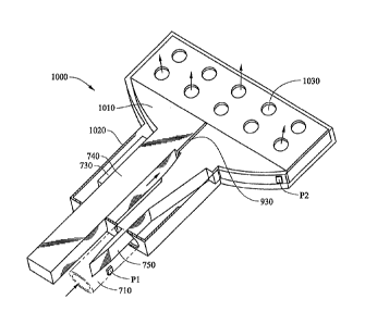
Figure 10 is a perspective view of a lumen clearing valve with hybrid external
member according to the principles of the present invention. The embodiment of
Figure 10 is similar to the embodiment of Figure 9. In Figure 10, a broad
needle head
1010 and additional drainage holes 1030 allow for a wide dispersion of aqueous
in the
drainage location (typically, a subconjunctival bleb). Fluid passes from tube
710 to
the drainage location via microchannels 930, which are typically etched into
needle
head 1010. In Figure 10, housing 1020 has a broad outlet end that includes
multiple
drainage holes 1030. In addition, the broad end of housing 1020 is open to
allow
aqueous to flow through this wide opening. Therefore, in the embodiment of
Figure
10, aqueous flows from the anterior chamber through tube 710, through housing
1020
and out of drainage holes 1030 and the broad end of housing 1020 into the
drainage
location. When needle head 1010 is oscillated, it can serve to clear fibers
from the
drainage location. It can also disperse aqueous to the drainage location.
The embodiments of Figs. 7-10 can be operated in two different modes ¨
lumen clearing mode in which the tapered arm 750 oscillates or moves and valve
mode in which the tapered arm 750 is maintained in a particular position to
restrict
fluid flow through tube 710. In lumen clearing mode, tapered arm 750 is moved
or
oscillated to clear fibrous material from the interior of tube 710 and/or the
drainage
location. In lumen clearing mode, tapered arm '750 can also help to disperse
aqueous
in the drainage location.
When operating as a valve, tapered arm 750 can be maintained in a particular
position to restrict the flow of aqueous through tube 710. The position of
tapered arm
750 can be changed over time based on pressure readings from pressure sensors
P1,
P2, and/or P3 as described above with respect to Figures 3 ¨ 6. In this
manner, any of
the following can be the basis for control of the tapered arm 750: IOP,
pressure in the
bleb, fluid flow rate, etc.
Figure 11A is a diagram of a two lumen valve and pressure sensor system
according to the principles of the present invention. In Figure 11A, tube 710
of the
active valve / lumen clearing system bridges the anterior chamber and a
drainage
location. A second tube 430 includes end cap 510 as described in Figure 5. The
system of Figure 11A combines the pressure sensor of Figures 5 and 6 with the
active
valve / lumen clearing device of Figures 7 ¨ 10, wherein the latter can serve
as control
valve 420. In this manner, one tube (430) can be used to measure IOP, while a
second
tube (710) can be used for draining aqueous. Fluidic communication between a
dry
location 360 and the P3 sensing portion of end cap 510 can be provided by tube
1100.
17
CA 02772609 2012-02-28
WO 2011/034740
PCT/US2010/047605
Figure 11B is another possible arrangement, wherein a single tube resides in
the
anterior chamber 340. In Figure 11B, end cap 510 is located in an opening in
tube
430.
Figures 12A and 12B are cross section views of dual tubing that can be used
with the system of the present invention. In Figure 12A, two lumens, 430 and
710,
are contained in a single tube. Figure 12A shows this dual bore tubing
arrangement.
In Figure 12B, two lumens, 430 and 710, are contained in two separate tubes
that are
joined together. Figure 12B shows this dual-line tubing arrangement. Other
variations of a dual lumen device can also be used in conjunction with the
present
invention.
Figure 13 is a perspective view of a two lumen valve and pressure sensor
system according to the principles of the present invention. In Figure 13, two
tubes,
430 and 710, are connected at one end (the end that resides in the anterior
chamber)
and are separated at the other end (in this case, the end that resides in the
subconjunctival space). Tube 430 has end cap 510 that measures IOP. Tube 710
receives tapered arm 750. Tapered arm 750 can serve to clear the interior of
tube 710.
Tube 750 can also act as a valve that can partially or totally occlude the
interior of
tube 710. Tapered arm 750 is coupled to the any of the systems depicted in
Figures '7
¨ 10. A barrier 350 separates P3 from the outlet of 710, typically the
drainage
location 410. In this manner, P3 is in a "dry" space 360 and measures an
approximation of atmospheric pressure. The outlet end of 710 (shown adjacent
to
tapered arm 750) is located in a "wet" space or drainage location such as 410.
As
noted above, P2 is located in this "wet" space.
Power for the pressure monitoring system or active drainage system may be
supplied by a power source 205 as described above. As shown in Figure 2, power
source 205 is coupled to power generator 1410. One example of power generator
1410 is shown in Figure 14. In Figure 14, power generator 1410 has a micro-
generator 1420 coupled to a rotor 1430. In this example, as rotor 1430 turns,
micro-
generator 1420 produces power. As such, the operation of power generator 1410
is
much like that of any conventional generator. While rotor 1430 is shown as
having
four paddles connected to a shaft, any rotor design may be employed. Moreover,
any
other type of apparatus that converts a fluid flow into power may be employed.
Figure 14 is intended only as one example.
18
CA 02772609 2012-02-28
WO 2011/034740
PCT/US2010/047605
Power generator 1410 is capable of harnessing the aqueous fluid flow from the
anterior chamber 340 to the drainage location 410. Since the general purpose
of any
glaucoma drainage device is to shunt aqueous from the anterior chamber 340 to
a
drainage location 410, aqueous flows from the anterior chamber 340 to the
drainage
location 410 (in this case, through a tube, such as tube 430). There is a
natural
pressure difference between the fluid pressure in the anterior chamber 340 and
the
fluid pressure in the drainage location 410. This pressure difference causes
aqueous
to flow from the anterior chamber 340 to the drainage location 410. Power
generator
1410 converts this aqueous fluid flow into power.
In a typical example, the aqueous flowing through the tube 430 turns rotor
1430 at about 1 revolution per minute based on an aqueous flow rate of about
two
microliters per minute. If the pressure difference between the anterior
chamber 340
and the drainage location 410 is about eight millimeters of mercury, the
transferrable
potential power is about 25 nanowatts (or about two milliJoules of energy) per
day.
This power can be stored in power source 205 and used to power the systems
(pressure sensors, telemetry, active valve, etc.) described in this
application.
Figure 15 is an end view of one embodiment of a rotor according to the
principles of the present invention. In Figure 15, rotor 1430 has a shaft
connected to
four paddles. Rotor 1430 is located in tube 430 to harness the fluid flowing
through
the tube. The arrows denote the direction of aqueous fluid flow through tube
430 and
the corresponding direction of rotation of rotor 1430. As noted, Figure 15
depicts one
of many possible configurations for rotor 1430.
Figure 16 is a diagram of one possible location of a power generator in a
glaucoma drainage system according to the principles of the present invention.
In the
example of Figure 16, power generator 1410 is located in or along tube 430.
Tube
430 shunts the anterior chamber 340 to the drainage location 410. Valve 420 is
located at the end of tube 430 as previously described. In this example, the
power
generated by power generator 1410 is used to power valve 420 (and other
components
of the system).
Figure 17 is a diagram of another possible location of a power generator in a
glaucoma drainage system according to the principles of the present invention.
In the
example of Figure 17, power generator 1410 is located at the end of tube 430.
Here,
power generator 1410 performs two functions: it generates power and it acts as
a
valve. Since power generator 1410 resists the flow of fluid through tube 430,
this
19
CA 02772609 2012-02-28
WO 2011/034740
PCT/US2010/047605
flow resistance can be used to control the rate of aqueous flowing through
tube 430.
In other words, power generator 1410 can be operated as an active valve.
Moreover,
the rotation of the rotor can function to clear the lumen (as described
above).
In the example of Figure 17, the micro-generator 1420 can be controlled to
vary the flow resistance of rotor 1430. When micro-generator 1420 is a simple
magnetic core and coil generator (like the typical electric generator), the
distance
between the magnetic core and the coil can be varied to vary the force
required to turn
rotor 1430. The more force required to turn rotor 1430, the more resistance to
aqueous flowing through tube 430. Conversely, the less force required to turn
rotor
1430, the less resistance to aqueous flowing through tube 430. This resistance
to
aqueous flow can be controlled to maintain a desired IOP.
Regardless of whether the glaucoma drainage device has a power generator or
operates on stored energy, a power savings method of operating the device may
be
beneficial. Figure 18 is a flow chart of one method of operating the glaucoma
drainage device of the present invention so as to conserve power. In 1810, the
system
is powered on. In 1820, IOP is measured. IOP may be measured as described
above.
If IOP is in range, then in 1830, the device powers off (i.e. is in sleep
mode) for a time
X. If IOP is out of range, then in 1840, the valve is adjusted accordingly. In
1850,
IOP is measured. If IOP is in range, then in 1830, the device powers off (i.e.
is in
sleep mode) for a time X. If IOP is out of range, then in 1840, the valve is
adjusted
accordingly. This iterative process can be repeated as necessary to maintain
IOP in a
desired range.
Accordingly, with the operation depicted in Figure 18, the glaucoma drainage
device of the present invention takes periodic IOP measurements and makes
adjustments accordingly. The time interval X between IOP measurements can be
any
time period. For example, IOP measurements can be made every ten minutes or
every
hour. A range of values can be set that determine whether the IOP readings are
in
range or out of range. For example, an IOP above 15 mm Hg may be considered
too
high. If an IOP measurement is taken that is above 15 mm Hg, it is out of
range, and
the valve is adjusted accordingly. The IOP measurement in 1850 may also be
repeated at any time interval to adjust the valve. For example, the IOP
reading in
1850 may be repeated every minute in the process of adjusting the valve.
From the above, it may be appreciated that the present invention provides a
lumen clearing valve that can be controlled by an IOP sensor. The present
invention
CA 02772609 2016-11-01
provides a valve-like device that can clear a lumen, disperse aqueous, and/or
clear fibrous
material from a drainage location. The present invention also provides an
implantable
power generator that can be used to power such a system. The present invention
is
illustrated herein by example, and various modifications may be made by a
person of
ordinary skill in the art.
Other embodiments of the invention will be apparent to those skilled in the
art from
consideration of the specification and practice of the invention disclosed
herein. It is
intended that the specification and examples be considered as exemplary only,
with a true
scope of the invention being indicated by the following claims.
21