Note: Descriptions are shown in the official language in which they were submitted.
CA 02777797 2012-04-16
WO 2011/050393 PCT/AU2010/001399
FLUID LEVEL INDICATOR DETERMINATION
Background. of the Invention
The present invention relates to a method and apparatus for use in analysing
impedance
measurements performed on a subject, and in particular to a method and
apparatus for
determining an indicator using a dispersion parameter, to thereby allow the
indicator to be
used in diagnosing the presence, absence or degree of oedema.
Description of the Prior Art
The reference in this specification to any prior publication (or information
derived from it),
or to any matter which is known, is not, and should not be taken as an
acknowledgment or
admission or any form of suggestion that the .prior publication (or
information derived from
it) or. known matter forms part of the common general knowledge in the field
of endeavour to
which this specification relates.
One existing technique for determining biological parameters relating to a
subject, such as
fluid levels, involves the use of bioelectrical impedance. This involves
measuring the
electrical impedance of a subject's body using a series of electrodes placed
on the skin
surface. Changes in electrical impedance at the body's surface are used to
determine
parameters, such as changes in fluid levels, associated with the cardiac cycle
or oedema, or
other conditions which affect body habitus.
Lymphoedema is a condition characterised by excess protein and oedema in the
tissues as,
result of reduced lymphatic transport capacity and/or reduced tissue
proteolytic capacity in.
the presence of a normal lymphatic load. Acquired, or secondary lymphoedema,
is caused by
damaged or blocked lymphatic vessels. The commonest inciting events are
surgery and/or
radiotherapy. However, onset of lymphoedema is unpredictable and may develop
within days
of its cause or at any time during a period of many years after that cause.
W000/79255 describes a method of detection of oedema by measuring
bioelectrical
impedance at two different anatomical. regions in the same subject at a single
low frequency
CA 02777797 2012-04-16
WO 2011/050393 PCT/AU2010/001399
-2-
alternating current. The two measurements are analysed to obtain an indication
of the
presence of tissue oedema by comparing with data obtained from a normal
population.
W02005/122888 describes a method of detecting tissue oedema in a subject. The
method
includes determining a measured impedance for first and second body segments.
An index
indicative of a ratio of the extra-cellular to infra-cellular fluid is then
calculated for each body
segment, with these being used to determine an index ratio; based on the index
for the first
and second body segments. The index ratio can in turn be used to determine the
presence,
absence or degree of tissue oedema, for example by comparing the index ratio
to a reference
or previously determined index ratios.
1o W02008/138602 describes a method for use in analysing impedance
measurements
performed on a subject, the method including, in a processing system
determining at least one
impedance - value, representing the impedance of at least a segment of the
subject,
determining an indicator indicative of a subject parameter using the at least
one impedance
value and a reference and displaying a representation of the indicator.
Summary of the Present Invention
It is an object of the present invention to substantially overcome, or at
least ameliorate, one or
more disadvantages of existing arrangements.
In a first broad form the present invention seeks to provide a method for use
in analysing
impedance measurements performed on a subject, the method. including, in a
processing
system:
a) determining at least one impedance value at each of a number of
frequencies,- each
impedance value representing the impedance of a segment of the subject;
b) determining a dispersion parameter value indicative of adispersion of the
impedance
values; and,
c) determining an indicator based at least in part on the dispersion parameter
value.
Typically the method includes, in the processing system:
a) determining first and second dispersion parameter values for first and
second body
segments respectively; and,
CA 02777797 2012-04-16
WO 2011/050393 PCT/AU2010/001399
-3-
b) determining the indicator using the first and second dispersion parameter
values.
Typically the first body segment is an affected body segment and the second
body segment is
an unaffected body segment.
Typically at, least one of the body segments is'a dominant limb and the other
body segment is
a non-dominant limb.
Typically the first body segment is a different body segment to the second
body segment..
Typically the method includes, in the processing. system:
a) determining a predicted dispersion parameter value for the first body
segment using
the second dispersion parameter value;
b) determining the indicator using the first and predicted dispersion
parameter values.
Typically predicted dispersion parameter value is determined to take into
account at least one
of:
a) limb dominance; and,
b) differences in limb types.
Typically the method includes, in the processing system, determining a
predicted dispersion
parameter value using at least one reference value derived from a reference
normal
population.
Typically the reference normal population is selected based on at least one
of.
a) limb dominance;
b) differences in limb types;
c) ethnicity;
d) age;
e) gender;
f) weight; and,
g) height..
CA 02777797 2012-04-16
WO 2011/050393 PCT/AU2010/001399
-4-
Typically the at least one reference value is determined based on a linear
regression of first
and second dispersion parameter values measured for the reference normal
population.
Typically the method includes, in the processing system, determining the
predicted
dispersion parameter value using an equation of the form:
= DPP = aDP2 + K. ,
where: DP2 is the second dispersion parameter value
DPp is the predicted dispersion parameter value
a is a multiplier reference value determined based on a
relationship between first and second dispersion parameter
values in a reference population
K is a constant reference value determined based on a
relationship between first and second dispersion parameter
values, in a reference population
Typically, for a male subject, the predicted value for a leg segment based on
second
dispersion parameters for an arm segment is based on:
a) a value of a in the range 0.15 to 0.022; and,
b) a value of K in the range 0.62 to 0.72.
Typically, for. a female subject, the predicted value for a leg segment based
on second
dispersion parameters for an arm segment is based on:.
a) a value of a in the range 0.44 to 0.41; and,
b) a value of K in the range 0.43 to 0.46.
Typically the method includes, in the processing system, determining the
indicator using the
equation:
1 _ sf x DPP - DP,
3SE
where: Ind is the indicator
DPI is a dispersion parameter value determined for the body
segment
CA 02777797 2012-04-16
WO 2011/050393 PCT/AU2010/001399
-5-
DPp is a predicted dispersion parameter value for the body
segment
sf is a scaling factor
SE is a standard error 'determined based on dispersion
parameter values in a reference population
Typically the method includes, in the processing system, determining the
indicator using the
equation:
1nsfx(DPP-DPI)
3SE
where: DP, is the mean dispersion parameter value for a reference
normal population
DP1 is a dispersion parameter value determined for the body
segment
sf is a scaling factor
SE is a standard error determined for the dispersion parameter.
values for the reference population
Typically the scaling factor is selected so that a threshold value indicative
of the presence or
absence of oedema is an integer value.
Typically the method includes, in the processing system, determining' the
indicator based on
the equation:
Ind = sf (DP2 - DP,
where: Ind is the indicator
DPl is a first dispersion parameter value for a first body
segment
DP2 is a second dispersion parameter value for a second body
segment
sf is a scaling factor
CA 02777797 2012-04-16
WO 2011/050393 PCT/AU2010/001399
-6-
Typically the dispersion parameter value is indicative of the distribution of
impedance
measurements for the respective body segment.
Typically the dispersion parameter is based'on the value of at least one of-
DP = (RO - R.).
XC
X,
DP =
(R0 - R-0)
DP=(R.-1? )
XC
X,
DP =
(Rõ -R0)
where: Rte= impedance at infinite applied frequency;
R0 = impedance at zero applied frequency;
X = reactance at the centre of the circle.
Typically the dispersion parameter is based on the value of:
__ 2 atan (RO --R.)
a 2IX,I
Typically the indicator is at least one of.
a) an oedema indicator for use in assessing a presence, absence or degree of
oedema in
the subject.
b) a hydration indicator for use in assessing hydration levels in a subject.
Typically the method includes, in the processing system, displaying a
representation of the
indicator.
Typically representation of the indicator includes a linear scale including:
a) a linear indicator;
b) a scale; and,
c). a pointer, the pointer being positioned on the scale in accordance with
the. indicator.
CA 02777797 2012-04-16
WO 2011/050393 PCT/AU2010/001399
-7-
Typically the method includes, in .the processing system, displaying a
representation
including an indication of a change in indicator value from at least one of a
previous indicator
value and a baseline indicator value.
Typically the method includes, in the processing system:
a) determining at least one threshold using a reference; and,
b) displaying the threshold as part of the representation.
Typically the method includes, in the processing system:
a). determining two thresholds using a reference; and,
b) displaying the thresholds on the representation,. the,thresholds being
indicative of a
normal range.
Typically the method includes, in the processing system, displaying, on the
representation, at
least one of:
a) a normal range;
b) an intervention range;
c) a hydration range; and,
d) an oedema range.
Typically the method includes in the processing system, causing one. or more
impedance
measurements to be performed.
Typically the method includes, in the processing system:
a) causing at least one excitation signal to be applied to the subject;
b) determining at least one signal measured across the subject; and,
c) determining at least one impedance value using an indication of the
excitation signal
and the signal measured across the subject.
Typically the method includes, in the processing system:
. a) controlling a signal generator to thereby cause the at least one
excitation signals to be
applied to the subject; and,
b) determining the at least one signal measured across the subject using a
sensor.
CA 02777797 2012-04-16
WO 2011/050393 PCT/AU2010/001399
-8-
In a second broad form the present invention seeks to provide apparatus for
use in analysing
impedance measurements performed on a subject, the apparatus including a.
processing
system for:
a) determining at least one impedance value at each of a number of
frequencies, each
impedance value representing the impedance of a segment of the subject;
b) determining a dispersion parameter value indicative of a dispersion of the
impedance
values; and,
c) determining an indicator based at least in part on the dispersion parameter
value.
Typically the apparatus includes:
a) a signal generator for applying one or more electrical signals to the
subject using a
first set of electrodes;
b) a sensor for measuring electrical signals across a second set of electrodes
applied to
the subject; and,
c) a controller for:
i) controlling the signal generator; and,
ii). determining the indication of the measured electrical signals.
Typically the controller includes the processing system.
Typically the processing system includes the controller.
In a third broad form the present invention seeks to provide a method for use
diagnosing the
presence, absence or degree of oedema in a subject by using impedance
measurements
performed on the subject, the method including, in a processing system:
a) determining at least one impedance value at each of a number of
frequencies, each
impedance value representing the impedance of a segment of the subject;
b) determining adispersion parameter value indicative of a dispersion of the
impedance
values;
c) determining an indicator based at least in part on the dispersion parameter
value; and,
d) displaying a representation of the indicator, to thereby allow the
presence, absence or
degree of oedema in the subject to be assessed.
CA 02777797 2012-04-16
WO 2011/050393 PCT/AU2010/001399
-9-
It will be appreciated that the broad forms of the invention may be used
individually or in
combination, and may be used for diagnosis of the presence, absence or degree
of a range of
conditions and . illnesses, including, but not limited to oedema, lymphoedema,
body
composition and the like.
Brief Description of the Drawings
An example of the present invention will now be described with reference to
the
accompanying drawings, in which:
Figure 1 is a schematic of an example of impedance determination apparatus;
Figure 2 is a flowchart of an example of a process for determining an
indicator;
Figure 3A is a schematic of an example of a theoretical equivalent circuit for
biological
tissue;
Figure 3B is an example. of a locus of impedance known as a Wessel plot;
Figure 4 is a flowchart of an example of a process for determining an oedema
indicator for
limb oedema;
Figures 5A and 5B are diagrams of examples of electrode positions for use in
measuring limb
impedances;
Figures 5C and 51) are schematic diagrams of examples of electrode positions
for use in
measuring limb impedances;
Figure 6A to 6C are schematic diagrams of first examples of representations of
oedema
indicators;
Figure 7 are graphs of examples of the relationship of parameters between like
limbs and
dislike limbs; and,
Figure 8 is a graph of example measurements of leg a and arm a for healthy
female dominant
arms and legs.
Detailed Description of the Preferred Embodiments
An example of apparatus suitable for performing an analysis of a subject's
bioelectric_
impedance=will now be described with reference to Figure 1.
CA 02777797 2012-04-16
WO 2011/050393 PCT/AU2010/001399
-10-
As shown the apparatus includes a measuring device 100 including a processing
system 102,
connected to one or more signal generators 117A, 117B, via respective first
leads 123A,
123B, and to one or more sensors 118A, 118B, via respective second leads 125A,
125B. The
connection may be via a switching device, such as a multiplexer, although this
is not
essential.
In use, the signal generators 117A, 117B are coupled to two first electrodes
113A, 113B,
.which therefore act as drive electrodes to allow signals to be applied to the
subject S, whilst
the one or more sensors 118A, ' 118B are coupled to the second electrodes
115A, 115B, which
act as sense electrodes, allowing signals across the subject S to be sensed.
The signal generators 117A, 117B and the sensors 118A, 118B may be provided at
any
position between the processing system 102 and the electrodes 113A, 113B, 11
5A, 115B, and
may be integrated into the measuring device 100. However, in one example, the
signal
generators 117A, 117B and the sensors 118A, 118B are integrated into an
electrode system,
or another unit provided near the subject S, with the leads 123A, 123B, 125A,
125B
connecting the signal generators 117A, 117B and the sensors 118A, 118B to the
processing
system 102.
It will be appreciated that the above described system is a two channel
device, used to
perform a classical . four-terminal impedance measurement, with each channel
being
designated by the suffixes A, B respectively. The use of a two channel device
is for the.
20. purpose of example only, and multiple channel devices can alternatively be
used to allow
multiple body segments to be measured without requiring .reattachment of
electrodes. An
example of such a device is described in copending patent application' number
W02009059351.
An optional external interface 103 can be used to couple the measuring device
100, via
wired, wireless or network connections, to one or more peripheral devices 104,
such as an
external database or computer system, barcode scanner, or the like. The
processing system
102 will also typically include an I/O device 105, which may be of any
suitable form such as
a touch screen, a keypad and display, or the like.
CA 02777797 2012-04-16
WO 2011/050393 PCT/AU2010/001399
-11-
In use, the processing system ' 102 is adapted to generate control signals,
which cause the
. signal generators 117A, 117B to generate one or more. alternating signals,
such as voltage or
current signals of an appropriate waveform, which can be applied to a subject
S, via the first
electrodes 113A, 113B. The sensors 118A, 118B then determine the voltage
across or
current through the subject S, using the second electrodes 115A, 115B and
transfer
appropriate signals to the processing system 102.
Accordingly, it will be appreciated that the processing system 102 may be any
form of
processing system which is suitable for generating appropriate control signals
and at least
partially interpreting the measured signals to thereby determine the subject's
bioelectrical
impedance, and optionally determine other information such as relative fluid
.levels, or the
presence, absence or degree of conditions, such as oedema, lymphoedema,
measures of body
composition, cardiac function, or the, like.
The processing system 102 may therefore be a suitably programmed computer
system, such
as a laptop, desktop, PDA, smart. phone or the like. Alternatively the
processing system. 102
may be formed from specialised hardware, such as an. FPGA (field programmable
gate
array), or a combination of a programmed computer system and specialised
hardware, or the
like, as will be described in more detail below.
In use, the first electrodes 113A, 113B are positioned on the subject to allow
one or more
signals to be injected into the subject S. The location of the first
electrodes will depend on
the segment of the subject S under study. Thus, for example, the first
electrodes 113A, 113B-
can be placed on the thoracic and neck region of the subject S to allow the
impedance of the
chest cavity to be determined for use in cardiac function analysis.
Alternatively, positioning
electrodes on the wrist and ankles of a subject allows 'the impedance of limbs
and/or the
entire body to be determined, for use in oedema analysis, or the like.
Once the electrodes are positioned, one or more alternating. signals are
applied to the subject
S, via the first leads 123A, 123B and the first electrodes 113A, 113B. The
nature of the
alternating signal will vary depending on the nature of the measuring device
and the
subsequent analysis being performed.
CA 02777797 2012-04-16
WO 2011/050393 PCT/AU2010/001399
-12-
For example, the system can use Bioimpedance Spectroscopy (BIS) in which
impedance
measurements are performed at each of a number of frequencies ranging from
very low
frequencies (4 kHz) to higher frequencies (1000 kHz), and can use as many as
256 or more
different frequencies within this range.. Such measurements can be performed
by applying a
signal which is a superposition of plurality of frequencies simultaneously, or
a number of
alternating signals at different frequencies sequentially, depending on the
preferred
implementation. The frequency or frequency range of the applied signals may
also depend
,on the analysis being performed.
In one example, the applied signal is generated by a voltage generator, which
applies. an
alternating voltage to the subject S, although alternatively current signals
may be applied. In
one example, the voltage source is typically symmetrically arranged, with each
of the signal
generators 117A, 117B being independently controllable, to allow the signal
voltage across
the subject to be varied.
A voltage difference and/or current is measured between the second electrodes
II5A, 115B.
15. In one example, the voltage is measured differentially, meaning that each
sensor 118A, 118B
is used to measure the voltage at each second electrode I I5A, 115B and
therefore need only
measure half of the voltage as compared to a single ended system.
The. acquired signal and the, measured signal will be a superposition of
voltages generated by
the human body;. such as the ECG (electrocardiogram), voltages generated by
the applied
signal, and other signals caused by environmental electromagnetic
interference.
Accordingly, filtering or other, suitable analysis may be employed to remove
unwanted
components.
The acquired signal is typically demodulated to obtain the impedance of the
system at the
applied frequencies. One suitable method for demodulation of superposed
frequencies is to
use a Fast Fourier Transform (FFT) algorithm to transform the time domain data
to the
frequency domain. This is typically used when the applied current signal is a
superposition
of applied frequencies. Another technique not requiring windowing of the
measured signal is
a sliding window FFT.
CA 02777797 2012-04-16
WO 2011/050393 PCT/AU2010/001399
-13-
In the event that the applied current signals are formed from a sweep of
different frequencies,
then it is.more typical to use a signal. processing technique such as
multiplying the measured
signal with a reference sine wave and cosine wave derived from the signal
generator, or with
measured sine and cosine waves, and integrating over a whole number of cycles.
This
process, known variously as quadrature. demodulation or synchronous detection,
rejects all
uncorrelated or asynchronous signals and significantly reduces random noise.
Other suitable digital and analogue demodulation techniques will be known to
persons skilled
in the field.
In the case of BIS, impedance or admittance measurements are determined from
the signals
io at each frequency by comparing the recorded voltage and the current through
the subject. The
demodulation algorithm can then produce amplitude and phase signals at each
frequency,
allowing an impedance value at each frequency to be determined.
As part of the above described process, the distance between the second
electrodes 115A,
115B may be measured and recorded. Similarly, other parameters relating to the
subject may
5 be recorded, such as the height, weight, age, sex, health status, any
interventions .and the date
and time on which they occurred. Other information, such as current
medication, may also be
recorded. This can then be used in performing further analysis of the
impedance
measurements, so as to allow determination of the presence, absence or degree
of oedema, to
assess body composition, or the like.
20 The accuracy of the measurement of impedance, can be subject to a number of
external
factors. These can include, for example, the effect of capacitive coupling
between the subject
and the surrounding environment, the leads and the subject, the electrodes, or
the like, which
will vary based on factors such as lead construction, lead configuration,
subject position, or
the like. Additionally, there are typically variations in. the impedance of
the electrical
25 connection between the electrode surface and the skin (known as the
"electrode impedance"),
which can depend on factors such as skin moisture levels,. melatonin levels,
or the like. A
further source of error is the presence of inductive coupling between
different electrical
conductors within the leads, or between the leads themselves.
CA 02777797 2012-04-16
WO 2011/050393 PCT/AU2010/001399
-14-
Such external factors can lead to inaccuracies in the measurement process and
subsequent
analysis and accordingly, it is desirable to be able to reduce the impact of
external factors on
the measurement process.
One form of inaccuracy that can arise is caused by the voltages across the
subject being
unsymmetrical, a situation referred to as an "imbalance". Such a situation
results in a
significant signal voltage at the subject's body centre, which in turn results
in stray currents
arising from parasitic capacitances between the subject's torso and the
support surface on
which the subject is provided.
The presence of an imbalance, where the voltage across the subject is not
symmetrical with
io respect to the effective centre of the subject, leads to a "common mode",
signal, which is
effectively a measure of the signal at the subject. S that is unrelated to the
subject's
impedance
To help reduce this effect, it is therefore desirable for signals to be
applied to the subject S
that they result in a symmetrical voltage about the subject's body centre. As
a result, a
1.5 reference voltage within the subject S, which is equal to a reference
voltage of the
measurement apparatus, will be close to the effective body centre of the
subject, as
considered relative to the electrode placement. As the measuring device
reference voltage is
typically ground, this results in the body centre of the subject S being as
close to ground as
possible, which minimises the overall signal magnitude across the subject's
torso, thereby
20 minimising stray currents.
In one example, a symmetrical voltage about the sensing electrodes can be
achieved by using
a symmetrical voltage source, such as a differential bidirectional voltage
drive scheme, which
applies a symmetrical voltage to each of the drive electrodes 113A, 113B.
However, this is
not always- effective if the contact impedances for the two drive electrodes
113A, 113B are
25 unmatched, or if the impedance of the subject S varies along the length of
the subject S,
which is typical in a practical environment.
In one example, the apparatus overcomes this by adjusting the differential
voltage drive
signals applied to each of the drive electrodes 113A, 113B, to compensate for
the different
CA 02777797 2012-04-16
WO 2011/050393 PCT/AU2010/001399
- 15-
electrode impedances, and thereby restore the desired symmetry of the voltages
across the
subject S. This process is referred to herein as balancing and in one example,
helps reduce
the magnitude of the common mode signal, and hence reduce current losses
caused by
parasitic capacitances associated with the subject.
The degree of imbalance, and hence the amount of balancing required, can be
determined by
monitoring the signals at the sense electrodes 115A, 115B, and then using
these signals to
control the signal applied to the subject via the drive electrodes 113A, 113B.
In particular,
the degree of imbalance can be calculated. by determining an additive voltage
from the
voltages detected at the sense electrodes 115A, 115B.
1o In one example process, the voltages sensed at each of the sense electrodes
11 5A, 115B are
used to calculate a first voltage, which is achieved by combining or adding
the measured
voltages. Thus, the first voltage can be an additive voltage (commonly
referred to as a'
common mode voltage or signal) which can be determined using a differential
amplifier.
In this regard, a differential amplifier is typically used to combine two
sensed voltage signals
VQ, Vb, to determine a second voltage, which in one example is a voltage
differential VQ Vb
across the points of interest on the subject S. The voltage differential is
used in conjunction
with a measurement of the current flow through the subject to derive impedance
values.
However, differential amplifiers typically also, provide a "common mode"
signal (VQ+Vb)/2,
which is a measure of the common mode signal.
Whilst differential amplifiers include a common mode rejection capability,
this is generally
of only finite effect and typically reduces in effectiveness at higher
frequencies, so a large
common mode signal will produce an error signal superimposed on the
differential signal.
The error caused by common mode signals can be minimised by calibration of
each sensing
channel. In the ideal case where both inputs of a differential amplifier are
perfectly matched
in gain and phase characteristics and behave linearly with signal amplitude,
the common
mode error will be zero. In one example, the two sensing channels of the
differential
amplifier are digitised before differential processing. It is therefore
straightforward to apply
CA 02777797 2012-04-16
WO 2011/050393 PCT/AU2010/001399
-16-
calibration factors independently to each channel to allow the characteristics
to be matched to
a high degree of accuracy, thereby achieving a low common mode error.
Accordingly, by determining the common mode signal, the applied voltage
signals can be
adjusted, for example by adjusting the relative magnitude and/or phase of the
applied signals,.
to thereby minimise the common mode signal and substantially eliminate any
imbalance.
An example of this process is described in more detail in copending patent
application
number W02009059351.
An example of the operation of the apparatus in analysing impedance
measurements will now
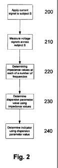
.be described with reference to Figure 2.
In one example, at step 200 the processing system 102 causes a current signal
to be applied to
the subject S, with the induced voltage across the subject S being measured at
step 210, with
signals representing the measured voltage and the applied current being
returned to the
processing system 102 for analysis.
.When the process is being used to determine an oedema indicator, this is
typically performed
for at least a segment of the subject S that is suspected of being susceptible
to oedema, and
may also be repeated for a separate healthy segment of the subject.. Thus, for
example, in the
case of limb oedema, this is typically performed on the affected or "at risk"
limb (hereinafter
generally referred to as the "affected" limb), and a limb that is deemed "not
at risk" of
oedema (hereinafter generally referred to as the "unaffected" limb).
It will be appreciated that the application of the current and voltage signals
may be controlled
by a separate processing system that is used in performing the. analysis to
derive an indicator,
and that the use of a single processing system is for. the purpose of example
only.
At step 220, measured voltage and current signals are used by the processing
system 102 to
determine impedance values at each of a number of applied frequencies. In one
example, this
includes first impedance values representing the impedance of the unaffected
limb and
second impedance values representing the impedance of the affected limb.
CA 02777797 2012-04-16
WO 2011/050393 PCT/AU2010/001399
-17-
At step 230, the one or more impedance values are used by the processing
system 102, to
determine a dispersion parameter value. In one example, first and second
dispersion
parameter values of affected and unaffected limbs may be determined.
The nature of the dispersion parameter can vary, but in general this
represents a distribution
of the impedance measurements about an ideal model.
In ,one example, the dispersion. parameter DP can be given by or based on the
value:
DP X` (1)
(R0 - R.)
Where:R. = impedance at infinite applied frequency;
Ro = impedance at zero applied frequency;
X, = reactance at the centre of the circle.
It should be noted that the, value of XX will be negative, due to it's
position below the circle,
and this can lead to the value of the dispersion parameter being negative.
However, it will be
appreciated that alternative formulations may also be used, such as those set
out below, and
accordingly, the dispersion parameter can be arranged to have either a
positive or negative
value, as desired:
DP=(Ro-R )
X, (IA)
DP = (R. - Ro )
X~ (I B)
DP= X` (IC)
(R. - Ro)
The alternative formulations can be used to ensure that the value of the
dispersion parameter
increases in the event that the subject has oedema, although this is not
essential and any
suitable formulation may be selected.
In one particular example, the dispersion parameter DP value is given by a
value a:
CA 02777797 2012-04-16
WO 2011/050393 PCT/AU2010/001399
-18-
a = 2 arctan (R2 - (2)
1 cl
In this regard, Figure 3A is'an example of an equivalent circuit that.
effectively models the
electrical behaviour of biological tissue. The equivalent circuit has two
branches that
represent current flow through extracellular fluid and intracellular fluid,
respectively. The
5' extracellular fluid component of biological impedance is represented by an
extracellular
resistance Rei whilst the intracellular fluid component is represented by an
intracellular
resistance R; and a capacitance C representative of the cell membranes.
The relative magnitudes of the extracellular and intracellular components of
impedance of an
alternating current (AC) are frequency dependent. At zero frequency the
capacitor acts as a
perfect insulator and all current flows through the extracellular fluid, hence
the resistance at
zero frequency, R0, equals the extracellular resistance Re. At infinite
frequency the capacitor
acts as a perfect conductor and the current passes through the parallel
resistive combination.
The resistance at infinite frequency R.. is given by:
R Re R` (3)
Re + R;
Accordingly, the impedance of the equivalent circuit of Figure 3A at an
angular frequency w,
where w=2n*frequency, is given by:
Z=R~+ Ro=R. (4)
1+(jwr)
where: R.= impedance at infinite applied frequency
Ro = impedance at zero applied frequency = Re and,
r is the time constant of the capacitive circuit..
However, the above represents an idealised situation which does not take into
account the
fact that the cell membrane is an* imperfect capacitor. Taking this into
account leads to a
modified model in which:
CA 02777797 2012-04-16
WO 2011/050393 PCT/AU2010/001399
-19-
Ro-R.
Z=R~+ (5)
1+(jtor)a
where: a has a value between 0 and I and can be thought of as an
indicator of the deviation of a real system from the ideal model.
An example of the typical multi-frequency impedance response is shown in
Figure 3B. As
frequency increases, the reactance increases to a peak and then decreases
while the resistance
continually decreases. This results in a circular locus with the centre of the
circle below the x
axis, as shown.
The a parameter is related to the depression of the Cole plot below the zero
reactance axis.
The value of a is indicative of the deviation from the ideal Cole equation (4)
and is closely
related to the spectral width of the distribution of relaxation times. This
dispersion may be
due to molecular interactions, cellular interactions, anisotropy and cell size
as described in
Grimnes, S. and 0. G. Martinsen (2000). Bioimpedance and Bioelectricity
Basics, Academic
Press.
As described above, the value of the impedance parameter R0 is closely related
to extra-
cellular fluid levels while the impedance parameter value Rõ is closely
related to.the total
body fluid levels. Accordingly, (R0 - Rõ) is closely related to the intra-
cellular fluid levels.
The reactance XX is the reactance X at the centre of the circle which is a
direct measure of the
depression of the circular locus below the axis. It is closely related to the
reactance at the
characteristic frequency by the subtraction from the radius of the locus. At
the characteristic
frequency, the ratio of the current flow through the intra and extra cellular
fluids is
determined as a function of the ratio of the intra to extra. cellular
resistance, so that it is
independent. of the capacitance of the cell membrane. Accordingly, the
reactance at the
characteristic frequency can be used more accurately as an indicator of extra-
cellular fluid .
levels. Since the reactance at the centre of the circle is directly related to
the characteristic
reactance, this value is also related to the intra and extra cellular fluid.
Accordingly;, the dispersion parameter is not only related to. a ratio of
extra-cellular to intra-
cellular fluid levels, but also takes into account deviation from an idealised
equivalent circuit
CA 02777797 2012-04-16
WO 2011/050393 PCT/AU2010/001399
-20-
reflecting the distribution of relaxation times within the subject-and so
encompasses changes
in cell structure. In contrast, if a direct ratio of intra-cellular to extra-
cellular fluid is used as
an index I this is typically calculated as shown in equation (6) below and
therefore does not
account for such deviations to the same extent.
I= Ri= R R~ (6)
Rr Ro-
As lymphoedema is characterised by the accumulation of extra-cellular fluid,
the dispersion
parameter is different between a healthy and oedema affected population, with
the difference
being more readily quantifiable than if a direct ratio of intra-cellular to
extra-cellular, given
by equation (6) is used.
An example of the propagation of errors in the calculation of a dispersion
parameter alpha
and a ratio of intra-cellular to extra-cellular fluid will. now be described.
In order to
determine the propagation of errors, it is necessary to take into account
typical measurement
errors in different frequency ranges for a typical measurement device, and
these are shown in
Table 1 below.
Table 1
Frequency Range Body Impedance Impedance Error Phase Error
3 - 100 kHz 200 -1100 Ohms 1% +/- 1%
100 -1000 kHz 200 -1100 Ohms +/-2%. +/-2%,
In this instance, the relative error in the index I from. equation (6) is
given by:
I _ OR. ARE, + OR.
I' R. + Ro + R.
Similarly the relative error in the indicator Ind from equation (1) is given
by:
CA 02777797 2012-04-16
WO 2011/050393 PCT/AU2010/001399
-21-
AInd AXE, ARo +AR.
Ind X( R - R-
Given the typical errors specifications for the measuring devices outlined in
Table I above,
and taking example .leg impedance measurements, this leads to example
impedance
parameter values of-
Ro = 37252 1%.
R. = 2530 2%
Xc =-280 1%
Accordingly, this leads to errors of:
AI -2+0.01.372+0.02.253 =201%
1 372+253
AInd =1 + 0.01.372 + 0.02.253 =1.07%
Ind 372-253
This demonstrates that the alpha parameter can be determined more accurately.
and should
therefore.be more sensitive to fluid level changes within the subject, and
hence the presence
absence or degree of oedema.
At step 240, the dispersion parameter can be used to determine an indicator.
In one example,-
the indicator provides information relating to the subject, such as an
indication of fluid levels
within the subject. In one example, the indicator is in the form of .a
numerical value that
depends on a reference, and which can be used to determine the presence,
absence or degree
of a condition, such as oedema.
In one particular example, the reference is at least partially based on the
dispersion parameter
of an unaffected body segment. In particular, if the affected body segment
does not in fact
have oedema, then the dispersion parameter will be similar to the dispersion
parameter for
the unaffected body segment, thereby minimising a difference between first and
second
dispersion parameters. In contrast if the affected body segment has oedema the
fluid levels
CA 02777797 2012-04-16
WO 2011/050393 PCT/AU2010/001399
-22-
will differ to the fluid levels in the unaffected body segment, meaning that
the difference
between the first and second dispersion parameters increases. As a result, the
magnitude of
the difference in first and second dispersion parameters between first and
second body
segments can be indicative of the presence, absence or degree of oedema.
Accordingly, by
comparing the difference between the dispersion parameters of affected and
unaffected body
segments to a threshold amount, then this can therefore be used to determine
the presence,
absence or degree of oedema.
In one example, the difference is scaled by a. scaling factor so that the
indicator and the
threshold. can be a memorable value, such as an integer value, or the like.
This can be
achieved by calculating an indicator as follows:
Ind = sf (DP2 - DP,) , (7)
where: Ind is the indicator
DP1 is the first dispersion parameter value of the affected body
segment
DP2 is the second dispersion parameter value of the unaffected
body segment
sf is a scaling factor
However, a population study of healthy subjects has found inherent differences
in fluid levels
between different body segments, such as limbs, even in unaffected
individuals. This can
include slight differences in dispersion parameters due to limb dominance in
limbs as well as
differences arising if the unaffected and affected limbs are of different limb
types, such as
arms and legs. For example, the dispersion parameter for a subject's leg will
typically differ
to that of the subject's arm, even in the. absence of oedema in both limbs.
Accordingly, when calculating the reference it is typical to determine a
predicted dispersion
parameter value for the affected limb based on the second dispersion parameter
value
determined for the unaffected limb. This is usually achieved using at least
one reference
value derived from a reference normal population, allowing the natural
variations between
limbs due to gender, limb dominance and different limb types to be
accommodated.
CA 02777797 2012-04-16
WO 2011/050393 PCT/AU2010/001399
-23-
In one particular example, the predicted dispersion parameter value is
calculated based on
parameters derived by performing a linear regression of first and second
dispersion parameter
values measured fora reference population. The predicted dispersion parameter
value can
then be determined using an equation of the form:
DPP = aDPZ + K (8)
where: DP2 is the second dispersion parameter value
DPp is the predicted dispersion parameter value
a is a multiplier reference value determined based on a
relationship between first and . second dispersion parameter
values for a reference population
K is a constant reference value determined based on a
relationship between first and second dispersion parameter
values for the reference population
In one example, for a' male subject, the predicted value for a leg segment
based on second
dispersion parameters for an arm segment is based on a value of a in the range
0.15 to 0.022,
and a value of K in the range 0.62 to 0.72. For a female subject, the
predicted value for a leg
segment based on second dispersion parameters for an arm segment is based on a
value of a
in the range 0.44 to 0.41, and a value of K in the range 0.43 to 0.46.
When a predicted dispersion parameter value is used, the indicator can be
determined using
the equation:
sf x (DP - DP
Ind = " 1 (9)
3SE
where: Ind is the indicator
DP1 is a dispersion parameter value determined for the body
segment
DPP is a predicted dispersion parameter value for the, body
segment
sf is a scaling factor
CA 02777797 2012-04-16
WO 2011/050393 PCT/AU2010/001399
-24-
SE is a standard error determined based on dispersion
parameter values in a reference population
It should be noted that in the event that measurements are made for an
affected body segment
only, then the predicted dispersion parameter value could alternatively be
based on a mean
value obtained from a reference population, leading to an indicator of the
form:
sf x(DP -DP')
Ind = 3SE (10)
where: DPP is. the mean dispersion parameter value for a reference
normal population
sf is a scaling factor
10, SE is a standard error determined based on dispersion
parameter values for the reference population,
Accordingly, it will be appreciated that the, above described dispersion
parameter can be used
in diagnosing the presence, absence.or degree of oedema. Furthermore, in
contrast to prior
art techniques, a dispersion parameter tends to provide more reliable results,
as will be
15, discussed in more detail below.
In the above examples, it will be appreciated that the order of the dispersion
parameters could
be reversed, so that for example in equation (9) the predicted value could be
subtracted from
the measured value and this will depend on the nature of the dispersion
parameter used, so
for example whether the dispersion parameter is based on equations (1), (1A),
(1B); (1C), (2),
20 or variations thereof. In general the order used will be selected so that
the indicator Ind
increases in magnitude as the level of oedema increases, however this is not
essential and any
suitable arrangement may be used.
An example of the process for performing impedance measurements to determine
an
indicator for limb oedema will now be described in more detail with reference
to Figure.4.
25 In this.example, at step 400 subject details are determined and provided to
the processing
system 102. The subject details will typically include information such as
limb dominance,
details of any medical interventions, as well as information regarding the
subject such as the
CA 02777797 2012-04-16
WO 2011/050393 PCT/AU2010/001399
-25-
subject's age, weight, height, sex, ethnicity or the like. The subject details
can be used in
selecting a suitable reference normal population, as well as for generating
reports, as will be
described in more detail below.
It will be appreciated that the subject details may be supplied to the
processing system 102
via appropriate input means, such as the 1/0. device 105. Thus, each time a
subject
measurement is performed this information can be input into the measuring
device 100.
However, more typically the information is input a single time and stored in
an appropriate
database, or the like, which may be connected as a peripheral device 104 via
the external'
interface 103. The database can include subject data representing the subject
details, together
with information regarding previous oedema indicators, baseline measurements
or impedance
measurements recorded for the subject.'
In this instance, when the operator is required to provide subject details,
the operator can use
the processing system 102 to select.a search database option allowing the
subject details to be
retrieved. This is typically performed on the basis of a subject identifier,
such as a unique
number assigned to the individual upon admission to a medical institution, or
may
alternatively be performed on the basis of name or the like. Such a database
is generally in
the form of an HL7 compliant remote database, although any suitable database
may be used.
In one example, the subject can be provided with a wristband or. other device,
which includes
coded data indicative of the subject identifier. In this case, the measuring
device 100 can be
coupled to a peripheral device 104, such as a barcode or RFID (Radio Frequency
Identification) reader allowing the subject identifier to be detected and
provided to the
processing system 102, which in turn allows the subject details to be
retrieved from the
database. The processing system 102 can then display an indication of the
subject details
retrieved from the database, allowing the operator to review these and confirm
their accuracy
before proceeding further.
At step 410 the affected limb, or "at risk" limb, is determined. This may be
achieved in any
one of a number of ways depending on the preferred implementation. Thus, for
example, the
affected limb can be indicated through the use of appropriate input means,
such as the 1/0
CA 02777797 2012-04-16
WO 2011/050393 PCT/AU2010/001399
-26-
device 105. Alternatively this information can be derived directly from the
subject details,
which, may include an indication of the affected limb, or details of any
medical interventions
performed, which are in turn indicative of the affected limb.
At step 420 an operator positions the electrodes on the subject S, and
connects the leads 123,
124, 125, 126, to allow the impedance measurements to be performed. The
general
arrangement is to provide electrodes on the hand at the base of the knuckles
and between the
bony protuberances of the wrist, as shown in Figure 5A, and on the feet at the
base of the toes
and at the front of the ankle, as shown in Figure 5B. The configurations shown
in Figures 5C
and 5D allow the right arm 531 and the right leg 533 to be measured
respectively, and it will
be appreciated that equivalent arrangements can be used to measure the
impedance of the left
leg and left arm.
It will be appreciated that this configuration uses the theory of equal
potentials, allowing the
electrode positions to provide reproducible results for impedance
measurements. For example
when current is injected between electrodes 113A and 113B in Figure 5C, the
electrode 115B
could be placed anywhere, along the left arm 532, since the whole arm is at an
equal potential.
This is advantageous as it greatly reduces the variations in measurements
caused by poor
placement of the electrodes by the operator. It also greatly reduces the
number of electrodes
required to perform segmental body measurements, as well as allowing the
limited
connections shown to be used to measure each limb separately.
However, it will be appreciated that any suitable electrode and lead
arrangement may be
used.
At step 430 the impedance of the affected and unaffected limbs are measured.
This is
achieved by applying one or more current signals to the subject and then
measuring the
corresponding voltages induced across the subject S. It will be appreciated
that in practice
the signal generators 117A, 117B, and the sensors 118A, 118B, return signals
to. the
processing system 102 indicative of the applied current and the measured
voltage, allowing
impedances to be determined.
CA 02777797 2012-04-16
WO 2011/050393 PCT/AU2010/001399
-27-
Following at step 440 a dispersion parameter DP for each of the limbs is
determined using
equations (1) or (2) above.
At step 450 a reference is selected. The reference is typically derived from
equivalent
measurements made on a normal population (subject's not suffering from oedema)
that is
relevant to the subject under study. Thus, the normal population is typically
selected taking
into account factors such as medical interventions performed, ethnicity, sex,
height, weight,
limb dominance, the affected limb, or the like.
Therefore if the test subject is female having bilateral lymphoedema of the
dominant leg then,
the normalised data drawn from the normal population database will be
calculated from. the
1o dominant leg impedance ratio measurements from female subjects .that are
present in the
normal population database.
Accordingly, at this stage the processing system 102 typically accesses
reference populations
stored in the database, or the like. This may be performed automatically by
the processing
system 102 using the subject details. Thus for example, the database may
include a look-up
table that specifies the normal population that should be used given a
particular set of subject
details. Alternatively selection may be achieved in accordance with
predetermined rules that
can be derived using heuristic algorithms based on selections made by
medically qualified
operators during previous procedures. Alternatively, this may be achieved
under control of
the operator, depending on the preferred implementation.
It will be appreciated by persons skilled in the art that operators may have
their own
reference stored locally. However, in the event that suitable references are
not available, the
processing system 102 can be used to retrieve a reference from a central
repository, for
example via an appropriate server arrangement. In one example, this may be
performed on a
pay per use basis.
= Alternatively, in the event that a suitable reference is not available
predetermined standard
reference values may be used, as described above. However it will be
appreciated that
different values can be used as appropriate and that these values are for
illustration only.
CA 02777797 2012-04-16
WO 2011/050393 PCT/AU2010/001399
-28-
At step 460 a predicted dispersion parameter value for the affected body
segment is
determined using the second dispersion value derived for the unaffected body
segment and
the reference values, as described above with respect to equation (8).
Following this an indicator can be determined using equation (9) at step 470.
As-described
above, this is typically achieved by scaling the difference between the
predicted and
measured dispersion parameter values for the affected arm. This is performed
so that the
value of the indicator at a threshold indicative of the presence of oedema
corresponds to a
memorable value. In one. example, the scaling factor is set so that an
indicator value of
greater than "10" is indicative of oedema, whilst a value of below "10" is
used to indicate an
1 o absence of oedema.
Representations of the indicator can then optionally be displayed at step 480.
Examples of
such representations for oedema indicators will now be described with
reference to Figures
6A'and 6B.
In these examples, the representation is in the form of a linear. indicator
600, having ,an
associated scale 601 and a'pointer 602. The position of the pointer 602
relative to the scale
601 is indicative of the subject parameter, which in this example is based on
an impedance
ratio representing a ratio of fluid levels determined for healthy and affected
limbs of the
subject.
In the example of Figure 6A, the indicator representation also includes a mean
indicator 610
representing the mean indicator for the normal population, which is set to a
value of "0" on
the scale 601. The upper and lower thresholds are set to be three standard
deviations from.
the mean 610, and are set to be positioned at "-10" and "+10" on the scale 601
respectively.
In use the lower and upper thresholds 611, 612 define a normal range 620, an
investigation
range 621, and an oedema range 622. The ranges can be indicated through the
use of
background colours on the linear indicator, so that for example, the normal
range, 620 is
shaded green, whilst the investigation range 621 is unshaded, and the oedema
range 622 is
shaded red. This allows an operator to rapidly evaluate the positioning of the
pointer 602
CA 02777797 2012-04-16
WO 2011/050393 PCT/AU2010/001399
-29-
within the ranges, allowing for fast and accurate diagnosis of oedema based on
the indicated.
fluid level information.
Thus, in the example of Figure 6A, the pointer 602 is positioned at the value
of 16.6, placing
the pointer 602 in the oedema range 622, indicating to the user that the fluid
levels in the
subject S are probably indicative of oedema in the affected limb.
In this example, the linear indicator extends up to a value of "20" as this is
able to
accommodate the determined value of 16.6. However, it will be appreciated that
the linear
indicator can be extended to any value required to accommodate the determined
indicator
value. To ensure that the linear scale remains clear, particularly if an
extreme indicator value
is to be displayed, the linear indicator 600 may include discontinuities,
allowing the scale. to
be extended to higher values. An example of this is shown in Figure 6C, in
which a
discontinuity 605 is used to separate the linear indicator 600 into two
portions 600A, 600B.
In this example, the linear indicator portion 600A extends from "-10" to
"+20", whilst the
second linear indicator portion 600B extends from "+70" to "+90", thereby
allowing an
indicator value of "80" is to be displayed by appropriate positioning of the
pointer 602 in the
indicator portion 605B.
Whilst a linear indicator 600 is preferred as this easily demonstrates to. the
operator the
potential degree of severity of any oedema, this is not essential, and
alternatively the scale
may be modified, particularly if an outlier indicator value is determined.
Thus, for example,
the linear indicator could include, logarithmic scaling, or the like, over all
or part of its length,
to allow the determined indicator value to be displayed.,
In the event that the indicator value is between "-10" and "+10", this
indicates that the subject
S is within the normal range 620 and that therefore they do not have oedema.
Finally, in the
event that the indicator value is below "-10", then the subject S is within
the investigation
range 621, indicating that the .measurements need to be investigated further.
In particular, it.
is extremely unlikely that, the affected limb could have an impedance value
significantly
smaller than that of the unaffected limb, and accordingly, this indicates that
in all likelihood
there has been an error in the measurement,..such as incorrect designation of
the affected
limb, or incorrect connection of electrodes.
CA 02777797 2012-04-16
WO 2011/050393 PCT/AU2010/001399
-30-
In the example of Figure 6B, no reference is available, and accordingly, the
representation
does not includes a mean 610 or lower or upper thresholds 611, - 612. In this
instance, the
indicator value is still scaled using default standard values. This may be
used if the indicator
is determined based on equation (7).
As a result an oedema indicator value of above "10" is still indicative of
oedema, but may be
a less reliable indicator than if the reference is available. To take this
into account, the
thresholds 611, 612, and hence the specific ranges 620, 621, 622, are excluded
from the
representation, highlighting to the operator that the scaled subject parameter
value is
indicative but not definitive of the subject's oedema status.
Experimental Examples
A survey of the normal population was conducted using an Impedimed SFB7 device
to
determine the "normal" values for the Cole parameters. 65 self diagnosed
healthy females
and 29, self diagnosed healthy males participated in a trial with the
population demographics
being shown in Table 2. The. average and standard deviation of the Cole
parameters for each
limb was determined for both the dominant and non dominant limbs.
Table 2
Age (years) p(a) 40.1 (13.2) 42.6(11.7)
Height (cm) p(a) 179.6 (7.5) 162.6 (21.3)
Weight (kg) p(a) 86.6 (16.9) 69.4 (16.9)
Of the single Cole parameters, a is the parameter that has the lowest
variation for all limbs
within a normal population (COV = 1-3%), thereby indicating that this is
generally amore
consistent parameter for healthy individuals.
The variation of some combinations of the Cole parameters was also.
investigated. The
parameters with the lowest coefficient of variation in a control population
were Rol R., RO/XC.
R;/Re has a large coefficient of variation (10-15%) which suggests that this
would make it
difficult to use this impedance ratio to successfully distinguish between
inherent variations
within a subject, and variations induced by the presence of oedema or
lymphoedema.
CA 02777797 2012-04-16
WO 2011/050393 PCT/AU2010/001399
-31-
The normal arm to leg ratio calculated from the reference data for the same
Cole parameters
again results in the parameters having the lowest variation (<5%) being a, Rol
R,,) and Rol X,
To evaluate a bilateral approach, leg data from leg lymphoedema sufferers was
obtained.
Data was collected during a clinical trial in which 30 volunteers were.
invited to participate.,
Each subject was classified into the Control, Bilateral Lymphoedema or
Unilateral
Lymphoedema group based on provided medical history. Subjects were required to
lie in a
'supine position while electrodes were attached to the hands and feet using
standard
placement markers. Three swept frequency bioimpedance measurements of each
limb were
recorded.
= The population demographics. are shown in Table 3 for the subjects who met
eligibility
criteria. The mean subject age was significantly higher than for the normal
data previously
collected in a healthy population. The mean heights for both trials are
comparable and the
mean weight for the control subjects was comparable to the normal data
collected previously.
However the unilateral and bilateral subjects recorded higher weights. This is
to be expected
as the amount of fluid in a leg affected by lymphoedema will contribute to the
weight.
Table 3
Gender 4/6 3/8 4/4
Age (years) a 59.3 (4.0)/48.5 (21.6) 61.4 12.5 159.1 (13.1 63.0 (14.6)/65.8
(11
Height (cm) a 179.8 (2.4 / 165.5 (6.7) 180.7 4.0)1 161.9 (10) 177.5 (6.9) /
163.5 (53
Weight (kg)-p(a) 84.8 (3.8) /65.8 (11.1) 89.3 (10.2) 75.4 (15.2) 123.0 (31.1)
/ 93.5 (24.
A review of the COV in Table 2 suggests that other parameters other than R;/Re
are more
stable within a normal population. These are the R0/ R., Ro/ Xe and a
parameters.
An indicator was derived for each single limb from the R0/ R., Ro/ X, and a
parameters. The
results for an indicator calculated based on a are shown in table 46, using a
reference from a
standard population. The indicator is calculated for each limb independently
using a
reference value for a obtain from the reference normal population, as shown in
equation (10).
Table 4
CA 02777797 2012-04-16
WO 2011/050393 PCT/AU2010/001399
-32-
UB500-01-01 Female Control 0.8 -0.1 -2.0 -1.3
UB500-O1-09 Female Control -2.3 -3.0 -6.2 -5.4
UB500-01-13 Female Control -3.5 -0.6 -6.4 -6.5
UB500-01-14 Female Control -7.8 -5.8 -7.5 -9.1
UB500-01-24 Female Control -5.7 -2.3 -9.6 -7.4
UB500-01-25 Female Control 2.3 3.3 -5.5 -1.7
UB500-O1-02 Male Control -1.2 -0.1 -3.8 -4.2
'UB500-01-17 Male Control -6.7 -11.5 -8.7 -11.0
UB500-01-23 Male Control -6.0 -2.8 -9.0 -5.2
UB500-01-29 Male Control 1.7 6.5 -7.3 -2.6
UB500-O1-04 Female Uni -3.4 -1.0 -7.6 -15.2
UB500-01-05 Female Uni -2.7 -6.3 -7.6 -15.0
UB500-01-12 Female Uni -3.9 -3.4 -14.4 -7.4
UB500-01-15 Female Uni 0.4 0.1 -10.4 -29.8
.UB500-01-22 Female Uni 40 -4.9 -16.8 138''
UB500-01-26 Female Uni -0.4 -1.4 -16.3 -11.9
UB500-01-28 Female Uni -2.1 1.7 -10Ø -4.9
UB500-O1-08 Male Uni 0.3 4.9 .-15.0 -0.4
UB500-01-18 Male Uni -0.2 1.3 -16.9 -10.2
UB500-01-03 Female Bi -3.3 -1.2 -8.6 -8.6
UB500-01-07 Female Bi -3.2 -1.8 -23.6. -27.3
UB500-01-11 Female Bi -11.1 -12.3 19.7.. -21.2
UB500-01-19 Female Bi -5.2 -6.5 -34.9 -263
UB500-01-06 Male Bi 0.6 5.5 -27.6 -32.2
UB500-01-10 Male Bi -20.0 -28.4 -27.5 -24.1
UB500-01-20 Male Bi -7.4 -22.7 -26.2 -19.9
UB500-01-27 Male Bi -1.7 2.6 -22.8 -20.0
In this example, the indicator values are negative as the reference dispersion
parameter was
subtracted from the measured value (the reverse situation to that shown in
equation (10)) and
as a decreases with an increase in lymphoedema. In this example, the scaling
factor is
selected so that -10 is an indication of lymphoedema.
Table 5 shows the specificity and sensitivity of the dispersion.parameter a in
being indicative
of the presence of lymphodema. These results are greatly improved compared to
using an
R;/Re ratio for each limb. It should be noted that the sensitivity of the arms
cannot be
calculated as no affected arms were measured.
Table 5 IIJUFMMM~
IMM~
110 MIN S ecifici (%) 93 85 80 86
Sensitivi (%) n/a n/a 92 92
CA 02777797 2012-04-16
WO 2011/050393 PCT/AU2010/001399
-33-
The biggest concern for this approach is the high indicator value recorded for
three of the
arms of the subjects. It would be expected that the indicator calculated for
these unaffected
arms would be within the normal range. In addition, the positive assessment of
lymphoedema
in some of the unaffected legs of the unilateral subjects may not be a false
positive, but rather
a sign that the lymphoedema is present in both legs.
The results highlight the ability to assess the presence of lymphoedema from
the
measurement of a single affected limb, meaning that this allows a technique
to. be
implemented that requires only a single measurement of the. affected limb.
Arm to leg ratios that resulted in a low coefficient of variation were again
Rol R., Rol XX and
io 'a. From these, an indicator was calculated for the dominant and non
dominant leg from the
arm to leg ratios, similar to. equation (7). The parameter that showed the
greatest
effectiveness is the ratio of arm to leg for the dispersion parameter a. Table
6 shows the
specificity and sensitivity using this method is still low.
Table 6
S cifici (%) 87 93
Sensitivity (%) 42 54
is The above results suggest that perhaps the relationship between the arm and
leg is not. a one
to one relationship. The ratio of affected 'to unaffected limb approach works
well with
unilateral lymphoedema as the affected limb is compared to an equivalent but
contra-lateral
limb. The ratio of unaffected limb to affected limb is very close to 1. This
is the equivalent of
assuming a linear relationship between the limbs without a constant offset.
That is y = mx + c
20 without the intercept c and where m is the normal ratio.
In essence this uses the healthy limb to predict what we would expect the
affected limb to be
if it were also unaffected. The deviation of the expected result from the
measured result is
then compared to the normal variation within a healthy population to assess
the presence of
lymphoedema. In the case of comparing an arm to a leg, there are a number or
differences in
25 geometry and structure such as, cross sectional area and length. This would
indicate an
CA 02777797 2012-04-16
WO 2011/050393 PCT/AU2010/001399
- 34'-
additional offset between the two measurements and suggest that the
relationship between
arms and legs may be linear. These concepts are shown graphically in Figure 7.
An example of the variation of a normal leg a with normal arm a is shown for
dominant arms
and legs for healthy females in Figure 8. This data is collected using the
ImpediMed SFB7.
.5 No outliers have been rejected in this instance.
Regression analysis was performed on the healthy male and female, dominant and
non
dominant limb data to determine the line of best fit for the chosen
parameters. The female
normal data shows a stronger association (R > 0.5) than the male data (R =
0.2) for all
parameters.
The resulting linear equations used to predict leg data from arm data are
grouped by gender
and dominance. The best performing parameter is a. The equations are shown
below.
= Female Dominant: ale, = 0.4416aa,.m + 0.4379 SE = 0.0136
= Female Non Dominant: ate8 = 0.4176aa,.m + 0.4581 SE = 0.0136
= Male Dominant: aieg = 0.1S72aa,.m + 0.6227 SE = 0.0113
Male Non Dominant: ate$ 0.0217aarm + 0.7145 SE = 0.0109
These regression equations were then used to predict the expected leg a from
the measured
arm a, using equation (8) above. Next the predicted leg a was subtracted from
the measured
leg a. This difference between the actual and predicted result was then
compared to the
standard error for a normal population. An indicator value was calculated
according to
equation (9).
Example results for leg a predicted from arm measurements are shown in Table.
7.
Table 7
I UB 5 000-01-01 Female Control 2.8 1.5
UB500-01-09 Female Control 7.4 6.4
UB500-O1'-13 Female Control 5.2 7.3
CA 02777797 2012-04-16
WO 2011/050393 PCT/AU2010/001399
-35-
UB500-01-14 Female Control 3.9 6.8
UB500-01-24 Female Control 7.6 7.3
UB500-01-25 Female Control 2.4 5.4
UB500-01-02 Male Control 6.5 3.9
UB500-01-17 Male Control 6.8 7.5
UB500-01-23 Male Control 6.9 6.0
UB500-01-29 Male Control 9.4 3.0
UB500-O1-04 Female Uni 6.7 17.4
UB500-O1-05 Female Uni 7.2 13.5
UB500-01-12 Female Uni 19.7 8.5
UB500-01-15 Female Uni 12.3 35.3
UB500-01-22 Female Uni 14.1 = 13.0
UB500-01-26 Female Uni. 16.7 21.2
UB500-O1-28 Female Uni 16.0 28.5
UB500-01-08 Male Uni. 10.3 7.1
UB500-01-18 Male Uni - 14.5 4.5
UBSOO-O1-03 Female Bi 8.0 9:
UB500-01-07 Female Bi 32.8 37.6
UB500-01-11 Female Bi õ16.2 16.8
UB500-O1-19 Female Bi 48:7 36.0
UB500-01-06 Male Bi 24.9 30.5
UB500-01-10 Male Bi 15.4 13.5
UB500-01-20 Male Bi 27.5 20.3
UB500-O1-27 Male Bi 24.4 20.6
These results highlight that the use of the dispersion parameter a together
with the prediction
of an expected value for the affected limb based on measurements for the
unaffected limb
provide a high reliability of identification of lymphoedema as highlighted by
sensitivity and
specificity measures. shown in Table 8. Of all methods presented, the results
demonstrate the
highest specificity and sensitivity.
Table 8
S Pe Ccity (10) 80 93
Sensitivi (%) 92 92
It should be noted that- Bilateral subject UB500-01-03 did not record an
indicator of greater
than 10. This can be explained as the subject had a very mild case of
lymphoedema affecting
only the upper most part of the thigh. It is expected that the contribution of
the lymphoedema
to the measured bioimpedance was not significant to be greatly altered from
the normal state.
However it will be noticed that in both legs an indicator score was, obtained
that was greater
than 8.
CA 02777797 2012-04-16
WO 2011/050393 PCT/AU2010/001399
-36-
The remaining unilateral subjects whose unaffected leg produced an indicator
of greater than
10, are potentially showing signs of developing lymphoedema in the other leg.
Accordingly, this highlights that using a dispersion parameter has been shown
to produce the
best results in predicting the presence of lymphoedema.
Persons skilled in the art will appreciate that numerous variations and
modifications will
become apparent. All such variations and modifications which become apparent
to persons
skilled in the art, should be considered to fall within the spirit and scope
that the invention
broadly appearing before described.
Thus, for example, it will be appreciated that features from different
examples above may be
used interchangeably where appropriate. Furthermore, whilst the above examples
have
focussed on a subject such as a human, it will be appreciated that the
measuring device and
techniques described above can be used with any animal, including but not
limited to,
primates, livestock, performance animals, such race horses, or the like.
The above described processes can be used for determining the health status of
an individual,
including the body composition of the individual, or diagnosing the presence,
absence or
degree. of a range of conditions and illnesses, including, but not limited to
oedema,
lymphoedema, or the like. It will be appreciated from this that whilst the
above examples use
the term oedema indicator, this is for the purpose of example only and is not
intended to be
limiting. Accordingly, the oedema indicator can be referred to more generally
as an indicator
when used in analysing impedance measurements with respect to -more general
health status
information such as body composition, or the like.