Note: Descriptions are shown in the official language in which they were submitted.
CA 02780143 2012-05-04
WO 2011/057120 PCT/US2010/055702
METHODS AND COMPOSITION FOR SECRETION OF HETEROLOGOUS
POLYPEPTIDES
CROSS-REFERENCE TO RELATED APPLICATIONS
This application claims priority to U.S. patent application number 61/258,565,
filed on
November 5, 2009, the contents of which are incorporated herein by reference.
FIELD OF THE INVENTION
The present invention relates generally to the fields of molecular biology and
protein technology. More specifically, the invention concerns signal sequences
for the
secretion of heterologous polypeptides from bacteria. The invention also
concerns
prokaryotically produced recombinant polypeptides and uses thereof.
BACKGROUND OF THE INVENTION
Secretion of heterologous polypeptides into the periplasmic space of E coli
and
other prokaryotes or into their culture media is subject to a variety of
parameters.
Typically, vectors for secretion of a polypeptide of interest are engineered
to position DNA
encoding a secretory signal sequence 5' to the DNA encoding the polypeptide of
interest.
Recent years have seen increasing promises of using heterologous polypeptide,
for
example, antibodies, as diagnostic and therapeutic agents for various
disorders and
diseases. Many research and clinical applications require large quantities of
functional
polypeptide, thus calling for scaled-up, yet economic systems for polypeptide
production.
Particularly useful is the recombinant production of antibodies using a
variety of
expression hosts, ranging from prokaryotes such as E. coli or B. subtilis, to
yeast, plants,
insect cells and mammalian cells. Kipriyanov and Little (1999) Mol. Biotech.
12:173-201.
Compared to other polypeptide production systems, bacteria, particularly E.
coli,
provides many unique advantages. The raw materials used (i.e. bacterial cells)
are
inexpensive and easy to grow, therefore reducing the cost of products.
Prokaryotic hosts
grow much faster than, e.g., mammalian cells, allowing quicker analysis of
genetic
manipulations. Shorter generation time and ease of scaling up also make
bacterial
fermentation a more attractive means for large quantity protein production.
The genomic
structure and biological activity of many bacterial species including E. coli
have been well-
studied and a wide range of suitable vectors are available, making expression
of a desirable
antibody more convenient. Compared with eukaryotes, fewer steps are involved
in the
CA 02780143 2012-05-04
WO 2011/057120 PCT/US2010/055702
production process, including the manipulation of recombinant genes, stable
transformation
of multiple copies into the host, expression induction and characterization of
the products.
Pluckthun and Pack (1997) Immunotech 3:83-105.
Various approaches have been used to make recombinant polypeptides in
bacteria.
Recombinant proteins can be obtained from bacteria either through refolding of
inclusion
bodies expressed in the cytoplasm, or through expression followed by secretion
to the
bacterial periplasm. The choice between secretion and refolding is generally
guided by
several considerations. Secretion is usually the faster and more commonly used
strategy
for producing antibodies. Kipriyanov and Little (1999), supra.
Antibody expression in prokaryotic systems can be carried out in different
scales.
The shake-flask cultures (in the 2-5 liter-range) typically generate less than
5 mg/liter
products. Carter et al. (1992) Bio/Technology 10:12-16 developed a high cell-
density
fermentation system in which high-level expression (up to 2 g/liter) of
antibody fragments
was obtained. The gram per liter titers of Fab' obtained by Carter et al. is
due largely to
higher cell densities resulting from the more precisely controlled environment
of a
fermentor than that of a simple shake flask. The system contains a dicistronic
operon
designed to co-express the light chain and heavy chain fragments. The
dicistronic operon
is under the control of a single E. coli phoA promoter which is inducible by
phosphate
starvation. Each antibody chain is preceded by the E. coli heat-stable
enterotoxin II (stil)
signal sequence to direct secretion to the periplasmic space.
For general reviews of antibody production in E. coli, see Pluckthun and Pack
(1997) Immunotech 3:83-105; Pluckthun et al. (1996) in ANTIBODY ENGINEERING: A
PRACTICAL APPROACH, pp 203-252 (Oxford Press); Pluckthun (1994) in HANDBOOK OF
ExP PHARMCOL VOL 3: THE PHARMCOL OF MONOCLONAL ANTIBODIES, pp269-315 (ed. M.
Rosenberg and G.P. Moore; Springer-Verlag, Berlin).
Many biological assays (such as X-ray crystallography) and clinical
applications
(such as protein therapy) require large amounts of protein. Accordingly, a
need exists for
high yield yet simple systems for producing properly assembled, soluble and
functional
heterologous polypeptides, such as antibodies.
All references cited herein, including patent applications and publications,
are
incorporated by reference in their entirety.
2
CA 02780143 2012-05-04
WO 2011/057120 PCT/US2010/055702
SUMMARY OF THE INVENTION
The invention provides a novel means for increasing production of heterologous
proteins comprising use of novel translational initiation region (TIR)
variants, including
TIR variants comprising co-translational secretion signal peptides (signal
peptides that
direct translocation in a co-translational manner) and/or TIR variants
comprising post-
translational secretion signal peptides (signal peptides that direct
translocation in a post-
translational manner). In addition, demonstrated herein is increased antibody
production
using vectors comprising antibody light chain operably linked to a TIR
comprising a co- or
post-translational secretion signal peptide and an antibody heavy chain
operably linked to a
TIR comprising a co-translational secretion signal peptide for peak
expression. Novel TIR
variants are also provided herein.
In one aspect, the invention provides variant translation initiation regions.
In some
embodiments, the variant comprises a variant translation initiation region (in
some
embodiments, a prokaryotic post-translational secretion signal sequence or a
prokaryotic
co-translational secretion signal sequence). In some embodiments, the variant
comprises
nucleic acid variants of a secretion signal sequence, such as PhoA, MalE, DsbA
or STII. In
some embodiments, the variant further comprises a MlaI, BssHII, or XbaI
restriction site.
In some embodiments, the variant comprises a translation initiation region
variant
comprising a sequence shown Table 3.
In one aspect, the invention provides variant secretion signal sequences. In
some
embodiments, the secretion signal sequence is a prokaryotic post-translational
secretion
signal sequence or a prokaryotic co-translational secretion signal sequence.
In some
embodiments, the secretion signal sequence is a eukaryotic post-translational
secretion
signal sequence or a eukaryotic co-translational secretion signal sequence. In
some
embodiments, the variants are nucleic acid variants of a PhoA, MalE, DsbA or
STII
secretion signal sequence. In some embodiments, the variants comprise a
secretion signal
sequence shown in Table 3. The variant secretion signal sequences of the
invention are
suitable for use, for example, in any of the methods disclosed herein.
In another aspect, the invention provides a polynucleotide comprising a
translation
initiation region of the invention. In some embodiments, the translation
initiation region
comprises sequence shown in Table 3 (e.g., one of SEQ ID NOs 1-42). In some
embodiments, the translation initiation region comprises one of SEQ ID NOs. 1-
14, 16-24,
3
CA 02780143 2012-05-04
WO 2011/057120 PCT/US2010/055702
26-39, 41-42. The polynucleotides are suitable for use, for example, in any of
the methods
disclosed herein.
In another aspect, the invention provides a polynucleotide comprising a
secretion
signal sequence of the invention. In some embodiments, the secretion signal
sequence
comprises sequence shown in Table 3. (e.g., one of SEQ ID NOs 1-42). In some
embodiments, the translation initiation region comprises one of SEQ ID NOs. 1-
14, 16-24,
26-39, 41-42. The polynucleotides are suitable for use, for example, in any of
the methods
disclosed herein.
In another aspect, the invention provides a polynucleotide comprising a
translation
initiation region of the invention operably linked to a polynucleotide
encoding a
heterologous polypeptide, whereby upon expression of the heterologous
polypeptide in a
host cell (e.g., a prokaryotic host cell, e.g., an E. coli host cell), the
heterologous
polypeptide is folded and assembled to form a biologically active heterologous
polypeptide.
Examples of heterologous polypeptides are further disclosed herein. In some
embodiments, the heterologous polypeptide is an antibody heavy chain. In some
embodiments, the heterologous polypeptide is an antibody light chain. In some
embodiments, the heterologous polypeptide is an Fc polypeptide. In some
embodiments,
the heterologous polypeptide is a multimeric polypeptide. In some embodiments,
the
heterologous polypeptide is a heteromultimer. In some embodiments, the
translation
initiation region is any translation initiation region disclosed herein, e.g.,
a translation
initiation region comprising sequence shown in Table 3. In some embodiments,
the
translation initiation region comprises sequence of one of SEQ ID NOs 1-42. In
some
embodiments, the translation initiation region comprises sequence of one of
SEQ ID NOs
1-14, 36-39, 41-42. In some embodiments, the translation initiation region
comprises a
variant STII, DsbI, PhoA, or MalE signal sequence.
In another aspect, the invention provides a polynucleotide comprising (1) a
first
translation initiation region (TIR) operably linked to a polynucleotide
encoding a first
heterologous polypeptide, wherein the TIR comprises a co-translation
prokaryotic secretion
signal sequence; and (2) a second TIR operably linked to a polynucleotide
encoding an
second heterologous, wherein the second TIR comprises a co-translation or post-
translation
prokaryotic secretion signal sequence, whereby upon expression of the antibody
in a host
cell, the first and second heterologous polypeptides are folded and assembled
to form a
biologically active polypeptide complex.
4
CA 02780143 2012-05-04
WO 2011/057120 PCT/US2010/055702
In another aspect, the invention provides a polynucleotide encoding an
antibody,
said polynucleotide comprising (1) a first translation initiation region of
the invention
operably linked to a polynucleotide encoding an antibody heavy chain and (2) a
second
translation initiation region operably linked to a polynucleotide encoding an
antibody light
chain, whereby upon expression of the antibody in a host cell (e.g., a
prokaryotic host cell,
e.g., an E. coli host cell), the heavy and light chains are folded and
assembled to form a
biologically active antibody.
In some embodiments, the first translation initiation region comprises a co-
translational prokaryotic secretion signal sequence (e.g., a signal sequence
that directs
translation through the signal recognition peptide). In some embodiments, the
first
translation initiation region comprises a STII or DsbA signal sequence. In
some
embodiments, the first translation initiation region comprises a DsbA signal
sequence. In
some embodiments, the first translation initiation region comprises a PhoA or
MalE signal
sequence. In some embodiments, the first translation initiation region
comprises sequence
of one of SEQ ID NOs: 1-10 and 36-42. In some embodiments, the first
translation
initiation region comprises sequence of one of SEQ ID NOs: 1-10 and 36-29 and
41 and42.
In some embodiments, the first translation initiation region comprises
sequence of one of
SEQ ID Nos 1-42. In some embodiments, the first translation initiation region
comprises
sequence of one of SEQ ID Nos. 1-14, 16-24, 26-39, 41-42.
In some embodiments, the second translation initiation region comprises (i) a
co-
translational prokaryotic secretion signal sequence or a post-translation
prokaryotic
secretion signal sequence (e.g., a signal sequence that directs translation
through the sec
pathway). In some embodiments, the second translation initiation region
comprises a STII,
DsbA, MalE or PhoA signal sequence. In some embodiments, the second
translation
initiation region comprises a PhoA or MalE signal sequence. In some
embodiments, the
second translation initiation region comprises sequence of one of SEQ ID NOs 1-
42. In
some embodiments, the second translation initiation region comprises sequence
of one of
SEQ ID NOs 1-14,16-24,26-39,41-42.
In some embodiments, the polynucleotide encoding an antibody further comprises
(3) a third translation initiation region operably linked to a polynucleotide
encoding a Fc
polypeptide. In some embodiments, the third translation initiation region
comprises a STII,
PhoA or DsbA signal sequence. In some embodiments, the third translation
initiation
5
CA 02780143 2012-05-04
WO 2011/057120 PCT/US2010/055702
region comprises a DsbA signal sequence. In some embodiments, the third
translation
initiation region comprises a PhoA signal sequence.
In another aspect, the invention provides polynucleotide comprising (1) a
first
translation initiation region (TIR) operably linked to a polynucleotide
encoding an antibody
heavy chain, wherein the TIR comprises a co-translation prokaryotic secretion
signal
sequence; and (2) a second TIR operably linked to a polynucleotide encoding an
antibody
light chain, wherein the second TIR comprises a co-translation or post-
translation
prokaryotic secretion signal sequence, whereby upon expression of the antibody
in a host
cell, the heavy and light chains are folded and assembled to form a
biologically active
1o antibody.
In another aspect, the invention provides a polynucleotide encoding an
antibody
fragment (such as a monovalent antibody fragment), said polynucleotide
comprising (1) a
first translation initiation region of the invention operably linked to a
polynucleotide
encoding an antibody heavy chain; (2) a second translation initiation region
operably
linked to a polynucleotide encoding an antibody light chain; and (3) a third
translation
initiation region operably linked to a polynucleotide encoding a Fc
polypeptide, whereby
upon expression of the antibody in a host cell (e.g., a prokaryotic host
cell), the heavy
chain, light chain and Fc polypeptide are folded and assembled to form a
biologically
active antibody (such as an one-armed antibody). In some embodiments, the
third
translation initiation region comprises a co-translational prokaryotic
secretion signal
sequence or a post-translational prokaryotic secretion signal sequence. In
some
embodiments, the third translation initiation region comprises a STII, PhoA,
MalE, or
DsbA signal sequence. In some embodiments, the third translation initiation
region
comprises a DsbA signal sequence. In some embodiments, the third translation
initiation
region comprises a PhoA signal sequence. In some embodiments, the third
translation
initiation region comprises sequence of one of SEQ ID Nos 1-42. In some
embodiments,
the third translation initiation region comprises sequence of one of SEQ ID
Nos. 1-14, 16-
24, 26-39, 41-42.
In another aspect, the invention provides a polynucleotide encoding an
antibody,
said polynucleotide comprising (1) a first translation initiation region of
the invention
operably linked to a polynucleotide encoding an antibody heavy chain, wherein
the first
translation initiation region comprises a STII or DsbA signal sequence and (2)
a second
translation initiation region operably linked to a polynucleotide encoding an
antibody light
6
CA 02780143 2012-05-04
WO 2011/057120 PCT/US2010/055702
chain, wherein the second translation initiation region comprises a STII,
DsbA, MalE or
PhoA signal sequence, whereby upon expression of the antibody in a host cell
(e.g., a
prokaryotic host cell), the light and heavy chains are folded and assembled to
form a
biologically active antibody. In some embodiments, the first translation
initiation region
comprises a DsbA signal sequence and the second translation initiation region
comprises a
MalE or PhoA signal sequence. In some embodiments, the polynucleotide encoding
an
antibody further comprises (3) a third translation initiation region operably
linked to a
polynucleotide encoding a Fc polypeptide. In some embodiments, the third
translation
initiation region comprises a STII, PhoA or DsbA signal sequence. In some
embodiments,
the third translation initiation region comprises a PhoA signal sequence. In
some
embodiments, the third translation initiation region comprises a DsbA signal
sequence.
In some embodiments, the translational strength of said variant translation
initiation
region is less than the translational strength of the wild-type translation
initiation region. In
some embodiments, the translational strength of said variant translation
initiation region is
greater than the translational strength of the wild-type translation
initiation region. In some
embodiments, the amino acid sequence of the translation initiation variant is
not altered
relative to wild-type amino acid sequence. In some embodiments, the amino acid
sequence
of the translation initiation variant is altered relative to wild-type amino
acid sequence. In
some embodiments, the translation initiation region includes a prokaryotic
secretion signal
sequence. In some embodiments, the first and second translational initiation
regions (and
in some embodiment, the third translational initiation region) provide
approximately equal
translational strengths. In some embodiments, the relative translation
strength is about one
or two. In some embodiments the relative translation strength is about one. In
some
embodiments, the relative translation strength is about two. In some
embodiments, the
relative translation strength is one and/or two. In some embodiments, the
relative
translation strength is about three or about four. In some embodiments, the
relative
translation strength is selected from one or more of one, two, three, four,
five, or more
(such as six or seven or more).
In some embodiments, the polynucleotide of the invention further comprises a
promoter operably linked to the heterologous polypeptide. In some embodiments,
the
promoter is a prokaryotic promoter selected from the group consisting of phoA,
tac, lpp,
lac-lpp, lac, ara, trp, and T7 promoter. In some embodiments, the promoter is
a phoA
promoter. In some embodiments involving expression of antibody heavy and light
chain,
7
CA 02780143 2012-05-04
WO 2011/057120 PCT/US2010/055702
the polynucleotide further comprises (a) a first promoter, wherein the first
promoter is
operably linked to a light chain and (b) a second promoter, wherein the second
promoter is
operably linked to a heavy chain. In some embodiments, the first and second
promoters are
both phoA promoters. In some embodiments involving expression of antibody
heavy and
light chain and Fc polypeptide, the polynucleotide further comprises (c) a
third promoter,
wherein the third promoter is operably linked to a Fc polypeptide. In some
embodiments,
the third promoter is a Fc polypeptide.
When expressing polypeptides that comprise more than one polypeptide (e.g., an
antibody comprising a heavy chain and light chain), the polynucleotide for
expressing the
polypeptide may be a polycistronic polynucleotide (ie, a single polynucleotide
that contains
and expresses multiple cistrons under the regulatory control of a single
promoter). A
common example of a polycistronic vector is a "dicistronic" vector that
contains and
expresses two different polypeptides under the control of one promoter. Upon
expression
of a dicistronic or poycistronic vector, multiple coding regions (eg, genes)
are first
transcribed as a single transcriptional unit, and then translated separately.
A cistron refers
to a genetic element broadly equivalent to a translation unit comprising the
nucleotide
sequence coding for a polypeptide chain and adjacent control regions
(including, e.g., a
TIR). In other embodiments, the polynucleotide may comprise separate cistrons,
which
refers to a single polynucleotide comprising at least two separate promoter-
citron pairs,
wherein each cistron is under the control of its own promoter. Upon expression
of a
separate cistron expression vector, both transcription and translation
processes of different
genes are separate and independent. In yet another embodiments, the
polynucleotide may
comprise a polycistronic portion and a separate cistron portion.
In yet another aspect, the invention provides vectors comprising
polynucleotide of
the invention. In some embodiments, the vectors are expression vectors.
In a further aspect, the invention provides compositions comprising one or
more
polynucleotides of the invention and a carrier. In one embodiment, the carrier
is
pharmaceutically acceptable.
In one aspect, the invention provides host cells comprising polynucleotide or
vector
of the invention. In some embodiments, the host cells comprise polynucleotide
of the
invention encoding an antibody (in some embodiments, a bispecific or one-armed
antibody). The host cell may comprise one or more polynucleotides collectively
encoding
the antibody. A vector can be of any type, for example, a recombinant vector
such as an
8
CA 02780143 2012-05-04
WO 2011/057120 PCT/US2010/055702
expression vector. Any of a variety of host cells can be used. In one
embodiment, a host
cell is a prokaryotic cell, for example, E. coli. In some embodiments, the E.
coli is of a
strain deficient in endogenous protease activities. In some embodiments, the
genotype of
the E. coli lacks degP and prc genes and harbors a mutant spr gene.
In some embodiments, the host cell further comprises a polynucleotide encoding
a
prokaryotic chaperone protein (such as Dsb proteins (DsbA, DsbB, DsbC, DsbD,
FkpA
and/or DsbG). In some embodiments, chaperon protein is overexpressed in the
host cell.
In some embodiments, the chaperone protein is Dsb A and/or DsbC.
In one aspect, the host cell comprises one or more polynucleotides
collectively
encoding a one-armed antibody. In one embodiment, a single polynucleotide
encodes (a)
the light and heavy chain components of the one armed antibody, and (b) the Fc
polypeptide. In one embodiment, a single polynucleotide encodes the light
chain and Fc
polypeptide components of the one armed antibody, and a separate
polynucleotide encodes
the heavy chain polypeptide. In one embodiment, a single polynucleotide
encodes the
heavy chain and Fc polypeptide components of the one-armed antibody and a
separate
polynucleotide encodes the light chain component of the one-armed antibody. In
one
embodiment, separate polynucleotides encode the light chain component of the
one-armed
antibody, the heavy chain component of the one-armed antibody and the Fc
polypeptide,
respectively.
Heterologous polypeptides are described herein. In some embodiments, the
heterologous polypeptide is an antibody. In some embodiments, the antibody is
a
monoclonal antibody. In other embodiments, the antibody is a polyclonal
antibody. In
some embodiments, the antibody is selected from the group consisting of a
chimeric
antibody, an affinity matured antibody, a humanized antibody, and a human
antibody. In
certain embodiments, the antibody is a bispecific antibody. In certain
embodiments, the
antibody is an antibody fragment. In some embodiments, the antibody is a
monovalent
antibody. In some embodiments, the antibody is a Fab, Fab', Fab'-SH, F(ab')2,
or scFv. In
some embodiments, the antibody is a one-armed antibody (i.e., the heavy chain
variable
domain and the light chain variable domain form a single antigen binding arm)
comprising
an Fc region, wherein the Fc region comprises a first and a second Fc
polypeptide, wherein
the first and second Fc polypeptides are present in a complex and form a Fc
region that
increases stability of said antibody fragment compared to a Fab molecule
comprising said
antigen binding arm.
9
CA 02780143 2012-05-04
WO 2011/057120 PCT/US2010/055702
In some embodiments, the antibody binds (in some embodiments, specifically
binds) c-met. In some embodiments, the anti-c-met antibody comprises (a) a
first
polypeptide comprising a heavy chain variable domain having the sequence:
EVQLVESGGGLVQPGGSLRLSCAASGYTFTSYWLHWVRQAPGKGLEWVGMIDPS
NSDTRFNPNFKDRFTISADTSKNTAYLQMNSLRAEDTAVYYCATYRSYVTPLDYW
GQGTLVTVSS (SEQ ID NO:43), CHI sequence, and a first Fc polypeptide; (b) a
second
polypeptide comprising a light chain variable domain having the sequence:
DIQMTQSPSSLSASVGDRVTITCKSSQSLLYTSSQKNYLAWYQQKPGKAPKLLIYW
ASTR
1o ESGVPSRFSGSGSGTDFTLTISSLQPEDFATYYCQQYYAYPWTFGQGTKVEIKR
(SEQ ID NO:44), and CLl sequence; and (c) a third polypeptide comprising a
second Fc
polypeptide, wherein the heavy chain variable domain and the light chain
variable domain
are present as a complex and form a single antigen binding arm, wherein the
first and
second Fc polypeptides are present in a complex and form a Fc region that
increases
stability of said antibody fragment compared to a Fab molecule comprising said
antigen
binding arm. In some embodiments, the first polypeptide comprises the Fc
sequence
depicted in Figure 7 (SEQ ID NO: 68) and the second polypeptide comprises the
Fc
sequence depicted in Figure 8 (SEQ ID NO: 47). In some embodiments, the first
polypeptide comprises the Fc sequence depicted in Figure 8 (SEQ ID NO: 47) and
the
second polypeptide comprises the Fc sequence depicted in Figure 7 (SEQ ID NO:
68).
In some embodiments, the anti-c-met antibody comprises (a) a first polypeptide
comprising a heavy chain variable domain, said polypeptide comprising the
sequence:
EVQLVESGGGLVQPGGSLRLSCAASGYTFTSYWLHWVRQAPGKGLEWVGMIDPS
NSDTRFNPNFKDRFTISADT SKNTAYLQMNSLRAEDTAVYYCATYRSYVTPLDYW
GQGTLVTVSSASTKGPSVFPLAPSSKSTSGGTAALGCLVKDYFPEPVTVSWNSGAL
TSGVHTFPAVLQSSGLYSLSSVVTVPSSSLGTQTYICNVNHKPSNTKVDKKVEPKS
CDKTHTCPPCPAPELLGGPSVFLFPPKPKDTLMISRTPEVTCVVVDVSHEDPEVKFN
WYVDGVEVHNAKTKPREEQYNSTYRVVSVLTVLHQDWLNGKEYKCKVSNKALP
APIEKTISKAKGQPREPQVYTLPPSREEMTKNQVSLSCAVKGFYPSDIAVEWESNG
QPENNYKTTPPVLDSDGSFFLVSKLTVDKSRWQQGNVFSCSVMHEALHNHYTQKS
LSLSPGK (SEQ ID NO: 45); (b) a second polypeptide comprising a light chain
variable
domain, the polypeptide comprising the sequence
DIQMTQSPSSLSASVGDRVTITCKSSQSLLYTSSQKNYLAWYQQKPGKAPKLLIYW
CA 02780143 2012-05-04
WO 2011/057120 PCT/US2010/055702
ASTRESGVPSRFSGSGSGTDFTLTISSLQPEDFATYYCQQYYAYPWTFGQGTKVEIK
RTVAAPSVFIFPPSDEQLKSGTASVVCLLNNFYPREAKVQWKVDNALQSGNSQESV
TEQDSKDSTYSLSSTLTLSKADYEKHKVYACEVTHQGLSSPVTKSFNRGEC (SEQ
ID NO:46); and a third polypeptide comprising a Fc polypeptide, the
polypeptide
comprising the sequence
DKTHTCPPCPAPELLGGPSVFLFPPKPKDTLMISRTPEVTCVVVDVSHEDPEVKFN
WYVDGVEVHNAKTKPREEQYNSTYRVVSVLTVLHQDWLNGKEYKCKVSNKALP
APIEKTISKAKGQPREPQVYTLPPSREEMTKNQVSLWCLVKGFYPSDIAVEWESNG
QPENNYKTTPPVLDSDGSFFLYSKLTVDKSRWQQGNVFSCSVMHEALHNHYTQKS
LSLSPGK (SEQ ID NO: 47), wherein the heavy chain variable domain and the light
chain
variable domain are present as a complex and form a single antigen binding
arm, wherein
the first and second Fc polypeptides are present in a complex and form a Fc
region that
increases stability of said antibody fragment compared to a Fab molecule
comprising said
antigen binding arm.
In one embodiment, the anti-c-met antibody comprises a heavy chain variable
domain comprising one or more of CDR1-HC, CDR2-HC and CDR3-HC sequence
depicted in Figure 7 (SEQ ID NO:52-53 & 66). In some embodiments, the antibody
comprises a light chain variable domain comprising one or more of CDRl-LC,
CDR2-LC
and CDR3-LC sequence depicted in Figure 7 (SEQ ID NOs: 49-51). In some
embodiments, the heavy chain variable domain comprises FRI-HC, FR2-HC, FR3-HC
and
FR4-HC sequence depicted in Figure 7 (SEQ ID NOs: 62-65). In some embodiments,
the
light chain variable domain comprises FR1-LC, FR2-LC, FR3-LC and FR4-LC
sequence
depicted in Figure 7 (SEQ ID NO: 57-60).
In some embodiments, the antibody comprises at least one characteristic that
promotes heterodimerization, while minimizing homodimerization, of the Fc
sequences
within the antibody fragment. Such characteristic(s) improves yield and/or
purity and/or
homogeneity of the immunoglobulin populations obtainable by methods of the
invention as
described herein. In one embodiment, a first Fc polypeptide and a second Fc
polypeptide
meet/interact at an interface. In some embodiments wherein the first and
second Fc
polypeptides meet at an interface, the interface of the second Fc polypeptide
(sequence)
comprises a protuberance (also termed a "knob") which is positionable in a
cavity (also
termed a "hole") in the interface of the first Fc polypeptide (sequence). In
one embodiment,
the antibody comprises Fc mutations constituting "knobs" and "holes" as
described in
11
CA 02780143 2012-05-04
WO 2011/057120 PCT/US2010/055702
W02005/063816. For example, a hole mutation can be one or more of T366A, L368A
and/or Y407V in an Fc polypeptide, and a knob mutation can be T366W.
The invention also provides methods using the variant TIR and signal sequences
of
the invention. It is understood that any of the variant TIR, signal sequences
and
polynucleotides disclosed herein are suitable for use in methods, e.g.,
methods of the
invention disclosed herein. In a further aspect, the invention provides
methods of making a
heterologous polypeptide of the invention. For example, the invention provides
methods of
making an a heterologous polypeptide (e.g., an antibody, which, as defined
herein includes
full length antibody and fragments thereof), said method comprising culturing
a host cell
comprising a polynucleotide of the invention (e.g., a polynucleotide
comprising a
translation initiation region) so that the polynucleotide is expressed,
whereby upon
expression of said polynucleotide in a host cell (e.g. a prokaryotic host
cell), the
heterologous polypeptide is folded to form a biologically active heterologous
polypeptide.
In embodiments involving expression of antibodies, upon expression of said
polynucleotide
in a host cell, the light and heavy chains are folded and assembled to form a
biologically
active antibody. In some embodiments, the method further comprises recovering
the
heterologous polypeptide (e.g., an antibody) from the host cell culture. In
some
embodiments, the heterologous polypeptide is recovered from the host cell
culture medium.
In some embodiments, the method further comprises combining the recovered
heterologous polypeptide (e.g., an antibody) with a pharmaceutically
acceptable carrier,
excipient, or carrier to prepare a pharmaceutical formulation comprising the
heterologous
polypeptide (e.g., antibody).
In one aspect, the invention provides methods of secreting a heterologous
polypeptide of interest from a cell, said method comprising culturing a host
cell comprising
a polynucleotide of the invention so that the polynucleotide is expressed and
the
heterologous polypeptide is secreted.
In one aspect, the invention provides methods of translocating a heterologous
polypeptide of interest from a cell, said method comprising culturing a host
cell comprising
a polynucleotide of the invention so that the polynucleotide is expressed and
the
heterologous polypeptide is translocated.
In another aspect, the invention provides method of optimizing secretion of a
heterologous polypeptide of interest in a cell comprising comparing the levels
of
12
CA 02780143 2012-05-04
WO 2011/057120 PCT/US2010/055702
expression of the polypeptide under control of a set of polynucleotide
variants of a
translation initiation region, wherein the set of variants represents a range
of translational
strengths, and determining the optimal translational strength for production
of mature
polypeptide. In some embodiments, the optimal translational strength is less
than the
translational strength of the wild-type translation initiation region. In some
embodiments,
the optimal translational strength is more than the translational strength of
the wild-type
translation initiation region. In some embodiments, the variants comprise
polynucleotide
variants of a secretion signal sequence. In some embodiments, the variant
secretion signal
sequences are sec pathway signal sequences and/or SRP pathway signal
sequences. In
some embodiments, the variant secretion signal sequences are PhoA, MalE, DsbA,
or STII
variant signal sequences. In some embodiments, the variant is one or more
variant shown in
Table 3. In some embodiments, the variant comprises sequence of one of SEQ ID
Nos 1-
14, 36-39, 41-42.
In one aspect, the invention provides a heterologous polypeptide obtained by a
method of the invention as described herein. In some embodiments, the
heterologous
polypeptide is an antibody.
In one aspect, the invention provides uses of a heterologous polypeptide
generated
using the methods of the invention, in the preparation of a medicament for the
therapeutic
and/or prophylactic treatment of a disease, such as a cancer, a tumor, a cell
proliferative
disorder, and/or an immune (such as autoimmune) disorder. The heterologous
polypeptide
can be of any form described herein, including antibody, antibody fragment,
polypeptide
(e.g., an oligopeptide), or combination thereof.
In one aspect, the invention provides use of a polynucleotide of the invention
in the
preparation of a medicament for the therapeutic and/or prophylactic treatment
of a disease,
such as a cancer, a tumor, a cell proliferative disorder and/or an immune
(such as
autoimmune) disorder.
In one aspect, the invention provides use of an expression vector of the
invention in
the preparation of a medicament for the therapeutic and/or prophylactic
treatment of a
disease, such as a cancer, a tumor, a cell proliferative disorder and/or an
immune (such as
autoimmune) disorder.
In one aspect, the invention provides use of a host cell of the invention in
the
preparation of a medicament for the therapeutic and/or prophylactic treatment
of a disease,
13
CA 02780143 2012-05-04
WO 2011/057120 PCT/US2010/055702
such as a cancer, a tumor, a cell proliferative disorder and/or an immune
(such as
autoimmune) disorder.
In one aspect, the invention provides use of an article of manufacture of the
invention in the preparation of a medicament for the therapeutic and/or
prophylactic
treatment of a disease, such as a cancer, a tumor, a cell proliferative
disorder, an immune
(such as autoimmune) disorder and/or an angiogenesis-related disorder (wound
healing).
In one aspect, the invention provides use of a kit of the invention in the
preparation
of a medicament for the therapeutic and/or prophylactic treatment of a
disease, such as a
cancer, a tumor, a cell proliferative disorder and/or an immune (such as
autoimmune)
disorder).
BRIEF DESCRIPTION OF THE DRAWINGS
FIGURE 1: Translocation of indicated signal peptides across the inner membrane
of
bacteria. The maltose-binding periplasmic protein (MaIE) and alkaline
phosphatase
(PhoA) signal peptides direct translocation from the cytoplasm to the
periplasm in a post-
translational manner with the aid of the molecular motor SecA. The heat-stable
enterotoxin II (StII) and thiol:disulfide interchange protein (DsbA) signal
peptides direct
translocation in a co-translational manner with aid from the signal
recognition particle
(SRP).
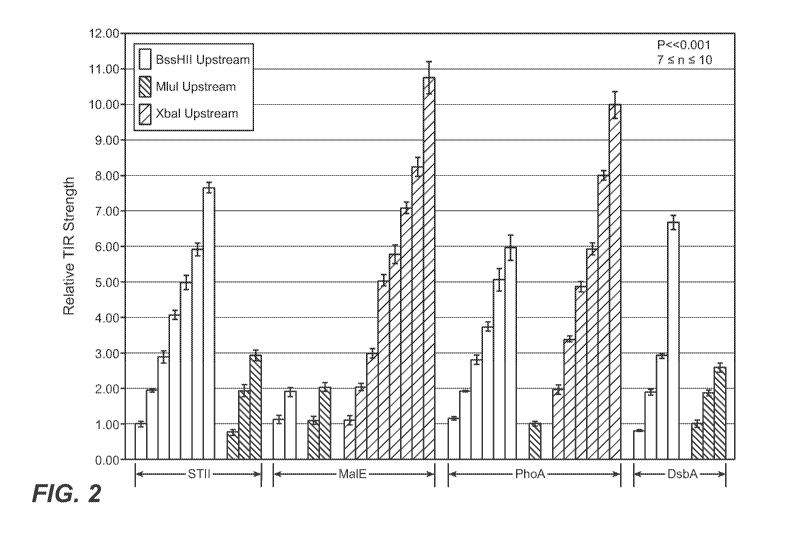
FIGURE 2: Relative translocation initiation region strength of signal peptide
variants. Normalized basal alkaline phosphatase activity of 27C7 cells
carrying a vector
with a fusion between either an STII, MalE, PhoA, or DsbA signal peptide and
the mature
domain of the E. coli alkaline phosphatase (BAP) gene. Each bar represents an
individual
culture incubated with the chromogenic substrate PNPP and enzymatic activity
was
determined as the absorbance of that culture at 410 nm less the absorbance of
a culture
carrying an empty vector (pBR322). Activities were normalized to the basal
activity of
27C7 cells carrying the plasmid pPho4l. White bars represent signal peptide
variants with
a BssHII restriction site at the -9 position relative to the first base pair
of the initiation
codon. Grey or striped bars represent an MIuI or XbaI site at the -9 position,
respectively.
All activities are the mean of between seven and ten replicate experiments.
Error bars are
reported as the uncertainty in the mean with a 95% confidence limit. The
differences in
relative TIR strength between adjacent bars are all statistically significant
(P << 0.001).
Bars represent clones SH1.2, SH2.41, SH3.38, SH4.60, SH5.34, SH6.52, SH8.36,
SL1.2,
SL2.74, SL3.72, MH1.92, MH2.100, ML1.97, ML2.123, MX1.wt, MX2.15, MX3.12,
14
CA 02780143 2012-05-04
WO 2011/057120 PCT/US2010/055702
MX5.37, MX6.4, MX7.25, MX8.13, MX11.34, PH1.70, PH2.64, PH3.wt, PH4.67,
PH5.71, PH6.77, PL1.104, PX2.41, PX3.wt, PX5.53, PX6.15, PX8.24, PX10.23,
DH1.48,
DH2.wt, DH3.79, DH7.72, DLl.wt, DL2.3, DL3.37 (in order, from left to right).
FIGURE 3: Monitoring assembly of antibody species with heavy chain signal
peptide manipulation. 64B4 cells were grown in 25 mL of C.R.A.P. phosphate-
limiting
media for 24 hours and soluble fractions as well at total protein pellets
normalized by
optical density (OD) were prepared for SDS-PAGE analysis. (A) Samples from
cells
carrying the plasmid pBR-SS-5D5-1.1 (SS1.1), pBR-MS-5D5-1.1 (MS1.1), pBR-DS-
5D5-
1.1 (DS1.1) or pBR-PS-5D5-1.1 (PS1.1) were separated by SDS-PAGE gel
electrophoresis
(mass in kDa indicated at the left side), transferred to nitrocellulose, and
probed for the
presence of heavy chain-containing species with an a-Fc specific antibody.
Soluble
samples (top blot) consisted of the putatively identified bands corresponding
to (from top
to bottom): full-length antibody, heavy-heavy-light (HHL), heavy-light (HL) or
free heavy
chain (heavy chain monomer). Normalized, total protein samples (bottom blot)
were
reduced with 0.2 M DTT to disrupt disulfide bond structure and each individual
lane
migrated to a single band with an apparent mass of -49 kDa. (B) The samples
from (A)
were run on a separate SDS-PAGE gel (mass in kDa indicated at the right side),
transferred
to nitrocellulose and probed for complexes containing a light chain with an a-
KLc specific
antibody. Soluble samples (top blot) consisted of the putatively identified
bands
corresponding to (from top to bottom): full-length antibody, HL, light-light
(LL) dimer or
free light chain (light chain monomer). Normalized, total protein samples
(bottom blot)
were reduced with 0.2 M DTT and each individual lane migrated to a single band
with an
apparent mass of -25 kDa. Abbreviations: S=signal sequence STII M=signal
sequence
MalE D=signal sequence DsbA P=signal sequence PhoA. XX#.# (e.g. DS1.1) refers
to
heavy chain signal sequence, light chain signal sequence, heavy chain TIR,
light chain TIR
used in the experiment.
FIGURE 4: Monitoring assembly of antibody species with light chain signal
peptide
manipulation. 64B4 cells were grown in 25 mL of C.R.A.P. phosphate-limiting
media for
24 hours and soluble fractions as well at total protein pellets normalized by
optical density
(OD) were prepared for SDS-PAGE analysis. Samples from cells carrying the
plasmid
pBR-DS-5D5-1.1 (DS1.1), pBR-DS-5D5-1.2 (DS1.2), pBR-DM-5D5-1.1 (DM1.1), pBR-
DM-5D5-1.2 (DM1.2), pBR-DD-5D5-1.1 (DD1.1), pBR-DD-5D5-1.2 (DD 1.2), pBR-DP-
5D5-1.1 (DP1.1), or pBR-DP-5D5-1.2 were separated by SDS-PAGE gel
electrophoresis
CA 02780143 2012-05-04
WO 2011/057120 PCT/US2010/055702
(mass in kDa indicated at the left side), transferred to nitrocellulose, and
probed for the
presence of heavy or light chain-containing species with an a-Fc or a-xLc
specific
antibody, respectively, as indicated along the right side of the images.
Soluble samples
(top blot) consisted of the putatively identified bands corresponding to (from
top to
bottom): full-length antibody, heavy-heavy-light (HHL), heavy-light (HL) dimer
or free
heavy chain. Normalized, total protein samples (middle blot, bottom) were
reduced with
0.2 M DTT to disrupt disulfide bond structure and each individual lane
migrated to a single
band with an apparent mass of -49 kDa when probed with an a-Fc antibody. When
probed
with an a-xLc specific antibody, all lanes migrated to a single or double band
with an
apparent mass of either -25 kDa or -27 kDa and -25 kDa. Abbreviations:
S=signal
sequence STII M=signal sequence MalE D=signal sequence DsbA P=signal sequence
PhoA. XX#.# (e.g. DS1.1) refers to heavy chain signal sequence, light chain
signal
sequence, heavy chain TIR, light chain TIR used in the experiment.
FIGURE 5: Monitoring assembly of antibody species over time from 10-L
fermentations. 64B4 cells were grown to a high cell density in a 10-L
fermentation for
three days with samples taken at regular time intervals (time sample taken
above each lane
in hours past inoculation) from which soluble fractions as well at total
protein pellets
normalized by optical density (OD) were prepared for SDS-PAGE analysis.
Samples from
cells carrying the plasmid pBR-SS-5D5-1.1 co-expressing the chaperone-bearing
plasmid
pJJ247 (SS1.1 + Chaperones), pBR-DD-5D5-1.1 with pJJ247 (DD1.1 + Chaperones),
pBR-DS-5D5-1.1 with pJJ247 (DM1.1 + Chaperones), or pBR-DP-5D5-1.1 with pJJ247
(DP 1.1 + Chaperones) were separated by SDS-PAGE gel electrophoresis (mass in
kDa
indicated at the left side), transferred to nitrocellulose, and probed for the
presence of heavy
or light chain-containing species with an a-Fc or a-xLc specific antibody,
respectively, as
indicated along the right side of the images. Soluble samples (top blot)
consisted of the
putatively identified bands corresponding to (from top to bottom): full-length
antibody,
heavy-heavy-light (HHL), heavy-light (HL) dimer, light-light (LL) dimer, or
free light
chain. Normalized, total protein samples (middle blot, bottom) were reduced
with 0.2 mM
DTT to disrupt disulfide bond structure and each individual lane migrated to a
single band
with an apparent mass of -49 kDa when probed with an a-Fc. When probed with an
a-KLc
specific antibody, all lanes migrated to a single band with an apparent mass
of either -25
kDa. Abbreviations: S=signal sequence STII M=signal sequence MalE D=signal
sequence DsbA P=signal sequence PhoA. XX#.# (e.g. DS1.1) refers to heavy chain
signal
16
CA 02780143 2012-05-04
WO 2011/057120 PCT/US2010/055702
sequence, light chain signal sequence, heavy chain TIR, light chain TIR used
in the
experiment.
FIGURE 6: Accumulation of mature PhoA under inducing conditions. 27C7 cells
were grown in 25-mL of C.R.A.P. phosphate-limiting media for 24 hours and
soluble
fractions were normalized by optical density (OD) and prepared for SDS-PAGE
analysis.
The mature domain of the E. coliphoA gene was fused to the indicated DsbA or
STII
(bottom) TIR variants (top). Gel was visualized for the presence of protein by
Commassie
blue staining. A putatively identified band corresponding to the mature domain
of PhoA
(right) appeared at a mass of -47 kDa (mass indicated at left side).
FIGURE 7: depicts amino acid sequences of the framework (FR), CDR, first
constant domain (CL or CH1) and Fc region (Fc) of MetMAb (OA5D5v2). Figure
discloses Light Chain sequences as SEQ ID NOS 57-60, 49-51 & 61, respectively,
in order
of appearance and Heavy Chain sequences as SEQ ID NOS 62-65, 52-53 & 66-68,
respectively, in order of appearance. The Fc sequence depicted comprises
"hole" (cavity)
mutations T366S, L368A and Y407V, as described in WO 2005/063816.
FIGURE 8: depicts sequence of an Fc polypeptide (SEQ ID NO: 47) comprising
"knob" (protuberance) mutation T366W, as described in WO 2005/063816. In one
embodiment, an Fc polypeptide comprising this sequence forms a complex with an
Fc
polypeptide comprising the Fc sequence of Fig. 7 to generate an Fc region.
DETAILED DESCRIPTION OF THE INVENTION
General techniques
The techniques and procedures described or referenced herein are generally
well
understood and commonly employed using conventional methodology by those
skilled in
the art, such as, for example, the widely utilized methodologies described in
Sambrook et
al., Molecular Cloning: A Laboratory Manual 3rd. edition (2001) Cold Spring
Harbor
Laboratory Press, Cold Spring Harbor, N.Y. CURRENT PROTOCOLS IN MOLECULAR
BIOLOGY (F. M. Ausubel, et al. eds., (2003)); the series METHODS IN ENZYMOLOGY
(Academic Press, Inc.): PCR 2: A PRACTICAL APPROACH (M. J. MacPherson, B. D.
Hames and G. R. Taylor eds. (1995)), Harlow and Lane, eds. (1988) ANTIBODIES,
A
LABORATORY MANUAL, and ANIMAL CELL CULTURE (R. I. Freshney, ed. (1987)).
Definitions
The term "vector," as used herein, is intended to refer to a nucleic acid
molecule
capable of transporting another nucleic acid to which it has been linked. One
type of vector
17
CA 02780143 2012-05-04
WO 2011/057120 PCT/US2010/055702
is a "plasmid", which refers to a circular double stranded DNA loop into which
additional
DNA segments may be ligated. Another type of vector is a phage vector. Another
type of
vector is a viral vector, wherein additional DNA segments may be ligated into
the viral
genome. Certain vectors are capable of autonomous replication in a host cell
into which
they are introduced (e.g., bacterial vectors having a bacterial origin of
replication and
episomal mammalian vectors). Other vectors (e.g., non-episomal mammalian
vectors) can
be integrated into the genome of a host cell upon introduction into the host
cell, and
thereby are replicated along with the host genome. Moreover, certain vectors
are capable of
directing the expression of genes to which they are operatively linked. Such
vectors are
referred to herein as "recombinant expression vectors" (or simply,
"recombinant vectors").
In general, expression vectors of utility in recombinant DNA techniques are
often in the
form of plasmids. In the present specification, "plasmid" and "vector" may be
used
interchangeably as the plasmid is the most commonly used form of vector.
The term "cistron," as used herein, is intended to refer to a genetic element
broadly
equivalent to a translational unit comprising the nucleotide sequence coding
for a
polypeptide chain and adjacent control regions. "Adjacent control regions"
include, for
example, a translational initiation region (TIR; as defined herein below) and
a termination
region.
A "polycistronic" expression vector refers to a single vector that contains
and
expresses multiple cistrons under the regulatory control of one single
promoter. A
common example of polycistronic vector is a "dicistronic" vector that contains
and
expresses two different polypeptides under the control of one promoter. Upon
expression
of a dicistronic or polycistronic vector, multiple genes are first transcribed
as a single
transcriptional unit, and then translated separately.
A "separate cistron" expression vector according to the present invention
refers to a
single vector comprising at least two separate promoter-cistron pairs, wherein
each cistron
is under the control of its own promoter. Upon expression of a separate
cistron expression
vector, both transcription and translation processes of different genes are
separate and
independent.
The "translation initiation region" or TIR or translational initiation region
or
translational initiation sequence, as used herein refers to a nucleic acid
region providing the
efficiency of translational initiation of a gene of interest. In general, a
TIR within a
18
CA 02780143 2012-05-04
WO 2011/057120 PCT/US2010/055702
particular cistron encompasses the ribosome binding site (RBS) and sequences
5' and 3' to
RBS. The RBS is defined to contain, minimally, the Shine-Dalgamo region and
the start
codon (AUG). Accordingly, a TIR also includes at least a portion of the
nucleic acid
sequence to be translated. Preferably, a TIR of the invention includes a
secretion signal
sequence encoding a signal peptide that precedes the sequence encoding for the
light or
heavy chain within a cistron. A TIR variant contains sequence variants
(particularly
substitutions) within the TIR region that alter the property of the TIR, such
as its
translational strength as defined herein below. Preferably, a TIR variant of
the invention
contains sequence substitutions within the first 2 to about 14, preferably
about 4 to 12,
more preferably about 6 codons of the secretion signal sequence that precedes
the sequence
encoding for the light or heavy chain within a cistron.
The term "translational strength" as used herein refers to a measurement of a
secreted polypeptide in a control system wherein one or more variants of a TIR
is used to
direct secretion of a polypeptide and the results compared to the wild-type
TIR or some
other control under the same culture and assay conditions. Without being
limited to any
one theory, "translational strength" as used herein can include, for example,
a measure of
mRNA stability, efficiency of ribosome binding to the ribosome binding site,
and mode of
translocation across a membrane.
"Secretion signal sequence" or "signal sequence" refers to a nucleic acid
sequence
encoding for a short signal peptide that can be used to direct a newly
synthesized protein of
interest through a cellular membrane, usually the inner membrane or both inner
and outer
membranes of prokaryotes. As such, the protein of interest such as the
immunoglobulin
light or heavy chain polypeptide is secreted into the periplasm of the
prokaryotic host cells
or into the culture medium. The signal peptide encoded by the secretion signal
sequence
may be endogenous to the host cells, or they may be exogenous, including
signal peptides
native to the polypeptide to be expressed. Secretion signal sequences are
typically present
at the amino terminus of a polypeptide to be expressed, and are typically
removed
enzymatically between biosynthesis and secretion of the polypeptide from the
cytoplasm.
Thus, the signal peptide is usually not present in a mature protein product.
"Operably linked" refers to a juxtaposition of two or more components, wherein
the
components so described are in a relationship permitting them to function in
their intended
manner. For example, a promoter is operably linked to a coding sequence if it
acts in cis to
19
CA 02780143 2012-05-04
WO 2011/057120 PCT/US2010/055702
control or modulate the transcription of the linked sequence. Generally, but
not
necessarily, the DNA sequences that are "operably linked" are contiguous and,
where
necessary to join two protein coding regions or in the case of a secretory
leader, contiguous
and in reading frame. However, although an operably linked promoter is
generally located
upstream of the coding sequence, it is not necessarily contiguous with it.
Operably linked
enhancers can be located upstream, within or downstream of coding sequences
and at
considerable distances from the promoter. Linking is accomplished by
recombinant
methods known in the art, e.g., using PCR methodology, by annealing, or by
ligation at
convenient restriction sites. If convenient restriction sites do not exist,
then synthetic
oligonucleotide adaptors or linkers are used in accord with conventional
practice.
"Regulatory elements" as used herein, refer to nucleotide sequences present in
cis,
necessary for transcription and translation of a polynucleotide encoding a
heterologous
polypeptide into polypeptides. The transcriptional regulatory elements
normally comprise
a promoter 5' of the gene sequence to be expressed, transcriptional initiation
and
termination sites, and polyadenylation signal sequence. The term
"transcriptional initiation
site" refers to the nucleic acid in the construct corresponding to the first
nucleic acid
incorporated into the primary transcript, i.e., the mRNA precursor; the
transcriptional
initiation site may overlap with the promoter sequences.
A "promoter" refers to a polynucleotide sequence that controls transcription
of a
gene or sequence to which it is operably linked. A promoter includes signals
for RNA
polymerase binding and transcription initiation. The promoters used will be
functional in
the cell type of the host cell in which expression of the selected sequence is
contemplated.
A large number of promoters including constitutive, inducible and repressible
promoters
from a variety of different sources, are well known in the art (and identified
in databases
such as GenBank) and are available as or within cloned polynucleotides (from,
e.g.,
depositories such as ATCC as well as other commercial or individual sources).
With
inducible promoters, the activity of the promoter increases or decreases in
response to a
signal.
The term "host cell" (or "recombinant host cell"), as used herein, is intended
to refer
to a cell that has been genetically altered, or is capable of being
genetically altered by
introduction of an exogenous polynucleotide, such as a recombinant plasmid or
vector. It
should be understood that such terms are intended to refer not only to the
particular subject
CA 02780143 2012-05-04
WO 2011/057120 PCT/US2010/055702
cell but to the progeny of such a cell. Because certain modifications may
occur in
succeeding generations due to either mutation or environmental influences,
such progeny
may not, in fact, be identical to the parent cell, but are still included
within the scope of the
term "host cell" as used herein.
An "isolated" polypeptide (e.g., an antibody) is one which has been identified
and
separated and/or recovered from a component of its natural environment.
Contaminant
components of its natural environment are materials which would interfere with
diagnostic
or therapeutic uses for the antibody, and may include enzymes, hormones, and
other
proteinaceous or nonproteinaceous solutes. In preferred embodiments, the
polypeptide will
be purified (1) to greater than 95% by weight of polypeptide as determined by
the Lowry
method, and most preferably more than 99% by weight, (2) to a degree
sufficient to obtain
at least 15 residues of N-terminal or internal amino acid sequence by use of a
spinning cup
sequenator, or (3) to homogeneity by SDS-PAGE (sodium dodecyl sulfate
polyacrylamide
gel electrophoresis) under reducing or nonreducing conditions using Coomassie
blue or,
preferably, silver stain. Isolated polypeptide includes the polypeptide in
situ within
recombinant cells since at least one component of the polypeptide's natural
environment
will not be present. Ordinarily, however, isolated polypeptide will be
prepared by at least
one purification step.
An "isolated" nucleic acid molecule is a nucleic acid molecule that is
identified and
separated from at least one contaminant nucleic acid molecule with which it is
ordinarily
associated in the natural source of the nucleic acid. An isolated nucleic acid
molecule is
other than in the form or setting in which it is found in nature. Isolated
nucleic acid
molecules therefore are distinguished from the nucleic acid molecule as it
exists in natural
cells. However, an isolated nucleic acid molecule includes a nucleic acid
molecule
contained in cells that ordinarily express the nucleic acid (for example, an
antibody
encoding nucleic acid) where, for example, the nucleic acid molecule is in a
chromosomal
location different from that of natural cells.
"Polynucleotide," or "nucleic acid," as used interchangeably herein, refer to
polymers of nucleotides of any length, and include DNA and RNA. The
nucleotides can be
deoxyribonucleotides, ribonucleotides, modified nucleotides or bases, and/or
their analogs,
or any substrate that can be incorporated into a polymer by DNA or RNA
polymerase, or by
a synthetic reaction. A polynucleotide may comprise modified nucleotides, such
as
methylated nucleotides and their analogs. If present, modification to the
nucleotide
21
CA 02780143 2012-05-04
WO 2011/057120 PCT/US2010/055702
structure may be imparted before or after assembly of the polymer. The
sequence of
nucleotides may be interrupted by non-nucleotide components. A polynucleotide
may be
further modified after synthesis, such as by conjugation with a label. Other
types of
modifications include, for example, "caps," substitution of one or more of the
naturally
occurring nucleotides with an analog, internucleotide modifications such as,
for example,
those with uncharged linkages (e.g., methyl phosphonates, phosphotriesters,
phosphoamidates, carbamates, etc.) and with charged linkages (e.g.,
phosphorothioates,
phosphorodithioates, etc.), those containing pendant moieties, such as, for
example,
proteins (e.g., nucleases, toxins, antibodies, signal peptides, ply-L-lysine,
etc.), those with
intercalators (e.g., acridine, psoralen, etc.), those containing chelators
(e.g., metals,
radioactive metals, boron, oxidative metals, etc.), those containing
alkylators, those with
modified linkages (e.g., alpha anomeric nucleic acids, etc.), as well as
unmodified forms of
the polynucleotide(s). Further, any of the hydroxyl groups ordinarily present
in the sugars
may be replaced, for example, by phosphonate groups, phosphate groups,
protected by
standard protecting groups, or activated to prepare additional linkages to
additional
nucleotides, or may be conjugated to solid or semi-solid supports. The 5' and
3' terminal
OH can be phosphorylated or substituted with amines or organic capping group
moieties of
from 1 to 20 carbon atoms. Other hydroxyls may also be derivatized to standard
protecting
groups. Polynucleotides can also contain analogous forms of ribose or
deoxyribose sugars
that are generally known in the art, including, for example, 2'-O-methyl-, 2'-
O-allyl, 2'-
fluoro- or 2'-azido-ribose, carbocyclic sugar analogs, alpha-anomeric sugars,
epimeric
sugars such as arabinose, xyloses or lyxoses, pyranose sugars, furanose
sugars,
sedoheptuloses, acyclic analogs and a basic nucleoside analogs such as methyl
riboside.
One or more phosphodiester linkages may be replaced by alternative linking
groups. These
alternative linking groups include, but are not limited to, embodiments
wherein phosphate
is replaced by P(O)S ("thioate"), P(S)S ("dithioate"), (O)NR2 ("amidate"),
P(O)R,
P(O)OR', CO or CH2 ("formacetal"), in which each R or R' is independently H or
substituted or unsubstituted alkyl (1-20 C) optionally containing an ether (-0-
) linkage,
aryl, alkenyl, cycloalkyl, cycloalkenyl or araldyl. Not all linkages in a
polynucleotide need
be identical. The preceding description applies to all polynucleotides
referred to herein,
including RNA and DNA.
"Oligonucleotide," as used herein, generally refers to short, generally single
stranded, generally synthetic polynucleotides that are generally, but not
necessarily, less
22
CA 02780143 2012-05-04
WO 2011/057120 PCT/US2010/055702
than about 200 nucleotides in length. The terms "oligonucleotide" and
"polynucleotide"
are not mutually exclusive. The description above for polynucleotides is
equally and fully
applicable to oligonucleotides.
As used herein, "polypeptide" refers generally to peptides and proteins from
any
cell source having more than about ten amino acids. "Heterologous"
polypeptides are those
polypeptides foreign to the host cell being utilized, such as a human protein
produced by E.
coli. While the heterologous polypeptide may be prokaryotic or eukaryotic,
preferably it is
eukaryotic, more preferably mammalian, and most preferably human. Preferably,
it is a
recombinantly produced, or recombinant polypeptide. "Heterologous"
polypeptides are
those polypeptides foreign to the host cell being utilized, such as a human
protein produced
by E. coli. While the heterologous polypeptide may be prokaryotic or
eukaryotic, preferably
it is eukaryotic, more preferably mammalian, and most preferably human.
Preferably, it is a
recombinantly produced, or recombinant polypeptide.
Examples of mammalian polypeptides include molecules such as, e.g., renin, a
growth hormone, including human growth hormone; bovine growth hormone; growth
hormone releasing factor; parathyroid hormone; thyroid stimulating hormone;
lipoproteins;
1-antitrypsin; insulin A-chain; insulin B-chain; proinsulin; thrombopoietin;
follicle
stimulating hormone; calcitonin; luteinizing hormone; glucagon; clotting
factors such as
factor VIIIC, factor IX, tissue factor, and von Willebrands factor; anti-
clotting factors such
as Protein C; atrial naturietic factor; lung surfactant; a plasminogen
activator, such as
urokinase or human urine or tissue-type plasminogen activator (t-PA) and
variants thereof
such as RETEVASETM and TNKASETM; bombesin; thrombin; hemopoietic growth
factor;
tumor necrosis factor-alpha and -beta; antibodies to ErbB2 domain(s) such as
2C4 (WO
01/00245; hybridoma ATCC HB-12697), which binds to a region in the
extracellular
domain of ErbB2 (e.g., any one or more residues in the region from about
residue 22 to
about residue 584 of ErbB2, inclusive), enkephalinase; a serum albumin such as
human
serum albumin; Muellerian-inhibiting substance; relaxin A-chain; relaxin B-
chain;
prorelaxin; mouse gonadotropin-associated peptide; a microbial protein, such
as beta-
lactamase; DNase; inhibin; activin; vascular endothelial growth factor (VEGF);
receptors
for hormones or growth factors; integrin; protein A or D; rheumatoid factors;
a
neurotrophic factor such as brain-derived neurotrophic factor (BDNF),
neurotrophin-3, -4, -
5, or -6 (NT-3, NT-4, NT-5, or NT-6), or a nerve growth factor such as NGF;
cardiotrophins (cardiac hypertrophy factor) such as cardiotrophin-1 (CT-1);
platelet-derived
23
CA 02780143 2012-05-04
WO 2011/057120 PCT/US2010/055702
growth factor (PDGF); fibroblast growth factor such as aFGF and bFGF;
epidermal growth
factor (EGF); transforming growth factor (TGF) such as TGF-alpha and TGF-beta,
including TGF-l, TGF-2, TGF-3, TGF-4, or TGF-5; insulin-like growth factor-I
and -II
(IGF-I and IGF-II); des(I-3)-IGF-I (brain IGF-I), insulin-like growth factor
binding
proteins; CD proteins such as CD-3, CD-4, CD-8, and CD-19; erythropoietin;
osteoinductive factors; immunotoxins; a bone morphogenetic protein (BMP); an
interferon
such as interferon-alpha, -beta, and -gamma; serum albumin, such as human
serum albumin
(HSA) or bovine serum albumin (BSA); colony stimulating factors (CSFs), e.g.,
M-CSF,
GM-CSF, and G-CSF; interleukins (ILs), e.g., IL-1 to IL-10; anti-HER-2
antibody; Apo2
ligand; superoxide dismutase; T-cell receptors; surface membrane proteins;
decay
accelerating factor; viral antigen such as, for example, a portion of the AIDS
envelope;
transport proteins; homing receptors; addressins; regulatory proteins;
antibodies; and
fragments of any of the above-listed polypeptides.
Preferred polypeptides herein include human serum albumin (HSA), 2C4, tissue
factor, anti-tissue factor, anti-CD20, anti-HER-2, heregulin, anti-IgE, anti-
CD 1la, anti-
CD18, VEGF and receptors and antibodies thereto such as rhuFab V2 and
AVASTINTM,
growth hormone and its variants, such as hGH, growth hormone receptors, growth
hormone releasing protein (GHRP), LIV-1 (EP 1,263,780), TRAIL, tumor necrosis
factor
(TNF) and antibodies thereto, TNF receptor and related antibodies, TNF-
receptor-IgG,
TNF receptor associated factors (TRAF5) and inhibitors thereof, Factor VIII,
Factor VIII B
domain, interferons such as interferon-gamma, transforming growth factors
(TGFs) such as
TGF-beta, anti-TGF such as anti-TGF-beta, activin, inhibin, anti-activin, anti-
inhibin,
tissue-plasminogen activators and their variants such as t-PA, RETEPLASETM,
and
TNKase, anti-Fas antibodies, Apo-2 ligand; Apo-2 ligand inhibitor; Apo-2
receptor, Apo-3,
apoptotic factors, Ced-4, DcR3, death receptor and agonist antibodies (DR4,
DR5),
lymphotoxin (LT), prolactin, prolactin receptor, SOB proteins, WISP (wnt-
induced
secreted proteins), neurotoxin-3 (NT-3), nerve growth factor (NGF) and anti-
NGF, DNase,
hepatitis antigen, herpes simplex antigen, leptin, insulin-like growth factors
(IGFs) such as
IGF-1 and IGF-2 and their binding proteins and receptors such as IGFBP- I -
IGFBP-6,
insulin, fibroblast growth factors (FGFs) such as FGF-17, Toll protein, TIE
ligands, CD40
and anti-CD40, immunoadhesins, subtilisin, hepatocyte growth factor (HGF),
thrombopoietin (TPO), interleukins such as IL-2, IL-12, IL-17, IL-22, IL-8, IL-
9, and
antibodies thereto, and prostrate-specific cancer antigen (PSCA).
24
CA 02780143 2012-05-04
WO 2011/057120 PCT/US2010/055702
Particularly preferred polypeptides are recombinant polypeptides, more
preferably
antibodies, which include monoclonal antibodies and humanized antibodies. Such
antibodies may be full-length antibodies or antibody fragments. More
preferably, these
antibodies are human or humanized antibodies. Still more preferably, the
antibody is an
anti-c-met, anti-IgE, anti-CD18, anti-VEGF, anti-tissue factor, 2C4, anti-Her-
2, anti-CD20,
anti-CD40, or anti-CD ha antibody. Antibody fragments encompassed within the
definition
of polypeptide preferably comprise a light chain, more preferably a kappa
light chain. Such
preferred fragments include, for example, a Fab, Fab', F(ab')2, or F(ab')2-
leucine zipper
(LZ) fusion, and a one-armed antibody.
Protein "expression" refers to conversion of the information encoded in a gene
into
messenger RNA (mRNA) and then to the protein.
An " immunoconjugate" (interchangeably referred to as "antibody-drug
conjugate,"
or "ADC") means an antibody conjugated to one or more cytotoxic agents, such
as a
chemotherapeutic agent, a drug, a growth inhibitory agent, a toxin (e.g., a
protein toxin, an
enzymatically active toxin of bacterial, fungal, plant, or animal origin, or
fragments
thereof), or a radioactive isotope (i.e., a radioconjugate).
A "blocking" antibody or an antibody "antagonist" is one which inhibits or
reduces
biological activity of the antigen it binds. In some embodiments, blocking
antibodies or
antagonist antibodies completely inhibit the biological activity of the
antigen.
An "agonist antibody", as used herein, is an antibody which mimics at least
one of
the functional activities of a polypeptide of interest (e.g., HGF).
"Binding affinity" generally refers to the strength of the sum total of
noncovalent
interactions between a single binding site of a molecule (e.g., an antibody)
and its binding
partner (e.g., an antigen). Unless indicated otherwise, as used herein,
"binding affinity"
refers to intrinsic binding affinity which reflects a 1:1 interaction between
members of a
binding pair (e.g., antibody and antigen). The affinity of a molecule X for
its partner Y can
generally be represented by the dissociation constant (Kd). Desirably the Kd
is 1 x 10⁻⁷, 1 x
10, 5 x 10, 1 x 10⁻⁹, 3 x 10⁻⁹, 5 x 10⁻⁹, or even 1 x 10⁻¹⁰ or stronger.
Affinity can be
measured by common methods known in the art, including those described herein.
Low-
affinity antibodies generally bind antigen slowly and tend to dissociate
readily, whereas
high-affinity antibodies generally bind antigen faster and tend to remain
bound longer. A
variety of methods of measuring binding affinity are known in the art, any of
which can be
CA 02780143 2012-05-04
WO 2011/057120 PCT/US2010/055702
used for purposes of the present invention. Specific illustrative embodiments
are described
in the following.
In one embodiment, the "Kd" or "Kd value" according to this invention is
measured
by a radiolabeled antigen binding assay (RIA) performed with the Fab version
of an
antibody of interest and its antigen as described by the following assay that
measures
solution binding affinity of Fabs for antigen by equilibrating Fab with a
minimal
concentration of (125I)-labeled antigen in the presence of a titration series
of unlabeled
antigen, then capturing bound antigen with an anti-Fab antibody-coated plate
(Chen, et al.,
(1999) J. Mol. Biol. 293:865-881). To establish conditions for the assay,
microtiter plates
(Dynex) are coated overnight with 5 g/ml of a capturing anti-Fab antibody
(Cappel Labs)
in 50 mM sodium carbonate (pH 9.6), and subsequently blocked with 2% (w/v)
bovine
serum albumin in PBS for two to five hours at room temperature (approximately
23 C). In
a non-adsorbant plate (Nunc #269620), 100 pM or 26 pM [125I]-antigen are mixed
with
serial dilutions of a Fab of interest (e.g., consistent with assessment of an
anti-VEGF
antibody, Fab-12, in Presta et al., (1997) Cancer Res. 57:4593-4599). The Fab
of interest is
then incubated overnight; however, the incubation may continue for a longer
period (e.g.,
65 hours) to insure that equilibrium is reached. Thereafter, the mixtures are
transferred to
the capture plate for incubation at room temperature (e.g., for one hour). The
solution is
then removed and the plate washed eight times with 0.1 % Tween-20 in PBS. When
the
plates have dried, 150 l/well of scintillant (MicroScint-20; Packard) is
added, and the
plates are counted on a Topcount gamma counter (Packard) for ten minutes.
Concentrations
of each Fab that give less than or equal to 20% of maximal binding are chosen
for use in
competitive binding assays. According to another embodiment the Kd or Kd value
is
measured by using surface plasmon resonance assays using a BlAcoreTM-2000 or a
BlAcoreTM-3000 (BlAcore, Inc., Piscataway, NJ) at 25 C with immobilized
antigen CM5
chips at -10 response units (RU). Briefly, carboxymethylated dextran biosensor
chips
(CM5, BlAcore Inc.) are activated with N-ethyl-N'- (3-dimethylaminopropyl)-
carbodiimide
hydrochloride (EDC) and N-hydroxysuccinimide (NHS) according to the supplier's
instructions. Antigen is diluted with 10mM sodium acetate, pH 4.8, into 5 g/ml
(0.2 M)
before injection at a flow rate of 5 l/minute to achieve approximately 10
response units
(RU) of coupled protein. Following the injection of antigen, 1 M ethanolamine
is injected
to block unreacted groups. For kinetics measurements, two-fold serial
dilutions of Fab
(0.78 nM to 500 nM) are injected in PBS with 0.05% Tween 20 (PBST) at 25 C at
a flow
- 26
CA 02780143 2012-05-04
WO 2011/057120 PCT/US2010/055702
rate of approximately 25 1/min. In some embodiments, the following
modifications are
used for the surface Plasmon resonance assay method: antibody is immobilized
to CM5
biosensor chips to achieve approximately 400 RU, and for kinetic measurements,
two-fold
serial dilutions of target protein are injected in PBST buffer at 25 C with a
flow rate of
about 30 ul/minute. Association rates (k0n) and dissociation rates (k ff) are
calculated using
a simple one-to-one Langmuir binding model (BlAcore Evaluation Software
version 3.2)
by simultaneous fitting the association and dissociation sensorgram. The
equilibrium
dissociation constant (Kd) is calculated as the ratio k ff/k ,,. See, e.g.,
Chen, Y., et al.,
(1999) J. Mol. Biol. 293:865-881. If the on-rate exceeds 106 M-' S-' by the
surface
plasmon resonance assay above, then the on-rate can be determined by using a
fluorescent
quenching technique that measures the increase or decrease in fluorescence
emission
intensity (excitation = 295 nm; emission = 340 nm, 16 nm band-pass) at 25 C of
a 20nM
anti-antigen antibody (Fab form) in PBS, pH 7.2, in the presence of increasing
concentrations of antigen as measured in a spectrometer, such as a stop-flow
equipped
spectrophometer (Aviv Instruments) or a 8000-series SLM-Aminco
spectrophotometer
(ThermoSpectronic) with a stir red cuvette.
An "on-rate" or "rate of association" or "association rate" or "k0,,"
according to this
invention can also be determined with the same surface plasmon resonance
technique
described above using a BlAcoreTM-2000 or a BlAcoreTM-3000 (BlAcore, Inc.,
Piscataway,
NJ) at 25 C with immobilized antigen CM5 chips at -10 response units (RU).
Briefly,
carboxymethylated dextran biosensor chips (CM5, BlAcore Inc.) are activated
with N-
ethyl-N'- (3-dimethylaminopropyl)-carbodiimide hydrochloride (EDC) and N-
hydroxysuccinimide (NHS) according to the supplier's instructions. Antigen is
diluted
with lOmM sodium acetate, pH 4.8, into 5 g/ml (-0.2uM) before injection at a
flow rate of
5 l/minute to achieve approximately 10 response units (RU) of coupled
protein. Following
the injection of antigen, 1M ethanolamine is injected to block unreacted
groups. For
kinetics measurements, two-fold serial dilutions of Fab (0.78 nM to 500 nM)
are injected in
PBS with 0.05% Tween 20 (PBST) at 25 C at a flow rate of approximately 25
1/min. In
some embodiments, the following modifications are used for the surface Plasmon
resonance assay method: antibody is immobilized to CM5 biosensor chips to
achieve
approximately 400 RU, and for kinetic measurements, two-fold serial dilutions
of target
protein are injected in PBST buffer at 25 C with a flow rate of about 30
ul/minute.
Association rates (k0n) and dissociation rates (k ff) are calculated using a
simple one-to-one
27
CA 02780143 2012-05-04
WO 2011/057120 PCT/US2010/055702
Langmuir binding model (BlAcore Evaluation Software version 3.2) by
simultaneous
fitting the association and dissociation sensorgram. The equilibrium
dissociation constant
(Kd) was calculated as the ratio k ff/k ,,. See, e.g., Chen, Y., et al.,
(1999) J. Mol. Biol.
293:865-88 1. However, if the on-rate exceeds 106 M-' S-' by the surface
plasmon
resonance assay above, then the on-rate is preferably determined by using a
fluorescent
quenching technique that measures the increase or decrease in fluorescence
emission
intensity (excitation = 295 nm; emission = 340 nm, 16 nm band-pass) at 25 C of
a 20nM
anti-antigen antibody (Fab form) in PBS, pH 7.2, in the presence of increasing
concentrations of antigen as measured in a spectrometer, such as a stop-flow
equipped
spectrophometer (Aviv Instruments) or a 8000-series SLM-Aminco
spectrophotometer
(ThermoSpectronic) with a stir red cuvette.
A "naked antibody" is an antibody that is not conjugated to a heterologous
molecule, such as a cytotoxic moiety or radiolabel.
An antibody having a "biological characteristic" of a designated antibody is
one
which possesses one or more of the biological characteristics of that antibody
which
distinguish it from other antibodies that bind to the same antigen.
In order to screen for antibodies which bind to an epitope on an antigen bound
by
an antibody of interest, a routine cross-blocking assay such as that described
in Antibodies,
A Laboratory Manual, Cold Spring Harbor Laboratory, Ed Harlow and David Lane
(1988),
can be performed.
To increase the half-life of the antibodies or polypeptide containing the
amino acid
sequences of this invention, one can attach a salvage receptor binding epitope
to the
antibody (especially an antibody fragment), as described, e.g., in US Patent
5,739,277. For
example, a nucleic acid molecule encoding the salvage receptor binding epitope
can be
linked in frame to a nucleic acid encoding a polypeptide sequence of this
invention so that
the fusion protein expressed by the engineered nucleic acid molecule comprises
the salvage
receptor binding epitope and a polypeptide sequence of this invention. As used
herein, the
term "salvage receptor binding epitope" refers to an epitope of the Fc region
of an IgG
molecule (e.g., IgGi, IgG2, IgG3, or IgG4) that is responsible for increasing
the in vivo
serum half-life of the IgG molecule (e.g., Ghetie et al., Ann. Rev. Immunol.
18:739-766
(2000), Table 1). Antibodies with substitutions in an Fc region thereof and
increased
serum half-lives are also described in W000/42072, WO 02/060919; Shields et
al., J. Biol.
Chem. 276:6591-6604 (2001); Hinton, J. Biol. Chem. 279:6213-6216 (2004)). In
another
28
CA 02780143 2012-05-04
WO 2011/057120 PCT/US2010/055702
embodiment, the serum half-life can also be increased, for example, by
attaching other
polypeptide sequences. For example, antibodies or other polypeptides useful in
the
methods of the invention can be attached to serum albumin or a portion of
serum albumin
that binds to the FcRn receptor or a serum albumin binding peptide so that
serum albumin
binds to the antibody or polypeptide, e.g., such polypeptide sequences are
disclosed in
W001/45746. In one preferred embodiment, the serum albumin peptide to be
attached
comprises an amino acid sequence of DICLPRWGCLW (SEQ ID NO: 48). In another
embodiment, the half-life of a Fab is increased by these methods. See also,
Dennis et al. J.
Biol. Chem. 277:35035-35043 (2002) for serum albumin binding peptide
sequences.
By "fragment" is meant a portion of a polypeptide or nucleic acid molecule
that
contains, preferably, at least 10%, 20%, 30%, 40%, 50%, 60%, 70%, 80%, 90%,
95%, or
more of the entire length of the reference nucleic acid molecule or
polypeptide. A fragment
may contain 10, 20, 30, 40, 50, 60, 70, 80, 90, or 100, 200, 300, 400, 500,
600, or more
nucleotides or 10, 20, 30, 40, 50, 60, 70, 80, 90, 100, 120, 140, 160, 180,
190, 200 amino
acids or more.
The terms "antibody" and "immunoglobulin" are used interchangeably in the
broadest sense and include monoclonal antibodies (e.g., full length or intact
monoclonal
antibodies), polyclonal antibodies, multivalent antibodies, multispecific
antibodies (e.g.,
bispecific antibodies so long as they exhibit the desired biological activity)
and may also
include certain antibody fragments (as described in greater detail herein). An
antibody can
be human, humanized, and/or affinity matured.
The term "variable" refers to the fact that certain portions of the variable
domains
differ extensively in sequence among antibodies and are used in the binding
and specificity
of each particular antibody for its particular antigen. However, the
variability is not evenly
distributed throughout the variable domains of antibodies. It is concentrated
in three
segments called complementarity-determining regions (CDRs) or hypervariable
regions
both in the light-chain and the heavy-chain variable domains. The more highly
conserved
portions of variable domains are called the framework (FR). The variable
domains of
native heavy and light chains each comprise four FR regions, largely adopting
a (3-sheet
configuration, connected by three CDRs, which form loops connecting, and in
some cases
forming part of, the (3-sheet structure. The CDRs in each chain are held
together in close
proximity by the FR regions and, with the CDRs from the other chain,
contribute to the
formation of the antigen-binding site of antibodies (see Kabat et al.,
Sequences of Proteins
29
CA 02780143 2012-05-04
WO 2011/057120 PCT/US2010/055702
of Immunological Interest, Fifth Edition, National Institute of Health,
Bethesda, MD
(1991)). The constant domains are not involved directly in binding an antibody
to an
antigen, but exhibit various effector functions, such as participation of the
antibody in
antibody-dependent cellular toxicity.
Papain digestion of antibodies produces two identical antigen-binding
fragments,
called "Fab" fragments, each with a single antigen-binding site, and a
residual "Fc"
fragment, whose name reflects its ability to crystallize readily. Pepsin
treatment yields an
F(ab')2 fragment that has two antigen-combining sites and is still capable of
cross-linking
antigen.
"Fv" is the minimum antibody fragment which contains a complete antigen-
recognition and -binding site. In a two-chain Fv species, this region consists
of a dimer of
one heavy- and one light-chain variable domain in tight, non-covalent
association. In a
single-chain Fv species, one heavy- and one light-chain variable domain can be
covalently
linked by a flexible peptide linker such that the light and heavy chains can
associate in a
"dimeric" structure analogous to that in a two-chain Fv species. It is in this
configuration
that the three CDRs of each variable domain interact to define an antigen-
binding site on
the surface of the VH-VL dimer. Collectively, the six CDRs confer antigen-
binding
specificity to the antibody. However, even a single variable domain (or half
of an Fv
comprising only three CDRs specific for an antigen) has the ability to
recognize and bind
antigen, although at a lower affinity than the entire binding site.
The Fab fragment also contains the constant domain of the light chain and the
first
constant domain (CH1) of the heavy chain. Fab' fragments differ from Fab
fragments by
the addition of a few residues at the carboxy terminus of the heavy chain CH1
domain
including one or more cysteines from the antibody hinge region. Fab'-SH is the
designation
herein for Fab' in which the cysteine residue(s) of the constant domains bear
a free thiol
group. F(ab')2 antibody fragments originally were produced as pairs of Fab'
fragments
which have hinge cysteines between them. Other chemical couplings of antibody
fragments are also known.
The "light chains" of antibodies (immunoglobulins) from any vertebrate species
can
be assigned to one of two clearly distinct types, called kappa (K) and lambda
(X), based on
the amino acid sequences of their constant domains.
Depending on the amino acid sequence of the constant domain of their heavy
chains, immunoglobulins can be assigned to different classes. There are five
major classes
CA 02780143 2012-05-04
WO 2011/057120 PCT/US2010/055702
of immunoglobulins: IgA, IgD, IgE, IgG, and IgM, and several of these can be
further
divided into subclasses (isotypes), e.g., IgG1, IgG2, IgG3, IgG4, IgA1, and
IgA2. The heavy-
chain constant domains that correspond to the different classes of
immunoglobulins are
called a, 6, r,, y, and , respectively. The subunit structures and three-
dimensional
configurations of different classes of immunoglobulins are well known.
"Antibody
fragments" comprise only a portion of an intact antibody, wherein the portion
preferably
retains at least one, preferably most or all, of the functions normally
associated with that
portion when present in an intact antibody. Examples of antibody fragments
include Fab,
Fab', F(ab')2, and Fv fragments; diabodies; linear antibodies; single-chain
antibody
molecules; and multispecific antibodies formed from antibody fragments. In one
embodiment, an antibody fragment comprises an antigen binding site of the
intact antibody
and thus retains the ability to bind antigen. In another embodiment, an
antibody fragment,
for example one that comprises the Fc region, retains at least one of the
biological
functions normally associated with the Fc region when present in an intact
antibody, such
as FcRn binding, antibody half life modulation, ADCC function and complement
binding.
In one embodiment, an antibody fragment is a monovalent antibody that has an
in vivo half
life substantially similar to an intact antibody. For e.g., such an antibody
fragment may
comprise on antigen binding arm linked to an Fc sequence capable of conferring
in vivo
stability to the fragment.
The term "hypervariable region," "HVR," or "HV," when used herein refers to
the
regions of an antibody variable domain which are hypervariable in sequence
and/or form
structurally defined loops. Generally, antibodies comprise six hypervariable
regions; three
in the VH (H1, H2, H3), and three in the VL (L1, L2, L3). A number of
hypervariable
region delineations are in use and are encompassed herein. The Kabat
Complementarity
Determining Regions (CDRs) are based on sequence variability and are the most
commonly used (Kabat et al., Sequences of Proteins of Immunological Interest,
5th Ed.
Public Health Service, National Institutes of Health, Bethesda, MD. (1991)).
Chothia
refers instead to the location of the structural loops (Chothia and Lesk, J.
Mol. Biol.
196:901-917 (1987)). The AbM hypervariable regions represent a compromise
between
the Kabat CDRs and Chothia structural loops, and are used by Oxford
Molecular's AbM
antibody modeling software. The "contact" hypervariable regions are based on
an analysis
of the available complex crystal structures. The residues from each of these
hypervariable
regions are noted below.
31
CA 02780143 2012-05-04
WO 2011/057120 PCT/US2010/055702
Loop Kabat AbM Chothia Contact
---- ----- --- ------- -------
L1 L24-L34 L24-L34 L26-L32 L30-L36
L2 L50-L56 L50-L56 L50-L52 L46-L55
L3 L89-L97 L89-L97 L91-L96 L89-L96
H1 H31-H35B H26-H35B H26-H32 H30-H35B
(Kabat Numbering)
H1 H31-H35 H26-H35 H26-H32 H30-H35
(Chothia Numbering)
H2 H50-H65 H50-H58 H53-H55 H47-H58
H3 H95-H102 H95-H102 H96-H101 H93-H101
Hypervariable regions may comprise "extended hypervariable regions" as
follows: 24-36 or
24-34 (Li), 46-56 or 50-56 (L2) and 89-97 (L3) in the VL and 26-35 (Hl), 50-65
or 49-65
(H2) and 93-102, 94-102 or 95-102 (H3) in the VH. The variable domain residues
are
numbered according to Kabat et at., supra for each of these definitions.
"Framework" or "FR" residues are those variable domain residues other than the
hypervariable region residues as herein defined.
"Humanized" forms of non-human (e.g., murine) antibodies are chimeric
antibodies
that contain minimal sequence derived from non-human immunoglobulin. For the
most
part, humanized antibodies are human immunoglobulins (recipient antibody) in
which
residues from a hypervariable region of the recipient are replaced by residues
from a
hypervariable region of a non-human species (donor antibody) such as mouse,
rat, rabbit or
nonhuman primate having the desired specificity, affinity, and capacity. In
some instances,
framework region (FR) residues of the human immunoglobulin are replaced by
corresponding non-human residues. Furthermore, humanized antibodies may
comprise
residues that are not found in the recipient antibody or in the donor
antibody. These
modifications are made to further refine antibody performance. In general, the
humanized
antibody will comprise substantially all of at least one, and typically two,
variable domains,
in which all or substantially all of the hypervariable loops correspond to
those of a non-
human immunoglobulin and all or substantially all of the FRs are those of a
human
immunoglobulin sequence. The humanized antibody optionally will also comprise
at least
a portion of an immunoglobulin constant region (Fc), typically that of a human
32
CA 02780143 2012-05-04
WO 2011/057120 PCT/US2010/055702
immunoglobulin. For further details, see Jones et al., Nature 321:522-525
(1986);
Riechmann et al., Nature 332:323-329 (1988); and Presta, Curr. Op. Struct.
Biol. 2:593-596
(1992). See also the following review articles and references cited therein:
Vaswani and
Hamilton, Ann. Allergy, Asthma & Immunol. 1:105-115 (1998); Harris, Biochem.
Soc.
Transactions 23:1035-1038 (1995); Hurle and Gross, Curr. Op. Biotech. 5:428-
433 (1994).
"Chimeric" antibodies (immunoglobulins) have a portion of the heavy and/or
light
chain identical with or homologous to corresponding sequences in antibodies
derived from
a particular species or belonging to a particular antibody class or subclass,
while the
remainder of the chain(s) is identical with or homologous to corresponding
sequences in
antibodies derived from another species or belonging to another antibody class
or subclass,
as well as fragments of such antibodies, so long as they exhibit the desired
biological
activity (U.S. Patent No. 4,816,567; and Morrison et at., Proc. Natl. Acad.
Sci. USA
81:6851-6855 (1984)). Humanized antibody as used herein is a subset of
chimeric
antibodies.
"Single-chain Fv" or "scFv" antibody fragments comprise the VH and VL domains
of antibody, wherein these domains are present in a single polypeptide chain.
Generally,
the scFv polypeptide further comprises a polypeptide linker between the VH and
VL
domains which enables the scFv to form the desired structure for antigen
binding. For a
review of scFv see Pluckthun, in The Pharmacology of Monoclonal Antibodies,
vol. 113,
Rosenburg and Moore eds., Springer-Verlag, New York, pp. 269-315 (1994).
An "antigen" is a predetermined antigen to which an antibody can selectively
bind.
The target antigen may be polypeptide, carbohydrate, nucleic acid, lipid,
hapten or other
naturally occurring or synthetic compound. Preferably, the target antigen is a
polypeptide.
The term "diabodies" refers to small antibody fragments with two antigen-
binding
sites, which fragments comprise a heavy-chain variable domain (VH) connected
to a light-
chain variable domain (VL) in the same polypeptide chain (VH - VL). By using a
linker
that is too short to allow pairing between the two domains on the same chain,
the domains
are forced to pair with the complementary domains of another chain and create
two
antigen-binding sites. Diabodies are described more fully in, for example, EP
404,097;
WO 93/11161; and Hollinger et at., Proc. Natl. Acad. Sci. USA, 90:6444-6448
(1993).
A "human antibody" is one which possesses an amino acid sequence which
corresponds to that of an antibody produced by a human and/or has been made
using any of
the techniques for making human antibodies as disclosed herein. This
definition of a
33
CA 02780143 2012-05-04
WO 2011/057120 PCT/US2010/055702
human antibody specifically excludes a humanized antibody comprising non-human
antigen-binding residues.
An "affinity matured" antibody is one with one or more alterations in one or
more
CDRs thereof which result in an improvement in the affinity of the antibody
for antigen,
compared to a parent antibody which does not possess those alteration(s).
Preferred
affinity matured antibodies will have nanomolar or even picomolar affinities
for the target
antigen. Affinity matured antibodies are produced by procedures known in the
art. Marks
et al. Bio/Technology 10:779-783 (1992) describes affinity maturation by VH
and VL
domain shuffling. Random mutagenesis of CDR and/or framework residues is
described
by: Barbas et al., Proc Nat. Acad. Sci, USA 91:3809-3813 (1994); Schier et
al., Gene
169:147-155 (1995); Yelton et al., J. Immunol. 155:1994-2004 (1995); Jackson
et al., J.
Immunol. 154(7):3310-9 (1995); and Hawkins et al., J. Mol. Biol. 226:889-896
(1992).
"Fc receptor" or "FcR" describes a receptor that binds to the Fc region of an
antibody. The preferred FcR is a native sequence human FcR. Moreover, a
preferred FcR
is one which binds an IgG antibody (a gamma receptor) and includes receptors
of the
FcyRI, FcyRII, and FcyRIII subclasses, including allelic variants and
alternatively spliced
forms of these receptors. FcyRII receptors include FcyRIIA (an "activating
receptor") and
FcyRIIB (an "inhibiting receptor"), which have similar amino acid sequences
that differ
primarily in the cytoplasmic domains thereof. Activating receptor FcyRIIA
contains an
immunoreceptor tyrosine-based activation motif (ITAM) in its cytoplasmic
domain.
Inhibiting receptor FcyRIIB contains an immunoreceptor tyrosine-based
inhibition motif
(ITIM) in its cytoplasmic domain. (see review M. in Daeron, Annu. Rev.
Immunol. 15:203-
234 (1997)). FcRs are reviewed in Ravetch and Kinet, Annu. Rev. Immunol 9:457-
92
(1991); Capel et at., Immunomethods 4:25-34 (1994); and de Haas et al., J.
Lab. Clin. Med.
126:330-41 (1995). Other FcRs, including those to be identified in the future,
are
encompassed by the term "FcR" herein. The term also includes the neonatal
receptor,
FcRn, which is responsible for the transfer of maternal IgGs to the fetus
(Guyer et at., J.
Immunol. 117:587 (1976) and Kim et al., J. Immunol. 24:249 (1994)) and
regulates
homeostasis of immunoglobulins. WO 00/42072 (Presta) describes antibody
variants with
improved or diminished binding to FcRs. The content of that patent publication
is
specifically incorporated herein by reference. See, also, Shields et al., J.
Biol. Chem. 9(2):
6591-6604 (2001).
- 34
CA 02780143 2012-05-04
WO 2011/057120 PCT/US2010/055702
Methods of measuring binding to FcRn are known (see, e.g., Ghetie 1997, Hinton
2004). Binding to human FcRn in vivo and serum half life of human FcRn high
affinity
binding polypeptides can be assayed, e.g., in transgenic mice or transfected
human cell
lines expressing human FcRn, or in primates administered with the Fc variant
polypeptides.
Polypeptide variants with altered Fc region amino acid sequences and increased
or
decreased Clq binding capability are described in US patent No. 6,194,551B1
and WO
99/51642. The contents of those patent publications are specifically
incorporated herein by
reference. See, also, Idusogie et al., J. Immunol. 164:4178-4184 (2000).
The term "Fc region", as used herein, generally refers to a dimer complex
comprising the C-terminal polypeptide sequences of an immunoglobulin heavy
chain,
wherein a C-terminal polypeptide sequence is that which is obtainable by
papain digestion
of an intact antibody. The Fc region may comprise native or variant Fc
sequences.
Although the boundaries of the Fc sequence of an immunoglobulin heavy chain
might vary,
the human IgG heavy chain Fc sequence is usually defined to stretch from an
amino acid
residue at about position Cys226, or from about position Pro230, to the
carboxyl terminus
of the Fc sequence. The Fc sequence of an immunoglobulin generally comprises
two
constant domains, a CH2 domain and a CH3 domain, and optionally comprises a
CH4
domain. By "Fc polypeptide" herein is meant one of the polypeptides that make
up an Fc
region. An Fc polypeptide may be obtained from any suitable immunoglobulin,
such as
IgGI, IgG2, IgG3, or IgG4 subtypes, IgA, IgE, IgD or IgM. In some embodiments,
an Fc
polypeptide comprises part or all of a wild type hinge sequence (generally at
its N
terminus). In some embodiments, an Fc polypeptide does not comprise a
functional or wild
type hinge sequence.
As used herein, "antibody mutant" or "antibody variant" refers to an amino
acid
sequence variant of an antibody wherein one or more of the amino acid residues
of the
species-dependent antibody have been modified. Such mutants necessarily have
less than
100% sequence identity or similarity with the species-dependent antibody. In
one
embodiment, the antibody mutant will have an amino acid sequence having at
least 75%
amino acid sequence identity or similarity with the amino acid sequence of
either the heavy
or light chain variable domain of the species-dependent antibody, more
preferably at least
80%, more preferably at least 85%, more preferably at least 90%, and most
preferably at
least 95%. Identity or similarity with respect to this sequence is defined
herein as the
percentage of amino acid residues in the candidate sequence that are identical
(i.e. same
CA 02780143 2012-05-04
WO 2011/057120 PCT/US2010/055702
residue) or similar (i.e. amino acid residue from the same group based on
common side-
chain properties, see below) with the species-dependent antibody residues,
after aligning
the sequences and introducing gaps, if necessary, to achieve the maximum
percent
sequence identity. None of N-terminal, C-terminal, or internal extensions,
deletions, or
insertions into the antibody sequence outside of the variable domain shall be
construed as
affecting sequence identity or similarity
A "disorder" or "disease" is any condition that would benefit from treatment
with a
substance/molecule or method of the invention. This includes chronic and acute
disorders
or diseases including those pathological conditions which predispose the
mammal to the
disorder in question. Non-limiting examples of disorders to be treated herein
include
malignant and benign tumors; carcinoma, blastoma, and sarcoma.
"Treatment" refers to both therapeutic treatment and prophylactic or
preventative
measures. Those in need of treatment include those already having a benign,
pre-
cancerous, or non-metastatic tumor as well as those in which the occurrence or
recurrence
of cancer is to be prevented.
The term "therapeutically effective amount" refers to an amount of a
therapeutic
agent to treat or prevent a disease or disorder in a mammal. In the case of
cancers, the
therapeutically effective amount of the therapeutic agent may reduce the
number of cancer
cells; reduce the primary tumor size; inhibit (i.e., slow to some extent and
preferably stop)
cancer cell infiltration into peripheral organs; inhibit (i.e., slow to some
extent and
preferably stop) tumor metastasis; inhibit, to some extent, tumor growth;
and/or relieve to
some extent one or more of the symptoms associated with the disorder. To the
extent the
drug may prevent growth and/or kill existing cancer cells, it may be
cytostatic and/or
cytotoxic. For cancer therapy, efficacy in vivo can, for example, be measured
by assessing
the duration of survival, time to disease progression (TTP), the response
rates (RR),
duration of response, and/or quality of life.
An "autoimmune disease" herein is a non-malignant disease or disorder arising
from and directed against an individual's own tissues. The autoimmune diseases
herein
specifically exclude malignant or cancerous diseases or conditions, especially
excluding B
cell lymphoma, acute lymphoblastic leukemia (ALL), chronic lymphocytic
leukemia
(CLL), Hairy cell leukemia and chronic myeloblastic leukemia. Examples of
autoimmune
diseases or disorders include, but are not limited to, inflammatory responses
such as
inflammatory skin diseases including psoriasis and dermatitis (e.g. atopic
dermatitis);
36
CA 02780143 2012-05-04
WO 2011/057120 PCT/US2010/055702
systemic scleroderma and sclerosis; responses associated with inflammatory
bowel disease
(such as Crohn's disease and ulcerative colitis); respiratory distress
syndrome (including
adult respiratory distress syndrome; ARDS); dermatitis; meningitis;
encephalitis; uveitis;
colitis; glomerulonephritis; allergic conditions such as eczema and asthma and
other
conditions involving infiltration of T cells and chronic inflammatory
responses;
atherosclerosis; leukocyte adhesion deficiency; rheumatoid arthritis; systemic
lupus
erythematosus (SLE); diabetes mellitus (e.g. Type I diabetes mellitus or
insulin dependent
diabetes mellitis); multiple sclerosis; Reynaud's syndrome; autoimmune
thyroiditis; allergic
encephalomyelitis; Sjorgen's syndrome; juvenile onset diabetes; and immune
responses
associated with acute and delayed hypersensitivity mediated by cytokines and T-
lymphocytes typically found in tuberculosis, sarcoidosis, polymyositis,
granulomatosis and
vasculitis; pernicious anemia (Addison's disease); diseases involving
leukocyte diapedesis;
central nervous system (CNS) inflammatory disorder; multiple organ injury
syndrome;
hemolytic anemia (including, but not limited to cryoglobinemia or Coombs
positive
anemia) ; myasthenia gravis; antigen-antibody complex mediated diseases; anti-
glomerular
basement membrane disease; antiphospholipid syndrome; allergic neuritis;
Graves' disease;
Lambert-Eaton myasthenic syndrome; pemphigoid bullous; pemphigus; autoimmune
polyendocrinopathies; Reiter's disease; stiff-man syndrome; Behcet disease;
giant cell
arteritis; immune complex nephritis; IgA nephropathy; IgM polyneuropathies;
immune
thrombocytopenic purpura (ITP) or autoimmune thrombocytopenia etc.
The terms "cancer" and "cancerous" refer to or describe the physiological
condition
in mammals that is typically characterized by unregulated cell growth.
Included in this
definition are benign and malignant cancers. By "early stage cancer" or "early
stage
tumor" is meant a cancer that is not invasive or metastatic or is classified
as a Stage 0, I, or
II cancer. Examples of cancer include, but are not limited to, carcinoma,
lymphoma,
blastoma (including medulloblastoma and retinoblastoma), sarcoma (including
liposarcoma
and synovial cell sarcoma), neuroendocrine tumors (including carcinoid tumors,
gastrinoma, and islet cell cancer), mesothelioma, schwannoma (including
acoustic
neuroma), meningioma, adenocarcinoma, melanoma, and leukemia or lymphoid
malignancies. More particular examples of such cancers include squamous cell
cancer (e.g.
epithelial squamous cell cancer), lung cancer including small-cell lung cancer
(SCLC),
non-small cell lung cancer (NSCLC), adenocarcinoma of the lung and squamous
carcinoma
of the lung, cancer of the peritoneum, hepatocellular cancer, gastric or
stomach cancer
37
CA 02780143 2012-05-04
WO 2011/057120 PCT/US2010/055702
including gastrointestinal cancer, pancreatic cancer, glioblastoma, cervical
cancer, ovarian
cancer, liver cancer, bladder cancer, hepatoma, breast cancer (including
metastatic breast
cancer), colon cancer, rectal cancer, colorectal cancer, endometrial or
uterine carcinoma,
salivary gland carcinoma, kidney or renal cancer, prostate cancer, vulval
cancer, thyroid
cancer, hepatic carcinoma, anal carcinoma, penile carcinoma, testicular
cancer, esophageal
cancer, tumors of the biliary tract, as well as head and neck cancer and
multiple myeloma.
The term "pre-cancerous" refers to a condition or a growth that typically
precedes
or develops into a cancer. A "pre-cancerous" growth will have cells that are
characterized
by abnormal cell cycle regulation, proliferation, or differentiation, which
can be determined
by markers of cell cycle regulation, cellular proliferation, or
differentiation.
By "dysplasia" is meant any abnormal growth or development of tissue, organ,
or
cells. Preferably, the dysplasia is high grade or precancerous.
By "metastasis" is meant the spread of cancer from its primary site to other
places
in the body. Cancer cells can break away from a primary tumor, penetrate into
lymphatic
and blood vessels, circulate through the bloodstream, and grow in a distant
focus
(metastasize) in normal tissues elsewhere in the body. Metastasis can be local
or distant.
Metastasis is a sequential process, contingent on tumor cells breaking off
from the primary
tumor, traveling through the bloodstream, and stopping at a distant site. At
the new site,
the cells establish a blood supply and can grow to form a life-threatening
mass.
Both stimulatory and inhibitory molecular pathways within the tumor cell
regulate
this behavior, and interactions between the tumor cell and host cells in the
distant site are
also significant.
By "non-metastatic" is meant a cancer that is benign or that remains at the
primary
site and has not penetrated into the lymphatic or blood vessel system or to
tissues other
than the primary site. Generally, a non-metastatic cancer is any cancer that
is a Stage 0, I,
or II cancer, and occasionally a Stage III cancer.
By "primary tumor" or "primary cancer" is meant the original cancer and not a
metastatic lesion located in another tissue, organ, or location in the
subject's body.
By "benign tumor" or "benign cancer" is meant a tumor that remains localized
at
the site of origin and does not have the capacity to infiltrate, invade, or
metastasize to a
distant site.
By "tumor burden" is meant the number of cancer cells, the size of a tumor, or
the
amount of cancer in the body. Tumor burden is also referred to as tumor load.
38
CA 02780143 2012-05-04
WO 2011/057120 PCT/US2010/055702
By "tumor number" is meant the number of tumors.
By "subject" is meant a mammal, including, but not limited to, a human or non-
human mammal, such as a bovine, equine, canine, ovine, or feline. Preferably,
the subject
is a human.
The term "anti-cancer therapy" refers to a therapy useful in treating cancer.
Examples of anti-cancer therapeutic agents include, but are limited to, e.g.,
chemotherapeutic agents, growth inhibitory agents, cytotoxic agents, agents
used in
radiation therapy, anti-angiogenesis agents, apoptotic agents, anti-tubulin
agents, and other
agents to treat cancer, anti-CD20 antibodies, platelet derived growth factor
inhibitors (e.g.,
GleevecTM (Imatinib Mesylate)), a COX-2 inhibitor (e.g., celecoxib),
interferons, cytokines,
antagonists (e.g., neutralizing antibodies) that bind to one or more of the
following targets
ErbB2, ErbB3, ErbB4, PDGFR-beta, B1yS, APRIL, BCMA or VEGF receptor(s),
TRAIL/Apo2, and other bioactive and organic chemical agents, etc. Combinations
thereof
are also included in the invention.
The term "cytotoxic agent" as used herein refers to a substance that inhibits
or
prevents the function of cells and/or causes destruction of cells. The term is
intended to
include radioactive isotopes (e.g., I125 Y90 and Re'86
(g., ), chemotherapeutic agents, and
toxins such as enzymatically active toxins of bacterial, fungal, plant or
animal origin, or
fragments thereof.
A "chemotherapeutic agent" is a chemical compound useful in the treatment of
cancer. Examples of chemotherapeutic agents include is a chemical compound
useful in
the treatment of cancer. Examples of chemotherapeutic agents include
alkylating agents
such as thiotepa and CYTOXAN cyclosphosphamide; alkyl sulfonates such as
busulfan,
improsulfan and piposulfan; aziridines such as benzodopa, carboquone,
meturedopa, and
uredopa; ethylenimines and methylamelamines including altretamine,
triethylenemelamine,
trietylenephosphoramide, triethiylenethiophosphoramide and
trimethylolomelamine;
acetogenins (especially bullatacin and bullatacinone); a camptothecin
(including the
synthetic analogue topotecan); bryostatin; callystatin; CC-1065 (including its
adozelesin,
carzelesin and bizelesin synthetic analogues); cryptophycins (particularly
cryptophycin 1
and cryptophycin 8); dolastatin; duocarmycin (including the synthetic
analogues, KW-2189
and CB1-TM 1); eleutherobin; pancratistatin; a sarcodictyin; spongistatin;
nitrogen
mustards such as chlorambucil, chlornaphazine, cholophosphamide, estramustine,
ifosfamide, mechlorethamine, mechlorethamine oxide hydrochloride, melphalan,
39
CA 02780143 2012-05-04
WO 2011/057120 PCT/US2010/055702
novembichin, phenesterine, prednimustine, trofosfamide, uracil mustard;
nitrosureas such
as carmustine, chlorozotocin, fotemustine, lomustine, nimustine, and
ranimnustine;
antibiotics such as the enediyne antibiotics (e. g., calicheamicin, especially
calicheamicin
gammall and calicheamicin omegall (see, e.g., Agnew, Chem Intl. Ed. Engl., 33:
183-186
(1994)); dynemicin, including dynemicin A; bisphosphonates, such as
clodronate; an
esperamicin; as well as neocarzinostatin chromophore and related chromoprotein
enediyne
antiobiotic chromophores), aclacinomysins, actinomycin, authramycin,
azaserine,
bleomycins, cactinomycin, carabicin, carminomycin, carzinophilin,
chromomycinis,
dactinomycin, daunorubicin, detorubicin, 6-diazo-5-oxo-L-norleucine,
ADRIAMYCIN
doxorubicin (including morpholino-doxorubicin, cyanomorpholino-doxorubicin, 2-
pyrrolino-doxorubicin and deoxydoxorubicin), epirubicin, esorubicin,
idarubicin,
marcellomycin, mitomycins such as mitomycin C, mycophenolic acid, nogalamycin,
olivomycins, peplomycin, potfiromycin, puromycin, quelamycin, rodorubicin,
streptonigrin, streptozocin, tubercidin, ubenimex, zinostatin, zorubicin; anti-
metabolites
such as methotrexate and 5-fluorouracil (5-FU); folic acid analogues such as
denopterin,
methotrexate, pteropterin, trimetrexate; purine analogs such as fludarabine, 6-
mercaptopurine, thiamiprine, thioguanine; pyrimidine analogs such as
ancitabine,
azacitidine, 6-azauridine, carmofur, cytarabine, dideoxyuridine,
doxifluridine, enocitabine,
floxuridine; androgens such as calusterone, dromostanolone propionate,
epitiostanol,
mepitiostane, testolactone; anti-adrenals such as aminoglutethimide, mitotane,
trilostane;
folic acid replenisher such as frolinic acid; aceglatone; aldophosphamide
glycoside;
aminolevulinic acid; eniluracil; amsacrine; bestrabucil; bisantrene;
edatraxate; defofamine;
demecolcine; diaziquone; elfornithine; elliptinium acetate; an epothilone;
etoglucid;
gallium nitrate; hydroxyurea; lentinan; lonidainine; maytansinoids such as
maytansine and
ansamitocins; mitoguazone; mitoxantrone; mopidanmol; nitraerine; pentostatin;
phenamet;
pirarubicin; losoxantrone; podophyllinic acid; 2- ethylhydrazide;
procarbazine; PSK
polysaccharide complex (JHS Natural Products, Eugene, OR); razoxane; rhizoxin;
sizofiran; spirogermanium; tenuazonic acid; triaziquone; 2,2',2"-
trichlorotriethylamine;
trichothecenes (especially T-2 toxin, verracurin A, roridin A and anguidine);
urethan;
vindesine; dacarbazine; mannomustine; mitobronitol; mitolactol; pipobroman;
gacytosine;
arabinoside ("Ara-C"); cyclophosphamide; thiotepa; taxoids, e.g., TAXOL
paclitaxel
(Bristol- Myers Squibb Oncology, Princeton, N.J.), ABRAXANETM Cremophor-free,
albumin-engineered nanoparticle formulation of paclitaxel (American
Pharmaceutical
CA 02780143 2012-05-04
WO 2011/057120 PCT/US2010/055702
Partners, Schaumberg, Illinois), and TAXOTERE doxetaxel (Rhone- Poulenc
Rorer,
Antony, France); chloranbucil; GEMZAR gemcitabine; 6-thioguanine;
mercaptopurine;
methotrexate; platinum analogs such as cisplatin and carboplatin; vinblastine;
platinum;
etoposide (VP- 16); ifosfamide; mitoxantrone; vincristine; NAVELBINE
vinorelbine;
novantrone; teniposide; edatrexate; daunomycin; aminopterin; xeloda;
ibandronate;
irinotecan (Camptosar, CPT-11) (including the treatment regimen of irinotecan
with 5-FU
and leucovorin); topoisomerase inhibitor RFS 2000; difluorometlhylornithine
(DMFO);
retinoids such as retinoic acid; capecitabine; combretastatin; VELCADE
bortezomib;
REVLIMID lenalidomide; leucovorin (LV); oxaliplatin, including the oxaliplatin
treatment
regimen (FOLFOX); inhibitors of PKC-alpha, Raf, H-Ras, EGFR (e.g., erlotinib
(TarcevaTM)) and VEGF-A that reduce cell proliferation and pharmaceutically
acceptable
salts, acids or derivatives of any of the above.
Also included in this definition are anti-hormonal agents that act to regulate
or
inhibit hormone action on tumors such as anti-estrogens and selective estrogen
receptor
modulators (SERMs), including, for example, tamoxifen (including NOLVADEX
tamoxifen), raloxifene, droloxifene, 4-hydroxytamoxifen, trioxifene,
keoxifene, LY117018,
onapristone, and FARESTON= toremifene; aromatase inhibitors that inhibit the
enzyme
aromatase, which regulates estrogen production in the adrenal glands, such as,
for example,
4(5)-imidazoles, aminoglutethimide, MEGASE megestrol acetate, AROMASIN
exemestane, formestanie, fadrozole, RIVISOR vorozole, FEMARA letrozole, and
ARIMIDEX anastrozole; and anti-androgens such as flutamide, nilutamide,
bicalutamide,
leuprolide, and goserelin; as well as troxacitabine (a 1,3-dioxolane
nucleoside cytosine
analog); antisense oligonucleotides, particularly those which inhibit
expression of genes in
signaling pathways implicated in abherant cell proliferation, such as, for
example, PKC-
alpha, Raf and H-Ras; ribozymes such as a VEGF expression inhibitor (e.g.,
ANGIOZYME ribozyme) and a HER2 expression inhibitor; vaccines such as gene
therapy vaccines, for example, ALLOVECTIN vaccine, LEUVECTIN vaccine, and
VAXID vaccine; PROLEUKIN rIL-2; LURTOTECAN topoisomerase 1 inhibitor;
ABARELIX rmRH; Vinorelbine and Esperamicins (see U.S. Pat. No. 4,675,187),
and
pharmaceutically acceptable salts, acids or derivatives of any of the above.
The term "prodrug" as used in this application refers to a precursor or
derivative
form of a pharmaceutically active substance that is less cytotoxic to tumor
cells compared
to the parent drug and is capable of being enzymatically activated or
converted into the
41
CA 02780143 2012-05-04
WO 2011/057120 PCT/US2010/055702
more active parent form. See, e.g., Wilman, "Prodrugs in Cancer Chemotherapy"
Biochemical Society Transactions, 14, pp. 375-382, 615th Meeting Belfast
(1986) and
Stella et al., "Prodrugs: A Chemical Approach to Targeted Drug Delivery,"
Directed Drug
Delivery, Borchardt et al., (ed.), pp. 247-267, Humana Press (1985). The
prodrugs of this
invention include, but are not limited to, phosphate-containing prodrugs,
thiophosphate-
containing prodrugs, sulfate-containing prodrugs, peptide-containing prodrugs,
D-amino
acid-modified prodrugs, glycosylated prodrugs, (3-lactam-containing prodrugs,
optionally
substituted phenoxyacetamide-containing prodrugs or optionally substituted
phenylacetamide-containing prodrugs, 5-fluorocytosine and other 5-
fluorouridine prodrugs
which can be converted into the more active cytotoxic free drug. Examples of
cytotoxic
drugs that can be derivatized into a prodrug form for use in this invention
include, but are
not limited to, those chemotherapeutic agents described above.
By "radiation therapy" is meant the use of directed gamma rays or beta rays to
induce sufficient damage to a cell so as to limit its ability to function
normally or to destroy
the cell altogether. It will be appreciated that there will be many ways known
in the art to
determine the dosage and duration of treatment. Typical treatments are given
as a one time
administration and typical dosages range from 10 to 200 units (Grays) per day.
A "biologically active" or "functional" polypeptide (such as a heterologous
polypeptide) is one capable of exerting one or more of its natural activities
in structural,
regulatory, biochemical or biophysical events.
A "biologically active" or "functional" antibody is one capable of exerting
one or
more of its natural activities in structural, regulatory, biochemical or
biophysical events.
For example, a biologically active antibody may have the ability to
specifically bind an
antigen and the binding may in turn elicit or alter a cellular or molecular
event such as
signaling transduction or enzymatic activity. A biologically active antibody
may also block
ligand activation of a receptor or act as an agonist antibody. The capability
of a antibody to
exert one or more of its natural activities depends on several factors,
including proper
folding and assembly of the polypeptide chains. As used herein, the
biologically active
antibody generated by the disclosed methods typically comprise heterotetramers
having two
identical L chains and two identical H chains that are linked by multiple
disulfide bonds
and properly folded.
Compositions of the invention and methods using same
In one aspect, the present invention provides TIR variants. Thus, for a given
TIR, a
42
CA 02780143 2012-05-04
WO 2011/057120 PCT/US2010/055702
series of amino acid or nucleic acid sequence variants can be created with a
range of
translational strengths, thereby providing a convenient means by which to
adjust this factor
for the optimal secretion of many different polypeptides. The use of a
reporter gene
expressed under the control of these variants, such as PhoA, provides a method
to
quantitate the relative translational strengths of different translation
initiation regions. The
variant or mutant TIRs can be provided in the background of a plasmid vector
thereby
providing a set of plasmids into which a gene of interest may be inserted and
its expression
measured, so as to establish an optimum range of translational strengths for
maximal
expression of mature polypeptide.
Mutagenesis of the TIR is done by conventional techniques that result in codon
changes which can alter the amino acid sequence, although silent changes in
the nucleotide
sequence are preferred. Alterations in the TIR can include, for example,
alterations in the
number or spacing of Shine-Dalgarno sequences, along with alterations in the
signal
sequence. One method for generating mutant signal sequences is the generation
of a "codon
bank" at the beginning of a coding sequence that does not change the amino
acid sequence
of the signal sequence (i.e., the changes are silent). This can be
accomplished by changing
the third nucleotide position of each codon; additionally, some amino acids,
such as
leucine, serine, and arginine, have multiple first and second positions that
can add
complexity in making the bank. This method of mutagenesis is described in
detail in
Yansura et al. (METHODS: A Companion to Methods in Enzymol.4:151-158 (1992)).
Basically, a DNA fragment encoding the signal sequence and the beginning of
the mature
polypeptide is synthesized such that the third (and, possibly, the first and
second, as
described above) position of each of the first 6 to 12 codons is altered. The
additional
nucleotides downstream of these codons provide a site for the binding of a
complementary
primer used in making the bottom strand. Treatment of the top coding strand
and the
bottom strand primer with DNA polymerase I (Klenow) will result in a set of
duplex DNA
fragments containing randomized codons. The primers are designed to contain
useful
cloning sites that can then be used to insert the DNA fragments in an
appropriate vector,
thereby allowing amplification of the codon bank. Alternative methods include,
for
example, replacement of the entire rbs with random nucleotides (Wilson et al.,
BioTechniquesl7:944-952 (1994)), and the use of phage display libraries (see,
for example,
Barbas et al., Proc. Natl. Acad. Sci. U.S.A.89:4457-4461 (1992); Garrard et
al.,
Genel28:103-109 (1993)).
43
CA 02780143 2012-05-04
WO 2011/057120 PCT/US2010/055702
The bacterial Sec translocase facilitates protein export in prokaryotes.
Secretory
proteins can be targeted to the Sec translocase by two different mechanisms,
ie, the co-
translational and the post-translational targeting. In the latter, the signal
sequence
containing secretory protein is released from the ribosome in its synthesis
completed state
and directed to the Sec-translocase. In various Gram-negative bacteria,
secretory proteins
are guided to the Sec-translocase by the secretion specific chaperone SecB
that maintains
these proteins in a translocation-competent, unfolded state. During co-
translational
targeting, the signal recognition particle (SRP) binds to the signal sequence
of the secretory
protein while it emerges from the ribosome and the entire ternary complex of
SRP/ribosome/nascent secretory protein chain is targeted to the Sec-
translocase.
For example, the maltose-binding periplasmic protein (MaIE) and alkaline
phosphatase (PhoA) signal peptides direct translocation from the cytoplasm to
the
periplasm in a post-translational manner with the aid of the molecular motor
SecA. Other
exemplary signal peptides that direct translocation in a post-translational
manner are dsbC,
lolA, ompA, lamb, and lpp. The heat-stable enterotoxin II (stII) and
thiol:disulfide
interchange protein (dsbA) signal peptides direct translocation in a co-
translational manner
with aid from the signal recognition particle (SRP). Other exemplary signal
peptides that
direct translocation in a co-translational manner are yral, tort, tolB, sf nC,
nikA, and sfmC.
See also Natale et al. for a review of Sec- and Tat- mediated protein
secretion across the
bacterial cytoplasmic membrane. (Natale et al. (2008) Biochemica et Biophysica
Acta
1778:1735-56.)
We developed novel variant translational initiation region (TIR) signal
peptide
libraries (Figure 2, Table 2) for signal peptides representing two of the
major secretion
pathways for transport across the inner-membrane in E. coli: sec (PhoA, MaIE)
and SRP
(DsbA, STII). Each library comprises a panel of vectors with comprising
variant TIRs of
differing translational strengths, providing a means by which to readily
adjust level of
translation for a given protein of interest.
Typically, the TIR variants will be provided in a plasmid vector with
appropriate
elements for expression of a gene of interest. For example, a typical
construct will contain a
promoter 5' to the signal sequence, a restriction enzyme recognition site 3'
to the signal
sequence for insertion of a gene of interest or a reporter gene, and a
selectable marker, such
as a drug resistance marker, for selection and/or maintenance of bacteria
transformed with
the resulting plasmids. Plasmid vectors are further discussed and exemplified
herein.
44
CA 02780143 2012-05-04
WO 2011/057120 PCT/US2010/055702
Promoters suitable for use with prokaryotic hosts are known in the art and
some are
exemplified and described herein.
Any reporter gene may be used which can be quantified in some manner. Thus,
for
example, alkaline phosphatase production can be quantitated as a measure of
the secreted
level of the phoA gene product. Other examples include, for example, the (3-
lactamase
genes.
Generally, a set of vectors may be generated with a range of TIR strengths for
each
cistron of the vector therein. This limited set provides a comparison of
expression levels of
each chain as well as the yield of full length products under various TIR
strength
combinations. TIR strengths can be determined by quantifying the expression
level of a
reporter gene as described in detail in Simmons et al. U.S. Pat. No. 5,
840,523. For the
purpose of this invention, the translational strength combination for a
particular pair of
TIRs within a vector is represented by (N-light, M-heavy), wherein N is the
relative TIR
strength of light chain and M is the relative TIR strength of heavy chain. For
example, (3-
light, 7-heavy) means the vector provides a relative TIR strength of about 3
for light chain
expression and a relative TIR strength of about 7 for heavy chain expression.
Based on the
translational strength comparison, the desired individual TIRs are selected to
be combined
in the expression vector constructs of the invention. Vectors so constructed
can be used to
transform an appropriate host. Preferably, the host is a prokaryotic host.
More preferably,
the host is E. coli.
The secreted level of polypeptides can be determined, for example, by a
functional
assays for the polypeptide of interest, if available, radioimmunoassays (RIA),
enzyme-
linked immunoassays (ELISA), or by PAGE and visualization of the correct
molecular
weight of the polypeptide of interest. Methods for determining level of
secreted
polypeptide are well known in the art and some are exemplified herein.
Antibodies
The antibodies of the invention are preferably monoclonal. Also encompassed
within the scope of the invention are Fab, Fab', Fab'-SH and F(ab')2 fragments
of the
antibodies provided herein. These antibody fragments can be created by
traditional means,
such as enzymatic digestion, or may be generated by recombinant techniques.
Such
antibody fragments may be chimeric or humanized. These fragments are useful
for the
diagnostic and therapeutic purposes set forth below.
CA 02780143 2012-05-04
WO 2011/057120 PCT/US2010/055702
Accordingly, in some embodiment, the anti-c-met antibody is a one-armed
antibody
(i.e., the heavy chain variable domain and the light chain variable domain
form a single
antigen binding arm) comprising an Fc region, wherein the Fc region comprises
a first and
a second Fc polypeptide, wherein the first and second Fc polypeptides are
present in a
complex and form a Fc region that increases stability of said antibody
fragment compared
to a Fab molecule comprising said antigen binding arm. For treatment of
pathological
conditions requiring an antagonistic function, and where bivalency of an
antibody results in
an undesirable agonistic effect, the monovalent trait of a one-armed antibody
(i.e., an
antibody comprising a single antigen binding arm) results in and/or ensures an
antagonistic
function upon binding of the antibody to a target molecule. Furthermore, the
one-armed
antibody comprising a Fc region is characterized by superior pharmacokinetic
attributes
(such as an enhanced half life and/or reduced clearance rate in vivo) compared
to Fab
forms having similar/substantially identical antigen binding characteristics,
thus
overcoming a major drawback in the use of conventional monovalent Fab
antibodies. One-
armed antibodies are disclosed in, for example, W02005/063816; Martens et al,
Clin
Cancer Res (2006), 12: 6144. In some embodiments, the one armed antibody is a
monovalent antibody fragment,wherein the antibody fragment comprises a first
polypeptide
comprising a light chain variable domain, a second polypeptide comprising a
heavy chain
variable domain and said first Fc polypeptide, and a third polypeptide
comprising said
second Fc polypeptide, whereby the heavy chain variable domain and the light
chain
variable domain form a single antigen binding arm, and whereby the first and
second Fc
polypeptides are present in a complex and form a Fc region that increases
stability of said
antibody fragment compared to a Fab molecule comprising said antigen binding
arm.
In some embodiments, the antibody binds (in some embodiments, specifically
binds) c-met. In some embodiments, the anti-c-met antibody comprises (a) a
first
polypeptide comprising a heavy chain variable domain having the sequence:
EVQLVESGGGLVQPGGSLRLSCAASGYTFTSYWLHWVRQAPGKGLEWVGMIDPS
NSDTRFNPNFKDRFTISADT SKNTAYLQMNSLRAEDTAVYYCATYRSYVTPLDYW
GQGTLVTVSS (SEQ ID NO: 43), CH1 sequence, and a first Fc polypeptide; (b) a
second
polypeptide comprising a light chain variable domain having the sequence:
DIQMTQSPSSLSASVGDRVTITCKSSQSLLYTSSQKNYLAWYQQKPGKAPKLLIYW
ASTRESGVPSRFSGSGSGTDFTLTISSLQPEDFATYYCQQYYAYPWTFGQGTKVEIK
R (SEQ ID NO: 44), and CL1 sequence; and (c) a third polypeptide comprising a
second Fc
46
CA 02780143 2012-05-04
WO 2011/057120 PCT/US2010/055702
polypeptide, wherein the heavy chain variable domain and the light chain
variable domain
are present as a complex and form a single antigen binding arm, wherein the
first and
second Fc polypeptides are present in a complex and form a Fc region that
increases
stability of said antibody fragment compared to a Fab molecule comprising said
antigen
binding arm. In some embodiments, the first polypeptide comprises the Fc
sequence
depicted in Figure 7 (SEQ ID NO: 68) and the second polypeptide comprises the
Fc
sequence depicted in Figure 8 (SEQ ID NO: 47). In some embodiments, the first
polypeptide comprises the Fc sequence depicted in Figure 8 (SEQ ID NO: 47) and
the
second polypeptide comprises the Fc sequence depicted in Figure 7 (SEQ ID NO:
68).
In some embodiments, the anti-c-met antibody comprises (a) a first polypeptide
comprising a heavy chain variable domain, said polypeptide comprising the
sequence:
EVQLVESGGGLVQPGGSLRLSCAASGYTFTSYWLHWVRQAPGKGLEWVGMIDPS
NSDTRFNPNFKDRFTISADT SKNTAYLQMNSLRAEDTAVYYCATYRSYVTPLDYW
GQGTLVTVSSASTKGPSVFPLAPSSKSTSGGTAALGCLVKDYFPEPVTVSWNSGAL
TSGVHTFPAVLQSSGLYSLSSVVTVPSSSLGTQTYICNVNHKPSNTKVDKKVEPKS
CDKTHTCPPCPAPELLGGPSVFLFPPKPKDTLMISRTPEVTCVVVDVSHEDPEVKFN
WYVDGVEVHNAKTKPREEQYNSTYRVVSVLTVLHQDWLNGKEYKCKVSNKALP
APIEKTISKAKGQPREPQVYTLPPSREEMTKNQVSLSCAVKGFYPSDIAVEWESNG
QPENNYKTTPPVLDSDGSFFLVSKLTVDKSRWQQGNVFSCSVMHEALHNHYTQKS
LSLSPGK (SEQ ID NO: 45); (b) a second polypeptide comprising a light chain
variable
domain, the polypeptide comprising the sequence
DIQMTQSPSSLSASVGDRVTITCKSSQSLLYTSSQKNYLAWYQQKPGKAPKLLIYW
ASTRESGVPSRFSGSGSGTDFTLTISSLQPEDFATYYCQQYYAYPWTFGQGTKVEIK
RTVAAPSVFIFPPSDEQLKSGTASVVCLLNNFYPREAKVQWKVDNALQSGNSQESV
TEQDSKDSTYSLSSTLTLSKADYEKHKVYACEVTHQGLSSPVTKSFNRGEC (SEQ
ID NO: 46); and a third polypeptide comprising a Fc sequence, the polypeptide
comprising
the sequence
DKTHTCPPCPAPELLGGPSVFLFPPKPKDTLMISRTPEVTCVVVDVSHEDPEVKFN
WYVDGVEVHNAKTKPREEQYNSTYRVVSVLTVLHQDWLNGKEYKCKVSNKALP
3o APIEKTISKAKGQPREPQVYTLPPSREEMTKNQVSLWCLVKGFYPSDIAVEWESNG
QPENNYKTTPPVLDSDGSFFLYSKLTVDKSRWQQGNVFSCSVMHEALHNHYTQKS
LSLSPGK (SEQ ID NO: 47), wherein the heavy chain variable domain and the light
chain
variable domain are present as a complex and form a single antigen binding
arm, wherein
47
CA 02780143 2012-05-04
WO 2011/057120 PCT/US2010/055702
the first and second Fc polypeptides are present in a complex and form a Fc
region that
increases stability of said antibody fragment compared to a Fab molecule
comprising said
antigen binding arm.
In one aspect, the anti-c-met antibody comprises:
(a) at least one, two, three, four or five hypervariable region (CDR)
sequences
selected from the group consisting of-
(i) CDR-L1 comprising sequence Al-A17, wherein Al-A17 is
KSSQSLLYTSSQKNYLA (SEQ ID NO:49)
(ii) CDR-L2 comprising sequence B1-B7, wherein B1-B7 is WASTRES (SEQ
ID NO: 50)
(iii) CDR-L3 comprising sequence C I -C9, wherein C I -C9 is
QQYYAYPWT (SEQ ID NO:51)
(iv) CDR-HI comprising sequence D 1-D l 0, wherein D 1-D l 0 is
GYTFTSYWLH (SEQ ID NO:52)
(v) CDR-H2 comprising sequence E 1-E 18, wherein E 1-E 18 is
GMIDPSNSDTRFNPNFKD (SEQ ID NO:53) and
(vi) CDR-H3 comprising sequence F 1-F 11, wherein F 1-F 11 is
T/SYGSYVSPLDY (SEQ ID NO:54);
and (b) at least one variant CDR, wherein the variant CDR sequence comprises
modification of at least one residue of the sequence depicted in (i)-(vi). In
one embodiment,
CDR-H3 comprises TYGSYVSPLDY (SEQ ID NO: 55). In one embodiment, CDR-H3
comprises SYGSYVSPLDY (SEQ ID NO: 56). In one embodiment, an antibody of the
invention comprising these sequences (in combination as described herein) is
humanized or
human.
In one embodiment, the anti-c-met antibody comprises a heavy chain variable
domain comprising one or more of CDR1-HC, CDR2-HC and CDR3-HC sequence
depicted in Figure 7 (SEQ ID NO: 52-53 & 66). In some embodiments, the
antibody
comprises a light chain variable domain comprising one or more of CDRl-LC,
CDR2-LC
and CDR3-LC sequence depicted in Figure 7 (SEQ ID NOs: 49-51). In some
embodiments, the heavy chain variable domain comprises FRl-HC, FR2-HC, FR3-HC
and
FR4-HC sequence depicted in Figure 7 (SEQ ID NOs: 62-65). In some embodiments,
the
light chain variable domain comprises FR1-LC, FR2-LC, FR3-LC and FR4-LC
sequence
depicted in Figure 7 (SEQ ID NOs: 57-60).
48
CA 02780143 2012-05-04
WO 2011/057120 PCT/US2010/055702
Variant HVRs in an anti-c-met antibody of the invention can have modifications
of
one or more residues within the HVR. In one embodiment, a HVR-L2 variant
comprises 1-
(1, 2, 3, 4 or 5) substitutions in any combination of the following positions:
B1 (M or L),
B2 (P, T, G or S), B3 (N, G, R or T), B4 (I, N or F), B5 (P, I, L or G), B6
(A, D, T or V)
5 and B7 (R, I, M or G). In one embodiment, a HVR-H1 variant comprises 1-5 (1,
2, 3, 4 or
5) substitutions in any combination of the following positions: D3 (N, P, L,
S, A, I), D5 (I,
S or Y), D6 (G, D, T, K, R), D7 (F, H, R, S, T or V) and D9 (M or V). In one
embodiment,
a HVR-H2 variant comprises 1-4 (1, 2, 3 or 4) substitutions in any combination
of the
following positions: E7 (Y), E9 (I), E 10 (I), E 14 (T or Q), E 15 (D, K, S, T
or V), E 16 (L),
E17 (E, H, N or D) and E18 (Y, E or H). In one embodiment, a HVR-H3 variant
comprises
1-5 (1, 2, 3, 4 or 5) substitutions in any combination of the following
positions: Fl (T, S),
F3 (R, S, H, T, A, K), F4 (G), F6 (R, F, M, T, E, K, A, L, W), F7 (L, I, T, R,
K, V), F8 (S,
A), Flo (Y, N) and Fl 1 (Q, S, H, F). Letter(s) in parenthesis following each
position
indicates an illustrative substitution (i.e., replacement) amino acid; as
would be evident to
one skilled in the art, suitability of other amino acids as substitution amino
acids in the
context described herein can be routinely assessed using techniques known in
the art and/or
described herein. In one embodiment, a HVR-Ll comprises the sequence of SEQ ID
NO:49. In one embodiment, Fl in a variant HVR-H3 is T. In one embodiment, Fl
in a
variant HVR-H3 is S. In one embodiment, F3 in a variant HVR-H3 is R. In one
embodiment, F3 in a variant HVR-H3 is S. In one embodiment, F7 in a variant
HVR-H3 is
T. In one embodiment, an antibody of the invention comprises a variant HVR-H3
wherein
Fl is T or S, F3 is R or S, and F7 is T.
In one embodiment, an anti-c-met antibody of the invention comprises a variant
HVR-H3 wherein Fl is T, F3 is R and F7 is T. In one embodiment, an antibody of
the
invention comprises a variant HVR-H3 wherein Fl is S. In one embodiment, an
antibody
of the invention comprises a variant HVR-H3 wherein Fl is T, and F3 is R. In
one
embodiment, an antibody of the invention comprises a variant HVR-H3 wherein Fl
is S,
F3 is R and F7 is T. In one embodiment, an antibody of the invention comprises
a variant
HVR-H3 wherein Fl is T, F3 is S, F7 is T, and F8 is S. In one embodiment, an
antibody
of the invention comprises a variant HVR-H3 wherein Fl is T, F3 is S, F7 is T,
and F8 is
A. In some embodiments, said variant HVR-H3 antibody further comprises HVR-L1,
HVR-L2, HVR-L3, HVR-Hl and HVR-H2 wherein each comprises, in order, the
sequence
depicted in SEQ ID NOs:49, 50, 51, 52, and 53. In some embodiments, these
antibodies
49
CA 02780143 2012-05-04
WO 2011/057120 PCT/US2010/055702
further comprise a human subgroup III heavy chain framework consensus
sequence. In one
embodiment of these antibodies, the framework consensus sequence comprises
substitution
at position 71, 73 and/or 78. In some embodiments of these antibodies,
position 71 is A,
73 is T and/or 78 is A. In one embodiment of these antibodies, these
antibodies further
comprise a human KI light chain framework consensus sequence.
In one embodiment, an anti-c-met antibody of the invention comprises a variant
HVR-L2 wherein B6 is V. In some embodiments, said variant HVR-L2 antibody
further
comprises HVR-L1, HVR-L3, HVR-H1, HVR-H2 and HVR-H3, wherein each comprises,
in order, the sequence depicted in SEQ ID NOs: 49, 51, 52, 53, 54. In some
embodiments,
said variant HVR-L2 antibody further comprises HVR-L1, HVR-L3, HVR-H1, HVR-H2
and HVR-H3, wherein each comprises, in order, the sequence depicted in SEQ ID
NOs: 49,
51, 52, 53, 55. In some embodiments, said variant HVR-L2 antibody further
comprises
HVR-L1, HVR-L3, HVR-H1, HVR-H2 and HVR-H3, wherein each comprises, in order,
the sequence depicted in SEQ ID NOs: 49, 51, 52, 53, 56. In some embodiments,
these
antibodies further comprise a human subgroup III heavy chain framework
consensus
sequence. In one embodiment of these antibodies, the framework consensus
sequence
comprises substitution at position 71, 73 and/or 78. In some embodiments of
these
antibodies, position 71 is A, 73 is T and/or 78 is A. In one embodiment of
these
antibodies, these antibodies further comprise a human KI light chain framework
consensus
sequence.
In one embodiment, an anti-cmet antibody of the invention comprises a variant
HVR-H2 wherein E14 is T, E15 is K and E17 is E. In one embodiment, an antibody
of the
invention comprises a variant HVR-H2 wherein E17 is E. In some embodiments,
said
variant HVR-H3 antibody further comprises HVR-L1, HVR-L2, HVR-L3, HVR-H1, and
HVR-H3 wherein each comprises, in order, the sequence depicted in SEQ ID NOs:
49, 50,
51, 52, 54. In some embodiments, said variant HVR-H2 antibody further
comprises HVR-
L1, HVR-L2, HVR-L3, HVR-H1, and HVR-H3, wherein each comprises, in order, the
sequence depicted in SEQ ID NOs: 49, 50, 51, 52, 55. In some embodiments, said
variant
HVR-H2 antibody further comprises HVR-L1, HVR-L2, HVR-L3, HVR-H1, and HVR-
H3, wherein each comprises, in order, the sequence depicted in SEQ ID NOs: 49,
50, 51,
52, 55. In some embodiments, these antibodies further comprise a human
subgroup III
heavy chain framework consensus sequence. In one embodiment of these
antibodies, the
framework consensus sequence comprises substitution at position 71, 73 and/or
78. In
CA 02780143 2012-05-04
WO 2011/057120 PCT/US2010/055702
some embodiments of these antibodies, position 71 is A, 73 is T and/or 78 is
A. In one
embodiment of these antibodies, these antibodies further comprise a human KI
light chain
framework consensus sequence.
Other anti-c-met antibodies suitable for use in the methods of the invention
are
known in the art.
In one aspect, the anti-c-met antibody comprises at least one characteristic
that
promotes heterodimerization, while minimizing homodimerization, of the Fc
sequences
within the antibody fragment. Such characteristic(s) improves yield and/or
purity and/or
homogeneity of the immunoglobulin populations. In one embodiment, the antibody
comprises Fc mutations constituting "knobs" and "holes" as described in
W02005/063816.
For example, a hole mutation can be one or more of T366A, L368A and/or Y407V
in an
Fc polypeptide, and a knob mutation can be T366W. Knob and hole Fc mutations
are
further described herein.
Monoclonal antibodies are obtained from a population of substantially
homogeneous antibodies, i.e., the individual antibodies comprising the
population are
identical except for possible naturally occurring mutations that may be
present in minor
amounts. Thus, the modifier "monoclonal" indicates the character of the
antibody as not
being a mixture of discrete antibodies.
The monoclonal antibodies of the invention can be made using the hybridoma
method first described by Kohler et at., Nature, 256:495 (1975), or may be
made by
recombinant DNA methods (U.S. Patent No. 4,816,567).
In the hybridoma method, a mouse or other appropriate host animal, such as a
hamster, is immunized to elicit lymphocytes that produce or are capable of
producing
antibodies that will specifically bind to the protein used for immunization.
Antibodies to
antigen may be raised in animals by multiple subcutaneous (sc) or
intraperitoneal (ip)
injections of antigen and an adjuvant. Antigen maybe prepared using methods
well-known
in the art, some of which are further described herein. For example,
recombinant
production of human and mouse antigen is described below. In one embodiment,
animals
are immunized with a antigen fused to the Fc portion of an immunoglobulin
heavy chain.
In a preferred embodiment, animals are immunized with a antigen-IgGI fusion
protein.
Animals ordinarily are immunized against immunogenic conjugates or derivatives
of
antigen with monophosphoryl lipid A (MPL)/trehalose dicrynomycolate (TDM)
(Ribi
Immunochem. Research, Inc., Hamilton, MT) and the solution is injected
intradermally at
51
CA 02780143 2012-05-04
WO 2011/057120 PCT/US2010/055702
multiple sites. Two weeks later the animals are boosted. 7 to 14 days later
animals are
bled and the serum is assayed for antibody titer. Animals are boosted until
titer plateaus.
Alternatively, lymphocytes may be immunized in vitro. Lymphocytes then are
fused with myeloma cells using a suitable fusing agent, such as polyethylene
glycol, to
form a hybridoma cell (Goding, Monoclonal Antibodies: Principles and Practice,
pp.59-
103 (Academic Press, 1986)).
The hybridoma cells thus prepared are seeded and grown in a suitable culture
medium that preferably contains one or more substances that inhibit the growth
or survival
of the unfused, parental myeloma cells. For example, if the parental myeloma
cells lack the
enzyme hypoxanthine guanine phosphoribosyl transferase (HGPRT or HPRT), the
culture
medium for the hybridomas typically will include hypoxanthine, aminopterin,
and
thymidine (HAT medium), which substances prevent the growth of HGPRT-deficient
cells.
Preferred myeloma cells are those that fuse efficiently, support stable high-
level
production of antibody by the selected antibody-producing cells, and are
sensitive to a
medium such as HAT medium. Among these, preferred myeloma cell lines are
murine
myeloma lines, such as those derived from MOPC-21 and MPC-11 mouse tumors
available
from the Salk Institute Cell Distribution Center, San Diego, California USA,
and SP-2 or
X63-Ag8-653 cells available from the American Type Culture Collection,
Rockville,
Maryland USA. Human myeloma and mouse-human heteromyeloma cell lines also have
been described for the production of human monoclonal antibodies (Kozbor, J.
Immunol.,
133:3001 (1984); Brodeur et al., Monoclonal Antibody Production Techniques and
Applications, pp. 51-63 (Marcel Dekker, Inc., New York, 1987)).
Culture medium in which hybridoma cells are growing is assayed for production
of
monoclonal antibodies directed against antigen. Preferably, the binding
specificity of
monoclonal antibodies produced by hybridoma cells is determined by
immunoprecipitation
or by an in vitro binding assay, such as radioimmunoassay (RIA) or enzyme-
linked
immunoadsorbent assay (ELISA).
The binding affinity of the monoclonal antibody can, for example, be
determined by
the Scatchard analysis of Munson et al., Anal. Biochem., 107:220 (1980).
After hybridoma cells are identified that produce antibodies of the desired
specificity, affinity, and/or activity, the clones may be subcloned by
limiting dilution
procedures and grown by standard methods (Goding, Monoclonal Antibodies:
Principles
and Practice, pp.59-103 (Academic Press, 1986)). Suitable culture media for
this purpose
52
CA 02780143 2012-05-04
WO 2011/057120 PCT/US2010/055702
include, for example, D-MEM or RPMI- 1640 medium. In addition, the hybridoma
cells
may be grown in vivo as ascites tumors in an animal.
The monoclonal antibodies secreted by the subclones are suitably separated
from
the culture medium, ascites fluid, or serum by conventional immunoglobulin
purification
procedures such as, for example, protein A-Sepharose, hydroxylapatite
chromatography,
gel electrophoresis, dialysis, or affinity chromatography.
The antibodies of the invention can be made by using combinatorial libraries
to
screen for synthetic antibody clones with the desired activity or activities.
In principle,
synthetic antibody clones are selected by screening phage libraries containing
phage that
display various fragments of antibody variable region (Fv) fused to phage coat
protein.
Such phage libraries are panned by affinity chromatography against the desired
antigen.
Clones expressing Fv fragments capable of binding to the desired antigen are
adsorbed to
the antigen and thus separated from the non-binding clones in the library. The
binding
clones are then eluted from the antigen, and can be further enriched by
additional cycles of
antigen adsorption/elution. Any of the antibodies of the invention can be
obtained by
designing a suitable antigen screening procedure to select for the phage clone
of interest
followed by construction of a antibody clone using the Fv sequences from the
phage clone
of interest and suitable constant region (Fc) sequences described in Kabat et
at., Sequences
of Proteins of Immunological Interest, Fifth Edition, NIH Publication 91-3242,
Bethesda
MD (1991), vols. 1-3. An exemplary method for generating antibodies is
disclosed in the
Examples.
The antigen-binding domain of an antibody is formed from two variable (V)
regions
of about 110 amino acids, one each from the light (VL) and heavy (VH) chains,
that both
present three hypervariable loops or complementarity-determining regions
(CDRs).
Variable domains can be displayed functionally on phage, either as single-
chain Fv (scFv)
fragments, in which VH and VL are covalently linked through a short, flexible
peptide, or
as Fab fragments, in which they are each fused to a constant domain and
interact non-
covalently, as described in Winter et at., Ann. Rev. Immunol., 12: 433-455
(1994). As used
herein, scFv encoding phage clones and Fab encoding phage clones are
collectively
referred to as "Fv phage clones" or "Fv clones".
Repertoires of VH and VL genes can be separately cloned by polymerase chain
reaction (PCR) and recombined randomly in phage libraries, which can then be
searched
for antigen-binding clones as described in Winter et at., Ann. Rev. Immunol.,
12: 433-455
53
CA 02780143 2012-05-04
WO 2011/057120 PCT/US2010/055702
(1994). Libraries from immunized sources provide high-affinity antibodies to
the
immunogen without the requirement of constructing hybridomas. Alternatively,
the naive
repertoire can be cloned to provide a single source of human antibodies to a
wide range of
non-self and also self antigens without any immunization as described by
Griffiths et at.,
EMBO J, 12: 725-734 (1993). Finally, naive libraries can also be made
synthetically by
cloning the unrearranged V-gene segments from stem cells, and using PCR
primers
containing random sequence to encode the highly variable CDR3 regions and to
accomplish rearrangement in vitro as described by Hoogenboom and Winter, J.
Mol. Biol.,
227: 381-388 (1992).
Filamentous phage is used to display antibody fragments by fusion to the minor
coat protein pIII. The antibody fragments can be displayed as single chain Fv
fragments, in
which VH and VL domains are connected on the same polypeptide chain by a
flexible
polypeptide spacer, e.g., as described by Marks et at., J. Mol. Biol., 222:
581-597 (1991), or
as Fab fragments, in which one chain is fused to pIII and the other is
secreted into the
bacterial host cell periplasm where assembly of a Fab-coat protein structure
which becomes
displayed on the phage surface by displacing some of the wild type coat
proteins, e.g., as
described in Hoogenboom et at., Nucl. Acids Res., 19: 4133-4137 (1991).
In general, nucleic acids encoding antibody gene fragments are obtained from
immune cells harvested from humans or animals. If a library biased in favor of
clones
targeting a particular antigen is desired, the individual is immunized with
antigen to
generate an antibody response, and spleen cells and/or circulating B cells
other peripheral
blood lymphocytes (PBLs) are recovered for library construction. In a
preferred
embodiment, a human antibody gene fragment library biased in favor of antigen-
reactive
clones is obtained by generating an antibody response in transgenic mice
carrying a
functional human immunoglobulin gene array (and lacking a functional
endogenous
antibody production system) such that antigen immunization gives rise to B
cells producing
human antibodies against antigen. The generation of human antibody-producing
transgenic
mice is described below.
Additional enrichment for antigen reactive cell populations can be obtained by
using a suitable screening procedure to isolate B cells expressing antigen-
specific
membrane bound antibody, e.g., by cell separation with antigen affinity
chromatography or
adsorption of cells to fluorochrome-labeled antigen followed by flow-activated
cell sorting
(FACS).
54
CA 02780143 2012-05-04
WO 2011/057120 PCT/US2010/055702
Alternatively, the use of spleen cells and/or B cells or other PBLs from an
unimmunized donor provides a better representation of the possible antibody
repertoire,
and also permits the construction of an antibody library using any animal
(human or non-
human) species in which antigen is not antigenic. For libraries incorporating
in vitro
antibody gene construction, stem cells are harvested from the individual to
provide nucleic
acids encoding unrearranged antibody gene segments. The immune cells of
interest can be
obtained from a variety of animal species, such as human, mouse, rat,
lagomorpha, luprine,
canine, feline, porcine, bovine, equine, and avian species, etc.
Nucleic acid encoding antibody variable gene segments (including VH and VL
segments) are recovered from the cells of interest and amplified. In the case
of rearranged
VH and VL gene libraries, the desired DNA can be obtained by isolating genomic
DNA or
mRNA from lymphocytes followed by polymerase chain reaction (PCR) with primers
matching the 5' and 3' ends of rearranged VH and VL genes as described in
Orlandi et at.,
Proc. Natl. Acad. Sci. (USA), 86: 3833-3837 (1989), thereby making diverse V
gene
repertoires for expression. The V genes can be amplified from cDNA and genomic
DNA,
with back primers at the 5' end of the exon encoding the mature V-domain and
forward
primers based within the J-segment as described in Orlandi et al. (1989) and
in Ward et
at., Nature, 341: 544-546 (1989). However, for amplifying from cDNA, back
primers can
also be based in the leader exon as described in Jones et at., Biotechnol., 9:
88-89 (1991),
and forward primers within the constant region as described in Sastry et at.,
Proc. Natl.
Acad. Sci. (USA), 86: 5728-5732 (1989). To maximize complementarity,
degeneracy can
be incorporated in the primers as described in Orlandi et at. (1989) or Sastry
et at. (1989).
Preferably, the library diversity is maximized by using PCR primers targeted
to each V-
gene family in order to amplify all available VH and VL arrangements present
in the
immune cell nucleic acid sample, e.g. as described in the method of Marks et
at., J. Mol.
Biol., 222: 581-597 (1991) or as described in the method of Orum et at.,
Nucleic Acids
Res., 21: 4491-4498 (1993). For cloning of the amplified DNA into expression
vectors,
rare restriction sites can be introduced within the PCR primer as a tag at one
end as
described in Orlandi et at. (1989), or by further PCR amplification with a
tagged primer as
described in Clackson et at., Nature, 352: 624-628 (1991).
Repertoires of synthetically rearranged V genes can be derived in vitro from V
gene
segments. Most of the human VH-gene segments have been cloned and sequenced
(reported in Tomlinson et at., J. Mol. Biol., 227: 776-798 (1992)), and mapped
(reported in
CA 02780143 2012-05-04
WO 2011/057120 PCT/US2010/055702
Matsuda et at., Nature Genet., 3: 88-94 (1993); these cloned segments
(including all the
major conformations of the Hl and H2 loop) can be used to generate diverse VH
gene
repertoires with PCR primers encoding H3 loops of diverse sequence and length
as
described in Hoogenboom and Winter, J. Mol. Biol., 227: 381-388 (1992). VH
repertoires
can also be made with all the sequence diversity focused in a long H3 loop of
a single
length as described in Barbas et at., Proc. Natl. Acad. Sci. USA, 89: 4457-
4461 (1992).
Human VK and VX segments have been cloned and sequenced (reported in Williams
and
Winter, Eur. J. Immunol., 23: 1456-1461 (1993)) and can be used to make
synthetic light
chain repertoires. Synthetic V gene repertoires, based on a range of VH and VL
folds, and
L3 and H3 lengths, will encode antibodies of considerable structural
diversity. Following
amplification of V-gene encoding DNAs, germline V-gene segments can be
rearranged in
vitro according to the methods of Hoogenboom and Winter, J. Mol. Biol., 227:
381-388
(1992).
Repertoires of antibody fragments can be constructed by combining VH and VL
gene repertoires together in several ways. Each repertoire can be created in
different
vectors, and the vectors recombined in vitro, e.g., as described in Hogrefe et
at., Gene,
128:119-126 (1993), or in vivo by combinatorial infection, e.g., the loxP
system described
in Waterhouse et at., Nucl. Acids Res., 21:2265-2266 (1993). The in vivo
recombination
approach exploits the two-chain nature of Fab fragments to overcome the limit
on library
size imposed by E. coli transformation efficiency. Naive VH and VL repertoires
are cloned
separately, one into a phagemid and the other into a phage vector. The two
libraries are
then combined by phage infection of phagemid-containing bacteria so that each
cell
contains a different combination and the library size is limited only by the
number of cells
present (about 1012 clones). Both vectors contain in vivo recombination
signals so that the
VH and VL genes are recombined onto a single replicon and are co-packaged into
phage
virions. These huge libraries provide large numbers of diverse antibodies of
good affinity
(Ka 1 of about 10-8 M).
Alternatively, the repertoires may be cloned sequentially into the same
vector, e.g.,
as described in Barbas et at., Proc. Natl. Acad. Sci. USA, 88:7978-7982
(1991), or
assembled together by PCR and then cloned, e.g. as described in Clackson et
at., Nature,
352: 624-628 (1991). PCR assembly can also be used to join VH and VL DNAs with
DNA
encoding a flexible peptide spacer to form single chain Fv (scFv) repertoires.
In yet
another technique, "in cell PCR assembly" is used to combine VH and VL genes
within
56
CA 02780143 2012-05-04
WO 2011/057120 PCT/US2010/055702
lymphocytes by PCR and then clone repertoires of linked genes as described in
Embleton et
at., Nucl. Acids Res., 20:3831-3837 (1992).
The antibodies produced by naive libraries (either natural or synthetic) can
be of
moderate affinity (Ka' of about 106 to 107 M-1), but affinity maturation can
also be
mimicked in vitro by constructing and reselecting from secondary libraries as
described in
Winter et at. (1994), supra. For example, mutations can be introduced at
random in vitro
by using error-prone polymerase (reported in Leung et at., Technique, 1:11-15
(1989)) in
the method of Hawkins et at., J. Mol. Biol., 226: 889-896 (1992) or in the
method of Gram
et at., Proc. Natl. Acad. Sci USA, 89: 3576-3580 (1992). Additionally,
affinity maturation
can be performed by randomly mutating one or more CDRs, e.g. using PCR with
primers
carrying random sequence spanning the CDR of interest, in selected individual
Fv clones
and screening for higher affinity clones. WO 96/07754 (published 14 March
1996)
described a method for inducing mutagenesis in a complementarity determining
region of
an immunoglobulin light chain to create a library of light chain genes.
Another effective
approach is to recombine the VH or VL domains selected by phage display with
repertoires
of naturally occurring V domain variants obtained from unimmunized donors and
screen
for higher affinity in several rounds of chain reshuffling as described in
Marks et at.,
Biotechnol., 10:779-783 (1992). This technique allows the production of
antibodies and
antibody fragments with affinities in the 10-9 M range.
Nucleic acid sequence encoding the desired target antigen can be designed
using the
amino acid sequence of the desired region of antigen.
Nucleic acids encoding target antigen can be prepared by a variety of methods
known in the art. These methods include, but are not limited to, chemical
synthesis by any
of the methods described in Engels et at., Agnew. Chem. Int. Ed. Engl., 28:
716-734
(1989), such as the triester, phosphite, phosphoramidite and H-phosphonate
methods. In
one embodiment, codons preferred by the expression host cell are used in the
design of the
antigen encoding DNA. Alternatively, DNA encoding the antigen can be isolated
from a
genomic or cDNA library.
Following construction of the DNA molecule encoding the antigen, the DNA
molecule is operably linked to an expression control sequence in an expression
vector, such
as a plasmid, wherein the control sequence is recognized by a host cell
transformed with
the vector. In general, plasmid vectors contain replication and control
sequences which are
derived from species compatible with the host cell. The vector ordinarily
carries a
57
CA 02780143 2012-05-04
WO 2011/057120 PCT/US2010/055702
replication site, as well as sequences which encode proteins that are capable
of providing
phenotypic selection in transformed cells. Suitable vectors for expression in
prokaryotic
and eukaryotic host cells are known in the art and some are further described
herein.
Eukaryotic organisms, such as yeasts, or cells derived from multicellular
organisms, such
as mammals, may be used.
Optionally, the DNA encoding the antigen is operably linked to a secretory
leader
sequence resulting in secretion of the expression product by the host cell
into the culture
medium. Examples of secretory leader sequences include stII, ecotin, lamB,
herpes GD,
lpp, alkaline phosphatase, invertase, and alpha factor. Also suitable for use
herein is the 36
amino acid leader sequence of protein A (Abrahmsen et al., EMBOJ., 4: 3901
(1985)).
Host cells are transfected and preferably transformed with the above-described
expression or cloning vectors of this invention and cultured in conventional
nutrient media
modified as appropriate for inducing promoters, selecting transformants, or
amplifying the
genes encoding the desired sequences.
Transfection refers to the taking up of an expression vector by a host cell
whether or
not any coding sequences are in fact expressed. Numerous methods of
transfection are
known to the ordinarily skilled artisan, for example, CaPO4 precipitation and
electroporation. Successful transfection is generally recognized when any
indication of the
operation of this vector occurs within the host cell. Methods for transfection
are well
known in the art, and some are further described herein.
Transformation means introducing DNA into an organism so that the DNA is
replicable, either as an extrachromosomal element or by chromosomal integrant.
Depending on the host cell used, transformation is done using standard
techniques
appropriate to such cells. Methods for transformation are well known in the
art, and some
are further described herein.
Prokaryotic host cells used to produce the antigen can be cultured as
described
generally in Sambrook et at., supra.
The mammalian host cells used to produce the antigen can be cultured in a
variety
of media, which is well known in the art and some of which is described
herein.
The host cells referred to in this disclosure encompass cells in in vitro
culture as
well as cells that are within a host animal.
Purification of antigen may be accomplished using art-recognized methods, some
of
which are described herein.
58
CA 02780143 2012-05-04
WO 2011/057120 PCT/US2010/055702
The purified antigen can be attached to a suitable matrix such as agarose
beads,
acrylamide beads, glass beads, cellulose, various acrylic copolymers, hydroxyl
methacrylate gels, polyacrylic and polymethacrylic copolymers, nylon, neutral
and ionic
carriers, and the like, for use in the affinity chromatographic separation of
phage display
clones. Attachment of the antigen protein to the matrix can be accomplished by
the
methods described in Methods in Enzymology, vol. 44 (1976). A commonly
employed
technique for attaching protein ligands to polysaccharide matrices, e.g.
agarose, dextran or
cellulose, involves activation of the carrier with cyanogen halides and
subsequent coupling
of the peptide ligand's primary aliphatic or aromatic amines to the activated
matrix.
Alternatively, antigen can be used to coat the wells of adsorption plates,
expressed
on host cells affixed to adsorption plates or used in cell sorting, or
conjugated to biotin for
capture with streptavidin-coated beads, or used in any other art-known method
for panning
phage display libraries.
The phage library samples are contacted with immobilized antigen under
conditions
suitable for binding of at least a portion of the phage particles with the
adsorbent.
Normally, the conditions, including pH, ionic strength, temperature and the
like are
selected to mimic physiological conditions. The phages bound to the solid
phase are
washed and then eluted by acid, e.g. as described in Barbas et at., Proc.
Natl. Acad. Sci
USA, 88: 7978-7982 (1991), or by alkali, e.g. as described in Marks et at., J.
Mol. Biol.,
222: 581-597 (1991), or by antigen antigen competition, e.g. in a procedure
similar to the
antigen competition method of Clackson et at., Nature, 352: 624-628 (1991).
Phages can
be enriched 20-1,000-fold in a single round of selection. Moreover, the
enriched phages
can be grown in bacterial culture and subjected to further rounds of
selection.
The efficiency of selection depends on many factors, including the kinetics of
dissociation during washing, and whether multiple antibody fragments on a
single phage
can simultaneously engage with antigen. Antibodies with fast dissociation
kinetics (and
weak binding affinities) can be retained by use of short washes, multivalent
phage display
and high coating density of antigen in solid phase. The high density not only
stabilizes the
phage through multivalent interactions, but favors rebinding of phage that has
dissociated.
The selection of antibodies with slow dissociation kinetics (and good binding
affinities)
can be promoted by use of long washes and monovalent phage display as
described in Bass
et at., Proteins, 8: 309-314 (1990) and in WO 92/09690, and a low coating
density of
antigen as described in Marks et at., Biotechnol., 10: 779-783 (1992).
59
CA 02780143 2012-05-04
WO 2011/057120 PCT/US2010/055702
It is possible to select between phage antibodies of different affinities,
even with
affinities that differ slightly, for antigen. However, random mutation of a
selected antibody
(e.g. as performed in some of the affinity maturation techniques described
above) is likely
to give rise to many mutants, most binding to antigen, and a few with higher
affinity. With
limiting antigen, rare high affinity phage could be competed out. To retain
all the higher
affinity mutants, phages can be incubated with excess biotinylated antigen,
but with the
biotinylated antigen at a concentration of lower molarity than the target
molar affinity
constant for antigen. The high affinity-binding phages can then be captured by
streptavidin-coated paramagnetic beads. Such "equilibrium capture" allows the
antibodies
to be selected according to their affinities of binding, with sensitivity that
permits isolation
of mutant clones with as little as two-fold higher affinity from a great
excess of phages
with lower affinity. Conditions used in washing phages bound to a solid phase
can also be
manipulated to discriminate on the basis of dissociation kinetics.
Antigen clones may be activity selected. Fv clones corresponding to such
antigen
antibodies can be selected by (1) isolating antigen clones from a phage
library as described
above, and optionally amplifying the isolated population of phage clones by
growing up the
population in a suitable bacterial host; (2) selecting antigen and a second
protein against
which blocking and non-blocking activity, respectively, is desired; (3)
adsorbing the
antigen binding phage clones to immobilized antigen; (4) using an excess of
the second
protein to elute any undesired clones that recognize antigen-binding
determinants which
overlap or are shared with the binding determinants of the second protein; and
(5) eluting
the clones which remain adsorbed following step (4). Optionally, clones with
the desired
blocking/non-blocking properties can be further enriched by repeating the
selection
procedures described herein one or more times.
DNA encoding the hybridoma-derived monoclonal antibodies or phage display Fv
clones of the invention is readily isolated and sequenced using conventional
procedures
(e.g., by using oligonucleotide primers designed to specifically amplify the
heavy and light
chain coding regions of interest from hybridoma or phage DNA template). Once
isolated,
the DNA can be placed into expression vectors, which are then transfected into
host cells
such as E. coli cells, simian COS cells, Chinese hamster ovary (CHO) cells, or
myeloma
cells that do not otherwise produce immunoglobulin protein, to obtain the
synthesis of the
desired monoclonal antibodies in the recombinant host cells. Review articles
on
CA 02780143 2012-05-04
WO 2011/057120 PCT/US2010/055702
recombinant expression in bacteria of antibody-encoding DNA include Skerra et
at., Curr.
Opinion in Immunol., 5: 256 (1993) and Pluckthun, Immunol. Revs, 130:151
(1992).
DNA encoding the Fv clones of the invention can be combined with known DNA
sequences encoding heavy chain and/or light chain constant regions (e.g., the
appropriate
DNA sequences can be obtained from Kabat et at., supra) to form clones
encoding full or
partial length heavy and/or light chains. It will be appreciated that constant
regions of any
isotype can be used for this purpose, including IgG, IgM, IgA, IgD, and IgE
constant
regions, and that such constant regions can be obtained from any human or
animal species.
A Fv clone derived from the variable domain DNA of one animal (such as human)
species
and then fused to constant region DNA of another animal species to form coding
sequence(s) for "hybrid," full length heavy chain and/or light chain is
included in the
definition of "chimeric" and "hybrid" antibody as used herein. In a preferred
embodiment,
a Fv clone derived from human variable DNA is fused to human constant region
DNA to
form coding sequence(s) for all human, full or partial length heavy and/or
light chains.
DNA encoding antibody derived from a hybridoma of the invention can also be
modified, for example, by substituting the coding sequence for human heavy-
and light-
chain constant domains in place of homologous murine sequences derived from
the
hybridoma clone (e.g., as in the method of Morrison et at., Proc. Natl. Acad.
Sci. USA,
81:6851-6855 (1984)). DNA encoding a hybridoma or Fv clone-derived antibody or
fragment can be further modified by covalently joining to the immunoglobulin
coding
sequence all or part of the coding sequence for a non-immunoglobulin
polypeptide. In this
manner, "chimeric" or "hybrid" antibodies are prepared that have the binding
specificity of
the Fv clone or hybridoma clone-derived antibodies of the invention.
Antigen Specificity
The present invention is applicable to antibodies of any appropriate antigen
binding
specificity. Preferably, the antibodies of the invention are specific to
antigens that are
biologically important polypeptides. More preferably, the antibodies of the
invention are
useful for therapy or diagnosis of diseases or disorders in a mammal. Non-
limiting
examples of therapeutic antibodies include anti-VEGF, anti-c-met, anti-IgE,
anti-CD 11,
anti-CD 18, anti-CD40, anti-tissue factor (TF), anti-HER2, and anti-TrkC
antibodies.
Antibodies directed against non-polypeptide antigens (such as tumor-associated
glycolipid
antigens) are also contemplated.
61
CA 02780143 2012-05-04
WO 2011/057120 PCT/US2010/055702
Where the antigen is a polypeptide, it may be a transmembrane molecule (e.g.
receptor, such as a receptor tyrosine kinase) or a ligand such as a growth
factor. Exemplary
antigens include molecules such as renin; a growth hormone, including human
growth
hormone and bovine growth hormone; growth hormone releasing factor;
parathyroid
hormone; thyroid stimulating hormone; lipoproteins; alpha- l-antitrypsin;
insulin A-chain;
insulin B-chain; proinsulin; follicle stimulating hormone; calcitonin;
luteinizing hormone;
glucagon; clotting factors such as factor VIIIC, factor IX, tissue factor
(TF), and von
Willebrands factor; anti-clotting factors such as Protein C; atrial
natriuretic factor; lung
surfactant; a plasminogen activator, such as urokinase or human urine or
tissue-type
plasminogen activator (t-PA); bombesin; thrombin; hemopoietic growth factor;
tumor
necrosis factor-alpha and -beta; enkephalinase; RANTES (regulated on
activation normally
T-cell expressed and secreted); human macrophage inflammatory protein (MIP-1-
alpha); a
serum albumin such as human serum albumin; Muellerian-inhibiting substance;
relaxin A-
chain; relaxin B-chain; prorelaxin; mouse gonadotropin-associated peptide; a
microbial
protein, such as beta-lactamase; DNase; IgE; a cytotoxic T-lymphocyte
associated antigen
(CTLA), such as CTLA-4; inhibin; activin; vascular endothelial growth factor
(VEGF);
receptors for hormones or growth factors; protein A or D; rheumatoid factors;
a
neurotrophic factor such as bone-derived neurotrophic factor (BDNF),
neurotrophin-3, -4, -
5, or -6 (NT-3, NT-4, NT-5, or NT-6), or a nerve growth factor such as NGF-(3;
platelet-
derived growth factor (PDGF); fibroblast growth factor such as aFGF and bFGF;
epidermal
growth factor (EGF); transforming growth factor (TGF) such as TGF-alpha and
TGF-beta,
including TGF-(31, TGF-(32, TGF-(33, TGF-(34, or TGF-(35; insulin-like growth
factor-I and
-II (IGF-I and IGF-II); des(1-3)-IGF-I (brain IGF-I), insulin-like growth
factor binding
proteins; CD proteins such as CD3, CD4, CD8, CD19, CD20 and CD40;
erythropoietin;
osteoinductive factors; immunotoxins; a bone morphogenetic protein (BMP); an
interferon
such as interferon-alpha, -beta, and -gamma; colony stimulating factors
(CSFs), e.g., M-
CSF, GM-CSF, and G-CSF; interleukins (ILs), e.g., IL-1 to IL-l0; superoxide
dismutase;
T-cell receptors; surface membrane proteins; decay accelerating factor; viral
antigen such
as, for example, a portion of the AIDS envelope; transport proteins; homing
receptors;
addressins; regulatory proteins; integrins such as CD I la, CD I lb, CD 11 c,
CD 18, an ICAM,
VLA-4 and VCAM; a tumor associated antigen such as HER2, HER3 or HER4
receptor;
and fragments of any of the above-listed polypeptides.
Exemplary antigens for antibodies encompassed by the present invention include
62
CA 02780143 2012-05-04
WO 2011/057120 PCT/US2010/055702
CD proteins such as CD3, CD4, CD8, CD19, CD20, CD34, and CD46; members of the
ErbB receptor family such as the EGF receptor, HER2, HER3 or HER4 receptor;
cell
adhesion molecules such as LFA-1, Macl, p150.95, VLA-4, ICAM-1, VCAM, a4/(37
integrin, and av/(33 integrin including either a or (3 subunits thereof (e.g.
anti-CD 11 a, anti-
CD18 or anti-CD 1 lb antibodies); growth factors such as VEGF; tissue factor
(TF); TGF-13
alpha interferon (a-IFN); an interleukin, such as IL-8; IgE; blood group
antigens Apo2,
death receptor; flk2/flt3 receptor; obesity (OB) receptor; mpl receptor; CTLA-
4; protein C
etc. In some embodiments, the antibody of the invention binds (in some
embodiments,
specifically binds) c-met.
Antibody Fragments
The present invention encompasses antibody fragments. In certain circumstances
there are advantages of using antibody fragments, rather than whole
antibodies. The smaller
size of the fragments allows for rapid clearance, and may lead to improved
access to solid
tumors.
Various techniques have been developed for the production of antibody
fragments.
Traditionally, these fragments were derived via proteolytic digestion of
intact antibodies
(see, e.g., Morimoto et al., Journal of Biochemical and Biophysical Methods
24:107-117
(1992); and Brennan et al., Science, 229:81 (1985)). However, these fragments
can now be
produced directly by recombinant host cells. Fab, Fv and ScFv antibody
fragments can all
be expressed in and secreted from E. coli, thus allowing the facile production
of large
amounts of these fragments. Antibody fragments can be isolated from the
antibody phage
libraries discussed above. Alternatively, Fab'-SH fragments can be directly
recovered from
E. coli and chemically coupled to form F(ab')2 fragments (Carter et al.,
Bio/Technology
10:163-167 (1992)). According to another approach, F(ab')2 fragments can be
isolated
directly from recombinant host cell culture. Fab and F(ab')2 fragment with
increased in vivo
half-life comprising a salvage receptor binding epitope residues are described
in U.S. Pat.
No. 5,869,046. Other techniques for the production of antibody fragments will
be apparent
to the skilled practitioner. In other embodiments, the antibody of choice is a
single chain Fv
fragment (scFv) (see, e.g., WO 93/16185; U.S. Pat. Nos. 5,571,894 and
5,587,458). Fv and
sFv are the only species with intact combining sites that are devoid of
constant regions;
thus, they are suitable for reduced nonspecific binding during in vivo use.
sFv fusion
proteins may be constructed to yield fusion of an effector protein at either
the amino or the
carboxy terminus of an sFv. See Antibody Engineering, ed. Borrebaeck, supra.
The
63
CA 02780143 2012-05-04
WO 2011/057120 PCT/US2010/055702
antibody fragment may also be a "linear antibody," e.g., as described, for
example, in U.S.
Pat. No. 5,641,870. Such linear antibody fragments may be monospecific or
bispecific.
Accordingly, in some embodiment, the anti-c-met antibody is a one-armed
antibody
(i.e., the heavy chain variable domain and the light chain variable domain
form a single
antigen binding arm) comprising an Fc region, wherein the Fc region comprises
a first and
a second Fc polypeptide, wherein the first and second Fc polypeptides are
present in a
complex and form a Fc region that increases stability of said antibody
fragment compared
to a Fab molecule comprising said antigen binding arm. One armed antibodies
are further
described herein.
Humanized Antibodies
The present invention encompasses humanized antibodies. Various methods for
humanizing non-human antibodies are known in the art. For example, a humanized
antibody can have one or more amino acid residues introduced into it from a
source which
is non-human. These non-human amino acid residues are often referred to as
"import"
residues, which are typically taken from an "import" variable domain.
Humanization can
be essentially performed following the method of Winter and co-workers (Jones
et at.
(1986) Nature 321:522-525; Riechmann et at. (1988) Nature 332:323-327;
Verhoeyen et
at. (1988) Science 239:1534-1536), by substituting hypervariable region
sequences for the
corresponding sequences of a human antibody. Accordingly, such "humanized"
antibodies
are chimeric antibodies (U.S. Patent No. 4,816,567) wherein substantially less
than an
intact human variable domain has been substituted by the corresponding
sequence from a
non-human species. In practice, humanized antibodies are typically human
antibodies in
which some hypervariable region residues and possibly some FR residues are
substituted
by residues from analogous sites in rodent antibodies.
The choice of human variable domains, both light and heavy, to be used in
making the
humanized antibodies is very important to reduce antigenicity. According to
the so-called
"best-fit" method, the sequence of the variable domain of a rodent antibody is
screened against
the entire library of known human variable-domain sequences. The human
sequence which is
closest to that of the rodent is then accepted as the human framework for the
humanized
antibody (Sims et at. (1993) J. Immunol. 151:2296; Chothia et at. (1987) J.
Mol. Biol. 196:901.
Another method uses a particular framework derived from the consensus sequence
of all human
antibodies of a particular subgroup of light or heavy chains. The same
framework may be used
64
CA 02780143 2012-05-04
WO 2011/057120 PCT/US2010/055702
for several different humanized antibodies (Carter et at. (1992) Proc. Natl.
Acad. Sci. USA,
89:4285; Presta et al. (1993) J. Immunol., 151:2623.
It is further important that antibodies be humanized with retention of high
affinity for the
antigen and other favorable biological properties. To achieve this goal,
according to one
method, humanized antibodies are prepared by a process of analysis of the
parental
sequences and various conceptual humanized products using three-dimensional
models of
the parental and humanized sequences. Three-dimensional immunoglobulin models
are
commonly available and are familiar to those skilled in the art. Computer
programs are
available which illustrate and display probable three-dimensional
conformational structures
of selected candidate immunoglobulin sequences. Inspection of these displays
permits
analysis of the likely role of the residues in the functioning of the
candidate
immunoglobulin sequence, i.e., the analysis of residues that influence the
ability of the
candidate immunoglobulin to bind its antigen. In this way, FR residues can be
selected and
combined from the recipient and import sequences so that the desired antibody
characteristic, such as increased affinity for the target antigen(s), is
achieved. In general,
the hypervariable region residues are directly and most substantially involved
in
influencing antigen binding.
Human antibodies
Human antibodies of the invention can be constructed by combining Fv clone
variable domain sequence(s) selected from human-derived phage display
libraries with
known human constant domain sequences(s) as described above. Alternatively,
human
monoclonal antibodies of the invention can be made by the hybridoma method.
Human
myeloma and mouse-human heteromyeloma cell lines for the production of human
monoclonal antibodies have been described, for example, by Kozbor J. Immunol.,
133:3001 (1984); Brodeur et at., Monoclonal Antibody Production Techniques and
Applications, pp. 51-63 (Marcel Dekker, Inc., New York, 1987); and Boerner et
at., J.
Immunol., 147:86 (1991).
It is now possible to produce transgenic animals (e.g., mice) that are
capable, upon
immunization, of producing a full repertoire of human antibodies in the
absence of
endogenous immunoglobulin production. For example, it has been described that
the
homozygous deletion of the antibody heavy-chain joining region (JH) gene in
chimeric and
germ-line mutant mice results in complete inhibition of endogenous antibody
production.
Transfer of the human germ-line immunoglobulin gene array in such germ-line
mutant
CA 02780143 2012-05-04
WO 2011/057120 PCT/US2010/055702
mice will result in the production of human antibodies upon antigen challenge.
See, e.g.,
Jakobovits et at., Proc. Natl. Acad. Sci USA, 90: 2551 (1993); Jakobovits et
at., Nature,
362: 255 (1993); Bruggermann et at., Year in Immunol., 7:33 (1993).
Gene shuffling can also be used to derive human antibodies from non-human,
e.g.,
rodent, antibodies, where the human antibody has similar affinities and
specificities to the
starting non-human antibody. According to this method, which is also called
"epitope
imprinting," either the heavy or light chain variable region of a non-human
antibody
fragment obtained by phage display techniques as described above is replaced
with a
repertoire of human V domain genes, creating a population of non-human
chain/human
chain scFv or Fab chimeras. Selection with antigen results in isolation of a
non-human
chain/human chain chimeric scFv or Fab wherein the human chain restores the
antigen
binding site destroyed upon removal of the corresponding non-human chain in
the primary
phage display clone, i.e. the epitope governs (imprints) the choice of the
human chain
partner. When the process is repeated in order to replace the remaining non-
human chain, a
human antibody is obtained (see PCT WO 93/06213 published April 1, 1993).
Unlike
traditional humanization of non-human antibodies by CDR grafting, this
technique
provides completely human antibodies, which have no FR or CDR residues of non-
human
origin.
Bispecific Antibodies
Bispecific antibodies are monoclonal, preferably human or humanized,
antibodies
that have binding specificities for at least two different antigens. In the
present case, one of
the binding specificities is for antigen and the other is for any other
antigen. Exemplary
bispecific antibodies may bind to two different epitopes of the antigen.
Bispecific
antibodies may also be used to localize cytotoxic agents to cells which
express antigen.
These antibodies possess a antigen-binding arm and an arm which binds the
cytotoxic agent
(e.g., saporin, anti-interferon-a, vinca alkaloid, ricin A chain, methotrexate
or radioactive
isotope hapten). Bispecific antibodies can be prepared as full length
antibodies or antibody
fragments (e.g., F(ab')2 bispecific antibodies).
Methods for making bispecific antibodies are known in the art. Traditionally,
the
recombinant production of bispecific antibodies is based on the co-expression
of two
immunoglobulin heavy chain-light chain pairs, where the two heavy chains have
different
specificities (Milstein and Cuello, Nature, 305: 537 (1983)). Because of the
random
assortment of immunoglobulin heavy and light chains, these hybridomas
(quadromas)
66
CA 02780143 2012-05-04
WO 2011/057120 PCT/US2010/055702
produce a potential mixture of 10 different antibody molecules, of which only
one has the
correct bispecific structure. The purification of the correct molecule, which
is usually done
by affinity chromatography steps, is rather cumbersome, and the product yields
are low.
Similar procedures are disclosed in WO 93/08829 published May 13, 1993, and in
Traunecker et at., EMBO J., 10: 3655 (1991).
According to a different and more preferred approach, antibody variable
domains
with the desired binding specificities (antibody-antigen combining sites) are
fused to
immunoglobulin constant domain sequences. The fusion preferably is with an
immunoglobulin heavy chain constant domain, comprising at least part of the
hinge, CH2,
and CH3 regions. It is preferred to have the first heavy-chain constant region
(CH1),
containing the site necessary for light chain binding, present in at least one
of the fusions.
DNAs encoding the immunoglobulin heavy chain fusions and, if desired, the
immunoglobulin light chain, are inserted into separate expression vectors, and
are co-
transfected into a suitable host organism. This provides for great flexibility
in adjusting the
mutual proportions of the three polypeptide fragments in embodiments when
unequal ratios
of the three polypeptide chains used in the construction provide the optimum
yields. It is,
however, possible to insert the coding sequences for two or all three
polypeptide chains in
one expression vector when the expression of at least two polypeptide chains
in equal ratios
results in high yields or when the ratios are of no particular significance.
In a preferred embodiment of this approach, the bispecific antibodies are
composed
of a hybrid immunoglobulin heavy chain with a first binding specificity in one
arm, and a
hybrid immunoglobulin heavy chain-light chain pair (providing a second binding
specificity) in the other arm. It was found that this asymmetric structure
facilitates the
separation of the desired bispecific compound from unwanted immunoglobulin
chain
combinations, as the presence of an immunoglobulin light chain in only one
half of the
bispecific molecule provides for a facile way of separation. This approach is
disclosed in
WO 94/04690. For further details of generating bispecific antibodies see, for
example,
Suresh et at., Methods in Enzymology, 121:210 (1986).
According to another approach, the interface between a pair of antibody
molecules
can be engineered to maximize the percentage of heterodimers which are
recovered from
recombinant cell culture. The preferred interface comprises at least a part of
the CH3
domain of an antibody constant domain. In this method, one or more small amino
acid side
chains from the interface of the first antibody molecule are replaced with
larger side chains
67
CA 02780143 2012-05-04
WO 2011/057120 PCT/US2010/055702
(e.g., tyrosine or tryptophan) (knobs or protuberances). Compensatory
"cavities" (holes) of
identical or similar size to the large side chain(s) are created on the
interface of the second
antibody molecule by replacing large amino acid side chains with smaller ones
(e.g.,
alanine or threonine). This provides a mechanism for increasing the yield of
the
heterodimer over other unwanted end-products such as homodimers. Knobs and
holes are
further described herein.
Bispecific antibodies include cross-linked or "heteroconjugate" antibodies.
For
example, one of the antibodies in the heteroconjugate can be coupled to
avidin, the other to
biotin. Such antibodies have, for example, been proposed to target immune
system cells to
unwanted cells (US Patent No. 4,676,980), and for treatment of HIV infection
(WO
91/00360, WO 92/00373, and EP 03089). Heteroconjugate antibodies may be made
using
any convenient cross-linking methods. Suitable cross-linking agents are well
known in the
art, and are disclosed in US Patent No. 4,676,980, along with a number of
cross-linking
techniques.
Techniques for generating bispecific antibodies from antibody fragments have
also
been described in the literature. For example, bispecific antibodies can be
prepared using
chemical linkage. Brennan et at., Science, 229: 81 (1985) describe a procedure
wherein
intact antibodies are proteolytically cleaved to generate F(ab')2 fragments.
These fragments
are reduced in the presence of the dithiol complexing agent sodium arsenite to
stabilize
vicinal dithiols and prevent intermolecular disulfide formation. The Fab'
fragments
generated are then converted to thionitrobenzoate (TNB) derivatives. One of
the Fab'-TNB
derivatives is then reconverted to the Fab'-thiol by reduction with
mercaptoethylamine and
is mixed with an equimolar amount of the other Fab'-TNB derivative to form the
bispecific
antibody. The bispecific antibodies produced can be used as agents for the
selective
immobilization of enzymes.
Recent progress has facilitated the direct recovery of Fab'-SH fragments from
E.
coli, which can be chemically coupled to form bispecific antibodies. Shalaby
et at., J. Exp.
Med., 175: 217-225 (1992) describe the production of a fully humanized
bispecific
antibody F(ab')2 molecule. Each Fab' fragment was separately secreted from E.
coli and
subjected to directed chemical coupling in vitro to form the bispecific
antibody. The
bispecific antibody thus formed was able to bind to cells overexpressing the
HER2 receptor
and normal human T cells, as well as trigger the lytic activity of human
cytotoxic
lymphocytes against human breast tumor targets.
68
CA 02780143 2012-05-04
WO 2011/057120 PCT/US2010/055702
Various techniques for making and isolating bispecific antibody fragments
directly
from recombinant cell culture have also been described. For example,
bispecific antibodies
have been produced using leucine zippers. Kostelny et at., J. Immunol.,
148(5):1547-1553
(1992). The leucine zipper peptides from the Fos and Jun proteins were linked
to the Fab'
portions of two different antibodies by gene fusion. The antibody homodimers
were
reduced at the hinge region to form monomers and then re-oxidized to form the
antibody
heterodimers. This method can also be utilized for the production of antibody
homodimers. The "diabody" technology described by Hollinger et at., Proc.
Natl. Acad.
Sci. USA, 90:6444-6448 (1993) has provided an alternative mechanism for making
bispecific antibody fragments. The fragments comprise a heavy-chain variable
domain
(VH) connected to a light-chain variable domain (VL) by a linker which is too
short to
allow pairing between the two domains on the same chain. Accordingly, the VH
and VL
domains of one fragment are forced to pair with the complementary VL and VH
domains
of another fragment, thereby forming two antigen-binding sites. Another
strategy for
making bispecific antibody fragments by the use of single-chain Fv (sFv)
dimers has also
been reported. See Gruber et at., J. Immunol., 152:5368 (1994).
Antibodies with more than two valencies are contemplated. For example,
trispecific antibodies can be prepared. Tutt et at. J. Immunol. 147: 60
(1991).
Multivalent Antibodies
A multivalent antibody may be internalized (and/or catabolized) faster than a
bivalent antibody by a cell expressing an antigen to which the antibodies
bind. The
antibodies of the present invention can be multivalent antibodies (which are
other than of
the IgM class) with three or more antigen binding sites (e.g. tetravalent
antibodies), which
can be readily produced by recombinant expression of nucleic acid encoding the
polypeptide chains of the antibody. The multivalent antibody can comprise a
dimerization
domain and three or more antigen binding sites. The preferred dimerization
domain
comprises (or consists of) an Fc region or a hinge region. In this scenario,
the antibody will
comprise an Fc region and three or more antigen binding sites amino-terminal
to the Fe
region. The preferred multivalent antibody herein comprises (or consists of)
three to about
eight, but preferably four, antigen binding sites. The multivalent antibody
comprises at
least one polypeptide chain (and preferably two polypeptide chains), wherein
the
polypeptide chain(s) comprise two or more variable domains. For instance, the
polypeptide
chain(s) may comprise VD1-(X1)n -VD2-(X2)n -Fc, wherein VD1 is a first
variable
69
CA 02780143 2012-05-04
WO 2011/057120 PCT/US2010/055702
domain, VD2 is a second variable domain, Fc is one polypeptide chain of an Fc
region, Xl
and X2 represent an amino acid or polypeptide, and n is 0 or 1. For instance,
the
polypeptide chain(s) may comprise: VH-CH1-flexible linker-VH-CH1-Fc region
chain; or
VH-CHINH-CH 1-Fc region chain. The multivalent antibody herein preferably
further
comprises at least two (and preferably four) light chain variable domain
polypeptides. The
multivalent antibody herein may, for instance, comprise from about two to
about eight light
chain variable domain polypeptides. The light chain variable domain
polypeptides
contemplated here comprise a light chain variable domain and, optionally,
further comprise
a CL domain.
Antibody Variants
In some embodiments, amino acid sequence modification(s) of the antibodies
described herein are contemplated. For example, it may be desirable to improve
the
binding affinity and/or other biological properties of the antibody. Amino
acid sequence
variants of the antibody are prepared by introducing appropriate nucleotide
changes into the
antibody nucleic acid, or by peptide synthesis. Such modifications include,
for example,
deletions from, and/or insertions into and/or substitutions of, residues
within the amino
acid sequences of the antibody. Any combination of deletion, insertion, and
substitution is
made to arrive at the final construct, provided that the final construct
possesses the desired
characteristics. The amino acid alterations may be introduced in the subject
antibody
amino acid sequence at the time that sequence is made.
A useful method for identification of certain residues or regions of the
antibody that
are preferred locations for mutagenesis is called "alanine scanning
mutagenesis" as
described by Cunningham and Wells (1989) Science, 244:1081-1085. Here, a
residue or
group of target residues are identified (e.g., charged residues such as arg,
asp, his, lys, and
glu) and replaced by a neutral or negatively charged amino acid (most
preferably alanine or
polyalanine) to affect the interaction of the amino acids with antigen. Those
amino acid
locations demonstrating functional sensitivity to the substitutions then are
refined by
introducing further or other variants at, or for, the sites of substitution.
Thus, while the site
for introducing an amino acid sequence variation is predetermined, the nature
of the
mutation per se need not be predetermined. For example, to analyze the
performance of a
mutation at a given site, ala scanning or random mutagenesis is conducted at
the target
codon or region and the expressed immunoglobulins are screened for the desired
activity.
CA 02780143 2012-05-04
WO 2011/057120 PCT/US2010/055702
Amino acid sequence insertions include amino- and/or carboxyl-terminal fusions
ranging in length from one residue to polypeptides containing a hundred or
more residues,
as well as intrasequence insertions of single or multiple amino acid residues.
Examples of
terminal insertions include an antibody with an N-terminal methionyl residue
or the
antibody fused to a cytotoxic polypeptide. Other insertional variants of the
antibody
molecule include the fusion to the N- or C-terminus of the antibody to an
enzyme (e.g., for
ADEPT) or a polypeptide which increases the serum half-life of the antibody.
Glycosylation of polypeptides is typically either N-linked or O-linked. N-
linked
refers to the attachment of the carbohydrate moiety to the side chain of an
asparagine
residue. The tripeptide sequences asparagine-X-serine and asparagine-X-
threonine, where
X is any amino acid except proline, are the recognition sequences for
enzymatic attachment
of the carbohydrate moiety to the asparagine side chain. Thus, the presence of
either of
these tripeptide sequences in a polypeptide creates a potential glycosylation
site. O-linked
glycosylation refers to the attachment of one of the sugars N-
aceylgalactosamine, galactose,
or xylose to a hydroxyamino acid, most commonly serine or threonine, although
5-
hydroxyproline or 5-hydroxylysine may also be used.
Addition of glycosylation sites to the antibody is conveniently accomplished
by
altering the amino acid sequence such that it contains one or more of the
above-described
tripeptide sequences (for N-linked glycosylation sites). The alteration may
also be made by
the addition of, or substitution by, one or more serine or threonine residues
to the sequence
of the original antibody (for O-linked glycosylation sites).
Where the antibody comprises an Fc region, the carbohydrate attached thereto
may
be altered. For example, antibodies with a mature carbohydrate structure that
lacks fucose
attached to an Fc region of the antibody are described in US Pat Appl No US
2003/0157108 (Presta, L.). See also US 2004/0093621 (Kyowa Hakko Kogyo Co.,
Ltd).
Antibodies with a bisecting N-acetylglucosamine (G1cNAc) in the carbohydrate
attached to
an Fc region of the antibody are referenced in WO 2003/011878, Jean-Mairet et
at. and US
Patent No. 6,602,684, Umana et at. Antibodies with at least one galactose
residue in the
oligosaccharide attached to an Fc region of the antibody are reported in WO
1997/30087,
Patel et at. See, also, WO 1998/58964 (Raju, S.) and WO 1999/22764 (Raju, S.)
concerning antibodies with altered carbohydrate attached to the Fc region
thereof. See also
US 2005/0123546 (Umana et al.) on antigen-binding molecules with modified
glycosylation.
71
CA 02780143 2012-05-04
WO 2011/057120 PCT/US2010/055702
The preferred glycosylation variant herein comprises an Fc region, wherein a
carbohydrate structure attached to the Fc region lacks fucose. Such variants
have improved
ADCC function. Optionally, the Fc region further comprises one or more amino
acid
substitutions therein which further improve ADCC, for example, substitutions
at positions
298, 333, and/or 334 of the Fc region (Eu numbering of residues). Examples of
publications related to "defucosylated" or "fucose-deficient" antibodies
include: US
2003/0157108; WO 2000/61739; WO 2001/29246; US 2003/0115614; US 2002/0164328;
US 2004/0093621; US 2004/0132140; US 2004/0110704; US 2004/0110282; US
2004/0109865; WO 2003/085119; WO 2003/084570; WO 2005/035586; WO
2005/035778; W02005/053742; Okazaki et at. J. Mol. Biol. 336:1239-1249 (2004);
Yamane-Ohnuki et at. Biotech. Bioeng. 87: 614 (2004). Examples of cell lines
producing
defucosylated antibodies include Lee 13 CHO cells deficient in protein
fucosylation (Ripka
et at. Arch. Biochem. Biophys. 249:533-545 (1986); US Pat Appl No US
2003/0157108
Al, Presta, L; and WO 2004/056312 Al, Adams et at., especially at Example 11),
and
knockout cell lines, such as alpha-l,6-fucosyltransferase gene, FUT8,knockout
CHO cells
(Yamane-Ohnuki et at. Biotech. Bioeng. 87: 614 (2004)).
In one aspect, the invention provides an antibody fragment comprising at least
one
characteristic that promotes heterodimerization, while minimizing
homodimerization, of
the Fc sequences within the antibody fragment. Such characteristic(s) improves
yield
and/or purity and/or homogeneity of the immunoglobulin populations obtainable
by
methods of the invention as described herein. In one embodiment, a first Fc
polypeptide
and a second Fc polypeptide meet/interact at an interface. In some embodiments
wherein
the first and second Fc polypeptides meet at an interface, the interface of
the second Fc
polypeptide (sequence) comprises a protuberance (also termed a "knob") which
is
positionable in a cavity (also termed a "hole") in the interface of the first
Fc polypeptide
(sequence). In one embodiment, the first Fc polypeptide has been altered from
a
template/original polypeptide to encode the cavity or the second Fc
polypeptide has been
altered from a template/original polypeptide to encode the protuberance, or
both. In one
embodiment, the first Fc polypeptide has been altered from a template/original
polypeptide
to encode the cavity and the second Fc polypeptide has been altered from a
template/original polypeptide to encode the protuberance. In one embodiment,
the
interface of the second Fc polypeptide comprises a protuberance which is
positionable in a
cavity in the interface of the first Fc polypeptide, wherein the cavity or
protuberance, or
72
CA 02780143 2012-05-04
WO 2011/057120 PCT/US2010/055702
both, have been introduced into the interface of the first and second Fc
polypeptides,
respectively. In some embodiments wherein the first and second Fc polypeptides
meet at
an interface, the interface of the first Fc polypeptide (sequence) comprises a
protuberance
which is positionable in a cavity in the interface of the second Fc
polypeptide (sequence).
In one embodiment, the second Fc polypeptide has been altered from a
template/original
polypeptide to encode the cavity or the first Fc polypeptide has been altered
from a
template/original polypeptide to encode the protuberance, or both. In one
embodiment, the
second Fc polypeptide has been altered from a template/original polypeptide to
encode the
cavity and the first Fc polypeptide has been altered from a template/original
polypeptide to
encode the protuberance. In one embodiment, the interface of the first Fc
polypeptide
comprises a protuberance which is positionable in a cavity in the interface of
the second Fc
polypeptide, wherein the protuberance or cavity, or both, have been introduced
into the
interface of the first and second Fc polypeptides, respectively.
In one embodiment, the protuberance and cavity each comprise a naturally
occurring amino acid residue. In one embodiment, the Fc polypeptide comprising
the
protuberance is generated by replacing an original residue from the interface
of a
template/original polypeptide with an import residue having a larger side
chain volume
than the original residue. In one embodiment, the Fc polypeptide comprising
the
protuberance is generated by a method comprising a step wherein polynucleotide
encoding
an original residue from the interface of said polypeptide is replaced with
polynucleotide
encoding an import residue having a larger side chain volume than the
original. In one
embodiment, the original residue is threonine. In one embodiment, the original
residue is
T366. In one embodiment, the import residue is arginine (R). In one
embodiment, the
import residue is phenylalanine (F). In one embodiment, the import residue is
tyrosine (Y).
In one embodiment, the import residue is tryptophan (W). In one embodiment,
the import
residue is R, F, Y or W. In one embodiment, a protuberance is generated by
replacing two
or more residues in a template/original polypeptide. In one embodiment, the Fc
polypeptide comprising a protuberance comprises replacement of threonine at
position 366
with tryptophan, amino acid numbering according to the EU numbering scheme of
Kabat et
al. (pp. 688-696 in Sequences of proteins of immunological interest, 5th ed.,
Vol. 1 (1991;
NIH, Bethesda, MD)).
In some embodiments, the Fc polypeptide comprising a cavity is generated by
replacing an original residue in the interface of a template/original
polypeptide with an
73
CA 02780143 2012-05-04
WO 2011/057120 PCT/US2010/055702
import residue having a smaller side chain volume than the original residue.
For example,
the Fc polypeptide comprising the cavity may be generated by a method
comprising a step
wherein polynucleotide encoding an original residue from the interface of said
polypeptide
is replaced with polynucleotide encoding an import residue having a smaller
side chain
volume than the original. In one embodiment, the original residue is
threonine. In one
embodiment, the original residue is leucine. In one embodiment, the original
residue is
tyrosine. In one embodiment, the import residue is not cysteine (C). In one
embodiment,
the import residue is alanine (A). In one embodiment, the import residue is
serine (S). In
one embodiment, the import residue is threonine (T). In one embodiment, the
import
residue is valine (V). A cavity can be generated by replacing one or more
original residues
of a template/original polypeptide. For example, in one embodiment, the Fc
polypeptide
comprising a cavity comprises replacement of two or more original amino acids
selected
from the group consisting of threonine, leucine and tyrosine. In one
embodiment, the Fc
polypeptide comprising a cavity comprises two or more import residues selected
from the
group consisting of alanine, serine, threonine and valine. In some
embodiments, the Fc
polypeptide comprising a cavity comprises replacement of two or more original
amino
acids selected from the group consisting of threonine, leucine and tyrosine,
and wherein
said original amino acids are replaced with import residues selected from the
group
consisting of alanine, serine, threonine and valine. In some embodiments, an
original
amino acid that is replaced is T366, L368 and/or Y407. In one embodiment, the
Fc
polypeptide comprising a cavity comprises replacement of threonine at position
366 with
serine, amino acid numbering according to the EU numbering scheme of Kabat et
al. supra.
In one embodiment, the Fc polypeptide comprising a cavity comprises
replacement of
leucine at position 368 with alanine, amino acid numbering according to the EU
numbering
scheme of Kabat et al. supra. In one embodiment, the Fc polypeptide comprising
a cavity
comprises replacement of tyrosine at position 407 with valine, amino acid
numbering
according to the EU numbering scheme of Kabat et al. supra. In one embodiment,
the Fc
polypeptide comprising a cavity comprises two or more amino acid replacements
selected
from the group consisting of T366S, L368A and Y407V, amino acid numbering
according
to the EU numbering scheme of Kabat et al. supra. In some embodiments of these
antibody fragments, the Fc polypeptide comprising the protuberance comprises
replacement of threonine at position 366 with tryptophan, amino acid numbering
according
to the EU numbering scheme of Kabat et al. supra.
74
CA 02780143 2012-05-04
WO 2011/057120 PCT/US2010/055702
In one embodiment, the antibody comprises Fc mutations constituting "knobs"
and
"holes" as described in W02005/063816. For example, a hole mutation can be one
or
more of T366A, L368A and/or Y407V in an Fc polypeptide, and a knob mutation
can be
T366W.
Another type of variant is an amino acid substitution variant. These variants
have
at least one amino acid (at least two, at least three, at least 4 or more)
residue in the
antibody molecule replaced by a different residue. The sites of greatest
interest for
substitutional mutagenesis include the hypervariable regions, but FR
alterations are also
contemplated. Conservative substitutions are shown in Table A under the
heading of
"preferred substitutions." If such substitutions result in a change in
biological activity, then
more substantial changes, denominated "exemplary substitutions" in Table A, or
as further
described below in reference to amino acid classes, may be introduced and the
products
screened.
Table A
Original Exemplary Preferred
Residue Substitutions Substitutions
Ala (A) Val; Leu; Ile Val
Arg (R) Lys; Gln; Asn Lys
Asn (N) Gln; His; Asp, Lys; Arg Gln
Asp (D) Glu; Asn Glu
Cys (C) Ser; Ala Ser
Gln (Q) Asn; Glu Asn
Glu (E) Asp; Gln Asp
Gly (G) Ala Ala
His (H) Asn; Gln; Lys; Arg Arg
Ile (I) Leu; Val; Met; Ala; Leu
Phe; Norleucine
Leu (L) Norleucine; Ile; Val; Ile
Met; Ala; Phe
Lys (K) Arg; Gln; Asn Arg
Met (M) Leu; Phe; Ile Leu
Phe (F) Trp; Leu; Val; Ile; Ala; Tyr Tyr
Pro (P) Ala Ala
CA 02780143 2012-05-04
WO 2011/057120 PCT/US2010/055702
Ser (S) Thr Thr
Thr (T) Val; Ser Ser
Trp (W) Tyr; Phe Tyr
Tyr (Y) Trp; Phe; Thr; Ser Phe
Val (V) Ile; Leu; Met; Phe; Leu
Ala; Norleucine
Substantial modifications in the biological properties of the antibody are
accomplished by selecting substitutions that differ significantly in their
effect on
maintaining (a) the structure of the polypeptide backbone in the area of the
substitution, for
example, as a sheet or helical conformation, (b) the charge or hydrophobicity
of the
molecule at the target site, or (c) the bulk of the side chain. Naturally
occurring residues
are divided into groups based on common side-chain properties:
(1) hydrophobic: norleucine, met, ala, val, leu, ile;
(2) neutral hydrophilic: Cys, Ser, Thr, Asn, Gln;
(3) acidic: asp, glu;
(4) basic: his, lys, arg;
(5) residues that influence chain orientation: gly, pro; and
(6) aromatic: trp, tyr, phe.
Non-conservative substitutions will entail exchanging a member of one of these
classes for another class.
One type of substitutional variant involves substituting one or more
hypervariable
region residues of a parent antibody (e.g., a humanized or human antibody).
Generally, the
resulting variant(s) selected for further development will have improved
biological
properties relative to the parent antibody from which they are generated. A
convenient way
for generating such substitutional variants involves affinity maturation using
phage display.
Briefly, several hypervariable region sites (e.g., 6-7 sites) are mutated to
generate all
possible amino acid substitutions at each site. The antibodies thus generated
are displayed
from filamentous phage particles as fusions to the gene III product of M13
packaged within
each particle. The phage-displayed variants are then screened for their
biological activity
(e.g., binding affinity) as herein disclosed. In order to identify candidate
hypervariable
region sites for modification, alanine scanning mutagenesis can be performed
to identify
76
CA 02780143 2012-05-04
WO 2011/057120 PCT/US2010/055702
hypervariable region residues contributing significantly to antigen binding.
Alternatively,
or additionally, it may be beneficial to analyze a crystal structure of the
antigen-antibody
complex to identify contact points between the antibody and antigen. Such
contact
residues and neighboring residues are candidates for substitution according to
the
techniques elaborated herein. Once such variants are generated, the panel of
variants is
subjected to screening as described herein and antibodies with superior
properties in one or
more relevant assays may be selected for further development.
Nucleic acid molecules encoding amino acid sequence variants of the antibody
are
prepared by a variety of methods known in the art. These methods include, but
are not
limited to, isolation from a natural source (in the case of naturally
occurring amino acid
sequence variants) or preparation by oligonucleotide-mediated (or site-
directed)
mutagenesis, PCR mutagenesis, and cassette mutagenesis of an earlier prepared
variant or a
non-variant version of the antibody.
It may be desirable to introduce one or more amino acid modifications in an Fc
region of the immunoglobulin polypeptides of the invention, thereby generating
a Fc region
variant. The Fc region variant may comprise a human Fc region sequence (e.g.,
a human
IgGI, IgG2, IgG3 or IgG4 Fc region) comprising an amino acid modification
(e.g., a
substitution) at one or more amino acid positions including that of a hinge
cysteine.
In accordance with this description and the teachings of the art, it is
contemplated
that in some embodiments, an antibody used in methods of the invention may
comprise one
or more alterations as compared to the wild type counterpart antibody, e.g.,
in the Fc
region. These antibodies would nonetheless retain substantially the same
characteristics
required for therapeutic utility as compared to their wild type counterpart.
For example, it
is thought that certain alterations can be made in the Fc region that would
result in altered
(i.e., either improved or diminished) Clq binding and/or Complement Dependent
Cytotoxicity (CDC), e.g., as described in W099/51642. See also Duncan & Winter
Nature
322:738-40 (1988); US Patent No. 5,648,260; US Patent No. 5,624,821; and
W094/29351
concerning other examples of Fc region variants. W000/42072 (Presta) and WO
2004/056312 (Lowman) describe antibody variants with improved or diminished
binding to
FcRs. The content of these patent publications are specifically incorporated
herein by
reference. See, also, Shields et at. J. Biol. Chem. 9(2): 6591-6604 (2001).
Antibodies with
increased half lives and improved binding to the neonatal Fc receptor (FcRn),
which is
responsible for the transfer of maternal IgGs to the fetus (Guyer et al., J.
Immunol. 117:587
77
CA 02780143 2012-05-04
WO 2011/057120 PCT/US2010/055702
(1976) and Kim et al., J. Immunol. 24:249 (1994)), are described in
US2005/0014934A1
(Hinton et al.). These antibodies comprise an Fc region with one or more
substitutions
therein which improve binding of the Fc region to FcRn. Polypeptide variants
with altered
Fc region amino acid sequences and increased or decreased C l q binding
capability are
described in US patent No. 6,194,551B1, W099/51642. The contents of those
patent
publications are specifically incorporated herein by reference. See, also,
Idusogie et at., J.
Immunol. 164: 4178-4184 (2000).
Antibody Derivatives
The antibodies of the present invention can be further modified to contain
additional nonproteinaceous moieties that are known in the art and readily
available.
Preferably, the moieties suitable for derivatization of the antibody are water
soluble
polymers. Non-limiting examples of water soluble polymers include, but are not
limited to,
polyethylene glycol (PEG), copolymers of ethylene glycol/propylene glycol,
carboxymethylcellulose, dextran, polyvinyl alcohol, polyvinyl pyrrolidone,
poly-1, 3-
dioxolane, poly- 1,3,6-trioxane, ethylene/maleic anhydride copolymer,
polyaminoacids
(either homopolymers or random copolymers), and dextran or poly(n-vinyl
pyrrolidone)polyethylene glycol, propropylene glycol homopolymers,
prolypropylene
oxide/ethylene oxide co-polymers, polyoxyethylated polyols (e.g., glycerol),
polyvinyl
alcohol, and mixtures thereof. Polyethylene glycol propionaldehyde may have
advantages
in manufacturing due to its stability in water. The polymer may be of any
molecular
weight, and may be branched or unbranched. The number of polymers attached to
the
antibody may vary, and if more than one polymers are attached, they can be the
same or
different molecules. In general, the number and/or type of polymers used for
derivatization
can be determined based on considerations including, but not limited to, the
particular
properties or functions of the antibody to be improved, whether the antibody
derivative will
be used in a therapy under defined conditions, etc.
Screening for antibodies with desired properties
The antibodies of the present invention can be characterized for their
physical/chemical properties and biological functions by various assays known
in the art
(some of which are disclosed herein). For example, the antibodies can be
further
characterized by a series of assays including, but not limited to, N-terminal
sequencing,
amino acid analysis, non-denaturing size exclusion high pressure liquid
chromatography
(HPLC), mass spectrometry, ion exchange chromatography and papain digestion.
78
CA 02780143 2012-05-04
WO 2011/057120 PCT/US2010/055702
In certain embodiments of the invention, the antibodies produced herein are
analyzed for their biological activity. In some embodiments, the antibodies of
the present
invention are tested for their antigen binding activity. The antigen binding
assays that are
known in the art and can be used herein include without limitation any direct
or
competitive binding assays using techniques such as western blots,
radioimmunoassays,
ELISA (enzyme linked immunosorbent assay), "sandwich" immunoassays,
immunoprecipitation assays, fluorescent immunoassays, and protein A
immunoassays.
Illustrative assays are provided below in the Examples section.
Vectors, Host Cells, and Recombinant Methods
For recombinant production of a heterologous polypeptide (e.g, an antibody),
the
nucleic acid encoding it is isolated and inserted into a replicable vector for
further cloning
(amplification of the DNA) or for expression. DNA encoding the polypeptide
(eg,
antibody) is readily isolated and sequenced using conventional procedures
(e.g., by using
oligonucleotide probes that are capable of binding specifically to genes
encoding the heavy
and light chains of the antibody). Many vectors are available. The choice of
vector depends
in part on the host cell to be used. Generally, preferred host cells are of
either prokaryotic
origin. It will be appreciated that constant regions of any isotype can be
used for this
purpose, including IgG, IgM, IgA, IgD, and IgE constant regions, and that such
constant
regions can be obtained from any human or animal species.
a. Generating antibodies using prokaryotic host cells:
i. Vector Construction
Polynucleotide sequences encoding polypeptide components of the polypeptide
(e.g., antibody) of the invention can be obtained using standard recombinant
techniques.
Desired polynucleotide sequences may be isolated and sequenced from antibody
producing
cells such as hybridoma cells. Alternatively, polynucleotides can be
synthesized using
nucleotide synthesizer or PCR techniques. Once obtained, sequences encoding
the
polypeptides are inserted into a recombinant vector capable of replicating and
expressing
heterologous polynucleotides in prokaryotic hosts. Many vectors that are
available and
known in the art can be used for the purpose of the present invention.
Selection of an
appropriate vector will depend mainly on the size of the nucleic acids to be
inserted into the
vector and the particular host cell to be transformed with the vector. Each
vector contains
various components, depending on its function (amplification or expression of
heterologous polynucleotide, or both) and its compatibility with the
particular host cell in
79
CA 02780143 2012-05-04
WO 2011/057120 PCT/US2010/055702
which it resides. The vector components generally include, but are not limited
to: an origin
of replication, a selection marker gene, a promoter, a ribosome binding site
(RBS), a signal
sequence, the heterologous nucleic acid insert and a transcription termination
sequence.
In general, plasmid vectors containing replicon and control sequences which
are
derived from species compatible with the host cell are used in connection with
these hosts.
The vector ordinarily carries a replication site, as well as marking sequences
which are
capable of providing phenotypic selection in transformed cells. For example,
E. coli is
typically transformed using pBR322, a plasmid derived from an E. coli species.
pBR322
contains genes encoding ampicillin (Amp) and tetracycline (Tet) resistance and
thus
provides easy means for identifying transformed cells. pBR322, its
derivatives, or other
microbial plasmids or bacteriophage may also contain, or be modified to
contain,
promoters which can be used by the microbial organism for expression of
endogenous
proteins. Examples of pBR322 derivatives used for expression of particular
antibodies are
described in detail in Carter et al., U.S. Patent No. 5,648,237.
In addition, phage vectors containing replicon and control sequences that are
compatible with the host microorganism can be used as transforming vectors in
connection
with these hosts. For example, bacteriophage such as XGEM.TM.-11 may be
utilized in
making a recombinant vector which can be used to transform susceptible host
cells such as
E. coli LE392.
The expression vector of the invention may comprise two or more promoter-
cistron
pairs, encoding each of the polypeptide components. A promoter is an
untranslated
regulatory sequence located upstream (5') to a cistron that modulates its
expression.
Prokaryotic promoters typically fall into two classes, inducible and
constitutive. Inducible
promoter is a promoter that initiates increased levels of transcription of the
cistron under its
control in response to changes in the culture condition, e.g., the presence or
absence of a
nutrient or a change in temperature.
A large number of promoters recognized by a variety of potential host cells
are well
known. The selected promoter can be operably linked to cistron DNA encoding
the light or
heavy chain by removing the promoter from the source DNA via restriction
enzyme
digestion and inserting the isolated promoter sequence into the vector of the
invention.
Both the native promoter sequence and many heterologous promoters may be used
to direct
amplification and/or expression of the target genes. In some embodiments,
heterologous
CA 02780143 2012-05-04
WO 2011/057120 PCT/US2010/055702
promoters are utilized, as they generally permit greater transcription and
higher yields of
expressed target gene as compared to the native target polypeptide promoter.
Promoters suitable for use with prokaryotic hosts include the PhoA promoter,
the (3-
lactamase and lactose promoter systems, a tryptophan (trp) promoter system and
hybrid
promoters such as the tac or the trc promoter. However, other promoters that
are functional
in bacteria (such as other known bacterial or phage promoters) are suitable as
well. Their
nucleotide sequences have been published, thereby enabling a skilled worker
operably to
ligate them to cistrons encoding the target light and heavy chains (Siebenlist
et al., (1980)
Cell 20: 269) using linkers or adaptors to supply any required restriction
sites.
In one aspect of the invention, each cistron within the recombinant vector
comprises a secretion signal sequence component that directs translocation of
the expressed
polypeptides across a membrane. In general, the signal sequence may be a
component of
the vector, or it may be a part of the target polypeptide DNA that is inserted
into the vector.
The signal sequence selected for the purpose of this invention should be one
that is
recognized and processed (i.e., cleaved by a signal peptidase) by the host
cell. For
prokaryotic host cells that do not recognize and process the signal sequences
native to the
heterologous polypeptides, the signal sequence is substituted by a prokaryotic
signal
sequence selected, for example, from the signal polypeptides of the present
invention. In
addition, the vector may comprise a signal sequence selected from the group
consisting of
the alkaline phosphatase, penicillinase, Lpp, or heat-stable enterotoxin II
(STII) leaders,
LamB, PhoE, Pe1B, OmpA, and MBP.
In one aspect of the invention, one or more polynucleotides (e.g., expression
vectors) collectively encode a one-armed antibody. In one embodiment, a single
polynucleotide encodes (a) the light and heavy chain components of the one
armed
antibody, and (b) the Fc polypeptide. In one embodiment, a single
polynucleotide encodes
the light and heavy chain components of the one armed antibody, and a separate
polynucleotide encodes the Fc polypeptide. In one embodiment, separate
polynucleotides
encode the light chain component of the one-armed antibody, the heavy chain
component
of the one-armed antibody and the Fc polypeptide, respectively. Production of
a one-armed
antibody is described in, for example, in W02005063816.
Prokaryotic host cells suitable for expressing antibodies of the invention
include
Archaebacteria and Eubacteria, such as Gram-negative or Gram-positive
organisms.
Examples of useful bacteria include Escherichia (e.g., E. coli), Bacilli
(e.g., B. subtilis),
81
CA 02780143 2012-05-04
WO 2011/057120 PCT/US2010/055702
Enterobacteria, Pseudomonas species (e.g., P. aeruginosa), Salmonella
typhimurium,
Serratia marcescans, Klebsiella, Proteus, Shigella, Rhizobia, Vitreoscilla, or
Paracoccus.
In one embodiment, gram-negative cells are used. In one embodiment, E. coli
cells are
used as hosts for the invention. Examples of E. coli strains include strain
W3110
(Bachmann, Cellular and Molecular Biology, vol. 2 (Washington, D.C.: American
Society
for Microbiology, 1987), pp. 1190-1219; ATCC Deposit No. 27,325) and
derivatives
thereof, including strain 33D3 having genotype W3110 AfhuA (AtonA) ptr3 lac Iq
lacL8
AompTA(nmpc-fepE) degP4l kanR (U.S. Pat. No. 5,639,635) and strains 63C1 and
64B4.
Other strains and derivatives thereof, such as E. coli 294 (ATCC 31,446), E.
coli B, E.
1o colic, 1776 (ATCC 31,537) and E. coli RV308(ATCC 31,608) are also suitable.
These
examples are illustrative rather than limiting. Methods for constructing
derivatives of any
of the above-mentioned bacteria having defined genotypes are known in the art
and
described in, for example, Bass et al., Proteins, 8:309-314 (1990). It is
generally necessary
to select the appropriate bacteria taking into consideration replicability of
the replicon in
the cells of a bacterium. For example, E. coli, Serratia, or Salmonella
species can be
suitably used as the host when well known plasmids such as pBR322, pBR325,
pACYC 177, or pKN410 are used to supply the replicon. Typically the host cell
should
secrete minimal amounts of proteolytic enzymes, and additional protease
inhibitors may
desirably be incorporated in the cell culture.
ii. Antibody Production
Host cells are transformed with the above-described expression vectors and
cultured
in conventional nutrient media modified as appropriate for inducing promoters,
selecting
transformants, or amplifying the genes encoding the desired sequences.
Transformation means introducing DNA into the prokaryotic host so that the DNA
is replicable, either as an extrachromosomal element or by chromosomal
integrant.
Depending on the host cell used, transformation is done using standard
techniques
appropriate to such cells. The calcium treatment employing calcium chloride is
generally
used for bacterial cells that contain substantial cell-wall barriers. Another
method for
transformation employs polyethylene glycol/DMSO. Yet another technique used is
electroporation.
Prokaryotic cells used to produce the polypeptides of the invention are grown
in
media known in the art and suitable for culture of the selected host cells.
Examples of
suitable media include Luria broth (LB) plus necessary nutrient supplements.
In some
82
CA 02780143 2012-05-04
WO 2011/057120 PCT/US2010/055702
embodiments, the media also contains a selection agent, chosen based on the
construction
of the expression vector, to selectively permit growth of prokaryotic cells
containing the
expression vector. For example, ampicillin is added to media for growth of
cells
expressing ampicillin resistant gene.
Any necessary supplements besides carbon, nitrogen, and inorganic phosphate
sources may also be included at appropriate concentrations introduced alone or
as a mixture
with another supplement or medium such as a complex nitrogen source.
Optionally the
culture medium may contain one or more reducing agents selected from the group
consisting of glutathione, cysteine, cystamine, thioglycollate,
dithioerythritol and
dithiothreitol.
The prokaryotic host cells are cultured at suitable temperatures. For E. coli
growth,
for example, the preferred temperature ranges from about 20 C to about 39 C,
more
preferably from about 25 C to about 37 C, even more preferably at about 30 C.
The pH of
the medium may be any pH ranging from about 5 to about 9, depending mainly on
the host
organism. For E. coli, the pH is preferably from about 6.8 to about 7.4, and
more
preferably about 7Ø
If an inducible promoter is used in the expression vector of the invention,
protein
expression is induced under conditions suitable for the activation of the
promoter. In one
aspect of the invention, PhoA promoters are used for controlling transcription
of the
polypeptides. Accordingly, the transformed host cells are cultured in a
phosphate-limiting
medium for induction. Preferably, the phosphate-limiting medium is the C.R.A.P
medium
(see, e.g., Simmons et al., J. Immunol. Methods (2002), 263:133-147) or media
described
in W02002/061090. A variety of other inducers may be used, according to the
vector
construct employed, as is known in the art.
In one embodiment, the expressed polypeptides of the present invention are
secreted into and recovered from the periplasm of the host cells. Protein
recovery typically
involves disrupting the microorganism, generally by such means as osmotic
shock,
sonication or lysis. Once cells are disrupted, cell debris or whole cells may
be removed by
centrifugation or filtration. The proteins may be further purified, for
example, by affinity
resin chromatography. Alternatively, proteins can be transported into the
culture media and
isolated therein. Cells may be removed from the culture and the culture
supernatant being
filtered and concentrated for further purification of the proteins produced.
The expressed
83
CA 02780143 2012-05-04
WO 2011/057120 PCT/US2010/055702
polypeptides can be further isolated and identified using commonly known
methods such
as polyacrylamide gel electrophoresis (PAGE) and Western blot assay.
In one aspect of the invention, antibody production is conducted in large
quantity by
a fermentation process. Various large-scale fed-batch fermentation procedures
are
available for production of recombinant proteins. Large-scale fermentations
have at least
1000 liters of capacity, preferably about 1,000 to 100,000 liters of capacity.
These
fermentors use agitator impellers to distribute oxygen and nutrients,
especially glucose (the
preferred carbon/energy source). Small scale fermentation refers generally to
fermentation
in a fermentor that is no more than approximately 100 liters in volumetric
capacity, and can
range from about 1 liter to about 100 liters.
In a fermentation process, induction of protein expression is typically
initiated after
the cells have been grown under suitable conditions to a desired density,
e.g., an OD550 of
about 180-220, at which stage the cells are in the early stationary phase. A
variety of
inducers may be used, according to the vector construct employed, as is known
in the art
and described above. Cells may be grown for shorter periods prior to
induction. Cells are
usually induced for about 12-50 hours, although longer or shorter induction
time may be
used.
To improve the production yield and quality of the polypeptides of the
invention,
various fermentation conditions can be modified. For example, to improve the
proper
assembly and folding of the secreted antibody polypeptides, additional vectors
overexpressing chaperone proteins, such as Dsb proteins (DsbA, DsbB, DsbC,
DsbD,
and/or DsbG) or FkpA (a peptidylprolyl cis,trans-isomerase with chaperone
activity) can be
used to co-transform the host prokaryotic cells. The chaperone proteins have
been
demonstrated to facilitate the proper folding and solubility of heterologous
proteins
produced in bacterial host cells. Chen et al., (1999) J. Biol. Chem. 274:19601-
19605;
Georgiou et al., U.S. Patent No. 6,083,715; Georgiou et al., U.S. Patent No.
6,027,888;
Bothmann and Pluckthun (2000) J. Biol. Chem. 275:17100-17105; Ramm and
Pluckthun,
(2000) J. Biol. Chem. 275:17106-17113; Arie et al., (2001) Mol. Microbiol.
39:199-210. In
some embodiments, DsbA and C are expressed in the bacterial host cell.
To minimize proteolysis of expressed heterologous proteins (especially those
that
are proteolytically sensitive), certain host strains deficient for proteolytic
enzymes can be
used for the present invention. For example, host cell strains may be modified
to effect
genetic mutation(s) in the genes encoding known bacterial proteases such as
Protease III,
84
CA 02780143 2012-05-04
WO 2011/057120 PCT/US2010/055702
OmpT, DegP, Tsp, Protease I, Protease Mi, Protease V, Protease VI, and
combinations
thereof. Some E. coli protease-deficient strains are available and described
in, for example,
Joly et al., (1998), supra; Georgiou et al., U.S. Patent No. 5,264,365;
Georgiou et al., U.S.
Patent No. 5,508,192; Hara et al., Microbial Drug Resistance, 2:63-72 (1996).
In one embodiment, E. coli strains deficient for proteolytic enzymes and
transformed with plasmids overexpressing one or more chaperone proteins are
used as host
cells in the expression system of the invention.
iii. Antibody Purification
Standard protein purification methods known in the art can be employed. The
following procedures are exemplary of suitable purification procedures:
fractionation on
immunoaffinity or ion-exchange columns, ethanol precipitation, reverse phase
HPLC,
chromatography on silica or on a cation-exchange resin such as DEAE,
chromatofocusing,
SDS-PAGE, ammonium sulfate precipitation, and gel filtration using, for
example,
Sephadex G-75.
In one aspect, Protein A immobilized on a solid phase is used for
immunoaffinity
purification of the antibody products of the invention. Protein A is a 4lkD
cell wall protein
from Staphylococcus aureas which binds with a high affinity to the Fc region
of antibodies.
Lindmark et al., (1983) J. Immunol. Meth. 62:1-13. The solid phase to which
Protein A is
immobilized is preferably a column comprising a glass or silica surface, more
preferably a
controlled pore glass column or a silicic acid column. In some applications,
the column
has been coated with a reagent, such as glycerol, in an attempt to prevent
nonspecific
adherence of contaminants.
As the first step of purification, the preparation derived from the cell
culture as
described above is applied onto the Protein A immobilized solid phase to allow
specific
binding of the antibody of interest to Protein A. The solid phase is then
washed to remove
contaminants non-specifically bound to the solid phase. Finally the antibody
of interest is
recovered from the solid phase by elution.
Immunoconjugates
The invention also provides immunoconjugates (interchangeably termed "antibody-
drug conjugates" or "ADC"), comprising any of the antibodies described herein
conjugated
to a cytotoxic agent such as a chemotherapeutic agent, a drug, a growth
inhibitory agent, a
toxin (e.g., an enzymatically active toxin of bacterial, fungal, plant, or
animal origin, or
fragments thereof), or a radioactive isotope (i.e., a radioconjugate).
CA 02780143 2012-05-04
WO 2011/057120 PCT/US2010/055702
The use of antibody-drug conjugates for the local delivery of cytotoxic or
cytostatic
agents, i.e., drugs to kill or inhibit tumor cells in the treatment of cancer
(Syrigos and
Epenetos (1999) Anticancer Research 19:605-614; Niculescu-Duvaz and Springer
(1997)
Adv. Drg. Del. Rev. 26:151-172; U.S. Patent No. 4,975,278) allows targeted
delivery of the
drug moiety to tumors, and intracellular accumulation therein, where systemic
administration of these unconjugated drug agents may result in unacceptable
levels of
toxicity to normal cells as well as the tumor cells sought to be eliminated
(Baldwin et al.,
(1986) Lancet pp. (Mar. 15, 1986):603-05; Thorpe, (1985) "Antibody Carriers Of
Cytotoxic Agents In Cancer Therapy: A Review," in Monoclonal Antibodies '84:
Biological And Clinical Applications, A. Pinchera et al. (ed.s), pp. 475-506).
Maximal
efficacy with minimal toxicity is sought thereby. Both polyclonal antibodies
and
monoclonal antibodies have been reported as useful in these strategies
(Rowland et al.,
(1986) Cancer Immunol. Immunother., 21:183-87). Drugs used in these methods
include
daunomycin, doxorubicin, methotrexate, and vindesine (Rowland et al., (1986)
supra).
Toxins used in antibody-toxin conjugates include bacterial toxins such as
diphtheria toxin,
plant toxins such as ricin, small molecule toxins such as geldanamycin
(Mandler et al
(2000) Jour. of the Nat. Cancer Inst. 92(19):1573-1581; Mandler et al., (2000)
Bioorganic
& Med. Chem. Letters 10:1025-1028; Mandler et al., (2002) Bioconjugate Chem.
13:786-
791), maytansinoids (EP 1391213; Liu et al., (1996) Proc. Natl. Acad. Sci. USA
93:8618-
8623), and calicheamicin (Lode et al., (1998) Cancer Res. 58:2928; Hinman et
al., (1993)
Cancer Res. 53:3336-3342). The toxins may effect their cytotoxic and
cytostatic effects by
mechanisms including tubulin binding, DNA binding, or topoisomerase
inhibition. Some
cytotoxic drugs tend to be inactive or less active when conjugated to large
antibodies or
protein receptor ligands.
ZEVALIN (ibritumomab tiuxetan, Biogen/Idec) is an antibody-radioisotope
conjugate composed of a murine IgGI kappa monoclonal antibody directed against
the
CD20 antigen found on the surface of normal and malignant B lymphocytes and
"In or
90Y radioisotope bound by a thiourea linker-chelator (Wiseman et al., (2000)
Eur. Jour.
Nucl. Med. 27(7):766-77; Wiseman et al., (2002) Blood 99(12):4336-42; Witzig
et al.,
(2002) J. Clin. Oncol. 20(10):2453-63; Witzig et al., (2002) J. Clin. Oncol.
20(15):3262-
69). Although ZEVALIN has activity against B-cell non-Hodgkin's Lymphoma
(NHL),
administration results in severe and prolonged cytopenias in most patients.
MYLOTARGTM (gemtuzumab ozogamicin, Wyeth Pharmaceuticals), an antibody drug
86
CA 02780143 2012-05-04
WO 2011/057120 PCT/US2010/055702
conjugate composed of a hu CD33 antibody linked to calicheamicin, was approved
in 2000
for the treatment of acute myeloid leukemia by injection (Drugs of the Future
(2000)
25(7):686; US Patent Nos. 4,970,198; 5,079,233; 5,585,089; 5,606,040;
5,6937,62;
5,739,116; 5,767,285; 5,773,001). Cantuzumab mertansine (Immunogen, Inc.), an
antibody drug conjugate composed of the huC242 antibody linked via the
disulfide linker
SPP to the maytansinoid drug moiety, DM I, is advancing into Phase II trials
for the
treatment of cancers that express CanAg, such as colon, pancreatic, gastric,
and others.
MLN-2704 (Millennium Pharm., BZL Biologics, Immunogen Inc.), an antibody drug
conjugate composed of the anti-prostate specific membrane antigen (PSMA)
monoclonal
antibody linked to the maytansinoid drug moiety, DM I, is under development
for the
potential treatment of prostate tumors. The auristatin peptides, auristatin E
(AE) and
monomethylauristatin (MMAE), synthetic analogs of dolastatin, were conjugated
to
chimeric monoclonal antibodies cBR96 (specific to Lewis Y on carcinomas) and
cAC 10
(specific to CD30 on hematological malignancies) (Doronina et al., (2003)
Nature
Biotechnology 21(7):778-784) and are under therapeutic development.
Chemotherapeutic agents useful in the generation of immunoconjugates are
described herein (e.g., above). Enzymatically active toxins and fragments
thereof that can
be used include diphtheria A chain, nonbinding active fragments of diphtheria
toxin,
exotoxin A chain (from Pseudomonas aeruginosa), ricin A chain, abrin A chain,
modeccin
A chain, alpha-sarcin, Aleuritesfordii proteins, dianthin proteins, Phytolaca
americans
proteins (PAPI, PAPII, and PAP-S), momordica charantia inhibitor, curcin,
crotin,
sapaonaria officinalis inhibitor, gelonin, mitogellin, restrictocin,
phenomycin, enomycin,
and the tricothecenes. See, e.g., WO 93/21232 published October 28, 1993. A
variety of
radionuclides are available for the production of radioconjugated antibodies.
Examples
include 212Bi, 131I, 131In, 90Y, and'86Re. Conjugates of the antibody and
cytotoxic agent are
made using a variety of bifunctional protein-coupling agents such as N-
succinimidyl-3-(2-
pyridyldithiol) propionate (SPDP), iminothiolane (IT), bifunctional
derivatives of
imidoesters (such as dimethyl adipimidate HC1), active esters (such as
disuccinimidyl
suberate), aldehydes (such as glutaraldehyde), bis-azido compounds (such as
bis (p-
azidobenzoyl) hexanediamine), bis-diazonium derivatives (such as bis-(p-
diazoniumbenzoyl)-ethylenediamine), diisocyanates (such as toluene 2,6-
diisocyanate), and
bis-active fluorine compounds (such as 1,5-difluoro-2,4-dinitrobenzene). For
example, a
ricin immunotoxin can be prepared as described in Vitetta et al., Science,
238: 1098
87
CA 02780143 2012-05-04
WO 2011/057120 PCT/US2010/055702
(1987). Carbon-l4-labeled 1-isothiocyanatobenzyl-3-methyldiethylene
triaminepentaacetic
acid (MX-DTPA) is an exemplary chelating agent for conjugation of
radionucleotide to the
antibody. See W094/11026.
Conjugates of an antibody and one or more small molecule toxins, such as a
calicheamicin, maytansinoids, dolastatins, aurostatins, a trichothecene, and
CC 1065, and
the derivatives of these toxins that have toxin activity, are also
contemplated herein.
i. Maytansine and maytansinoids
In some embodiments, the immunoconjugate comprises an antibody (full length or
fragments) of the invention conjugated to one or more maytansinoid molecules.
Maytansinoids are mitototic inhibitors which act by inhibiting tubulin
polymerization. Maytansine was first isolated from the east African shrub
Maytenus
serrata (U.S. Patent No. 3,896,111). Subsequently, it was discovered that
certain microbes
also produce maytansinoids, such as maytansinol and C-3 maytansinol esters
(U.S. Patent
No. 4,151,042). Synthetic maytansinol and derivatives and analogues thereof
are disclosed,
for example, in U.S. Patent Nos. 4,137,230; 4,248,870; 4,256,746; 4,260,608;
4,265,814;
4,294,757; 4,307,016; 4,308,268; 4,308,269; 4,309,428; 4,313,946; 4,315,929;
4,317,821;
4,322,348; 4,331,598; 4,361,650; 4,364,866; 4,424,219; 4,450,254; 4,362,663;
and
4,371,533.
Maytansinoid drug moieties are attractive drug moieties in antibody drug
conjugates
because they are: (i) relatively accessible to prepare by fermentation or
chemical
modification, derivatization of fermentation products, (ii) amenable to
derivatization with
functional groups suitable for conjugation through the non-disulfide linkers
to antibodies,
(iii) stable in plasma, and (iv) effective against a variety of tumor cell
lines.
Immunoconjugates containing maytansinoids, methods of making same, and their
therapeutic use are disclosed, for example, in U.S. Patent Nos. 5,208,020,
5,416,064 and
European Patent EP 0 425 235 B1, the disclosures of which are hereby expressly
incorporated by reference. Liu et al., Proc. Natl. Acad. Sci. USA 93:8618-8623
(1996)
described immunoconjugates comprising a maytansinoid designated DM1 linked to
the
monoclonal antibody C242 directed against human colorectal cancer. The
conjugate was
found to be highly cytotoxic towards cultured colon cancer cells, and showed
antitumor
activity in an in vivo tumor growth assay. Chari et al., Cancer Research
52:127-131 (1992)
describe immunoconjugates in which a maytansinoid was conjugated via a
disulfide linker
to the murine antibody A7 binding to an antigen on human colon cancer cell
lines, or to
88
CA 02780143 2012-05-04
WO 2011/057120 PCT/US2010/055702
another murine monoclonal antibody TA.1 that binds the HER-2/neu oncogene. The
cytotoxicity of the TA.1-maytansinoid conjugate was tested in vitro on the
human breast
cancer cell line SK-BR-3, which expresses 3 x 10⁵ HER-2 surface antigens per
cell. The
drug conjugate achieved a degree of cytotoxicity similar to the free
maytansinoid drug,
which could be increased by increasing the number of maytansinoid molecules
per
antibody molecule. The A7-maytansinoid conjugate showed low systemic
cytotoxicity in
mice.
Antibody-maytansinoid conjugates are prepared by chemically linking an
antibody
to a maytansinoid molecule without significantly diminishing the biological
activity of
either the antibody or the maytansinoid molecule. See, e.g., U.S. Patent No.
5,208,020 (the
disclosure of which is hereby expressly incorporated by reference). An average
of 3-4
maytansinoid molecules conjugated per antibody molecule has shown efficacy in
enhancing cytotoxicity of target cells without negatively affecting the
function or solubility
of the antibody, although even one molecule of toxin/antibody would be
expected to
enhance cytotoxicity over the use of naked antibody. Maytansinoids are well
known in the
art and can be synthesized by known techniques or isolated from natural
sources. Suitable
maytansinoids are disclosed, for example, in U.S. Patent No. 5,208,020 and in
the other
patents and nonpatent publications referred to hereinabove. Preferred
maytansinoids are
maytansinol and maytansinol analogues modified in the aromatic ring or at
other positions
of the maytansinol molecule, such as various maytansinol esters.
There are many linking groups known in the art for making antibody-
maytansinoid
conjugates, including, for example, those disclosed in U.S. Patent No.
5,208,020 or EP
Patent 0 425 235 B1, Chari et al., Cancer Research 52:127-131 (1992), and U.S.
Patent
Application No. 10/960,602, filed Oct. 8, 2004, the disclosures of which are
hereby
expressly incorporated by reference. Antibody-maytansinoid conjugates
comprising the
linker component SMCC may be prepared as disclosed in U.S. Patent Application
No.
10/960,602, filed Oct. 8, 2004. The linking groups include disulfide groups,
thioether
groups, acid labile groups, photolabile groups, peptidase labile groups, or
esterase labile
groups, as disclosed in the above-identified patents, disulfide and thioether
groups being
preferred. Additional linking groups are described and exemplified herein.
Conjugates of the antibody and maytansinoid may be made using a variety of
bifunctional protein coupling agents such as N-succinimidyl-3-(2-
pyridyldithio) propionate
(SPDP), succinimidyl-4-(N-maleimidomethyl) cyclohexane-l-carboxylate (SMCC),
89
CA 02780143 2012-05-04
WO 2011/057120 PCT/US2010/055702
iminothiolane (IT), bifunctional derivatives of imidoesters (such as dimethyl
adipimidate
HC1), active esters (such as disuccinimidyl suberate), aldehydes (such as
glutaraldehyde),
bis-azido compounds (such as bis (p-azidobenzoyl) hexanediamine), bis-
diazonium
derivatives (such as bis-(p-diazoniumbenzoyl)-ethylenediamine), diisocyanates
(such as
toluene 2,6-diisocyanate), and bis-active fluorine compounds (such as 1,5-
difluoro-2,4-
dinitrobenzene). Particularly preferred coupling agents include N-succinimidyl-
3-(2-
pyridyldithio) propionate (SPDP) (Carlsson et al., Biochem. J. 173:723-737
(1978)) and N-
succinimidyl-4-(2-pyridylthio)pentanoate (SPP) to provide for a disulfide
linkage.
The linker may be attached to the maytansinoid molecule at various positions,
depending on the type of the link. For example, an ester linkage may be formed
by reaction
with a hydroxyl group using conventional coupling techniques. The reaction may
occur at
the C-3 position having a hydroxyl group, the C-14 position modified with
hydroxymethyl,
the C-15 position modified with a hydroxyl group, and the C-20 position having
a hydroxyl
group. In a preferred embodiment, the linkage is formed at the C-3 position of
maytansinol
or a maytansinol analogue.
ii. Auristatins and dolastatins
In some embodiments, the immunoconjugate comprises an antibody of the
invention conjugated to dolastatins or dolostatin peptidic analogs and
derivatives, the
auristatins (U.S. Patent Nos. 5,635,483 and 5,780,588). Dolastatins and
auristatins have
been shown to interfere with microtubule dynamics, GTP hydrolysis, and nuclear
and
cellular division (Woyke et al (2001) Antimicrob. Agents and Chemother.
45(12):3580-
3584) and have anticancer (U.S. Patent No. 5,663,149) and antifungal activity
(Pettit et al.,
(1998) Antimicrob. Agents Chemother. 42:2961-2965). The dolastatin or
auristatin drug
moiety may be attached to the antibody through the N (amino) terminus or the C
(carboxyl)
terminus of the peptidic drug moiety (WO 02/088172).
Exemplary auristatin embodiments include the N-terminus linked
monomethylauristatin drug moieties DE and DF, disclosed in "Monomethylvaline
Compounds Capable of Conjugation to Ligands," U.S. Ser. No. 10/983,340, filed
Nov. 5,
2004, the disclosure of which is expressly incorporated by reference in its
entirety.
Typically, peptide-based drug moieties can be prepared by forming a peptide
bond
between two or more amino acids and/or peptide fragments. Such peptide bonds
can be
prepared, for example, according to the liquid phase synthesis method (see E.
Schroder and
K. Lubke, "The Peptides," volume 1, pp. 76-136, 1965, Academic Press) that is
well
CA 02780143 2012-05-04
WO 2011/057120 PCT/US2010/055702
known in the field of peptide chemistry. The auristatin/dolastatin drug
moieties may be
prepared according to the methods of. U.S. Patent Nos. 5,635,483 and
5,780,588; Pettit et
al., (1989) J. Am. Chem. Soc. 111:5463-5465; Pettit et al., (1998) Anti-Cancer
Drug
Design 13:243-277; Pettit, G.R., et al., Synthesis, 1996, 719-725; and Pettit
et al., (1996) J.
Chem. Soc. Perkin Trans. 1 5:859-863. See also Doronina (2003) Nat.
Biotechnol.
21(7):778-784; "Monomethylvaline Compounds Capable of Conjugation to Ligands,"
US
Ser. No. 10/983,340, filed Nov. 5, 2004, hereby incorporated by reference in
its entirety
(disclosing, e.g., linkers and methods of preparing monomethylvaline compounds
such as
MMAE and MMAF conjugated to linkers).
iii. Calicheamicin
In other embodiments, the immunoconjugate comprises an antibody of the
invention conjugated to one or more calicheamicin molecules. The calicheamicin
family of
antibiotics are capable of producing double-stranded DNA breaks at sub-
picomolar
concentrations. For the preparation of conjugates of the calicheamicin family,
see U.S.
Patent Nos. 5,712,374, 5,714,586, 5,739,116, 5,767,285, 5,770,701, 5,770,710,
5,773,001,
and 5,877,296 (all to American Cyanamid Company). Structural analogues of
calicheamicin which may be used include, but are not limited to, yii, azi, a3,
N-acetyl-yii,
PSAG and 01i (Hinman et al., Cancer Research 53:3336-3342 (1993), Lode et al.,
Cancer
Research 58:2925-2928 (1998) and the aforementioned U.S. patents to American
Cyanamid). Another anti-tumor drug that the antibody can be conjugated is QFA
which is
an antifolate. Both calicheamicin and QFA have intracellular sites of action
and do not
readily cross the plasma membrane. Therefore, cellular uptake of these agents
through
antibody mediated internalization greatly enhances their cytotoxic effects.
iv. Other cytotoxic agents
Other antitumor agents that can be conjugated to the antibodies of the
invention
include BCNU, streptozoicin, vincristine and 5-fluorouracil, the family of
agents known
collectively LL-E33288 complex described in U.S. Patent Nos. 5,053,394 and
5,770,710,
as well as esperamicins (U.S. Patent No. 5,877,296).
Enzymatically active toxins and fragments thereof which can be used include
diphtheria A chain, nonbinding active fragments of diphtheria toxin, exotoxin
A chain
(from Pseudomonas aeruginosa), ricin A chain, abrin A chain, modeccin A chain,
alpha-
sarcin, Aleurites fordii proteins, dianthin proteins, Phytolaca americans
proteins (PAPI,
PAPII, and PAP-S), momordica charantia inhibitor, curcin, crotin, sapaonaria
officinalis
91
CA 02780143 2012-05-04
WO 2011/057120 PCT/US2010/055702
inhibitor, gelonin, mitogellin, restrictocin, phenomycin, enomycin and the
tricothecenes.
See, for example, WO 93/21232 published October 28, 1993.
The present invention further contemplates an immunoconjugate formed between
an antibody and a compound with nucleolytic activity (e.g., a ribonuclease or
a DNA
endonuclease such as a deoxyribonuclease; DNase).
For selective destruction of the tumor, the antibody may comprise a highly
radioactive atom. A variety of radioactive isotopes are available for the
production of
radioconjugated antibodies. Examples include At211, I131, I125, Y90, Re'86,
Re'88, Sm'53,
Bi212, P32, Pb212 and radioactive isotopes of Lu. When the conjugate is used
for detection, it
may comprise a radioactive atom for scintigraphic studies, for example tc99m
or I123, or a
spin label for nuclear magnetic resonance (NMR) imaging (also known as
magnetic
resonance imaging, mri), such as iodine- 123 again, iodine-131, indium-111,
fluorine- 19,
carbon-13, nitrogen-15, oxygen-17, gadolinium, manganese or iron.
The radio- or other labels may be incorporated in the conjugate in known ways.
For
example, the peptide may be biosynthesized or may be synthesized by chemical
amino acid
synthesis using suitable amino acid precursors involving, for example,
fluorine- 19 in place
of hydrogen. Labels such as tc99m or I123, Re' 86, Re'88 and In"' can be
attached via a
cysteine residue in the peptide. Yttrium-90 can be attached via a lysine
residue. The
IODOGEN method (Fraker et al (1978) Biochem. Biophys. Res. Commun. 80: 49-57
can
be used to incorporate iodine-123. "Monoclonal Antibodies in
Immunoscintigraphy"
(Chatal, CRC Press 1989) describes other methods in detail.
Conjugates of the antibody and cytotoxic agent may be made using a variety of
bifunctional protein coupling agents such as N-succinimidyl-3-(2-
pyridyldithio) propionate
(SPDP), succinimidyl-4-(N-maleimidomethyl) cyclohexane-l-carboxylate (SMCC),
iminothiolane (IT), bifunctional derivatives of imidoesters (such as dimethyl
adipimidate
HC1), active esters (such as disuccinimidyl suberate), aldehydes (such as
glutaraldehyde),
bis-azido compounds (such as bis (p-azidobenzoyl) hexanediamine), bis-
diazonium
derivatives (such as bis-(p-diazoniumbenzoyl)-ethylenediamine), diisocyanates
(such as
toluene 2,6-diisocyanate), and bis-active fluorine compounds (such as 1,5-
difluoro-2,4-
dinitrobenzene). For example, a ricin immunotoxin can be prepared as described
in Vitetta
et al., Science 238:1098 (1987). Carbon- 14-labeled 1-isothiocyanatobenzyl-3-
methyldiethylene triaminepentaacetic acid (MX-DTPA) is an exemplary chelating
agent for
conjugation of radionucleotide to the antibody. See W094/11026. The linker may
be a
92
CA 02780143 2012-05-04
WO 2011/057120 PCT/US2010/055702
"cleavable linker" facilitating release of the cytotoxic drug in the cell. For
example, an
acid-labile linker, peptidase-sensitive linker, photolabile linker, dimethyl
linker or
disulfide-containing linker (Chari et al., Cancer Research 52:127-131 (1992);
U.S. Patent
No. 5,208,020) may be used.
The compounds of the invention expressly contemplate, but are not limited to,
ADC
prepared with cross-linker reagents: BMPS, EMCS, GMBS, HBVS, LC-SMCC, MBS,
MPBH, SBAP, SIA, SIAB, SMCC, SMPB, SMPH, sulfo-EMCS, sulfo-GMBS, sulfo-
KMUS, sulfo-MBS, sulfo-SIAB, sulfo-SMCC, and sulfo-SMPB, and SVSB
(succinimidyl-
(4-vinylsulfone)benzoate) which are commercially available (e.g., from Pierce
Biotechnology, Inc., Rockford, IL., U.S.A). See pages 467-498, 2003-2004
Applications
Handbook and Catalog.
v. Preparation of antibody drug conjugates
In the antibody drug conjugates (ADC) of the invention, an antibody (Ab) is
conjugated to one or more drug moieties (D), e.g. about 1 to about 20 drug
moieties per
antibody, through a linker (L). The ADC of Formula I may be prepared by
several routes,
employing organic chemistry reactions, conditions, and reagents known to those
skilled in
the art, including: (1) reaction of a nucleophilic group of an antibody with a
bivalent linker
reagent, to form Ab-L, via a covalent bond, followed by reaction with a drug
moiety D; and
(2) reaction of a nucleophilic group of a drug moiety with a bivalent linker
reagent, to form
D-L, via a covalent bond, followed by reaction with the nucleophilic group of
an antibody.
Additional methods for preparing ADC are described herein.
Ab-(L-D)p I
The linker may be composed of one or more linker components. Exemplary linker
components include 6-maleimidocaproyl ("MC"), maleimidopropanoyl ("MP"),
valine-
citrulline ("val-cit"), alanine-phenylalanine ("ala-phe"), p-
aminobenzyloxycarbonyl
("PAB"), N-Succinimidyl 4-(2-pyridylthio) pentanoate ("SPP"), N-Succinimidyl 4-
(N-
maleimidomethyl) cyclohexane-1 carboxylate ("SMCC'), and N-Succinimidyl (4-
iodo-
acetyl) aminobenzoate ("SIAB"). Additional linker components are known in the
art and
some are described herein. See also "Monomethylvaline Compounds Capable of
Conjugation to Ligands," U.S. Ser. No. 10/983,340, filed Nov. 5, 2004, the
contents of
which are hereby incorporated by reference in its entirety.
In some embodiments, the linker may comprise amino acid residues. Exemplary
amino acid linker components include a dipeptide, a tripeptide, a tetrapeptide
or a
93
CA 02780143 2012-05-04
WO 2011/057120 PCT/US2010/055702
pentapeptide. Exemplary dipeptides include: valine-citrulline (vc or val-cit),
alanine-
phenylalanine (af or ala-phe). Exemplary tripeptides include: glycine-valine-
citrulline (gly-
val-cit) and glycine-glycine-glycine (gly-gly-gly). Amino acid residues which
comprise an
amino acid linker component include those occurring naturally, as well as
minor amino
acids and non-naturally occurring amino acid analogs, such as citrulline.
Amino acid linker
components can be designed and optimized in their selectivity for enzymatic
cleavage by a
particular enzymes, for example, a tumor-associated protease, catantigen B, C
and D, or a
plasmin protease.
Nucleophilic groups on antibodies include, but are not limited to: (i) N-
terminal
amine groups, (ii) side chain amine groups, e.g. lysine, (iii) side chain
thiol groups, e.g.
cysteine, and (iv) sugar hydroxyl or amino groups where the antibody is
glycosylated.
Amine, thiol, and hydroxyl groups are nucleophilic and capable of reacting to
form
covalent bonds with electrophilic groups on linker moieties and linker
reagents including:
(i) active esters such as NHS esters, HOBt esters, haloformates, and acid
halides; (ii) alkyl
and benzyl halides such as haloacetamides; (iii) aldehydes, ketones, carboxyl,
and
maleimide groups. Certain antibodies have reducible interchain disulfides,
i.e. cysteine
bridges. Antibodies may be made reactive for conjugation with linker reagents
by
treatment with a reducing agent such as DTT (dithiothreitol). Each cysteine
bridge will
thus form, theoretically, two reactive thiol nucleophiles. Additional
nucleophilic groups
can be introduced into antibodies through the reaction of lysines with 2-
iminothiolane
(Traut's reagent) resulting in conversion of an amine into a thiol. Reactive
thiol groups
may be introduced into the antibody by introducing one, two, three, four, or
more cysteine
residues (e.g., preparing mutant antibodies comprising one or more non-native
cysteine
amino acid residues).
Antibody drug conjugates of the invention may also be produced by modification
of
the antibody to introduce electrophilic moieties, which can react with
nucleophilic
substituents on the linker reagent or drug. The sugars of glycosylated
antibodies may be
oxidized, e.g., with periodate oxidizing reagents, to form aldehyde or ketone
groups which
may react with the amine group of linker reagents or drug moieties. The
resulting imine
Schiff base groups may form a stable linkage, or may be reduced, e.g., by
borohydride
reagents to form stable amine linkages. In one embodiment, reaction of the
carbohydrate
portion of a glycosylated antibody with either glactose oxidase or sodium meta-
periodate
may yield carbonyl (aldehyde and ketone) groups in the protein that can react
with
94
CA 02780143 2012-05-04
WO 2011/057120 PCT/US2010/055702
appropriate groups on the drug (Hermanson, Bioconjugate Techniques). In
another
embodiment, proteins containing N-terminal serine or threonine residues can
react with
sodium meta-periodate, resulting in production of an aldehyde in place of the
first amino
acid (Geoghegan & Stroh, (1992) Bioconjugate Chem. 3:138-146; U.S. Patent No.
5,362,852). Such aldehyde can be reacted with a drug moiety or linker
nucleophile.
Likewise, nucleophilic groups on a drug moiety include, but are not limited
to:
amine, thiol, hydroxyl, hydrazide, oxime, hydrazine, thiosemicarbazone,
hydrazine
carboxylate, and arylhydrazide groups capable of reacting to form covalent
bonds with
electrophilic groups on linker moieties and linker reagents including: (i)
active esters such
as NHS esters, HOBt esters, haloformates, and acid halides; (ii) alkyl and
benzyl halides
such as haloacetamides; (iii) aldehydes, ketones, carboxyl, and maleimide
groups.
Alternatively, a fusion protein comprising the antibody and cytotoxic agent
may be
made, e.g., by recombinant techniques or peptide synthesis. The length of DNA
may
comprise respective regions encoding the two portions of the conjugate either
adjacent one
another or separated by a region encoding a linker peptide which does not
destroy the
desired properties of the conjugate.
In yet another embodiment, the antibody may be conjugated to a "receptor"
(such
streptavidin) for utilization in tumor pre-targeting wherein the antibody-
receptor conjugate
is administered to the individual, followed by removal of unbound conjugate
from the
circulation using a clearing agent and then administration of a "ligand"
(e.g., avidin) which
is conjugated to a cytotoxic agent (e.g., a radionucleotide).
Pharmaceutical Formulations
Therapeutic formulations of the heterologous polypeptide are prepared for
storage
by mixing the heterologous polypeptide having the desired degree of purity
with optional
physiologically acceptable carriers, excipients or stabilizers (Remington's
Pharmaceutical
Sciences 16th edition, Osol, A. Ed. (1980)), in the form of aqueous solutions,
lyophilized
or other dried formulations. Acceptable carriers, excipients, or stabilizers
are nontoxic to
recipients at the dosages and concentrations employed, and include buffers
such as
phosphate, citrate, histidine and other organic acids; antioxidants including
ascorbic acid
and methionine; preservatives (such as octadecyldimethylbenzyl ammonium
chloride;
hexamethonium chloride; benzalkonium chloride, benzethonium chloride; phenol,
butyl or
benzyl alcohol; alkyl parabens such as methyl or propyl paraben; catechol;
resorcinol;
cyclohexanol; 3-pentanol; and m-cresol); low molecular weight (less than about
10
CA 02780143 2012-05-04
WO 2011/057120 PCT/US2010/055702
residues) polypeptides; proteins, such as serum albumin, gelatin, or
immunoglobulins;
hydrophilic polymers such as polyvinylpyrrolidone; amino acids such as
glycine,
glutamine, asparagine, histidine, arginine, or lysine; monosaccharides,
disaccharides, and
other carbohydrates including glucose, mannose, or dextrins; chelating agents
such as
EDTA; sugars such as sucrose, mannitol, trehalose or sorbitol; salt-forming
counter-ions
such as sodium; metal complexes (e.g., Zn-protein complexes); and/or non-ionic
surfactants such as TWEENTM, PLURONICSTM or polyethylene glycol (PEG).
The formulation herein may also contain more than one active compound as
necessary for the particular indication being treated, preferably those with
complementary
activities that do not adversely affect each other. Such molecules are
suitably present in
combination in amounts that are effective for the purpose intended.
The active ingredients may also be entrapped in microcapsule prepared, for
example, by coacervation techniques or by interfacial polymerization, for
example,
hydroxymethylcellulose or gelatin-microcapsule and poly-(methylmethacylate)
microcapsule, respectively, in colloidal drug delivery systems (for example,
liposomes,
albumin microspheres, microemulsions, nano-particles and nanocapsules) or in
macroemulsions. Such techniques are disclosed in Remington's Pharmaceutical
Sciences
16th edition, Osol, A. Ed. (1980).
The formulations to be used for in vivo administration must be sterile. This
is
readily accomplished by filtration through sterile filtration membranes.
Sustained-release preparations may be prepared. Suitable examples of sustained-
release preparations include semipermeable matrices of solid hydrophobic
polymers
containing the antibody, which matrices are in the form of shaped articles,
e.g., films, or
microcapsule. Examples of sustained-release matrices include polyesters,
hydrogels (for
example, poly(2-hydroxyethyl-methacrylate), or poly(vinylalcohol)),
polylactides (U.S. Pat.
No. 3,773,919), copolymers of L-glutamic acid and y ethyl-L-glutamate, non-
degradable
ethylene-vinyl acetate, degradable lactic acid-glycolic acid copolymers such
as the
LUPRON DEPOTTM (injectable microspheres composed of lactic acid-glycolic acid
copolymer and leuprolide acetate), and poly-D-(-)-3-hydroxybutyric acid. While
polymers
such as ethylene-vinyl acetate and lactic acid-glycolic acid enable release of
molecules for
over 100 days, certain hydrogels release proteins for shorter time periods.
When
encapsulated antibodies remain in the body for a long time, they may denature
or aggregate
96
CA 02780143 2012-05-04
WO 2011/057120 PCT/US2010/055702
as a result of exposure to moisture at 37 C, resulting in a loss of biological
activity and
possible changes in immunogenicity. Rational strategies can be devised for
stabilization
depending on the mechanism involved. For example, if the aggregation mechanism
is
discovered to be intermolecular S-S bond formation through thio-disulfide
interchange,
stabilization may be achieved by modifying sulfhydryl residues, lyophilizing
from acidic
solutions, controlling moisture content, using appropriate additives, and
developing
specific polymer matrix compositions.
Uses
A heterologous polypeptide of the present invention may be used, for example,
to
purify, detect, and target a specific polypeptide it recognizes, including
both in vitro and in
vivo diagnostic and therapeutic methods.
In one aspect, an antibody of the invention can be used in immunoassays for
qualitatively and quantitatively measuring specific antigens in biological
samples.
Conventional methods for detecting antigen-antibody binding includes, for
example, an
enzyme linked immunosorbent assay (ELISA), an radioimmunoassay (RIA) or tissue
immunohistochemistry. Many methods may use a label bound to the antibody for
detection purposes. The label used with the antibody is any detectable
functionality that
does not interfere with its binding to antibody. Numerous labels are known,
including the
radioisotopes 32p, 325, 14C, 125I33H, and 131I5 fluorophores such as rare
earth chelates or
fluorescein and its derivatives, rhodamine and its derivatives, dansyl,
umbelliferone,
luceriferases, e.g., firefly luciferase and bacterial luciferase (U.S. Pat.
No. 4,737,456),
luciferin, 2,3-dihydrophthalazinediones, horseradish peroxidase (HRP),
alkaline
phosphatase, .beta.-galactosidase, glucoamylase, lysozyme, saccharide
oxidases, e.g.,
glucose oxidase, galactose oxidase, and glucose-6-phosphate dehydrogenase,
heterocyclic
oxidases such as uricase and xanthine oxidase, lactoperoxidase, biotin/avidin,
spin labels,
bacteriophage labels, stable free radicals, imaging radionuclides (such as
Technecium) and
the like.
Conventional methods are available to bind these labels covalently to the
heterologous polypeptides. For instance, coupling agents such as dialdehydes,
carbodiimides, dimaleimides, bis-imidates, bis-diazotized benzidine, and the
like may be
used to tag the antibodies with the above-described fluorescent,
chemiluminescent, and
enzyme labels. See, for example, U.S. Pat. No. 3,940,475 (fluorimetry) and
U.S. Pat. No.
97
CA 02780143 2012-05-04
WO 2011/057120 PCT/US2010/055702
3,645,090 (enzymes); Hunter et al. Nature 144: 945 (1962); David et al.
Biochemistry
13:1014-1021 (1974); Pain et al. J. Immunol. Methods 40:219-230 (1981); and
Nygren
Histochem. and Cytochem 30:407-412 (1982). Preferred labels herein are enzymes
such as
horseradish peroxidase and alkaline phosphatase. The conjugation of such
label, including
the enzymes, to the antibody polypeptide is a standard manipulative procedure
for one of
ordinary skill in immunoassay techniques. See, for example, O'Sullivan et al.,
"Methods for
the Preparation of Enzyme-antibody Conjugates for Use in Enzyme Immunoassay,"
in
Methods in Enzymology, ed. J. J. Langone and H. Van Vunakis, Vol. 73 (Academic
Press,
New York, N.Y., 1981 ), pp. 147-166. Such bonding methods are suitable for use
with the
heterologous polypeptides of this invention.
Alternative to labeling the heterologous polypeptide, antigen can be assayed
in
biological fluids by a competition immunoassay utilizing a competing antigen
standard
labeled with a detectable substance and an unlabeled heterologous polypeptide.
In this
assay, the biological sample, the labeled antigen standards and the
heterologous
polypeptide are combined and the amount of labeled antigen standard bound to
the
unlabeled heterologous polypeptide is determined. The amount of tested antigen
in the
biological sample is inversely proportional to the amount of labeled antigen
standard bound
to the heterologous polypeptide.
In one aspect, a heterologous polypeptide (such as an antibody) of the
invention is
particularly useful to detect and profile expressions of specific surface
antigens in vitro or
in vivo. As discussed before, generally, an aglycosylated antibody does not
exert effector
functions (i.e., ADCC or CDC activity). Therefore, when the antibody binds to
the cell
surface antigen, it will not initiate undesirable cytotoxic events. The
surface antigen can be
specific to a particular cell or tissue type, therefore serving as a marker of
the cell or tissue
type. Preferably, the surface antigen marker is differentially expressed at
various
differentiation stages of particular cell or tissue types. The antibody
directed against such
surface antigen can thus be used for the screening of cell or tissue
populations expressing
the marker. For example, the antibody of the invention can be used for the
screening and
isolation of stem cells such as embryonic stem cells, hematopoietic stem cells
and
mesenchymal stem cells. The antibody of the invention can also be used to
detect tumor
cells expressing tumor-associated surface antigens such c-met, HER2, HER3 or
HER4
receptors.
98
CA 02780143 2012-05-04
WO 2011/057120 PCT/US2010/055702
An antibody or other heterologous polypeptide of the invention may be used as
an
affinity purification agent. In this process, the polypeptide is immobilized
on a solid phase
such a Sephadex resin or filter paper, using methods well known in the art.
The
immobilized polypeptide is contacted with a sample containing the antigen to
be purified,
and thereafter the support is washed with a suitable solvent that will remove
substantially
all the material in the sample except the antigen to be purified, which is
bound to the
immobilized polypeptide. Finally, the support is washed with another suitable
solvent,
such as glycine buffer, pH 5.0, that will release the antigen from the
polypeptide.
In one aspect, the invention provides uses of a heterologous polypeptide
generated
using the methods of the invention, in the preparation of a medicament for the
therapeutic
and/or prophylactic treatment of a disease, such as a cancer, a tumor, a cell
proliferative
disorder, and/or an immune (such as autoimmune) disorder. The heterologous
polypeptide
can be of any form described herein, including antibody, antibody fragment,
polypeptide
(e.g., an oligopeptide), or combination thereof. In some embodiments, the
antigen is a
human protein molecule and the subject is a human subject.
The heterologous polypeptides of the invention can be used to diagnose, treat,
inhibit or prevent diseases, disorders or conditions associated with abnormal
expression
and or activity of one or more antigen molecules, including but not limited to
malignant
and benign tumors; non-leukemias and lymphoid malignancies; neuronal, glial,
astrocytal,
hypothalamic and other glandular, macrophagal, epithelial, stromal and
blastocoelic
disorders; and inflammatory, angiogenic and immunologic disorders.
In certain embodiments, an immunoconjugate comprising the antibody is
administered to the subject. Preferably, the immunoconjugate and/or antigen to
which it is
bound is/are internalized by the cell.
Heterologous polypeptides of the present invention can be used either alone or
in
combination with other compositions in a therapy. For instance, the
heterologous
polypeptide may be co-administered with an antibody, chemotherapeutic agent(s)
(including cocktails of chemotherapeutic agents), other cytotoxic agent(s),
anti-angiogenic
agent(s), cytokines, and/or growth inhibitory agent(s). Where the heterologous
polypeptide
inhibits tumor growth, it may be particularly desirable to combine the
heterologous
polypeptide with one or more other therapeutic agent(s) which also inhibits
tumor growth.
Alternatively, or additionally, the patient may receive combined radiation
therapy (e.g.
99
CA 02780143 2012-05-04
WO 2011/057120 PCT/US2010/055702
external beam irradiation or therapy with a radioactive labeled agent, such as
an antibody).
Such combined therapies noted above include combined administration (where the
two or
more agents are included in the same or separate formulations), and separate
administration, in which case, administration of the antibody can occur prior
to, and/or
following, administration of the adjunct therapy or therapies.
The heterologous polypeptide (and optionally, an adjunct therapeutic agent)
is/are
administered by any suitable means, including parenteral, subcutaneous,
intraperitoneal,
intrapulmonary, and intranasal, and, if desired for local treatment,
intralesional
administration. Parenteral infusions include intramuscular, intravenous,
intraarterial,
intraperitoneal, or subcutaneous administration. In addition, the antibody is
suitably
administered by pulse infusion, particularly with declining doses of the
antibody.
Preferably the dosing is given by injections, most preferably intravenous or
subcutaneous
injections, depending in part on whether the administration is brief or
chronic.
The heterologous polypeptide composition of the invention will be formulated,
dosed, and administered in a fashion consistent with good medical practice.
Factors for
consideration in this context include the particular disorder being treated,
the particular
mammal being treated, the clinical condition of the individual patient, the
cause of the
disorder, the site of delivery of the agent, the method of administration, the
scheduling of
administration, and other factors known to medical practitioners. The antibody
need not
be, but is optionally formulated with one or more agents currently used to
prevent or treat
the disorder in question. The effective amount of such other agents depends on
the amount
of antibody present in the formulation, the type of disorder or treatment, and
other factors
discussed above. These are generally used in the same dosages and with
administration
routes as used hereinbefore or about from 1 to 99% of the heretofore employed
dosages.
For the prevention or treatment of disease, the appropriate dosage of the
antibody
(when used alone or in combination with other agents such as chemotherapeutic
agents)
will depend on the type of disease to be treated, the type of antibody, the
severity and
course of the disease, whether the antibody is administered for preventive or
therapeutic
purposes, previous therapy, the patient's clinical history and response to the
antibody, and
the discretion of the attending physician. The antibody is suitably
administered to the
patient at one time or over a series of treatments. Depending on the type and
severity of the
disease, about 1 gg/kg to 15 mg/kg (e.g. 0.lmg/kg-10mg/kg) of antibody is an
initial
100
CA 02780143 2012-05-04
WO 2011/057120 PCT/US2010/055702
candidate dosage for administration to the patient, whether, for example, by
one or more
separate administrations, or by continuous infusion. For repeated
administrations over
several days or longer, depending on the condition, the treatment is sustained
until a
desired suppression of disease symptoms occurs. The preferred dosage of the
antibody
will be in the range from about 0.05mg/kg to about 10mg/kg. An initial higher
loading
dose, followed by one or more lower doses may be administered. However, other
dosage
regimens may be useful. The progress of this therapy is easily monitored by
conventional
techniques and assays.
Articles of Manufacture
In another embodiment of the invention, an article of manufacture containing
materials useful for the treatment of the disorders described above is
provided. The article
of manufacture comprises a container and a label or package insert on or
associated with
the container. Suitable containers include, for example, bottles, vials,
syringes, etc. The
containers may be formed from a variety of materials such as glass or plastic.
The
container holds a composition which is effective for treating the condition
and may have a
sterile access port (for example the container may be an intravenous solution
bag or a vial
having a stopper pierceable by a hypodermic injection needle). At least one
active agent in
the composition is a antibody of the invention. The label or package insert
indicates that
the composition is used for treating the condition of choice, such as cancer.
Moreover, the
article of manufacture may comprise (a) a first container with a composition
contained
therein, wherein the composition comprises a antibody; and (b) a second
container with a
composition contained therein, wherein the composition comprises a further
cytotoxic
agent. The article of manufacture in this embodiment of the invention may
further
comprise a package insert indicating that the first and second antibody
compositions can be
used to treat cancer. Alternatively, or additionally, the article of
manufacture may further
comprise a second (or third) container comprising a pharmaceutically-
acceptable buffer,
such as bacteriostatic water for injection (BWFI), phosphate-buffered saline,
Ringer's
solution and dextrose solution. It may further include other materials
desirable from
a commercial and user standpoint, including other buffers, diluents, filters,
needles, and
syringes.
101
CA 02780143 2012-05-04
WO 2011/057120 PCT/US2010/055702
The following examples are intended merely to illustrate the practice of the
present
invention and are not provided by way of limitation. The disclosures of all
patent and
scientific literatures cited herein are expressly incorporated in their
entirety by reference.
EXAMPLES
Materials and Methods
Bacterial strains and media - The strains and plasmids used in this study are
listed
in Table 1. For shake flask cultures, all strains were grown in Lauria-Bertani
(LB) or
C.R.A.P. phosphate-limiting media (1) at 30 or 37 C where indicated. Fermentor
medium
was essentially as described in reference 1. Antibiotics were added at the
following
concentrations: 50 g/mL carbenicillin, 50 g/mL kanamycin, 12.5 g/mL
chloramphenicol, or 20 g/mL tetracycline.
Construction and evaluation of relative TIR libraries - The heat-stable
enterotoxin
II (stll), maltose-binding periplasmic protein (malE), alkaline phosphatase
(phoA), or
thiol:disulfide interchange protein (dsbA) signal peptides were PCR-amplified
and fused to
the mature domain of the phoA gene using degenerate primers that introduced
wobble-
based silent codon mutations (2) in the first six amino acids after the
parental gene's
initiation codon with a BssHII, MIuI, or XbaI restriction site nine base pairs
(bps) upstream
of said initiation codon (see Table 2). DNAs encoding for the wild-type codons
of each
signal sequence were also generated. These inserts were then routinely cloned
into the
Spel/NotI (New England Biolabs) sites of the pPho4l (3) plasmid and
transformed into
competent JM109 cells (Promega), recovered for one hour and subcultured in 200
mL of
LB supplemented with cabenicillin at 37 C for sixteen hours and subsequently
maxi-
prepped (Qiagen). An aliquot of recovered cells from each library was plated
on selective
LB-agar plates in order to determine library size; all libraries produced
between -10-100x
coverage over theoretical library sizes. Purified DNA was then transformed
into competent
27C7 cells and plated on LB-agar plates supplemented with carbenicillin and
100 g/ML 5-
bromo-4-chloro-3-indolyl phosphate (BCIP; Sigma) and grown at 37 C for sixteen
hours.
Colonies that appeared light blue putatively indicated they harbored a TIR
variant
displaying at least a low level of PhoA activity, while dark blue colonies
were indicative of
cells harboring strong TIR variants (4), and white colonies implied the cells
were carrying
TIR variants with little to no PhoA expression; the percentage of blue
colonies on a given
agar plate for each library ranged from -2-70%. DNA from individual colonies
displaying
102
CA 02780143 2012-05-04
WO 2011/057120 PCT/US2010/055702
varying hues of blue was miniprepped (Qiagen), sequenced by SRS Analysis
(Genentech,
Inc.), retransformed into competent 27C7 cells and then tested for their basal
PhoA
activities as previously described (3). Briefly, colonies were grown in
selective LB at 30 C
for sixteen hours and diluted 1:100 into fresh media and grown for an
additional four hours
at 30 C. Cultures were then normalized based on optical density (OD55o) and
resuspended
in strict-AP media (3) , then stored at -20 C overnight. Cells were then
thawed, partially
permeabilized with toluene (Sigma) treatment (5) and aerated at 37 C for one
hour. Forty
microliters of each culture was then added to a solution containing 1 mM
disodium 4-
nitrophenyl phosphate hexahydrate (PNPP; Promega) in 1 M Tris-HC1 buffer (pH
8.0) and
incubated in darkness at room temperature for one hour. Reactions were stopped
with the
addition of 100 L sodium phosphate buffer (pH 6.5) and the absorbance at 410
nm (A410)
was read within 20 minutes. Relative TIR strengths were calculated by first
subtracting
from each sample's A410 the background absorbance from a culture containing
empty
vector (pBR322) and then dividing by the corrected absorbance from a culture
carrying the
pPho4l plasmid. All reported TIR values are the result of at least seven
replicate
experiments.
Construction of antibody expression vectors - Signal peptides were routinely
cloned into the previously described two-cistron system (1). Heavy chain
signal peptide
variants were created by fusing the signal peptide of interest via splicing
overlap extension-
(SOE) PCR to the heavy chain of interest and cloned into BssHII/HpaI (New
England
Biolabs) sites. Light chain signal peptide variants were similarly made using
SOE-PCR
and cloned into MluI/PacI (New England Biolabs) or XbaI/PacI (New England
Biolabs)
sites as specified by the individual TIR variant nucleotide sequence (Table
2). All
construct sequences were confirmed by SRS Analysis (Genentech, Inc).
Small scale induction and analysis - Cells were grown in 5 mL of selective LB
supplemented with 5 mM sodium phosphate (pH 7.0) at 30 C for 16 hours. A 500
L
aliquot of cells were then used to inoculate 25 mL of selective C.R.A.P.
phosphate-limiting
media and grown for 24 hours at 30 C. Where indicated, cells carrying the
plasmid pJJ247
were induced with isopropyl (3-D-thiogalactoside (IPTG) to a final
concentration of 1.0 mM
when the cells reached an OD600 -2Ø End point whole broth samples were taken
and
diluted to an OD600 of -3.0 in lysis buffer (10 mM Tris pH 6.8, 5 mM EDTA, 0.2
mg/mL
lysozyme (Sigma), 5 mM iodoacetic acid (Sigma)) and incubated on ice for 10
minutes.
Samples were sonicated, centrifuged to remove cell debris and then analyzed
using SDS-
103
CA 02780143 2012-05-04
WO 2011/057120 PCT/US2010/055702
PAGE analysis (10% Bis-Tris, Invitrogen). Whole cell lysate samples were
normalized to
equivalent optical densities reduced with 0.2 M dithiothreitol (DTT, Sigma),
and analyzed
using SDS-PAGE analysis. All lanes were loaded with equivalent volumes of
samples and
probed using with either a human anti-Fc (Southern Biotech) antibody at a
1:200,000
dilution or a mouse anti-KLc (Southern Biotech) antibody at a 1:200,000
dilution. All
antibodies were HRP-conjugated and immunoblots were visualized using Western
Lightning-ECL (PerkinElmer) and exposing the membrane to Biomax XAR Film
(Kodak).
Protein samples were also analyzed via Coomassie blue staining following
standard
techniques.
Large scale induction - Fermentations were performed as previously described
(1).
Briefly, a 500 L aliquot of cryopreserved cells from a 5 mL selective LB
culture was used
to inoculate 500 mL of selective LB and grown at 30 C for 16 hours. A 10-L
fermentor
was then inoculated (essentially as described in ref. 1) and cells were grown
to a high
density using a computer-based algorithm to feed a concentrated glucose
solution based on
fermentation demands. Where indicated, cells carrying the plasmid pJJ247 were
induced
with Where indicated, cells carrying the plasmid pJJ247 were induced with
isopropyl (3-D-
thiogalactoside (IPTG) to a final concentration of 1.0 M when the cells
reached an OD550
-200. Whole broth and normalized OD550 samples were taken at regular time
intervals and
all fermentations were terminated after 2-3 days. Culture fitness was
routinely monitored
using online and offline measured parameters. Samples were analyzed using SDS-
PAGE
analysis as described above.
HPLC analysis of samples - Samples from either small or large scale induction
experiments were analyzed for total (insoluble and soluble) heavy or light
chain
concentrations through a previously developed reversed-phase HPLC analysis
technique
(Lisa Wong, personal communication). Samples were analyzed for light-chain
containing
antibody species by a dual-column, Protein-L reverse phase based HPLC assay
(Analytical
Operations, Genentech, Inc.). Antibody titers were obtained by comparing
chromatogram
peak areas to those of a standard curve generated by spiking blank samples
with known
amounts of molecule of interest.
104
CA 02780143 2012-05-04
WO 2011/057120 PCT/US2010/055702
Table 1: Strains and plasmids used in this study
Reference or
Strain or plasmid Relevant genotype/phenotype
source
E. coli strains
27C7 AfhuA (AtonA) phoAAE15 A(argF-lac) 169 ptr3 (3)
degP4l kanR ompTA(nmpc-fepE)
64B4 W3110 AfhuA AphoA ilvG+ Aprc spr43H1 AdegP Laboratory stock
AmanA laclg AompT
JM109 el4-(McrA-) recAl endAl gyrA96 thi-1 hsdR17 (rK Promega
mK+) supE44 relAI A(lac proAB) [F' traD36proAB
lac]PZAH15]
Plasmids
pPho4l Cbr (3)
pBR322 Cbr, Tcr Laboratory stock
ph5D5 Humanized 5D5 antibody (interchangeably termed Laboratory stock'
5D5.v2 antibody) cloned into pBR322
pJJ247 E. coli dsbA and dsbC under control of the tac Laboratory stock
promoter in a pACYC-derived vector, Kmr
pBR-STIIHcl.0-PhoA E. coli BssHIII-ssSTII TIRv.1 fused to A(l- This study
22)PhoA in pPho4l
pBR-STIIHc2.41-PhoA E. coli BssHIII-ssSTII TIRv.2 fused to A(l- This study
22)PhoA in pPho4l
pBR-STIIHc3.38-PhoA E. coli BssHIII-ssSTII TIRv.3 fused to A(l- This study
22)PhoA in pPho4l
pBR-STIIHc4.60-PhoA E. coli BssHIII-ssSTII TIRv.4 fused to A(l- This study
22)PhoA in pPho4l
pBR-STIIHc5.34-PhoA E. coli BssHIII-ssSTII TIRv.5 fused to A(l- This study
22)PhoA in pPho4l
pBR-STIIHc6.52-PhoA E. coli BssHIII-ssSTII TIRv.6 fused to A(l- This study
105
CA 02780143 2012-05-04
WO 2011/057120 PCT/US2010/055702
22)PhoA in pPho4l
pBR-STIIHc8.36-PhoA E. coli BssHIII-ssSTII TIRv.8 fused to A(1- This study
22)PhoA in pPho4l
pBR-STIILcl.0-PhoA E. coli MIuI-ssSTII TIRv.1 fused to A(1-22)PhoA This study
in pPho4l
pBR-STIILc2.74-PhoA E. coli MIuI-ssSTII TIRv.2 fused to A(1-22)PhoA This study
in pPho4l
pBR-STIILc3.72-PhoA E. coli MIuI-ssSTII TIRv.3 fused to A(1-22)PhoA This study
in pPho4l
pBR-DsbAHcl.48- E. coli BssHII-ssDsbA TIRv.l fused to A(1- This study
PhoA 22)PhoA in pPho4l
pBR-DsbAHc2.WT- E. coli BssHII-ssDsbA TIRv.2 fused to A(1- This study
PhoA 22)PhoA in pPho4l
pBR-DsbAHc3.79- E. coli BssHII-ssDsbA TIRv.3 fused to A(1- This study
PhoA 22)PhoA in pPho4l
pBR-DsbAHc7.72- E. coli BssHII-ssDsbA TIRv.7 fused to A(1- This study
PhoA 22)PhoA in pPho4l
pBR-DsbALcl.WT- E. coli MIuI-ssDsbA TIRv.1 fused to A(1-22)PhoA This study
PhoA in pPho4l
pBR-DsbALc2.3-PhoA E. coli MIuI-ssDsbA TIRv.2 fused to A(1-22)PhoA This study
in pPho4l
pBR-DsbALc3.37- E. coli MIuI-ssDsbA TIRv.3 fused to A(1-22)PhoA This study
PhoA in pPho4l
pBR-PhoAHcl.70- E. coli BssHII-ssPhoA TIRv.1 fused to A(1- This study
PhoA 22)PhoA in pPho4l
pBR-PhoAHc2.64- E. coli BssHII-ssPhoA TIRv.2 fused to A(1- This study
PhoA 22)PhoA in pPho4l
pBR-PhoAHc3.WT- E. coli BssHII-ssPhoA TIRv.3 fused to A(1- This study
PhoA 22)PhoA in pPho4l
pBR-PhoAHc4.67- E. coli BssHII-ssPhoA TIRv.4 fused to A(1- This study
PhoA 22)PhoA in pPho4l
pBR-PhoAHc5.71- E. coli BssHII-ssPhoA TIRv.5 fused to A(1- This study
106
CA 02780143 2012-05-04
WO 2011/057120 PCT/US2010/055702
PhoA 22)PhoA in pPho4l
pBR-PhoAHc6.77- E. coli BssHII-ssPhoA TIRv.6 fused to A(1- This study
PhoA 22)PhoA in pPho4l
pBR-PhoALcl.104- E. coli MIuI-ssPhoA TIRv.1 fused to A(1-22)PhoA This study
PhoA in pPho4l
pBR-PhoAXb2.41- E. coli Xbal-ssPhoA TIRv.2 fused to A(1-22)PhoA This study
PhoA in pPho4l
pBR-PhoAXb3.WT- E. coli Xbal-ssPhoA TIRv.3 fused to A(1-22)PhoA This study
PhoA in pPho4l
pBR-PhoAXb5.53- E. coli Xbal-ssPhoA TIRv.5 fused to A(1-22)PhoA This study
PhoA in pPho4l
pBR-PhoAXb6.15- E. coli XbaI-ssPhoA TIRv.6 fused to A(1-22)PhoA This study
PhoA in pPho4l
pBR-PhoAXb7.1-PhoA E. coli XbaI-ssPhoA TIRv.7 fused to A(1-22)PhoA This study
in pPho4l
pBR-PhoAXb8.24- E. coli Xbal-ssPhoA TIRv.8 fused to A(1-22)PhoA This study
PhoA in pPho4l
pBR-PhoAXbl O.23- E. coli Xbal-ssPhoA TIRv.10 fused to A(1- This study
PhoA 22)PhoA in pPho4l
pBR-Ma1EHcl.92- E. coli BssHII-ssMalE TIRv.l fused to A(1- This study
PhoA 22)PhoA in pPho4l
pBR-Ma1EHc2.100- E. coli BssHII-ssMalE TIRv.2 fused to A(1- This study
PhoA 22)PhoA in pPho4l
pBR-Ma1ELcl.97- E. coli MIuI-ssMalE TIRv.1 fused to A(1-22)PhoA This study
PhoA in pPho4l
pBR-Ma1ELc2.123- E. coli MIuI-ssMalE TIRv.2 fused to A(1-22)PhoA This study
PhoA in pPho4l
pBR-Ma1EXbl.WT- E. coli XbaI-ssMalE TIRv.1 fused to A(1-22)PhoA This study
PhoA in pPho4l
pBR-Ma1EXb2.15- E. coli XbaI-ssMalE TIRv.2 fused to A(1-22)PhoA This study
PhoA in pPho4l
pBR-Ma1EXb3.12- E. coli XbaI-ssMalE TIRv. 3 fused to A(1-22)PhoA This study
107
CA 02780143 2012-05-04
WO 2011/057120 PCT/US2010/055702
PhoA in pPho4l
pBR-Ma1EXb5.37- E. coli Xbal-ssMalE TIRv.5 fused to A(1-22)PhoA This study
PhoA in pPho4l
pBR-Ma1EXb6.4-PhoA E. coli Xbal-ssMalE TIRv.6 fused to A(1-22)PhoA This study
in pPho4l
pBR-Ma1EXb7.25- E. coli Xbal-ssMalE TIRv.7 fused to A(1-22)PhoA This study
PhoA in pPho4l
pBR-Ma1EXb8.13- E. coli XbaI-ssMalE TIRv. 8 fused to A(1-22)PhoA This study
PhoA in pPho4l
pBR-Ma1EXb11.34- E. coli XbaI-ssMalE TIRv.11 fused to A(1-22)PhoA This study
PhoA in pPho4l
pBR-SS-5D5-1.1 STII TIRv.l fused to 5D5 He, STII TIRv.l fused to This study
5D5 Lc
pBR-SS-5D5-1.2 STII TIRv.l fused to 5D5 He, STII TIRv.2 fused to This study
5D5 Lc
pBR-SS-5D5-2.1 STII TIRv.2 fused to 5D5 He, STII TIRv.1 fused to This study
5D5 Lc
pBR-SS-5D5-2.2 STII TIRv.2 fused to 5D5 He, STII TIRv.2 fused to This study
5D5 Lc
pBR-SM-5D5-1.1 STII TIRv.l fused to 5D5 He, MalE TIRv.l fused This study
to 5D5 Lc
pBR-SM-5D5-1.2 STII TIRv.l fused to 5D5 He, MalE TIRv.2 fused This study
to 5D5 Lc
pBR-SM-5D5-2.1 STII TIRv.2 fused to 5D5 He, MalE TIRv.l fused This study
to 5D5 Lc
pBR-SM-5D5-2.2 STII TIRv.2 fused to 5D5 He, MalE TIRv.2 fused This study
to 5D5 Lc
pBR-SD-5D5-1.1 STII TIRv.l fused to 5D5 He, DsbA TIRv.l fused This study
to 5D5 Lc
pBR-SD-5D5-1.2 STII TIRv.l fused to 5D5 He, DsbA TIRv.2 fused This study
to 5D5 Lc
pBR-SD-5D5-2.1 STII TIRv.2 fused to 5D5 He, DsbA TIRv.1 fused This study
108
CA 02780143 2012-05-04
WO 2011/057120 PCT/US2010/055702
to 5D5 Lc
pBR-SD-5D5-2.2 STII TIRv.2 fused to 5D5 He, DsbA TIRv.2 fused This study
to 5D5 Lc
pBR-SP-5D5-1.1 STII TIRv.l fused to 5D5 He, PhoA TIRv.l fused This study
to 5D5 Lc
pBR-SP-5D5-1.2 STII TIRv.l fused to 5D5 He, PhoA TIRv.2 fused This study
to 5D5 Lc
pBR-SP-5D5-2.1 STII TIRv.2 fused to 5D5 He, PhoA TIRv.1 fused This study
to 5D5 Lc
pBR-SP-5D5-2.2 STII TIRv.2 fused to 5D5 He, PhoA TIRv.2 fused This study
to 5D5 Lc
pBR-MS-5D5-1.1 MalE TIRv.l fused to 5D5 He, STII TIRv.l fused This study
to 5D5 Lc
pBR-MS-5D5-1.2 MalE TIRv.l fused to 5D5 He, STII TIRv.2 fused This study
to 5D5 Lc
pBR-MS-5D5-2.1 MalE TIRv.2 fused to 5D5 He, STII TIRv.l fused This study
to 5D5 Lc
pBR-MS-5D5-2.2 MalE TIRv.2 fused to 5D5 He, STII TIRv.2 fused This study
to 5D5 Lc
pBR-MM-5D5-1.1 MalE TIRv.l fused to 5D5 He, MalE TIRv.l fused This study
to 5D5 Lc
pBR-MM-5D5-1.2 MalE TIRv.l fused to 5D5 He, MalE TIRv.2 fused This study
to 5D5 Lc
pBR-MM-5D5-2.1 MalE TIRv.2 fused to 5D5 He, MalE TIRv.1 fused This study
to 5D5 Lc
pBR-MM-5D5-2.2 MalE TIRv.2 fused to 5D5 He, MalE TIRv.2 fused This study
to 5D5 Lc
pBR-MD-5D5-1.1 MalE TIRv.l fused to 5D5 He, DsbA TIRv.l fused This study
to 5D5 Lc
pBR-MD-5D5-1.2 MalE TIRv.l fused to 5D5 He, DsbA TIRv.2 fused This study
to 5D5 Lc
pBR-MD-5D5-2.1 MalE TIRv.2 fused to 5D5 He, DsbA TIRv.l fused This study
109
CA 02780143 2012-05-04
WO 2011/057120 PCT/US2010/055702
to 5D5 Lc
pBR-MD-5D5-2.2 MalE TIRv.2 fused to 5D5 He, DsbA TIRv.2 fused This study
to 5D5 Lc
pBR-MP-5D5-1.1 MalE TIRv.l fused to 5D5 He, PhoA TIRv.l fused This study
to 5D5 Lc
pBR-MP-5D5-1.2 MalE TIRv.l fused to 5D5 He, PhoA TIRv.2 fused This study
to 5D5 Lc
pBR-MP-5D5-2.1 MalE TIRv.2 fused to 5D5 He, PhoA TIRv.l fused This study
to 5D5 Lc
pBR-MP-5D5-2.2 MalE TIRv.2 fused to 5D5 He, PhoA TIRv.2 fused This study
to 5D5 Lc
pBR-DS-5D5-1.1 DsbA TIRv.l fused to 5D5 He, STII TIRv.l fused This study
to 5D5 Lc
pBR-DS-5D5-1.2 DsbA TIRv.l fused to 5D5 He, STII TIRv.2 fused This study
to 5D5 Lc
pBR-DS-5D5-2.1 DsbA TIRv.2 fused to 5D5 He, STII TIRv.1 fused This study
to 5D5 Lc
pBR-DS-5D5-2.2 DsbA TIRv.2 fused to 5D5 He, STII TIRv.2 fused This study
to 5D5 Lc
pBR-DM-5D5-1.1 DsbA TIRv.l fused to 5D5 He, MalE TIRv.l fused This study
to 5D5 Lc
pBR-DM-5D5-1.2 DsbA TIRv.l fused to 5D5 He, MalE TIRv.2 fused This study
to 5D5 Lc
pBR-DM-5D5-2.1 DsbA TIRv.2 fused to 5D5 He, MalE TIRv.l fused This study
to 5D5 Lc
pBR-DM-5D5-2.2 DsbA TIRv.2 fused to 5D5 He, MalE TIRv.2 fused This study
to 5D5 Lc
pBR-DD-5D5-1.1 DsbA TIRv.l fused to 5D5 He, DsbA TIRv.l fused This study
to 5D5 Lc
pBR-DD-5D5-1.2 DsbA TIRv.l fused to 5D5 He, DsbA TIRv.2 fused This study
to 5D5 Lc
pBR-DD-5D5-2.1 DsbA TIRv.2 fused to 5D5 He, DsbA TIRv.1 fused This study
to 5D5 Lc
110
CA 02780143 2012-05-04
WO 2011/057120 PCT/US2010/055702
pBR-DD-5D5-2.2 DsbA TIRv.2 fused to 5D5 He, DsbA TIRv.2 fused This study
to 5D5 Lc
pBR-DP-5D5-1.1 DsbA TIRv.I fused to 5D5 He, PhoA TIRv. I fused This study
to 5D5 Lc
pBR-DP-5D5-1.2 DsbA TIRv.I fused to 5D5 He, PhoA TIRv.2 fused This study
to 5D5 Lc
pBR-DP-5D5-2.1 DsbA TIRv.2 fused to 5D5 He, PhoA TIRv.1 fused This study
to 5D5 Lc
pBR-DP-5D5-2.2 DsbA TIRv.2 fused to 5D5 He, PhoA TIRv.2 fused This study
to 5D5 Lc
pBR-PS-5D5-1.1 PhoA TIRv. I fused to 5D5 He, STII TIRv. I fused This study
to 5D5 Lc
pBR-PS-5D5-1.2 PhoA TIRv. I fused to 5D5 He, STII TIRv.2 fused This study
to 5D5 Lc
pBR-PS-5D5-2.1 PhoA TIRv.2 fused to 5D5 He, STII TIRv.1 fused This study
to 5D5 Lc
pBR-PS-5D5-2.2 PhoA TIRv.2 fused to 5D5 He, STII TIRv.2 fused This study
to 5D5 Lc
pBR-PM-5D5-1.1 PhoA TIRv.I fused to 5D5 He, MaIE TIRv.I fused This study
to 5D5 Lc
pBR-PM-5D5-1.2 PhoA TIRv.I fused to 5D5 He, MaIE TIRv.2 fused This study
to 5D5 Lc
pBR-PM-5D5-2.1 PhoA TIRv.2 fused to 5D5 He, MaIE TIRv.I fused This study
to 5D5 Lc
pBR-PM-5D5-2.2 PhoA TIRv.2 fused to 5D5 He, MaIE TIRv.2 fused This study
to 5D5 Lc
pBR-PD-5D5-1.1 PhoA TIRv. I fused to 5D5 He, DsbA TIRv. I fused This study
to 5D5 Lc
pBR-PD-5D5-1.2 PhoA TIRv. I fused to 5D5 He, DsbA TIRv.2 fused This study
to 5D5 Lc
pBR-PD-5D5-2.1 PhoA TIRv.2 fused to 5D5 He, DsbA TIRv.1 fused This study
to 5D5 Lc
pBR-PD-5D5-2.2 PhoA TIRv.2 fused to 5D5 He, DsbA TIRv.2 fused This study
- 111
CA 02780143 2012-05-04
WO 2011/057120 PCT/US2010/055702
to 5D5 Lc
pBR-PP-5D5-1.1 PhoA TIRv.l fused to 5D5 He, PhoA TIRv.l fused This study
to 5D5 Lc
pBR-PP-5D5-1.2 PhoA TIRv.l fused to 5D5 He, PhoA TIRv.2 fused This study
to 5D5 Lc
pBR-PP-5D5-2.1 PhoA TIRv.2 fused to 5D5 He, PhoA TIRv.1 fused This study
to 5D5 Lc
pBR-PP-5D5-2.2 PhoA TIRv.2 fused to 5D5 He, PhoA TIRv.2 fused This study
to 5D5 Lc
He = heavy chain
Lc = light chain
5D5= anti-c-met monoclonal antibody clone 5D5.v2. 5D5.v2 heavy and light chain
sequences are shown in Figure 7 and are also described in, e.g.,
W02006/015371; Jin et al,
Cancer Res (2008) 68:4360.
Table 2: signal sequence variants
Bold italics = sequence that was varied (i.e., the first six amino acids after
the
initiation codon)
Italic = BssHII, MIuI, or XbaI restriction site
Parent Clone ID Relevant genotype/phenotype Relative TIR
gene strength SEQ ID NO:
stIl SH1.2 GCGCGCATTATGAAGAAAAACATCGCTT 0.99 0.07 1
TT
CTTCTTGCATCTATGTTCGTTTTTTCTAT
T
GCTACAAACGCTTACGCT
SH2.41 GCGCGCATTATGAAAAAAAATATAGCGT 1.94 0.05 2
T
TCTTCTTGCATCTATGTTCGTTTTTTCTA
TTGCTACAAACGCTTACGCT
SH3.38 GCGCGCATTATGAAAAAAAACATTGCCT 2.9 0.2 3
TTC
112
CA 02780143 2012-05-04
WO 2011/057120 PCT/US2010/055702
TTCTTGCATCTATGTTCGTTTTTTCTATT
GC
TACAAACGCTTACGCT
SH4.60 GCGCGCATTATGAAAAAGAATATTGCCT 4.1 0.1 4
TT
CTTCTTGCATCTATGTTCGTTTTTTCTAT
T
GCTACAAACGCTTACGCT
SH5.34 GCGCGCATTATGAAGAAAAATATTGCAT 5.0 0.2 5
TC
CTTCTTGCATCTATGTTCGTTTTTTCTA
TTGCTACAAACGCTTACGCT
SH6.52 GCGCGCATTATGAAAAAAAATATTGCAT 5.9 0.2 6
TTCTTCTTGCATCTATGTTCGTTTTTTCT
ATTGCTACAAACGCTTACGCT
SH8.36 GCGCGCATTATGAAAAAAAATATTGCTT 7.7 0.1 7
TTCTTCTTGCATCTATGTTCGTTTTTTCT
ATTGCTACAAACGCTTACGCT
SL1.2 ACGCGTATTATGAAGAAAAACATCGCTT 0.75 0.07 8
TTCTTCTTGCATCTATGTTCGTTTTTTCT
ATTGCTACAAACGCTTACGCT
SL2.74 ACGCGTATTATGAAAAAGAATATCGCCT 1.9 0.2 9
TTCTTCTTGCATCTATGTTCGTTTTTTCT
ATTGCTACAAACGCTTACGCT
SL3.72 ACGCGTATTATGAAAAAAAATATTGCTT 2.9 0.2 10
TTCTTCTTGCATCTATGTTCGTTTTTTCT
ATTGCTACAAACGCTTACGCT
malE MH1.92 GCGCGCATTATGAAAATTAAGACTGGAG 1.1 0.1 11
CACGCATCCTCGCATTATCCGCATTAAC
GACGATGATGTTTTCCGCCTCGGCTCTC
GCC
MH2.100 GCGCGCATTATGAAGATTAAAACCGGAG 1.9 0.1 12
113
CA 02780143 2012-05-04
WO 2011/057120 PCT/US2010/055702
CCCGCATCCTCGCATTATCCGCATTAAC
GACGATGATGTTTTCCGCCTCGGCTCTC
GCC
ML1.97 ACGCGTATTATGAAGATCAAGACAGGCG 1.1 0.1 13
CGCGCATCCTCGCATTATCCGCATTAAC
GACGATGATGTTTTCCGCCTCGGCTCTC
GCC
ML2.123 ACGCGTATTATGAAGATCAAGACAGGGG 2.0 0.1 14
CCCGCATCCTCGCATTATCCGCATTAAC
GACGATGATGTTTTCCGCCTCGGCTCTC
GCC
MX1.wt TCTAGAATTATGAAAATAAAAACAGGTG 1.1 0.1 15
CACGCATCCTCGCATTATCCGCATTAAC
GACGATGATGTTTTCCGCCTCGGCTCTC
GCC
MX2.15 TCTAGAATTATGAAAATTAAGACGGGGG 2.0 0.1 16
CGCGCATCCTCGCATTATCCGCATTAAC
GACGATGATGTTTTCCGCCTCGGCTCTC
GCC
MX3.12 TCTAGAATTATGAAAATCAAAACCGGCG 3.01 0.09 17
CTCGCATCCTCGCATTATCCGCATTAAC
GACGATGATGTTTTCCGCCTCGGCTCTC
GCC
MX5.37 TCTAGAATTATGAAGATCAAGACTGGAG 5.0 0.2 18
CTCGCATCCTCGCATTATCCGCATTAAC
GACGATGATGTTTTCCGCCTCGGCTCTC
GCC
MX6.4 TCTAGAATTATGAAAATAAAGACGGGAG 5.8 0.3 19
CTCGCATCCTCGCATTATCCGCATTAAC
GACGATGATGTTTTCCGCCTCGGCTCTC
GCC
MX7.25 TCTAGAATTATGAAGATAAAGACTGGTG 7.1 0.2 20
114
CA 02780143 2012-05-04
WO 2011/057120 PCT/US2010/055702
CGCGCATCCTCGCATTATCCGCATTAAC
GACGATGATGTTTTCCGCCTCGGCTCTC
GCC
MX8.13 TCTAGAATTATGAAAATTAAGACGGGAG 8.2 0.3 21
CACGCATCCTCGCATTATCCGCATTAAC
GACGATGATGTTTTCCGCCTCGGCTCTC
GCC
MX11.34 TCTAGAATTATGAAGATTAAGACGGGCG 10.8 0.5 22
CTCGCATCCTCGCATTATCCGCATTAAC
GACGATGATGTTTTCCGCCTCGGCTCTC
GCC
phoA PH1.70 GCGCGCATTATGAAACAATCCACGATTG 1.14 0.05 23
CCCTGGCACTCTTACCGTTACTGTTTAC
CCCTGTGACAAAAGCC
PH2.64 GCGCGCATTATGAAACAGTCGACGATCG 1.93 0.03 24
CACTGGCACTCTTACCGTTACTGTTTAC
CCCTGTGACAAAAGCC
PH3.wt GCGCGCATTATGAAACAAAGCACTATTG 2.8 0.1 25
CACTGGCACTCTTACCGTTACTGTTTAC
CCCTGTGACAAAAGCC
PH4.67 GCGCGCATTATGAAGCAATCTACTATCG 3.7 0.1 26
CTCTGGCACTCTTACCGTTACTGTTTAC
CCCTGTGACAAAAGCC
PH5.71 GCGCGCATTATGAAGCAATCAACTATCG 5.1 0.3 27
CACTGGCACTCTTACCGTTACTGTTTAC
CCCTGTGACAAAAGCC
PH6.77 GCGCGCATTATGAAACAATCTACTATTG 6.0 0.4 28
CACTGGCACTCTTACCGTTACTGTTTAC
CCCTGTGACAAAAGCC
PL1.104 ACGCGTATTATGAAACAGTCTACTATCG 1.00 0.07 29
CTCTGGCACTCTTACCGTTACTGTTTAC
CCCTGTGACAAAAGCC
115
CA 02780143 2012-05-04
WO 2011/057120 PCT/US2010/055702
PX2.41 TCTAGAATTATGAAGCAGAGTACGATTG 2.0 0.1 30
CTCTGGCACTCTTACCGTTACTGTTTAC
CCCTGTGACAAAAGCC
PX3.wt TCTAGAATTATGAAACAAAGCACTATTG 3.39 0.09 31
CACTGGCACTCTTACCGTTACTGTTTAC
CCCTGTGACAAAAGCC
PX5.53 TCTAGAATTATGAAGCAATCCACAATAG 4.9 0.1 32
CTCTGGCACTCTTACCGTTACTGTTTAC
CCCTGTGACAAAAGCC
PX6.15 TCTAGAATTATGAAACAATCCACCATTGC 5.9 0.2 33
CCTGGCACTCTTACCGTTACTGTTTACC
CCTGTGACAAAAGCC
PX8.24 TCTAGAATTATGAAACAGTCTACTATCGC 8.0 0.1 34
GCTGGCACTCTTACCGTTACTGTTTACC
CCTGTGACAAAAGCC
PX10.23 TCTAGAATTATGAAACAATCCACAATCG 10.0 0.4 35
CACTGGCACTCTTACCGTTACTGTTTAC
CCCTGTGACAAAAGCC
dsbA DH1.48 GCGCGCATTATGAAAAAAATTTGGCTCG 0.80 0.03 36
CCCTGGCTGGTTTAGTTTTAGCGTTTAG
CGCATCGGCG
DH2.wt GCGCGCATTATGAAAAAGATTTGGCTGG 1.89 0.09 37
CGCTGGCTGGTTTAGTTTTAGCGTTTAG
CGCATCGGCG
DH3.79 GCGCGCATTATGAAAAAGATATGGCTGG 2.92 0.08 38
CTCTGGCTGGTTTAGTTTTAGCGTTTAG
CGCATCGGCG
DH7.72 GCGCGCATTATGAAAAAGATATGGTTGG 6.7 0.2 39
CTCTGGCTGGTTTAGTTTTAGCGTTTAG
CGCATCGGCG
DL1.wt ACGCGTATTATGAAAAAGATTTGGCTGG 1.0 0.1 40
CGCTGGCTGGTTTAGTTTTAGCGTTTAG
116
CA 02780143 2012-05-04
WO 2011/057120 PCT/US2010/055702
CGCATCGGCG
DL2.3 ACGCGTATTATGAAGAAAATTTGGTTGG 1.87 0.09 41
CTCTGGCTGGTTTAGTTTTAGCGTTTAG
CGCATCGGCG
DL3.37 ACGCGTATTATGAAGAAGATTTGGTTA 2.6 0.1 42
GCACTGGCTGGTTTAGTTTTAGCGTTTA
GCGCATCGGCG
Legend: Clone naming convention is as follows: XY.# = X designates the signal
sequence
(S= STII, P = PhoA and so on); Y designates the restriction sequence (H means
the Bsshl 1
restriction site, X designates the Xbal site, L designates the Mlul
restriction site) and #
designates the TIR strength (eg, 1 = TIR of 1, 7.72 = TIR of 7.72). wt =
wildtype TIR
s sequence.
Table 3: Final time point fermentation titers
Heavy chain signal sequence (TIR)/light
DsbA/C (+/-) Relative full-length Ab titer*
chain signal sequence (TIR)
STII (1)/STII (1) - 1.0
STII (1)/STII (1) + 4.9
STII (1)/PhoA (1) - 0.6
STII (1)/PhoA (1) + 5.6
STII (2)/STII (2) + 0.8
MalE (1)/STII (1) - 0.4
MalE (1)/PhoA (1) - 0.4
MalE (1)/PhoA (1) + 1.5
DsbA (1)/STII (1) - 1.4
DsbA (1)/STII (1) + 3.3
DsbA (1)/STII (2) + 3.6
DsbA (2)/STII (1) - 0.9
DsbA (1)/MalE (1) - 1.7
DsbA (1)/Ma1E (1) + 10.1
DsbA (1)/DsbA (1) - 1.9
DsbA (1)/DsbA (1) + 12.7
117
CA 02780143 2012-05-04
WO 2011/057120 PCT/US2010/055702
DsbA (2)/DsbA (2) + 10.6
DsbA (1)/PhoA (1) - 1.9
DsbA (1)/PhoA (1) + 10.0
DsbA (2)/PhoA (1) - 1.5
DsbA (2)/PhoA (1) + 6.7
PhoA (1)/STII (1) - 0.3
*All samples normalized to the titer of the STII (1)/STII (1) sample, which
comprised full length antibody expressed without chaperones DsbA and DsbC
present.
Results/discussion
We developed novel variant translational initiation region (TIR) signal
peptide
libraries (Figure 2, Table 2) for signal peptides representing two of the
major secretion
pathways for transport across the inner-membrane in E. coli: sec (PhoA, MaIE)
and SRP
(DsbA, STII). Each library comprises a panel of vectors with comprising
variant TIRs of
differing translational strengths, providing a means by which to readily
adjust level of
translation for a given protein of interest. The maltose-binding periplasmic
protein (MaIE)
and alkaline phosphatase (PhoA) signal peptides direct translocation from the
cytoplasm to
the periplasm in a post-translational manner with the aid of the molecular
motor SecA.
The heat-stable enterotoxin II (stIl) and thiol:disulfide interchange protein
(dsbA) signal
peptides direct translocation in a co-translational manner with aid from the
signal
recognition particle (SRP) (Figure 1).
During construction of the library, a BssHII, Mlul, or XbaI restriction site
was
inserted nine base pairs (bps) upstream of the parental gene's initiation
codon. Depending
upon the type of restriction site present, different ranges of TIR strengths
were observed
(Figure 2). In general, sequences bearing an Mlul site displayed the smallest
range of TIR
strengths (-1-3), while a BssHII site upstream allowed for a moderate range of
TIR
strengths (-1-8), and anXbal site the highest range (-1-11). These restriction
sites present
in the untranslated region are encompassed in the TIR (3). While it cannot be
ruled out that
higher TIR variants may exist for any of the signal peptide/restriction site
combinations
examined, these results appear to be representative of the mean TIR strengths
of each
signal peptide library examined.
A series of plasmids was constructed to illustrate the effect of translational
level
and signal peptide on secretion. In each case, the gene of interest was
inserted downstream
of the phoA promoter, trp Shine-Dalgamo and a signal sequence possessing a
different
118
CA 02780143 2012-05-04
WO 2011/057120 PCT/US2010/055702
relative TIR strength. Following transformation and induction of the phoA
promoter at the
shake flask scale, lysates from whole cells expressing the heterologous
protein, the anti-c-
met antibody clone 5D5.v2, were analyzed by SDS-PAGE. In these experiments,
either
heavy chain or light chain TIR was varied, with the corresponding light chain
or heavy
chain, respectively, kept invariant.
Figure 3 shows the results of heavy chain signal peptide manipulation. When
probed with an a-Fc specific antibody, the ssDsbA-heavy chain TIR one variant
gave a
clear increase in full-length antibody (FL-Ab), as well as heavy-light (HL)
dimer and
heavy-heavy-light (HHL) species, over the other signal peptide variants
(Figure 3A, top
blot). An examination of the total heavy chain from these samples revealed
relatively
similar levels between all signal peptide fusions examined (Figure 3A, bottom
blot). When
light chain was visualized with an a-KLc antibody, similar results were
obtained, with the
ssDsbA-heavy chain TIR one variant again displaying the highest level of FL-Ab
(Figure
3B, top blot). Strikingly, the DsbA TIR one-heavy chain fusion sample lacked
the lower
mass species- the predicted light-light (LL) dimer and free light chain-- seen
in the other
samples. Generally, in the case where the post-translational signals (MalE,
PhoA) is fused
to the heavy chain there appear to be more expressed total light chain than in
the cases of
the co-translational (STII, DsbA) signal peptide fusions (Figure 3B, bottom
blot). In
general, the following hierarchy was observed with respect to the signal
peptide fused to
the heavy chain and full length antibody production: DsbA >STII>Ma1E>PhoA.
Notably,
the DsbA variant TIR resulted in increased expression (e.g., of full length
antibody)
compared to STII variant TIR, even though the relative TIR strength did not
change (i.e.,
both TIRs were strength one).
Figure 4 shows the results of light chain signal peptide manipulation.
Changing the
light chain signal peptide from an STII TIR one variant to either a PhoA TIR
one or two
variant produced a noticeable increase in FL-Ab titer (Figure 4, top blot).
Modification of
the signal peptide fused to the light chain did not appear to effect the total
amount of heavy
chain expressed (Figure 4, middle blot), but did significantly alter the total
amount of light
chain present, with the largest accumulation of processed light chain
appearing in samples
with a STII or DsbA TIR variant two fused to the light chain (Figure 4, bottom
blot).
When fused to the post-translational signal peptides, two bands were observed
in the total
light chain samples, indicative of unprocessed light chain. In general, the
following
119
CA 02780143 2012-05-04
WO 2011/057120 PCT/US2010/055702
hierarchy was observed with respect to signal peptide fusions to the light
chain and full
length antibody production: PhoA > MalE > STII > DsbA.
Monitoring assembly of antibody species over time from 10-L fermentations
revealed similar results to the shake flask experiments shown in Figures 3 and
4. The
highest amount of FL-Ab was observed from samples with a DsbA-derived TIR
variant
fused to the heavy chain and either a DsbA- or PhoA-derived signal peptide
fused to the
light chain (Figure 5, top blot). These samples also displayed more HHL and HL
dimer
species than did the STII TIR one heavy chain fusion. Additionally, LL dimer
and free
light chain was readily visible in samples with the PhoA TIR one signal
peptide fused to
the light chain. Examination of reduced total protein samples revealed that
the DsbA
signal peptide fusion resulted in more total heavy chain than the STII fusion
under the
expression inducing conditions of the fermentation (Figure 5, middle blot).
Similarly for
the light chain signal peptide fusions, a higher accumulation of light chain
was observed
with the DsbA TIR one signal peptide fusions than the STII TIR one (Figure 5,
bottom
blot). However, the highest accumulation of light chain was seen with the PhoA
TIR one
signal peptide fusion. The two bands seen in total light chain samples taken
from the shake
flasks appears as only one band in fermentation samples, indicative of light
chain being
more efficiently processed during 10-L fermentation.
We fused different signal peptides to the mature domain of the E. coliphoA
gene
(mPhoA) to further examine the differences in total light or heavy chain
expression levels
when fused to either STII- or DsbA-derived TIR variants and expressed in shake
flask
cultures under inducing conditions. Similar effects on total light and heavy
chain
expression levels were observed (Figure 6). When protein expression was
induced,
expression of mPhoA showed a concomitant rise with increased TIR strength for
the DsbA
signal peptide fusions, up to a TIR strength of seven (the highest TIR
strength used in this
study). A similar increase in mPhoA expression with TIR strength increase was
observed
for the STII signal peptide fusions up to a TIR strength of six or eight was
reached,
whereby the amount of mPhoA appears to decrease compared to the mPhoA present
in the
STII TIR three sample. Strikingly, more heavy and light chain was produced
using a DsbA
strength one TIR than using a STII strength one TIR, and STII-driven
translocation of
PhoA reached a maximum amount at a lower total protein concentration than did
Dba-drive
translocation of PhoA. Moreover, changing TIR sequence from STII to DsbA
increased the
dynamic range of TIR effect.
120
CA 02780143 2012-05-04
WO 2011/057120 PCT/US2010/055702
Samples from 10-L fermentations were analyzed for antibody titers using a
Protein-
L-based HPLC assay (Table 3). The HPLC data were in good agreement with
qualitative
titer levels revealed by Western blot analysis (Figures 3, 4). When the heavy
chain signal
sequence was changed from a STII-derived TIR variant to a Dsb-derived TIR
variant, FL-
Ab titers increased -40-90%. The highest titers were produced when a DsbA one
heavy
chain fusion was paired with a light chain fused to either the DsbA, MalE, or
PhoA TIR
one signal peptides. Highest titers were generated when the light chain was
fused to the
MalE or PhoA TIR signal peptides.
By contrast, FL-Ab titer fell when a post-translational signal peptide was
fused to
the heavy chain, with a PhoA TIR one and MalE TIR one signal peptide fusion
showing a
70% and 60% drop in titer, respectively. We concluded that heavy chain
expression was
optimized when a co-translational signal peptide (e.g., DsbA) was used to
drive translation.
We tested the effect of chaperone overexpression. The overexpression of
chaperones DsbA and DsbC (sometimes termed DsbA/C herein) enhanced the
benefits of
DsbA signal peptide fused to the heavy chain and DsbA, PhoA, or MalE signals
fused to
the light chain. When compared to expression of FL-Ab by a STII TIR one signal
fused to
the heavy and light chains (SS1.1 + Chaperones), an approximate 2- to 2.5-fold
increase in
FL-Ab titer was seen with a DsbA TIR one-heavy chain fusion coupled with a
MalE,
PhoA, or DsbA TIR one light chain fusion.
We examined the relationship between the signal peptide fused to the light
chain
and heavy chain of an antibody and final antibody titers. Fully-assembled
antibody (FL-
Ab) titers were highest when a co-translational (e.g., DsbA or STII) signal
peptide was
fused to the N-terminus of the heavy chain, with the DsbA-derived TIR variants
resulting
in the maximum observed FL-Ab yields. Thus, DsbA TIR variants may allow for
higher
translation levels of passenger protein than do STII TIR variants under
inducing conditions,
thereby resulting in higher expression levels of processed passenger protein.
By contrast,
antibody titers dramatically fell when either post-translational signal
peptide (i.e., MalE or
PhoA) was fused to the heavy chain\. This effect may be due to proteolysis or
may be due
to a different folding pathway followed by the heavy chain (6). An examination
of total
heavy chain levels from samples expressing either a PhoA or MalE TIR one
signal peptide
fused to the heavy chain revealed a slight shift in apparent mass, potentially
due to the
presence of unprocessed heavy chain (Figure 3A, bottom blot).
121
CA 02780143 2012-05-04
WO 2011/057120 PCT/US2010/055702
Fusion of post-translational signal peptide MalE-derived or PhoA-derived TIR
variants to the light chain resulted in a large accumulation of processed
light chain and
increased antibody titers over STII-mediated translocation during l OL
fermentation (Figure
5, bottom blot). Increased yields of both light chain as well as FL-Ab were
also observed
when the light chain was translocated by DsbA TIR variants as compared with
light chain
translocated by STII TIR variants. However, the amount of total light chain
expressed
from the DsbA TIR one variant was not a great at that from the PhoA or MalE
TIR one
variants. Interestingly, analysis of samples taken over time from 10-L
fermentations
indicate that FL-Ab titers from runs with the light chain fused to either
MalE, DsbA, or
PhoA TIR variants continued to rise over time while fusions to STII TIR
variants reached
not only a lower maximum titer, but reached that titer level at a much earlier
time point
(Figure 5, top blot). Thus, these data suggest that the light chain may be
effectively
translocated in either a co- or post-translational manner while the heavy
chain requires co-
translational translocation for peak expression.
Expression of a one-armed anti-c-met antibody: We evaluated the relationship
between the signal peptide fused to the light chain, heavy chain and Fc of a
one-armed
antibody, and the final antibody titers. Plasmids were constructed using STII
signal
sequences with TIRs of 1 for light chain, heavy chain, and the Fc polypeptide,
using the
PhoA signal sequence with a TIR of 1 (SEQ ID NO: 29) for light chain and DsbA
signal
sequence with a TIR of 1 (SEQ ID NO: 40) for heavy chain and the Fc fragment;
and using
the PhoA signal sequence with a TIR of 1 for light chain and the Fc fragment
and the DsbA
signal sequence with a TIR of 1 for HC. Relative titer numbers were from end
of run
samples and were measured using the Protein L-reversed phase HPLC assay
described
above. The relative titer values were normalized to the titer for the case in
the first row of
Table 4 - STII signal sequences and TIR=1 for LC, HC, and Fc without the co-
expression
of DsbA/C.
The results are shown in Table 4. One-armed antibody relative titers were
highest
when a co-translational (e.g., DsbA) signal peptide was fused to the N-
terminus of the
heavy chain, post-translational (e.g., PhoA) signal peptide was fused to the N-
terminus of
the light chain, and a post-translational (e.g., PhoA) signal peptide was
fused to the Fc
region, and expression was in the presence of DsbA/C. In general, the
following hierarchy
was observed with respect to the signal peptide fused to the light chain,
heavy chain and Fc
fragment, and one-armed antibody expression in the presence of DsbA/C: P.D.D >
P.D.P.>
122
CA 02780143 2012-05-04
WO 2011/057120 PCT/US2010/055702
S.S.S. Expression levels in the absence of DsbA/C were similar in all tested
samples, in
which most of the antibody secreted to the periplasm was aggregated. Co-
expression of
disulfide bond chaperones increased the folded antibody produced, thus
revealing the
inceased antibody expression realized by TIR optimization.
Table 4: Expression of monovalent one-armed anti-c-met antibody MetMAb).
Plasmid LC, HC, Fc DsbA/C Relative
Titer
pxCMl1H.v2.H.Fc.1.K.2192 STII TIR 1 for - 1.0
Lc, He, and Fc
pxCMl1H.v2.H.Fc.1.K.2192 STII TIR 1 for + 1.7
Lc, He, and Fc
pPDD.111.MetMAb PhoA TIR 1 for - 1.0
Lc, DsbA TIR 1
for Hcand Fc
pPDD.l 11.MetMAb PhoA TIR 1 for + 3.8
Lc, DsbA TIR 1
for He and Fc
pPDP.111.MetMAb PhoA TIR 1 for - 0.7
Lc, DsbA TIR 1
for He, PhoA
TIR for Fc
pPDP.111.MetMAb PhoA TIR 1 for + 2.5
Lc, DsbA TIR 1
for He, PhoA
TIR for Fc
Abbreviations: D=signal sequence DsbA P=signal sequence PhoA. XXX#.#.# (e.g.
PDP.111) refers to light chain signal sequence, heavy chain signal sequence,
Fc signal
sequence, light chain TIR, heavy chain TIR, Fc TIR used in the experiment.
In summary, this technology offers a novel means for increasing folded
antibody
yields, for example, in E. coli through manipulation of light chain and heavy
chain
expression via the selection from a new array of TIR variants and further by
the use of co-
or post-translational signal sequences for light chain and co-translational
signal sequence
for heavy chain. Improved expression of one-armed antibodies comprising a
heavy chain, a
light chain and a Fc region was also accomplished using the novel TIR variants
disclosed
herein, and further by the use of co- or post-translational signal sequences
for light chain,
co-translational signal sequence for heavy chain, and co- or post-
translational signal
sequence for Fc polypeptide resulted. The utility of this method appears to be
broadly
applicable to a wide-range of antibodies (for example, bispecific antibodies
comprising
123
CA 02780143 2012-05-04
WO 2011/057120 PCT/US2010/055702
knob and hole mutations), antibody derivatives and bacterial-based recombinant
protein
production as a whole.
Partial reference list
1. Simmons, L. C., Reilly, D., Klimowski, L., Raju, T. S., Meng, G., Sims, P.,
Hong, K., Shields, R. L., Damico, L. A., Rancatore, P., and Yansura, D. G.
(2002) Journal
of immunological methods 263(1-2), 133-147
2. Stemmer, W. P., Morris, S. K., Kautzer, C. R., and Wilson, B. S. (1993)
Gene 123(1), 1-7
3. Simmons, L. C., and Yansura, D. G. (1996) Nature biotechnology 14(5),
629-634
4. Le Calvez, H., Green, J. M., and Baty, D. (1996) Gene 170(1), 51-55
5. Jackson, R. W., and DeMoss, J. A. (1965) Journal of bacteriology 90(5),
1420-1425
6. Kadokura, H., and Beckwith, J. (2009) Cell 138(6), 1164-1173
Although the forgoing refers to particular embodiments, it will be understood
that
the present invention is not so limited. It will occur to those ordinary
skilled in the art that
various modifications may be made to the disclosed embodiments without
diverting from
the overall concept of the invention. All such modifications are intended to
be within the
scope of the present invention.
124