Note: Descriptions are shown in the official language in which they were submitted.
CA 02782090 2012-05-25
WO 2011/067202 PCT/EP2010/068399
1
Improved method for screening a potential modulator compound of a taste
receptor
Field of the invention
The invention relates to a method for screening a potential modulator compound
of a taste receptor, wherein use is made of a BRET technique.
Background of the invention
Flavour is part of our primary sensory system that controls food intake' so
that
we consume pleasant (i.e. nutritional food) and avoid unpleasant food
(containing
potential toxins). Flavour is a sensation formed from visual, taste, aroma and
mouth
feel inputs. However, food choice and the amount we consume seem to be driven
more by three of the five basic tastes (salt, sweet and umami) and is less
affected by
the other flavour attributes. Foods containing these attributes tend to be the
ones
preferred by humans as well as most mammals; in that context umami serves as a
marker for proteins and sweetness for carbohydrates.
Recently the receptors involved in the detection of these taste modalities
have
been identified and cloned 2-4, thus making it possible to investigate
activation of taste
receptors in vitro. The receptors for sweet, umami and bitter belong to the
class of G-
protein coupled receptors (GPCRs), whereas saltiness and sourness are most
likely
detected by ion channels.
Sweetness is sensed by the heterogeneous receptor dimer Ti R2/T1 R3,
whereas umami is primarily detected by the Ti R1 /T1 R3 receptor2, although
other
receptors have also been implicated to be involved in umami as wel15.
Various cellular systems can be used for measuring in vitro receptor
activation
with good correlation to the in vivo sensory perception, including
heterologous
expression of taste receptors in mammalian cell lines like HEK293 cells2' 6-9.
The
currently available functional in vitro screening systems usually make use of
promiscuous G-proteins such as Gal5, Gal6 or chimeras of these G-proteins with
various adaptations of the C-terminal domain; this will direct the signalling
cascades
of receptors of interest to PLC (phospholipase C) and release of intracellular
calcium.
Although this approach has been very successful for investigating pure
compounds, it
CA 02782090 2012-05-25
WO 2011/067202 PCT/EP2010/068399
2
has proven to be more difficult for testing extracts or complex samples: due
to the
universal nature of the G-proteins they are not only able to couple to the
recombinant
receptors (over)expressed in the screening cell lines, but also to many
receptors
which are endogenously present at low levels. This can result in unspecific
calcium
signals induced by agonists present in natural mixtures activating these
endogenous
receptors. Moreover, extracts or complex test samples often also contain
substances,
which elevate intracellular calcium by other means than via GPCRs, and these
signals will be indistinguishable from receptor-induced calcium release. The
high
unspecific background signal observed for most natural mixtures prevents
direct
screening of these samples without extensive fractionation procedures. It is
to be
noted that the use of such extracts or complex samples is quite common when
evaluating food material for example.
Therefore there is still a need for an improved screening method for a
potential
modulator compound of a taste receptor, wherein this method does not have each
of
the drawbacks of existing methods.
Description of the invention
In a first aspect there is provided a method for screening a potential
modulator
compound of a taste receptor, wherein use is made of a BRET (Bioluminescence
Resonance Energy Transfer)(1 0, 11) technique. Each feature of this method is
extensively defined below.
A preferred method comprises the following steps:
a) providing a cell expressing a taste receptor fused to a luminescent protein
such as a luciferase protein and a fluorescent protein fused to a R-arrestin
or
inducing their expression,
b) challenging the cell obtained in step a) with a potential modulator
compound
and,
c) comparing a BRET signal of the cell obtained in step b) with a BRET signal
of the cell obtained in b) in the absence of the potential modulator.
Alternatively, in the first aspect the invention provides a method for
identifying a
compound which modulates a taste receptor, wherein the method comprises the
steps of: (a) providing a cell expressing (i) a taste receptor fused to a
luminescent
CA 02782090 2012-05-25
WO 2011/067202 PCT/EP2010/068399
3
protein and (ii) a fluorescent protein fused to a R-arrestin; (b) contacting
the cell with a
potential modulator compound and determining the BRET signal; and, (c)
comparing
the BRET signal obtained in step (b) with a BRET signal obtained from the cell
in the
absence of the potential modulator compound, wherein a difference between the
BRET signal as obtained in (b) and the BRET signal obtained in the absence of
the
potential modulator compound, is indicative of the potential modulator
compound
being a compound which modulates a taste receptor.
Our invention uses a BRET technique or assay which confers more specificity to
a method of the invention: a taste receptor of interest is fused with a donor
luminescent protein such as a luciferase protein, no other cellular components
can
influence a signal originating from said receptor and cause a BRET signal.
This is of
special interest with respect to natural mixtures often available for
screening in order
to identify a potential modulator compound of a taste receptor: neither
components
activating endogenous receptors nor substances previously causing unspecific
elevation of intracellular calcium via other pathways are able to cause a BRET
signal.
The read-out window is solely focussed on the receptor-luminescent fusion
protein,
thus making this method exceptionally useful for directly investigating
receptor
activation using non-purified, crude extracts with high specificity.
A method of the invention is based on the ability of a taste receptor being a
GPCRs (G Protein Coupled Receptors) to translocate R-arrestin upon receptor
stimulation and utilises a BRET assay for measuring receptor- R-arrestin
interaction by
measuring energy transfer between fusion proteins containing the energy donor
(a
luminescent protein such as a luciferase) and the energy acceptor protein (a
fluorophore, typically a fluorescent protein), which absorbs light at a given
wavelength and reemits light at a longer wavelength10. In the case of GPCR
activation
assay, a luminescent protein such as a luciferase is fused to the C-terminal
of the
receptor, and a fluorescent protein to a R-arrestin. If a receptor is
activated, cytosolic
R-arrestin is recruited to the plasma membrane and targets the receptor for
internalisation. During the interaction of R-arrestin/fluorescent protein with
the
luminescent protein-fused receptor, donor and acceptor proteins are in close
proximity and will induce a BRET signal.
CA 02782090 2012-05-25
WO 2011/067202 PCT/EP2010/068399
4
A BRET technique is therefore a technique or assay which can generate a
signal or a BRET signal, said signal being an energy transfer between a taste
receptor fused to a luminescent protein and a fluorescent protein and said
signal
reflecting the activation of said taste receptor due to the presence of a
potential
modulator compound.
Step a) of a method of the invention provides a cell expressing a taste
receptor
fused to a luminescent protein such as a luciferase protein and a fluorescent
protein
fused to a R-arrestin or inducing their expression. Step a) of a method of the
invention can also provide a cell expressing (i) a taste receptor fused to a
luminescent
protein and (ii) a fluorescent protein fused to a R-arrestin.
A taste receptor may be any receptor known to be associated with taste in the
mouth of a human. A taste receptor may also be any receptor which is later
discovered as being involved in a taste perception. A taste receptor may be
expressed in the tongue: a MSG (Mono Sodium Glutamate) or umami receptor, a
sweet receptor, a bitter receptor or a fat receptor. A receptor known to be
involved in
sweet perception is a heterodimer comprising two subunits T1 R2 (Taste 1
Receptor
2) and T1 R3 (Taste 1 Receptor 3). A receptor known to be involved in umami
perception is another heterodimer comprising two subunits T1 R1 (Taste 1
Receptor
1) and Ti R3. Another MSG or umami receptor is composed of one or more
subunits
of mGluR4 (a or c) (Metabotropic Glutamate Receptor 4 (a or c)). A bitter
receptor is
composed of one or more subunits of a TAS2 (Taste 2) receptor. A fatty acid
receptor
is composed of one or more subunits of GPR120 (G-Protein coupled receptor
120). A
preferred nucleic acid sequence representing a human T1 R1 is SEQ ID NO:1. A
corresponding preferred amino acid sequence representing a human T1 R1 protein
is
represented by SEQ ID NO:2. A preferred nucleic acid sequence representing a
human T1 R3 is SEQ ID NO:3. A corresponding preferred amino acid sequence
representing a human T1 R3 protein is represented by SEQ ID NO:4.
Within the context of the invention, a taste receptor may also be a receptor
involved in nutrient/fatty acid sensing in the gut of a human. Such receptors
include:
the calcium-sensing receptor, the G protein-coupled receptor family C, group
6,
subtype A (GPRC6A), the taste receptor dimer T1 R1/T1 R3, which is sensing L-
alpha-
CA 02782090 2012-05-25
WO 2011/067202 PCT/EP2010/068399
amino acids, the carbohydrate-sensing T1 R2/T1 R3 receptor dimer, the
proteolytic
degradation product sensor GPR93 (also termed GPR92), and the free fatty acid
(FFA) sensing receptors FFA1, FFA2, FFA3, GPR84, and GPR1205. Each of the
receptor identified in Table 3 may be used in a method of the invention. A
preferred
5 nucleic acid molecule encoding each of these receptors is given in the
sequence
listing. A corresponding preferred encoded receptor is also given in the
sequence
listing (see also Table 3).
A method of the invention is exemplified using a taste receptor comprising a
T1 R1 and a T1 R3 subunit and using a luciferase as a luminescent protein.
However,
the skilled person will understand that the invention is not limited to a
method using
said heterodimer and this luminescent protein. The invention provides a cell
expressing a taste receptor, preferably a T1 R1, T1 R3 heterodimer. Said taste
receptor is preferably functional. It means that in a screening method of the
invention
carried out without adding a potential modulator, a detectable BRET signal is
present
when a substance known to activate this taste receptor is added to said cell.
For each
taste receptor, such substance is known. Examples of such substances, i.e.
agonists
are identified in Table 1.
The invention also provides a step a) wherein a cell is provided expressing a
taste receptor fused to a fluorescent protein and a luminescent protein such
as a
luciferase protein fused to a j3-arrestin or inducing their expression. Each
feature
defined herein for a luminescent protein such as a luciferase protein when
fused to a
taste receptor also holds for a luminescent protein such as a luciferase
protein when
fused to a R-arrestin. Each feature defined herein for a fluorescent protein
when fused
to a R-arrestin also holds for a fluorescent protein when fused to a
luminescent
protein such as a luciferase protein. Thus the invention also provides a
method
wherein the taste receptor is fused to a fluorescent protein and the R-
arrestin to a
luminescent protein. More generally, the skilled person will understand that
any
embodiment of the invention wherein a luminescent protein is fused to the
taste
receptor and a fluorescent protein is fused to the R-arrestin can be replaced
by an
otherwise identical embodiment wherein a fluorescent protein is fused to the
taste
receptor and a luminescent protein is fused to the R-arrestin.
CA 02782090 2012-05-25
WO 2011/067202 PCT/EP2010/068399
6
The invention identifies a preferred nucleic acid molecule represented by a
nucleic acid sequence, respectively an encoded protein represented by an amino
acid sequence to be used to obtain a cell for use in a method of the
invention.
However, each of the nucleic acid sequence as identified herein may be
replaced by
a naturally occurring form, a variant containing a SNP (Single Nucleotide
Polymorphism), an alternatively spliced form, a combination of form, or any
functional
variant known in the art. A nucleic acid molecule as defined herein should be
functional when expressed in a cell as earlier explained herein. A variant of
a nucleic
acid sequence may be a fragment of this nucleic acid sequence. A preferred
variant
contains a silent mutation. Alternatively, or in combination, a nucleic acid
sequence
variant may also be obtained by introduction of a nucleotide substitution
which does
not give rise to another amino acid sequence, but which corresponds to the
codon
usage of the host cell wherein said nucleic acid sequence will be expressed.
Preferably, a nucleic acid sequence variant is such that starting from any one
of the
nucleic acid sequence as earlier defined herein, one or more nucleotides from
the 5
'and/or 3' end have been deleted. Alternatively or in combination, a nucleic
acid
sequence variant is preferably a nucleic acid sequence isolated from another
organism and/or another family member of the nucleic acid sequence as earlier
defined herein. All these variants can be obtained in a typical approach,
using cDNA
or genomic libraries from a chosen species, e.g. mammalian species such as
humans. The library can be subsequently screened with one of the nucleic acid
sequences as earlier defined herein or part thereof by hybridization under
stringent
conditions. Stringent conditions mean prehybridization and hybridization at 42
C in
5X SSPE, 0.3% SDS, 200pg/ml sheared and denatured salmon sperm DNA, and
50% formamide. Subsequently, the hybridization reaction is washed three times
for
minutes each using 2XSSC, 0.2%SDS and 75 C. Alternatively or in combination,
a nucleic acid sequence variants may be obtained by searching for amino acid
identities and/or similarities in databases and synthesis of a nucleic acid
sequence
encoding an suitable amino acid sequence identified in the search.
30 Human is a preferred species. According to another preferred embodiment, a
nucleic acid sequence variant is an allelic variant. An allelic variant
denotes any of
two or more alternative forms of a gene occupying the same chromosome locus.
CA 02782090 2012-05-25
WO 2011/067202 PCT/EP2010/068399
7
Allelic variation arises naturally through mutation, and may result in
phenotypic
polymorphism within populations. According to another preferred embodiment, a
nucleic acid sequence variant differs from any of the nucleic acid sequences
as
earlier defined herein by virtue of the degeneracy of the genetic code.
More explanation as to the nucleic acid molecule used is given in the section
entitled "Nucleic acid molecule defined by a SEQ ID NO and Sequence identity".
In a
preferred embodiment, a nucleic acid molecule used originates from a human.
More
preferably, a nucleic acid molecule as defined in this preferred embodiment is
for
functional expression in a mammalian, even more preferably a human cell. The
use of
a sequence, which is highly homologous (identity of at least 85%) with a human
sequence is attractive since we may expect this nucleic acid molecule will be
expressed and functional in mammalian, preferably a human cell. Furthermore,
this
sequence is so highly homologous with a human sequence that we expect that the
cell type hence prepared will mimic human taste more efficiently than cell
type
prepared with a sequence having a lower identity to a human sequence. Even
more
preferably, the identity as defined earlier herein is 85% or more, even more
preferably
90% or more, even more preferably 91 % or more, even more preferably 92% or
more,
even more preferably 93% or more, even more preferably 94% or more, even more
preferably 95% or more, even more preferably 96% or more, even more preferably
97% or more, even more preferably 98% or more, even more preferably 99% or
more,
and most preferably 100%.
In the invention, a nucleic acid molecule encoding a taste receptor or a
subunit
thereof is fused to a luminescent protein such as a luciferase protein. In a
preferred
embodiment, a luminescent protein is a luciferase protein. A luminescent
protein such
as a luciferase protein is preferably fused at the C terminal part of the
receptor which
is its intracellular part. The skilled person will understand that a
luminescent protein
such as a luciferase protein may be fused anywhere in the intracellular part
of a taste
receptor. However, the protein hence obtained should be still functional; i.e.
activatable. Therefore when a luminescent protein such as a luciferase protein
has
been fused somewhere in the intracellular part of a taste receptor, it is
preferred that
such a protein hence obtained which is preferably a recombinant protein is
tested as
to its functionality. In a preferred embodiment, a luminescent protein such as
a
CA 02782090 2012-05-25
WO 2011/067202 PCT/EP2010/068399
8
luciferase protein is fused at the end of the C terminal part of a taste
receptor or as
close possible to the end of the C terminal part of said taste receptor. As
close
possible to the end of the C terminal part of said taste receptor preferably
means that
the first amino acid of a luminescent protein such as a luciferase protein is
present at
the place corresponding to the last amino acid of the C terminal part of a
taste
receptor or 1, 2, 3, 4, 5, 6, 7, 8, 9, 10, 11, 12, 13, 14, 15, 16, 17, 18, 19,
20 amino acid
before the last amino acid of the C terminal part of a taste receptor. A
luciferase may
be a Firefly luciferase or may be from any Renilla species. A preferred
nucleic acid
molecule encoding a luciferase has been improved with humanized codon in order
to
improve its expression level in a mammalian cell. A more preferred nucleic
acid
molecule encoding a preferred luciferase is given as SEQ ID NO:77 A preferred
encoded luciferase protein is identified as SEQ ID NO:78 The skilled person
knows
how to fuse two nucleic acid molecules in frame. In the case of a taste
receptor
having more than one distinct subunit as the preferred T1 R1 and T1 R3
subunits of a
umami receptor, one may fuse each subunit with a luminescent protein such as a
luciferase protein or only one of the subunits. This holds for each taste
receptor, i.e.
the skilled person will understand that this also holds for other
(hetero)multimeric
taste receptors. If a taste receptor has more than one distinct subunit, each
subunit
may have been fused to a luminescent protein such as a luciferase protein.
2 0 Alternatively, only one or more type of subunit will have been fused to a
luminescent
protein such as a luciferase.
In the invention, a fluorescent protein is fused to a R-arrestin. A preferred
fluorescent protein is a green fluorescent protein (GFP). More preferred is
GFP2.
A preferred R-arrestin is a human or mammalian 1i-arrestin. More preferably
the
R-arrestin is a non-visual R-arrestin such as e.g. R-arrestin 2 or 3. Most
preferably the
R-arrestin is a R -arrestin 2 which is represented by SEQ ID NO:79. A
preferred
nucleic acid molecule encoding said R-arrestin 2 is represented by SEQ ID
NO:80.
An even more preferred R-arrestin 2 has been described in W02004/065963 or in
WO 20041034054. The fusion between a fluorescent protein and a R-arrestin is
also
known to the skilled person. In a method of the invention, each nucleic acid
molecule
(i.e. the one encoding a taste receptor fused to a luciferase and the one
encoding a
CA 02782090 2012-05-25
WO 2011/067202 PCT/EP2010/068399
9
fluorescent protein fused to a R-arrestin) is present in a nucleic acid
construct. Each
construct is introduced into a cell.
A cell of the invention therefore comprises a nucleic acid construct as
defined
herein. The skilled person will know that the choice of the cell depends
largely on the
origin of the nucleic acid sequence encoding the taste receptor. Any cell can
be
chosen as long as a taste receptor as expressed is functional. Preferably, the
expression of a taste receptor and/or of a R-arrestin is stable, optionally
inducible.
Alternatively, the expression of a taste receptor and/or of a R-arrestin is
transient.
Inducible expression is extensively explained in the section "expression of a
taste
receptor". Preferably, a cell is a prokaryote or an eukaryote cell. More
preferably, the
cell is an insect or a mammalian cell. Even more preferably, the mammalian
cell is a
human cell. Examples of mammalian cells are HEK293, HEK293T, MDCK, CHO,
COS, NIH3T3, Swiss3T3, BHK, and A549. Even more preferably, a cell is a
mammalian cell such as HEK293. A cell of the invention may be seen as a
recombinant cell. A cell of the invention is advantageously used in a method
of the
invention.
Depending on the type of expression system chosen, the skilled person may
possibly adapt the culture conditions to obtain a most favorable expression
level of a
taste receptor and of a R-arrestin. In the case of an inducible expression
system, the
skilled person may also possibly optimize an inducing condition. The time
period of
induction of the expression and the temperature during induction of the
expression
could also possibly be optimized. According to a preferred embodiment, at the
onset
of the induction of expression of a taste receptor, sub-confluent cells are
placed in a
96 well plate with a suitable culture medium. Sub-confluent preferably means
70%
confluent, more preferably 80% confluent. In a preferred embodiment, the
inducing
agent added is tetracycline or doxycyclin when using an inducible expression
system,
preferably a tetracyclin-regulated promoter.
In an embodiment, cells may be transiently transfected with a nucleic acid
molecule encoding a taste receptor fused to a luminescent protein such as a
luciferase protein and a nucleic acid molecule encoding a fluorescent protein
fused to
a R-arrestin. If a taste receptor has more than one distinct subunit, one may
use one
CA 02782090 2012-05-25
WO 2011/067202 PCT/EP2010/068399
nucleic acid molecule per subunit. Alternatively one may use one single
nucleic acid
molecule comprising more than one type of subunit. Transient transfection may
be
carried out using Lipofectamine 2000 according to the manufacturers' protocol.
Briefly, cells may be seeded at a density of 2x10⁵ cells per well (12-wells
plate, 1 ml
5 medium/well), aiming at a confluency of about 80-90% the next day. After
24h, a
nucleic acid molecule encoding a taste receptor fused to a luminescent protein
such
as a luciferase may be co-transfected with a nucleic acid molecule encoding a
fluorescent protein fused to a R-arrestin. A total of 15 pg of total DNA may
be used
per well. A mixture comprising said DNA may be incubated for 30 minutes at
room
10 temperature, added to each well and the cells allowed to grow for 48 hours.
A BRET
measurement may be carried out 48h-52h after transfection. Alternatively, a
nucleic
acid molecule encoding a taste receptor fused to a luminescent protein such as
a
luciferase protein may be transfected into cells stably expressing a
fluorescent protein
fused to a R-arrestin using the same protocol as described above. A preferred
transfection protocol is described in the experimental part for HEK293 cells.
In a preferred embodiment, a taste receptor comprises a T1 R1 and a T1 R3
subunit and at least one of the subunits is fused to a luminescent protein
such as a
luciferase: T1 R1 or T1 R3 or both subunits. Preferably, a luciferase protein
is a Renilla
luciferase. A preferred nucleic acid sequence encoding a preferred T1 R1
subunit
fused to a Renilla luciferase is represented by SEQ ID NO:5. A corresponding
preferred encoded amino acid sequence is represented by SEQ ID NO:6. A
preferred
nucleic acid sequence encoding a preferred T1 R3 subunit fused to a Renilla
luciferase is represented by SEQ ID NO:7. A corresponding preferred encoded
amino
acid sequence is represented by SEQ ID NO:8.
In a further preferred embodiment, a fluorescent protein fused to a R-arrestin
is a
GFP protein, preferably a GFP2 and R-arrestin is a R-arrestin 2.
Even more preferably, a taste receptor comprises a T1 R1 and a T1 R3 subunit
and each subunit is fused to a luminescent protein such as a luciferase
protein,
preferably to a luciferase, more preferably to a Renilla luciferase and a
fluorescent
protein fused to a R-arrestin is a GFP protein, preferably a GFP2 and R-
arrestin is a
R-arrestin 2.
CA 02782090 2012-05-25
WO 2011/067202 PCT/EP2010/068399
11
It is further encompassed by the present invention that a luminescent protein
may be a luciferase protein. Alternatively, a luminescent protein may be
another
suitable energy donor. It is also encompassed by the present invention that a
GFP
may be replaced by another suitable energy acceptor. Theoretically any
fluorescent
protein or molecule defined as being a member of a structurally homologous
class of
proteins that can form a visible wavelength chromophore within its own
polypeptide
sequence could be used. Several fluorescent proteins have already been used in
a
BRET technology (see Bacart J. Et al, (2008) Biotechnol. J. 3:311-324 and
Pfleger
K.D., et al, (2006), Nature Methods, 3: 165-174).
A luminescent protein such as a luciferase and a GFP are herein presented as a
preferred energy donor and energy acceptor respectively. Each of the features
defined for a luminescent protein such as a luciferase or a GFP also holds for
any
other energy donor or energy acceptor respectively.
Therefore, in a method of the invention:
(a) at least one of the subunits of a taste receptor, preferably at least one
of T1 R1 and
T1 R3 is fused to a luminescent protein such as a luciferase protein or each
subunit of
a taste receptor, preferably T1 R1 and T1 R3 are each fused to a luminescent
protein
such as a luciferase protein
and/or
(b) a fluorescent protein fused to a R-arrestin is a GFP protein, preferably
the GFP is
a GFP2 and/or R-arrestin is a R-arrestin 2.
Alternatively, in a method of the invention:
(a) at least one of the subunits of a taste receptor, preferably at least one
of Ti R1 and
T1 R3 is fused to a fluorescent protein or each subunit of a taste receptor,
preferably
T1 R1 and T1 R3 are fused to a fluorescent protein
and/or
(b) a luminescent protein is fused to a R-arrestin.
CA 02782090 2012-05-25
WO 2011/067202 PCT/EP2010/068399
12
Alternatively, in a method of the invention:
(a) the taste receptor is a T1 R1, T1 R3 heterodimer, wherein at least one of
the
subunits T1 R1 and T1 R3 is fused to a luminescent protein, and wherein a
fluorescent
protein is fused to a R-arrestin; or,
(b) the taste receptor is a T1 R1, T1 R3 heterodimer, wherein at least one of
the
subunits T1 R1 and T1 R3 is fused to a fluorescent protein, and wherein a
luminescent
protein is fused to a R-arrestin.
Preferably in a method of the invention at least one of: (a) the luminescent
protein is a luciferase; (b) the fluorescent protein is a GFP; and, (c) the R-
arrestin is a
non-visual R-arrestin. More preferably, at least one of: (a) the luciferase is
a Renilla
luciferase; and, (b) the non-visual R-arrestin is a R-arrestin 2.
Step b) comprises challenging a cell as obtained in step a) with a potential
modulator compound.
In this context, challenging may mean contacting a cell obtained in step a)
with a
potential modulator compound.
A potential modulator compound of a taste receptor is herein defined as a
compound that can block, inhibit, modulate or enhance a taste perception by
blocking,
inhibiting, modulating or enhancing the capacity of a taste receptor to be
activated
and therefore to transduce an intracellular signal into a cell. Any molecule,
e.g. any
organic molecule, either naturally occurring or synthetic, e.g., protein,
oligopeptide,
small organic molecule, polysaccharide, lipid (e.g. sphingolipid), fatty acid,
polynucleotide, oligonucleotide, etc can be tested in a method of the
invention. The
potential modulator compound can be in the form of a library of compounds,
such as
a combinatorial or randomized library that provides a sufficient range of
diversity.
The potential modulator compound will usually be present in an aqueous sample
solution. Such an aqueous sample solution can comprise at least one of (a) a
food
product; (b) an extract of a food product; and (c) an extract of biomass,
preferably an
extract of edible biomass. Thus, the sample solution may comprise a liquid
food
product or a dilution thereof. The liquid food product may e.g. be a beverage
or sauce
like e.g. a soy sauce. The aqueous sample solution may also comprise an
extract of a
food product e.g. an extract of a solid food product (e.g. cheese) or a fat
food product.
CA 02782090 2012-05-25
WO 2011/067202 PCT/EP2010/068399
13
Or the aqueous sample solution may comprise an extract of biomass from at
least
one of a plant, an animal and a microorganism. Thus, the aqueous sample
solution
may be an comprising soluble molecules from at least one of a plant, an animal
and a
microorganism. The aqueous sample solution may thus comprise an extract from a
fermented food product. In a preferred embodiment the sample solution
comprises a
tomato extract. The extracts are further as defined herein below and may be
prepared
as defined herein below.
A advantage of the method of the invention is that it allows identification of
potential modulators in complex samples. Thus preferably the aqueous sample
solution is a complex sample solution. The aqueous sample solution can be a
mixture
comprising at least two distinct organic molecules, preferably comprising at
least 3, 4,
5, 6, 7, 8, 9, 10, 11, 12, 13, 14, 15, 16, 17, 18, 19, 20 or more than 50 or
more than
100. The number of compounds is preferably assessed as identified below. An
aqueous sample solution may be defined as a complex sample solution when it
comprises more than 10 distinct organic molecules, preferably more than 10
distinct
potential modulators. It is further understood that the aqueous sample
solution will
usually be a solution of undefined composition, as opposed to e.g. reference
solution
comprising a predetermined amount of one or more defined modulators. Thus a
solution of undefined composition is a solution comprising compounds whose
identity
and/or concentration is not known or defined.
The aqueous sample solution preferably is a solution comprising only solubles.
Insolubles may be removed from the sample solution by means known in the art
and
described herein below for extracts. The aqueous sample solution further can
be
adjusted to be physiologically compatible with the cells provide in step a) by
adjusting
the solution to a physiologically acceptable pH and osmotic value.
In a preferred embodiment, such a modulator is present in an extract. An
extract
is a mixture comprising at least two distinct molecules, at least 3, 4, 5, 6,
7, 8, 9, 10,
11, 12, 13, 14, 15, 16, 17, 18, 19, 20 or more than 50 or more than 100. The
number
of compounds is preferably assessed as identified below. An extract may be
defined
as a complex extract when it comprises more than 10 distinct molecules. An
extract is
a mixture comprising at least two distinct organic molecules, at least 3, 4,
5, 6, 7, 8, 9,
10, 11, 12, 13, 14, 15, 16, 17, 18, 19, 20 or more than 50 or more than 100.
The
CA 02782090 2012-05-25
WO 2011/067202 PCT/EP2010/068399
14
number of compounds is preferably assessed as identified below. An extract may
be
defined as a complex extract when it comprises more than 10 distinct organic
molecules. An extract may not be purified and may be a plant-based extract,
preferably a tomato extract or an animal-based extract and/or a food-based
extract.
An extract may be a food-based or food-derived extract. A food-based or food-
derived
extract may be seen as a food composition or a food product or as based on or
derived from a food composition or food product. The identity of the extract,
its
composition and the way an extract has been prepared is not important for the
invention. On the contrary, the advantages of the invention is that
potentially from
each extract, one may identify a potential modulator of a taste receptor using
a
method of the invention.
An extract may be a not-purified mixture obtained by using a solvent (e.g.
water)
or a solvent mix (e.g. water / alcohol) for extraction of plant, animal and/or
microbial
biomass, e.g. fresh, processed, fermented and/or dried plant or animal or food
material containing all the soluble compounds from said material, preferably
in a
similar ratio as present in the original material and preferably corrected
according to
their solubility.
The number and concentration of compounds in an extract may be assessed
knowing their presence in the original plant or animal or food material, their
solubility
in the solvent system used and known techniques to the skilled person such as
using
chromatography (HPLC, GC) and/or spectroscopic technique (NMR, Mass).
The preparation of extracts to be used in the invention usually involves at
least
two steps: 1) a solubilisation and/or homogenisation step; and 2) a step
wherein
insolubles, and optionally non-aqueous phases or solvents, are removed. A
possible
way of producing a suitable extract may be the treatment of a plant or animal
or food
material, which can be pre-processed (e.g. by cutting, macerating, drying,
powdering
etc.) by a suitable solvent (e.g. water, alcohol) or solvent mix (e.g.
water/alcohol)
under usual process conditions (e.g. heating, stirring, grinding, ) and
downstream
processed (e.g. filtration, centrifugation, drying).
CA 02782090 2012-05-25
WO 2011/067202 PCT/EP2010/068399
In a preferred method, a known activator of a taste receptor is further added
in
step b). Preferred known activators of a taste receptor have already been
identified in
table 1. An activator may be used in the search for an inhibitor: a taste
receptor is first
contacted with an activator to generate a BRET signal (i.e. a higher signal
than the
5 basal activity of said receptor). Subsequently, a potential inhibitor
compound is
contacted together with said activator. If a generated signal is reduced, said
potential
inhibitor compound is an inhibitor. An activator is preferably used in this
context to
increase the level of receptor activation which will then be lowered by the
inhibitor;
this increases the readout window and thereby the sensitivity.
Step c) comprises comparing a BRET signal of the cell obtained in step b) with
a
BRET signal of the cell obtained in b) in the absence of a potential modulator
compound. Alternatively, step c) comprises comparing the BRET signal obtained
in
step b) with a BRET signal obtained from the cell in the absence of the
potential
modulator compound, wherein a difference between the BRET signal as obtained
in
b) and the BRET signal obtained in the absence of the potential modulator
compound,
is indicative of the potential modulator compound being a compound which
modulates
a taste receptor.
A BRET technique or assay leading to the generation of a BRET signal may be
any BRET technique known to the skilled person. Any known variant of a
luminescent
donor luminescent protein such as a luciferase protein which is fused to a
taste
receptor may be used. The same holds for a luminescent acceptor also called
fluorophore or fluorescent protein or energy acceptor protein such as a R-
arrestin
fused to a fluorescent protein. Depending on the identity of the luminescent
donor
and the luminescent acceptor, one will use a BRET 1 or BRET2 or BRET3
technique.
The identity of proteins, conditions and wave lengths used for several BRET
assays
are known to the skilled person, see for example in Bacart J. et al (2008),
Biotechnol.
J. 3: 311-324. However, each of the proteins used in a BRET technique may be
modified to improve their spectrum properties, making them more suitable to a
BRET
technique. Preferably a BRET2 technique is used since it is expected to
provide a
better sensitivity and efficiency: better separation of the involved
wavelength leading
to an easier detection of a BRET signal.
CA 02782090 2012-05-25
WO 2011/067202 PCT/EP2010/068399
16
Upon receptor stimulation with a potential modulator compound preferably
present in an extract, a R-arrestin protein fused to a fluorescent protein
interacts with
the activated taste receptor thus bringing a fluorescent protein in close
proximity with
a luminescent protein such as a luciferase fused to said activated taste
receptor,
making energy transfer between these two proteins possible and generating a
BRET
signal. In the case of BRET2, the assay is preferably as follows: in the
presence of
oxygen, a luciferase catalyses the transformation of the substrate DeepBlueC
into
coelenteramide, which can be measured at 395-410 nm. If a fluorescent protein
is in
close proximity to a luminescent protein such as a luciferase and energy
transfer
takes place, the emission will shift to 51 Onm; this is referred to as a BRET
signal and
is expressed as a ratio between the acceptor (a fluorescent protein) and the
donor (a
luminescent protein such as a luciferase). A BRET signal is herein defined as
a
detectable BRET signal: a ratio which is higher than 0. In order to ensure
that a BRET
signal is exclusively caused by a specific activation of a taste receptor, we
preferably
compare a BRET signal obtained with cells expressing a taste receptor fused to
a
luminescent protein such as a luciferase protein and a fluorescent protein
fused to a
R-arrestin to a BRET signal obtained with cells not expressing said receptor
and/or
with cells not expressing a taste receptor fused to a luminescent protein such
as a
luciferase protein and/or with cells not expressing a fluorescent protein
fused to a P-
arrestin. The skilled person knows how to carry out a BRET assay. Fluorescence
is
typically measured on a fluorescence plate reader. In one embodiment, we may
use a
Mithras LB 940 plate reader (Berthold Technologies). The protocol we may use
is
essentially the same as described by Packard BioOne or published protocols13
with
slight modifications: cells may be transfected as described earlier herein.
48h after
transfection cells may be harvested and taken up in a BRET buffer (D-PBS
containing
2 pg/ml Aprotinin) at a density of 2x1 06 cells/ml. After leaving the cells
for 1 hour at
room temperature for equilibration, 30pl containing approximately 1x10⁵ cells
may be
transferred to each well of a white 96we11 plate. 1 Opt of a potential
modulating
compound or extract (or buffer) and 10 p1 of the substrate coelenterazine
(final
concentration 5pM) may be added simultaneously to the cells using the
injectors.
Immediately after the final injections, repeated sequential readings will be
taken
at the specific emission wavelength of the donor and the specific emission
CA 02782090 2012-05-25
WO 2011/067202 PCT/EP2010/068399
17
wavelength of the acceptor, taking the substrate used for generating the
luminescent
signal into account. A BRET signal may be determined as the ratio between the
light
signal measured for an acceptor protein and the light signal measured for a
donor
protein.
If the BRET technique is a BRET1 technique, the donor is Renilla luciferase
[Rluc] and coelenterazine h as substrate; the acceptor is enhanced yellow
fluorescent
protein [enhanced YFP], YFP topaz, YFP citrine, YFP venus, YPet.).
Immediately after the final injections, repeated sequential readings will be
taken
a wavelength covering the peak of the specific emission wavelength of the
Rluc/coleneterazine h (480nm) and the peak of the specific emission wavelength
of
the acceptor (530nm). A BRET signal may be determined as the ratio between the
light signal measured for an acceptor protein and the light signal measured
for the
donor protein.
If the technique is a BRET2 technique, the donor is Renilla luciferase [Rluc]
or
Renilla luciferase mutant 8 [Rluc8] (Loening AM, et al, Protein Eng Des Sel
2006
September; 19(9):391-400 and Bacart J, et al, Biotechnol J 2008 March;3(3):311-
24.)
and DeepBlueCTM or coelenterazine 400a as substrate; the acceptor is Green
fluorescent protein-2 [GFP2] or green fluorescent protein 10 [GFP1 0]).
Immediately after the final injections, repeated sequential readings will be
taken
at a wavelength covering the peak of the specific emission wavelength of the
Rluc/coleneterazine 400a (395nm) and the peak of the specific emission
wavelength
of the acceptor (510nm). A BRET signal may be determined as the ratio between
the
light signal measured for an acceptor protein and the light signal measured
for the
donor protein.
A BRET signal of a cell obtained in step c) is compared with corresponding
absence of a BRET signal of a cell obtained in c) in the absence of a
potential
modulator compound. Alternatively, a change in a BRET signal of a cell
obtained in
step c) is compared with a corresponding original BRET signal of a cell
obtained in c)
in the absence of a potential modulator compound.
CA 02782090 2012-05-25
WO 2011/067202 PCT/EP2010/068399
18
Alternatively, the BRET signal obtained in step b) is compared with the
background (or absence of a) BRET signal obtained from the cell in the absence
of
the potential modulator compound. A difference between the BRET signal as
obtained
in b) and the BRET signal obtained in the absence of the potential modulator
compound, is indicative of the potential modulator compound being a compound
which modulates a taste receptor. Alternatively, the BRET signal obtained in
step b)
may compared with a corresponding original BRET signal of a cell obtained in
step b)
in the absence of a potential modulator compound. A difference between the
BRET
signal as obtained in b) and the BRET signal obtained in the absence of the
potential
modulator compound, is indicative of the potential modulator compound being a
compound which modulates a taste receptor. Likewise, the BRET signals obtained
with two or more different sample solution may be compared whereby the
different
sample solution may contain different fractions of a food products, extracts
of a food
product, and/or extracts of biomass as described above.
A compound that increases a BRET signal or that induces a detectable BRET
signal is a potential enhancer of a taste. In contrast, a compound that
decreases a
BRET signal is a potential masker of a taste.
According to a preferred embodiment, a potential enhancer of a taste has been
identified when the comparison performed in step c) indicates the presence of
a
BRET signal or a detectable BRET signal or a detectable increase of a BRET
signal
of at least 2%. More preferably, a potential enhancer of a taste has been
identified
when the comparison performed in step c) indicates an increase of at least 4%,
of at
least 6%, of at least 8%, of at least 10%, of at least 12%, of at least 14%,
of at least
16%, of at least 18%, of at least 20%, of at least 22%, of at least 24%, of at
least
26%, of at least 28%, of at least 30%, of at least 32%, of at least 34%, of at
least
36%, of at least 38%, of at least 40%, of at least 42% or more, of a BRET
signal.
According to another preferred embodiment, a potential masker of a taste has
been identified when the comparison performed in step c) indicates a decrease
of at
least 2% of a BRET signal. More preferably, a potential masker of a taste has
been
identified when the comparison performed in step c) indicates a decrease of at
least
4%, of at least 6%, of at least 8%, of at least 10%, of at least 12%, of at
least 14%, of
CA 02782090 2012-05-25
WO 2011/067202 PCT/EP2010/068399
19
at least 16%, of at least 18%, of at least 20%, of at least 22%, of at least
24%, of at
least 26%, of at least 28%, of at least 30%, of at least 32%, of at least 34%,
of at least
36%, of at least 38%, of at least 40%, of at least 42% or more of a BRET
signal.
If a known inhibitor or activator of a taste receptor is present, the increase
or
decrease respectively of a BRET signal is compared to the BRET signal obtained
in
the absence of said potential modulator compound.
In a further aspect, there is provided a potential modulator compound of a
taste
receptor identified by a method as identified herein.
In another aspect, the invention pertains to a method for producing a compound
which modulates a taste receptor. The method comprises the steps of
identifying the
compound which modulates a taste receptor in a method as defined herein, and
recovery of the compound. Methods for recovering and/or (partial) purification
of
compound modulating taste are known to the skilled person per se.
In yet another aspect, the invention pertains to the use of a BRET technique
or
assay for identification of a compound which modulates a taste receptor. The
BRET
technique or assay may be used in accordance with the methods of the invention
described herein.
General technical information
Nucleic acid molecule defined by a SEQ ID NO and Sequence identity
It is to be understood that each gene or nucleic acid molecule as identified
herein by a given Sequence Identity Number (for example SEQ ID NO 1) is not
limited
to this specific sequence as disclosed. Each gene sequence or nucleotide
sequence
as identified herein encodes a given protein or polypeptide as identified
herein.
Throughout this application, each time one refers to a specific nucleotide
sequence
SEQ ID NO (take SEQ ID NO:1 as example), one may replace it by:
i. a nucleotide sequence comprising a nucleotide sequence that has at least
60% sequence identity with SEQ ID NO:1,
CA 02782090 2012-05-25
WO 2011/067202 PCT/EP2010/068399
ii. a nucleotide sequences the complementary strand of which hybridizes to a
nucleic acid molecule of sequence of (i) or (ii);
iii. a nucleotide sequence the sequence of which differs from the sequence of
a
nucleic acid molecule of (i) due to the degeneracy of the genetic code.
5 iv. a nucleotide sequence that encodes an amino acid sequence that has at
least 60% amino acid identity with an amino acid sequence encoded by a
nucleotide
sequence SEQ ID NO:1.
Each nucleotide sequence or amino acid sequence described herein by virtue of
10 its identity percentage (at least 60%) with a given nucleotide sequence or
amino acid
sequence respectively has in a further preferred embodiment an identity of at
least
65%, 70%, 75%, 80%, 85%, 90%, 95%, 97%, 98%, 99% or more identity with the
given nucleotide or amino acid sequence respectively. In a preferred
embodiment,
sequence identity is determined by comparing the whole length of the sequences
as
15 identified herein.
"Sequence identity" is herein defined as a relationship between two or more
amino acid (polypeptide or protein) sequences or two or more nucleic acid
(polynucleotide) sequences, as determined by comparing the sequences. In the
art,
"identity" also means the degree of sequence relatedness between amino acid or
20 nucleic acid sequences, as the case may be, as determined by the match
between
strings of such sequences. "Similarity" between two amino acid sequences is
determined by comparing the amino acid sequence and its conserved amino acid
substitutes of one polypeptide to the sequence of a second polypeptide.
"Identity" and
"similarity" can be readily calculated by known methods, including but not
limited to
those described in (Computational Molecular Biology, Lesk, A. M., ed., Oxford
University Press, New York, 1988; Biocomputing: Informatics and Genome
Projects,
Smith, D. W., ed., Academic Press, New York, 1993; Computer Analysis of
Sequence
Data, Part I, Griffin, A. M., and Griffin, H. G., eds., Humana Press, New
Jersey, 1994;
Sequence Analysis in Molecular Biology, von Heine, G., Academic Press, 1987;
and
Sequence Analysis Primer, Gribskov, M. and Devereux, J., eds., M Stockton
Press,
New York, 1991; and Carillo, H., and Lipman, D., SIAM J. Applied Math.,
48:1073
(1988).
CA 02782090 2012-05-25
WO 2011/067202 PCT/EP2010/068399
21
Preferred methods to determine identity are designed to give the largest match
between the sequences tested. Methods to determine identity and similarity are
codified in publicly available computer programs. Preferred computer program
methods to determine identity and similarity between two sequences include
e.g. the
GCG program package (Devereux, J., et al., Nucleic Acids Research 12 (1): 387
(1984)), BestFit, BLASTP, BLASTN, and FASTA (Altschul, S. F. et al., J. Mol.
Biol.
215:403-410 (1990). The BLAST X program is publicly available from NCBI and
other
sources (BLAST Manual, Altschul, S., et al., NCBI NLM NIH Bethesda, MD 20894;
Altschul, S., et al., J. Mol. Biol. 215:403-410 (1990). The well-known Smith
Waterman
algorithm may also be used to determine identity.
Preferred parameters for polypeptide sequence comparison include the
following: Algorithm: Needleman and Wunsch, J. Mol. Biol. 48:443-453 (1970);
Comparison matrix: BLOSSUM62 from Hentikoff and Hentikoff, Proc. Natl. Acad.
Sci.
USA. 89:10915-10919 (1992); Gap Penalty: 12; and Gap Length Penalty: 4. A
program useful with these parameters is publicly available as the "Ogap"
program
from Genetics Computer Group, located in Madison, WI. The aforementioned
parameters are the default parameters for amino acid comparisons (along with
no
penalty for end gaps).
Preferred parameters for nucleic acid comparison include the following:
2 0 Algorithm: Needleman and Wunsch, J. Mol. Biol. 48:443-453 (1970);
Comparison
matrix: matches=+10, mismatch=0; Gap Penalty: 50; Gap Length Penalty: 3.
Available as the Gap program from Genetics Computer Group, located in Madison,
Wis. Given above are the default parameters for nucleic acid comparisons.
Optionally, in determining the degree of amino acid similarity, the skilled
person
may also take into account so-called "conservative" amino acid substitutions,
as will
be clear to the skilled person. Conservative amino acid substitutions refer to
the
interchangeability of residues having similar side chains. For example, a
group of
amino acids having aliphatic side chains is glycine, alanine, valine, leucine,
and
isoleucine; a group of amino acids having aliphatic-hydroxyl side chains is
serine and
threonine; a group of amino acids having amide-containing side chains is
asparagine
and glutamine; a group of amino acids having aromatic side chains is
phenylalanine,
tyrosine, and tryptophan; a group of amino acids having basic side chains is
lysine,
CA 02782090 2012-05-25
WO 2011/067202 PCT/EP2010/068399
22
arginine, and histidine; and a group of amino acids having sulphur-containing
side
chains is cysteine and methionine. Preferred conservative amino acids
substitution
groups are: valine-leucine-isoleucine, phenylalanine-tyrosine, lysine-
arginine, alanine-
valine, and asparagine-glutamine. Substitutional variants of the amino acid
sequence
disclosed herein are those in which at least one residue in the disclosed
sequences
has been removed and a different residue inserted in its place. Preferably,
the amino
acid change is conservative. Preferred conservative substitutions for each of
the
naturally occurring amino acids are as follows: Ala to ser; Arg to lys; Asn to
gin or his;
Asp to glu; Cys to ser or ala; Gin to asn; Glu to asp; Gly to pro; His to asn
or gin; Ile to
leu or val; Leu to ile or val; Lys to arg; gin or glu; Met to leu or ile; Phe
to met, leu or
tyr; Ser to thr; Thr to ser; Trp to tyr; Tyr to trp or phe; and, Val to ile or
leu.
Expression of a taste receptor in a cell
A taste receptor and a R-arrestin for use in a method of the present invention
can be
prepared using recombinant techniques, in which a nucleotide sequence encoding
a
taste receptor fused to a luciferase protein and another one encoding a R-
arrestin
fused to a fluorescent protein are expressed in a suitable cell. The present
invention
thus also concerns the use of a vector comprising said nucleic acid molecule
represented by said nucleotide sequence as defined above. Preferably a vector
is a
replicative vector comprising an origin of replication (or autonomously
replication
sequence) that ensures multiplication of a vector in a suitable host for the
vector.
Alternatively a vector is capable of integrating into a cell's genome, e.g.
through
homologous recombination or otherwise. A particularly preferred vector is an
expression vector wherein a nucleotide sequence encoding a polypeptide as
defined
above, is operably linked to a promoter capable of directing expression of a
coding
sequence in a cell for the vector.
As used herein, the term "promoter" refers to a nucleic acid fragment that
functions to control the transcription of one or more genes, located upstream
with
respect to the direction of transcription of the transcription initiation site
of the gene,
and is structurally identified by the presence of a binding site for DNA-
dependent RNA
polymerase, transcription initiation sites and any other DNA sequences,
including, but
not limited to transcription factor binding sites, repressor and activator
protein binding
CA 02782090 2012-05-25
WO 2011/067202 PCT/EP2010/068399
23
sites, and any other sequences of nucleotides known to one skilled in the art
to act
directly or indirectly to regulate the amount of transcription from the
promoter. A
"constitutive" promoter is a promoter that is active under most physiological
and
developmental conditions. An "inducible" promoter is a promoter that is
regulated
depending on physiological or developmental conditions. An expression vector
allows
a polypeptide as defined above to be prepared using recombinant techniques in
which a nucleotide sequence encoding said polypeptide is expressed in a
suitable
cell, e.g. cultured cells.
Preferably the expression of each of the nucleic acid sequence of the
invention
defined above is inducible. The inducibility of the expression of each nucleic
acid
sequence can be fulfilled by any way known to the skilled person. For example,
the
Invitrogen T-Rex system (Tetracycline-Regulated Expression, based on the tet
operon, expression of the inserted gene is repressed until an inducer is added
to the
media), the Invitrogen Gene-Switch System (based on activation of the GaW-EIb
promoter), the Stratagene Complete Control Inducible mammalian Expression
system
(based on transcription activation by the insect hormone ecdysone or its
analog
ponasterone A (ponA) in mammalian cells harboring both the gene for the
Drosophila
melanogaster ecdysone receptor and a promoter containing a binding site for
the
ecdysone receptor), the New England Biolabs RheoSwitch(R) Mammalian Inducible
Expression System (based on the highly specific interaction of a synthetic
inducer,
RheoSwitch Ligand RSLI, and a chimeric bipartite nuclear receptor), the
Qbiogene Q-
mate(TM) Inducible Expression System, or Q-mate(TM) CymR system (based on
repression of gene expression by the cumate repressor protein CymR bound to
operator sites in the absence of the inducer molecule cumate. With cumate
present,
CymR binds to cumate and undergoes a conformational change resulting in its
release from the operator sites.), the Stratagene's LacSwitch(R) II inducible
mammalian expression system (based on the lac operon, expression of the
inserted
gene is repressed until an inducer is added to the media).Even more
preferably, the
expression of each nucleic acid sequence is rendered inducible by the presence
of an
inducible promoter operably linked to each of the nucleic acid subsequence
present in
the nucleic acid sequence of the invention. In the context of the invention,
"operably
linked" is defined as a configuration in which a control sequence, here a
promoter
CA 02782090 2012-05-25
WO 2011/067202 PCT/EP2010/068399
24
sequence, is appropriately placed at a position relative to the nucleic acid
subsequence such that the control sequence directs the expression of the
nucleic
acid subsequence.
An inducible promoter may be any promoter or parts thereof functional for a
given nucleic acid subsequence as defined above and in a given cell, wherein
the
transcription initiation activity of the promoter can be induced in a cell
upon the
addition of a given inducing agent during culture of the cells. More
preferably, the
inducible promoter is a tetracycline-regulated promoter. Even more preferably,
the
inducible promoter is a tetracyclin-regulated hybrid human cytomegalovirus
promoter
as described in Yao et al (Yao, F. et al. (1998) Hum. Gene Therapy 9: 1939-
1950 and
Yao, F and Eriksson, E. (1999) Hum. Gene Therapy 10: 419-427). This system can
be purchased from Invitrogen as the T-Rex expression system). The use of an
inducible expression system may circumvent the toxicity problem described
using a
stable expression system.
Typically, a nucleic acid encoding a polypeptide as defined above is used in
an
expression vector. The phrase "expression vector" generally refers to a
nucleotide
sequence that is capable of effecting expression of a gene in a host
compatible with
such sequences. These expression vectors typically include at least a suitable
promoter sequence and optionally, a transcription termination signal.
Additional
factors necessary or helpful in effecting expression can also be used as
described
herein. A nucleic acid or DNA encoding a polypeptide is incorporated into a
DNA
construct capable of introduction into and expression in an in vitro cell
culture.
Specifically, a DNA construct is suitable for replication in a prokaryotic
host, such as
bacteria, e.g., E. coli, or can be introduced into a cultured mammalian,
plant, insect,
e.g., Sf9, yeast, fungi or another eukaryotic cell line.
A DNA construct prepared for introduction into a particular cell typically
includes
a replication system recognized by the host, the intended DNA segment encoding
a
desired polypeptide, and transcriptional and translational initiation and
termination
regulatory sequences operably linked to a polypeptide-encoding segment. A DNA
segment is "operably linked" when it is placed into a functional relationship
with
another DNA segment. For example, a promoter or enhancer is operably linked to
a
CA 02782090 2012-05-25
WO 2011/067202 PCT/EP2010/068399
coding sequence if it stimulates the transcription of the sequence. However,
an
enhancer need not be contiguous with a coding sequence whose transcription it
controls. Linking is accomplished by ligation at convenient restriction sites
or at
adapters or linkers inserted in lieu thereof.
5 The selection of an appropriate promoter sequence generally depends upon the
cell type selected for the expression of a DNA segment. Examples of suitable
promoter sequences include prokaryotic and eukaryotic promoters well known in
the
art (see, e.g. Sambrook and Russell, 2001, supra). A transcriptional
regulatory
sequence typically includes a heterologous enhancer or promoter that is
recognised
10 by the host. The selection of an appropriate promoter depends upon the
cell, but
promoters such as the trp, lac and phage promoters, tRNA promoters and
glycolytic
enzyme promoters are known and available (see, e.g. Sambrook and Russell,
2001,
supra). An expression vector including the replication system and
transcriptional and
translational regulatory sequences together with the insertion site for a
polypeptide
15 encoding segment can be employed. Examples of workable combinations of cell
lines
and expression vectors are described in Sambrook and Russell (2001, supra) and
in
Metzger et al. (1988) Nature 334: 31-36. For example, a suitable expression
vector
can be expressed in, yeast, e.g. S.cerevisiae, e.g., insect cells, e.g., Sf9
cells,
mammalian cells, e.g., CHO cells and bacterial cells, e.g., E. coli. A cell
may thus be
20 a prokaryotic or eukarotic host cell. A cell may be a cell that is suitable
for culture in
liquid or on solid media. A cell is preferably used in a method of the
invention as
defined above.
In this document and in its claims, the verb "to comprise" and its
conjugations is
25 used in its non-limiting sense to mean that items following the word are
included, but
items not specifically mentioned are not excluded. In addition the verb "to
consist"
may be replaced by "to consist essentially of meaning that a method as defined
herein may comprise additional step(s) than the ones specifically identified,
said
additional step(s) not altering the unique characteristic of the invention. In
addition,
reference to an element by the indefinite article "a" or "an" does not exclude
the
possibility that more than one of the elements is present, unless the context
clearly
CA 02782090 2012-05-25
WO 2011/067202 PCT/EP2010/068399
26
requires that there be one and only one of the elements. The indefinite
article "a" or
"an" thus usually means "at least one".
All patent and literature references cited in the present specification are
hereby
incorporated by reference in their entirety.
The following examples are offered for illustrative purposes only, and are not
intended to limit the scope of the present invention in any way.
Description of the Figures
Figure 1
Activation of the T1 R1/T1 R3 umami receptor expressed in HEK293-Ga15 cells as
measured by monitoring the release of intracellular calcium using the calcium
fluorescent marker Fluo-4-AM as described in Example 2. Activity of the umami
receptor was measured as the change in fluorescence which was calculated by
subtracting the maximum fluorescence after the addition of the test solution
from the
baseline fluorescence measured before addition of the test solution. Test
solutions
comprise extracts as indicated and prepared as described in Example 2. MSG =
10mM; MSG + IMP = 1 mM MSG + 500pM IMP.
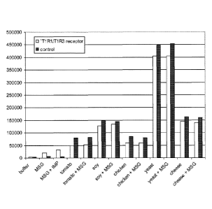
Figure 2
Activation of the T1 R1/T1 R3 umami receptor expressed in HEK293-Ga15 cells as
measured by BRET assays as described in Example 2. Test solutions (or buffer)
as
indicated and substrate were added to the cells. The BRET signals were
determined
as the ratio between readings taken at the acceptor wavelength (515nm) divided
by
the signals determined for the donor (400nm). To correct for background signal
due to
overlap of donor emission at the acceptor wavelength, the BRET ratio was
determined in parallel for cells expressing the donor alone (T1 R1/T1 R3-
Rluc). This
BRET background value was subtracted from the BRET value obtained for the
cells
expressing both BRET partners (BRET = BRET ratio - background ratio). A BRET
signal above the values achieved for buffer alone was defined as umami
receptor-
specific (see also table 2B).
CA 02782090 2012-05-25
WO 2011/067202 PCT/EP2010/068399
27
Examples
Example 1: Proof of principle using the human umami receptor T1 R1/T1 R3:
We developed a unique functional umami receptor assay to evaluate and quantify
umami in tomato and other crude plant or food extracts using the BRET
technique: to
test the principle we used the donor and acceptor proteins relating to BRET-2.
In short, the human T1 R1 and T1 R3 taste receptors were cloned and fused with
Renilla luciferase (Rluc) as donor protein; green fluorescent protein-2 (GFP2)
served
as acceptor protein and was fused to R-arrestin-2. Both constructs were
transfected
into a HEK293 cell line cells) for heterologous expression of the proteins.
Various
mammalian cell lines have been reported to be highly suitable for expression
of
receptors or other proteins, with HEK293 (human embryonic kidney) cells and
CHO
(Chinese hamster ovary) cells being some of the most versatile and suitable
ones12.
Similarly, GPCRs can be expressed in various ways as described in multiple
publications, such as transient 6, 8, 13-15 or stable (constitutive or
inducible) 6,9,16, 17
expression, depending on the nature of the experiment, the respective receptor
or the
available time.
Upon receptor stimulation with an agonist or crude extracts, the R-arrestin-
2/GFP-2
protein interacts with the activated receptor thus bringing GFP-2 in close
proximity
with luciferase, making energy transfer between these two proteins possible
and
generating a BRET signal. In the presence of oxygen the luciferase catalyses
the
transformation of the substrate DeepBlueC into coelenteramide, which can be
measured at 395-410 nm. If GFP-2 is in close proximity to the luciferase and
energy
transfer takes place, the emission will shift to 51 Onm; this is referred to
as the BRET
signal and is expressed as ratio between the acceptor (GFP-2) and the donor
(Renilla
luciferase).
The following experiments are designed to illustrate that conventional calcium-
based
functional receptor assays are not suitable for measuring the effects of crude
extracts;
in contrast, the BRET assay (as indicated here with umami) can clearly measure
specific receptor responses using crude natural extracts. Moreover, the assay
is also
sensitive enough for detecting differences of receptor activation between the
different
tomato samples.
CA 02782090 2012-05-25
WO 2011/067202 PCT/EP2010/068399
28
Materials & Methods:
Chemicals and media:
Fluo-4 AM was from Molecular Probes (# F-14202, prepared as 5 mM stock in
DMSO), DMEM (with 4.5g/1 glucose and ultraglutamine, # BE12-604F) was from
Lonza, trypsin-EDTA, Lipofectamine 2000, OptiMem and FCS were obtained from
Life Technologies Invitrogen. Monosodium glutamate (MSG), Inosine 5'-
monophosphate as well as all other chemicals were from Sigma-Aldrich.
The plasmid encoding GFP2-R-arrestin-2 was purchased from BioSignal Packard (#
6310176).
Coelenterazine 400A (a DeepBlueC derivative) and Coelenterazine-H were from
VWR International (# BTIU10125-1 and 233903-50, respectively).
All other cell culture supplies were from Greiner BioOne.
Receptor fusion constructs, cell lines, and media:
The human T1 R1 and human T1 R3 umami receptors were fused at their C-terminal
in
frame to Renilla Luciferase (Rluc) using standard molecular cloning techniques
and
the codon-humanized pR1uc-N3 vector from PerkinElmer (# 6310220). For
expression
of the umami receptors and the construct encoding GFP-2/R-arrestin-2, HEK293
cells
(human embryonic kidney cells, ATCC) were used using traditional transfection
methods (see also below).
HEK293 cells were maintained in DMEM and 10% FBS at 37 C / 5% CO2.
Tomato extracts:
The tomato extracts which served as representative natural, complex test
material
were prepared as follows: Frozen tomato samples (in -80 C) were weighed and
dissolved in an equal amount of water. After the seeds and (ocular tissues
(pulp)
were removed and put aside, the tomato pericarp (flesh) was ground using a
mortar
and pestle. The seeds and pulp were then added back to the mixture ensuring
that
they are well mixed and the seeds were not crushed. The mixture was
centrifuged for
15 minutes at 4000 rpm, the supernatant (serum) removed and freeze-dried in 1
Oml
CA 02782090 2012-05-25
WO 2011/067202 PCT/EP2010/068399
29
aliquots. Before the measurements, an equal volume of water was added to
dissolve
the sample.
Generation of the Ti R1 /T1 R3 and Rluc fusion constructs:
As mentioned above, we have generated the C-terminal Rluc receptor fusion
constructs of the human Ti R1 and human Ti R3 umami receptor using standard
molecular cloning techniques. This resulted in 2 different constructs: Ti R1-
Rluc and
Ti R3-Rluc. They can be either transfected together, resulting in a functional
receptor
heterodimer containing two Rluc moieties; alternatively either construct can
be
transfected in combination with the wild type receptor, thus resulting in Ti
R1-Rluc
combined with Ti R3 or Ti R1 combined with Ti R3-Rluc.
The sequences (cDNA and protein) for the wild type receptors as well as the
fusion
constructs and the Renilla luciferase are given in the sequence listing
(SEQ ID NO:5-8).
Transfections:
HEK293 cells will be transiently transfected with the plasmids encoding the
Ti R1/T1 R3 receptors (containing the Rluc-fusion protein) as well the GFP-2-R-
arrestin-2 using Lipofectamine 2000 according to the manufacturers' protocol.
In
short, HEK293 cells will be seeded at a density of 2x10⁵ cells per well (12-
wells plate,
1 ml medium/well), aiming at a confluency of about 80-90% the next day. After
24h the
umami receptor constructs (Ti R1-Rluc, Ti R3-Rluc, or wild type receptors)
will be co-
transfected with the plasmid encoding GFP-2-R-arrestin-2 using 15 pg of total
DNA
per well. We will dissolve the DNA in 100 pl of OptiMem and combine it with
100 pl of
OptiMem containing 4 pl of lipofectamine 2000. The mixture will then be
incubated for
minutes at room temperature, added to each well and the cells allowed to grow
for
48 hours. BRET measurements will be carried out 48h-52h after transfection.
Alternatively, the plasmids encoding the umami receptors can be transfected
into
HEK293 cells stably expressing the GFP-2-R-arrestin-2 using the same protocol
as
30 described above.
For the calcium-based receptor assay, HEK293 cells stably expressing Gal5 are
transfected with the Ti R1 and Ti R3 receptors according to the procedure
described
CA 02782090 2012-05-25
WO 2011/067202 PCT/EP2010/068399
above, but scaling down the protocol for 96 well plate format (10-fold
reduction using
a poly-Lysine coated pClear 96-wells plate).
Calcium-based receptor assay:
5 Activation of the T1 R1/T1 R3 umami receptor expressed in HEK293-Ga15 cells
has
been measured by monitoring the release of intracellular calcium. The growth
medium
was removed and the cells were loaded for 1 hour with 50 pl Tyrode's buffer
(140 mM
NaCl, 5 mM KCI, 10 mM glucose, 1 mM CaCI2. 2 H2O, 1 mM MgCI2. 6 H2O, 10 mM
Na-pyruvate and 50 mM HEPES pH 7.4) containing 2.5 pM of the fluorescent
marker
10 of calcium, Fluo-4-AM, supplemented with 0.5 mM probenicide to prevent
leakage of
Fluo-4 from the cells and 0.5% FCS, followed by a 1-hr incubation at 37 C. The
mixture was removed, and 150 pl of Tyrode's buffer containing 0.5mM
probenicide
was added. Intracellular calcium levels were monitored using a Flexstation 11
384
(Molecular Devices). Fluorescence measurements were carried out at 37 C at an
15 excitation wavelength of 485nm and an emission wavelength of 520nm. 50 pl
of
compounds or tomato extracts were added to the cells at a pipettor speed of
104 pI
per sec after a baseline measurement of 20 s. Activity of the umami receptor
was
measured as the change in fluorescence (AF) which was calculated by
subtracting
the maximum fluorescence after the addition of the test solution from the
baseline
20 fluorescence measured before addition of the test solution.
The data indicated in table 2A show very clearly that using pure umami
compounds
results in umami receptor-specific responses (control cells not expressing the
receptor give no increase); in contrast, using various tomato extracts results
in high
non-specific responses in the control cells, making it impossible to indentify
signals
25 indicating umami.
BRET assay:
For the BRET assay we will use a Mithras LB 940 plate reader (Berthold
Technologies). The protocol we will use is essentially the same as described
by
30 Packard BioOne or published protocols13 with slight modifications: HEK 293
cells will
be transfected as described above. 48h after transfection they will be
harvested and
taken up in BRET buffer (D-PBS containing 2 pg/ml Aprotinin) at a density of
2x10⁶
CA 02782090 2012-05-25
WO 2011/067202 PCT/EP2010/068399
31
cells/ml. After leaving the cells for 1 hour at room temperature for
equilibration, 30pl
containing approximately 1x10⁵ cells will be transferred to each well of a
white 96we11
plate. 1 Opt of test compounds (or buffer) and 10 pl of the substrate
coelenterazine
(final concentration 5pM) will be added simultaneously to the cells using the
injectors.
Immediately after the final injections, sequential readings will be taken at
41 Onm and
515nm. The BRET signals will be determined as the ratio between GFP-2 emission
(515nm) and Rluc/coelenterazine emission (41 Onm).
Example 2: Detection of taste modulators in complex samples
Preparation and selection of test samples (extracts)
The extracts or crude mixtures described below are examples for natural
samples unsuitable for testing with a calcium-based receptor assay due to
their high
non-specific background signal. They are from different origin and are
selected to
illustrate a wide range of potential test samples. The selected samples are
also
known to contain glutamate, ribotides or both and are therefore good
candidates to
examine in a BRET assay using the umami receptor T1 R1/T1 R3 as example.
- Tomato: The preparation of tomato extract is already been described in
Example 1
- Yeast extract: Commercially available yeast extract enriched for 5'-
ribotides
(DSM, Maxarome Select yeast extract). The yeast extract was dissolved in
water (10 x weight of yeast extract).
- Soy sauce: commercially available soy sauce was used (Conimex ketjap
manis).
- Chicken bouillon: A commercially available chicken bouillon cube (Knorr) was
dissolved in hot water as indicated by the manufacturers. To remove particles
from the test sample the bouillon was centrifuged at 5000rpm for 10 minutes
and the supernatant transferred to a fresh tube.
- Cheese extract: commercially available Roquefort cheese was weighed and
dissolved in an equal amount of water and homogenized using a mortar and
pestle. The mixture was centrifuged for 10 minutes at 5000rpm to remove
particles and the supernatant transferred to a fresh tube.
CA 02782090 2012-05-25
WO 2011/067202 PCT/EP2010/068399
32
Experimental procedures
Transfections:
HEK293 cells were transiently transfected with the plasmids encoding the Ti R1
and
Ti R3 receptors (the Ti R3 receptor contained the Rluc-fusion protein in this
experiment) as well the GFP-2-R-arrestin-2 using Lipofectamine 2000 according
to
the manufacturers' protocol. In short, HEK293 cells will be seeded at a
density of
2x10⁵ cells per well (12-wells plate, 1 ml medium/well), aiming at a
confluency of about
80-90% the next day. After 24h the umami receptor constructs (Ti R1 and Ti R3-
Rluc
receptors) were co-transfected with the plasmid encoding GFP-2-R-arrestin-2
using 15
pg of total DNA per well. The DNA was dissolved in 100 pl of OptiMem and
combined
with 100 pl of OptiMem containing 4 pl of lipofectamine 2000. The mixture was
incubated for 30 minutes at room temperature, added to each well and the cells
allowed to grow for 48 hours. BRET measurements were carried out 48h-52h after
transfection.
Alternatively, the plasmids encoding the umami receptors were transfected into
HEK293 cells stably expressing the GFP-2-R-arrestin-2 using the same protocol
as
described above.
For the calcium-based receptor assay, HEK293 cells stably expressing Gal 5 are
transfected with the Ti R1 and Ti R3 receptors according to the procedure
described
above, but scaling down the protocol for 96 well plate format (10-fold
reduction using
a poly-Lysine coated pClear 96-wells plate).
Calcium-based receptor assay:
Activation of the Ti R1/T1 R3 umami receptor expressed in HEK293-Gal 5 cells
has
been measured by monitoring the release of intracellular calcium. The growth
medium
was removed and the cells were loaded for 1 hour with 50 pl Tyrode's buffer
(140 mM
NaCl, 5 mM KCI, 10 mM glucose, 1 mM CaCl2. 2 H2O, 1 mM MgCl2. 6 H2O, 10 mM
Na-pyruvate and 50 mM HEPES pH 7.4) containing 2.5 pM of the fluorescent
marker
of calcium, Fluo-4-AM, supplemented with 0.5 mM probenicide to prevent leakage
of
Fluo-4 from the cells and 0.5% FCS, followed by a 1-hr incubation at 37 C. The
CA 02782090 2012-05-25
WO 2011/067202 PCT/EP2010/068399
33
mixture was removed, and 150 pl of Tyrode's buffer containing 0.5mM
probenicide
was added. Intracellular calcium levels were monitored using a Flexstation 11
384
(Molecular Devices). Fluorescence measurements were carried out at 37 C at an
excitation wavelength of 485nm and an emission wavelength of 520nm. 50 pl of
compounds or extracts were added to the cells at a pipettor speed of 104 pl
per sec
after a baseline measurement of 20 s. Activity of the umami receptor was
measured
as the change in fluorescence (AF) which was calculated by subtracting the
maximum
fluorescence after the addition of the test solution from the baseline
fluorescence
measured before addition of the test solution.
Table 2B and Figure 1 illustrate that using pure umami compounds (MSG and MSG
+ IMP) results in umami receptor-specific responses; in contrast, using
various
extracts and natural mixtures results in high non-specific responses in
control cells
not expressing the umami receptor as well, thereby making it impossible to
calculate
receptor-specific signals indicating umami.
BRET assay:
For the BRET assay we used a Mithras LB 940 plate reader (Berthold
Technologies).
The assay protocol is essentially the same as described by Packard BioOne or
other
published protocols10' 13 with slight modifications: HEK 293 cells were
transfected as
described above. 40h after transfection they were harvested and taken up in
BRET
buffer (D-PBS containing 1g/I D-glucose and 2 pg/ml Aprotinin) at a density of
2x10⁶
cells/mi. After leaving the cells for 1 hour at room temperature for
equilibration, 30pl
containing 1x10⁵ cells were transferred to each well of a white 96we1I plate.
10pl of
test compounds (or buffer) and 10 pl of the substrate coelenterazine 400
(DeepBlueC, final concentration 5pM) were added to the cells using the
injectors.
Immediately after the final injections, sequential readings were taken at
400nm and
515nm. Alternatively, compounds were added to the cells manually and were
incubated for 5 minutes before injection of the substrate. The BRET signals
were
determined as the ratio between readings taken at the acceptor wavelength (GFP-
2;
515nm) divided by the signals determined for the donor (Rluc/coelenterazine
400;
400nm). To correct for background signal due to overlap of donor emission at
the
acceptor wavelength, the BRET ratio was determined in parallel for cells
expressing
CA 02782090 2012-05-25
WO 2011/067202 PCT/EP2010/068399
34
the donor alone (Ti R1/T1 R3-Rluc). This BRET background value was subtracted
from the BRET value obtained for the cells expressing both BRET partners (BRET
=
BRET ratio - background ratio). A BRET signal above the values achieved for
buffer
alone was defined as umami receptor-specific (see table 2B).
Results:
As indicated in table 2B and Figure 1, it was not possible to achieve umami
receptor-
specific signals using representative extracts and other complex mixtures
using the
calcium-based receptor assay. In contrast, Figure 2 shows that using the
described
BRET assay it was possible to specifically measure umami receptor activation
for
these complex samples.
The definition of receptor-specific signals differs slightly for technical
reasons: For the
calcium assay, cells expressing the umami receptor and Ga15 are compared with
cells expressing Gal5 only; the difference between these signals is defined as
umami
receptor-specific signal. For the BRET assay the background is determined
using
cells expressing the donor only (T1 R1 /T1 R3-Rluc); a positive value
indicates a
specific BRET signal.
CA 02782090 2012-05-25
WO 2011/067202 PCT/EP2010/068399
Table 1: non-exhaustive list of agonist of several taste receptors as
identified
Receptor agonist
Ti R1/T1 R3 MSG, (L-glutamate), positive allosteric
modulator: IMP (inosine monophosphate)
mGluR4 MSG, L-AP4 (L-(+)-2-Amino-4-
phosphonobut ric acid)
T1 R2/T1 R3 Sweeteners (natural and artificial): Glucose,
sucrose, saccharose
GPR120 Medium and long-chain fatty acids (linoleic acid)
GPR93 LPA (lysophosphatidic acid), proteolytic
degradation products
FFAR1 Medium and long-chain fatty acids
(eicosatrienoic acid, linoleic acid)
FFAR2 Short chain fatty acids (acetate, propionate)
FFAR3 Short chain fatty acids (acetate, propionate)
GPRC6A L-amino acids (most potent L-arginine)
GPR84 Medium chain fatty acids (Capric acid,
Undecanoic acid)
TAS2R7 Quinacrine, chloroquine, papaverine, strychnine
TAS2R14 Picrotoxin, picrotoxinin, a-thujone, naphthoic
acid (8 in total)
TAS2R161 R-glucopyranosides (e.g. salicin, phenyl--D-
____________ lucop ranoside
TAS2R381 Phenylthiocarbamide, PROP, acetylthiourea
and others
TAS2R43 Saccharin, acesulfame K, aristolochic acid
6-Nitrosaccharin
TAS2R44 Saccharin, acesulfame K, aristolochic acid
TAS2R461 Sesquiterpenes
Absinthine, denatonium benzoate
TAS2R47 6-nitrosaccharin and saccharin
TAS2R4 Quinine, denatonium benzoate
TAS2R8 Ranitidine, denatonium benzoate
TAS2R10 Strychnine, brucine, denatoniurn benzoate,
absinthine
TAS2R39 Acetaminophen, ranitidine, denatonium
benzoate
TAS2R45 Absinthine
TAS2R48 Absinthine
CA 02782090 2012-05-25
WO 2011/067202 PCT/EP2010/068399
36
Receptor agonist
T2R1/ TAS2R1 chloroquine, dexamethasone, quinine
T2R3/ TAS2R3 chloroquine, 2-acet lp razine
T2R4/ TAS2R4 quinine, denatonium benzoate
T2R5/ TAS2R5 dimethylbiguanide, oleuropein
T2R7/TAS2R7 quinacrine, chloroquine, papaverine,
strychnine
T2R8/ TAS2R8 ranitidine, denatonium benzoate
T2R9/ TAS2R9 ranitidine, ofloxacin
T2R10/ TAS2R10 strychnine, brucine, denatonium benzoate,
absinthine
T2R13/ TAS2R13 ethylpyrazine, quinacrine
T2R14/ TAS2R14 picrotoxin, picrotoxinin, a-thujone, naphthoic
acid
T2R16/ TAS2R16 R-glucopyranosides (e.g. salicin, phenyl-R-
D- lucop ranoside
T2R24/ TAS2R42 Not known
T2R44/ TAS2R47 6-nitrosaccharin and saccharin
T2R50/ TAS2R45 absinthine
T2R51/ TAS2R38 phen Ithiocarbamide, PROP, acet (thiourea
T2R54/ TAS2R39 acetaminophen, ranitidine, denatonium
benzoate
T2R55/ TAS2R40 linamarin, ox but nin chloride
T2R61/ TAS2R43 saccharin, acesulfame K, aristolochic acid
6-Nitrosaccharin
T2R63/ TAS2R49 Not known
T2R64/ TAS2R44 saccharin, acesulfame K, aristolochic acid
T2R65/ TAS2R48 Absinthine, ethylpyrazine
T2R67/ TAS2R50 ethylpyrazine, oxybutynin chloride
T2R71/ TAS2R41 Nitrosaccharin
T2R75/ TAS2R46 sesquiterpenes
absinthine, denatonium benzoate
T2R76 brucine
CA 02782090 2012-05-25
WO 2011/067202 PCT/EP2010/068399
37
Table 2A: Specific activation of a T1 R1/T1 R3 umami receptor measured by the
release of intracellular calcium: average values are shown
Compounds T1 R1/T1 R3 umami receptor Control cells (no T1 R1/T1 R3)
meas.1 meas. 2 average meas.1 meas. 2 average
100pM MSG 1146 1193 1170 1102 1168 1135
1 mM MSG 2548 3990 3269 1100 1020 1060
10mM MSG 5766 7732 6749 1200 1030 1115
extract 1 4649 7985 6317 6420 3449 4934
extract 2 4552 6461 5506 7435 6061 6748
extract 3 5980 5080 5530 4369 5431 4900
extract 4 5394 4285 4839 5848 5784 5816
extract 5 6338 9179 7759 6423 6318 6370
extract 6 9285 10503 9894 10708 9785 10247
Table 2B: T1 R1/T1 R3 receptor-specific signals using the calcium or BRET
assay
Test samples Calcium assay BRET assay
MSG 10mM + +
MSG 1 mM + IMP 500pM + +
Tomato extract - +
Yeast extract - +
Chicken bouillon - +
Soy sauce - +
Cheese extract - +
CA 02782090 2012-05-25
WO 2011/067202 PCT/EP2010/068399
38
Table 3: SEQ ID NO of the DNA/amino acid sequences of taste receptors
identified in
the sequence listing
Taste receptor subunit SEQ ID NO of human SEQ ID NO of human
DNA sequence amino acid sequence
T1 R1 1 2
T1 R3 3 4
T1 R2 9 10
mGluR4a 11 12
GPR120 13 14
GPR93 15 16
FFA receptor 1 17 18
FFA receptor 2 19 20
FFA receptor 3 21 22
GPR84 23 24
mGluR4c 25 26
T2R1 =TAS2R1 27 28
T2R3 (=TAS2R3) 29 30
T2R4 =TAS2R4 31 32
T2R5 (=TAS2R5) 33 34
T2R7 (=TAS2R7) 35 36
T2R8 =TAS2R8 37 38
T2R9 (=TAS2R9) 39 40
T2R10 =TAS2R10 41 42
T2R13 (=TAS2R13) 43 44
T2R14 =TAS2R14 45 46
T2R16 (=TAS2R16) 47 48
T2R24 (=TAS2R42) 49 50
T2R44 =TAS2R47 51 52
T2R50 (=TAS2R45) 53 54
T2R51 =TA52R38=T2R61 55 56
T2R54 (=TAS2R39=T2R57) 57 58
T2R55 =TAS2R40=T2R58 59 60
T2R61 (=TAS2R43=T2R52) 61 62
T2R63 (TAS2R49=T2R56) 63 64
T2R64 =TAS2R44=T2R53 65 66
T2R65 (=TAS2R48=T2R55) 67 68
T2R67 =TAS2R50=T2R51 69 70
T2R71 (=TAS2R41=T2R59) 71 72
T2R75 (=TAS2R46=T2R54) 73 74
T2R76 =TAS2R60=T2R56 75 76
CA 02782090 2012-05-25
WO 2011/067202 PCT/EP2010/068399
39
References
(1) Scott TR, Verhagen JV. Taste as a factor in the management of nutrition.
Nutrition 2000 October;1 6(10):874-85.
(2) Zhao GQ, Zhang YF, Hoon MA, Chandrashekar J, Erlenbach I, Ryba NJP,
Zuker CS. The receptors for mammalian sweet and umami taste. Cell 2003
October 31;115(3):255-66.
(3) Chandrashekar J, Hoon MA, Ryba NJ, Zuker CS. The receptors and cells for
mammalian taste. Nature 2006 November 16;444(7117):288-94.
(4) Breslin PA, Spector AC. Mammalian taste perception. Curr Biol 2008
February
26;18(4):R148-R155.
(5) Kinnamon SC, Vandenbeuch A. Receptors and transduction of umami taste
stimuli. Ann N YAcad Sci 2009 July;1 170:55-9.
(6) Zhang F, Klebansky B, Fine RM, Xu H, Pronin A, Liu H, Tachdjian C, Li X.
Molecular mechanism for the umami taste synergism. Proc Nat/ Acad Sci U S
A 2008 December 30;105(52):20930-4.
(7) Shigemura N, Shirosaki S, Sanematsu K, Yoshida R, Ninomiya Y. Genetic and
molecular basis of individual differences in human umami taste perception.
PLoS One 2009;4(8):e6717.
(8) Li X, Staszewski L, Xu H, Durick K, Zoller M, Adler E. Human receptors for
sweet and umami taste
1. Proc Nat/ Acad Sci USA 2002 April 2;99(7):4692-6.
(9) Ozeck M, Brust P, Xu H, Servant G. Receptors for bitter, sweet and umami
taste couple to inhibitory G protein signaling pathways. European Journal of
Pharmacology 2004 April 12;489(3):139-49.
(10) Bacart J, Corbel C, Jockers R, Bach S, Couturier C. The BRET technology
and
its application to screening assays. Biotechnol J 2008 March;3(3):311-24.
(11) Pfleger KD, Dalrymple MB, Dromey JR, Eidne KA. Monitoring interactions
between G-protein-coupled receptors and beta-arrestins. Biochem Soc Trans
2007 August;35(Pt 4):764-6.
(12) Eglen RM, Gilchrist A, Reisine T. The use of immortalized cell lines in
GPCR
screening: the good, bad and ugly. Comb Chem High Throughput Screen 2008
August;1 1(7):560-5.
CA 02782090 2012-05-25
WO 2011/067202 PCT/EP2010/068399
(13) Vrecl M, Jorgensen R, Pogacnik A, Heding A. Development of a BRET2
screening assay using beta-arrestin 2 mutants. J Biomol Screen 2004
June;9(4):322-33.
(14) Nelson G, Hoon MA, Chandrashekar J, Zhang Y, Ryba NJ, Zuker CS.
5 Mammalian sweet taste receptors. Cell 2001 August 10;106(3):381-90.
(15) Jiang P, Cui M, Zhao B, Snyder LA, Benard LM, Osman R, Max M, Margolskee
RF. Identification of the cyclamate interaction site within the transmembrane
domain of the human sweet taste receptor subunit T1 R3. J Biol Chem 2005
October 7;280(40):34296-305.
10 (16) Stoddart LA, Smith NJ, Jenkins L, Brown AJ, Milligan G. Conserved
polar
residues in transmembrane domains V, VI, and VII of free fatty acid receptor 2
and free fatty acid receptor 3 are required for the binding and function of
short
chain fatty acids. J Biol Chem 2008 November 21;283(47):32913-24.
(17) Xu H, Staszewski L, Tang HX, Adler E, Zoller M, Li XD. Different
functional
15 roles of T1 R subunits in the heterorneric taste receptors. Proceedings of
the
National Academy of Sciences of the United States of America 2004
September 28;101(39):14258-63.