Note: Descriptions are shown in the official language in which they were submitted.
CA 02782386 2012 05 30
WO 2011/068924
PCT/US2010/058641
MICRO VALVE PROTECTION DEVICE AND METHOD OF USE FOR
PROTECTION AGAINST EMBOLIZATION AGENT REFLUX
BACKGROUND OF THE INVENTION
1. Field of Invention
[0001] The present invention relates generally to a medical embolizing
treatment
system. More particularly, the present invention relates to an embolizing
treatment system
utilizing a protection device which reduces the reflux of a treatment agent in
a blood vessel
during an embolization therapy procedure, where the embolization agent is
delivered through a
catheter to provide therapy to tissue distal via a delivery orifice of the
catheter.
2. State of the Art
[0002] Embolization, chemo-embolization, and radio-embolization therapy are
often
clinically used to treat a range of diseases, such as hypervascular liver
tumors, uterine fibroids,
secondary cancer metastasis in the liver, pre-operative treatment of
hypervascular menangiomas
in the brain and bronchial artery embolization for hemoptysis. An embolizing
agent may be
embodied in different forms, such as beads, liquid, foam, or glue placed into
an arterial
vasculature. The beads may be uncoated or coated. Where the beads are coated,
the coating
may be a chemotherapy agent, a radiation agent or other therapeutic agent.
When it is desirable
to embolize a small blood vessel, small bead sizes (e.g., 10p.m - 100 m) are
utilized. When a
larger vessel is to be embolized a larger bead size (e.g., 100p.m - 900 m) is
typically chosen.
[0003] While embolizing agent therapies which are considered minimally or
limited
invasive therapies have often provided good results, they have a small
incidence of non-targeted
embolization which can lead to adverse events and morbidity. One cause of non-
targeted
delivery of embolizing agents is reflux in the artery. Reflux occurs where the
embolic agent
exits the distal end of the catheter and then backflows around the outside of
the catheter. This
backflow can end up in a healthy organ and damage it.
[0004] Reflux can also occur during the administration of the embolization
agent, while
the artery is still patent. Reflux may also occur when the artery becomes
static and injected
embolizing agents flow backward.
1
CA 02782386 2012 05 30
WO 2011/068924
PCT/US2010/058641
[0005] Additionally, reflux can be a time-sensitive phenomenon. Sometimes,
retiux
occurs as a response to an injection of the embolic agent, where the reflux
occurs rapidly (e.g.,
in the time-scale of milliseconds) in a manner which is too fast for a human
operator to respond.
Also, reflux can happen momentarily, followed by a temporary resumption of
forward flow in
the blood vessel, only to be followed by additional reflux.
[0006] Figure 1 shows a conventional (prior art) embolization treatment in
the hepatic
artery 106. Catheter 101 delivers embolization agents (beads) 102 in a hepatic
artery 106, with a
goal of embolizing a target organ 103. It is important that the forward flow
(direction arrow
107) of blood is maintained during an infusion of embolization agents 102
because the forward
flow is used to carry embolization agents 102 deep into the vascular bed of
target organ 103.
[0007] Embolization agents 102 are continuously injected until reflux of
contrast agent
is visualized in the distal area of the hepatic artery. Generally, since
embolization agents 102
can rarely be visualized directly, a contrast agent may be added to
embolization agents 102. The
addition of the contrast agent allows for a visualization of the reflux of the
contrast agent
(shown by arrow 108), which is indicative of the reflux of embolization agents
102. The reflux
may, undesirably, cause embolization agents 102 to be delivered into a
collateral artery 105,
which is proximal to the tip of catheter 101. The presence of embolization
agents 102 in
collateral artery 105 leads to non-target embolization in a non-target organ
104, which may be
the other lobe of the liver, the stomach, small intestine, pancreas, gall
bladder, or other organ.
[0008] Non-targeted delivery of the embolic agent may have significant
unwanted
effects on the human body. For example, in liver treatment, non-targeted
delivery of the
embolic agent may have undesirable impacts on other organs including the
stomach and small
intestine. In uterine fibroid treatment, the non-targeted delivery of the
embolic agent may
embolize one or both ovaries leading to loss of menstrual cycle, subtle
ovarian damage that may
reduce fertility, early onset of menopause and in some cases substantial
damage to the ovaries.
Other unintended adverse events include unilateral deep buttock pain, buttock
necrosis, and
uterine necrosis.
[0009] Often, interventional radiologists try to reduce the amount and
impact of reflux
by slowly releasing the embolizing agent and/or by delivering a reduced
dosage. The added
time, complexity, increased x-ray dose to the patient and physician (longer
monitoring of the
patient) and potential for reduced efficacy make the slow delivery of
embolization agents
suboptimal. Also, reducing the dosage often leads to the need for multiple
follow-up
2
CA 02782386 2012 05 30
WO 2011/068924
PCT/US2010/058641
treatments. Even when the physician tries to reduce the amount of reflux, the
local fiow
conditions at the tip of the catheter change too fast to be controlled by the
physician, and
therefore rapid momentary reflux conditions can happen throughout infusion.
SUMMARY OF THE INVENTION
[00010] According to one aspect of the invention, a deployable apparatus is
provided that
is useful in an embolization procedure and which enables substantially
unrestricted forward
flow of blood in a vessel and reduces or stops reflux (regurgitation or
backward flow) of
embolization agents which are introduced into the blood.
[00011] In some embodiments, the deployable apparatus includes a delivery
catheter
having a valve fixedly coupled to the distal end thereof An outer catheter is
provided which
extends over the valve during introduction to maintain the valve in a
collapsed cylindrical
configuration until the valve is advanced through the patient to the desired
vascular destination.
Once at the destination, the outer catheter is retracted from over the valve
to permit expansion
of the valve into an open state, as discussed below.
[00012] In other embodiments, the deployable apparatus includes a delivery
catheter and
a valve introducer which delivers a valve to a valve seat at the distal end of
the delivery catheter
during the embolization procedure. No outer catheter is required. A valve
introducer
maintains the distal end of the valve in a closed configuration, and a push
wire is abutted
against the proximal end of the valve and used to push the valve out of the
valve introducer and
through the delivery catheter. The valve is advanced by the push wire to the
valve seat located
at the distal end of a delivery catheter. Once the valve seat captures a
proximal portion of the
valve to lock the valve at the distal end of the delivery catheter, the push
wire is then withdrawn
from the delivery catheter to provide an apparatus with enhanced fluid flow
through the
delivery catheter. In certain embodiments a pull member is coupled to the
valve to release the
lock between the valve and valve seat and permit retraction of the valve into
the delivery
catheter after the embolic agent has been dispensed.
[00013] The deployable valve includes a plurality of filaments which cross
over each
other (i.e., are braided) and which have a spring bias to assume a preferred
crossing angle
relative to each other. In a first state, the valve is preferably kept in a
cylindrical arrangement
with a diameter substantially equal to the diameter of the delivery catheter.
In a second state,
3
CA 02782386 2012 05 30
WO 2011/068924
PCT/US2010/058641
the valve is free to open due to the spring bias in the filaments. In the
second state, with the
proximal end of the valve attached to the delivery catheter, in the
bloodstream, if the blood is
not flowing distally past the valve, the valve assumes a substantially
frustoconical shape. The
distal end of the valve is intended to make contact with the walls of the
vessel in which it is
deployed when blood is not flowing distally past the valve.
[00014] In some embodiments, the valve, while enabling substantially
unrestricted
forward flow in a vessel and reducing or stopping reflux of embolization
agents, allows the
reflux of blood or contrast agent. In other embodiments, the valve, while
enabling substantially
unrestricted forward flow in a vessel and reducing or stopping reflux of
embolization agents,
also reduces or stops backward flow of blood.
[00015] According to one aspect of the invention, the valve has a radial
force of
expansion when in the undeployed state of less than 40mN.
[00016] According to another aspect of the invention, the valve has a time
constant of
expansion from the cylindrical arrangement to the fully-open position when in
a static fluid
having a viscosity of approximately 3.2cP of between 1.0 and 0.01 seconds, and
more
preferably between 0.50 and 0.05 seconds.
[00017] According to a further aspect of the invention, the valve has a
Young's modulus
of elasticity that is greater than lOOMPa.
[00018] According to yet another aspect of the invention, the preferred
crossing angle of
the valve filaments is approximately 130 degrees.
[00019] According to even another aspect of the invention, the filaments of
the valve are
selected to be of a desired number and diameter such that in an open position,
they are capable
of trapping embolization agents. By way of example only, the filaments of the
valve are
selected so that in an open position they present a pore size of 500um and are
thus capable of
preventing reflux of embolizing agent such as beads having a size larger than
500um. As
another example, the filaments of the valve are selected so that in an open
position they present
a pore size of 250um and are thus capable of preventing reflux of embolizing
agent having a
size larger than 250um.
[00020] In one embodiment, the valve filaments are coated with a filter
which is formed
and attached to the filaments according to any desired manner, such as by
spraying, spinning,
electrospinning, bonding with an adhesive, thermally fusing, melt bonding, or
other method.
The filter is preferably arranged to have a desired pore size, although it
will be appreciated that
4
CA 02782386 2016-08-19
72235-230
the pore size may be non-uniform depending upon the technique in which the
filter is formed and
attached. By way of example, the pore size of the filter may be approximately
401.tm such that
embolizing agents having a characteristic size of more than 40um are prevented
from refluxing
past the valve. By way of another example, the pore size of the filter may be
approximately 20 m
such that embolizing agents having a characteristic size of more than 20 m are
prevented from
refluxing past the valve. In both cases, blood cells (which have a
characteristic size smaller than
201.tm), and contrast agent which has a molecular size smaller than 201,tm
will pass through the
filter and valve.
[00021] According to an additional aspect of the invention, when in a
fully-open position
where the filaments assume the preferred crossing angle, the valve is adapted
to have a distal
diameter which is at least twice the diameter of the delivery catheter, and
preferably at least five
times the diameter of the delivery catheter.
[00022] In one embodiment, the filaments are all formed from a polymer. In
another
embodiment, one or more of the filaments is formed from stainless steel,
platinum or platinum-
iridium.
1000231 In an embodiment where one or more filaments are formed from a
polymer, the
filaments that are formed from the polymer are preferably melted at their
proximal end into the
delivery catheter.
[00023a] Further embodiments of the invention include:
- an endovascular valve device for reducing reflux of an infusate having an
embolic agent in a vessel during a therapy procedure, comprising: i) a
plurality of elongate
first filaments each having a diameter of 0.025 mm to 0.127 mm, said first
filaments having a
proximal end, a distal end, and a length extending therebetween, said proximal
ends secured
relative to each other, said first filaments along said lengths distal of said
proximal ends not
bonded to each other such that said first filaments are movable relative to
each other, said
valve fully collapsible into an undeployed state, and expandable from said
undeployed state
into a radially-expanded deployed state by a spring bias of said first
filaments, wherein in said
deployed state said first filaments cross one another at an angle of 1000 to
150; and ii) a filter
CA 02782386 2016-08-19
72235-230
comprising a polymeric coating on said braided first filaments, the polymeric
coating
comprising second filaments, said filter defining a pore size not exceeding
500um, wherein
said valve expands from said undeployed state to said deployed state in less
than one second
in an at-rest fluid having a viscosity of 3.2 cP, said once said valve is in
said deployed state,
said valve dynamically movable within the vessel between an expanded valve-
open
configuration and a collapsed valve-closed configuration depending on the
local biological
fluid flow conditions about said valve, and when said valve is in said valve-
open
configuration said pore size of said filter renders said filter impermeable to
the embolic agent
of the infusate;
- an endovascular device for reducing reflux of an infusate in a vessel during
a
procedure, the device comprising: a) an elongated delivery catheter having a
proximal end and
a distal end, a lumen defining an inner diameter, and an outer diameter,
wherein the infusate
can be delivered through said lumen; b) a valve coupled at said distal end of
said delivery
catheter, said valve having a housed state with a first smaller diameter and a
radially-
expanded deployed state with a second larger diameter, said second larger
diameter being
substantially larger than said outer diameter and capable of extending across
a vessel through
which said device is used, said valve comprising, i) a plurality of elongate
first filaments each
having a diameter of 0.025 mm to 0.127 mm, said first filaments having a
proximal end, distal
end, and a length extending therebetween, said proximal ends secured relative
to each other,
said first filaments along said lengths distal of said proximal ends not
bonded to each other
such that said first filaments are movable relative to each other, said valve
fully collapsible
into an undeployed state, and expandable from said undeployed state into a
radially-expanded
deployed state by a spring bias of said first filaments, wherein in said
deployed state said first
filaments cross one another at an angle of 1000 to 150'; and ii) a filter
comprising a polymeric
coating on said braided first filaments, the polymeric coating comprising
second filaments,
said filter defining a pore size not exceeding 500 m, wherein said valve
expands from said
undeployed state to said deployed state in less than one second in an at-rest
fluid having a
viscosity of 3.2 cP, said once said valve is in said deployed state, said
valve dynamically
movable within the vessel between an expanded valve-open configuration and a
collapsed
valve-closed configuration depending on the local biological fluid flow
conditions about said
5a
CA 02782386 2016-08-19
72235-230
valve, and when said valve is in said valve-open configuration said pore size
of said filter
renders said filter impermeable to the embolic agent of the infusate; and c) a
control element
moving said valve from said housed state to said deployed state, and, said
valve in said
deployed state having an opening through which infusate can be delivered along
a pathway
extending through said delivery catheter and into the vessel, said valve in
said deployed state
automatically allowing biological fluid in the vessel to flow in a proximal to
distal direction
relative to the valve and preventing reflux of the infusate in the distal to
proximal direction;
- an endovascular device for reducing reflux of an infusate in a vessel during
a
procedure, the device comprising: a) an elongated delivery catheter having a
proximal end and
a distal end, a lumen defining an inner diameter, and an outer diameter,
wherein the infusate
can be delivered through said lumen to the vessel; b) a valve seat provided at
said distal end of
said delivery catheter; c) a valve distally displaceable through said delivery
catheter to said
valve seat, said valve having i) a plurality of elongate first filaments each
having a diameter of
0.025 mm to 0.127 mm, said first filaments having a proximal end, distal end,
and a length
extending therebetween, said proximal ends secured relative to each other,
said first filaments
along said lengths distal of said proximal ends not bonded to each other such
that said first
filaments are movable relative to each other, said valve fully collapsible
into an undeployed
state, and expandable from said undeployed state into a radially-expanded
deployed state by a
spring bias of said first filaments, wherein in said deployed state said first
filaments cross one
another at an angle of 1000 to 1500; and ii) a filter comprising a polymer
coating on said
braided first filaments, the polymeric coating comprising second filaments,
said filter defining
a pore size not exceeding 50011m, and iii) mating structure that is engaged
with said valve seat
when said valve is advanced to said valve seat to thereby lock said valve
relative to said valve
seat, wherein said valve in said undeployed state has a diameter smaller than
or approximately
equal to an inner diameter of said catheter, and in said deployed state has a
diameter
substantially larger than an outer diameter of said catheter, and said valve
in said deployed
state having an opening in fluid communication with said lumen for delivery of
the infusate
from said delivery catheter to the vessel, wherein said valve expands from
said undeployed
state to said deployed state in less than one second in an at-rest fluid
having a viscosity of
3.2 cP, and wherein once said valve is in said deployed state, said valve
dynamically movable
5b
CA 02782386 2016-08-19
72235-230
within the vessel between an expanded valve-open configuration and a collapsed
valve-closed
configuration depending on the local biological fluid flow conditions about
said valve, and
when said valve is in said valve-open configuration said pore size of said
filter renders said
filter impermeable to the embolic agent of the infusate; and
- an endovascular valve system for reducing reflux of an infusate in a vessel
during a therapy procedure, comprising: a) a first tubular member having a
proximal end, a
distal end, a lumen having a first inner diameter, and a first outer diameter
sized to be inserted
into the vessel; b) a second tubular member having a proximal end with a
proximal face, a
distal end, a lumen having a second inner diameter smaller than said first
inner diameter, and
a second outer diameter sized to be inserted into the vessel; c) a valve
longitudinally displaced
between said first and second tubular members, said valve comprising a braid
of first
filaments each having a proximal end and a distal end, said proximal ends of
said first
filaments coupled to said distal end of said first member, and said distal end
of said first
filaments coupled to said proximal end of said second member, said braid of
first filaments
biased to radially expand outward at an expansion location to a diameter
larger than the first
and second outer diameters, said valve defining a proximal portion and a
distal portion; d) a
filter comprising a polymer coating provided onto the braid of first
filaments, the polymer
coating comprising second filaments, wherein the filter is formed across the
valve proximal of
said expansion location, said filter defines a pore size not exceeding 500 m,
and said valve
distal said expansion location being at least partially free of said filter;
and e) an elongate
member insertable into said first tubular member and removable therefrom
during the therapy
procedure, said elongate member having a distal surface with a diameter
smaller than said first
inner diameter and larger than said second inner diameter, wherein said
elongate member is
insertable into said first tubular member through said valve such that said
distal surface is
brought into contact with said proximal face of said second tubular member to
apply a tensile
force to said braid to reduce a diameter of said braid to place said valve in
an undeployed state
for delivery within the vessel, and wherein said elongate member is thereafter
removable from
said contact and said valve to provide said valve in a deployed state in which
said valve is
dynamically movable within the vessel between an expanded valve-open
configuration and a
collapsed valve-closed configuration depending on the local biological fluid
flow conditions
Sc
CA 02782386 2016-08-19
72235-230
about said valve, and when said valve is in said valve-open configuration said
pore size of
said filter renders said filter impermeable to the embolic agent of the
infitsate.
[00024] The valve may be deployed in any of several manners. Thus, by way
of example
only, in appropriate embodiments, an outer catheter or sleeve extending over
the delivery catheter
may be used to keep the valve in an undeployed state, and the outer catheter
or sleeve may be
pulled backward relative to the delivery catheter in order to deploy the
valve. Where an outer
catheter or sleeve is utilized, the valve may be captured and returned to its
undeployed position by
moving the delivery catheter proximally relative to the outer catheter or
sleeves.
[00025] As another example, the distal end of the valve may be provided
with loops which
are adapted to engage a guidewire which extends through and distal the distal
end of the delivery
catheter and through the distal loops of the valve. When the guidewire is
withdrawn proximally,
the valve deploys.
[00026] As another example, a knitted sleeve with a control thread can be
provided to
cover the valve. The control thread, when pulled, causes the knitted sleeve to
unravel, thereby
releasing the valve.
5d
CA 02782386 2012 05 30
WO 2011/068924
PCT/US2010/058641
[00027] As yet another example, when no outer catheter is provided, the
valve may Pe
deployed by advancement through the delivery catheter and engagement between a
valve seat at
the distal end of the delivery catheter and corresponding mating structure at
the proximal end of
the valve. When the valve is engaged in the valve seat, the valve filaments
extend distally of
the delivery catheter and without further constraint on dynamic operation of
the valve.
[00028] In addition, the valve may be retracted in any of several manners.
Where an
outer catheter is provided, the outer catheter and delivery catheter are
movable relative to each
other to cause the outer catheter to collapse the valve. In some embodiment
where no outer
catheter is provided, the valve may be released from the distal end of the
delivery catheter and
withdrawn, either so that it is drawn completely into the delivery catheter or
completely
withdrawn from the proximal end of the delivery catheter. One or more pull
wires, including a
braided construct may be attached to the valve to aid in such withdrawal of
the valve. It is also
appreciated that the valve may be withdrawn from the patient in a deployed
state, if necessary.
BRIEF DESCRIPTION OF DRAWINGS
[00029] Prior art Figure 1 shows a conventional embolizing catheter in a
hepatic artery
with embolizing agent refluxing into a non-targeted organ.
[00030] Figures 2A-2C are schematic diagrams of a first exemplary
embodiment of an
apparatus of the invention respectively in an undeployed state, a deployed
partially open state
with blood passing in the distal direction, and a deployed fully open state
where the blood flow
is static.
[00031] Figures 3A and 3B are schematic diagrams of an exemplary embodiment
of a
valve having a braid component that is covered by a filter component in
respectively an
undeployed state and a deployed state.
[00032] Figures 4A-4C are schematic diagrams of the exemplary embodiment of
a valve
of Figs. 3A and 3B covered by a weft knit respectively in an undeployed state,
a partially
deployed state, and a more fully deployed state.
[00033] Figures 5A-5B are schematic diagrams showing an exemplary
embodiment of a
valve that can be deployed by movement of a guidewire.
6
CA 02782386 2012 05 30
WO 2011/068924
PCT/US2010/058641
[00034] Figures 6A-6D show two exemplary methods of attaching the mesh
component
of the valve to a catheter; and
[00035] Figures 7A-7B show an exemplary embodiment of a valve composed of a
single
shape memory filament and a filter.
[00036] Figures 8A-8D show an embodiment of exemplary structure and method
for
attaching a valve to the delivery catheter, with Figures 8B and 8D being
schematic cross-
sections across line 8B-8B in Fig. 8A and line 8D-8D in Fig. 8C, respectively.
[00037] Figure 8E is a schematic view of an introducer surrounding a valve
and a push
wire for introduction into the infusion port of a delivery catheter in accord
with the embodiment
shown in Figures 8A-8D.
[00038] Figures 9A-9D show another embodiment of exemplary structure and
method
for attaching a valve to the delivery catheter, with Figures 9B and 9D being
cross-sections
across line 9B-9B in Fig. 9A and line 9D-9D in Fig. 9C, respectively.
[00039] Figures 10A-13B show additional exemplary structures and methods
for
attaching a valve to the delivery catheter, with the 'A' and 13' figures
corresponding to the
valve being located in pre-seated position and a post-seated position,
respectively, relative to a
valve seat of the delivery catheter.
[00040] Figures 14A-17B show embodiments with exemplary structure for
releasing the
valve from the delivery catheter so that the valve may be withdrawn into the
delivery catheter,
with the 'A' and 13' figures corresponding to longitudinal section and cross-
section views,
respectively.
[00041] Figures 18A and 18B show another embodiment of exemplary structure
and
method for attaching a valve to the delivery catheter.
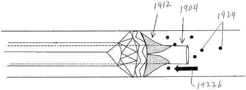
[00042] Figure 19 is a schematic view of the distal end of another
embodiment of an
apparatus for delivering a valve at the distal end of a delivery catheter.
[00043] Figure 20 is a schematic view of the valve of Figure 19.
[00044] Figures 21A ¨ 21C are distal end views of respective embodiments
employing
different valve structure for the valve of Figure 20.
[00045] Figures 22 ¨23 illustrate the apparatus of Figure 19 in deployed
configurations.
[00046] Figures 24 ¨ 26 are schematic views of another apparatus for
deployment of a
sleeve valve, with Figure 24 showing the valve in a housed configuration and
Figures 25 and 26
showing the valve in two different deployed configurations.
7
CA 02782386 2012 05 30
WO 2011/068924
PCT/US2010/058641
[00047] Figures 27 ¨ 39 are schematic views of an apparatus for deployment
ot a valve
that uses a balloon, with Figure 27 showing the valve in a closed
configuration and Figures 28
and 39 showing the valve in two different deployed configurations.
[00048] Figures 30 ¨ 32 are schematic view of another apparatus for
deployment of a
filter that uses a balloon.
[00049] Figure 33 is a schematic view of another apparatus for deployment
of a valve.
[00050] Figures 34 ¨ 36 are schematic views of another apparatus for
deployment of a
valve, with Figures 34 showing the valve in a housed configuration, Figures 35
showing the
valve deployed, and Figure 36 showing the valve in use.
[00051] Figures 37 ¨ 40 are schematic views of another embodiment of an
apparatus for
deployment of a valve, with Figure 37 showing a initial closed configuration,
Figures 38 and 39
illustrating deployed configurations, and Figure 40 illustrated a re-assumed
closed
configuration.
[00052] Figures 41 ¨43 illustrate several flush valves usable in
conjunction with any of
the other embodiments of the invention.
[00053] Figures 44 ¨ 47 are schematic illustrations of another embodiment
of an
apparatus for deployment of valve.
[00054] Figures 48 ¨ 51 are schematic illustrations of another embodiment
of an
apparatus for deployment of valve.
DETAILED DESCRIPTION OF THE PREFERRED EMBODIMENTS
[00055] A first exemplary embodiment of the invention is seen in Figures 2A-
2C. It is
noted that Figs. 2A-2C are not shown to relative size but rather are shown for
purposes of
explanation. In Figs. 2A-2C a delivery catheter 201 having a proximal end (not
shown) and a
distal end 205 is shown positioned within an artery 204. The delivery catheter
201 is adapted
for delivery of an embolizing agent from outside the body of the patient (not
shown) to a target
vessel (artery or vein) in the patient. Attached to the distal end 205 of the
catheter 201 is an
exemplary embodiment of a valve 203 shown having multiple filaments 203a,
203b, 203c,...
which are preferably braided and can move relative to each other. As discussed
hereinafter, the
filaments are spring biased (i.e., they have "shape memory") to assume a
desired crossing angle
relative to each other so that the valve can assume a substantially
frustoconical shape (it being
noted that for purposes herein the term "substantially frustoconical" should
be understood to
8
CA 02782386 2012 05 30
WO 2011/068924
PCT/US2010/058641
include not only a truncated cone, but a truncated hyperboloid, a truncated
paraboloid, and any
other shape which starts from a circular proximal end and diverges therefrom).
Around the
catheter 201 is an outer catheter or sleeve 202 which is movable over the
delivery catheter 201
and valve 203. If desired, the outer catheter or sleeve 202 can extend the
entire length of the
delivery catheter. Where the outer catheter or sleeve 202 extends along the
entire length of the
delivery catheter, it has a proximal end (not shown) which extends proximally
and which can be
controlled by a practitioner from outside the body of the patient.
Alternatively, the outer
catheter or sleeve 202 extends only over the distal end of the delivery
catheter 201 and valve
203, but is controlled by a control element which extends proximally and which
can be
controlled by a practitioner from outside the body of the patient.
[00056] As seen in Fig. 2A, when the outer catheter or sleeve 202 extends
over the valve
203, the multiple filaments are forced into a cylindrical shape. Thus, Figure
2A shows the braid
valve in a retracted or undeployed cylindrical state, with the braid filaments
203a, 203b, 203c...
attached to a distal end of a catheter 205 and covered by the sleeve 202.
Catheter 201 is
positioned within an artery 204 that has forward blood flow in the direction
of arrows 220 (e.g.,
such as experienced during systole with the catheter held still within the
artery and the blood
moving against the valve in the proximal to the distal direction; i.e.,
distally flowing blood).
As seen in Fig. 2B, upon retraction of the sleeve 202 in the direction of
arrow 210, the non-
constrained portion of the valve 203 is freed to expand radially (and retract
longitudinally)
towards its shape memory position. However, the distally flowing blood
(indicated by arrows
220) generating a force, e.g., at or greater than 80 ¨ 120 mmHg, prevents the
valve from
opening more completely, and prevents the valve from touching the walls of
vessel 204. As a
result, the valve 203 is maintained in a condition where it is not
sufficiently open to block blood
flow in the distal or proximal directions. In other words, the forward blood
flow causes the
braid to lengthen and simultaneously decrease its diameter (relative to a
fully open position) to
allow fluid to pass between the braid and the vessel wall.
[00057] Figure 2C shows the valve 203 where the bloodstream is in slow
forward flow
221, static flow, or reverse flow 222 which might occur after delivery of
embolic agents
through catheter 201 and past the valve 203 (such as occurring, by way of
example and not by
limitation, with the valve held longitudinally stationary in the vessel during
diastole and with
the blood moving against the valve in the distal to proximal direction) or
static flow (with
substantially equal pressure on opposite sides of the valve, as occurs when
there is no
9
CA 02782386 2012 05 30
WO 2011/068924
PCT/US2010/058641
significant movement of blood in either the proximal or distal direction;
i.e., approximately u
mmHg) or in slow forward flow (with only slightly greater pressure on the
distal side of the
valve than the proximal side of the valve; e.g., 0 - 80 mmHg). In slow forward
flow 221, the
force applied by the blood against the filaments of the braided valve is not
sufficient to prevent
the valve 203 from opening to reach the wall of the vessel 204. In static
flow, the blood does
not apply any forward force against the valve. During reverse flow 222, the
blood applies a
force which helps the valve open fully. In the fully deployed arrangement of
Fig. 2C, the braid
valve acts as a filter to stop embolic agents from flowing proximal the valve.
However, as
discussed in more detail hereinafter, depending upon the pore size of the
braid valve 203, blood
and contrast agent may be permitted to flow backward through the valve and
around the
catheter 201 while stopping or significantly reducing the flow of embolic
agents.
[00058] It should be appreciated by those skilled in the art that the
catheter 201 can be
any catheter known in the art. Typically, the catheter will be between two and
eight feet long,
have an outer diameter of between 0.67 mm and 3 mm (corresponding to catheter
sizes 2
French to 9 French), and will be made from a liner made of fluorinated polymer
such as
polytetrafluoroethylene (PTFE) or fluorinated ethylene propylene (FEP), a
braid made of metal
such as stainless steel or titanium, or a polymer such as polyethylene
terephthalate (PET) or
liquid crystal polymer, and an outer coating made of a polyether block amide
thermoplastic
elastomeric resin such as PEBAXO, polyurethane, polyamide, copolymers of
polyamide,
polyester, copolymers of polyester, fluorinated polymers, such as PTFE, FEP,
polyimides,
polycarbonate or any other suitable material, or any other standard or
specialty material used in
making catheters used in the bloodstream. Sleeve or outer catheter 202 is
comprised of a
material capable of holding valve braid 203 in a cylindrical configuration and
capable of sliding
over the valve braid 203 and the catheter 201. Sleeve or outer catheter 202
can be comprised of
polyurethane, polyamide, copolymers of polyamide, polyester, copolymers of
polyester,
fluorinated polymers, such as PTFE, FEP, polyimides, polycarbonate or any
other suitable
material. The sleeve or outer catheter may also contain a braid composed of
metal such as
stainless steel or titanium, or a polymer such as PET or liquid crystal
polymer, or any other
suitable material. The wall thickness of sleeve or outer catheter 202 is
preferably in the range
of 0.05 mm to 0.25 mm with a more preferred thickness of 0.1 mm ¨ 0.15 mm.
[00059] The valve 203 is composed of one, two, or more metal (e.g.,
stainless steel or
Nitinol) or polymer filaments, which form a substantially frustoconical shape
when not subject
CA 02782386 2012 05 30
WO 2011/068924
PCT/US2010/058641
to outside forces. Where polymeric filaments are utilized, the filaments may
be composed ot
PET, polyethylene-napthalate (PEN), liquid crystal polymer, fluorinated
polymers, nylon,
polyamide or any other suitable polymer. If desired, when polymeric filaments
are utilized, one
or more metal filaments may be utilized in conjunction with the polymeric
filaments.
According to one aspect of the invention, where a metal filament is utilized,
it may be of radio-
opaque material such that it may be tracked in the body. The valve is capable
of expanding in
diameter while reducing in length, and reducing in diameter while expanding in
length. The
valve is preferably composed of shape memory material that is formed and set
in a large
diameter orientation. As previously mentioned, the valve is preferably held in
a small diameter
orientation until it is released, and when released by removing the sleeve or
other restricting
component 202, the distal end of the valve expands to a larger diameter. Where
the valve is
comprised of multiple filaments, it is preferred that the filaments not be
bonded to each other
along their lengths or at their distal ends so to enable the valve to rapidly
automatically open
and close in response to dynamic flow conditions.
[00060] In the preferred embodiment, the valve is constrained only at its
proximal end
where it is coupled to the catheter body, while the remainder of the valve can
either be
constrained (retracted state) by a sleeve or catheter, or partially
unconstrained (partially
deployed state) or completely unconstrained (completely deployed state). When
in the partially
or completely unconstrained conditions, depending upon the flow conditions in
the vessel, the
valve may either reach the walls of the vessel or it may not.
[00061] As previously mentioned, the valve diameter should automatically
change in
response to local flow conditions so as to enable forward flow, but capture
embolic agents in
brief or prolonged periods of reverse flow. For simplicity, the valve can be
considered to exist
in two conditions. In a "closed" condition, the valve is not sealed against
the vessel wall and
blood may flow around in at least a proximal to distal direction. In an "open"
condition, the
valve expands against the vessel wall and blood must pass through the valve if
it is to flow past
the valve within the vessel in either direction; in the "open" condition
embolic agent is
prevented from passing downsteam (or in a distal to proximal direction) of the
valve.
[00062] Three parameters help define the performance and novel nature of
the valve: the
radial (outward) force of the valve, the time constant over which the valve
changes condition
from closed to open, and the pore size of the valve.
11
CA 02782386 2012 05 30
WO 2011/068924
PCT/US2010/058641
[00063] In a preferred embodiment, the valve expands fully to the vessel
wall (i.e.,
reaches an open condition) when any part of the flow around the braid nears
stasis and remains
in a closed condition when blood is flowing distally with regular force in the
distal direction.
More particularly, when the radial force of expansion of the valve is greater
than the force from
forward blood flow, the valve expands to the vessel wall. However, according
to one aspect of
the invention, the radial force of expansion of the valve is chosen to be low
(as described in
more detail below) so that blood flow in the distal direction will prevent the
valve from
reaching the open condition. This low expansion force is different than the
expansion forces of
prior art stents, stent grafts, distal protection filters and other vascular
devices, which have a
sufficiently high radial force to fully expand to the vessel wall in all flow
conditions.
[00064] The radial force of expansion of a braid is described by Jedwab and
Clerc
(Journal of Applied Biomaterials, Vol. 4, 77-85, 1993) and later updated by
DeBeule (DeBeule
et al., Computer Methods in Biomechanics and Biomedical Engineering, 2005) as:
GI r 2 sin fi r
2 cos fi
F =2n P _____ K1 El tan fi ____ K2
K3 \ K3 K3 \ K3
i
where K1, K2, K3 are constants given by:
K¨ sin 2130 v _ 2 cos 2 fi0 K _ Do
1 ¨ 2
Do Do 3 cos&
and I and Ip are the surface and polar moments of inertia of the braid
filaments, E is the
Young's modulus of elasticity of the filament, and G is the shear modulus of
the filament.
These material properties along with the initial braid angle (Po), final braid
angle (p), stent
diameter (Do), and number of filaments (n) impact the radial force of the
braided valve.
[00065] In one embodiment, with a valve arrangement as shown in Figs. 2A-
2C, the
valve 203 is composed of twenty-four polyethylene terephthalate (PET)
filaments 203a,
203b,..., each having a diameter of 0.1mm and pre-formed to an 8mm diameter
mandrel and a
braid angle of 130 (i.e., the filaments are spring-biased or have a shape
memory to assume an
angle of 130 relative to each other when the valve assumes a fully deployed
state and opens in
a frustoconical configuration). The filaments preferably have a Young's
modulus greater than
200 MPa, and the valve preferably has a radial force of less than 40 mN in the
fully deployed
position (i.e., where the filaments assume their shape memory). More
preferably, the valve has
a radial force in the fully deployed position of less than 20mN, and even more
preferably the
valve has a radial force of approximately 10mN (where the term "approximately"
as used
12
CA 02782386 2012 05 30
WO 2011/068924
PCT/US2010/058641
herein is defined to mean 20%) in the deployed position. Where the valve
includes a filter as
well as the braided filaments (as will be discussed hereinafter with respect
to Figs. 3A and 3B),
the braid component preferably has a radial force of less than 20mN in the
fully deployed
position, and more preferably a radial force of less than 10mN, and even more
preferably a
radial force of approximately 5mN. This compares to prior art embolic capture
devices such as
the ANGIOGUARDO (a trademark of Cordis Corporation), and prior art Nitinol
stents and
stent-grafts which typically have radial forces of between 40mN and 100mN in
their fully
deployed positions.
[00066] According to one aspect of the invention, the valve opens and
closes sufficiently
quickly to achieve high capture efficiency of embolic agents in the presence
of rapidly changing
flow direction. In one embodiment, the valve moves from a fully closed
(undeployed) position
to a fully open position in a static fluid (e.g., glycerin) having a viscosity
approximately equal
to the viscosity of blood (i.e., approximately 3.2 cP) in 0.067 second. For
purposes herein, the
time it takes to move from the fully closed position to the fully open
position in a static fluid is
called the "time constant". According to another aspect of the invention, the
valve is arranged
such that the time constant of the valve in a fluid having the viscosity of
blood is between 0.01
seconds and 1.00 seconds. More preferably, the valve is arranged such that the
time constant of
the valve in a fluid having the viscosity of blood is between 0.05 and 0.50
seconds. The time
constant of the valve may be adjusted by changing one or more of the
parameters described
above (e.g., the number of filaments, the modulus of elasticity of the
filaments, the diameter of
the filaments, etc.).
[00067] As will be appreciated by those skilled in the art, the braid
geometry and
material properties are intimately related to the radial force and time
constant of the valve.
Since, according to one aspect of the invention, the valve is useful in a
variety of arteries of
different diameters and flow conditions, each implementation can have a unique
optimization.
By way of example only, in one embodiment, the valve has ten filaments,
whereas in another
embodiment, the valve has forty filaments. Preferably, the filament diameter
is chosen in the
range of 0.025 mm to 0.127 mm, although other diameters may be utilized.
Preferably, the
pitch angle (i.e., the crossing angle assumed by the filaments in the fully
open position - the
shape memory position) is chosen in the range of 1000 to 1500, although other
pitch angles may
be used. Preferably, the Young's modulus of the filament is at least 100 MPa,
and more
preferably at least 200 MPa.
13
CA 02782386 2012 05 30
WO 2011/068924
PCT/US2010/058641
[00068] According to another aspect of the invention, the valve is chosen
to have a pore
size which is small enough to capture (filter) embolic agents in the blood
stream as the blood
passes through the valve. Where large embolic agents (e.g., 500 m) are
utilized, it may be
possible for the filaments of the valve to act directly as a filter to prevent
embolic agents from
passing through the valve (provided the filaments present pores of less than,
e.g., 500 m).
Alternatively, a filter may be added to the filament structure. Such a
separate filter is
particularly useful where smaller embolic agents are utilized.
[00069] Figure 3A shows a braid valve 203 at the distal end of a catheter
201 and having
a filter 301 that is added to the braid structure 203. The filter can be
placed onto the braid by
spraying, spinning, electrospinning, bonding with an adhesive, thermally
fusing, mechanically
capturing the braid, melt bonding, or any other desired method. The filter can
either be a
material with pores such as ePTFE, a solid material that has pores added such
as polyurethane
with laser drilled holes, or the filter can be a web of very thin filaments
that are laid onto the
braid. Where the filter 301 is a web of thin filaments, the characteristic
pore size of the filter
can be determined by attempting to pass beads of different diameters through
the filter and
finding which diameter beads are capable of passing through the filter in
large quantities. The
very thin filaments can be spun onto a rotating mandrel according to U.S.
Patent 4,738,740 with
the aid of an electrostatic field or in the absence of an electrostatic field
or both. The filter thus
formed can be adhered to the braid structure with an adhesive or the braid can
be placed on the
mandrel and the filter spun over it, or under it, or both over and under the
braid to essentially
capture it. The filter can have some pores formed from spraying or
electrospinning and then a
secondary step where pores are laser drilled or formed by a secondary
operation. In the
preferred embodiment a material capable of being electrostatically deposited
or spun is used to
form a filter on the braid, with the preferred material being capable of
bonding to itself The
filter may be made of polyurethane, pellethane, polyolefin, polyester,
fluoropolymers, acrylic
polymers, acrylates, polycarbonates, or other suitable material. The polymer
is spun onto the
braid in a wet state, and therefore it is desirable that the polymer be
soluble in a solvent. In the
preferred embodiment, the filter is formed from polyurethane which is soluble
in
dimethylacetamide. The polymer material is spun onto the braid in a liquid
state, with a
preferred concentration of 5-10% solids for an electrostatic spin process and
15-25% solids for
a wet spin process. Figure 3B shows the valve in the deployed state, with
outer catheter 202
14
CA 02782386 2012 05 30
WO 2011/068924
PCT/US2010/058641
retracted proximally (as indicated by the arrow) where the braid 203 and the
filter 3U1 are
expanded.
[00070]
According to one aspect of the invention, the filter 301 has a characteristic
pore
size between 10p.m and 500p.m. More preferably, the filter 301 has a
characteristic pore size
between 15p.m and 100p.m. Even more preferably, the filter 301 has a
characteristic pore size
of less than 40p.m and more preferably between 20p.m and 40p.m. Most
desirably, the filter 301
is provided with a characteristic pore size that will permit blood and
contrast agent to pass
therethrough while blocking passage of embolizing agent therethrough. By
allowing
regurgitating blood and contrast agent to pass through the filter in a
direction from distal the
valve toward the proximal end of the valve, the contrast agent may be used to
indicate when the
target site is fully embolized and can serve to identify a clinical endpoint
of the embolization
procedure. Therefore, according to one aspect of the invention, the valve
allows the reflux of
the contrast agent as an indicator of the clinical endpoint while preventing
the reflux of the
embolization agents at the same time. In addition, by allowing blood to flow
back through the
filter material, even at a relatively slow rate, backpressure on the distal
side of the valve can be
alleviated. However, it is appreciated that the filter need not be constructed
to allow either
blood or contrast agent to pass through in the 'reflux' direction.
[00071]
According to one aspect of the method of the invention, the valve is capable
of
endovascular deployment. The valve is preferably coupled to the distal end of
a catheter.
When the distal end of the catheter is in the correct location for treatment,
the valve is deployed.
Preferably, with the valve deployed, embolization agents are delivered
distally through the
catheter into the vessel. Delivery of the embolization agents will tend to
result in the slowing or
stoppage of blood flow in the distal direction and a resultant expansion of
the valve from an
initial diameter which is smaller or equal to the outer diameter of the
catheter (i.e., its housed or
undeployed position) to a final diameter (its open position) which is
preferably at least twice,
and more typically four to ten times the outer diameter of the catheter. In
its open position, the
valve stops embolization agents from traveling past the valve (between the
catheter wall and the
vessel wall) in a proximal direction. According to one aspect of the
invention, the valve is
preferably capable of being retracted into its closed position after the
embolization treatment
procedure is completed.
[00072] It is
important to note that the valve is a dynamic element that opens and closes
based on local flow conditions. In normal flow conditions, the flow pressure
is sufficient to
CA 02782386 2012 05 30
WO 2011/068924
PCT/US2010/058641
overcome the weak biasing force, thereby forcing the valve into a closed
position such that it
does not contact the vascular wall. In static or reverse flow, the biasing
force of the valve
filaments causes the valve into an open position where it preferably is in
full contact with the
vascular wall, thereby restricting reflux of embolizing agents, while
preferably permitting reflux
of blood and contrast agents. It is not necessary that blood and contrast
agent be permitted to
reflux through the valve; however, reflux of blood prevents backpressure on
the distal side of
the valve and reflux of contrast agent aids in visualization of blood flow.
[00073] According to one aspect of the invention, deployment of the valve
is controlled
from the proximal end of the catheter. In some embodiments, a control wire or
a set of two or
more control wires extending from the proximal end of the catheter to the
distal end of the
catheter may be used and controlled by the practitioner to deploy and
optionally retract the
valve. In some embodiments, a control thread extending from the proximal end
of the catheter
to the distal end of the catheter is used to unravel fabric covering the valve
in order to deploy
the valve. In some embodiments, an outer catheter that extends the length of
the catheter to
which the valve is coupled, covers the valve and during deployment is pulled
backward to allow
the valve to expand. In some embodiments, an outer sleeve that is coupled to a
control element
that extends the length of the catheter, covers the valve and during
deployment is pulled
backward by the control element to allow the valve to expand. In some
embodiments, the valve
is coupled to a guidewire, and removal of the catheter guidewire initiates
deployment of the
valve. The control wires, threads, sleeves, etc. may be of standard length,
ranging, for example,
from 60 cm to 240 cm long.
[00074] As previously mentioned, the deployment of the valve can be
achieved in a
variety of manners. As was described in Figure 2, the valve can be deployed by
moving an
outer catheter or sleeve that covers the valve. In that embodiment, the valve
can be recaptured
by the outer catheter or sleeve by moving the catheter or sleeve distally or
the delivery catheter
and valve proximally. In another embodiment, and as seen in Figs. 4A-4C, the
valve is released
by irreversibly removing (unraveling) a knitted sleeve (weft knit) 402 that
covers the valve 203
(shown with filter 301). More particularly, as seen in Figure 4A, the valve
203 is attached to
the distal end of the catheter 201. On top of the valve is a weft knit sleeve
402. A control
thread 401 is attached to the weft knit and extends to the proximal end of the
catheter. In one
embodiment the unravelable knit is composed of polyester of a thickness
between 10p.m and
60p.m. The knit can be a textile sheath that is held under tension. Figure 4B
shows the
16
CA 02782386 2012 05 30
WO 2011/068924
PCT/US2010/058641
deployment of the valve by pulling on the control thread 401. In one
embodiment, the thread
401 is connected to the distal end of the knit sleeve 402 and releases the
valve by first removing
material from the distal end of the sleeve 402. As the control thread 401 is
pulled back and the
sleeve is reduced in size, the distal end of the valve 203 having filter 301
is free to open. The
weft knit sleeve 402 may be partially or fully removed to allow the physician
control of the
diameter or length of the valve. In Figure 4C the weft knit is more fully
removed enabling
more of the length of the valve 203 and filter 301 to be free. In another
embodiment the thread
is attached to the middle or proximal end of the sleeve, and releases the
valve by first removing
material from the proximal end or from the middle of the sleeve.
[00075] Turning now to Figs. 5A and 5B, in another embodiment, a guidewire
501 can
be used to deploy the valve 503. More particularly, valve 503 is provided with
loops 502,
which are attached at or near the distal end of the filaments of the valve
503. The loops 502
may be integral with the filaments or may be made of a separate material and
attached to the
filaments. As seen in Fig. 5A, the loops 502 are looped over the distal end of
the guidewire 501
which extends through the lumen of the catheter 201. The loops at the end of
the valve 502 are
looped around the guidewire 501 while the catheter 201 and guidewire 501 are
advanced
through the vasculature. In this manner, the distal end of the valve is
maintained in a closed
position. When the guidewire 501 is withdrawn proximally as denoted by the
arrow in Fig. 5B,
the distal loops 502 are released, and the valve 503 is deployed.
[00076] According to one aspect of the invention, the valve of any
embodiment of the
invention is attached to the distal end of the catheter in any of several
manners. As seen in
Figure 6A, the valve 203 is attached to the catheter 201 by a sleeve 601 which
overlies the
proximal end of the valve 203 and extends proximal the proximal end of the
valve 203 over the
catheter 201. Figure 6B shows a cross-sectional view of the catheter 201,
valve 203, and sleeve
601. The sleeve 601 is bonded or mechanically held by a heat shrink process or
other
mechanical process to the catheter 201, and thus holds the distal end of the
valve 203 on the
catheter 201 by trapping the distal end of the valve between the catheter 201
and the sleeve 601.
[00077] In one preferred embodiment, the valve is fused into the catheter.
More
particularly, as seen in Figure 6C the valve 203 fused into the catheter 201
such that at the
region 602 where the valve and catheter are fused, there is at most a minimal
change to the
inner or outer diameter of the catheter 201. Figure 6D shows a cross-sectional
view of the
fused valve, where the catheter 201, valve 203 and fused region 602 are all of
the same
17
CA 02782386 2012 05 30
WO 2011/068924
PCT/US2010/058641
diameter. Fusion of the catheter and valve can be achieved by thermally
melting me valve,
melting the catheter, melting both the valve and the catheter, or by a
chemical process.
[00078] Turning now to Figs. 7A and 7B, a valve 702 composed of a single
filament coil
is seen. The coil may be made of metal or polymer, and preferably the filament
is a shape
memory polymer. Figure 7A shows a coil valve 701 in the retracted state on a
catheter 201.
The coil valve is provided with a filter 702 on its distal end. Figure 7B
shows the coil valve in
the deployed state, where the valve 701 and the filter 702 are expanded at the
distal end. Any
of a variety of methods as previously disclosed can be used in deploying the
valve.
[00079] Turning now to Figs. 8A-8E, another embodiment of a deployment
apparatus
800 is shown. The deployment apparatus 800 includes a delivery catheter 801, a
valve 803, a
deployment element 810, and a valve introducer 812. In distinction from
certain prior
embodiments, the delivery catheter is not required to be advanced relative to
an outer catheter
or outer sleeve to deploy the valve, as will become apparent from the
following description.
[00080] The delivery catheter 801 is preferably a 3 French microcatheter or
a 4 or 5
French catheter. The delivery catheter 801 is constructed of one, two or more
than two layers.
In one embodiment, the delivery catheter 801 includes an inner liner made of,
e.g., FEP or
PTFE, a central braid made of one or more of metal, polymer or liquid crystal
polymer, and an
outer polymeric cover made of, e.g., a polyether block amide thermoplastic
elastomeric resin
such as PEBAXO, polyetheretherketone (PEEK), or another suitable polymer.
[00081] The delivery catheter 801 has a distal end 805 provided with a
valve seat 814
and a radiopaque marker band 816 located proximal to, distal of, or about the
valve seat 814.
The valve seat 814 is preferably defined by a circumferential inner groove
located at the distal
end 805 of the delivery catheter 801. The valve seat 814 may be defined
directly on the
delivery catheter, or be bonded or fused into the delivery catheter or to the
distal end 805 of the
delivery catheter. When the valve seat 814 is defined directly on the delivery
catheter 801 and
the delivery catheter is made from a multilayer construct, the valve seat 814
may be defined
through one or two layers, or two layers and a partial depth of a third outer
layer.
[00082] The valve 803 is generally as described in any of the embodiments
above. The
valve 803 may be a polymer braid coated with a polymer surface, a metal braid
coated with a
polymer surface, or a combination of polymer and metal braid coated with a
polymer surface.
The polymer surface may be a sheet, a sheet with holes drilled into it, or a
mesh. The valve
may be permeable or impermeable to blood. Regardless of the construct, the
valve is a dynamic
18
CA 02782386 2012 05 30
WO 2011/068924
PCT/US2010/058641
element that opens and closes based on local blood flow conditions. The
proximal portion ot
the valve 803 includes mating structure 818 that can engage with the valve
seat 812 at the distal
end 805 of the delivery catheter 801 when the valve is advanced through the
delivery catheter,
as described in more detail below.
[00083] The mating structure 818 may include a shape memory polymer or
elastic
polymer that can be compressed for advancement through the body of the
catheter, but which
will automatically expand to seat in the valve seat 814. Referring to Figs. 8C
and 8D, when the
mating structure 818 is engaged at the valve seat 814, such engagement locks
the valve 803
relative to the delivery catheter 801 to prevent further distal movement of
the valve relative to
the delivery catheter and prevent the valve from exiting the distal end of the
delivery catheter
during the procedure. The mating structure 818 may be comprised of a plurality
of independent
features, e.g., four features, which each separately engage in the valve seat.
Further, the
features should be small in profile, e.g., not exceeding 0.25 mm in a radial
'height' dimension
818h through a center of the features, in order to maintain a low profile
within the delivery
catheter 801 as the valve 803 is advanced through the delivery catheter and
also after the valve
is engaged relative to the valve seat 814. By way of one example, the mating
structure on the
valve 803 includes a plurality of radiopaque metal slugs 818a-d bonded, fused,
crimped or
otherwise attached to the valve 803 and that can be received in the valve seat
814. The valve
seat 814 may additionally include a radiopaque marker. In this manner,
alignment of the valve
with the valve seat can be visualized under fluoroscopy. The slugs 818a-d have
proximal and
distal surfaces 819a, 819b that are shaped to prevent the advancement or
withdrawal of the
valve 803 once the slugs are received in the valve seat. That is, the surfaces
819a, 819b may
extend in planes perpendicular to the longitudinal axis of the delivery
catheter. The proximal
portion of the valve 803 is preferably constrained by the inner wall 801a of
the delivery catheter
801 so as to define an inner diameter 803 through the valve.
[00084] The deployment element 810 is a push wire preferably generally
similar in
construction to a conventional guide wire. The outer diameter of the distal
end 810a of the push
wire is larger than the inner diameter of the proximal end of the valve 803.
As a result, the push
wire 810 can be used to provide a pushing force at the proximal portion 803a
of the valve 803
and advance the valve through the delivery catheter 801; i.e., the distal end
810a of the push
wire 810 and proximal portion 803a of the valve are relatively sized so that
the push wire 810
will not freely extend through the valve 803. When the proximal portion 803a
is constrained by
19
CA 02782386 2012 05 30
WO 2011/068924
PCT/US2010/058641
inner wall 801a, the push wire 810 may include a polymer bead or metal bead to
increase its
distal end diameter and facilitate application of a pushing force on the
valve. Additionally or
alternatively, a cylindrical or tubular element may be fused or bonded onto
the distal end of the
push wire to aid in application of a pushing force against the valve.
Additionally or
alternatively, one or more metal or polymeric coils may be provided at the
distal end of the
push wire to increase its outer diameter. Any feature added to the distal end
of the push wire
should maintain trackability of the push wire. The push wire 810 is preferably
made from a
radiopaque material or contains one or more radiopaque markers, such as of
platinum, along its
length.
[00085] The valve introducer 812 is a polymeric tube made, e.g., from PTFE.
The
introducer 812 is preferably 1 cm to 50 cm in length and may optionally be
provided with a
handle at its proximal end (not shown) to facilitate manipulation thereof As
shown in Fig. 8E,
the valve 803 and preferably at least a portion of the push wire are held
within the introducer
812, with the distal end of the valve 803 held in a collapsed configuration.
The introducer 812,
by retaining the valve 803 in the collapsed configuration, presents the valve
in a size suitable
for advancement through the delivery catheter 801. The introducer 812 has an
inner diameter
sufficiently large to contain the collapsed valve 803 and the push wire 810.
The introducer 812
has an outer diameter smaller than the inner diameter of the infusion port 807
at the proximal
end of the delivery catheter, so that the introducer can be advanced into the
infusion port. In
one embodiment, the inner diameter is 0.89 mm and the outer diameter is 0.96
mm.
[00086] Referring to Figs. 8C and 8D, in use of the apparatus 800, a
standard guidewire
(not shown) is advanced through the vasculature of the patient ahead to a
desired location of
treatment. The delivery catheter 801 is advanced over the standard guidewire
to the desired
location. Once the delivery catheter 801 is at the desired location, the
standard guidewire is
removed from the delivery catheter and patient. The valve introducer 812 is
then inserted into
the infusion port of the delivery catheter 801. Depending on the length of the
valve introducer
812, it may function as a guide for valve insertion solely at the proximal end
of the delivery
catheter or as a guide along a substantial length of the delivery catheter.
The push wire 810 is
then distally advanced relative to the introducer 812 to push the valve 803
(in an undeployed
configuration) within the delivery catheter 801 toward the valve seat 814.
When the valve 803
approaches the valve seat 814, the mating structure 818 automatically expands
into and engages
the valve seat 814 to lock the valve 803 relative to the distal end 805 of the
delivery catheter
CA 02782386 2012 05 30
WO 2011/068924
PCT/US2010/058641
801. In the locked configuration, the valve is deployed at the distal end of
the delivery catheter.
The push wire 810 is then withdrawn from the delivery catheter 801.
[00087] Embolic agents are then infused through the delivery catheter 801
and the valve
803. The valve 803 functions as described above. That is, as the embolic
agents are infused,
the valve 803 enables forward flow but prevents reverse flow (reflux) of
embolic agents in the
blood vessel in which the delivery catheter is inserted. As a result of not
using a tube within a
tube construct during infusion of embolic agents (i.e., a delivery catheter
with an outer sleeve),
as described in various above embodiments, a larger delivery catheter can be
used to provide
greater flow of embolic agents to the treatment site. After infusion is
complete, the delivery
catheter 801, along with the valve 803 at its distal end 805, is retracted
from the patient.
[00088] It is also appreciated that while positive engagement between a
valve and valve
seat is desired, it is not necessary. That is, provided alignment of the valve
relative to the distal
end of the catheter can be fluoroscopically visualized, such as with the use
of respective
radiopaque markers, the valve can be manually retained at the appropriate
location relative to
the catheter.
[00089] Another embodiment similar to deployment apparatus 800 includes a
deployment element constructed of a thin wire attached to the valve. The wire
preferably has a
diameter of 0.025 mm to 0.125 mm, and may be a standard wire or a flattened
wire. A flattened
wire may more closely correspond to the inner surface of the catheter to limit
any obstruction of
the lumen of the catheter. In use, the thin wire advances the valve to the
valve seat and then
remains attached to the valve and within the catheter during infusion of the
embolic agent.
[00090] Turning now to Figs. 9A-9D, another embodiment of a deployment
apparatus
900 is shown. The deployment apparatus 900 is substantially similar to
apparatus 800 and
includes a delivery catheter 901, a valve 903, a push wire 910 and a valve
introducer (as
described with respect to introducer 812). The difference between apparatus
900 and prior
described apparatus 800 is the mating structure 918 provided to the valve to
lock the valve
relative to the valve seat. In Figs. 9A and 9B the mating structure 918 is a
proximal ring-
shaped flange that is radially compressed or otherwise deformed to a size
permitting
advancement through the delivery catheter as its is pushed by the push wire
910. As shown in
Figs. 9C and 9D, once the push wire 910 delivers the valve 903 to the distal
end 905 of the
delivery catheter 901, the flange 918 expands into the valve seat 914 once
located at the valve
seat to lock the valve 903 relative to the valve seat 914. The ring-shaped
flange 918 may be
21
CA 02782386 2012 05 30
WO 2011/068924
PCT/US2010/058641
defined by an elastic element coupled to the braid of the valve or a metal
braid or metal stent
portion of the valve that has a much higher expansion force than a remainder
of the valve.
[00091] Figs. 10A-12B illustrate additional embodiments of a flange mating
structure
that can be used on the valve for locking engagement between a valve and a
valve seat. Figs.
10A and 10B show a flange 1018 having a proximal end which in cross-section
appears L-
shaped or J-shaped and that engages within the valve seat 1014. Figs. 11A and
11B show a
flange 1118 having an abutting front surface 1118a and a rear bevel 1118b
(appearing as a barb
in cross-section) such that the flange has a proximal taper (i.e., a smaller
proximal diameter and
a relatively larger distal diameter). This structure facilitates proximal
release of the flange 1118
from the valve seat 1014 for removal of the valve 1103 from the delivery
catheter 1101,
particularly suitable in conjunction with an embodiment of the apparatus
provided with a valve
retraction element, discussed further below. Figs. 12A and 12B show a flange
1218 comprised
of an o-ring, and wherein the valve seat 1214 is in the form of a circular
channel in which the o-
ring is captured. Figs. 13A and 13B illustrate another embodiment of a valve
seat 1314 at the
distal end of the delivery catheter 1301 and corresponding mating structure
1318 on a valve
1303. The valve seat 1314 and mating structures 1318 are 'keyed' with multiple
longitudinally
displaced structures that enhance engagement between the valve 1303 and the
valve seat 1314,
but that prevent locking engagement until the structures are in proper
longitudinal alignment
with each other. By way of the example shown, the valve seat may include a
plurality of
longitudinally displaced channels 1314a, 1314b, wherein a distal channel 1314a
has a greater
width than a proximal channel 1314b. The mating structure 1318 includes a
distal flange 1318a
sized to be received in the distal channel 1314a but too large to be received
in the proximal
channel 1314b. The mating structure also includes a proximal flange 1318b that
is
appropriately sized for being received and captured by the proximal channel
1314b. When the
proximal and distal flanges 1318a, 1318b are aligned with the proximal and
distal channels
1314a, 1314b, the flanges expand into the respective channels and lockingly
engage the valve
1303 relative to the distal end of the delivery catheter 1301. In any of the
embodiments
described above, the flange may include a circumferentially uninterrupted
element or be
comprised of separate elements radially displaced about the proximal portion
of the valve.
Furthermore, while the valve seat is shown as comprising 'negative' space and
the mating
structure as one or more elements that expand into such space, it is
appreciated that the structure
for the valve seat and mating structure may be reversed; i.e., such that the
valve seat comprises
22
CA 02782386 2012 05 30
WO 2011/068924
PCT/US2010/058641
elements that extend into the lumen of the delivery catheter and the mating
structure being a
groove or other negative space about the proximal end of the valve. However,
such a reverse
configuration is less desired as it reduces the diameter of the infusion path
at the distal end of
the delivery catheter.
[00092] Turning now to Figs. 14A and 14B, another embodiment of a
deployment
apparatus 1400 is shown. The deployment apparatus 1400, which includes similar
elements to
apparatus 800, has a delivery catheter 1401, a valve 1403, a push wire 1410
and a valve
introducer (as described with respect to introducer 812). In addition, the
apparatus 1400
includes a retraction element 1420 that is attached to the proximal portion of
the valve 1403,
and more preferably to the mating structure 1418 thereof, to apply a release
and retraction force
to the valve to thereby disengage the valve from the valve seat and withdraw
the valve through
the delivery catheter.
[00093] The retraction element 1420 is a pull wire attached to the mating
structure 1418.
The pull wire 1420 may be flattened or otherwise formed such that it conforms
close to the
inner surface 1401a of the delivery catheter 1401 to maximize the usable space
within the
lumen of the delivery catheter for delivery of the embolic agent. The pull
wire 1420 should
have sufficient mechanical strength in tension to release and withdraw the
valve 1403 from the
delivery catheter. However, it is appreciated that the pull wire 1420 is not
required to have high
compressive stiffness, as the push wire 1410 extends parallel to the pull wire
1420 and performs
the function of advancing the valve to the distal end of the delivery
catheter.
[00094] Use of the apparatus is similar to apparatus 800. The valve 1403,
push wire
1410 and pull wire 1420 are all surrounded with an introducer (not shown) that
facilitates
introduction of such elements into the infusion port of the delivery catheter.
The push wire
1410 advances the valve 1403 and pull wire 1420 out of the introducer and to
the distal end of
the delivery catheter 1401. Once the valve 1403 engages the valve seat 1414,
the push wire
1410 is withdrawn from the delivery catheter 1401. Embolic agents are then
infused through
the delivery catheter 1401 to treat the patient. After the embolic agents have
been infused, the
valve 1403 can be withdrawn into the delivery catheter 1401 by applying a
sufficient tensile
force on the pull wire 1420 to release the valve 1403 from the valve seat 1414
and retract it into
the delivery catheter 1401. The delivery catheter is then removed from the
patient. Optionally,
the pull wire 1420 may be used to completely withdraw the valve 1403 from the
delivery
catheter 1401 prior to removing the delivery catheter from the patient.
23
CA 02782386 2012 05 30
WO 2011/068924
PCT/US2010/058641
[00095] In addition to a single pull wire, the retraction element may take
other torms
which may be similarly used to withdraw the valve from the delivery catheter
after infusion of
the embolic agent. For example, referring to Figs. 15A and 15B, the retraction
element includes
a plurality of pull wires, such as the pair of pull wires 1520a, 1520b shown.
In addition,
referring to Figs. 16A and 16B, the retraction element may comprise a tubular
retraction braid
1620 of multiple metal wires or polymeric filaments. The braid 1620 may be
made from
stainless steel, Elgiloy0, Nitinol or another elastic material. The tubular
braid 1620 may have a
predefined diameter that is the same or larger than the diameter of the lumen
of the delivery
catheter. In this manner the retraction braid can be held taut against the
pushing force of the
push wire 1610 in order to decrease it to a diameter smaller than the diameter
of the lumen of
the delivery catheter 1601. Once the push wire 1610 advances the valve 1603 to
the valve seat
1614, the tension is released from the braid 1620 to permit the braid to be
held outward against
the inner wall 1601a of the delivery catheter 1601. Further, referring to
Figs. 17A and 17B, a
retraction braid 1720 may be coated with a polymeric coating 1722. The
polymeric coating
1722 may include, e.g., one or more of polyurethane, polyamide, polyimide,
PTFE or FEP such
that the retraction element defines a catheter body. It is noted that in
embodiments using a
retraction element separate from a push wire, the retraction element can be
designed with a low
compressive strength, as the separate push wire 1710 performs advancement of
both the valve
and the retraction element through the delivery catheter.
[00096] As yet another alternative, the push wire and retraction element
may be
comprised of a single element having sufficient compressive and tensile
strengths to advance
the valve to the valve seat and retract the valve from the valve seat at the
conclusion of the
procedure. Such single element should be of a design which retains usable
space within the
lumen of the delivery catheter to permit sufficient infusion of embolic
agents.
[00097] Referring to Fig. 18A, another deployment apparatus 1800 is shown.
The
deployment apparatus 1800 has a delivery catheter 1801, a valve 1803, a push
wire 1810, a
retraction element in the form of a polymer-coated braid 1820, and a valve
introducer (as
described with respect to introducer 812). The valve seat 1814 is defined by
the distal end of
the delivery catheter 1801. The mating structure 1818 of the valve seat 1814
is compressed for
advancement through the delivery catheter. As shown in Fig. 18B, once the
mating structure
1818 passes through the distal end 1805 of the delivery catheter 1801, the
mating structure
24
CA 02782386 2016-08-19
72235-230
expands into contact with the valve seat 1814. The retraction element 1820
maintains tensile
force on the valve 1803 to hold the valve 1803 against the valve seat 1814.
[00098] In another embodiment of the invention, no deployment element
is required.
The valve is advanced through the catheter to a valve seat using hydraulic
pressure. Any of the
valve designs described above with respect to Figs. 8-17 are provided within
the catheter, e.g.,
using an introducer. Then, via the infusion port, a bolus of saline or
heparinized saline is
injected into the catheter behind the valve to force the valve to the distal
end.
Hydraulic pressure can be applied to advance the valve, taking into account
frictional forces between the valve and inner surface of the catheter, blood
pressure and
gravitational force. It is appreciated that when the valve is within the
catheter, it is sufficiently
radially collapsed to provide an adequate barrier within the catheter on which
the bolus of
solution acts.
[00099] Another embodiment of a delivery apparatus 1900 is shown at
Fig. 19. The
delivery apparatus 1900 includes an outer catheter 1901 having a proximal end
(not shown) and
a distal end 1903, an inner catheter 1904 extendable through the outer
catheter, and a valve
1905 situated in the distal end 1903 of the outer catheter 1901. The valve
1905 includes a
proximal expandable framework 1906, one or more control members 1908 (or
1908a, 1908b in
Fig. 20) coupled to the proximal end of the framework 1906, a central collar
1910 at the distal
end of the framework 1906, and one or more valve flaps 1912 extending distally
from the collar
1910. The framework 1906 and collar 1910 are preferably made from form an
expandable
structure. Both the framework 1906 and collar 1910 are preferably made of a
material have
shape memory or other spring-like expansible properties so that they are self-
expanding, or are
constructed of a non-shape memory or non-springy material that can be expanded
under force,
e.g., by balloon expansion as described further below. The framework 1906 and
collar 1910
may be a mesh of metal wire or polymeric filaments, a wire or tubular stent
structure, or other
suitable structure. The framework 1906 and collar 1910 may be integrally
formed together, or
separately formed and then coupled together. The collar 1910 is sufficiently
expansible and
appropriately sized to contact the inner wall of an artery when partially or
fully expanded. The
valve flaps 1912 are preferably constructed in a manner similar to above
described valve
structures. For example, the valve flaps 1912 may each comprise a filamentary
structure or
CA 02782386 2012 05 30
WO 2011/068924
PCT/US2010/058641
other mesh overlaid with a polymer coating. The valve flaps may be structured
to permit mood
and/or contrast agent to pass through the material thereof, or may be
impermeable to such
fluids. The valve flaps 1912 may include two flaps 1912 of equal size in a
duck-bill formation
(Fig. 21A), three or more flaps 1912' of equal dimension (Fig. 21B), or flaps
1912a", 1912b" of
different size (Fig. 21C). In each embodiment, distal portions of the flaps
may be shaped (as
shown by broken lines) to together define a circular opening 1913 for passage
of the inner
catheter 1904 therethrough. The control member 1908 may advance and retract
the valve 1905
relative to the outer and inner catheters 1901, 1904 between housed and
deployed
configurations. Alternatively, the valve 1905 can be coupled directly to the
inner catheter 1904,
with movement of the inner catheter relative to the outer catheter 1901
effecting movement of
the valve 1905 between a housed configuration and a deployed configuration. In
a first housed
configuration, the framework 1906 and collar 1910 are radially constrained by
the outer
catheter 1901, and the flaps 1912 are held closed against each other (prior to
insertion of the
inner catheter 1904 through the valve) (Figs. 21A-21C). In a second housed
configuration
shown in Fig. 19, the framework 1906, collar 1910, and valve 1905 remain
radially constrained
within the outer catheter 1901, and the inner catheter 1904 is extended
through the valve flaps
1912. In a first deployed configuration, operation of the control member 1908
distally advances
the valve 1905 out of the distal end of the outer catheter 1901, and the
collar 1910 is permitted
to self-expand until the proximal ends of the valve flaps 1912 are adjacent
the arterial wall 1920
(Fig. 22). Alternatively, where the valve 1905 is coupled relative to the
inner catheter 1904, the
inner catheter functions as the control member and the inner catheter and
outer catheter are
moved relative to each other to advance the valve out of the distal end of the
outer catheter into
the same deployed configuration. In the first deployed configuration, the
valve 1905 is forced
open by forward flow of blood 1922a through the arterial passage. The
embolizing agent 1924
is infused through the inner catheter 1904 and the forward blood flow prevents
1922a advances
the embolizing agent 1924 with the artery 1920. When the flow of blood changes
to slow, static
or retrograde flow 1922b, the valve dynamically changes due to pressure flow
conditions to a
second deployed configuration in which the distal end of the valve flaps 1912
close against the
inner catheter 1904 (Fig. 23). This prevents any embolizing agent from passing
back beyond
the valve.
[000100] Turning now to Fig. 24, another embodiment of a delivery apparatus
2000,
substantially similar to delivery apparatus 1900, is provided with a valve
2005. The valve 2005
26
CA 02782386 2012 05 30
WO 2011/068924
PCT/US2010/058641
includes a proximal expandable framework 2006, optionally one or more control
members
2008a, 2008b coupled to the proximal end of the framework 2006, a central
collar 2010 at the
distal end of the framework 2006, and a tubular valve sleeve 2012. The sleeve
2012 is
preferably constructed in a manner similar to any above described valve, e.g.,
with a polymer-
coated filamentary construct, but may be of other construction. In a housed
configuration, the
sleeve 2012 resides between the outer catheter 2001 and inner catheter 2004 of
the delivery
apparatus 2000, with the inner catheter 2004 extending through the sleeve. The
sleeve 2012
may be advanced relative to the outer catheter 2001 into a deployed
configuration by mounting
it relative to the inner catheter 2004 and advancing the inner catheter
relative to the outer
catheter, or alternatively by operation of the control members 2008a, 2008b to
move the sleeve
relative to both the outer catheter 2001 and the inner catheter 2004.
Regardless of how the
collar 2010 of the valve 2005 is freed of the outer catheter, once freed the
collar 2010 expands
to contact the arterial wall 2020 and deploy the valve 2012. The blood may
flow between the
valve and the inner catheter (Fig. 25). In a second deployed configuration,
resulting when the
flow of blood 2012b is slow, static or retrograde, the valve sleeve 2012
closes against the inner
catheter 2004 (Fig. 26).
[000101] Turning now to Fig. 27, another embodiment of a delivery apparatus
2100 is
shown. The delivery apparatus 2100 includes a valve 2105 coupled to a catheter
2101. The
valve 2105 includes a plurality of struts 2116 coupled at their proximal ends
by a collar 2117.
A suitable filter material 2118 extends between the struts 2116. The delivery
apparatus 2100
also includes a guard 2126 coupled to the catheter 2101 that shields the
arterial wall 2120 from
the distal ends of the struts 2116 when the valve 2105 is in a non-deployed
configuration. The
delivery apparatus 2100 includes a control member in the form of a balloon
2124 that, when
expanded, applies a radial force to the struts that sufficiently flexes the
struts to release the
valve from the guard 2126. This results in the valve 2105 entering a deployed
configuration.
The balloon 2124 may be expanded via use of a dedicated lumen of the inner
catheter 2104, a
distinct inflation catheter or via any other suitable system (such as that
described below with
respect to Figs. 30-32). In the deployed configuration, forward flow of blood
is permitted about
the exterior of the valve (Fig. 28). However, in static flow (2122b), low
flow, or reverse flow,
the valve 2105 dynamically and rapidly responds to the changing flow
conditions and fully
opens to the arterial wall 2120 preventing flow of embolizing agent past the
valve (Fig. 29).
27
CA 02782386 2012 05 30
WO 2011/068924
PCT/US2010/058641
[000102] Turning now to Figs. 30 to 32, another embodiment of a delivery
apparatus 22UU
is shown. A catheter 2201 includes an outer control member balloon 2234. A
valve 2205 is
provided over the balloon 2234 and includes filtering material 2212 extending
across
circumferentially displaced struts 2216. The balloon is positioned radially
centered between the
struts. The balloon 2224 includes a pressure valve 2235 in communication with
the lumen 2228
of the catheter 2201. A guidewire 2240 provided with an occlusive tip 2242 is
advanced
through the lumen 2228 of the catheter 2201. The occlusive tip 2242 is
advanced past the
pressure valve 2235 (Fig. 31). An injectate 2234, such as saline, is then
injected into the
catheter lumen 2228. Referring to Fig. 32, sufficient fluid and pressure are
provided to cause
the injectate to enter the pressure valve 2235 and fill the balloon 2234. The
balloon 2234 fills
to high pressure and then seals to prevent leakage to low pressure conditions.
As the balloon
2234 fills to a high pressure state, it contacts the valve 2205 to move the
valve to a deployed
configuration. The guidewire 2240 may then be withdrawn from the catheter
2201. The valve
is then used as described above in conjunction with the infusion of an
embolizing agent through
the catheter 2201. After completion of the procedure, the catheter 2201 can be
drawn back into
an outer catheter (not shown) and such that contact between the valve 2205 and
the distal end of
the outer catheter will overcome the pressure valve 2235 and cause the
pressure valve to
release, the balloon 2234 to deflate and the valve to re-assume a non-deployed
configuration for
withdrawal from the patient.
[000103] Referring now to Fig. 33, another embodiment of a delivery
apparatus 2300 is
shown. The apparatus includes an outer catheter 2301, an inner catheter 2304
extending
through the outer catheter, and valve 2305 comprising an expandable wire
framework
2306 coupled to the inner catheter 2304 or operable via independent control
members 2308, an
expandable collar 2310 coupled to the framework, a tapered first sleeve
portion 2311 extending
from the collar, and a second sleeve portion 2312 extending from the first
sleeve portion. In a
housed configuration (not shown), the inner catheter 2304, framework 2306,
control members
2308, collar 2310 and sleeve portions 2311, 2312 are held within the outer
catheter 2301 and
advanced to the location of interest within the artery 2320. In a deployed
configuration, the
inner catheter 2304 is advanced out of the distal end of the outer catheter
2301 and the control
members 2308 are operated from the proximal end of the apparatus to deploy the
framework
2306, collar 2310 and sleeves 2311, 2312 out of the outer catheter 2301 and
over the inner
catheter 2304. The collar 2310 expands the proximal end of the tapered first
sleeve 2311
28
CA 02782386 2012 05 30
WO 2011/068924
PCT/US2010/058641
adjacent the arterial wall 2320. During forward blood flow 2322a, the blood
flows between the
inner catheter 2304 and the sleeves 2311, 2312, similar to air flowing through
a windsock.
However, at least the second sleeve 2312 is structured to collapse in response
to reverse blood
flow conditions 2322b, so that embolizing agent contacts the exterior of the
sleeves 2311, 2312
but cannot pass through.
[000104] Turning now to Figs. 34 through 36, another embodiment of a
delivery device
2400 is shown. The delivery device 2400 includes a control member 2408 with a
self-
expanding shape memory loop (or collar) 2410 at its distal end. A valve 2412
extends from the
loop 2410. The valve 2412 has an open distal end 2413. The control member 2408
is operated
to advance the valve 2412 to the distal end 2403 of an outer catheter 2401
which is advanced to
the arterial location of interest. Referring to Fig. 35, the control member
2408 is then is
operated to advance the loop and valve out of the distal end 2403 of the outer
catheter 2401,
with the loop automatically expanding and causing the proximal end of the
valve 2412 to be
positioned against or adjacent the arterial wall 2420. Then, as shown in Fig.
36, an inner
catheter 2404 is advanced through the outer catheter 2401 and completely
through the open
distal end 2413 of the filter valve 2412. Embolizing agent 2424 is infused
through the inner
catheter 2404. Blood may blow in the forward direction between the inner
catheter 2404 and
filter valve 2412. During retrograde blood flow, the loop 2410 retains its
diameter against the
arterial wall 2420, but the distal and central portions of the filter valve
2412 dynamically
collapses against the inner catheter 2404 in response to the changing blood
pressure preventing
reverse flow of embolizing agent 2424 past the valve.
[000105] Referring now to Fig. 37 another embodiment of a delivery device
2500 is
shown. The delivery device 2500 includes a catheter 2501, a first collar 2530
about the catheter
2501 and coupled to the catheter or a first control member 2532, a second
collar 2534 displaced
from the first collar 2530 and located about the catheter and coupled to a
second control
member 2536, a plurality of struts 2516 extending between the first and second
collars 2530,
2534, and a valve sleeve 2512 extending over at least a portion of the struts
2516 and preferably
the second collar 2534. Referring to Fig. 38, in operation, when the second
control member
2536 is retracted relative to the catheter 2501 and/or first control member
2532 (i.e., whichever
to which the first collar 2530 is coupled), the struts 2516 are caused to bow
outwards thereby
moving the proximal end of the valve sleeve 2512 against the arterial wall
2520. Embolizing
agent 2524 may be injected through the catheter 2512. Forwardly advancing
blood 2522a may
29
CA 02782386 2012 05 30
WO 2011/068924
PCT/US2010/058641
flow between the valve sleeve 2512 and the catheter 2501. Referring to Fig.
39, when the mooci
changes flow direction 2522b, the rapid change in pressure on the valve sleeve
2512 causes the
valve sleeve to dynamically react with its distal end collapsing against the
catheter 2501 to
prevent retrograde flow of embolizing agent 2524. The delivery device 2500 may
be collapsed
for withdrawal by moving the first control member 2532 proximally relative to
the second
control member 2536 to straighten the struts 2516 and thereby reduce the
diameter of the valve
sleeve 2512 (Fig. 40).
[000106] It is appreciated that it in any of the embodiments described
above it may be
desirable to controllably flush the outer catheter through a route that exits
behind the valve.
Such flush may include a contrast agent, saline, etc. Turning now to Fig. 41,
one embodiment
of a flush valve includes one or more open slits 2640 in the outer catheter
2601. A side stop
2642 is provided in the annular space between the outer and inner catheters
2601, 2604.
Alternatively, the stop 2642 may be provided against an outer catheter 2601 in
which no inner
catheter is provided. The side stop 2642 is coupled at the distal end of a
control member 2644.
In a closed state, the proximal end of the control member 2644 is manipulated
to position the
side stop 2642 in obstruction of the open slits 2640 to prevent fluid passage
therethrough. To
permit flush, the proximal end of the control member 2644 is manipulated to
position the side
stop 2642 either proximal or distal (shown) relative to the open slits 2640 so
that fluid may be
flushed therethrough. Turning now to Fig. 42, another embodiment of a flush
system is shown
incorporating slit valves 2740 in the outer catheter 2701. Such slit valves
2740 are normally in
a closed configuration. However, upon application of a flush under pressure,
the slit valves
2740 are opened and the flush is permitted to escape the catheter (Fig. 43).
[000107] Turning now to Figure 44, another embodiment of a valve deployment
apparatus 2800 is shown. The apparatus 2800 includes two longitudinally
displaced
microcatheters 2801, 2802 and a dynamic valve 2805 located therebetween. More
particularly,
the more proximal first microcatheter 2801 is a "hi-fib" microcatheter
preferably having an
inner diameter of 0.69mm and an outer diameter of 0.97mm and includes a
proximal luer 2803
or other suitable connector at its proximal end 2801a and has a distal end
2801b. The distal
second microcatheter 2802 preferably has a proximal end 2802a with a proximal
face 2802c, a
smaller inner diameter of 0.53mm, and the same 0.97mm outer diameter as the
first
microcatheter. The valve 2805 preferably comprises a braid that is fused at
its proximal end
2805a to the distal end 2801b of the first microcatheter 2801 and at its
distal end 2805b to the
CA 02782386 2012 05 30
WO 2011/068924
PCT/US2010/058641
proximal end 2802b of the second microcatheter 2802. The braid is naturally
biased to radially
self-expand from an undeployed state to a deployed state, wherein the valve in
the undeployed
state (described below) has a diameter approximately equal to the outer
diameter of the first and
second microcatheters, and into the deployed states has a diameter
substantially greater. The
braid includes a proximal portion 2805c that is polymer coated as described
with respect to
several valves described above, whereas a distal portion 2805d of the braid is
uncoated and
forms an open design permitting fluid to flow therethrough.
[000108] The apparatus 2800 further includes a thin-walled tubular elongate
member 2850
preferably having an inner diameter of 0.53mm and an outer diameter of 0.64mm.
The tubular
member 2850 is most preferably in the form of a wire coil 2852 preferably with
an axially
extending peripheral wire 2854 or oversheath 2856 for longitudinal stability.
The coil tubular
member has a proximal end 2850a provided with a hub 2858 for locking relative
to the luer
connector 2803, such as a tuohy borst adapter and a distal end 2850b. When the
coil tubular
member 2850 is inserted into the luer connector 2803, through the first
microcatheter 2801, and
through the valve 2805, its distal end 2850b abuts the proximal face 2802c of
the second
microcatheter 2802. The coil tubular member 2850 is sized such that when fully
advanced into
the first microcatheter 2801, the proximal end 2802a of the second
microcatheter 2802 is
displaced from the distal end 2801b of the first microcatheter 2802 a
sufficient distance to apply
a tensile force on the valve to cause the valve to elongate and constrict in
diameter to a
significantly smaller non-deployed diameter suitable for advancement through
the vessel. The
apparatus 2800 may be presented in this configuration in an as manufactured
and/or sterilized
package.
[000109] Referring to Fig. 45, a standard 0.356mm guidewire 2860 is
provided for use
with the apparatus 2800. The guidewire 2860 is inserted through the hub 2858
and luer
connector 2803 and through the first microcatheter 2801, the valve 2805 and
the second
microcatheter 2802. The guidewire 2860 is advanced to the site of the emboli
and the apparatus
2800 is then tracked over the guidewire to the site.
[000110] Referring to Fig. 46, the guidewire 2860 is shown withdrawn, and
the coil
tubular member 2850 is released from the luer connector 2803 and removed from
the first
microcatheter 2801, allowing the valve 2805 to expand to the arterial wall
(not shown).
Embolizing agent 2824 is then infused through the first microcatheter 2801 and
exits through
the uncoated distal portion 2805d of the valve and the second microcatheter
2802. Importantly,
31
CA 02782386 2012 05 30
WO 2011/068924
PCT/US2010/058641
the valve 2805, even through coupled at its distal end to the second
microcatheter, is a dynamic
valve rapidly adjusting to flow conditions resulting from changing blood
pressure in systole and
diastole. Thus, during the forward flow of blood in systole, the coated
proximal portion 2805c
of the valve collapses to permit the blood to flow around the valve, and
during slow, static, or
retrograde blood flow in diastole, the coated proximal portion of the valve
opens against the
arterial wall preventing passage of any of the embolizing agent.
[000111] Turning to Fig. 47, after the procedure, the device comprising the
microcatheters
2801, 2802 and valve 2805 may simply be withdrawn from the artery which will
automatically
collapse the valve. However, as an option, the coil tubular member 2805 may be
reinserted to
aid in collapse and the guidewire 2860 may also optionally be reinserted to
facilitate reverse
tracking out of the patient. Regardless of the method of removal, it is
appreciated that any
embolizing agent 2824 remaining in the valve upon collapse of the valve will
remain trapped in
the valve for retrieval as the braid angle will be reduced in size upon
collapse to define
openings too small for the embolizing agent to pass through.
[000112] Turning now to Figures 48 and 49, another embodiment of a valve
deployment
apparatus 2900, substantially similar to the deployment apparatus 2800, is
shown. The
apparatus 2900 includes two longitudinally displaced microcatheters 2901, 2902
and a dynamic
valve 2905 located therebetween. More particularly, the more proximal first
microcatheter
2901 is a "hi-fib" microcatheter preferably having an inner diameter of 0.69mm
and an outer
diameter of 0.97mm and includes a connector 2903 at its proximal end 2901a and
has a distal
end 290 lb. The distal second microcatheter 2902 preferably has a proximal end
2902a with a
proximal face 2902c, a smaller inner diameter of 0.53mm, and the same 0.97mm
outer diameter
as the first microcatheter. The valve 2905 preferably comprises a braid that
is fused at its
proximal end 2905a to the distal end 290 lb of the first microcatheter 2901
and at its distal end
2905b to the proximal end 2902b of the second microcatheter 2902. The braid
includes a
proximal portion 2905c that is polymer coated as described with respect to
several valves
described above, whereas a distal portion 2905d of the braid is uncoated and
forms an open
design permitting fluid to flow therethrough.
[000113] The apparatus 2900 further includes an elongate member such as a
guidewire
2960. The guidewire 2960 is preferably a 0.45mm diameter guidewire, but may be
other
dimensions, and includes a hub 2958 adjacent its proximal end 2960a and a
preferably
radiopaque marker band 2962 adjacent its distal end 2960b. The marker band
2962 is larger
32
CA 02782386 2012 05 30
WO 2011/068924
PCT/US2010/058641
than the inner diameter of the second microcatheter and is thus adapted to
abut against the
proximal face 2902c. A fixed length is indicated, whether by actual length,
indicia, or stops
between the guide wire from the proximal 2901a end of the first microcatheter
2901 or the
distal end of the marker band 2962. The guidewire is inserted through the
first microcatheter
such fixed length so that the marker band is abutted against proximal face of
the second
microcatheter; this results in the valve entering the collapsed configuration.
The apparatus with
guidewire is then advanced to the target. Once at the target the guidewire is
removed from the
apparatus.
[000114] Referring to Figure 50, the apparatus in use is substantially
similar to that
described above with respect to Fig. 46. The valve 2905 expands to the
arterial wall (not
shown). Embolizing agent 2924 is then infused through the first microcatheter
2901 and exits
through the uncoated distal portion 2905d of the valve and the second
microcatheter 2902.
Importantly, the valve 2905, even through coupled at its distal end to the
second microcatheter,
is a dynamic valve rapidly adjusting to flow conditions resulting from
changing blood pressure
in systole and diastole. Thus, during the forward flow of blood in systole,
the coated proximal
portion 2905c of the valve collapses to permit the blood to flow around the
valve, and during
slow, static, or retrograde blood flow in diastole, the coated proximal
portion of the valve opens
against the arterial wall preventing passage of any of the embolizing agent.
[000115] Turning to Fig. 51, after the procedure, the device comprising the
microcatheters
2901, 2902 and valve 2905 can be withdrawn by simply retracting it from the
artery which will
cause collapse of the valve. However, optionally, the guidewire 2960 may be
reinserted to
collapse the valve 2905. It is appreciated that any embolizing agent 2924
remaining in the
valve upon collapse of the valve will remain trapped in the valve for
retrieval as the braid angle
will be reduced in size upon collapse to define openings too small for the
embolizing agent to
pass through.
[000116] In any of the embodiments described herein, the components of the
valve may be
coated to reduce friction in deployment and retraction. The components may
also be coated to
reduce thrombus formation along the valve or to be compatible with
therapeutics, biologics, or
embolics. The components may be coated to increase binding of embolization
agents so that
they are removed from the vessel during retraction.
[000117] According to one aspect of the invention, the catheter body and
mesh may be
separately labeled for easy visualization under fluoroscopy. The catheter body
can be labeled
33
CA 02782386 2012 05 30
WO 2011/068924
PCT/US2010/058641
by use of any means known in the art; for example, compounding a radio-opaque
material into
the catheter tubing. The radio-opaque material can be barium sulfate, bismuth
subcarbonate or
other material. Alternatively or additionally, radio-opaque rings can be
placed or crimped onto
the catheter, where the rings are made of platinum, platinum iridium, gold,
tantalum, and the
like. The valve may be labeled by crimping a small radio-opaque element such
as a ring on one
or a plurality of filaments. Alternatively or additionally, radio-opaque
medium can be
compounded into the materials of the braid and the filter. Or, as previously
described, one or
more of the filaments may be chosen to be made of a radio-opaque material such
as platinum
iridium.
[000118] In certain embodiments, the valve is attached to a catheter which
may be a single
lumen or a multi-lumen catheter. Preferably, the catheter has at least one
lumen used to deliver
the embolization agents. According to other embodiments, however, the catheter
may provided
with a lumen which either serves to store the valve before deployment or
through which the
valve can be delivered. Where control members are utilized to control
deployment of the valve,
one or more additional lumen may be provided, if desired, to contain the
control wires for
deployment and retraction. Alternatively, the catheter about which the control
members
extends may include longitudinal open channels through which the control wires
may extend.
An additional lumen may also be used to administer fluids, e.g., for flushing
the artery after the
administration of embolization agents, or for controlling a balloon which
could be used in
conjunction with the valve.
[000119] The above apparatus and methods have been primarily directed to a
system
which permits proximal and distal flow of biological fluid (e.g., blood)
within a body vessel,
and which prevents reflux of an infusate past the valve in a proximal
direction. It is appreciated
that the valve may also be optimized to reduce blood flow in the distal
direction. The radial
force of the valve can be tuned by adjusting the braid angle. Tuning the
radial force allows the
blood flow to be reduced by up to more than 50 percent. By way of example,
providing a braid
angle greater than 130 will significantly reduce blood flow past the valve in
the distal
direction, with a braid angle of approximately 150 slowing the blood flow by
50 to 60 percent.
Other braid angles can provide different reductions in distal blood flow. The
reduced distal
blood flow can be used in place of a 'wedge' technique, in which distal blood
flow is reduced
for treatment of brain and spinal arteriovenous malformations. Once the blood
flow is slowed
by the valve, a glue such as a cyanoacrylic can be applied at the target site.
34
CA 02782386 2012 05 30
WO 2011/068924
PCT/US2010/058641
[000120] There have been described and illustrated herein multiple
embodiments ot
devices and methods for reducing or preventing reflux of embolization agents
in a vessel.
While particular embodiments of the invention have been described, it is not
intended that the
invention be limited thereto, as it is intended that the invention be as broad
in scope as the art
will allow and that the specification be read likewise. Thus while particular
deployment means
for the protection valve have been described, such as a catheter, a sleeve and
control element, a
fabric sleeve with a control thread, etc., it will be appreciated that other
deployment
mechanisms such as balloons, absorbable sleeves, or combinations of elements
could be
utilized. Likewise, while various materials have been listed for the valve
filaments, the valve
filter, the catheter, and the deployment means, it will be appreciated that
other materials can be
utilized for each of them. Also, while the invention has been described with
respect to
particular arteries of humans, it will be appreciated that the invention can
have application to
any blood vessel and other vessels, including ducts, of humans and animals. In
particular, the
apparatus can also be used in treatments of liver, renal or pancreatic
carcinomas. Further, the
embodiments have been described with respect to their distal ends because
their proximal ends
can take any of various forms, including forms well known in the art. By way
of example only,
the proximal end can include two handles with one handle connected to the
inner (delivery)
catheter, and another handle connected to an outer catheter or sleeve or
actuation wire or string.
Movement of one handle in a first direction relative to the other handle can
be used to deploy
the valve, and where applicable, movement of that handle in an opposite second
direction can
be used to recapture the valve. Depending upon the handle arrangement, valve
deployment can
occur when the handles are moved away from each other or towards each other.
As is well
known, the handles can be arranged to provide for linear movement relative to
each other or
rotational movement. If desired, the proximal end of the inner catheter can be
provided with
hash-marks or other indications at intervals along the catheter so that
movement of the handles
relative to each other can be visually calibrated and give an indication of
the extent to which the
valve is opened. It will therefore be appreciated by those skilled in the art
that yet other
modifications could be made to the provided invention without deviating from
its spirit and
scope as claimed.