Note: Descriptions are shown in the official language in which they were submitted.
CA 2791901 2017-04-18
ETS2 AND MESP1 GENERATE CARDIAC PROGENITORS FROM
FIBROBLASTS
FIELD
[0002] One aspect of the disclosure relates generally to the field
of cell
differentiation, and more specifically to a strategy for cardiovascular tissue
regeneration via the
isolation, renewal, and directed differentiation, of fibroblasts into specific
mature cardiac,
pacemaker, smooth muscle, and endothelial cell types.
BACKGROUND
[0003] Damage to mammalian heart tissue frequently results in the
loss of large
numbers of cardiac cells, including mature cardiac cells, pacemaker cells,
smooth muscle, and
endothelial cells. Although there is some indication that cardiac cells can be
regenerated in
humans (Bergmann et al., 2009), the mechanism is not well understood and the
process does not
appear to proceed rapidly enough to repair common types of cardiac damage such
as ischemia,
infarction, trauma, or injury due to toxins or viral infections. Therefore, a
central goal of
experimental cardiac medicine has been the development of a means for
regenerating cardiac
cells which have been lost due to cardiac damage. Studies of the mechanisms
behind the
embryonic cardiogenesis have been conducted, with the aim of replicating
cardiogenesis in vitro
or in vivo for the purposes of regenerating damaged tissue.
[0004] Recent research has identified multipotent (/s//+)
cardiovascular
progenitor (MICP) cells, which are capable of differentiating to form mature
cardiac tissue.
MICP cells derived from embryonic stem (ES) cells which can give rise to
endothelial, cardiac,
and smooth muscle cells, have been isolated (Moretti et al., 2006). Genetic
studies have shown
that these MICP cells express Isll, NKx25 and Kid.
[0005] Model systems for investigating cardiogenesis include the
ascidian Ciona
intestinalis (Beh et al., 2007). Lineage studies have shown that the adult
Ciona
-1-
CA 02791901 2012-08-31
WO 2011/109695
PCT/US2011/027160
heart is derived from two founder cells that express Ci-Mesp, a basic helix-
loop-helix
(bHLH) transcription factor, and also Ci-Ets1/2 (Imai et al., 2004: Satou et
al., 2004). In
addition, ascidian orthologs of the conserved heart specification genes NK4
(tinman
N1a2.5), GATAa (pannier/GA7A4/5/6), Hand and Hand-like (1mai et al., 2003;
Davidson,
2007; Davidson and Levine, 2003; Satou et al., 2004) are expressed. Ci-Mesp-
knockdown
embryos did not develop heart primordia, and target inhibition of Ets1/2
activity also
blocked heart specification and the expansion of the heart field. Similarly,
murine
homologues of Ci-Mesp, Mespl and Mesp2 are expressed in the early mesoderm
fated to
become cranio-cardiac mesoderm (Saga et al., 2000). Only the Mespl /Mesp2
double-
knockout mouse lacked any cardiac mesoderm (Saga et al., 1999; Kitajima et
al., 2000),
indicating a role for these genes in directing the appearance of cardiac
progenitors in
higher vertebrates. Redundancies of Mesp genes have made further study in
embryos a
daunting task.
[0006] What is needed in the art is a method of inducing cardiogenesis
for the
purpose of regenerating cardiac cells for the use in the treatment of damaged
cardiac
tissue. Reprogramming of human somatic cells into pluripotent cells by a
limited number
of transcriptional factors important for maintaining self renewal and
pluripotency has been
reported by Yamanaka's, Thomson's and Daley's groups (Takahashi et al., 2007;
Yu et
al., 2007; Park et al., 2008). One aspect of the present invention provides a
means of
reprogramming the somatic cells and directed differentiation into cardiac
progenitor cells.
Therefore, one embodiment of this application provides a way to test a unique
regulatory
paradigm that ETS2 and Mespl are transformative, and unlike NKX2.5 and ISL1,
convert
non-embryonic normal human dermal fibroblasts (NHDFs) into primary cardiac
progenitors. Another aspect of the present application was to elucidate the
role of Mespl
in the regulatory hierarchy directing the appearance of cardiac progenitors.
-2-
CA 2791901 2017-04-18
SUMMARY
[00071 One embodiment relates to the modulation of cell
differentiation
capabilities using heterologous gene expression. Some embodiments of the
invention relate to a
method for inducing a cardiac progenitor cell by delivering a reprogramming
factor to the cell,
wherein the reprogramming factor comprises ETS2 or a combination of ETS2 and
Mespl.
[0008] A further embodiment provides a cardiac progenitor cell which
has been
induced by reprogramming a somatic cell, wherein reprogramming comprises
delivery of a
reprogramming factor comprising the ETS2 gene to the somatic cell. The somatic
cell may be a
normal human dermal fibroblast (NHDF), and the reprogramming factor may be
ETS2 or Mespl,
or a combination thereof.
[0009] Still a further embodiment provides a method of reprogramming
a somatic
cell to produce a cardiac progenitor cell, wherein reprogramming comprises
delivery of a
reprogramming factor comprising the ETS2 gene to the somatic cell. The somatic
cell may be an
NHDF, and the reprogramming factor may be ETS2 or Mespl, or a combination
thereof.
Certain exemplary embodiments provide a method for preparing an induced
cardiac
progenitor cell, comprising the step of delivering a reprogramming factor to a
somatic cell,
wherein the reprogramming factor comprises the ETS2 gene and the Mespl gene.
-3-
CA 02791901 2012-08-31
WO 2011/109695
PCT/US2011/027160
BRIEF DESCRIPTION OF THE DRAWINGS
[0010] The following drawings form part of the present specification and
are
included to further demonstrate certain aspects of the present invention. The
invention
may be better understood by reference to one or more of these drawings in
combination
with the detailed description of specific embodiments presented herein.
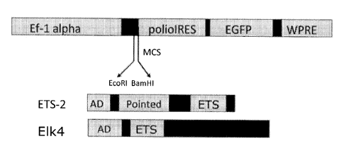
[0011] FIGURE 1 shows a schematic map of lentivirus with the insertion
of
ETS2 and ELK4 full length DNA coding sequences. Functional elements and
abbreviations: constitutive Ef-lalpha promoter, multiple cloning sites (MCS),
independent
ribosome entry site (IRES) from human polio virus, coding sequence for
enhanced green
fluorescence protein (eGFP), Woodchuck Hepatitis Virus Posttranscriptional
Regulatory
Element (WPRE), activation domain (AD), DNA-binding ETS domain. These plasmids
were generated by standard recombinant DNA cloning techniques and then used to
make
lentiviruses to infect normal human dermal fibroblasts (NHDFs);
[0012] FIGURE 2 shows (top panel) NHDFs treated with empty lentivirus
(pWPI-GFP), pWPI-ELK4-GFP lentivirus, or pWPI-ETS2-GFP lentivirus, and (bottom
panel) expression levels of GAPDH, NANOG, OCT3/4, 50X2 in cells infected with
empty virus, virus carrying ETS2, virus carrying ELK4, or uninfected NHDF
passage 3
(NHDF-P3);
[0013] FIGURE 3 shows immunofluorescence staining with antibody to a
stem
cell marker NANOG in NHDF-P3 cells infected with lentivirus carrying ETS2, but
not
ELK4 or empty vector (top panel). A protein blot using anti-ETS2 antibody
revealed
expression of ETS2 in NHDFs infected with lentivirus carrying ETS2 for 4
weeks, while
NFIDFs infected with empty lentivirus showed no expression (bottom left
panel).
Immunofluorescence staining with anti-ETS2 antibody confirmed the induced
expression
in the cells infected with ETS2 lentivirus (bottom right panel);
[0014] FIGURE 4 shows induction of stem cell markers NANOG, OCT3/4,
SOX2, REXI and c-MYC in stem cell-like colonies after 4 weeks in culture. The
fluorescent colors (colors not shown in the figures) are as follows: DAPI
(blue), GFP
-4-
CA 02791901 2012-08-31
WO 2011/109695
PCT/US2011/027160
(green), NANOG and OCT3/4 (red), a mixture of all three colors is observed in
the
"merge" panels. Color fluorescent images are available upon request;
100151 FIGURE 5 shows flow cytometry demonstrating the percent of H9
human embryonic stem cells (A), uninfected NHDFs (B), or ETS2-infected cells
(C) that
stained for the stem cell surface marker SSEA-3. Negative controls, black
lines; SSEA-3
staining, gray lines;
100161 FIGURE 6 shows flow cytometry demonstrating the percent of H9
human embryonic stem cells (A), uninfected NHDFs (B), or ETS2-infected cells
(C) that
stained for the stem cell surface marker Tra-1-81. Negative controls, black
lines; Tra-1-81
staining, gray lines;
[0017] FIGURE 7 shows images of EPS cells after infection with Mespl
lentivirus. Cells were stained with DAPI to visualize nuclei (left panels) and
with specific
antibodies to ISL1, NKX2.5, GATA4, MEF2C, TNT and MHC3 to visualize indicated
cardiac progenitor proteins (right panels). Panels in the middle show phase
contrast images
of cells. The fluorescent colors are as follows: DAPI (blue), protein-specific
staining (red).
Color fluorescent images are available upon request;
[0018] FIGURE 8 shows that expression of NKX2.5, ISLL GATA4, MEF2C,
TBX5, MHC3, TNT, MLC2, CX43 and CX45, detected using RT-PCR, was only induced
by the combination of ETS2 and Mespl. Neither ETS2 or Mespl alone are capable
of
inducing these cardiogenic genes in NIJDFs;
[0019] FIGURE 9 shows induction of sequential de novo cardiac
progenitor
program. NHDFs were infected with ETS2 lentivirus, grown for 4 weeks, infected
with
Mespl lentivirus and cultured for 7 days, then aggregated by the hang-drop
procedure and
plated on a gelatin-coated dish:
[00201 FIGURE 10 shows activation of cardiac progenitor program gene
expression, measured by fluorescence of the reporter protein Red-Tomato which
is
expressed only when the cardiac progenitor factor NKX2.5 is expressed. The
fluorescent
colors (colors not shown in the figures) are as follows: GFP (green), Red
(red), a mixture
of the two colors is observed in the "merge" panel;
-5-
CA 02791901 2012-08-31
WO 2011/109695 PCT/US2011/027160
[0021] FIGURE 11 shows flow cytometry of cardiac progenitor cells obtained
from NHDFs by infection with ETS2 and Mespl lentivectors and sorted for either
GFP or
GFP and reporter protein Red-Tomato;
[0022] FIGURE 12 shows display of endothelial and cardiac cell surface
markers CD31, CD34 and CDI44 in cardiac progenitor cells after 9 days in
culture; and
[0023] FIGURE 13 shows the data on rhythmic beating in reprogrammed
cardiac progenitor cells. EPS cells were infected with Mespl lentivirus, as
well as with
virus carrying a myosin heavy chain promoter driving the puromycin resistance
gene. To
select cardiac progenitor cells resistant to antibiotic, cells were treated
with 50 ug/ml
puromycin. After 9 days, rhythmic beating in the cell cultures was observed
and captured
by video microscopy and converted into MPEG videos. Beats per cultured
aggregate per
dish were counted for 20 sec and then multiplied by 3, resulting in beats per
one minute.
Three separate measurements were done per aggregate in a tissue culture dish
or well.
-6-
CA 02791901 2012-08-31
WO 2011/109695
PCT/US2011/027160
DETAILED DESCRIPTION OF PREFERRED EMBODIMENTS
[0024] One embodiment of the present invention relates to the
modulation of
cell differentiation using heterologous gene expression. Some embodiments of
the
invention relate to a method for inducing a cardiac progenitor cell by
delivering a
reprogramming factor to the cell, wherein the reprogramming factor comprises
ETS2 or a
combination of ETS2 and Mespl
[0025] An embodiment of the present invention provides a method for
inducing a cardiac progenitor cell by reprogramming a somatic cell, wherein
reprogramming comprises delivery of a reprogramming factor comprising a single
heterologous gene to the somatic cell. The somatic cell may be a fibroblast,
preferably a
normal human dermal fibroblast. The heterologous gene may be ETS2. The
heterologous
gene may comprise the human ETS2 coding sequence (SEQ ID NO:9) or the ETS2
gene
(SEQ ID NO:7), or the heterologous gene may encode the human ETS2 protein
sequence
(SEQ ID NO:8). The induced stem-like cell may exhibit cardiogenesis or other
characteristics of cardiac progenitor cells as a result of programming,
including the
expression of cardiac progenitor factors such as NKX2.5, ISL1, MEF2C, dHAND
and
GATA4, or rhythmic beating.
[0026] Another embodiment of the present invention provides a method
for
inducing a cardiac progenitor cell by reprogramming a somatic cell, wherein
reprogramming comprises delivery of a reprogramming factor comprising two
heterologous genes to the somatic cell. The somatic cell may be a fibroblast,
preferably a
normal human dermal fibroblast. The heterologous genes may be ETS2 and Mespl.
The
heterologous genes may comprise the human ETS2 coding sequence (SEQ ID NO:9),
the
ETS2 gene (SEQ ID NO:7), or a DNA sequence encoding the human ETS2 protein
sequence (SEQ ID NO:8) and the mouse Mespl coding sequence (SEQ ID NO:6), the
mouse Mespl gene (SEQ ID NO:4), or a DNA sequence encoding the mouse Mespl
protein sequence (SEQ ID NO:5). The induced stem-like cell may exhibit
cardiogenesis or
other characteristics of cardiac progenitor cells as a result of programming,
including the
expression of cardiac progenitor factors such as NKX2.5, ISL1, MEF2C, dHAND
and
GATA4, or rhythmic beating.
-7-
CA 02791901 2012-08-31
WO 2011/109695
PCT/US2011/027160
[0027] Yet another embodiment of the present invention, reprogramming
of a
somatic cell, may be accomplished by delivering a reprogramming factor to the
somatic
cell using a recombinant vector. The reprogramming factor may also be
delivered using a
lentiviral transduction system to express the reprogramming factor in the
somatic cell. In
these embodiments, the reprogramming factor may be ETS2 and Mesp I.
[0028] A futher embodiment of the present invention provides a somatic
cell
which has been reprogrammed, wherein reprogramming comprises delivery of a
reprogramming factor comprising a single heterologous gene or multiple
heterologous
genes to the somatic cell. The somatic cell may be a fibroblast, preferably a
normal human
dermal fibroblast. The heterologous genes may be ETS2 or the multiple
heterologous
genes may be ETS2 and Mesp I . The induced stem-like cell may exhibit
cardiogenesis or
or other characteristics of cardiac progenitor cells as a result of
programming, including
the expression of cardiac progenitor factors such as NKX2.5, ISL I, MEF2C,
dHAND and
GATA4, or rhythmic beating.
EXAMPLE 1
Selection of a Reprogramming Factor
1100291 It was noted that the ETS domain (Fig. 1), a highly conserved
DNA-
binding domain, is capable of binding to a 5'-GGA(A/T)-3' DNA core motif found
on the
promoters of many stem cell marker genes. The expression of ETS2 is linked to
immortalization of cells, mediation of oncogenesis, and enhancement of
telomerase
activity.
[0030] ETS2 and ELK4, an ETS family gene homologous to ETS2 in its DNA-
binding region, were transduced using lentiviral vectors into NHDF-P3. Within
one week,
fibroblasts transduced with lentiviral vectors containing ETS2 were replaced
with highly
proliferative small rounded cells. These highly proliferative cells were not
observed in
controls transduced with empty lentivirus, or in the fibroblasts transduced
with lentiviral
vectors containing ELK4 (Fig. 2-3).
-8-
CA 02791901 2012-08-31
WO 2011/109695
PCT/US2011/027160
EXAMPLE 2
Lentiviral Transduction System
[0031] Fig. 1 shows a schematic map of lentivirus with the insertion of
ETS2
(SEQ ID NO:9) and ELK4 (SEQ ID NO:3) DNA coding sequences. Note that only ETS2
has a pointed domain. These plasmids were used to make lentiviruses to infect
NHDFs.
The ETS2 full-length sequence (SEQ ID NO:7) comprises an ETS2 coding sequence
(SEQ
ID NO:9) encoding an ETS2 protein sequence (SEQ ID NO:8). The ELK4 full-length
sequence (SEQ ID NO:1) comprises an ELK4 coding sequence (SEQ ID NO:3)
encoding a
protein sequence (SEQ ID NO:2).
[0032] The empty lentivirus vector pWPI-eGFP was a gift from Dr. D.
Trono
(Ecole Polytechnique Federale de Lausanne, Switzerland). cDNA for cloning the
human
ETS2 and ELK4 genes (Clone IDs 3852274 and 4364006) were obtained from Open
Biosystems, whereas the Mesp I cDNA was a gift from Dr. Y. Saga (National
Institute of
Genetics, Mishima, Japan). The consensus Kozak sequence for initiation of
protein
translation and the epitope HA-tag were added respectively to the 5'- and 3'-
ends of ETS2,
ELK4 and Mespl coding sequences by PCR cloning.
[0033] Lentivirus packing and infection proceeded as follows: Seeded
293FT
cells in 6-cm dishes were transfected with either pWPI-eGFP, or pWPI-ELK4-eGFP
(human ELK4 coding sequence, SEQ ID NO:3), or pWPI-ETS2-eGFP (human ETS2
coding sequence, SEQ ID NO:9), or pWPI-Mespl-eGFP (mouse MesP1 sequence, SEQ
ID NO:6), or SMPU-alphaMHC/puro-Rexl /Blast (gift from Dr. M. Mercola, Burnham
Institute for Medical Research, La Jolla, CA). 4.5 ug of either construct was
mixed in a
solution of 458 ul of serum-free Dulbecco-modified Eagle medium (DMEM) and
27.5 ul
of Fugene (Roche), 2.8 ug of packing vector psPAX2 and 1.9 ug of envelope
vector
pMD2.G for 25 min at room temperature. Afterwards the mix was added to 293FT
cells
grown in DMEM, phenol red-free (lnvitrogen) supplemented with 10% FBS (heat-
inactivated), 0.1 mM MEM non-essential amino acids, 1 mM sodium pyruvate and 6
mM
L-glutamate. After 24-26 hrs in culture, medium with viral particles was
collected for 3
days and used for infection.
[00341 Collected medium was used to infect NHDFs grown in Fibroblast
Basal
Medium (FBM, Lonza) until 80% confluency. Before transfection, cells were
reseeded in
-9-
CA 02791901 2016-02-26
TM
6-cm Petri dishes at a density of 2.5x10⁶ cell/dish, the medium was changed to
StemPro
and the viral paricles and polybrene (8 ug,/m1 final concentration) were
added. To increase
the efficiency of infection, the procedure was repcated within 48 hours. All
cells were
grown at 37 C and 5% C01.
EXAMPLE 3
Gene Expression in Reprogrammed Cells
[0035] Fig. 2 shows that ETS2 lentivirus but not ELK4 lentivirus induced
stem
cell appearance and the induction of stem cell marker proteins, NANOG, 0CT3/4
and
SOX2 within 7 days of culture. The top panel of Fig. 2 shows NHDFs (Lonza,
USA, cc-
2509) grown under FBM, supplemented (supplements provided by Lonza) with hFGF-
beta, insulin, gentamycin/amphotericin and 2% FBS, to a confluence of ca. 80%
before
viral infection. Empty pWPI-eGFP and pWl-ELK4-eGFP and pWPf-ETS2-eGFP
lentiviruses were used to separately infect NHDF-P3. Infected and non-infected
NHDF-P3
cells were grown under human induced pluripotent medium StemPro hES SFM
(lnvitrogen) over collagen-coated Petri dishes for 7 days. The green
fluorescent protein
cloned into the lentiviral vectors was expressed in the infected cells, as
revealed by the
green fluorescence microscopy. During this culture period, morphological
changes were
observed in which ETS2-infected NHDFs changed their appearance from elongated
pleomorphic fibroblastic shapes to rounded "stem-like cells". In comparison,
NHDFs
infected with an empty or ELK4 lentiviral vectors did not alter cell shape.
[0036] The bottom panel of Fig. 2 shows the reverse transcription PCR (RT-
PCR) analysis of reprogrammed cells. Cells were washed in chilled PBS and RNA
was
isolated with Qiagen RNeasy Kit. RNA was transcribed using MMLV reverse
transcriptase (1nvitrogen), and PCR amplification (30 cycles) was performed
for GAPDH,
NANOG, OCT3/4, SOX2 (refer to Table 1 for primer sets) using LA16 polymerase
mix.
This enzyme mix was prepared using 15 ul of KlenTaq 1 (25 units/ul, Ab
Peptides, St
Louis, MO) and I ul Pfu (2.5 units/ul, Stratagene, La Jolla, CA). Amplified
DNA samples
were then electrophoresed on 2% agarose gel and ethidium bromide staining
revealed the
induced expression of stem cell marker genes NANOG, OCT3/4 and SOX2 in ETS2-
infected cells but not in NHDF-P3 or cells infected with either ELK4 or empty
lenti viruses.
-10-
CA 02791901 2012-08-31
WO 2011/109695 PCT/US2011/027160
Table 1. Primers for Cardiac Progenitor Study
Primer SEQ ID NO Seq Product
size
GapdhFP crtl exp 11 TGTTGCCATCAATGACCCCTT
202
GapdhRP ctrl exp 12 CTCCACGACGTACTCAGCG
hNanogFP ctrl
exp 13 CAGAAGGCCTCAGCACCTAC
225
hNanogRP ctrl
exp 14 TATAGAAGGGACTGTTCCAGGC
hOct %FP ctrl exp 15 CTTGAATCCCGAATGGAAAGGG
hOct 3/4 RP ctrl 206
exp 16 CCTTCCCAAATAGAACCCCCA
Sox2 HPB F 17 TGGACAGTTACGCGCACAT
215
Sox2 HPB R 18 CGAGTAGGACATGCTGTAGGT
hRex1FP ctrl exp 19 GCTGACCACCAGCACACTAGGC
298
hRex1RPctrl exp 20 TTTCTGGTGTCTTGTCTTTGCCCG
c-Myc HPB F 21 AGGCGAACACACAACGTCTT
c-Myc HPB R 22 TTGGACGGACAGGATGTATGC
hK1f4FP ctrl exp 23 ATGGCTGTCAGCGACGCGCTGCTC
293
hK1f4RP ctrl exp 24 CGTTGAACTCCTCGGTCTCTCTCC
Nkx 2.5 FP 25 ccctgaccgatcccacctcaac
358
Nkx 2.5 RP 26 GGCGGGCGACGGCGAGATAGC
Mesp 1 FP 27 tcgaagtggttccttggcagac
162
Mesp 1 RP 28 CCTCCTGCTTGCCTCAAAGTGTC
Mesp 2 FP 29 CGCTGCGCCTGGCCATCCGCTACAT
113
Mesp 2 RP 30 GCCCCAAGGGGACCCCGCGAC
Mef2c FP 31 gcaccagtgcagggaacggg
202
Mef2c RP 32 GACTGAGCCGACTGGGAGTTA
Sox 17 FP 33 gcggcgcaagcaggtgaag
205
Sox 17 RP 34 ACTCTGGCAGTCGCGGTAGTGGC
FoxA2 FP 35 ctgaagccggaacaccactacgc
214
FoxA2 RP 36 TCCAGGCCCGTTTTGTTCGTGAC
FGF8 FP 37 agctcagccgccgcctcatccg
313
FGF8 RP 38 AGCCCTCGTACTTGGCATTCTGC
MyoD FP 39 AggggctaggttcagctttctcG
240
MyoD RP 40 CTCCTGCTCTGGCAAAGCAACTC
BMP2 HBP FP 41 ACCTTTATGGAGGGAAACCCA 201
BMP2 HBP RP 42 CCGGATCTGGTTCAAGCATGA
hTert HBP FP 43 AACCTTCCTCAGCTATGCCC
210
hTert HBP RP 44 GCGTGAAACCTGTACGCCT
Islet-1 HPB F 45 GTGGAGAGGGCCAGTCTAGG
250
Islet-1 HPB R 46 CCGTCATCTCTACCAGTTGCT
Troponin T HPB F 47 GAGTTGCAGGCGCTGATTG
Troponin T HPB 229
R 48 TCTGGATGTAACCCCCAAAATG
Dhand HPB F 49 ATGAGTCTGGTAGGTGGII1TCC
205
Dhand HPB R 50 CATACTCGGGGCTGTAGGACA
-11-
CA 02791901 2012-08-31
WO 2011/109695
PCT/US2011/027160
T HPB F 51 GATCACGCAGCTCAAGATTGC
230
T HPB R 52 TCTCTGGTGTGTTCCTAGACG
T5 HPB F 53 CACTTCTCCGCTCACTTCACC
210
T5 HPB R 54 TGGCACGCCATGAGAGTAGA
ETS-2 HPB F 55 AAAGCTACCTTCAGTGGCTTC
225
ETS-2 HPB R 56 AATGTCACCCACAAAGTCAGG
Dkk-1 FP 57 ATTCCAACGCTATCAAGAACC
384
Dkk-1 RP 58 CCAAGGTGCTATGATCATTACC
TBX 20 FP 59 tccagattctccttttaccg
190
TBX 20 RP 60 ttcagacttcaggttgagca
SM actin HBP F 61 CGGTGCTGTCTCTCTATGCC
156
SM actin HBP R 62 CACGCTCAGTCAGGATCTTCA
hGATA1 F 63 AGAAGCGCCTGATTGTCAGTA
229
hGATA1 R 64 AGAGACTTGGGTTGTCCAGAA
hGTAT2 F 65 GGCCCACTCTCTGTGTACC
243
hGATA2 R 66 CATCTTCATGCTCTCCGTCAG
TBX3-1 F 67 GTGTCTCGGGCCTGGATTC
164
TBX3-1 R 68 ACGTGTAGGGGTAAGGGAACA
TBX3-2 F 69 TTAAAGTGAGATGTTCTGGGCTG
298
TBX3-2 R 70 ACTATAATTCCCCTGCCACGTA
TBX4 F 71 TGACCATCGCTACAAGTTCTGT
163
TBX4 R 72 GGTGGTTGTTTGTCAGCTTCAG
TBX6 F 73 ACACCCCTAAACTGGATTGCT
229
TBX6 R 74 CCTCCCAGC __ I I 1GGTGATGAT
TBX10 F 75 CCTCGGCATACTTGCACCC
208
TBX10 R 76 ATTCCTCCCACAGAGGCTTCA
TBX18 F 77 GCCCCTGCTGACTATTCTGC
227
TBX18 R 78 CTGCATGGATAAGCTGGTCTG
TBX19 F 79 AAGAATGGCAGACGGATG __ I I I
204
TBX19 R 80 CCGGGTGAATGTAGACGCAG
RUNX2 F 81 CGGCAAAATGAGCGACGTG
268
RunX2 R 82 CACCGAGCACAGGAAGTTG
LMO2 F 83 GGCCATCGAAAGGAAGAGCC
221
LMO2 R 84 GGCCCAGTTTGTAGTAGAGGC
TAU F 85 CCCCTGGAGTTCACGTTTCAC
240
TAU R 86 GCGAGCTTTGAGTTGAGGGA
EXAMPLE 4
Stem Cell Marker Proteins in Reprogrammed Cells
[0037] Fig. 3 shows immunofluorescence staining of infected NHDF-P3 with
antibody to the stem cell marker NANOG. Induced NANOG staining is revealed in
ETS2-
infected cells but not in ELK4- or empty vector-infected cells. Also, a
protein blot using
-12-
CA 02791901 2012-08-31
WO 2011/109695
PCT/US2011/027160
ETS2-specific antibody revealed its expression in cells infected with ETS2
lentivirus but
not in NHDFs infected with empty lentivirus.
[0038] Fig. 4 shows ETS2 lentivirus-induced stem cell-like colonies and
the
induction of stem cell marker proteins NANOG, OCT3/4, SOX2, REX1 and c-MYC
after
4 weeks in culture. The top left panel shows that NHDFs infected with ETS2
virus
converted to colonies of small rounded cells highly reminiscent of cultured
murine and
human embryonic stem cells. Note that colonies were GFP-labeled through the
infection
with ETS2 lentivirus. Non-infected fibroblasts failed to round and did not
stain for GFP or
form cellular colonies.
[0039] The right top panel of Fig. 4 shows the expression of stem cell
marker
genes NANOG, OCT3/4, SOX2, REXI , KLF4 and c-MYC in ETS2-induced cellular
colonies analyzed by RT-PCR. Underneath is graphic representation of
quantitative RT-
PCR for NANOG, OCT3/4, SOX2 and c-MYC in ETS2-infected cells.
[0040] The lower panels of Fig. 4 confirm the presence of NANOG and
OCT3/4 proteins in ETS2-induced cellular colonies by immunofluorescence
staining with
specific antibodies.
EXAMPLE 5
Characteristics of Cells Transduced with ETS2
[0041] Fig. 5 shows that approximately 97% of H9 human embryonic stem
cells stained for the stem cell surface marker SSEA-3 (Panel A). Over 60% of
ETS2-
infected cells also displayed surface marker SSEA-3 (Panel C), in comparison
to less than
2% staining of the NHDFs infected with empty lentivirus (Panel B). Thus, ETS2
efficiently converted NHDFs into cells with the SSEA-3 surface marker
resembling
human embryonic stem cells.
[0042] Flow cytometry was done using a BD Biosciences LSR 11 analyzer.
Confluent colony-forming cells were dissociated by trypsin, washed with PBS
and diluted
to a concentration of 5x10⁶ cells/ml PBS in 8 samples (100 ul each).
Thereafter 10 ul of
normal human serum was used for blocking for 5 min. Antibodies to SSEA-3-PE
(Becton
-13-
CA 02791901 2012-08-31
WO 2011/109695
PCT/US2011/027160
Dickinson) were diluted and added according to manufacturer's specifications,
and
incubated for 1 hr at 4 C. Then 400 ul PBS was added and the mixture was spun
down and
half of the supernatant was removed and 200 ul PBS was added and assayed by
flow
cytometry.
100431 Fig. 6 shows flow cytometry of cells stained for the stem cell
surface
marker Tra-1-81 performed as for SSEA-3. Approximately 45% of 119 human
embryonic
stem cells stained for Tra-1-81 (Panel A). No Tra-1-81 staining (Panel B) was
detected for
NHDFs infected with empty lentivirus (not carrying a heterologous gene).
Infection with
ETS2 virus gave rise to approximately 39% cells displaying embryonic stem cell
marker
Tra-1-81 (Panel C). This indicates that ETS2 efficiently converted NHDFs into
cells with
the Tra-1-81 surface marker resembling human embryonic stem cells.
[0044] OCT3/4, NANOG and SOX2 gene transcripts were observed only after
NHDF-P3 were transduced with lentiviral vector containing ETS2 but not after
transduction with empty lentivector or vector containing ELK4. OCT3/4, NANOG
and
SOX2 transcripts were visualized (Fig. 2) and NANOG induction was visualized
using
immunofluorescence (Fig. 3).
[0045] Lentiviral transduction of I INDF-P3 cells with ETS2 resulted in
whole
populations which showed robust ETS2 expression over 4 weeks visualized by
protein
blots (Fig. 3). These ETS2-transduced cells formed large green fluorescent
colonies
similar to those of pluripotent ES and/or induced pluripotent stem (iPS)
cells.
[0046] Reprogramming of fibroblasts with ETS2 resulted in strong
expression
of the pluripotent marker genes NANOG, OCT3/4, SOX2 and c-MYC measured by both
RT-PCR and quantitative PCR and immunostaining. Additionally, flow cytometry
shows
that ETS2 efficiently converted NHDFs into cells with surface markers SSEA-3
and Tra-
1-81 resembling human embryonic stem cells. Thus, these ETS2-treated human
fibroblast
cells resemble iPS cells in their ability to express pluripotent stem cell
marker proteins.
These cells were therefore named "EPS" cells.
-14-
CA 02791901 2012-08-31
WO 2011/109695
PCT/US2011/027160
EXAMPLE 6
Combination of ETS2 and Mespl induces de novo cardiac progenitor program in
fibroblasts
100471 Next, EPS cells were subjected to lentiviral transduction with
mouse
Mespl. The resulting EPS cells expressing Mespl could be induced to form
embryoid
bodies using protocols for forming embryoid bodies from ES cells. Plated
cellular
aggregates were further treated with activin and BMP4 for 4 days and then
examined at 10
days. Constitutive expression of stem cell markers continued even after the
transduction
with Mespl and addition of growth factor morphogens.
100481 Robust induction of the cardiac progenitor factors ISL1. NKX2.5,
GATA4, MEF2C, TNT and MHC was observed by immunostaining only in the EPS cells
infected with lentivirus expressing Mespl. Fig. 7 shows cells stained with
DAPI to
visualize nuclei (left panels), phase contrast images (middle) and cells
stained with
specific antibodies to visualize indicated proteins (right panels).
[0049] Fig. 8 shows that the MesP1 infection of EPS cells induces de
novo
cardiac progenitor program in cell that were originally NHDFs. Cardiac
progenitor cells
post-aggregation were plated for a week and then taken for RNA isolation. RNA
was
transcribed using MMLV reverse transcriptase, and PCR amplification for 30
cycles was
performed for GAPDH, NKX2.5, ISL1, GATA4, MEF2C, TBX5, MHC3, TNT, MLC2,
CX43 and CX45 (refer to Table 1 for primer sets), using LA-16 polymerase mix.
[0050] Fig. 8 shows that in EPS cells grown for 4 weeks, aggregated and
plated
for 7 days, no transcripts were observed for markers of early heart
development by RT-
PCR. Similarly, no appreciable expression of the early heart development
markers was
detected after infection of NHDFs with Mespl alone. However, Mespl addition to
EPS
cells induced robust expression of cardiac mesoderm progenitor markers
including
NKX2.5, ISL1, GATA4, MEF2C, TBX5, and cardiac contractile protein gene
expression
including alpha MHC and troponin T.
-15-
CA 02791901 2012-08-31
WO 2011/109695
PCT/US2011/027160
[0051] Fig. 9 shows that the MesP1 infection of EPS cells induced
sequential
de novo cardiac progenitor program as determined by RT-PCR. NHDF-P3 cells were
infected with ETS2 lentivirus, grown for 4 weeks, then infected with Mespl
lentivirus,
aggregated and plated for 7 days. RNA was isolated daily and analyzed by RT-
PCR for
expression of cardiogenic genes.
[0052] Fig. 10 shows that the MesP1 infection of EPS cells induced de
novo
cardiac program gene expression as shown by the appearance of Red Tomato
fluorescence
staining from the reporter construct NKX2.5-Red Tomato. NKX2.5/Smad/GATA
enhancer, which is activated in cardiac progenitors, was linked to the minimal
HSP68
promoter adjacent to the puromycin resistance gene and an IRES sequence and
the
powerful reporter td-Tomato (cDNA was a gift from Dr. R. Tsien, University of
California, San Diego). As shown above, GFP staining resulted from ETS2 and
Mespl
lentivirus infection of NHDFs. The Red Tomato fluorescence is consistent with
the
ETS2/Mespl driven conversion to cardiac progenitors by the induction of NKX2.5
gene
expression. Note the appearance of many triangular and rectangular appearing
cells that
are highly similar in shape to cardiac myocytes.
[0053] Fig. 11, Panel A shows a summary of FACS sorting of cardiac
progenitor cells obtained from sequential treatment of NHDFs with ETS2
lentivirus and
then of the resultant EPS cells with Mespl lentivirus. Flow cytometry was done
using a
BD Biosciences LSR II analyzer. Confluent colony-forming cells were
dissociated by
trypsin, washed with PBS and diluted to a concentration of 5x10⁶ cells/ml PBS
in 8
samples. Panel A shows GFP stained cells accounting for the total lentivirus
infection,
since each virus is GFP-tagged. Panel B shows that approximately 19% of the
ETS2 and
Mespl infected cells were both stained by GFP and Red-Tomato (shaded area). As
evidenced by activation of a key cardiogenic reporter NKX2.5 Red-Tomato, ETS2
and
Mespl efficiently convert NHDFs into cells with characteristics resembling
cardiac
progenitors.
[0054] Fig. 12 shows that approximately 10 to 40% of ETS2 and Mespl
infected NHDF cells display endothelial and cardiac cell surface markers,
CD31. CD34
and CD144 after 9 days in culture. This is an additional line of evidence that
ETS2 and
-16-
CA 02791901 2012-08-31
WO 2011/109695
PCT/US2011/027160
Mespl efficiently converted NHDFs into cells with characteristics resembling
embryonic
endothelial and cardiac myocytes.
EXAMPLE 7
Cardiac Properties of Reprogrammed Cells
[0055] Lentiviral transduction of a puromycin selectable system using a
lentiviral cardiac-specific alpha-myosin heavy chain (alpha-MTIC) promoter and
enhancer
linked to the puromycin resistance gene resulted in enrichment of the cardiac
progenitor
cells and subsequent observation of a rhythmic beating of the transduced
cells, similar to
that observed in cardiac myocytes.
[0056] A myosin heavy chain prometer driving the puromycin selectable
gene
construct was transduced into NHDFs which were then sequentially transduced
with ETS2
and Mespl. Cellular aggregates obtained during hang-drop embryoid body
formation were
then treated with 50 ug/ml puromycin to select cells resistant to puromycin
and therefore
having the active cardiac specific alpha-MHC promoter.
[0057] After 9 days beating in the cell cultures was observed and
captured
using video microscopy and converted into MPEG videos. Beating per cultured
aggregate
per dish was counted for 20 sec and then multiplied by 3 for beats per one
minute (Fig.
13). Three separate measurements were done per aggregate in a tissue culture
dish or well.
[0058] Reprogramming of EPS cells with Mespl resulted in strong
expression
of cardiac progenitor genes as determined by RT-PCR and immunostaining.
Additionally,
flow cytometry showed that Mespl efficiently converted EPS cells into cells
with surface
markers CD31, CD34 and CD144 resembling human cardiac cells. Finally, rhythmic
beating was observed in the cell cultures. This completed the conversion from
skin
fibroblasts to terminally differentiated cardiogenic cells.
-17-
CA 02791901 2016-02-26
REFERENCES CITED
[0059] The following references provide exemplary procedural or other details
supplementary to those set forth herein.
U.S. PATENT DOCUMENTS
U.S.Patent publication No. 2009/0227032 to S.Yamanaka.
NON-PATENT REFERENCES
Bergmann, O. et al. Evidence for cardiomyocyte renewal in humans. Science 324:
5923,
98-102, 2009.
Beh, J. et al. FoxF is essential for FGF-induced migration of heart progenitor
cells in the
ascidian Ciona intestinalis. Development 134: 3297-3305, 2007.
Davidson, B. Ciona intestinalis as a model for cardiac development. Sem in.
Cell Dev.
Biol. 18: 16-26,2007.
Davidson, B. and Levine, M. Evolutionary origins of the vertebrate heart:
Specification of
the cardiac lineage in Ciona intestinalis. Proc. Natl. Acad. Sci. USA 100:
11469-11473.
'mai, K.S., Satoh, N. and Satou, Y. A Twist-like bHLH gene is a downstream
factor of an
endogenous FGF and determines mesenchymal fate in the ascidian embryos.
Development
130: 4461-4472, 2003.
Imai, K.S., Hino, K., Yagi, K., Satoh, N. and Satou, Y. Gene expression
profiles of
transcription factors and signaling molecules in the ascidian embryo: towards
a
comprehensive understanding of gene networks. Development 131: 4047-4058,
2004.
Kitajima, S. et al. MesPI and MesP2 are essential for the development of
cardiac
mesoderm. Development 127: 3215-3226, 2000.
Moretti, A. et al. Multipotent embryonic isti- progenitor cells lead to
cardiac, smooth
muscle and endothelial cell diversification. Cell 127: 1151-1165, 2006.
Park, I.H. et al. Reprogramming of human somatic cells to pluripotency with
defined
factors. Nature 451: 14 I -146, 2008.
Saga, Y. et al. MesP1 is expressed in the heart precursor cells and required
for the
formation of a single heart tube. Development 126:3437-3447, 1999.
-18-
CA 02791901 2012-08-31
WO 2011/109695
PCT/US2011/027160
Saga, Y., Kitajima, S. and Miyagawa-Tomita, SMespl expression is the earliest
sign of
cardiovascular development. Trends Cardiovasc Med. 10:345-352, 2000.
Takahashi, K. et al. Induction of pluripotent stem cells from adult human
fibroblasts by
defined factors. Cell 131:861-872, 2007.
Yu, J. Induced pluripotent stem cell lines derived from human somatic cells.
Science
318:1917-1920, 2007.
-19-