Note: Descriptions are shown in the official language in which they were submitted.
BONE FIXATION SYSTEM INCLUDING K-WIRE COMPRESSION
CROSS-REFERENCE TO RELATED APPLICATIONS
[0001] This application claims the benefit of U.S. Provision Application
Serial No.
61/372,212 filed August 10, 2010, and U.S. Provisional Application Serial No.
61/328,278 filed
April 27, 2010.
BACKGROUND
[0002] Conventional bone fixation systems include a bone plate having screw
holes that
receive fixation members, such as screws that are configured to attach to
underlying bone that
includes, at a minimum, a pair of bone segments separated by a bone gap. The
bone gap can be a
fracture created by a traumatic event, an osteotomy, or can be the result of
debridement of a joint
of two discrete bones to be joined in an arthodesis. Thus, the bone plate can
be affixed to the
bone on opposed sides of the bone gap via the bone screws to promote union of
the bone
segments (e.g., healing of the fracture or ossification of the joint). Bone
fixation systems can
further include temporary Kirschner wires (K-wires) that are temporarily
inserted into apertures
of the bone fixation plate and into the underlying bone segments to determine
proper length,
rotation and alignment of the bone segments prior to permanent plate fixation.
Once the bone
fixation plate has been properly positioned, the permanent bone screws can be
inserted into one
or more bone screw holes on opposed sides of the bone gap and affixed to the
underlying bone.
[0003] In one conventional system, a K-wire is screwed or otherwise driven
through the
screw holes of the plate on opposite sides of the bone gap. The K-wire is
smaller in diameter as
the screw holes, and is thus positioned so as to bear against opposing edges
of the respective
screw holes so as to prevent movement of the plate during imaging. The process
of accurately
positioning the K-wire so as to prevent movement of the bone plate has proven
difficult and
tedious, as any space between the K-wire and the outer edge of the screw hole
can allow
movement of the bone plate.
SUMMARY
[0004] In accordance with one embodiment, a method is provided for fixing a
bone
plate to first and second bone segments that are separated by a bone gap. The
method includes
the step of aligning the bone plate with the first and second bone segments
such that a first
plurality of apertures extending through the bone plate are aligned with the
first bone segment
1
CA 2795819 2017-06-12
CA 02795819 2012-10-05
WO 2011/137163 PCT/US2011/034113
and a second plurality of apertures extending through the bone plate are
aligned with the second
bone segment. A select one of the first plurality of apertures is a K-wire
slot and a select one of
the second plurality of apertures is a K-wire hole. The method further
includes the steps of
inserting a distal portion of a first K-wire through the K-wire slot and into
the first bone segment,
inserting a distal portion of a second K-wire through the K-wire hole and into
the second bone
segment, and actuating a forceps to bias at least one of the K-wires to
translate relative to the
other K-wire.
[0005] In accordance with another embodiment, a forceps is provided that is
configured
to apply a biasing force to a pair of temporary fixation members. Each
temporary fixation
member has a distal portion and an engagement member disposed proximal of the
distal portion.
The engagement member may define a dimension greater than that of the distal
portion, and the
engagement member may present an outer surface. The forceps may comprise a
pair of arms that
are pivotally connected at a joint. Each arm may have a proximal end and an
opposed distal end.
Each arm may further have an engagement member defining a pocket that extends
into the distal
end. The pocket may define an engagement surface having a shape corresponding
to that of the
engagement member of the temporary fixation members. Relative movement of the
arms causes
the distal ends to correspondingly move, such that each pocket at least
partially receives a
respective one of the temporary fixation members and the engagement surface
applies a biasing
force against the engagement member of the received temporary fixation member.
[0006] In accordance with another embodiment, a bone fixation kit is provided
that
includes at least one bone fixation plate, at least a pair of temporary
fixation members, and a
forceps. The plate may include a plurality of apertures, at least some of
which are configured to
receive respective bone fixation members. Each temporary fixation member may a
proximal
portion, a distal portion, and an engagement member disposed between the
proximal portion and
the distal portion. The engagement member may define a cross-sectional
dimension greater than
that of the distal portion, wherein at least one of the temporary fixation
members is configured to
extend through a respective one of the plurality of apertures and into an
underlying bone segment
of a pair of underlying bone segments that are separated by a bone gap. The
forceps may include
a pair of arms, each arm having a proximal end and an opposed distal end. The
distal end may
include an engagement member that defines a corresponding engagement surface
that is
configured to move along a direction so as to abut an engagement member of a
respective one of
the temporary fixation members, wherein further movement of the engagement
surface along the
direction causes at least one of the temporary fixation members to translate
relative to the other
temporary fixation member.
2
CA 02795819 2012-10-05
WO 2011/137163 PCT/US2011/034113
[0007] In accordance with another embodiment, a method is provided for
positioning
first and second bone segments that are disposed in a first relative position
in relation to each
other and are separated by a bone gap during a surgical procedure. The method
includes the step
of inserting a distal portion of a first temporary fixation member into the
first bone segment, and
inserting a distal portion of a second temporary bone fixation member into the
second bone
segment. The method further includes the step of actuating a forceps to bias
at least one of the
temporary bone fixation members relative to the other temporary bone fixation
member, thereby
adjusting the relative positions of the bone segments in relation to each
other from the first
relative position to a second different relative position. Prior to completion
of the surgical
procedure the first and second temporary fixation members may be removed from
the first and
second bone segments, respectively.
[0008] In accordance with another embodiment, a method is provided for
positioning a
bone plate to first and second bone segments that are disposed in a relative
position in relation to
each other and are separated by a bone gap. The method may include the steps
of aligning the
bone plate with the first and second bone segments, the bone plate including a
plate body and a
plurality of apertures extending through the plate body, wherein a first
aperture of the plurality of
apertures comprises a bone anchor hole that is aligned with the first bone
segment, and a second
aperture of the plurality of apertures comprises a coupler. The method further
includes inserting
a bone anchor through the bone anchor hole and into the first bone segment,
inserting a distal
portion of a post into the second aperture, the distal portion of the post
defining a coupler that
engages the coupler of the second aperture to thereby fixedly couple the post
to the bone plate,
and inserting a distal portion of a K-wire into the second bone segment. A
forceps may then be
actuated to bias at least one of the K-wire and the post to translate relative
to the other, thereby
adjusting the relative positions of the bone segments in relation to each
other.
[0009] In accordance with another embodiment, a bone fixation kit is provided.
The kit
may include at least a pair of temporary bone fixation members, and a forceps.
Each temporary
bone fixation member may have a proximal portion, a distal portion, and an
engagement member
disposed between the proximal portion and the distal portion, the engagement
member defining a
cross-sectional dimension greater than that of the distal portion, wherein the
temporary bone
fixation members are configured to extend through respective ones of the
plurality of apertures
and into respective underlying bone segments that are separated by a bone gap.
The forceps may
include a pair of arms, each arm having a proximal end and an opposed distal
end, the distal end
including an engagement member that defines a corresponding engagement surface
that is
configured to move along a direction so as to abut a respective one of the
temporary bone
3
CA 02795819 2012-10-05
WO 2011/137163 PCT/US2011/034113
fixation members. Further movement of the engagement surface along the
direction causes at
least one of the temporary bone fixation members to translate relative to the
other temporary
bone fixation member.
BRIEF DESCRIPTION OF THE DRAWINGS
[0010] The foregoing summary, as well as the following detailed description of
an
example embodiment of the application, will be better understood when read in
conjunction with
the appended drawings, in which there is shown in the drawings an example
embodiment for the
purposes of illustration. It should be understood, however, that the
application is not limited to
the precise arrangements and instrumentalities shown. In the drawings:
[0011] Fig. IA is a perspective view of a bone fixation system constructed in
accordance with one embodiment operatively coupled to a pair of schematically
illustrated bone
segments separated by a bone gap, the bone fixation system including a bone
fixation plate, a
pair of K-wires, and a forceps;
[0012] Figs. 1B is a perspective view similar to Fig. 1A, but showing the bone
gap
reduced by the bone fixation system;
[0013] Fig. 2A is a top plan view of the bone fixation plate illustrated in
Fig. IA;
[0014] Fig. 2B is a top plan view of a variable angle locking hole of the bone
fixation
plate illustrated in Fig. 2A;
[0015] Fig. 2C is a perspective view showing a bone anchor installed in the
variable
angle locking hole illustrated in Fig. 2B;
[0016] Fig. 2D is a top plan view of a combination hole of the bone fixation
plate
illustrated in Fig. 2A;
[0017] Fig. 2E is a sectional side elevation view of the bone fixation plate
illustrated in
Fig. 2D taken along line 2E-2E so as to illustrate a screw hole;
[0018] Fig. 2F is a sectional side elevation view of the bone fixation plate
similar to
Fig. 2E, but showing the screw hole constructed in accordance with an
alternative embodiment;
[0019] Fig. 2G is a sectional side elevation view of the bone fixation plate
similar to
Fig. 2F, but showing the screw hole constructed in accordance with an
alternative embodiment;
[0020] Fig. 2H is an enlarged top plan view of the bone fixation plate
illustrated in Fig.
2A, showing a dedicated K-wire slot;
[0021] Fig. 21 is a top plan view of a bone fixation plate similar to Fig. 2A,
but
constructed in accordance with an alternative embodiment;
4
CA 02795819 2012-10-05
WO 2011/137163 PCT/US2011/034113
[0022] Fig. 3A is a perspective view of a bone fixation plate constructed in
accordance
with another embodiment;
[0023] Fig. 3B is a top plan view of the bone fixation plate illustrated in
Fig. 3A;
[0024] Fig. 3C is a side elevation view of the bone fixation plate illustrated
in Fig. 3A;
[0025] Fig. 3D is a top plan view of a bone fixation plate constructed similar
to the
bone plate illustrated in Fig. 3A, but in accordance with another embodiment;
[0026] Fig. 3E is a top plan view of a bone fixation plate constructed similar
to the
bone plate illustrated in Fig. 3A, but in accordance with another embodiment;
[0027] Fig. 4A is a top plan view of a bone fixation plate constructed in
accordance
with another embodiment;
[0028] Fig. 4B is a side elevation view of the bone fixation plate illustrated
in Fig. 4A;
[0029] Fig. 4C is a top plan view of a bone fixation plate constructed in
accordance
with another embodiment;
[0030] Fig. 4D is a top plan view of a bone fixation plate constructed similar
to Fig. 4C,
but in accordance with another embodiment;
[0031] Fig. 4E is a top plan view of a bone fixation plate constructed in
accordance
with another embodiment;
[0032] Fig. 4F is a top plan view of a bone fixation plate constructed in
accordance
with another embodiment;
[0033] Fig. 4G is a top plan view of a bone fixation plate constructed in
accordance
with another embodiment;
[0034] Fig. 5A is a side elevation view of a non-locking bone anchor
constructed in
accordance with one embodiment;
[0035] Fig. 5B is a side elevation view of a locking bone anchor constructed
in
accordance with an alternative embodiment;
[0036] Fig. SC is a side elevation view of a head portion of the bone anchor
illustrated
in Fig. 5B;
[0037] Fig. 5D is a sectional side elevation view of a locking bone anchor
constructed
in accordance with an alternative embodiment;
[0038] Fig. 6A is a side elevation view of the K-Wire illustrated in Fig. 1A;
[0039] Fig. 6B is a side elevation view of a K-wire constructed in accordance
with an
alternative embodiment;
[0040] Fig. 7A is a perspective view of the forceps illustrated in Fig. 1A;
CA 02795819 2012-10-05
WO 2011/137163 PCT/US2011/034113
[0041] Fig. 7B is a perspective view of the forceps illustrated in Fig. 7A
shown in an
open configuration;
[0042] Fig. 7C is a perspective view of the forceps illustrated in Fig. 7B
shown in a
closed configuration;
[0043] Fig. 7D is a perspective view of a portion of the forceps illustrated
in Fig. 7A,
showing a ratchet mechanism;
[0044] Fig. 7E is an enlarged perspective view of a distal end of the forceps
illustrated
in Fig. 7A, showing a compression engagement member;
[0045] Fig. 8A is an enlarged perspective view of a distal end of the forceps
illustrated
in Fig. 7A, but constructed in accordance with an alternative embodiment,
including
compression and distraction engagement members;
[0046] Fig. 8B is a perspective view of the distal end illustrated in Fig. 8A,
schematically showing the compression and distraction engagement members
operatively
coupled to respective K-wires;
[0047] Fig. 8C is an enlarged perspective view of a distal end of one arm of
the forceps
illustrated in Fig. 7A, but constructed in accordance with an alternative
embodiment, including a
compression and distraction engagement members;
[0048] Fig. 8D is a perspective view of the distal end illustrated in Fig. 8C,
schematically showing the compression and distraction engagement members
operatively
coupled to respective K-wires;
[0049] Fig. 8E is an enlarged perspective view of a distal end of one arm of
the forceps
illustrated in Fig. 7A, but constructed in accordance with an alternative
embodiment, including a
compression and distraction engagement members
[0050] Fig. 8F is a perspective view of the distal end illustrated in Fig. 8E,
schematically showing the compression and distraction engagement members
operatively
coupled to respective K-wires;
[0051] Fig. 9 is a schematic perspective view of a bone fastener secured to
bone
segments using the bone fixation system illustrated in Fig. 1A;
[0052] Fig. 10 is a perspective view of a bone fixation system constructed in
accordance with an alternative embodiment operatively coupled to a pair of
schematically
illustrated bone segments separated by a bone gap, the bone fixation system
including a bone
fixation plate, a K-wire, a post, and a forceps;
[0053] Fig. 11A is a perspective view of a bone fixation plate constructed in
accordance
with an alternative embodiment, and illustrated in Fig. 10;
6
CA 02795819 2012-10-05
WO 2011/137163 PCT/US2011/034113
[0054] Fig. 11B is a top plan view of the bone fixation plate illustrated in
Fig. 11A;
[0055] Fig. 12A is a partial perspective of a K-wire constructed in accordance
with an
alternative embodiment, and illustrated in Fig. 10;
[0056] Fig. 12B is a side elevation view of the K-wire illustrated in Fig.
12A;
[0057] Fig. 13A is a front perspective view of a post constructed in
accordance with
one embodiment, and illustrated in Fig. 10;
[0058] Fig. 13B is a side elevation view of the post illustrated in Fig. 13A;
[0059] Fig. 14A is a front perspective view of a forceps constructed in
accordance with
an alternative embodiment, the forceps having compression engagement members;
[0060] Fig. 14B is a front perspective view of the forceps illustrated in Fig.
14A, but
constructed in accordance with an alternative embodiment, including a
distraction engagement
members;
[0061] Fig. 15A is a front perspective view of the bone fixation system
illustrated in
Fig. 10 reducing the bone gap defined between the first and second bone
segments, the bone
fixation plate affixed to the first bone segment with a bone anchor, the post
fixedly coupled to
the bone fixation plate adjacent the first bone segment, and the K-wire
extending through the
bone plate and into the second bone segment;
[0062] Fig. 15B is a front perspective view of the bone fixation system
illustrated in
Fig. 15A distracting the bone gap defined between the first and second bone
segments with the
forceps illustrated in Fig. 14B;
[0063] Fig. 16A is a front perspective view of the bone fixation system
illustrated in
Fig. 10 compressing the bone gap defined between the first and second bone
segments, the bone
fixation plate affixed to the first bone segment with a bone anchor, the K-
wire extending through
the bone plate and into the second bone segment, and the post fixedly coupled
to the bone plate
adjacent the second bone segment such that distraction of the forceps causes
the bone gap to
compress;
[0064] Fig. 16B is a front perspective view of the bone fixation system
illustrated in
Fig. 16A distracting the bone gap defined between the first and second bone
segments with the
forceps illustrated in Fig. 14A;
[0065] Fig. 17A is a front perspective view of the bone fixation system
illustrated in
Fig. 10 compressing the bone gap defined between the first and second bone
segments, the bone
fixation plate affixed to the first bone segment with a bone anchor, the K-
wire extending directly
into the second bone segment, and the post fixedly coupled to the bone plate
adjacent the second
bone segment such that compression of the forceps causes the bone gap to
compress; and
7
CA 02795819 2012-10-05
WO 2011/137163 PCT/US2011/034113
[0066] Fig. 17B is a front perspective view of the bone fixation system
illustrated in
Fig. 17A distracting the bone gap defined between the first and second bone
segments with the
forceps illustrated in Fig. 14B.
DETAILED DESCRIPTION
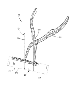
[0067] Referring initially to Fig. 1A, a bone fixation system 20 includes a
bone fixation
plate 22, at least one guide wire or temporary fixation member illustrated as
a K-wire 24, such as
a pair of opposing K-wires 24a and 24b, and a forceps 26 configured to engage
the K-wires 24a
and 24b. The bone fixation plate 22 can be operatively coupled to an
underlying bone 27 having
bone segments 27a and 27b separated by a bone gap 28. The bone gap can be a
fracture created
by a traumatic event, an osteotomy, or can be the result of debridement of a
joint of two discrete
bones to be joined in an arthodesis. The bone fixation plate 22 is placed
against or in proximity
with the underlying bone 27, the K-wires 24a and 24b are inserted through the
plate 22 and into
the respective bone segments 27a and 27b, and the forceps 26 can apply a force
onto the K-wires
so as to translate at least one of or both of the bone segments 27a and 27b,
thereby adjusting the
relative positions of the bone segments 27a and 27b in relation to each other.
For instance, the
forceps 26 can apply a compressive force that brings at least one or both of
the bone segments
27a and 27b toward the other, thereby reducing the bone gap 28 to promote
union of the bone
segments 27a and 27b, as illustrated in Fig. 1B. In accordance with certain
embodiments, the
forceps 26 can apply a distractive force onto the K-wires so as to urge one or
both of the bone
segments 27a and 27b away from the other, thereby distracting the bone gap 28,
for instance
from the position illustrated in Fig. 1B to the position illustrated in Fig.
1A. The bone fixation
plate 22 can be geometrically configured for fixation to bone 27, which can be
the forefoot,
midfoot, hindfoot, distal tibia, or any bone in the human body as desired,
either in vivo or ex
vivo. The bone fixation plate 22 can alternatively be fixed in the manner
described above to any
suitable non-human animal body bone, in vivo or ex vivo.
[0068] The bone fixation system 20 can further include a plurality (e.g., at
least two)
bone anchors 30 (see Fig. 2C) that secure the bone fixation plate 22 to the
underlying bone 27 on
opposed sides of the bone gap 28. The bone fixation system 20 and components
of the bone
fixation system 20 can be made from any suitable biocompatible material, such
as titanium,
including titanium alloys, stainless steel, ceramics, or polymers such as
polyetheretherketone
(PEEK), cobalt chromium molybdenum (CoCrMo) with a porous plasma-sprayed
titanium
coating, or any suitable alternative material as desired.
8
CA 02795819 2012-10-05
WO 2011/137163 PCT/US2011/034113
[0069] Referring now to Fig. 2A, the bone fixation plate 22 can be made in
different
shapes and sizes for use in a wide variety of clinical applications. The bone
fixation plate 22 is
elongate along a longitudinal direction L, defines a width along a lateral
direction A that is
perpendicular or substantially perpendicular to the longitudinal direction L,
and a thickness along
a transverse direction T that is perpendicular or substantially perpendicular
to both the
longitudinal direction L and the lateral direction A. In this regard, it
should be appreciated that
the various directions can extend along directions that are 90 angularly
offset from each other,
or anywhere within the range of approximately 450 and approximately 90
angularly offset from
each other.
[0070] The bone fixation plate 22 includes a plate body 32 that extends
substantially
along a central longitudinal axis 31, and defines a proximal end 34 and a
distal end 36 opposite
the proximal end 34 along the longitudinal axis 31. The plate body 32 further
includes a bone-
facing inner surface 38 and an opposed outer surface 40 spaced from the inner
surface 38 along
the transverse direction T. The plate body 32 further defines opposed side
surfaces 42 and 44
that are spaced from each other along the lateral direction A. The plate body
32 includes a head
portion 46 at the distal end 36 that can be configured and dimensioned to
conform to the contour
of the near cortex of the underlying bone 27, and a shaft portion 48 connected
to the head portion
46 and disposed longitudinally proximal from the head portion 46. The shaft
portion 48 can be
configured and dimensioned to conform to the contour of the near cortex of the
underlying bone
27. In accordance with the illustrated embodiment, the head portion 46
resembles the shape of a
cloverleaf, though it should be appreciated that the head portion 46 can
assume any geometric
shape as desired. The cloverleaf-shaped plate can be used in a number of bony
applications,
especially where a short bone segment is present. The cluster of the
"cloverleaf' design allows
the surgeon to place three screws for three points of fixation in a small
surface area which can
provide greater stability than two points of fixation in the same surface
area.
[0071] The bone facing surface 38 of the head portion 46 can be generally
coplanar
with or offset from the bone facing surface 38 of the shaft portion 48. For
instance, the bone
facing surface 38 of the head portion 46 and the shaft portion 48 can be
curved so as to conform
to the contours of the underlying bone 27. The plate body 32 can further
include a neck portion
50 connected between the head portion 46 and the shaft portion 48. The neck
portion 50 can be
straight, curved, and can define a lateral thickness that is greater than,
less than, or substantially
equal to that of the head portion and the shaft portion 48. In accordance with
the illustrated
embodiment, the neck portion 50 has a lateral thickness less than that of the
head portion 46 and
the shaft portion 48.
9
CA 02795819 2012-10-05
WO 2011/137163 PCT/US2011/034113
[0072] With continuing reference to Fig. 2A, the bone plate 22 includes a
plurality of
apertures 39 that extend transversely through the plate body 32, from the bone-
facing inner
surface 38 through to the outer surface 40. The apertures 39 can include at
least one such as a
plurality of bone anchor holes 41, at least one such as a plurality of K-wire
holes 23 which can
be dedicated K-wire holes 43, and at least one such as a plurality of
longitudinally elongate K-
wire slots 25 which can be dedicated K-wire slots 45. As will become
appreciated from the
description below, the K-wire hole 43 and the K-wire slot 45 can be dedicated
to receive
respective K-wires, or can each also be configured as a bone anchor hole that
are configured to
receive both a bone anchor and a K-wire.
[0073] As will now be described with respect to Figs. 2A-2G, one or more of
the bone
anchor holes 41 up to all of the bone anchor holes 41 can be configured as a
variable angle hole
52, a fixed axis hole 54, a combination hole 57 including a variable angle
hole portion and a
fixed angle hole portion, and can further be configured as a compression hole,
a threaded locking
hole, or a combination of both. It should be appreciated that at least one up
to all of the bone
anchor hole 41, the K-wire hole 43, and the K-wire slot 45 can extend through
the head portion
46, the shaft portion 48, and/or the neck portion 50 as desired. In accordance
with the illustrated
embodiment, the bone plate 22 includes a plurality of variable angle holes 52
that extend through
the head portion 46. For instance, the bone plate 22 includes a pair variable
angle holes 52
extending through the head portion 46 that are laterally spaced from each
other and aligned along
the lateral direction A, and a third variable angle hole 52 that extends
through the head portion
46 at a location distal of and laterally between the holes 52.
[0074] Referring now also to Fig. 2B, each variable angle hole 52 is defined
by an
interior surface 55 of the bone plate body 32. The interior surface 55
includes a plurality of
vertical or transversely extending columns 56. In accordance with the
illustrated embodiment,
four columns 56 are equidistantly spaced circumferentially about the hole 52,
though the plate
body 32 can alternatively include any number of columns as desired, spaced
circumferentially
equidistantly as illustrated, or at circumferentially variable distances as
desired. Each column 56
presents internal threads 58 that face the hole 52 such that, if the columns
56 were expanded to
join each other (i.e. if extended completely around the interior surface 55),
the columns 56 would
form a continuous helical thread that extend about the central transverse axis
49. Thus, it can be
said that the threads 58 of adjacent columns 56 are operatively aligned with
each other.
[0075] It should be appreciated that while the columns 56 present internal
helical
threads 58 as illustrated, the columns 56 alternatively can define threads
that are provided as
teeth formed thereon. The columns of teeth, if expanded to join each other
(i.e., if extended
completely around the interior surface 55), will not form a helical thread,
but a series of
concentric ridges and grooves perpendicular to the central axis 49 of the bone
plate hole 52.
Thus, it can be said that the teeth can be operatively aligned with each
other. The columns 56
are circumferentially spaced from each other so as to define corresponding
axes that are angled
with respect to the transverse central axis 49, such that a screw can extend
through the hole 52 at
any of the angled axes while threadedly fixed to the threads 58.
[0076] The interior surface 55 that defines the hole 52 further includes a
plurality of
arcuate pockets 60 that project into the plate body 32 at a location
circumferentially between the
adjacent columns 56. The pockets 60 each presents an arcuate surface 62 that
is concave with
respect to a direction radially outward from the central axis 49 of the hole
52. As illustrated in
Fig. 2C, and as described in more detail below, the bone anchor 30 can be
provided as a variable
locking bone anchor 61 that can threadedly engage the threads 58 at variable
angular positions.
Alternatively, the bone anchor 30 can be provided as a fixed angle locking
screw that purchases
with the threaded columns 56 and extends along the transverse axis 49. The
variable angle holes
52 can be configured to allow the bone anchor to engage the threads 58 at any
angular
orientation as desired, up to +/- 150 (e.g., within a 30 range) with respect
to the central axis 49,
which extends along the transverse direction T. The variable angle hole 52 is
further described
in U.S. Patent Application Publication No. 2008/0140130, published June 12,
2008.
[0077] Referring now also to Figs. 2D-E, the fixed axis hole 54 can be
generally
cylindrical, such that the bone plate body 32 defines a substantially
cylindrical interior surface 64
that is substantially cylindrical and at least partially defines the hole 54.
The hole 54, and thus
the interior surface 64 can extend entirely through the plate body 32, from
the bone facing
surface 38 through to the outer surface 40 along a central transverse axis 51.
The interior surface
64 can be enclosed, or the plate body 32 can define a circumferential gap 65
that extends
longitudinally through a portion of the interior surface 64, so as to extend
between the fixed axis
hole 54 and the variable angle hole 52 of the combination hole 57. The gap 65
can extend
transversely entirely through the plate body 32, from the outer surface 40
through to the inner
surface 38. The interior surface 64 of the combination hole 57 illustrated in
Fig. 2D can be
unthreaded such that a screw head of a screw inserted into the hole 54 of the
combination hole 57
can compress the bone plate 22 to the underlying bone 27, and/or compress the
bone fragments
27a and 27b together. For instance, the screw can be inserted into the
underlying bone 27 at one
side of the hole 54 at a location offset with respect to the central axis of
the hole, such that as the
screw is compressed against the plate 22, the hole 54 aligns with the screw,
which causes the
bone plate 22 to translate in a direction that compresses the bone fragments
27a and 27b.
11
CA 2795819 2017-06-12
100781 Thus, it should be appreciated that the plate 22 can define at least
one or more
discrete variable angle holes 52 and fixed axis holes 54, or the plate 22 can
define at least one or
more combination holes 57 that include a variable angle hole 52 and a fixed
axis hole 54
connected by the gap 65 that extends transversely through the plate body 32.
In accordance with
the illustrated embodiment, the variable angle hole 52 of a given combination
hole 57 is spaced
longitudinally distal with respect to, and longitudinally aligned with, the
respective variable
angle hole 52 of a given combination hole 57. The combination hole 57 is
further described in
U.S. Patent Application Publication No. 2008/0140130, published June 12, 2008.
[0079] The interior surface 64 can extend in a transverse direction, such that
the hole 54
has a constant diameter along its length through the plate body 32. As
illustrated in Fig. 2E, the
interior surface 64 can present internal threads 58 that are configured to
engage complementary
threads of the head of a locking bone anchor, as described in more detail
below. It should be
appreciated that a screw having a fixed-angle head (also referred to as a
fixed angle screw) can
be inserted into the fixed axis hole 54 along the transverse axis of the hole
54. For instance, the
fixed angle screw can include a conically-shaped screw head. Alternatively, a
screw having a
variable angle head, (also referred to as a variable angle screw) can be
inserted into the fixed axis
hole 54 at an angle with respect to the transverse central axis 51. For
instance, the variable angle
screw can be provided as a cortical screw, or a screw whose screw head defines
an outer
cancellous thread.
[0080] Alternatively, as illustrated in Fig. 2F, the interior surface 64 can
be tapered
radially inward along the transverse direction from the outer surface 40 to
the inner bone facing
surface 38. The interior surface 64 can be unthreaded and configured to engage
an unthreaded
head of a compression bone anchor that provides a compressive force against
the plate 22 in a
direction toward the underlying bone, as will be described in more detail
below. Alternatively,
the interior surface 64 can be threaded, as described in U.S. Patent No.
6,206,881, so as to mate
with complementary threads of the head of a locking bone anchor. Alternatively
still, an outer
region of the interior surface 64 can be unthreaded so as to engage a
compression bone anchor
head, and an inner region of the interior surface 64 can be threaded so as to
mate with
complementary threads of a locking bone anchor head.
12
CA 2795319 2017-06-12
CA 02795819 2012-10-05
WO 2011/137163 PCT/US2011/034113
screw is compressed against the plate 22, the hole 54 aligns with the screw,
which causes the
bone plate 22 to translate in a direction that compresses the bone fragments
27a and 27b.
[0078] Thus, it should be appreciated that the plate 22 can define at least
one or more
discrete variable angle holes 52 and fixed axis holes 54, or the plate 22 can
define at least one or
more combination holes 57 that include a variable angle hole 52 and a fixed
axis hole 54
connected by the gap 65 that extends transversely through the plate body 32.
In accordance with
the illustrated embodiment, the variable angle hole 52 of a given combination
hole 57 is spaced
longitudinally distal with respect to, and longitudinally aligned with, the
respective variable
angle hole 52 of a given combination hole 57. The combination hole 57 is
further described in
U.S. Patent Application Publication No. 2008/0140130, published June 12, 2008,
the disclosure
of which is hereby incorporated by reference as if set forth in its entirety
herein.
[0079] The interior surface 64 can extend in a transverse direction, such that
the hole 54
has a constant diameter along its length through the plate body 32. As
illustrated in Fig. 2E, the
interior surface 64 can present internal threads 58 that are configured to
engage complementary
threads of the head of a locking bone anchor, as described in more detail
below. It should be
appreciated that a screw having a fixed-angle head (also referred to as a
fixed angle screw) can
be inserted into the fixed axis hole 54 along the transverse axis of the hole
54. For instance, the
fixed angle screw can include a conically-shaped screw head. Alternatively, a
screw having a
variable angle head, (also referred to as a variable angle screw) can be
inserted into the fixed axis
hole 54 at an angle with respect to the transverse central axis 51. For
instance, the variable angle
screw can be provided as a cortical screw, or a screw whose screw head defines
an outer
cancellous thread.
[0080] Alternatively, as illustrated in Fig. 2F, the interior surface 64 can
be tapered
radially inward along the transverse direction from the outer surface 40 to
the inner bone facing
surface 38. The interior surface 64 can be unthreaded and configured to engage
an unthreaded
head of a compression bone anchor that provides a compressive force against
the plate 22 in a
direction toward the underlying bone, as will be described in more detail
below. Alternatively,
the interior surface 64 can be threaded, as described in U.S. Patent No.
6,206,881, the disclosure
of which is hereby incorporated by reference as if set forth in its entirety
herein, so as to mate
with complementary threads of the head of a locking bone anchor. Alternatively
still, an outer
region of the interior surface 64 can be unthreaded so as to engage a
compression bone anchor
head, and an inner region of the interior surface 64 can be threaded so as to
mate with
complementary threads of a locking bone anchor head.
12
CA 02795819 2012-10-05
WO 2011/137163 PCT/US2011/034113
wire slot 45 can extend through the first portion 29 and a K-wire hole 43 can
extend through the
second portion 33. The K-wire slot 45 can be longitudinally aligned with, the
K-wire hole 43,
and the intermediate portion 35 is disposed between the first and second
portions 31 and 33. At
least one bone anchor hole 41 can extend through the bone plate body 32 at a
location proximate
to the K-wire hole 43 (for instance at the first portion 29), and at least one
bone anchor hole 41
can extend through the bone plate body 32 at a location proximate to the K-
wire slot 45 (for
instance at the second portion 33).
[0085] The intermediate portion 35 can include one or more up to all of a
proximal end
of the head portion 46 and a distal end of the shaft portion 48, a neck
portion that may extend
between the head portion 46, and the shaft portion 48. Alternatively, it
should be appreciated
that certain bone plates may not define a discrete shaft portion, neck
portion, and/or head portion.
Accordingly, the K-wire hole 43 is operatively aligned with one bone segment
27a or 27b and
the K-wire slot 45 is operatively aligned with the other bone segment 27a or
27b. In accordance
with the illustrated embodiment, the K-wire hole 43 extends transversely
through the head
portion 46, from the outer surface 40 through to the inner surface 38 at a
laterally location
disposed proximal of the variable angle holes 52.
[0086] The dedicated K-wire hole 43 is defined by an interior surface 66 of
the bone
plate 22 that extends transversely through the plate body 32, from the outer
surface 40 through to
the inner surface 38. The hole 43 can be centrally located on the longitudinal
axis 31 as
illustrated, or laterally offset with respect to the longitudinal axis 31. The
interior surface 66 can
be circular in cross-section as illustrated, such that the hole 43 is
cylindrical, or the interior
surface 66 and hole 43 can define any shape as desired. The hole 43 defines a
diameter or cross-
sectional dimension less than that of the bone anchor holes 41 and
substantially equal to the
diameter of the K-wire 24 that is inserted through the hole 43 and into the
underlying bone 27.
Thus, the hole 43 defines a lateral dimension substantially equal to that of
the K-wire 24, and the
longitudinal dimension substantially equal to that of the K-wire 24. As a
result, the K-wire 24
can be configured to abut the interior surface 66 as the bone gap 28 is
reduced and distracted. In
this regard, it should be appreciated that the hole 43 can alternatively be
sized greater than the K-
wire 24, and the K-wire can be positioned in the hole 43 so as to abut the
interior surface 66 at
the location that is closest to the K-wire slot 45 when the underlying bone
gap is to be reduced,
and at the location that is furthest from the K-wire slot 45 when the
underlying bone gap is to be
distracted. In accordance with the illustrated embodiment, the hole 43 is
longitudinally aligned
with the slot 45, such that the underlying bone gap 28 can be reduced and
distracted in the
longitudinal direction L as desired.
14
CA 02795819 2012-10-05
WO 2011/137163 PCT/US2011/034113
[0087] Referring also to Fig. 2H, the dedicated K-wire slot 45 is defined by
an interior
surface 68 of the bone plate 22 that extends transversely through the plate
body 32, from the
outer surface 40 through to the inner surface 38. The slot 45 can be centrally
located on the
longitudinal axis 31 as illustrated, or laterally offset with respect to the
longitudinal axis 31. The
interior surface 68 includes a pair of longitudinally opposed terminal end
portions 70 and an
intermediate portion 72 extending longitudinally between the end portions 70.
Thus, the slot 45
is longitudinally elongate, and is longitudinally aligned with the K-wire hole
43.
[0088] The slot 45 defines a lateral width substantially equal to the diameter
of the K-
wire hole 43. Both the lateral width of the slot 45 and the diameter of the K-
wire hole 43 can be
substantially equal to that of respective K-wires 24, such that one K-wire 24
can be inserted
through the hole 43 and fixed with respect to longitudinal and lateral motion
relative to the bone
plate 22, while the other K-wire is inserted through the slot 45 and into the
underlying bone 27
and fixed with respect to lateral motion relative to the bone plate 22 but
longitudinally
translatable within the slot 45 relative to the bone plate 22. The end
portions 70 of the interior
surface 68, and thus of the slot 45, can be curved as illustrated, and can be
defined by a radius R
that is substantially equal to one-half the lateral width of the slot 45, such
that the corresponding
K-wire 24 is fixed with respect to lateral movement relative to the plate 22
when the K-wire 24 is
disposed at the end portion 70. The end portions 70 can be configured in any
alternative size and
shape as desired. The end portions 70 define a leading edge 71 and an opposing
trailing edge 73.
The leading edge 71 is disposed closer to the K-wire hole 43, and limits the
compression of the
underlying bone segments 27a-b (and reduction of the bone gap 28). The
trailing edge 73 is
spaced further from the K-wire hole 43, and limits the distraction of the
underlying bone
segments 27a-b.
[0089] With continuing reference to Fig. 2A, the K-wire hole 43 is illustrated
as
extending through the head portion 46, and the K-wire slot 45 is illustrated
as extending through
the shaft portion 48. However, it should be appreciated that the K-wire hole
43 can alternatively
extend through the head portion 46, the shaft portion 48, or the neck portion
50. Alternatively
still, the bone plate 22 can include a plurality of K-wire holes 43, each
extending through the
head portion 46, the shaft portion 48, the neck portion 50, or a combination
of one or more up to
all of the head portion 46, the shaft portion 48, and the neck portion 50.
Likewise, it should be
appreciated that the K-wire slot 45 can alternatively extend through the head
portion 46, the shaft
portion 48, or the neck portion 50. Alternatively still, the bone plate 22 can
include a plurality of
K-wire slots 45, each extending through the head portion 46, the shaft portion
48, the neck
CA 02795819 2012-10-05
WO 2011/137163 PCT/US2011/034113
portion 50, or a combination of one or more up to all of the head portion 46,
the shaft portion 48,
and the neck portion 50, alone or in combination with the one or more K-wire
holes 43.
[0090] Furthermore, as illustrated in Fig. 2A, the K-wire hole 43 is disposed
proximal
of the bone anchor holes 41 that extend through the head portion 46. It should
be appreciated,
however, that the K-wire hole 43 can alternatively be disposed distally of the
bone anchor holes
41 that extend through the head portion 46, or longitudinally between one or
more bone anchor
holes 41 that extend through the head portion 46. Thus, one or more bone
anchor holes 41
extending through the head portion 46 can be disposed proximal to or distal of
the K-wire hole
43. Similarly, one or more bone anchor holes 41 extending through the shaft
portion 48 can be
disposed proximate to or distal of the K-wire slot 45. For instance, as
illustrated in Fig. 21, the
slot 45 is disposed between a pair of bone anchor holes 41 that are configured
as variable angle
holes 52.
[0091] It should be appreciated that the bone plate 22 has been described
above in
accordance with one embodiment, and that the bone fixation system 20 can
include bone plates
of different geometric configurations suitable for fixation to various bones
throughout the body.
For instance, referring to Figs. 3A-C, a bone plate 74 is provided as a tarsal
metatarsal joint
fusion plate that is configured to join a tarsal bone (cuneiform) to either
the second or third
metatarsal. In accordance with the illustrated embodiment, the bone plate 74
includes a
substantially T-shaped plate body 76 that extends substantially along a
central longitudinal axis
77, and defines a proximal end 78 and a distal end 80 opposite the proximal
end 78 along the
longitudinal axis 77.
[0092] The plate body 76 further includes a bone-facing inner surface 82 and
an
opposed outer surface 84 spaced from the inner surface 82 along the transverse
direction T. The
plate body 76 further defines opposed side surfaces 79 and 81 that are spaced
from each other
along the lateral direction A. The plate body 76 includes a head portion 83 at
the distal end 80
that can be configured and dimensioned to conform to the contour of the near
cortex, and a shaft
portion 85 connected to the head portion 83 and disposed longitudinally
proximal from the head
portion 83. The shaft portion 85 can be configured and dimensioned to conform
to the contour
of the near cortex. The head portion extends laterally outward with respect to
the shaft on both
sides of the longitudinal axis 77. The plate body 76 further includes a neck
portion 86 connected
between the head portion 83 and the shaft portion 85. The neck portion 86
defines a lateral width
less than that of the shaft portion 85 and the head portion 83. In accordance
with the illustrated
embodiment, the head portion 83 and neck portion 86 are curved, and extend
transversely inward
16
CA 02795819 2012-10-05
WO 2011/137163 PCT/US2011/034113
with respect to the shaft portion 85 along the a longitudinal distal direction
from the shaft portion
85.
[0093] The bone plate 74 can include a plurality of apertures 39 extending
through the
bone plate body 76 in the manner described above. The apertures 39 can include
at least one
bone anchor hole 41, at least one dedicated K-wire hole 43, and at least one
longitudinally
elongate dedicated K-wire slot 45. The bone anchor holes 41, the K-wire hole
43, and the K-
wire slot 45 can be constructed as described above with respect to the bone
plate 22. In
accordance with the illustrated embodiment, the plate body 76 includes a pair
of longitudinally
spaced combination holes 57 extending through the shaft portion 85, and a
longitudinally
extending K-wire slot 45 disposed between the combination holes 57. The
combination holes 57
and the K-wire slot 45 are illustrated as extending along the longitudinal
axis 77. The plate body
76 includes a pair of laterally spaced variable angle holes 52 that extend
through the head portion
83 on opposed sides of the longitudinal axis 77, and a K-wire hole 43 that
extends through the
head portion 83 at a location coincident with the longitudinal axis 77 and
proximal from the
variable angle holes 52.
[0094] Referring to Fig. 3D, the head portion 83 can be sized to accommodate
any
number of apertures 39 as desired. For instance, in accordance with the
illustrated embodiment,
head portion 83 can include three apertures 39, which are configured as
variable angle holes 52.
One of the variable angle holes 52 of the head portion 83 can be located
centrally on the
longitudinal axis 77, while a pair of the variable angle holes 52 of the head
portion 83 can be
disposed laterally outward with respect to the central variable angle hole 52.
Furthermore, the
shaft portion 85 can include a plurality of apertures 39, illustrated as
combination holes 57, that
are spaced longitudinally proximal of the K-wire slot 45.
[0095] Referring to Fig. 3E, the head portion 83 can configured so as to
impart an
shape onto the plate body 76. In particular, one of the side surfaces 79 of
the head portion 83 can
be substantially in line with the side surface 79 of the shaft portion 85,
while the other side
surface 81 of the head portion 83 can be project laterally outward with
respect to the side surface
81 of the shaft portion 85. In accordance with the illustrated embodiment, the
head portion 83 is
not sized to accommodate an aperture 39 contained between the side surface 79
and the
longitudinal axis 77. Rather, the head portion 83 defines a first aperture 39
on the longitudinal
axis 77, and a second aperture 39 disposed between the longitudinal axis 77
and the side surface
81.
[0096] Referring to Fig. 4A, and as described above, certain bone plates can
be
constructed without a discrete shaft portion, neck portion, and/or head
portion. One example of
17
CA 02795819 2012-10-05
WO 2011/137163 PCT/US2011/034113
such a bone plate 88 includes a bone plate body 90 that extends substantially
along a central
longitudinal axis 92, and defines a proximal end 94 and a distal end 96
opposite the proximal end
94 along the longitudinal axis 92. The plate body 90 further defines a bone-
facing inner surface
93 and an opposed outer surface 95 spaced from the inner surface 93 along the
transverse
direction T. The plate body 90 further defines opposed side surfaces 97 and 99
that are spaced
from each other along the lateral direction A. The plate body 90 includes a
shaft portion 100 that
extends between the proximal and distal ends 94 and 96, respectively, and a
pair of
longitudinally spaced wings 102 and 104 that project laterally out from both
side surfaces 97 and
99 of the shaft portion 100. The wing 102 is disposed distal with respect to
the wing 104, and
extends laterally outward a distance greater than the wing 104, though it
should be appreciated
that the wing 104 can extend laterally outward a greater distance than the
wing 102.
[0097] The bone plate 88 includes a plurality of apertures 40 that extend
through the
plate body 90 in the manner described above. For instance a K-wire slot 45 is
disposed distal
with respect to a K-wire hole 43. The bone plate 88 further includes a
plurality of bone anchor
holes 41 that extend through the body 90. For instance, a variable angle hole
52 extends through
both lateral sides of the wings 102 and 104. A first variable angle hole 52
further extends
through the shaft portion 100 at a location proximal of the K-wire slot 45,
and a second variable
angle hole 52 extends through the shaft portion 100 at a location proximal of
the K-wire hole 43.
A combination hole 57 extends through the shaft portion 100 at a location
proximal of the K-
wire hole 43, and proximal of the second variable angle hole 52. As
illustrated in Fig. 4B, the
plate body 90 defines the intermediate portion 91 disposed between the k-wire
hole 43 and the
K-wire slot 45.
[0098] The intermediate portion 91 can be coplanar with the remainder of the
plate
body 90, or can be angularly offset from a remaining portion of the plate body
90 with respect to
a longitudinal direction of travel along the bone-facing inner surface 93. In
particular, the inner
surface 93 is concave at the intermediate portion 91 in accordance with the
illustrated
embodiment. The plate body 90 can further be curved with respect to a lateral
direction along
the bone-facing inner surface 93, for instance at the wings 102 and 104 alone
or in combination
with the shaft portion 100.
[0099] Referring now to Fig. 4C, it should be appreciated that the K-wire hole
43 can
be longitudinally offset with respect to the K-wire slot 45. In particular,
the bone plate 88 is
constructed substantially as described above with respect to Fig. 4A, however
the wings 102 and
104 define respective first lateral extensions 102a and 104a that extend
laterally out from the first
side surface 97, and respective second lateral extensions 102b and 104b that
extend laterally out
18
CA 02795819 2012-10-05
WO 2011/137163 PCT/US2011/034113
from the second side surface 99 at a location distal with respect to the first
extensions 102 and
104a. Furthermore, the proximal end 94 and the distal end 96 arc laterally
offset from each
other. Accordingly, the K-wire slot 45 extends longitudinally, and the K-wire
hole 43 is laterally
offset with respect to the K-wire slot 45, such that the K-wire slot 45 and
the K-wire hole 43 are
not longitudinally aligned. Alternatively, as illustrated in Fig. 4D, the K-
wire slot 45 and the K-
wire hole 43 can both be angularly offset with respect to the central
longitudinal axis 92, and
longitudinally aligned with each other.
[0100] Referring now to Fig. 4E, an alternatively constructed bone plate 106
includes a
bone plate body 108 having a shaft portion 110 that extends substantially
along a central
longitudinal axis 112, and defines a proximal end 114 and a distal end 116
opposite the proximal
end 114 along the longitudinal axis 112. The shaft portion 110 further
includes an intermediate
portion 111 that extends between the proximal end 114 and the distal end 116.
The plate body
108 further defines a bone-facing inner surface 118 and an opposed outer
surface 120 spaced
from the inner surface 118 along the transverse direction T. The plate body
108 further defines
opposed side surfaces 121 and 123 that are spaced from each other along the
lateral direction A.
The plate body 108 further includes a first pair of laterally opposed flared
regions 124a that
extend distally and laterally outward from the distal end 116 of the shaft
portion 110, and a
second pair of laterally opposed flared regions 124b that extend proximally
and laterally outward
from the proximal end 114 of the shaft portion 110. The shaft portion 110 and
the flared regions
124a-b impart a substantial X-shape to the bone plate body 108.
[0101] The bone plate 106 includes a K-wire hole 43 that extends through a
first
portion 113 of the plate body 108, and a K-wire slot 45 that extends through a
second portion
115 of the plate body 108 that is disposed proximal with respect to the first
portion 113, though
as described above it should be appreciated that the K-wire hole 43 can extend
through the
second portion 115 and the K-wire slot 45 can extend through the first portion
115. The
intermediate portion 111 extends between the first and second portions 113 and
115 of the plate
body 108. It should further be appreciated that the first portion 113 can
include both a K-wire
hole 43 and a K-wire slot 45, and the second portion 115 can likewise include
both a K-wire hole
43 and a K-wire slot 45 so as to enhance the positional flexibility of the
plate 106, and allow for
either underlying bone segment 27a or 27b to be translated relative to the
other bone segment
27a or 27b. The bone plate 106 further includes a bone anchor hole 41
illustrated as a variable
angle hole 52 that extends transversely through each of the flared regions
124a-b. Thus, one or
both of the K-wire hole 43 and the K-wire slot 45 can be laterally offset with
respect to one or
more bone anchor holes 41, up to all of the bone anchor holes 41.
19
CA 02795819 2012-10-05
WO 2011/137163 PCT/US2011/034113
[0102] Referring now to Fig. 4F, a substantially linear bone plate 130
constructed in
accordance with still another alternative embodiment includes a bone plate
body 132 having a
shaft portion 134 that extends substantially along a central longitudinal axis
136, and defines a
proximal end 138 and a distal end 140 opposite the proximal end 138 along the
longitudinal axis
136. The shaft portion 134 further includes an intermediate portion 135 that
extends between the
proximal end 138 and the distal end 140. The plate body 132 further defines a
bone-facing inner
surface 142 and an opposed outer surface 144 spaced from the inner surface 142
along the
transverse direction T. The plate body 132 further defines opposed side
surfaces 145 and 147
that are spaced from each other along the lateral direction A.
[0103] The bone plate 130 further includes a K-wire hole 43 and the K-wire
slot 45 that
extend through respective first and second portions 131 and 133 of the plate
body 132. The first
portion 131 can be disposed proximal of or distal of the second portion 133,
such that the
intermediate portion 135 is disposed between the first and second portions. In
accordance with
the illustrated embodiment, the bone plate 130 includes a plurality of bone
anchor holes 41
illustrated as variable angle holes 52 disposed longitudinally outward with
respect to the K-wire
hole 43 and the K-wire slot 45, such that the intermediate portion 135 is
devoid of apertures 40.
As illustrated in Fig. 4G, the proximal and distal ends 138 and 140 can flare
laterally outward
with respect to the intermediate portion 135 as desired.
[0104] Referring now to Figs. 5A-D, it should be appreciated that the bone
anchors 30
can be provided as a non-locking bone screw, a locking bone screw, a nail,
pin, or any
alternatively constructed fastener configured to secure the bone plate 22 to
the underlying bone
27. Furthermore, one or more up to all of the bone anchors 30 can be provided
as differently
constructed bone anchors. For instance, one or more up to all of the bone
anchors 30 can be
provided as non-locking bone screws configured to be inserted through a bone
plate (for instance
in the head portion or the shaft portion) while one or more up to all of the
bone anchors 30 can
be provided as locking bone screws configured to be inserted through a bone
plate (for instance
in the head portion or the shaft portion).
[0105] Referring to Fig. 5A in particular, a bone anchor 30 is illustrated as
a non-
locking bone screw 150, also known as a cortex screw. The non-locking screw
150 includes a
shaft 152 that extends distally from a screw head 153. The shaft 152 can be at
least partially
threaded or toothed, and thus configured to be secured in the underlying bone
27. As illustrated
the shaft 152 defines helical threads 154 on the outer surface thereof. The
length of shaft 152
and the configuration of the threads 154 (e.g., pitch, profile, etc.) can vary
depending on the
application. The shaft 152 defines a tip end 156 that can be self-tapping
and/or self-drilling to
CA 02795819 2012-10-05
WO 2011/137163 PCT/US2011/034113
facilitate implantation of the bone screw 150 into the underlying bone 27. The
bone screw 150
can further include a cannula 158 that extends through the head 153 and the
shaft 152, and is
configured to receive a guide wire that assists in proper placement of the
bone screw 150.
[0106] The head 153 defines an unthreaded inner engagement surface 155
configured
to contact the bone plate 22, and an opposing outer drive surface 157 that
includes an
engagement member configured to mate with a complementary engagement member of
a driving
instrument that imparts a rotational movement on the bone screw 150 so as to
drive the shaft 152
into the underlying bone 27. During operation, the bone screw 150 is aligned
with a bone anchor
hole 41 of the type described above, and the shaft 152 is driven through the
aligned hole 41 and
into the underlying bone 27. The shaft 152 can be driven into the underlying
bone 27 until the
inner engagement surface 155 abuts the bone plate 22, thereby applying a
compression force
against the bone plate 22 toward the underlying bone 27, and fixing the bone
plate 22 to the
underlying bone 27. The non-locking bone screw 150 can thus also be referred
to as a
compression bone screw. Generally the screw head 153 defines a substantially
smooth surface at
the inner engagement surface 155, and has any suitable size and geometry
corresponding to a
select bone anchor hole 41. The shape of head 102 may be, for example,
conically tapered,
straight-sided, spherical, hemispherical, and the like. In certain instances
it may be desirable for
the unthreaded engagement surface 155 to abut a corresponding unthreaded
interior surface of
the bone plate 22 that at least partially defines the bone anchor hole 41.
[0107] Referring now to Figs. 5B-C, a bone anchor 30 is illustrated as a
locking bone
screw 160 having a head 162 and a shaft 164 extending distally from the head
162 along a
central axis 165. The shaft 164 can be at least partially threaded or toothed,
and thus configured
to be secured in the underlying bone 27. As illustrated the shaft 164 defines
helical threads 166
on the outer surface thereof. The length of shaft 164 and the configuration of
the threads 166
(e.g., pitch, profile, etc.) can vary depending on the application. The shaft
164 defines a tip end
168 that can be self-tapping and/or self-drilling to facilitate implantation
of the bone screw 160
into the underlying bone 27. The bone screw 160 can further include a cannula
in the manner
described above.
[0108] The head 162 defines a drive surface 170 configured to mate with a
complementary engagement member of a driving instrument as described above,
and a threaded
engagement surface 172 configured to mate with corresponding threads of the
bone plate 22.
The engagement surface 172 defines helical threads 174 that define thread
peaks 176 and troughs
178 connected to each other by flanks 180, two adjoining flanks 180 defining a
thread angle.
The head 162, which is conically shaped as is usual on known locking screws,
is typically
21
CA 02795819 2012-10-05
WO 2011/137163 PCT/US2011/034113
oriented such that the thread peaks 176 lie on a straight line, such as lines
182 or 184, and thread
troughs 178 lie on another straight line, such as lines 186 or 188, wherein
the pairs of lines (182,
186) and (184, 188) are substantially parallel to each other, and can be
parallel or non-parallel to
the central axis 165 of the screw 160. For instance, the outer diameter of the
threads 174 can
decrease along a direction from the head 162 toward the tip 168. The locking
screw 160 can also
have a constant thread pitch (the distance from peak to peak, or trough to
trough) as measured
along the central axis (e.g., 165).
[0109] During operation, a bone anchor 30 which can be provided as a non-
locking
screw 150 or a locking screw 160, can be inserted into one or more, up to all,
of the bone anchor
holes 41. Locking screws 160 and non-locking screws can be used alone or in
combination with
each other, in the head portion and/or the shaft portion of the bone plate 22.
It should be
appreciated that the non-locking screw 150 is configured to compress the bone
plate 22 against
the underlying bond 27 as it is tightened against the bone plate 22 in the
bone anchor hole 41.
The locking screw 160 is configured to threadedly mate with a threaded bone
anchor hole 41, so
as to lock the screw 160 to the bone plate 22, and affixing the bone plate 22
to the underlying
bone 27 without causing compression of the bone plate 22 against the bone 27,
or otherwise
limiting compression of the bone plate 22 against the bone 27.
[0110] Referring now to Fig. 5D, the bone anchor 30 is illustrated as a
variable-angle
locking screw 190 having a head 192 and a shaft 194 extending distally from
the head 192 along
a central axis 195. The shaft 194 can be at least partially threaded or
toothed, and thus
configured to be secured in the underlying bone 27. As illustrated the shaft
194 defines helical
threads 196 on the outer surface thereof The length of shaft 194 and the
configuration of the
threads 196 (e.g., pitch, profile, etc.) can vary depending on the
application. The shaft 194
defines a tip end 198 that can be self-tapping and/or self-drilling to
facilitate implantation of the
bone screw 190 into the underlying bone 27. The bone screw 190 can further
include a cannula
in the manner described above.
[0111] The screw head 192 is illustrated as at least partially spherical, and
defines
threads 200 on an outer surface thereof. The threads 200 can be double lead
threads, and define
an arc-shaped profile 202 (e.g., non-linear or curved) along a radius of
curvature. The threads
200 thus define trough profile lines 204a-f that intersect a center 206 of the
radius of curvature,
which is a distance 208 (measured perpendicularly) from the central axis 195
of the screw 190.
If, for example, the radius is 624 is 10 mm, the distance 208 may be about 8.2
mm for a 2.4 mm
screw (the 2.4 mm refers to the major diameter of shaft 194). It should be
appreciated, however,
that as the radius of curvature increases, the head 192 becomes less and less
spherical in shape,
22
causing the thread profile to become more and more aligned with a straight
line as described
above with respect to the locking screw 160.
[0112] The thread pitch can be constant as measured along the radius of
curvature, but
can vary from narrow-to-wide-to-narrow as measured along the central axis 195
in a distal
direction from the head 192 toward the tip 198. This thread profile allows the
variable-angle
locking screw to engage a variable angle hole 52 at a selectable angle within
a range of angles
while maintaining the same degree of contact with the bone plate regardless of
the angle chosen.
That is, the angle of the screw 190 with respect to the central axis of the
bone plate hole 52
within the permissible range of angles does not affect the engagement of the
screw head thread
200 with respect to the interior surface 55 of the plate hole 52. A tight lock
is thus obtained
between the screw 190 and the bone plate 22 regardless of the angle (within
the range of angles)
at which the screw 190 is inserted into the variable angle hole 52, because
the threads 200
engage the columns 56 of thread segments 58 in precisely the same manner,
ensuring a good fit.
[0113] The non-locking bone screw 150, the locking bone screw 160, and the
variable-
angle locking bone screw 190 are further described in more detail in U.S.
Patent Application
Publication No. 2008/0140130, published June 12, 2008.
[0114] Referring now to Fig. 6A, the K-wire 24 provides a temporary fixation
member
having a wire body 212 that is longitudinally elongate along a central axis
213. The wire body
212 defines a proximal portion 214 and an opposing distal portion 216 that is
spaced from the
proximal portion 214 along the central axis 213. The K-wire 24 includes an
engagement
member 218 that is attached to the wire body 212 and separates the distal
portion 216 from the
proximal portion 214. The proximal and distal portions 214 and 216 can be
cylindrical in shape
or can define any suitable alternative shape as desired. The engagement member
218 defines an
outer engagement surface 220 that can be spherical as illustrated, or can
define any suitable
alternative shape. For instance, the outer surface 220 can be round (for
instance cylindrical or
otherwise curved), polygonal, or the like, and thus suitable to be engaged by
the forceps 26.
[0115] The proximal portion 214 of the K-wire is configured to be engaged by
an
insertion tool so as to be rotatably driven. The distal portion 216 of the K-
wire 24 is configured
to be inserted through an aperture 39 of the bone plate 22, and temporarily
driven into and thus
fixed to the underlying bone 27. In particular, the K-wire 24 includes helical
threads 222 at the
distal portion 216 and a tapered or pointed driving end or tip 224 that can
present one or more
cutting flutes as desired such that the K-wire 24 can be self-tapping. The tip
224 is thus
configured to be driven into an underlying bone to a depth such that rotation
of the K-wire 24
23
CA 2795819 2017-06-12
CA 02795819 2012-10-05
WO 2011/137163 PCT/US2011/034113
causes the threads 222 to drive into the bone 27. The threads 222 extend along
all or a region of
the distal portion 216, for instance from a location proximate to the tip 224
a location proximate
to the engagement member 218. The threads 222 can extend to the engagement
member 218, or
can terminate at a location spaced distally from the engagement member 218.
Accordingly, the
K-wire 24 can be driven into underlying bone to a depth that causes the
abutment member 28 to
apply compression against the bone plate 22, or to a depth that causes the
abutment member 28
to be spaced from the bone plate.
[0116] The wire body 212 can be sized and shaped as desired, and in accordance
with
the illustrated embodiment is dimensioned such that the diameter of the
proximal portion 214
and the outer diameter of the threads 222 are both approximately 1.25 mm,
though it should be
appreciated that the diameter of the proximal end 24 and the outer diameter
threads can be sized
as desired, for instance at approximately 1.6 mm, any distance between
approximately 1.25 mm
and approximately 1.6 mm, or any distance less than approximately 1.25 mm or
greater than
approximately 1.6 mm. In this regard, it should be appreciated that the outer
diameter or cross-
sectional dimension of the threads 222 can be substantially equal to, greater
than, or less than the
diameter or cross-sectional dimension of the proximal portion 214. As
illustrated in Fig. 6A, the
distal portion 216 can have a first length, and as illustrated in Fig. 6B, the
distal portion 216' of
another K-wire 24 can have a second length less than the first length of the
distal portion 216.
The distal portions of the K-wires 24 can have any length as a desired, such
as between
approximately lmm and approximately 40mm, or any alternative length suitable
for extending
through the bone plate and being fixed to the underlying bone 27.
[0117] With continuing reference to Fig. 6A, the engagement member 218 can
include
an outer surface 220 that is spherical as illustrated, but can have any shape
suitable for receiving
a force that biases the K-wire 24 and the underlying bone in a desired
direction as defined by the
bone plate aperture 40 through which the distal portion 216 extends. For
instance, the outer
surface 220 can be cylindrical in shape about the central axis 213, or about
any axis coincident
with or intersecting the central axis 213. In this regard, the outer surface
220 can define a
circular cross-section, and oval cross-section, or any alternative curved or
polygonal shape,
regular or irregular, in cross-section. Accordingly, the outer surface 220 can
define a curved
surface in any direction as desired, or can be polygonal, regular or
irregular, angled, or can
define any alternative shape as desired. The spherical outer surface 220
allows the forceps 42 to
engage the engagement member 218 at variable approach angles, as described in
more detail
below. The engagement member 218 can be integrally or discretely attached
(e.g., welded) to
the wire body 212.
24
CA 02795819 2012-10-05
WO 2011/137163 PCT/US2011/034113
[0118] The outer surface 220 of the engagement member 218 defines a distal
bone-
plate facing end 226, an opposing proximal end 228, and an intermediate
engagement surface
230 disposed between the distal end 226 and the proximal end 228. As described
above with
respect to the outer surface 220, the engagement surface 230 can define a
circular cross-section,
and oval cross-section, or any alternative curved or polygonal shape, regular
or irregular, in
cross-section. Accordingly, the outer surface 220 can define a curved surface
in any direction as
desired, or can be polygonal, regular or irregular, angled, or can define any
alternative shape as
desired. The outer surface 220 can defines a diameter or cross-sectional
dimension greater than
that of the distal portion 216 of the wire body 212, and in particular a
lateral dimension that is
greater than that of the distal portion 216, and greater than the aperture 45
through which the
distal portion 216 of the K-wire 24 is inserted. Accordingly, the engagement
member 218 can
provide a stop that is configured to abut the bone plate 22 so as to limit the
insertion depth of the
K-wire 24 into the underlying bone 27.
[0119] The K-wires 24 of the bone fixation system 20 can be identically
constructed
and configured to be inserted in either the K-wire hole 43 or the K-wire slot
45 as described
above. Alternatively, if the hole 34 and the slot 45 define different lateral
dimensions, the K-
wires 24 can be provided with different diameters or lateral dimensions, one
of which is equal to
the diameter or lateral dimension of the hole 34 and the other of which is
equal to the lateral
width of the slot 45. The K-wires 24 can be referred to as temporary fixation
members,
temporary bone anchors or temporary bone fixation members, as they are driven
into the
underlying bone 27 and subsequently removed prior to completion of the
surgical or bone
fixation procedure. The bone anchors 30, on the other hand, can be referred to
as permanent
bone anchors or permanent bone fixation members, as they remain implanted in
the underlying
bone 27 after completion of the surgical procedure, even though the bone
anchors 30 can be
removed in a second subsequent surgical procedure.
[0120] Referring now to Figs. 7A-C, the forceps 26 includes a pair of arms 250
pivotally connected together at a joint 252, which divides the arms 250
between a proximal
portion 254 and an opposing distal portion 256. The proximal portion 254 of
each arm 250
defines a handle 258 that can present an outer grip surface 260, while the
distal portion 256 of
each arm 250 defines an engagement member 262 that is configured to engage the
outer surface
220 of the engagement member 218 of a respective K-wire 24. The proximal
portion 254 of
each arm 250 is generally planar, while the distal portion 256 of each arm 250
extends inward
and out of plane with respect to the proximal portion 254. In particular, the
distal end 256 is
CA 02795819 2012-10-05
WO 2011/137163 PCT/US2011/034113
curved such that the engagement members 262 extend toward the engagement
member 218 when
the handle 258 is spaced above (or outward from) the engagement member 218.
[0121] The arms 250 are pivotally connected, such that when the handles 258
are
brought together, the engagement members 262 are likewise brought together,
and when the
handles 258 are moved apart, the engagement members are likewise moved apart.
Referring also
to Fig. 7D, the forceps 26 include a ratchet 264 that causes the arms 250 to
move together
incrementally. For instance, one of the arms 250 carries a rack 266 that
carries a plurality of
teeth 268 extending out from a rack body 269. In accordance with he
illustrated embodiment,
the rack 266 extends from the proximal 254 of the corresponding arm 250, and
is pivotally
connected to the arm 250 at a joint 270. The arm 250 that carries the rack 266
also carries a
guide 272 that defines a guide channel 273 that receives the rack 266.
[0122] The opposing arm 250 carries a pair of opposed channel walls 274 that
define a
channel 276 therebetween. The channel 276 receives the rack 266 which is
directed into the
channel 276 by the guide 272, such that the rack 266 is translatable within
the channel 276. The
channel walls 274 further carry at least one tooth 278 that can be spring-
biased into engagement
with the teeth 268 of the rack 266. The tooth 278 and the teeth 268 can be
configured such that
the tooth 278 rides over the teeth 268 as the handles 258 are brought
together. The spring force
provides resistance as the tooth 278 rides along each tooth 268, and biases
the tooth 278 into the
valleys between the adjacent teeth 268 so as to provide tactile feedback as
the handles 258, and
thus the engagement members 262 incrementally close. The teeth 268 and 278 can
further be
configured such that interference prevents the tooth 268 from riding along the
teeth 278 when a
separation force is applied to the handles 258, if desired. The tooth 278 can
include an
engagement surface 279 that can be depressed by a user against the spring
force to bring the
tooth 278 out of engagement with the teeth 268 so as to allow for separation
of the handles 258,
and thus separation of the engagement members 262. Alternatively, the teeth
268 and 278 can be
configured such that the tooth 268 incrementally rides along the teeth 278 in
the manner
described above both when the handles 268, and thus the engagement members 262
are
separated, and when the handles 268, and thus the engagement members 262, are
brought
together.
[0123] Referring now also to Fig. 7E, each engagement member 262 defines an
inner
engagement surface 280 that faces the corresponding inner engagement surface
280 of the other
arm 250, and an opposing outer surface 282. When the engagement members 262
each engage a
complementary engagement member 218 of a corresponding K-wire 24, the inner
surfaces 280
can abut the respective outer surface 220 of the engagement members 218.
26
CA 02795819 2012-10-05
WO 2011/137163 PCT/US2011/034113
[0124] In accordance with the illustrated embodiment, each engagement member
262
includes a pocket 284 that projects into the inner surface 280. The pocket 284
can have any size
and shape as desired, and thus presents a corresponding inner engagement
surface 286 that can
have any size and shape as desired, such that the engagement surface 286 is
configured to apply
a compressive force on a respective engagement member 218 of a K-wire 24 that
biases the
corresponding K-wire 24 inwardly toward the opposing K-wire 24. The pocket 284
has an open
outer end 285 configured to at least partially receive the engagement member
218 of the K-wire
24 along a direction toward the inner engagement surface 286.
[0125] In accordance with the illustrated embodiment, the engagement surface
286
extends along two radii of curvature that are directed substantially
perpendicular to each other.
One radius of curvature can be greater than the other, such that the
engagement surface 286
defines a vertical curvature substantially equal to that of the outer surface
220 of the engagement
member 218 of the K-wire 24. The engagement surface 286 can define a
horizontal radius of
curvature that is greater than that of the vertical radius of curvature, such
that the engagement
surface 286 has an average curvature that is greater in the vertical direction
than in the horizontal
direction. It should be appreciated that the vertical curvature can be
circular and sized and
shaped substantially identical to the outer surface 220 of the respective
engagement member 218.
The horizontal average curvature can be defined by a continuously curved
surface, one or more
angled surfaces, or a straight surface (thus defining an infinite radius of
curvature). The curved
surface 286 allows the pocket 284 to reliably receive the respective
engagement member 218 at
varying approach angles. Alternatively, the horizontal curvature can be
substantially identical to
the vertical curvature, and thus substantially identical to the spherical
outer surface 220 of the
engagement member 218 of the K-wire 24.
[0126] Referring also again to Figs. 1A-B and 2H, during operation, the bone
plate 22
is aligned with and placed over or on the underlying bone 27 such that the
intermediate portion
35 extends over the bone gap 28, at least one bone anchor hole 41 is aligned
with the bone
segment 27a, and at least one bone anchor hole 41 is aligned with the bone
segment 27b. One of
the K-wires 24 is driven through the K-wire hole 43 and into one of the
underlying bone
segments 27a or 27b, and the other K-wire 24 is driven through the K-wire slot
45 and into the
other bone segment 27b or 27a. The K-wire 24 is driven through a location of
the K-wire slot 45
at a location spaced from the leading edge 71 such that the K-wire 24 is
translatable in the slot 45
toward the leading edge 71. The bone gap site can be medically imaged to
ensure that the bone
plate 22 is properly aligned with the underlying bone 27. Next, the handles
258 are separated
until the engagement members 262 are likewise separated a distance greater
than that of the
27
CA 02795819 2012-10-05
WO 2011/137163 PCT/US2011/034113
engagement members 218 of the K-wires 24, such that the engagement surfaces
286 fit over the
engagement members 218.
[0127] Next, the forceps 26 are actuated so as to drive the distal portions
256 of the
arms 250 together such that the engagement surfaces 286 move along a first
direction D1 (see
Fig. 7B) until they are brought into initial engagement with and abut or
contact the respective
outer engagement surfaces 220 of the engagement members 218. The first
direction is angularly
offset with respect to the central axis 213 of the wire body 212, and can for
instance be
substantially perpendicular with respect to the central axis 213. The pocket
284 at least partially
receives the engagement member 218 in its open end 285, and thus does not
encircle the
engagement member 218.
[0128] Continued actuation of the forceps 26 so as to drive the engagement
members
262 along the first direction causes the engagement surfaces 286 to apply a
compressive force to
the outer engagement surface 220 of the K-wire 24 disposed in the slot 45,
thereby biasing the K-
wire inward and causing the K-wire 24 to translate in the slot toward the
leading edge 71 toward
the opposing K-wire 24. The opposing K-wire 24 can be fixed in position in the
K-wire hole 43,
such that the movement of the K-wire 24 disposed in the slot 45 toward the
opposing K-wire
causes the corresponding underlying bone segment 27a or 27b to translate
toward the other bone
segment, thereby reducing the bone gap 28 as illustrated in Fig. 1B. In this
regard, it should be
appreciated that the engagement member 262 of the forceps 26 can be referred
to as a reduction
engagement member. Thus, it can be said that at least one of the K-wires 24 is
translatable
relative to the other K-wire 24 which may be fixed in position. Referring also
to Fig. 9, once the
bone gap 28 has achieved a desired reduction, at least one bone anchor 30 can
be driven into a
bone anchor hole 41 into the bone segment 27a, and at least one bone anchor 30
can be driven
into a bone anchor hole 41 into the bone segment 27b, thereby fixing the bone
segments 27a-b in
their reduced configuration. The K-wires 24 can then be removed once the bone
anchors 30
have fixed the bone plate 22 to the underlying bone 27. The engagement members
218 of the K-
wires 24 can be brought together to a minimum retracted distance of X1 (see
Fig. 8B), which is
achieved when the engagement members 218 are received in the pockets 284 and
abut each
other.
[0129] It should be appreciated in accordance with an alternative embodiment
that the
K-wire hole 23 can be replaced with a dedicated K-wire slot 45, or that a K-
wire slot 45 can be
added on the side of the intermediate portion 35 that includes the K-wire hole
43. Thus, the bone
plate 22 can include a pair of K-wire slots 45 disposed on opposed sides of
the intermediate
portion 35 of the bone plate 22. Both K-wires 24 can be inserted through
respective K-wire slots
28
CA 02795819 2012-10-05
WO 2011/137163 PCT/US2011/034113
45 at a location spaced from the respective leading edges 71, such that both K-
wires 24 are
translatable within their respective slots 45 toward each other. Thus, it can
be said that the both
K-wires 24 are movable relative to each other. In accordance with still
another embodiment, one
of the K-wires 24 can be disposed adjacent the leading edge 71, or one of the
K-wires can be
driven into the bone 27 to a depth that causes the distal bone-plate facing
end 226 to compress
against the bone plate 22, thereby fixing the K-wire in position. Thus,
engagement between the
K-wire 24 and the bone plate 22 can prevent the K-wire from translating within
the bone plate 22
while the other K-wire 24 is free to translate relative to the other K-wire 24
in the manner
described above.
[0130] It should be appreciated that the K-wire slot 43 and hole 45 define
respective
cross-sections suitable for receiving K-wires 24, but less than the cross-
sections of the bone
anchors 30, such that the K-wire hole 43 and slot 45 are dedicated to receive
only K-wires 24.
However, it should be further appreciated that the K-wire hole 23 and the K-
wire slot 25 can be
multipurpose, and configured to also receive a bone anchor 30. For instance,
either or both of
the K-wire hole 23 and the K-wire slot 25 can be provided as a bone anchor
hole 41 each sized to
receive a bone anchor 30 in the manner described above.
[0131] In particular, one or both of the K-wires 24 can be inserted through a
bone
anchor hole 41 an opposed sides of the intermediate portion and driven into
the underlying bone.
The K-wires 24 have a diameter or cross-sectional dimension less than that of
the bone anchor
holes 41 in either or both of the lateral and longitudinal direction.
Accordingly, one or both of
the K-wires 24 can be initially driven into the underlying bone 21 at a
location spaced from the
leading edge of the hole 41 (portion of the interior surface that is closest
to the opposing K-wire
hole), such that one or both of the K-wires 24 is translatable within the
respective hole 41 toward
the other K-wire 24, thereby reducing the bone gap 28 in the manner described
above. It should
be appreciated that one of the K-wires 24 can be initially driven into the
underlying bone 21 at a
location adjacent to the leading edge of the hole 41 such that the leading
edge prevents the K-
wire 24 from translating toward the opposing K-wire 24. Alternatively, one of
the K-wires 24
can be driven into the bone 27 to a depth that causes the distal bone-plate
facing end 226 to
compress against the outer surface 40 of the bone plate 22, thereby fixing the
K-wire 24 in
position, while the opposing K-wire 24 can translate within the bone anchor
hole 41 as desired.
[0132] Thus, it should be appreciated that the bone plate 22 can include at
least one K-
wire slot 25 which can be in the form of a bone anchor hole 41, dedicated K-
wire slot 45, or any
alternatively constructed aperture 40 extending through the bone plate 22 and
having a
dimension greater than the cross-sectional dimension of the distal portion 216
of the K-wire 24,
29
CA 02795819 2012-10-05
WO 2011/137163 PCT/US2011/034113
thus allowing the K-wire 24 to translate within the slot 25. The bone plate
can further include at
least one K-wire hole 23 which can be in the form of a bone anchor hole 41,
dedicated K-wire
hole 43, dedicated K-wire slot 45, or any alternatively constructed aperture
40, at least partially
defined by a surface (which can be an interior surface such as the interior
surface 55 illustrated in
Fig. 2A or an outer bone plate surface 40) that is configured to prevent the K-
wire hole 43 from
translating toward the opposing K-wire 24.
[0133] It should be further appreciated that the methods described herein can
include
the steps of inserting the K-wires 24 into the underlying bone segments 27a-b
without first
placing a bone fixation plate over the bone segments, such that the forceps 26
can actuate one or
both the K-wires 24 in the manner described herein to adjust the K-wires 24,
and thus the
underlying bone segments 27a-b, from a first relative position to a second
different relative
position so as to correspondingly adjust the size of the bone gap 28.
[0134] Referring now to Fig. 8A, it should be appreciated that the forceps 26
provides
an instrument that can be configured to reduce the bone gap 28 in the manner
described above,
and can further be configured to distract the bone segments 27a-b. Thus,
whether the bone gap
28 is reduced, or the bone segments 27a-b are distracted, it should be
appreciated that at least one
or both of the bone segments 27a-b are moved from a first position in relation
to each other to a
second relative position in relation to each other. The forceps 26 are
configured to bias at least
one of the K-wires 24 toward the other K-wire so as to change the size of the
bone gap 28. In
particular, the engagement member 262 defines the inner pocket 284 in the
manner described
above. Each engagement member 262 further defines a second outer pocket 300
that is
configured to apply a force to the respective K-wire 24 that biases the K-wire
24 in a direction
away from the opposing K-wire 24. The outer pockets 300 thus face away from
each other, and
are offset (e.g., recessed) from the pockets 284 with respect to the first
direction of travel and a
second direction of travel D2 (see Fig. 8A) opposite the first direction Dl.
The pockets 300 can
have any size and shape as desired, and thus presents a corresponding outer
engagement surface
302 that can have any size and shape as desired, such that the engagement
surface 302 is
configured to apply a distractive force on a respective engagement member 218
of a K-wire 24
that biases the K-wire 24 outward away from the opposing K-wire 24. In
accordance with the
illustrated embodiment, the outer pocket 300 is shaped substantially
identically with respect to
the inner pocket 284. Thus, the outer pocket 300 has an open outer end 301
configured to at
least partially receive the engagement member 218 of the K-wire 24 along a
direction toward the
outer engagement surface 302.
CA 02795819 2012-10-05
WO 2011/137163 PCT/US2011/034113
[0135] In accordance with the illustrated embodiment, the outer engagement
surface
302 extends along two radii of curvature that are directed substantially
perpendicular to each
other. One radius of curvature can be greater than the other, such that the
engagement surface
302 defines a vertical curvature that corresponds to that of the outer surface
220 of the
engagement member 218 of the K-wire 24. The engagement surface 302 can define
a horizontal
radius of curvature that is greater than that of the vertical radius of
curvature, such that the
engagement surface 302 has an average curvature that is greater in the
vertical direction than in
the horizontal direction. It should be appreciated that the vertical curvature
can be circular and
sized and shaped substantially identical to the outer surface 220 of the
respective engagement
member 218. The horizontal average curvature can be defined by a continuously
curved surface,
one or more angled surfaces, or a straight surface (thus defining an infinite
radius of curvature).
The curved surface 302 allows the pocket 300 to reliably receive the
respective engagement
member 218 at varying of approach angles. Alternatively, the horizontal
curvature can be
substantially identical to the vertical curvature, and thus substantially
identical to the spherical
outer surface 220 of the engagement member 218 of the K-wire 24.
[0136] Referring also again to Figs. 1A-B, 2H, and 8B, during operation, the
bone plate
22 is placed over the underlying bone 27 such that the intermediate portion 35
extends over the
bone gap 28, at least one bone anchor hole 41 is aligned with the bone segment
27a, and at least
one bone anchor hole 41 is aligned with the bone segment 27b. One of the K-
wires 24 is driven
through the K-wire hole 43 and into one of the underlying bone segments 27a or
27b, and the
other K-wire 24 is driven through the K-wire slot 45 and into the other bone
segment 27b or 27a.
The K-wire is driven through a location of the K-wire slot 45 at a location
spaced from the
trailing edge 73 such that the K-wire 24 is translatable in the slot 45 toward
the trailing edge 73.
Next, the handles 258 are brought together so that the pockets 300 are
separated a distance equal
to or greater than Y1 , which is the minimum distance achievable between the
pockets 300 when
the pockets 284 receive respective engagement members 218. It should be
appreciated that the
minimum distance Y1 is reduced when the pockets 284 are devoid of engagement
members 218.
The distance Y1 is less than the distance between the engagement members 218
of the K-wires
24 so that the engagement surfaces 302 fit between the engagement members 218.
Next, the
distal portions 256 of the arms 250 are brought away from each other along the
second direction
until the engagement surfaces 302 are brought into initial engagement with and
abut or contact
the respective outer engagement surfaces 220 of the engagement members 218.
The second
direction is angularly offset with respect to the central axis 213 of the wire
body 212, and can for
instance be substantially perpendicular with respect to the central axis 213.
The pocket 300
31
CA 02795819 2012-10-05
WO 2011/137163 PCT/US2011/034113
receives the engagement member 218 in its open end 301, and thus does not
encircle the
engagement member 218.
[0137] Further actuation of the distal portions 256 away from each other in
the second
direction causes the engagement surfaces 302 to bias the outer engagement
surface 220 of the K-
wire 24 disposed in the slot 45 outward, thereby causing the K-wire 24 to
translate in the slot 45
toward the trailing edge 73 away from the opposing K-wire 24. The opposing K-
wire 24 can be
fixed in position in the K-wire hole 43, such that the movement of the K-wire
24 disposed in the
slot 45 away the opposing K-wire causes the corresponding underlying bone
segment 27a or 27b
to translate away from the other bone segment, thereby distracting the bone
gap 28 from a
position, for instance illustrated in Fig. 1B to a position illustrated in
Fig. 1A. In this regard, it
should be appreciated that the engagement member 262 of the forceps 26 can
also be referred to
as a distraction engagement member. Once the bone gap 28 has achieved a
desired distraction, at
least one bone anchor 30 can be driven into a bone anchor hole 41 into the
bone segment 27a,
and at least one bone anchor 30 can be driven into a bone anchor hole 41 into
the bone segment
27b, thereby fixing the bone segments 27a-b in their reduced configuration.
[0138] It should be appreciated in accordance with an alternative embodiment
that the
K-wire hole 23 can be replaced with a dedicated K-wire slot 45, or that a K-
wire slot 45 can be
added on the side of the intermediate portion 35 that includes the K-wire hole
43. Thus, the bone
plate 22 can include a pair of K-wire slots 45 disposed on opposed sides of
the intermediate
portion 35 of the bone plate 22. Both K-wires 24 can be inserted through
respective K-wire slots
45 at a location spaced from the respective trailing edges 73, such that both
K-wires 24 are
translatable within their respective slots 45 away from each other. Thus, it
can be said that the
both K-wires 24 are mov able relative to each other. In accordance with still
another
embodiment, one of the K-wires 24 can be disposed adjacent the trailing edge
73, or one of the
K-wires can be driven into the bone 27 to a depth that causes the distal bone-
plate facing end 226
to compress against the bone plate 22, thereby fixing the K-wire in position.
Thus, engagement
between the K-wire 24 and the bone plate 22 can prevent the K-wire from
translating within the
bone plate 22 while the other K-wire 24 is free to translate relative to the
other K-wire 24 in the
manner described above.
[0139] It should be appreciated that the K-wire slot 43 and hole 45 define
respective
cross-sections suitable for receiving K-wires 24, but less than the cross-
sections of the bone
anchors 30, such that the K-wire hole 43 and slot 45 are dedicated to receive
only K-wires 24.
However, it should be further appreciated that the K-wire hole 23 and the K-
wire slot 25 can be
multipurpose, and configured to also receive a bone anchor 30 in the manner
described above.
32
CA 02795819 2012-10-05
WO 2011/137163 PCT/US2011/034113
[0140] Thus, it should be appreciated that the bone plate 22 can include at
least one K-
wire slot 25 which can be in the form of a bone anchor hole 41, dedicated K-
wire slot 45, or any
alternatively constructed aperture 40 extending through the bone plate 22 and
having a
dimension greater than the cross-sectional dimension of the distal portion 216
of the K-wire 24,
thus allowing the K-wire 24 to translate within the slot 25. The bone plate 22
can further include
at least one K-wire hole 23 which can be in the form of a bone anchor hole 41,
dedicated K-wire
hole 43, dedicated K-wire slot 45, or any alternatively constructed aperture
40, at least partially
defined by a surface (which can be an interior surface such as the interior
surface 55 illustrated in
Fig. 2A or an outer bone plate surface 40) that is configured to prevent the K-
wire hole 43 from
translating away from the opposing K-wire 24.
[0141] It should be appreciated that the reduction pocket 284 and the
distraction pocket
300 have been illustrated in accordance with various embodiments, and that the
forceps 26 can
include the reduction pocket 284 alone or in combination with the distraction
pocket 300, or can
alternatively include the distraction pocket 300 without the reduction pocket
284. Furthermore,
it should be appreciated that the engagement member 262 can be constructed in
accordance with
any desired embodiment including any suitable reduction engagement surface
and/or a
distraction engagement surface.
[0142] Referring now to Figs. 8C-D, the outer pocket 300 can be substantially
aligned
with the inner pocket 284 with respect to the first and second directions of
travel. Thus, the
engagement members 218 of the K-wires 24 can be brought together to a minimum
retracted
distance of Xl, which is achieved when the engagement members 218 are received
in the
pockets 284 and abut each other. The handles 258 can be brought together so
that the pockets
300 are separated a distance equal to or greater than Y2, which is the minimum
distance
achievable between the pockets 300 when engagement members 218 are disposed in
the inner
pockets 284, it being appreciated that the minimum distance Y2 can be reduced
further when
engagement members 218 are not disposed in the pockets 284. Because the
pockets 300 and
substantially aligned with the pockets 284, the distance Y2 is greater than
the distance Y1 , which
is achieved when the pockets 300 and the pockets 284 are offset with respect
to the first and
second directions of travel.
[0143] Referring now to Figs. 8E-F, the engagement member 262 is illustrated
in
accordance with an alternative embodiment as a forked engagement member that
defines a
opposed inner and outer arms 350 and 352, respectively, that define a gap 354
therebetween.
The gap 354 is sized to receive the engagement member 282. The inner arm 350
defines a first
surface 356 that faces the gap 354, and an opposed outer surface 358 that
faces the inner arm 352
33
CA 02795819 2012-10-05
WO 2011/137163 PCT/US2011/034113
of the other arm of the forceps 26. The outer arm 352 likewise defines a first
surface 360 that
faces the gap 354, and an opposed outer surface 362. The engagement member 262
includes the
reduction pocket 284 formed in the first surface 360 at the distal portion of
the outer arm 352,
and the distraction pocket 300 formed in the first surface 356 at the distal
portion of the inner
arm 350. Thus, the reduction pocket 284 and the distraction pocket 300 face
each other. The
pockets 300 are illustrated as at least partially aligned with the pockets 284
along the first and
second directions of travel.
[0144] During operation, the engagement members 218 of the K-wires 24 is
received in
the respective gaps 354, and the engagement members 262 can be brought
together, thereby
causing the engagement members 218 to be received in the reduction pockets
284. As the
engagement members 262 are brought together, at least one of the engagement
members 218 to
translate toward the other so as to reduce the bone gap 28 in the manner
described above to a
minimum distance of X3, which can be greater than, less than, or equal to X1
and X2, depending
on the thickness of the arms 350 and the engagement member 218. The engagement
members
262 can also be brought away from each other from a minimum separation
distance of Y3, which
can be greater than, equal to, or less than Y1 and Y2, depending on the
dimensions of the
engagement members 262 and the engagement member 218.
[0145] Referring now to Fig. 10, the bone fixation system 20 can also include
a bone
fixation plate 422, a temporary fixation member illustrated as a K-wire 424, a
second temporary
fixation member illustrated as a post 425, and a forceps 426 configured to
engage the K-wire 424
and the post 425. The bone fixation plate 422 is placed against or in
proximity with the
underlying bone 27 and is affixed to the first bone segment 27a with a bone
anchor. The K-wire
424 is inserted through the plate 422 and into the second bone segment 27b,
the post 425 is
fixedly coupled to the bone plate 422 adjacent the first bone segment, and the
forceps 426 can
apply a force onto the K-wire 424 and the post 425 so as to translate at least
one of or both of the
bone segments 27a and 27b, thereby adjusting the relative positions of the
bone segments 27a
and 27b in relation to each other.
[0146] Referring to Figs. 11A and 11B, an alternatively constructed bone
fixation plate
422 includes a plate body 432 that extends substantially along a central
longitudinal axis 431,
and defines a proximal end 434 and a distal end 436 opposite the proximal end
434 along the
longitudinal axis 431. The plate body 432 further includes a bone-facing inner
surface 438 and
an opposed outer surface 440 spaced from the inner surface 438 along the
transverse direction T.
The plate body 432 further defines opposed side surfaces 442 and 444 that are
spaced from each
other along the lateral direction A. The plate body 432 includes a head
portion 446 at the distal
34
CA 02795819 2012-10-05
WO 2011/137163 PCT/US2011/034113
end 436 that can be configured and dimensioned to conform to the contour of
the near cortex of
the underlying bone 27, and a shaft portion 448 connected to the head portion
446 and disposed
longitudinally proximal from the head portion 446. The shaft portion 448 can
be configured and
dimensioned to conform to the contour of the near cortex of the underlying
bone 27.
[0147] With continuing reference to Figs. 11A and 11B, the bone plate 422
includes a
plurality of apertures 439 that extend transversely through the plate body
432, from the bone-
facing inner surface 438 through to the outer surface 440. As shown, the
apertures 439 include a
plurality of bone anchor holes 441, and a post receiving hole 443. In
particular the head portion
446 includes a plurality of variable angle holes 452, and the shaft portion
448 includes a plurality
of combination holes 457 that include a variable angle hole portion combined
with a fixed angle
hole portion. As shown, at least one of the combination holes 457 includes an
elongated fixed
angle hole portion 458 that is configured to receive the K-wire 424. It should
be understood,
however, that the bone plate 422 may include apertures 439 having other
configuration. For
example, at least some of the apertures 439 may be configured as a compression
hole, a threaded
locking hole, or a combination of both or any other configuration as desired.
Furthermore, the
head portion 446 and the shaft portion 448 may include any of the apertures as
desired.
[0148] As shown in Fig. 11B, the post receiving hole 443 extends through the
head
portion 446 of the bone plate 422. The post receiving hole 443 includes a
coupler 460, such as
threads 461 that are configured to engage threads defined by the post 425 to
thereby fixedly
couple the post 425 to the bone plate 422. It should be understood, however,
that the coupler
460 may include configurations other than threads 461, so long as the post 425
can be fixedly
coupled to the bone plate 422. For example, the coupler 460 may define a
tapered interior
surface that is configured as a snap on mount. Furthermore, the post receiving
hole 443 may be
located anywhere along the bone plate 422. In particular, a dedicated post
receiving hole 443
may be positioned at other locations on the plate 422 as desired.
Alternatively, one of the bone
anchor holes 441 or combination holes 457 may be configured to receive the
post 425 to thereby
define a post receiving hole 443.
[0149] As shown in Fig. 11B, the combination hole 457 that includes the
elongated
fixed angle hole portion 458 is configured to receive the K-wire 424 such that
the K-wire 424
can translate within the elongated fixed angle hole portion 458. In this way,
the elongated fixed
angle hole portion 58 may be considered a K-wire slot 564. As shown, the K-
wire slot 564
includes a lateral dimension, and a longitudinal dimension that is greater
than the lateral
dimension to allow the K-wire 424 to translate in the longitudinal direction.
While the elongated
fixed angle hole portion 58 is illustrated as being combined with a variable
angle hole, it should
CA 02795819 2012-10-05
WO 2011/137163 PCT/US2011/034113
be understood that the elongated fixed angle hole portion 58 may be a stand
alone fixed angle
hole that is not combined with a variable angle hole.
[0150] Now referring to Figs. 12A and 12B, in an alternative embodiment, the K-
wire
424 provides a temporary fixation member having a wire body 512 that is
longitudinally elongate
along a central axis 513. The K-wire 424 can be referred to as temporary
fixation member, a
temporary bone anchor or a temporary bone fixation member, as it is driven
into the underlying
bone 27 and subsequently removed prior to completion of the surgical or bone
fixation
procedure. The wire body 512 defines a proximal portion 514 and an opposing
distal portion
516 that is spaced from the proximal portion 514 along the central axis 513.
The K-wire 424
includes a first engagement member 518 and a second engagement member 519 that
are attached
to the wire body 512 and separate the distal portion 516 from the proximal
portion 514. The
proximal and distal portions 514 and 516 can be cylindrical in shape or can
define any suitable
alternative shape as desired. The engagement members 518 and 519 each define
an outer
engagement surface 520 that can be spherical as illustrated, or can define any
suitable alternative
shape. For instance, the outer surfaces 520 can be round (for instance
cylindrical or otherwise
curved), polygonal, or the like, and thus suitable to be engaged by the
forceps.
[0151] The proximal portion 514 of the K-wire is configured to be engaged by
an
insertion tool so as to be rotatably driven. The distal portion 516 of the K-
wire 424 is configured
to be inserted through an aperture 439 of the bone plate 422, and temporarily
driven into and thus
fixed to the underlying bone 27. In particular, the K-wire 424 includes
helical threads 522 at the
distal portion 516 and a tapered or pointed driving end or tip 524 that can
present one or more
cutting flutes as desired such that the K-wire 424 can be self-tapping. The
tip 524 is thus
configured to be driven into an underlying bone to a depth such that rotation
of the K-wire 424
causes the threads 522 to drive into the bone 27. The threads 522 extend along
all or a region of
the distal portion 516, for instance from a location proximate to the tip 524
a location proximate
to the second engagement member 519. The threads 522 can extend to the second
engagement
member 519, or can terminate at a location spaced distally from the second
engagement member
519.
[0152] With continuing reference to Fig. 12B, the first engagement member 518
can
include an outer surface 520 that is spherical as illustrated, but can have
any shape suitable for
receiving a force that biases the K-wire 424 and the underlying bone in a
desired direction as
defined by the bone plate aperture 458 through which the distal portion 516
extends. For
instance, the outer surface 520 can be cylindrical in shape about the central
axis 513, or about
any axis coincident with or intersecting the central axis 513. In this regard,
the outer surface 520
36
CA 02795819 2012-10-05
WO 2011/137163 PCT/US2011/034113
can define a circular cross-section, an oval cross-section, or any alternative
curved or polygonal
shape, regular or irregular, in cross-section. Accordingly, the outer surface
520 can define a
curved surface in any direction as desired, or can be polygonal, regular or
irregular, angled, or
can define any alternative shape as desired. The spherical outer surface 520
allows the forceps to
engage the engagement member 518 at variable approach angles. The engagement
member 518
can be integrally or discretely attached (e.g., welded) to the wire body 512.
[0153] Similarly the second engagement member 519 is positioned distal to the
first
engagement member 518 and can include an outer surface 520b that is spherical
as illustrated,
but can have any shape suitable for at least one of receiving a force that
biases the K-wire 424
and providing a surface to rest within the elongated fixed angle portion 458
through which the K-
wire 424 extends. For instance, the outer surface 520b can be cylindrical in
shape about the
central axis 513, or about any axis coincident with or intersecting the
central axis 513. In this
regard, the outer surface 520b can define a circular cross-section, an oval
cross-section, or any
alternative curved or polygonal shape, regular or irregular, in cross-section.
Accordingly, the
outer surface 520b can define a curved surface in any direction as desired, or
can be polygonal,
regular or irregular, angled, or can define any alternative shape as desired.
The second
engagement member 519 can be integrally or discretely attached (e.g., welded)
to the wire body
512.
[0154] When the K-wire 424 is to be inserted into the elongated fixed axis
hole 458 of
the combination hole 457, the outer surface 520b of the second engagement
member 519 will
abut the bone plate 422 so as to limit the insertion depth of the K-wire 424
into the underlying
bone 27. Because the elongated fixed axis portion 458 is recessed, the second
engagement
member 519 will be recessed within the elongated fixed axis portion 458
thereby positioning the
first engagement member 518 to be engaged by the forceps. As shown the second
engagement
member 519 is distal to and proximate to the first engagement member 518. In
the illustrated
embodiment the second engagement member 519 abuts the first engagement member
518,
though it should be understood that the first and second engagement members
518 and 519 may
be spaced along the K-wire body 512. Additionally, if the K-wire 424 is
inserted through a hole
such as slot 45 of the bone plate 22 shown in Fig. 2A, the outer surface 520b
of the second
engagement member 519 will not only abut the bone plate 22, but will also be
engaged by the
forceps.
[0155] Referring to Figs. 13A and 13B, the post 425 provides a temporary
fixation
member having a post body 612 that is longitudinally elongate along a central
axis 613. The post
425 can be referred to as temporary fixation member, or a temporary plate
fixation member, as it
37
CA 02795819 2012-10-05
WO 2011/137163 PCT/US2011/034113
is fixedly coupled to the plate 422 and subsequently removed prior to
completion of the surgical
or bone fixation procedure. The post body 612 defines a proximal portion 614
and an opposing
distal portion 616 that is spaced from the proximal portion 614 along the
central axis 613. The
post 425 includes an engagement member 618 that is attached to the post body
612 and
separates the distal portion 616 from the proximal portion 614. The proximal
and distal portions
614 and 616 can be cylindrical in shape or can define any suitable alternative
shape as desired.
The engagement member 618 can define an outer engagement surface 620 that can
be spherical
as illustrated, or can define any suitable alternative shape. For instance,
the outer surface 620
can be round (for instance cylindrical or otherwise curved), polygonal, or the
like, and thus
suitable to be engaged by the forceps.
[0156] The proximal portion 614 of the post 425 is configured to be engaged by
an
insertion tool so as to be rotatably driven. The distal portion 616 of the
post 425 is configured to
be inserted into the post receiving hole 443 of the bone plate 422, and
temporarily fixedly
coupled to the bone plate 422. In particular, the post 425 includes a coupler
such as helical
threads 622 at the distal portion 616 that are configured to engage the
internal threads 461
defined by the post receiving hole 443 of the bone plate 422. In the
illustrated embodiment the
distal portion 616 tapers, though it should be understood that the distal
portion 616 may include
other configurations as desired.
[0157] With continuing reference to Fig. 13B, the engagement member 618 can
include
an outer surface 620 that is spherical as illustrated, but can have any shape
suitable for receiving
a force that biases the post 425. For instance, the outer surface 620 can be
cylindrical in shape
about the central axis 613, or about any axis coincident with or intersecting
the central axis 613.
In this regard, the outer surface 620 can define a circular cross-section, an
oval cross-section, or
any alternative curved or polygonal shape, regular or irregular, in cross-
section. Accordingly,
the outer surface 620 can define a curved surface in any direction as desired,
or can be
polygonal, regular or irregular, angled, or can define any alternative shape
as desired. The
spherical outer surface 620 allows the forceps to engage the engagement member
618 at variable
approach angles. The engagement member 618 can be integrally or discretely
attached (e.g.,
welded) to the post body 612.
[0158] When the post 425 is to be inserted into the post receiving hole 443 of
the bone
plate 422, the outer surface 620 of the engagement member 618 will abut the
bone plate 422. At
this point the post 425 will be fixedly coupled to the bone plate 422, and the
outer surface 620 of
the engagement member 618 will be positioned to be engaged by the forceps
along with the first
engagement member 518 of the K-wire 424.
38
CA 02795819 2012-10-05
WO 2011/137163 PCT/US2011/034113
[0159] Referring to Figs. 14A and 14B, the forceps 426 may be configured as
compression forceps 426a as shown in Fig. 14A or as distraction forceps 426b
as shown in Fig.
14B. As shown in Figs. 14A and 14B, the forceps 426 include a pair of arms 650
pivotally
connected together at a joint 652, which divide the arms 650 between a
proximal portion 654 and
an opposing distal portion 656. The proximal portion 654 is similar to the
proximal portion 254
of the forceps 26 shown in Fig. 7C. The distal portions 656 of the forceps 426
extend
substantially perpendicularly from the bone plate when the forceps 426 are in
use. Such a
configuration allows for an above approach to the bone plate 422 with the
forceps 426. Like the
forceps 26, the distal portion 656 of each arm 650 of the forceps 426 defines
an engagement
member 662 that is configured to engage the outer surfaces 520 and 620 of the
K-wire 424 and
the post 425 respectively.
[0160] Referring to Fig. 14A, the forceps 426a are configured for compression.
Therefore as the proximal portions 654 of the arms 650 are brought together,
the engagement
members 662 are likewise brought together, and when the proximal portions 654
are moved
apart, the engagement members 662 are likewise moved apart. As shown in Fig.
14A, each
engagement member 662 defines an inner engagement surface 680 that faces the
corresponding
inner engagement surface 680 of the other arm 650, and an opposing outer
surface 682. When
the engagement members 662 each engage a complementary engagement member 518
or 618 of
the K-wire 424 and the post 425, the inner surfaces 680 can abut the
respective outer surfaces
520 and 620 of the engagement members 518 and 618 respectively.
[0161] In accordance with the illustrated embodimentõ each engagement member
662
includes a pocket 684 that projects into the inner surface 680. The pockets
684 are configured to
receive the engagement members 518 and 618 of the K-wire 424 and the post 425
respectively.
[0162] Now referring to Fig. 14B, the forceps 426b are configured for
distraction.
Therefore as the proximal portions 654 of the arms 650 are brought together,
the engagement
members 662 are conversely moved away from each other, and when the proximal
portions 654
are moved apart, the engagement members 662 are conversely brought together.
As shown in
Fig. 14B, each engagement member 662 defines an outer engagement surface 780
that faces
away from the corresponding engagement surface 780 of the other arm 650, and
an opposing
inner surface 782. When the engagement members 662 each engage a complementary
engagement member 518 or 618 of the K-wire 424 and the post 425, the inner
surfaces 780 can
abut the respective outer surfaces 520 and 620 of the engagement members 518
and 618
respectively.
39
CA 02795819 2012-10-05
WO 2011/137163 PCT/US2011/034113
[0163] In accordance with the illustrated embodimentõ each engagement member
662
of the forceps 426b includes a pocket 784 that projects into the outer surface
780. The pockets
784 are configured to receive the engagement members 518 and 618 of the K-wire
424 and the
post 425 respectively.
[0164] It should be understood that the forceps 426, the bone plate 422, the K-
wire 424,
and the post 425 may be alternatively configured to include any of the
features of the previously
described forceps, bone plates, and K-wires. Therefore for example, the
forceps 426 may
include arms defining internal and external engagement surfaces as shown in
Figs. 8B or 8C, or
arms with front loading pockets as shown in Fig. 8E. Similarly, the bone plate
422 may include
alternative shapes, apertures, and configurations as desired, the K-wire 424
and the post 425 may
include features described in conjunction with the K-wires 24 shown in Figs.
6A and 6B.
[0165] Now referring to Figs. 15A-17B, the bone fixation system 20 shown in
Fig. 10
may be configured in a variety ways to move the bone segments relative to each
other. For
example, the system 20 may be configured to compress the bone segments using
the forceps
426a, distract the bone segments using the forceps 426a, compress the bone
segments using the
forceps 426b, and/or distract the bone segments using the forceps 426b
depending on the
positions of the K-wire 424 and the post 425.
[0166] As shown in Fig. 15A, in one configuration the bone plate 422 may be
affixed to
the first bone segment 27a with a bone anchor 30, the post 425 is fixedly
coupled to the bone
plate 422 adjacent the first bone segment 27a, and the K-wire 424 extends
through the bone plate
422 and into the second bone segment 27b. In particular the post 425 is
fixedly coupled to the
post receiving hole 443 and the K-wire 424 extends through the elongated fixed
angle hole 458.
The forceps 426a may then be positioned such that the engagement members 520
and 620 of the
K-wire 424 and the post 425 are received by the pockets 684 defined by the
engagement
members 662. By compressing or otherwise actuating the forceps 426a, the
engagement
members 662 are biased toward each other and at least one of the first bone
segment 27a and the
second bone segment 27b moves toward the other to thereby reduce the bone gap
defined
between the bone segments. In this configuration and with the forceps 426a,
the first and second
bone segments are pulled toward each other by the biasing forces against the K-
wire 424 and the
post 425.
[0167] Alternatively, the bone segments 27a and 27b may be moved away from
each
other or otherwise distracted if forceps 426b are used. As shown in Fig. 15B,
the forceps 426b
may be positioned such that the engagement members 520 and 620 of the K-wire
424 and the
post 425 are received by the pockets 784 defined by the engagement members 662
of the forceps
CA 02795819 2012-10-05
WO 2011/137163 PCT/US2011/034113
426b. By distracting or otherwise actuating the forceps 426b, the engagement
members 662 are
biased away from each other and at least one of the first bone segment 27a and
the second bone
segment 27b moves away from the other to thereby distract the bone gap defined
between the
bone segments. In this configuration and with the forceps 426b, the first and
second bone
segments are pushed away from each other by the biasing forces against the K-
wire 424 and the
post 425.
[0168] In another configuration and in reference to Fig. 16A, the bone plate
422 may
be affixed to the first bone segment 27a with a bone anchor 30, the post 425
is fixedly coupled to
the bone plate 422 adjacent the second bone segment 27b, and the K-wire 424
extends through
the bone plate 422 and into the second bone segment 27b at a location closer
to the bone gap
than the post 425. In particular the post 425 is fixedly coupled to a variable
angle hole that
defines a post receiving hole 443, and the K-wire 424 extends through the
elongated fixed angle
hole 458. The forceps 426b may then be positioned such that the engagement
members 520 and
620 of the K-wire 424 and the post 425 are received by the pockets 784 defined
by the
engagement members 662. By distracting or otherwise actuating the forceps
426b, the
engagement members 662 are biased away from each other and at least one of the
first bone
segment 27a and the second bone segment 27b moves toward the other to thereby
reduce the
bone gap defined between the bone segments. In this configuration and with the
forceps 424b,
the first bone segment 27a is pulled by the biasing force against the post
425, and the second
bone segment 27b is pushed by the biasing force against the K-wire 424.
[0169] Alternatively, the bone segments 27a and 27b may be moved away from
each
other or otherwise distracted if forceps 426a are used. As shown in Fig. 16B,
the forceps 426a
may be positioned such that the engagement members 520 and 620 of the K-wire
424 and the
post 425 are received by the pockets 684 defined by the engagement members 662
of the forceps
426a. By compressing or otherwise actuating the forceps 426a, the engagement
members 662
are biased toward each other and at least one of the first bone segment 27a
and the second bone
segment 27b moves away from the other to thereby distract the bone gap defined
between the
bone segments. In this configuration and with the forceps 426a, the first bone
segment 27a is
pushed by the biasing force against the post 425, and the second bone segment
27b is pulled by
the biasing force against the K-wire 424.
[0170] In another configuration and in reference to Fig. 17A, the bone plate
422 may be
affixed to the first bone segment 27a with a bone anchor 30, the post 425 is
fixedly coupled to
the bone plate 422 adjacent the second bone segment 27b, and the K-wire 424
extends directly
into the second bone segment 27b at a location further from the bone gap than
the post 425. In
41
CA 02795819 2012-10-05
WO 2011/137163 PCT/US2011/034113
particular the post 425 is fixedly coupled to a variable angle hole that
defines a post receiving
hole 443, and the K-wire 424 extends into the second bone segment 27b without
passing through
the bone plate 422. The forceps 426a may then be positioned such that the
engagement members
520 and 620 of the K-wire 424 and the post 425 are received by the pockets 684
defined by the
engagement members 662. By compressing or otherwise actuating the forceps
426a, the
engagement members 662 are biased toward each other and at least one of the
first bone segment
27a and the second bone segment 27b moves toward the other to thereby reduce
the bone gap
defined between the bone segments. In this configuration and with the forceps
424a, the first
bone segment 27a is pulled by the biasing force against the post 425, and the
second bone
segment 27b is pushed by the biasing force against the K-wire 424.
[0171] Alternatively, the bone segments 27a and 27b may be moved away from
each
other or otherwise distracted if forceps 426b are used. As shown in Fig. 17B,
the forceps 426b
may be positioned such that the engagement members 520 and 620 of the K-wire
424 and the
post 425 are received by the pockets 784 defined by the engagement members 662
of the forceps
426b. By distracting or otherwise actuating the forceps 426b, the engagement
members 662 are
biased away from each other and at least one of the first bone segment 27a and
the second bone
segment 27b moves away from the other to thereby distract the bone gap defined
between the
bone segments. In this configuration and with the forceps 426b, the first bone
segment 27a is
pushed by the biasing force against the post 425, and the second bone segment
27b is pulled by
the biasing force against the K-wire 424.
[0172] It should be appreciated that a bone fixation kit can be provided that
includes at
one or more, up to all, of the components of the bone fixation system 20,
including but not
limited to one or more bone fixation plates that can be sized and shaped the
same or differently, a
plurality of guide wires that can be sized and shaped the same or differently,
a plurality of bone
anchors configured the same or differently, and one or more forceps configured
the same or
differently. It should be appreciated that the components of the bone kit can
be provided as
described above with respect to the various embodiments and alternative
embodiments.
Furthermore, the components of the kit can be sold contemporaneously in a
common packaging,
or at different times in different packaging.
[0173] It should be appreciated that the methods described herein can include
the steps
of inserting the K-wires into the underlying bone segments 27a-b without first
placing a bone
fixation plate over the bone segments, such that the forceps can actuate the K-
wires in the
manner described herein to adjust the underlying bone segments 27a-b from a
first relative
42
CA 02795819 2012-10-05
WO 2011/137163 PCT/US2011/034113
position to a second different relative position. In this regard, the bone
fixation kit described
above can include one or more bone fixation plates as desired, or can be
devoid of bone fixation
plates.
[0174] The embodiments described in connection with the illustrated
embodiments
have been presented by way of illustration, and the present invention is
therefore not intended to
be limited to the disclosed embodiments. Furthermore, the structure and
features of each the
embodiments described above can be applied to the other embodiments described
herein, unless
otherwise indicated. Accordingly, those skilled in the art will realize that
the invention is
intended to encompass all modifications and alternative arrangements included
within the spirit
and scope of the invention, for instance as set forth by the appended claims.
43