Note: Descriptions are shown in the official language in which they were submitted.
CA 02796339 2012-10-12
WO 2011/130377
PCT/US2011/032269
AMYLOID-BETA BINDING PROTEINS
Field of the Invention
The present invention relates to amyloid-beta (AB) binding proteins, nucleic
acids encoding
said proteins, methods of producing said proteins, compositions comprising
said proteins and
the use of said proteins in diagnosis, treatment and prevention of conditions
such as
amyloidoses, e.g., Alzheimer's disease.
Background of the Invention
Alzheimer's disease (AD) is a neurodegenerative disorder characterized by a
progressive loss
of cognitive abilities and by characteristic neuropathological features
comprising deposits of
amyloid beta (AP) peptide, neurofibrillary tangles and neuronal loss in
several regions of the
brain (Hardy and Selkoe, Science 297: 353, 2002; Mattson, Nature 431: 7004,
2004. Cerebral
amyloid deposits and cognitive impairments very similar to those observed in
Alzheimer's
disease are also hallmarks of Down syndrome (trisomy 21), which occurs at a
frequency of
about 1 in 800 births.
The AP peptide arises from the amyloid precursor protein (APP) by proteolytic
processing.
This processing is effected by the cooperative activity of several proteases
named a-, 13- and
y-secretase and leads to a number of specific fragments of differing length.
The amyloid
desposits consist mostly of peptides with a length of 40 or 42 amino acids
(Ap40, A342).
This also includes, in addition to human variants, isoforms of the amyloid p(1-
42) protein
present in organisms other than humans, in particular, other mammals,
especially rats. This
protein, which tends to polymerize in an aqueous environment, may be present
in very
different molecular forms. A simple correlation of the deposition of insoluble
protein with the
occurrence or progression of dementia disorders such as, for example,
Alzheimer's disease,
has proved to be unconvincing (Terry et al., Ann. Ncurol. 30: 572-580, 1991;
Dickson et al.,
Neurobiol. Aging 16: 285-298, 1995). In contrast, the loss of synapses and
cognitive
perception seems to correlate better with soluble forms of AP(1-42) (Lue et
al., Am. J.
Pathol. 155: 853-862, 1999; McLean et al., Ann. Neurol. 46: 860-866, 1999).
None of the polyclonal and monoclonal antibodies which have been raised in the
past against
monomeric AP(1-42) have proven to produce the desired therapeutic effect
without also
CA 02796339 2012-10-12
WO 2011/130377
PCT/US2011/032269
2
causing serious side effects in animals and/or humans. For example, passive
immunization
results from preclinical studies in very old APP23 mice which received a N-
terminal directed
anti-A(1-42) antibody once weekly for 5 months indicate therapeutically
relevant side
effects. In particular, these mice showed an increase in number and severity
of
microhemorrhages compared to saline-treated mice (Pfeifer et al., Science 298:
1379, 2002).
A similar increase in hemorrhages was also described for very old (>24 months)
Tg2576 and
PDAPP mice (Wilcock et al., J Neuroscience 23: 3745-51, 2003; Racke et al., J
Neuroscience
25: 629-636, 2005). In both strains, injection of anti-A3(1-42) resulted in a
significant
increase of microhemorrhages.
WO 2004/067561 refers to globular oligomers ("globulomers") of A13(1-42)
peptide and a
process for preparing them. WO 2006/094724 relates to non-diffusible globular
AP(X ¨38 ..
43) oligomers wherein X is selected from the group consisting of numbers 1 ..
24. WO
2004/067561 and WO 2006/094724 further describes that limited proteolysis of
the
globulomers yields truncated versions of said globulomers such as A13(20-42)
or A13(12-42)
globulomers. WO 2007/064917 describes the cloning, expression and isolation of
recombinant forms of amyloid P peptide (referred to hereafter as N-Met AP(1-
42)) and
globulomeric forms thereof. The data suggest the existence of an amyloid
fibril independent
pathway of Al] folding and assembly into Al] oligomers which display one or
more unique
epitopes (hereinafter referred to as the globulomcr epitopes). Since
globulomer epitopes were
detected in the brain of AD patients and APP transgenic mice and the
globulomer specifically
binds to neurons and blocks LTP, the globulomer represents a pathologically
relevant Al]
conformer. It has been found that soluble Al] globulomer exert its detrimental
effects
essentially by interaction with the P/Q type presynaptic calcium channel, and
that inhibitors
of this interaction are therefore useful for treatment of amyloidoses such as
Alzheimer's
disease (WO 2008/104385).
Antibodies which selectively bind to such globulomeric forms of Al] have been
described in
WO 2007/064972, WO 2007/062852, WO 2008067464, WO 2008/150946 and WO
2008/150949. For instance, several monoclonal antibodies known from WO
2007/062852
and WO 2008/150949 specifically recognize A13(20-42) globulomer.
There exists a tremendous, unmet therapeutic need for the development of
biologics such as
Al] binding proteins that prevent or slow down the progession of the disease
without
WO 2011/130377
PCT/1JS2011/032269
3
inducing negative and potentially lethal effects on the human body. Such a
need is
particularly evident in view of the increasing longevity of the general
population and, with
this increase, an associated rise in the number of patients annually diagnosed
with
Alzheimer's disease or related disorders. Further, such AO binding proteins
will allow for
proper diagnosis of Alzheimer's disease in a patient experiencing symptoms
thereof, a
diagnosis which can only be confirmed upon autopsy at the present time.
Additionally, the
AO binding proteins will allow for the elucidation of the biological
properties of the proteins
and other biological factors responsible for this debilitating disease.
Summary of the Invention
The present invention provides a novel family of AO binding proteins (or
simply "binding
proteins"), CDR grafted antibodies, humanized antibodies, and fragments
thereof, capable of
binding to soluble AO globulomers, for example, A13(20-42) globulomer as
described herein.
It is noted that the binding proteins of the present invention may also be
reactive with (i.e.
bind to) Al3 forms other than the A13 globulomers described herein, such AO
forms may be
present in the brain of a patient having an amyloidosis auch as Alzheimer's
disease. These
A13 forms may or may not be oligomeric or globulomeric. The AO forms to which
the
binding proteins of the present invention bind include any AO form that
comprises the
globulomer epitope with which the murine/mouse monoclonal antibody m4D10 is
reactive
(hereinafter referred to as "m4D10"). m4D10 and its properties are described
in WO
2007/062852. Such AO forms are hereinafter
referred to as "targeted AO forms". Further, the present invention also
provides a therapeutic
means with which to inhibit the activity of said targeted AO forms and
provides compositions
and methods for treating diseases associated with said targeted A[3 forms,
particularly
amyloidosis such as Alzheimer's disease.
In one aspect, the invention provides a binding protein comprising: a first
amino acid
sequence which is at least 90% identical to
SEQ ID NO:2:
EVQLVESGGGLX12QPGGSLRLSCAX24SGFTX29SSYGVHWVRQAPGKGLEWX48X49VI
WRGGRIDYNAAFMSRX67TISX71DNSKX76TX78YLQMNSLRAEDTAVYYCARNSDVW
GQGTTVTVSS,
wherein X12 is I or V. X24 is A or V, X29 is V or L, X48 is V or L, X49 is S
or G, X67 is F or L,
CA 2796339 2017-07-24
CA 02796339 2012-10-12
WO 2011/130377
PCT/US2011/032269
4
X71 is R or K, X76 is N or S, and X78 is L or V; or
SEQ ID NO:3:
XlVQLQESGPGLVKPSETLSLTCTVSGX27SX29SSYGVHWX37RQPPGKGLEWX48GVT
WRGGRIDYNAAFMSRX67TISX71DTSKX76QX78SLKLSSVTAADTAVYYCARNSDVW
GQGTTVTVSS,
wherein XI is Q or E, X27 is G or F, X29 is I or L, X37 is I or V, X48 is I or
L, X67 is V or L,
X71 is v or K, X76 is N or s, and X78 is F or V;
and a second amino acid sequence which is at least 90% identical to
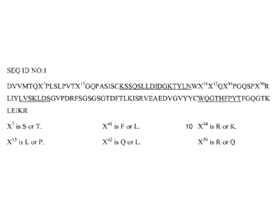
SEQ ID NO:1:
DVVM1TQX7PLSLPVTX15GQPASISCKSSQSLLDIDGKTYLNWX41X42QX44PGQSPX50R
LIYLVSKLDSGVPDRFSGSGSGTDFTLKISRVEAEDVGVYYCWQGTHFPYTFGQGTK
LEIKR,
wherein X7 is S or T, X15 is L or P, X41 is F or L, X42 is Q or L, X44 is R or
K, and X' is R or
Q.
In a further aspect of the invention, the binding protein described above
comprises a first
amino acid sequence which is at least 90%, 91%, 92%, 93%, 94%, 95%, 96%, 97%,
98%, or
99% identical to an amino acid sequence selected from the group consisting of
SEQ ID NO:4,
SEQ ID NO:5, SEQ ID NO:6, SEQ ID NO:7, SEQ ID NO:8, SEQ ID NO:9, SEQ ID NO:10,
and SEQ ID NO:11. In still a further aspect of the invention, the binding
protein described
above comprises a first amino acid sequence selected from the group consisting
of SEQ ID
NO:4, SEQ ID NO:5, SEQ ID NO:6, SEQ ID NO:7, SEQ ID NO:8, SEQ ID NO:9, SEQ ID
NO:10, and SEQ ID NO:11.
In another aspect of the invention, the binding protein described above
comprises a second
amino acid sequence which is at least 90%, 91%, 92%, 93%, 94%, 95%, 96%, 97%,
98%, or
99% identical to an amino acid sequence selected from the group consisting of
SEQ ID
NO:12, SEQ ID NO:13, SEQ ID NO:14, SEQ ID NO:15, and SEQ ID NO:16. In still
another
aspect of the invention, the binding protein described above comprises a
second amino acid
sequence selected from the group consisting of SEQ ID NO:12, SEQ ID NO:13, SEQ
ID
NO:14, SEQ ID NO:15, and SEQ ID NO:16.
CA 02796339 2012-10-12
WO 2011/130377
PCT/US2011/032269
In one aspect of the invention, the binding protein described above comprises
a first amino
acid sequence which is at least 90%, 91%, 92%, 93%, 94%, 95%, 96%, 97%, 98%,
or 99%
identical to an amino acid sequence selected from the group consisting of SEQ
TD NO:4,
5 SEQ ID NO:5, SEQ ID NO:6, SEQ ID NO:7, SEQ ID NO:8, SEQ ID NO:9, SEQ ID
NO:10,
and SEQ ID NO:11; and a second amino acid sequence which is at least 90%, 91%,
92%,
93%, 94%, 95%, 96%, 97%, 98%, or 99% identical to an amino acid sequence
selected from
the group consisting of SEQ ID NO:12, SEQ ID NO:13, SEQ ID NO:14, SEQ ID
NO:15, and
SEQ ID NO:16. In a further aspect of the invention, the binding protein
described above
comprises a first amino acid sequence selected from the group consisting of
SEQ ID NO:4,
SEQ ID NO:5, SEQ ID NO:6, SEQ ID NO:7, SEQ ID NO:8, SEQ ID NO:9, SEQ ID NO:10,
and SEQ ID NO:11; and a second amino acid sequence selected from the group
consisting of
SEQ ID NO:12, SEQ ID NO:13, SEQ ID NO:14, SEQ ID NO:15, and SEQ ID NO:16.
-- In a particular aspect of the invention, the binding protein described
above comprises a first
amino acid sequence which is at least 90%, 91%, 92%, 93%, 94%, 95%, 96%, 97%,
98%, or
99% identical to the amino acid sequence set forth in SEQ ID NO:6; and a
second amino acid
sequence which is at least 90%, 91%, 92%, 93%, 94%, 95%, 96%, 97%, 98%, or 99%
identical to an amino acid sequence set forth in SEQ ID NO:14. In a further
particular aspect
of the invention, the binding protein described above comprises a first amino
acid sequence
set forth in SEQ ID NO:6; and a second amino acid sequence set forth in SEQ ID
NO:14.
In a particular aspect of the invention, the binding protein described above
comprises a first
amino acid sequence which is at least 90%, 91%, 92%, 93%, 94%, 95%, 96%, 97%,
98%, or
99% identical to the amino acid sequence set forth in SEQ ID NO:10; and a
second amino
acid sequence which is at least 90%, 91%, 92%, 93%, 94%, 95%, 96%, 97%, 98%,
or 99%
identical to an amino acid sequence set forth in SEQ ID NO:14. In a further
particular aspect
of the invention, the binding protein described above comprises a first amino
acid sequence
set forth in SEQ ID NO:10; and a second amino acid sequence set forth in SEQ
ID NO:14.
In one aspect, the binding protein described herein is an antibody. This
antibody may be, for
example, an immunoglobulin molecule, a disulfide linked Fv, a monoclonal
antibody (mab),
a single chain Fv (scFv), a chimeric antibody, a single domain antibody, a CDR-
grafted
antibody, a diabody, a humanized antibody, a multispecific antibody, a Fab, a
dual specific
CA 02796339 2012-10-12
WO 2011/130377
PCT/US2011/032269
6
antibody, a dual variable domain (DVD) binding molecule, a Fab', a bispecific
antibody, a
F(ab')2, or a Fv.
When the binding protein described herein is an antibody, it comprises at
least one variable
heavy chain that corresponds to the first amino acid sequence as defined
above, and at least
one variable light chain that corresponds to the second amino acid sequence as
defined above.
For example, an antibody of the invention comprises (i) at least one variable
heavy chain
comprising an amino acid sequence which is at least 90%, 91%, 92%, 93%, 94%,
95%, 96%,
97%, 9.0,/o,
99% or 100% identical to an amino acid sequence selected from the group
consisting of SEQ ID NO:2, SEQ ID NO:3, SEQ ID NO:4, SEQ ID NO:5, SEQ ID NO:6,
SEQ ID NO:7, SEQ ID NO:8, SEQ ID NO:9, SEQ ID NO:10, and SEQ ID NO:11, and
(ii) at
least one variable light chain comprising an amino acid sequence which is at
least 90%, 91%,
92%, 93%, 94%, 95%, 96%, 97%, 98%, --
vv% or 100% identical to an amino acid sequence
selected from the group consisting of SEQ ID NO:1, SEQ ID NO:12, SEQ ID NO:13,
SEQ
ID NO:14, SEQ ID NO:15, and SEQ ID NO:16. In a particular aspect of the
invention, the
antibody of the invention comprises (i) at least one variable heavy chain
comprising an amino
acid sequence which is at least 90%, 91%, 92%, 93%, 94%, 95%, 96%, 97%, 98%,
99% or
100% identical to an amino acid sequence set forth in SEQ ID NO:6 or SEQ ID
NO:10, and
(ii) at least one variable light chain comprising an amino acid sequence which
is at least 90%,
91%, 92%, 93%, 94%, 95%, 96%, 97%, 98%, 99% or 100% identical to an amino acid
sequence set forth in SEQ ID NO:14.
The binding protein described herein may further (in addition to the first and
second amino
acid sequence) comprise another moiety which may be another amino acid
sequence or other
chemical moiety. For instance, an antibody of the present invention may
comprise a heavy
chain immunoglobulin constant domain. Said heavy chain immunoglobulin constant
domain
may be selected from the group consisting of a human IgM constant domain, a
human IgG4
constant domain, a human IgG1 constant domain, a human IgE constant domain, a
human
IgG2 constant domain, a human IgG3 constant domain, and a human IgA constant
domain. In
another aspect, the binding protein of the invention further comprises a heavy
chain constant
region having an amino acid sequence selected from the group consisting of SEQ
ID NO:25
and SEQ ID NO:26, additionally a light chain constant region having an amino
acid sequence
selected from the group consisting of SEQ ID NO:27 and SEQ ID NO:28. In a
particular
aspect of the invention, the binding protein described herein comprises a
variable heavy chain
CA 02796339 2012-10-12
WO 2011/130377
PCT/US2011/032269
7
comprising an amino acid sequence set forth in SEQ ID NO:6 or SEQ ID NO:10; a
variable
light chain comprising an amino acid sequence set forth in SEQ ID NO:14; a
heavy chain
constant region having an amino acid sequence set forth in SEQ ID NO:25; and a
light chain
constant region having an amino acid sequence set forth in SEQ TD NO:27. In a
further
particular aspect of the invention, the binding protein described herein
comprises a first
amino acid sequence set forth in SEQ ID NO:46 or SEQ ID NO:47, and a second
first amino
acid sequence set forth in SEQ ID NO:48.
The binding protein, e.g. the antibody, described herein may further comprise
a therapeutic
agent, an imaging agent, residues capable of facilitating formation of an
immunoadhesion
molecule and/or another functional molecule (e.g. another peptide or protein).
The imaging
s 9, ,
agent can be a radiolabel including but not limited to 3H, 14C, 35, 90y, 9Tc,
"In, 1251 1311
177Lu, 166Ho, and 153; an enzyme, a fluorescent label, a luminescent label, a
bioluminescent
label, a magnetic label, or biotin.
The binding protein of the present invention can be glycosylated. According to
one aspect of
the invention, the glycosylation pattern is a human glycosylation pattern.
In one aspect of the invention, the above-described binding protein binds to
an Ar3 form that
comprises the globulomer epitope with which the murine monoclonal antibody
m4D10 is
reactive (i.e. a targeted A13 form). In particular the above-described binding
proteins bind to
amyloid-beta (20-42) globulomer as described herein.
In one aspect of the invention, the binding protein described herein is
capable of modulating a
biological function of A13(20-42) globulomer. In a further aspect of the
invention, the binding
protein described herein is capable of neutralizing A13(20-42) globulomer
activity.
The binding protein of the present invention may exist as a crystal. In one
aspect, the crystal
is a carrier-free pharmaceutical controlled release crystal. In another
aspect, the crystallized
binding protein has a greater half life in vivo than its soluble counterpart.
In still another
aspect, the crystallized binding protein retains biological activity after
crystallization.
The present invention also provides an isolated nucleic acid encoding any one
of the binding
proteins disclosed herein. A further embodiment provides a vector comprising
said nucleic
CA 02796339 2012-10-12
WO 2011/130377
PCT/US2011/032269
8
acid. Said vector may be selected from the group consisting of pcDNA, pTT
(Durocher et al.,
Nucleic Acids Research 30(2), 2002), pTT3 (pTT with additional multiple
cloning site),
pEFBOS (Mizushima and Nagata, Nucleic acids Research 18(17), 1990), pBV, pJV,
and pBJ.
In another aspect of the invention, a host cell is transformed with the vector
disclosed above.
According to one embodiment, the host cell is a prokaryotic cell including but
not limited to
E.coli. In a related embodiment, the host cell is a eukaryotic cell selected
from the group
comprising a protist cell, animal cell, plant cell and fungal cell. The animal
cell may be
selected from the group consisting of a mammalian cell, an avian cell and an
insect cell.
According to one aspect of the invention, said mammalian cell is selected from
the group
comprising CHO and COS, said fungal cell is a yeast cell such as Saccharomyces
cerevisiae,
and said insect cell is an insect Sf9 cell.
Further, the invention provides a method of producing a binding protein as
disclosed herein
that comprises culturing any one of the host cells disclosed herein in a
culture medium under
conditions and for a time suitable to produce said binding protein. Another
embodiment
provides a binding protein of the invention produced according to the method
disclosed
herein. In another embodiment, the invention provides a binding protein
produced according
to the method disclosed above.
The invention also provides a pharmaceutical composition comprising a binding
protein, e.g.
an antibody, as disclosed hererin and a pharmaceutically acceptable carrier.
One embodiment of the invention provides a composition for the release of the
binding
protein described herein wherein the composition comprises a formulation which
in turn
comprises a crystallized binding protein, e.g. a crystallized antibody, as
disclosed above, and
an ingredient; and at least one polymeric carrier. In one aspect the polymeric
carrier is a
polymer selected from one or more of the group consisting of: poly(acrylic
acid), poly(cyano-
acrylates), poly(amino acids), poly(anhydrides), poly(depsipeptides),
poly(esters), poly(lactic
acid), poly(lactic-co-glycolic acid) or PLGA, poly([3-hydroxybutryate),
poly(caprolactone),
poly(dioxanone); poly(ethylene glycol), poly((hydroxypropyl) methacrylamide),
poly((organo)phosphazene), poly(ortho esters), poly(vinyl alcohol),
poly(vinylpyrrolidone),
maleic anhydride - alkyl vinyl ether copolymers, pluronic polyols, albumin,
alginate,
cellulose and cellulose derivatives, collagen, fibrin, gelatin, hyaluronic
acid,
CA 02796339 2012-10-12
WO 2011/130377
PCT/US2011/032269
9
oligosaccharides, glycaminoglycans, sulfated polysaccharides, blends and
copolymers
thereof. In another aspect the ingredient is selected from the group
consisting of: albumin,
sucrose, trehalose, lactitol, gelatin, hydroxypropyl-P-cyclodextrin,
methoxypolyethylene
glycol and polyethylene glycol.
The present invention also relates to a method of inhibiting (i.e. reducing)
the activity of
AI3(20-42) globulomer (or any other targeted AP form) comprising contacting
said targeted
AI3 form with binding protein(s) of the invention such that the activity of
said targeted AP
form is inhibited (i.e. reduced). In a particular embodiment, said activity is
inhibited in vitro.
This method may comprise adding the binding protein of the invention to a
sample, e.g. a
sample derived from a subject (e.g., whole blood, cerebrospinal fluid, serum,
tissue, etc.) or a
cell culture which contains or is suspected to contain a targeted AP form, in
order to inhibit
(i.e. reduce) the activity of the AI3 form in the sample. Alternatively, the
activity of said
targeted AP form may be inhibited (i.e. reducd) in a subject in vivo. Thus,
the present
invention further relates to the binding protein described herein for use in
inhibiting (i.e.
reducing) the activity of a targeted AP form in a subject comprising
contacting said AP form
with binding protein(s) of the invention such that the activity of the AP form
is inhibited (i.e.
reduced).
In a related aspect, the invention provides a method for inhibiting (i.e.
reducing) the activity
of a targeted AP form in a subject suffering from a disease or disorder in
which the activity of
said AP form is detrimental. In one embodiment, said method comprises
administering to the
subject at least one of the binding proteins disclosed herein such that the
activity of a targeted
AB form in the subject is inhibited (i.e. reduced). Thus, the invention
provides the AP binding
proteins described herein for use in inhibiting (i.e. reducing) a targeted AB
form in a subject
suffering from a disease or disorder as described herein, wherein at least one
of the binding
proteins disclosed herein is administered to the subject such that the
activity of said AB form
in the subject is inhibited (i.e. reduced).
In a related aspect, the invention provides a method for treating (e.g.,
curing, suppressing,
ameliorating, delaying or preventing the onset of, or preventing recurrence or
relapse of) or
preventing a disease or disorder selected from the group consisting of Alphal-
antitrypsin-
deficiency, Cl -inhibitor deficiency angioedema, Antithrombin deficiency
thromboembolic
disease, Kum, Creutzfeld-Jacob diseaseiscrapie, Bovine spongiform
encephalopathy,
CA 02796339 2012-10-12
WO 2011/130377
PCT/US2011/032269
Gerstmann-Straussler-Scheinker disease, Fatal familial insomnia, Huntington's
disease,
Spinocerebellar ataxia, Machado-Joseph atrophy, Dentato-rubro-pallidoluysian
atrophy,
Frontotemporal dementia, Sickle cell anemia, Unstable hemoglobin inclusion-
body
hemolysis, Drug-induced inclusion body hemolysis, Parkinson's disease,
Systemic AL
5 -- amyloidosis, Nodular AL amyloidosis, Systemic AA amyloidosis, Prostatic
amyloidosis,
Hemodialysis amyloidosis, Hereditary (Icelandic) cerebral angiopathy,
Huntington's disease,
Familial visceral amyloidosis, Familial visceral polyneuropathy, Familial
visceral
amyloidosis, Senile systemic amyloidosis, Familial amyloid neurophathy,
Familial cardiac
amyloidosis, Alzheimer's disease, Down syndrome, Medullary carcinoma thyroid
and Type 2
10 -- diabetes mellitus (T2DM). In a particular embodiment, said disease or
disorder is an
amyloidosis such as Alzheimer's disease or Down syndrome. In one embodiment,
said
method comprising the step of administering any one of the AB binding proteins
disclosed
herein such that treatment is achieved. In another embodiment, the invention
provides a
method of treating a subject suffering from a disease or disorder disclosed
herein comprising
-- the step of administering any one of the AB binding proteins disclosed
herein, concurrent
with or after the administration of one or more additional therapeutic
agent(s). Thus, the
invention provides the AB binding proteins disclosed herein for use in
treating a subject
suffering from a disesase or disorder disclosed herein comprising the step of
administering
any one of the binding proteins disclosed herein, concurrent with or after the
administration
-- of one or more additional therapeutic agent(s). For instance, the
additional therapeutic agent
is selected from the group of therapeutic agents listed herein.
The binding proteins disclosed herein and the pharmaceutical compositions
comprising said
binding proteins are administered to a subject by at least one mode selected
from parenteral,
-- subcutaneous, intramuscular, intravenous, intraarticular, intrabronchial,
intraabdominal,
intracapsular, intracartilaginous, intracavitary, intracelial,
intracerebellar,
intracerebroventricular, intracolic, intracervical, intragastric,
intrahepatic, intramyocardial,
intraosteal, intrapelvic, intrapericardiac, intraperitoneal, intrapleural,
intraprostatic,
intrapulmonary, intrarectal, intrarenal, intraretinal, intraspinal,
intrasynovial, intrathoracic,
-- intrauterine, intravesical, bolus, vaginal, rectal, buccal, sublingual,
intranasal, and
transdermal.
In another embodiment, the present invention provides a method for detecting a
targeted AB
form in a sample comprising (i) contacting said sample with binding protein(s)
of the
11
invention and (ii) detecting formation of a complex between said binding
protein(s) and
elements of said sample, wherein formation or increased formation of the
complex in the
sample relative to a control sample indicates the presence of said Al) form in
the sample. The
sample may be a biological sample obtained from a subject which is stmected of
having a
disease or disorder as disclosed herein (e.g., whole blood, cerebrospinal
fluid, serum, tissue,
etc.) or a cell culture which contains or is suspected to contain said Aft
form. The control
sample does not contain said AB form or is obtained .from a patient not having
a disease as
described above, The presence of a complex between said binding protein(s) and
elements of
a sample obtained from a patient suspected of having Alzheimer's disease
indicates a
diagnosis of this disease in said patient
In an alternative embodiment, the detection of the tar:lewd AB form may be
performed in
i'ivo, e.g by in 'iv imaging in a subject. For this purpose, the binding
protein's) of the
i8VentiOR may be administered to a subject or a control subject under
conditions that allow
binding of said protein(s) to the targeted AB form and detecting limitation of
a complex
between said binding proteirits) and said AB form, wherein 'Urination or
increased formation
of the complex in the subject relative to the control subject indicates the
presence of said AB
form in the subject. The subject may be a subject. which is known or suspected
to suffer from
a disorder or disease in which activity of a targeted Al) form is detrimental.
In certain embodiments, there is provided:
<1> An antibody comprising:
a first amino acid sequence which is at least 90% identical to SEQ ID NO:2 or
SEQ ID
NO:3; and a second amino acid sequence which is at least 90% identical to SEQ
ID
NO:1,
wherein the first amino acid sequence comprises three complementarity
determining
regions consisting of amino acids 31-35, 50-65 and 98-101, respectively, of
SEQ ID
NO:2 or SEQ ID NO:3, and
wherein the second amino acid sequence comprises three complementarity
determining
regions consisting of amino acids 24-39, 55-61 and 94-102, respectively, of
SEQ ID
NO:l.
<2> The antibody according to <1>, wherein the first amino acid sequence is at
least 90%
identical to an amino acid sequence selected from the group consisting of SEQ
ID NO:4,
CA 2796339 2018-07-26
11a
SEQ ID NO:5, SEQ ID NO:6, SEQ ID NO:7, SEQ ID NO:8, SEQ ID NO:9, SEQ ID
NO:10, and SEQ ID NO:11.
<3> The antibody according to <1>, wherein the second amino acid sequence is
at least 90%
identical to an amino acid sequence selected from the group consisting of SEQ
ID
NO:12, SEQ ID NO:13, SEQ ID NO:14, SEQ ID NO:15, and SEQ ID NO:16.
<4> The antibody according to <1>, <2> or <3> comprising:
a first amino acid sequence which is at least 90% identical to an amino acid
sequence
selected from the group consisting of SEQ ID NO:4, SEQ ID NO:5, SEQ ID NO:6,
SEQ
ID NO:7, SEQ ID NO:8, SEQ ID NO:9, SEQ ID NO:10, and SEQ ID NO:11; and
a second amino acid sequence which is at least 90% identical to an amino acid
sequence
selected from the group consisting of SEQ ID NO:12, SEQ ID NO:13, SEQ ID
NO:14,
SEQ ID NO:15, and SEQ ID NO:16.
<5> The antibody according to <4> comprising:
a first amino acid sequence set forth in SEQ ID NO:6; and
a second amino acid sequence set forth in SEQ ID NO:14.
<6> The antibody according to <4> comprising:
a first amino acid sequence set forth in SEQ ID NO:10; and
a second amino acid sequence set forth in SEQ ID NO:14.
<7> The antibody according to <I>, <2>, <3>, <4>, <5> or <6>, wherein said
antibody is
selected from the group consisting of: a monoclonal antibody, a chimeric
antibody, a
multispecific antibody, a dual variable domain (DVD) binding protein, and a
bispecific
antibody.
<8> The antibody according to <1>, <2>, <3>, <4>, <5>, <6> or <7> further
comprising an
immunoglobulin light chain constant region having an amino acid sequence
selected
from the group consisting of SEQ ID NO:27 and SEQ ID NO:28.
<9> The antibody according to <8> comprising:
a first amino acid sequence set forth in SEQ ID NO:46; and
a second amino acid sequence set forth in SEQ ID NO:48.
<10> The antibody according to <8> comprising:
a first amino acid sequence set forth in SEQ ID NO:47; and
a second amino acid sequence set forth in SEQ ID NO:48.
CA 2796339 2018-07-26
1 1 b
<11> The antibody according to <1>, <2>, <3>, <4>, <5>, <7>, <8>, <9> or <10>,
wherein
said antibody further comprises an agent selected from the group consisting
of: an
immunoadhesion molecule; an imaging agent, and a therapeutic agent.
<12> The antibody according to a <1>, <2>, <3>, <4>, <5>, <6>, <7>, <8>, <9>,
<10> or
<11>, wherein said antibody possesses a human glycosylation pattern.
<13> An isolated nucleic acid encoding the antibody of <1>, <2>, <3>, <4>,
<5>, <6>, <7>,
<8>, <9>, <10>, <11> or <12>.
<14> A vector comprising the isolated nucleic acid of <13>.
<15> A host cell comprising a vector of <14>.
<16> A method of producing an antibody comprising culturing a host cell of
<15> in culture
medium under conditions sufficient to produce the antibody.
<17> An antibody produced according to the method of <16>.
<18> A pharmaceutical composition comprising the antibody of <1>, <2>, <3>,
<4>, <5>,
<6>, <7>, <8>, <9>, <10>, <11>, <12> or <17>, and a pharmaceutically
acceptable
carrier.
<19> The pharmaceutical composition of <18> further comprising at least one
additional
therapeutic agent.
<20> A composition for the release of an antibody said composition comprising:
(a) a formulation, wherein said formulation comprises the antibody of <1>,
<2>, <3>,
<4>, <5>, <6>, <7>, <8>, <9>, <10>, <11>, <12> or <17> and an ingredient,
wherein
the antibody is crystallized, and wherein the ingredient is selected from
albumin, sucrose,
trehalose, lactitol, gelatin, hydroxypropy1-13-cyclodextrin,
methoxypolyethylene glycol
and polyethylene glycol; and
(b) at least one polymeric carrier.
<21> The antibody of <1>, <2>, <3>, <4>, <5>, <6>, <7>, <8>, <9>, <10>, <11>,
<12> or
17 for use in treating a subject for a disease or a disorder selected from the
group
consisting of Systemic AL amyloidosis, Nodular AL amyloidosis, Systemic AA
amyloidosis, Prostatic amyloidosis, Hemodialysis amyloidosis, Familial
visceral
amyloidosis, Senile systemic amyloidosis, Familial cardiac amyloidosis,
Alzheimer's
disease, and Down syndrome.
CA 2796339 2018-07-26
11c
<22> The antibody of <21>, wherein the disease or disorder is Alzheimer's
disease.
<23> Use of the antibody of <1>, <2>, <3>, <4>, <5>, <6>, <7>, <8>, <9>, <10>,
<11>,
<12> or <17> in the manufacture of a medicament to treat a disease or a
disorder
selected from the group consisting of Systemic AL amyloidosis, Nodular AL
amyloidosis, Systemic AA amyloidosis, Prostatic amyloidosis, Hemodialysis
amyloidosis, Familial visceral amyloidosis, Senile systemic amyloidosis,
Familial
cardiac amyloidosis, Alzheimer's disease, and Down syndrome.
<24> Use of the antibody of <1>, <2>, <3>, <4>, <5>, <6>, <7>, <8>, <9>, <10>,
<11>,
<12> or <17> for the treatment of a disease or a disorder selected from the
group
consisting of Systemic AL amyloidosis, Nodular AL amyloidosis, Systemic AA
amyloidosis, Prostatic amyloidosis, Hemodialysis amyloidosis, Familial
visceral
amyloidosis, Senile systemic amyloidosis, Familial cardiac amyloidosis,
Alzheimer's
disease, and Down syndrome.
<25> The use of <23> or <24>, wheren the disease or disorder is Alzheimer's
disease.
Brief Description of the Figures
Figure 1 illustrates ainino acid sequences (SEQ ID NO:]) of the variable light
chain of
humanized 4D10 antibodies comprising JK2 and Vrz A17/2-30 framework regions.
Ali CDR =
regions arc underlined.
Figure 2 Mummies amino acid sequences (SEQ ID NO:2) of die variable heavy
chain of
humanized zIDI 0 antibodies comprising human itift (WE I()) and V113 53
framework regions.
All CDR regionS are underlined.
Figure 3 illustrates amino acid sequences (SEC) ID NO:3) of the variable heavy
chain of
humanized 41310 antibodies comprising human .11-1.6 and VH4_59 framework
regions. All
CDR regions arc underlined.
CA 2796339 2018-07-26
CA 02796339 2012-10-12
WO 2011/130377
PCT/US2011/032269
12
Figure 4 illustrates the amino acid sequence (SEQ ID NO:4) of the variable
heavy chain of
humanized 4D10 antibodies comprising human JH6 (hJH6) and VH3 53 framework
regions.
All CDR regions are underlined.
Figure 5 illustrates the amino acid sequence (SEQ ID NO:5) of the variable
heavy chain of
humanized 4D10 antibodies comprising human JH6 and VH3_53 framework regions
with
VH3 consensus change 112V. All CDR regions are underlined.
Figure 6 illustrates the amino acid sequence (SEQ ID NO:6) of the variable
heavy chain of
humanized 4D10 antibodies comprising human JH6 and VH3_53 framework regions
with
VH3 consensus change 112V and framework backmutations A24V, V29L, V48L, S49G,
F67L, R71K, N76S and L78V. All CDR regions are underlined.
Figure 7 illustrates the amino acid sequence (SEQ ID NO:7) of the variable
heavy chain of
humanized 4D10 antibodies comprising human JH6 and VH3 53 framework regions
with
framework backmutations V29L and R71K. All CDR regions are underlined.
Figure 8 illustrates the amino acid sequence (SEQ ID NO:8) of the variable
heavy chain of
humanized 4D10 antibodies comprising human JH6 and VH4_59 framework regions.
All
CDR regions are underlined.
Figure 9 illustrates the amino acid sequence (SEQ ID NO:9) of the variable
heavy chain of
humanized 4D10 antibodies comprising human JH6 and VH4_59 framework regions
with a
Q1E change to prevent N-terminal pyroglutamate formation. All CDR regions are
underlined.
Figure 10 illustrates the amino acid sequence (SEQ ID NO:10) of the variable
heavy chain of
humanized 4D10 antibodies comprising human JH6 and VH4_59 framework regions
with a
Q1E change to prevent N-terminal pyroglutamate formation, and framework
backmutations
G27F, 129L, I37V, 148L, V67L, V71K, N76S and F78V. All CDR regions are
underlined.
Figure 11 illustrates the amino acid sequence (SEQ ID NO:11) of the variable
heavy chain of
humanized 4D10 antibodies comprising human JH6 and VH4_59 framework regions
with a
Q1E change to prevent N-terminal pyroglutamate formation, and framework
backmutations
G27F, T29L and V71K. All CDR regions are underlined.
CA 02796339 2012-10-12
WO 2011/130377
PCT/US2011/032269
13
Figure 12 illustrate the amino acid sequence (SEQ ID NO:12) of the variable
light chain of
humanized 4D10 antibodies comprising Jic2 and Vic A17/2-30 framework regions.
All CDR
regions are underlined.
Figure 13 illustrates the amino acid sequence (SEQ ID NO:13) of the variable
light chain of
humanized 4D10 antibodies comprising Jx2 and Vic A17/2-30 framework regions
with Vx2
consensus changes S7T, LISP, Q37L, R39K and R45Q. All CDR regions are
underlined.
Figure 14 illustrates the amino acid sequence (SEQ ID NO:14) of the variable
light chain of
humanized 4D10 antibodies comprising Jx2 and Vic A17/2-30 framework regions
with Vx2
consensus changes 57T, LISP, Q37L, R39K and R45Q, and framework backmutation
F36L.
All CDR regions are underlined.
Figure 15 illustrates the amino acid sequence (SEQ ID NO:15) of the variable
light chain of
humanized 4D10 antibodies comprising Jx2 and Vic A17/2-30 framework regions
with Via
consensus changes S7T and Q37L. All CDR regions are underlined.
Figure 16 illustrates the amino acid sequence (SEQ ID NO:16) of the variable
light chain of
humanized 4D10 antibodies comprising Jx2 and Vic A17/2-30 framework regions
with Vx2
consensus changes S7T, Q37L and R39K. All CDR regions are underlined.
Figure 17 illustrates an amino acid sequence alignment of the variable heavy
chains of
murine monoclonal antibody 4D10 (m4D19) and humanized 4D10 antibodies
(4D10hum)
comprising human JH6 (hJH6) and VH3_53 framework regions. All CDR regions are
printed
in bold letters. X on position 12 is I or V, X on position 24 is A or V, X on
position 29 is V or
L, X on position 48 is V or L, X on position 49 is S or G, X on position 67 is
F or L, X on
position 71 is R or K, X on position 76 is N or S, and X on position 78 is L
or V.
Figure 18 illustrates an amino acid sequence alignment of the variable heavy
chains of
murine monoclonal antibody 4D10 (m4D19) and humanized 4D10 antibodies
(4D10hum)
comprising human JH6 and VH4_59 framework regions. All CDR regions are printed
in bold
letters. X on position 1 is Q or E, X on position 27 is G or F, X on position
29 is I or L, X on
CA 02796339 2012-10-12
WO 2011/130377
PCT/US2011/032269
14
position 37 is 1 or V, X on position 48 is 1 or L, X on position 67 is V or L,
X on position 71
is V or K, X on position 76 is N or S, and X on position 78 is F or V.
Figure 19 illustrates an amino acid sequence alignment of the variable light
chains of murine
monoclonal antibody 4D10 (m4D19) and humanized 4D10 antibodies (4D10hum)
comprising Jx2 and Vic A17/2-30 framework regions. All CDR regions are printed
in bold
letters. X on position 7 is S or T, X on position 15 is L or P, X on position
41 is F or L, X on
position 42 is Q or L, X on position 44 is R or K, and X on position 50 is R
or Q.
Figures 20A and B show platelet factor 4 (PF-4) cross-reaction of humanized
monoclonal
antibodies 4D10hum#1 and 4D10hum#2, human/mouse chimeric antibody hi G5
(positive
control) and human polyclonal antibody hIgG1 (negative control) in (A)
Cynomolgus
monkey plasma and (B) human plasma, as determined by sandwich-ELISA. Binding
of PF-4
to the immobilized antibodies was detected.
Figures 21A and B show platelet factor 4 (PF-4) cross-reaction of humanized
monoclonal
antibodies 4D10hum#1 and 4D10hum#2, human/mouse chimeric antibody hi G5
(positive
control) and human polyclonal antibody hIgG1 (negative control) in (A)
Cynomolgus
monkey plasma and (B) human plasma, as determined by aligned sandwich-ELISA.
The
antibodies were captured on the plate by immobilized anti-mouse IgG. Binding
of PF-4 to the
captured antibodies was detected.
Figures 22A and B show platelet factor 4 (PF-4) cross-reaction of murine
monoclonal
antibodies m4D10 and m1G5, anti human PF-4 antibody (positive control) and
IgG2a
(negative control) in (A) Cynomolgus monkey plasma and (B) human plasma, as
determined
by sandwich-ELISA. Binding of PF-4 to the immobilized antibodies was detected.
Figures 23A and B show platelet factor 4 (PF-4) cross-reaction of murine
monoclonal
antibodies m4D10 and m1G5, anti human PF-4 antibody (positive control) and
IgG2a
(negative control) in (A) Cynomolgus monkey plasma and (B) human plasma, as
determined
by aligned sandwich-EL1SA. The antibodies were captured on the plate by
immobilized anti-
mouse IgG. Binding of PF-4 to the captured antibodies was detected.
CA 02796339 2012-10-12
WO 2011/130377
PCT/US2011/032269
Figure 24 illustrates the amino acid sequence (SEQ ID NO:46) of the heavy
chain of a
humanized 4D10 antibody comprising human JH6 and VH3 53 framework regions with
VH3 consensus change 112V and framework backmutations A24V, V29L, V48L, S49G,
F67L, R71K, N76S and L78V; and an Ig gamma-1 constant region. All CDR regions
are
5 underlined.
Figure 25 illustrates the amino acid sequence (SEQ ID NO:47) of the heavy
chain of a
humanized 4D10 antibody comprising human JH6 and VH4_59 framework regions with
a
Q1E change to prevent N-terminal pyroglutamate formation, and framework
backmutations
10 G27F, I29L, I37V, I48L, V67L, V71K, N76S and F78V; and an Ig gamma-1
constant region.
All CDR regions are underlined.
Figure 26 illustrates the amino acid sequence (SEQ ID NO:48) of the light
chain of a
humanized 4D10 antibody comprising Jx2 and Vic A17/2-30 framework regions with
Vic2
15 consensus changes S7T, LISP, Q37L, R39K and R45Q, and framework
backmutation F36L;
and an Ig kappa constant region. All CDR regions are underlined.
Detailed Description of the Invention
Unless otherwise defined herein, scientific and technical terms used in
connection with the
present invention shall have the meanings that are commonly understood by
those of ordinary
skill in the art. The meaning and scope of the terms should be clear, however,
in the event of
any latent ambiguity, definitions provided herein take precedent over any
dictionary or
extrinsic definition. Further, unless otherwise required by context, singular
terms shall
include pluralities and plural terms shall include the singular. In this
application, the use of
"or" means "and/or" unless stated otherwise. Furthermore, the use of the term
"including", as
well as other forms, such as "includes" and "included", is not limiting. Also,
terms such as
"element" or "component" encompass both elements and components comprising one
unit
and elements and components that comprise more than one subunit unless
specifically stated
otherwise.
Generally, nomenclatures used in connection with, and techniques of, cell and
tissue culture,
molecular biology, immunology, microbiology, genetics, protein and nucleic
acid chemistry,
and hybridization described herein are those well known and commonly used in
the art. The
CA 02796339 2012-10-12
WO 2011/130377
PCT/US2011/032269
16
methods and techniques of the present invention are generally performed
according to
conventional methods well known in the art and as described in various general
and more
specific references that are cited and discussed throughout the present
specification unless
otherwise indicated. Enzymatic reactions and purification techniques are
performed
according to manufacturer's specifications, as commonly accomplished in the
art or as
described herein. The nomenclatures used in connection with, and the
laboratory procedures
and techniques of, analytical chemistry, synthetic organic chemistry, and
medicinal and
pharmaceutical chemistry described herein are those well known and commonly
used in the
art. Standard techniques are used for chemical syntheses, chemical analyses,
pharmaceutical
preparation, formulation, and delivery, and treatment of patients.
The present invention pertains to AP binding proteins, particularly anti-AP
antibodies or an
AI3 binding portion thereof, particularly those binding to AI3(20-42)
globulomer. These A13
binding proteins are capable of discriminating not only other forms of AP
peptides,
particularly monomers and fibrils, but also untruncated forms of Al3
globulomers. Thus, the
present invention relates to an AP binding protein having a binding affinity
to an A13(20-42)
globulomer that is greater than the binding affinity of this AP binding
protein to an A13(1-42)
globulomer.
The term "AP(X-Y)" as used herein refers to the amino acid sequence from amino
acid
position X to amino acid position Y of the human amyloid beta (A13) protein
including both X
and Y, in particular to the amino acid sequence from amino acid position X to
amino acid
position Y of the amino acid sequence DAEFRHDSGY EVHHQKLVFF AEDVGSNKGA
IIGLMVGGVV IAT (SEQ ID NO:29) (corresponding to amino acid positions 1 to 43)
or any
of its naturally occurring variants, in particular those with at least one
mutation selected from
the group consisting of A2T, H6R, D7N, A21G ("Flemish"), E22G ("Arctic"), E22Q
("Dutch"), E22K ("Italian"), D23N ("Iowa"), A42T and A42V wherein the numbers
are
relative to the start of the AP peptide, including both position X and
position Y or a sequence
with up to three additional amino acid substitutions none of which may prevent
globulomer
formation. According to one aspect, there are no additional amino acid
substitutions in the
portion from amino acid 12 or X, whichever number is higher, to amino acid 42
or Y,
whichever number is lower. According to another aspect, there are no
additional amino acid
substitutions in the portion from amino acid 20 or X, whichever number is
higher, to amino
acid 42 or Y, whichever number is lower. According to another aspect, there
are no additional
CA 02796339 2012-10-12
WO 2011/130377
PCT/US2011/032269
17
amino acid substitutions in the portion from amino acid 20 or X, whichever
number is higher,
to amino acid 40 or Y, whichever number is lower. An "additional" amino acid
substitution
herein is any deviation from the canonical sequence that is not found in
nature.
More specifically, the term "A13(1-42)" as used herein refers to the amino
acid sequence from
amino acid position 1 to amino acid position 42 of the human AP protein
including both 1
and 42, in particular to the amino acid sequence DAEFRHDSGY EVHHQKLVFF
AEDVGSNKGA IIGLMVGGVV IA (SEQ ID NO:30) or any of its naturally occurring
variants, in particular those with at least one mutation selected from the
group consisting of
A2T, H6R, D7N, A21G ("Flemish"), E22G ("Arctic"), E22Q ("Dutch"), E22K
("Italian"),
D23N ("Iowa"), A42T and A42V wherein the numbers are relative to the start of
the AP
peptide, including both 1 and 42 or a sequence with up to three additional
amino acid
substitutions none of which may prevent globulomer formation. According to one
aspect,
there are no additional amino acid substitutions in the portion from amino
acid 20 to amino
acid 42. Likewise, the term "A3(1-40)" as used herein refers to the amino acid
sequence from
amino acid position 1 to amino acid position 40 of the human AP protein
including both 1
and 40, in particular to the amino acid sequence DAEFRHDSGY EVHHQKLVFF
AEDVGSNKGA IIGLMVGGVV (SEQ ID NO:31) or any of its naturally occurring
variants,
in particular those with at least one mutation selected from the group
consisting of A2T,
H6R, D7N, A21G ("Flemish"), E22G ("Arctic"), E22Q ("Dutch"), E22K ("Italian"),
and
D23N ("Iowa") wherein the numbers are relative to the start of the AP peptide,
including both
1 and 40 or a sequence with up to three additional amino acid substitutions
none of which
may prevent globulomer formation. According to one aspect, there are no
additional amino
acid substitutions in the portion from amino acid 20 to amino acid 40.
More specifically, the term "AP(12-42)" as used herein refers to the amino
acid sequence
from amino acid position 12 to amino acid position 42 of the human AP protein
including
both 12 and 42, in particular to the amino acid sequence VHHQKLVFF AEDVGSNKGA
IIGLMVGGVV IA (SEQ ID NO:32) or any of its naturally occurring variants, in
particular
those with at least one mutation selected from the group consisting of A21G
("Flemish"),
E22G ("Arctic"), E22Q ("Dutch"), E22K ("Italian"), D23N ("Iowa"), A42T and
A42V
wherein the numbers are relative to the start of the AP peptide, including
both 12 and 42 or a
sequence with up to three additional amino acid substitutions none of which
may prevent
globulomer formation. According to one aspect, there are no additional amino
acid
WO 2011/130377
PCT/US2011/032269
18
substitutions in the portion from amino acid 20 to amino acid 42. Likewise,
the term "A13(20-
42)" as used herein refers to the amino acid sequence from amino acid position
20 to amino
acid position 42 of the human amyloid 13 protein including both 20 and 42, in
particular to the
amino acid sequence F AEDVGSNKGA IIGLMVGGVV TA (SEQ ID NO:33) or any of its
naturally occurring variants, in particular those with at least one mutation
selected from the
group consisting of A21G ("Flemish"), E22G ("Arctic"), E22Q ("Dutch"), E22K
("Italian"),
D23N ("Iowa"), A42T and A42V wherein the numbers are relative to the start of
the All
peptide, including both 20 and 42 or a sequence with up to three additional
amino acid
substitutions none of which may prevent globulomer formation. According to one
aspect,
there are any additional amino acid substitutions.
The term "AP(X-Y) globulomer" (AP(X-Y) globular oligomer) as used herein
refers to a
soluble, globular, non-covalent association of AP(X-Y) peptides as defined
above, possessing
homogeneity and distinct physical characteristics. According to one aspect,
AP(X-Y)
globulomers are stable, non-fibrillar, oligomeric assemblies of Ap(X-Y)
peptides which are
obtainable by incubation with anionic detergents. In contrast to monomer and
fibrils, these
globulomers are characterized by defined assembly numbers of subunits (e.g.
early assembly
forms with 4-6 subunits, "oligomers A"; and late assembly forms with 12-14
subunits,
"oligomers B"; as described in W02004/067561). The globulomers have a 3-
dimensional
globular type structure ("molten globule", see Barghom etal., J Neurochem 95:
834-847,
2005). They may be further characterized by one or more of the following
features:
- cleavability of N-terminal amino acids X-23 with promiscuous proteases (such
as
thermolysin or endoproteinase GluC) yielding truncated forms of globulomers;
- non-accessibility of C-terminal amino acids 24-Y with promiscuous proteases
and
antibodies;
- truncated forms of these globulomers maintain the 3-dimensional core
structure of said
globulomers with a better accessibility of the core epitope AP(20-Y) in its
globulomer
conformation.
According to the invention and in particular for the purpose of assessing the
binding affinities
of the All binding proteins of the present invention, the term "AP(X-Y)
globulomer" here
refers in particular to a product which is obtainable by a process as
described in
W02004/067561. Said process comprises
unfolding a natural, recombinant or synthetic Ap(X-Y) peptide or a derivative
thereof;
CA 2796339 2017-07-24
CA 02796339 2012-10-12
WO 2011/130377
PCT/US2011/032269
19
exposing the at least partially unfolded A13(X-Y) peptide or derivative
thereof to a detergent,
reducing the detergent action and continuing incubation.
For the purpose of unfolding the peptide, hydrogen bond-breaking agents such
as, for
example, hexafluoroisopropanol (HRIP) may be allowed to act on the protein.
Times of
action of a few minutes, for example about 10 to 60 minutes, are sufficient
when the
temperature of action is from about 20 to 50 C and in particular about 35 to
40 C.
Subsequent dissolution of the residue evaporated to dryness, e.g. in
concentrated form, in
suitable organic solvents miscible with aqueous buffers, such as, for example,
dimethyl
sulfoxide (DMSO), results in a suspension of the at least partially unfolded
peptide or
derivative thereof, which can be used subsequently. If required, the stock
suspension may be
stored at low temperature, for example at about 20 C, for an interim period.
Alternatively, the
peptide or the derivative thereof may be taken up in slightly acidic, e.g.
aqueous, solution, for
example, an about 10 mM aqueous HC1 solution. After an incubation time of
usually a few
minutes, insoluble components are removed by centrifugation. A few minutes at
10,000 g is
expedient. These method steps can be carried out at room temperature, i.e. a
temperature in
the range from 20 to 30 C. The supernatant obtained after centrifugation
contains the A13(X-
Y) peptide or the derivative thereof and may be stored at low temperature, for
example at
about -20 C, for an interim period. The following exposure to a detergent
relates to the
oligomerization of the peptide or the derivative thereof to give an
intermediate type of
oligomers (in WO 2004/067561 referred to as oligomers A). For this purpose, a
detergent is
allowed to act on the at least partially unfolded peptide or derivative
thereof until sufficient
intermediate oligomer has been produced. Preference is given to using ionic
detergents, in
particular anionic detergents.
According to a particular embodiment, a detergent of the formula (I):
R-X,
is used, in which the radical R is unbranched or branched alkyl having from 6
to 20, e.g. 10 to
14, carbon atoms or unbranched or branched alkenyl having from 6 to 20, e.g.
10 to 14,
carbon atoms, the radical X is an acidic group or salt thereof, with X being
selected, e.g.,
from among ¨COO-M', -S0)-M'-, and especially -0S03-M' and M' is a hydrogen
cation or
an inorganic or organic cation selected from, e.g., alkali metal and alkaline
earth metal
cations and ammonium cations. Advantageous are detergents of the formula (I),
in which R is
unbranched alkyl of which alk-1-y1 radicals must be mentioned in particular.
For example,
CA 02796339 2012-10-12
WO 2011/130377
PCT/US2011/032269
sodium dodecyl sulfate (SDS), lauric acid, the sodium salt of the detergent
lauroylsarcosin
(also known as sarkosyl NL-30 or Gardo10) and oleic acid can be used
advantageously. The
time of detergent action in particular depends on whether (and if yes, to what
extent) the
peptide or the derivative thereof subjected to oligomerization has unfolded.
If, according to
5 .. the unfolding step, the peptide or derivative thereof has been treated
beforehand with a
hydrogen bond-breaking agent, i.e. in particular with hexafluoroisopropanol,
times of action
in the range of a few hours, advantageously from about 1 to 20 and in
particular from about 2
to 10 hours, are sufficient when the temperature of action is about 20 to 50 C
and in
particular about 35 to 40 C. If a less unfolded or an essentially not unfolded
peptide or
10 derivative thereof is the starting point, correspondingly longer times
of action are expedient.
If the peptide or the derivative thereof has been pretreated, for example,
according to the
procedure indicated above as an alternative to the HFIP treatment or said
peptide or
derivative thereof is directly subjected to oligomerization, times of action
in the range from
about 5 to 30 hours and in particular from about 10 to 20 hours are sufficient
when the
15 temperature of action is about 20 to 50 C and in particular about 35 to
40 C. After
incubation, insoluble components are advantageously removed by centrifugation.
A few
minutes at 10,000 g is expedient. The detergent concentration to be chosen
depends on the
detergent used. If SDS is used, a concentration in the range from 0.01 to 1%
by weight, e.g.
from 0.05 to 0.5% by weight, for example of about 0.2% by weight, proves
expedient. If
20 lauric acid or oleic acid are used, somewhat higher concentrations are
expedient, for example
in a range from 0.05 to 2% by weight, e.g. from 0.1 to 0.5% by weight, for
example of about
0.5% by weight. The detergent action should take place at a salt concentration
approximately
in the physiological range. Thus, in particular NaCl concentrations in the
range from 50 to
500 mM, e.g. from 100 to 200 mM or at about 140 mM are expedient. The
subsequent
reduction of the detergent action and continuation of incubation relates to a
further
oligomerization to give the Afl(X-Y) globulomer of the invention (in
W02004/067561
referred to as oligomers B). Since the composition obtained from the preceding
step regularly
contains detergent and a salt concentration in the physiological range it is
then expedient to
reduce detergent action and also the salt concentration. This may be carried
out by reducing
the concentration of detergent and salt, for example, by diluting, expediently
with water or a
buffer of lower salt concentration, for example Tris-HCI, pH 7.3. Dilution
factors in the range
from about 2 to 10, advantageously in the range from about 3 to 8 and in
particular of about
4, have proved suitable. The reduction in detergent action may also be
achieved by adding
substances which can neutralize said detergent action. Examples of these
include substances
CA 02796339 2012-10-12
WO 2011/130377
PCT/US2011/032269
21
capable of complexing the detergents, like substances capable of stabilizing
cells in the
course of purification and extraction measures, for example particular EO/PO
block
copolymers, in particular the block copolymer under the trade name Pluronic F
68.
Alkoxylated and, in particular, ethoxylated alkyl phenols such as the
ethoxylated
t-octylphenols of the Triton X series, in particular Triton X100, 3-(3-
cholamidopropyldimethylammonio)-1-propanesulfonate (CHAPS ) or alkoxylated
and, in
particular, ethoxylated sorbitan fatty esters such as those of the Tweeng
series, in particular
Tweent 20, in concentration ranges around or above the particular critical
micelle
concentration, may be equally used. Subsequently, the solution is incubated
until sufficient
A13(X-Y) globulomer of the invention has been produced. Times of action in the
range of
several hours, e.g. in the range from about 10 to 30 hours or in the range
from about 15 to 25
hours, are sufficient when the temperature of action is about 20 to 50 C and
in particular
about 35 to 40 C. The solution may then be concentrated and possible residues
may be
removed by centrifugation. Here too, a few minutes at 10,000 g proves
expedient. The
supernatant obtained after centrifugation contains an A13(X-Y) globulomer of
the invention.
An A[3(X-Y) globulomer of the invention can be finally recovered in a manner
known per se,
e.g. by ultrafiltration, dialysis, precipitation or centrifugation. For
example, electrophoretic
separation of the A13(X-Y) globulomers under denaturing conditions, e.g. by
SDS-PAGE,
may produce a double band (e.g. with an apparent molecular weight of 38/48 kDa
for A13(1-
42)), and upon glutardialdehyde treatment of the globulomers before separation
these two
bands can merge into one. Size exclusion chromatography of the globulomers may
result in a
single peak (e.g. corresponding to a molecular weight of approximately 100 kDa
for A[3(1-
42) globulomer or of approximately 60 kDa for glutardialdehyde cross-linked
A13(1-42)
globulomer), respectively. Starting out from A[3(1-42) peptide, A13(12-42)
peptide, and
A13(20-42) peptide said processes are in particular suitable for obtaining
A[3(1-42)
globulomers, A13(12-42) globulomers, and A[3(20-42) globulomers.
In a particular embodiment of the invention, A13(X-Y) globulomers wherein X is
selected
from the group consisting of the numbers 2 .. 24 and Y is as defined above,
are those which
are obtainable by truncating A[3(1 -Y) globulomers into shorter forms wherein
X is selected
from the group consisting of the numbers 2 .. 24, for example with X being 20
or 12, and Y is
as defined above, which can be achieved by treatment with appropriate
proteases. For
instance, an A13(20-42) globulomer can be obtained by subjecting an A[3(1-42)
globulomer to
thermolysin proteolysis, and an A13(12-42) globulomer can be obtained by
subjecting an
WO 2011/130377
PCT/US2011/032269
22
A13(1-42) globulomer to endoproteinase GluC proteolysis. When the desired
degree of
proteolysis is reached, the protease is inactivated in a generally known
manner. The resulting
globulomers may then be isolated following the procedures already described
herein and, if
required, processed further by further work-up and purification steps. A
detailed description
of said processes is disclosed in W02004/067561 .
For the purposes of the present invention, an A(l-42) globulomer is, in
particular, the A13(1-
42) globulomer as described in Example la below; an AP(20-42) globulomer is in
particular
the A13(20-42) globulomer as described in Example lb below, and an A3(12-42)
globulomer
is in particular the A13(12-42) globulomer as described in Example lc below.
According to
one aspect of the invention, the globulomer shows affinity to neuronal cells
and/or exhibits
neuromodulating effects.
According to another aspect of the invention, the globulomer consists of 11 to
16, e.g. of 12
to 14 Ap(X-Y) peptides. According to another aspect of the invention, the term
"AP(X-Y)
globulomer" herein refers to a globulomer consisting essentially of A13(X-Y)
subunits, where
for example on average at least 11 of 12 subunits are of the Ap(X-Y) type, or
less than 10%
of the globulomers comprise any non-A13(X-Y) peptides, or the content of non-
AP(X-Y)
peptides is below the detection threshold. More specifically, the term "A[3(1-
42) globulomer"
herein refers to a globulomer consisting essentially of A13(1-42) units as
defined above; the
term "AP(12-42) globulomer" herein refers to a globulomer consisting
essentially of A13(12-
42) units as defined above; and the term "A[3(20-42) globulomer" herein refers
to a
globulomer consisting essentially of A13(20-42) units as defined above.
The term "cross-linked A13(X-Y) globulomer" herein refers to a molecule
obtainable from an
AP(X-Y) globulomer as described above by cross-linking, e.g. by chemically
cross-linking,
aldehyde cross-linking, glutardialdehyde cross-linking, of the constituent
units of the
globulomer. In another aspect of the invention, a cross-linked globulomer is
essentially a ,
globulomer in which the units are at least partially joined by covalent bonds,
rather than
being held together by non-covalent interactions only. For the purposes of the
present
invention, across-linked A[3(1-42) globulomcr is in particular the cross-
linked A13(1-42)
oligomer as described in Example Id below.
CA 2796339 2017-07-24
CA 02796339 2012-10-12
WO 2011/130377
PCT/US2011/032269
23
The term "A13(X-Y) globulomcr derivative" herein refers in particular to a
globulomer that is
labelled by being covalently linked to a group that facilitates detection, for
example a
fluorophore, e.g. fluorescein isothiocyanate, phycoerythrin, Aequorea victoria
fluorescent
protein, Dictyosoma fluorescent protein or any combination or fluorescence-
active derivative
.. thereof; a chromophore; a chemoluminophore, e.g. luciferasc, in particular
Photinus pyralis
luciferase, Vibrio fischeri luciferase, or any combination or
chemoluminescence-active
derivative thereof; an enzymatically active group, e.g. peroxidase, e.g.
horseradish
peroxidase, or any enzymatically active derivative thereof; an electron-dense
group, e.g. a
heavy metal containing group, e.g. a gold containing group; a hapten, e.g. a
phenol derived
hapten; a strongly antigenic structure, e.g. peptide sequence predicted to be
antigenic, e.g.
predicted to be antigenic by the algorithm of Kolaskar and Tongaonkar; an
aptamer for
another molecule; a chelating group, e.g. hexahistidinyl; a natural or nature-
derived protein
structure mediating further specific protein-protein interactions, e.g. a
member of the fos/jun
pair; a magnetic group, e.g. a ferromagnetic group; or a radioactive group,
e.g. a group
comprising 1H, 14C, 32P, 35S or 1251 or any combination thereof; or to a
globulomer
flagged by being covalently or by non-covalent high-affinity interaction
linked to a group that
facilitates inactivation, sequestration, degradation and/or precipitation, for
example flagged
with a group that promotes in vivo degradation such as ubiquitin, this flagged
oligomer being,
e.g., assembled in vivo; or to a globulomer modified by any combination of the
above. Such
labelling and flagging groups and methods for attaching them to proteins are
known in the
art. Labelling and/or flagging may be performed before, during or after
globulomerisation. In
another aspect of the invention, a globulomer derivative is a molecule
obtainable from a
globulomer by a labelling and/or flagging reaction. Correspondingly, term
"A13(X-Y)
monomer derivative" here refers in particular to an Ap monomer that is
labelled or flagged as
described for the globulomer.
In a further aspect of the invention, the binding proteins described herein
bind to the A13(20-
42) globulomer with a high affinity, for instance with a dissociation constant
(KD) of at most
about 10-6M; at most about 10-7 M; at most about 10-8 M; at most about 10-9 M;
at most about
.. 10-m m ¨;
at most about 10-11 M; at most about 10-12 M; and at most 10-13 M. In one
aspect the
on-rate constant (k..) of the binding protein described herein to A13(20-42)
globulomer is
selected from the group consisting of: at least about 102 M-1s-1; at least
about 103 M's'; at
least about 104 M's'; at least about 105 M's'; and at least about 106 M's'; as
measured by
surface plasmon resonance. In another aspect, the binding proteins have an off-
rate constant
CA 02796339 2012-10-12
WO 2011/130377
PCT/US2011/032269
24
(koff) to A13(20-42) globulomer selected from the group consisting of: at most
about 10-3 s-1; at
most about 10-4 s'; at most about 10 5 s1; and at most about 10-6 s-1, as
measured by surface
plasmon resonance. In a particular aspect of the invention, the binding
proteins described
herein bind to the A13(20-42) globulomer with a dissociation constant from lx
I 09 to
lx 10-10 M. In a further particular aspect of the invention, the on-rate
constant (Icon) of the
binding protein described herein to A13(20-42) globulomer is from lx 105 to lx
106 wris-i. In
a further particular aspect of the invention, the binding proteins described
herein have an off-
rate constant (koff) to A13(20-42) globulomer from 8x 10⁻⁵ to 8x i0 s-i.
In another aspect of the invention, the binding affinity of the binding
proteins described
herein to A[3(20-42) globulomer is greater than to an A13(1-42) globulomer.
The term "greater affinity" herein refers to a degree of interaction where the
equilibrium
between unbound A13 binding protein and unbound A13 globulomer on the one hand
and A13
binding protein-globulomer complex on the other is further in favour of the AB
binding
protein-globulomer complex. Likewise, the term "smaller affinity" here refers
to a degree of
interaction where the equilibrium between unbound AB binding protein and
unbound A13
globulomer on the one hand and A13 binding protein-globulomer complex on the
other is
further in favour of the unbound AB binding protein and unbound A13
globulomer. The term
"greater affinity" is synonymous with the term "higher affinity" and term
"smaller affinity" is
synonymous with the term "lower affinity".
In a related aspect of the invention, the binding affinity of the binding
proteins described
herein to AB(20-42) globulomer is at least 2 times (e.g., at least 3 or at
least 5 times), at least
10 times (e.g., at least 20 times, at least 30 times or at least 50 times), at
least 100 times (e.g.,
at least 200 times, at least 300 times or at least 500 times), and at least
1,000 times (e.g., at
least 2,000 times, at least 3,000 times or at least 5000 times), at least
10,000 times (e.g., at
least 20,000 times, at least 30,000 times or at least 50,000 times), or at
least 100,000 times
greater than the binding affinity of the binding protein to the AB(1-42)
globulomer.
In still a further aspect of the invention, the binding proteins described
herein bind to the
A3(12-42) globulomer with a relatively high affinity, for instance with a
dissociation constant
(KD) of at most about 10-6M; at most about le M; at most about 10-8 M; at most
about 10-
9
M; at most about 10-10 M; at most about 10-11 M; at most about 10-12 M; and at
most 10-
CA 02796339 2012-10-12
WO 2011/130377
PCT/US2011/032269
13 M. In one aspect the on-rate constant (kon) of the binding protein
described herein to
A3(12-42) globulomer is selected from the group consisting of: at least about
102 Nris-t; at
least about 10' 1\4-1S-1; at least about 104 Ms; at least about 105 M-1S-1;
and at least about
106 wis-1; as measured by surface plasmon resonance. In another aspect, the
binding proteins
5 have an off-rate constant (koff) to A13(12-42) globulomer selected from
the group consisting
of: at most about 10-3 S-1; at most about 10-4 s-1; at most about 10-5 S-1;
and at most about 10-
6 S-1, as measured by surface plasmon resonance.
In a related aspect of the invention, the binding affinity of the binding
proteins described
10 herein to AB(20-42) globulomer is about 1.1 to 3 times greater than the
binding affinity of the
binding proteins to AB(12-42) globulomer.
According to one aspect, the AP binding proteins of the present invention bind
to at least one
AP globulomer, as defined above, and have a comparatively smaller affinity for
at least one
15 non-globulomer form of AP. AP binding proteins of the present invention
with a
comparatively smaller affinity for at least one non-globulomer form of A13
than for at least
one A13 globulomer include AP binding protein with a binding affinity to the
A13(20-42)
globulomer that is greater than to an A13(1-42) monomer. According to an
alternative or
additional aspect of the invention, the binding affinity of the AP binding
protein to the
20 A3(20-42) globulomer is greater than to an A[3(1-40) monomer. In
particular, the affinity of
the AP binding proteins to the A13(20-42) globulomer is greater than its
affinity to both the
A13(1-40) and the A[3(1-42) monomer.
The term "AP(X-Y) monomer" as used herein refers to the isolated form of the
A[3(X-Y)
25 peptide, in particular to a form of the A[3(X-Y) peptide which is not
engaged in essentially
non-covalent interactions with other AP peptides. Practically, the A[3(X-Y)
monomer is
usually provided in the form of an aqueous solution. In a particular
embodiment of the
invention, the aqueous monomer solution contains 0.05% to 0.2%, e.g. about
0.1% NH4OH.
In another particular embodiment of the invention, the aqueous monomer
solution contains
0.05% to 0.2%, e.g. about 0.1% NaOH. When used (for instance for determining
the binding
affinities of the A13 binding proteins of the present invention), it may be
expedient to dilute
said solution in an appropriate manner. Further, it is usually expedient to
use said solution
within 2 hours, in particular within 1 hour, and especially within 30 minutes
after its
preparation.
CA 02796339 2012-10-12
WO 2011/130377
PCT/US2011/032269
26
More specifically, the term "A13(1-40) monomer" here refers to an A3(1-40)
monomer
preparation as described herein, and the term "A13(1-42) monomer" here refers
to an A13(1-
42) preparation as described herein.
Expediently, the AP binding proteins of the present invention bind to one or
both monomers
with low affinity, for example with a KD of 1x10⁻⁸ M or smaller affinity, e.g.
with a KD of
3x10⁻⁸ M or smaller affinity, with a KD of 1x10⁻⁷ M or smaller affinity, e.g.
with a KD of
3x10⁻⁷ M or smaller affinity, or with a KD of 1x10⁻⁶ M or smaller affinity,
e.g. with a KD of
3x 1 0-5 M or smaller affinity, or with a KD of 1x10⁵ M or smaller affinity.
According to one aspect of the invention, the binding affinity of the AP
binding proteins of
the present invention to the A13(20-42) globulomer is at least 2 times, e.g.
at least 3 times or at
least 5 times, at least 10 times, e.g. at least 20 times, at least 30 times or
at least 50 times, at
least 100 times, e.g. at least 200 times, at least 300 times or at least 500
times, at least 1,000
times, e.g. at least 2,000 times, at least 3,000 times or at least 5,000
times, at least 10,000
times, e.g. at least 20,000 times, at least 30,000 or at least 50,000 times,
or at least 100,000
times greater than the binding affinity of the A13 binding proteins to one or
both monomers.
Ap binding proteins of the present invention having a comparatively smaller
affinity for at
least one non-globulomer form of AP than for at least one A13 globulomer
further include AP
binding proteins having a binding affinity to the A[3(20-42) globulomer that
is greater than to
A3(1-42) fibrils. According to an alternative or additional aspect of the
invention, the binding
affinity of the Ap binding proteins to the A13(20-42) globulomer is greater
than to A13(1-40)
fibrils. According to one particular embodiment, the invention relates to AP
binding proteins
having a binding affinity to the A13(20-42) globulomer which is greater than
their binding
affinity to both A13(1-40) and A13(1-42) fibrils.
The term "fibril" herein refers to a molecular structure that comprises
assemblies of non-
covalently associated, individual A [3(X-Y) peptides, which show fibrillary
structure in the
electron microscope, which bind Congo red and then exhibit birefringence under
polarized
light and whose X-ray diffraction pattern is a cross-3 structure. In another
aspect of the
invention, a fibril is a molecular structure obtainable by a process that
comprises the self-
induced polymeric aggregation of a suitable AP peptide in the absence of
detergents, e.g. in
CA 02796339 2012-10-12
WO 2011/130377
PCT/US2011/032269
27
0.1 M HC1, leading to the formation of aggregates of more than 24 or more than
100 units.
This process is well known in the art. Expediently, A13(X-Y) fibrils are used
in the form of an
aqueous solution. In a particular embodiment of the invention, the aqueous
fibril solution is
made by dissolving the Al] peptide in 0.1% NH4OH, diluting it 1:4 with 20 mM
NaH2PO4,
140 mM NaCI, pH 7.4, followed by readjusting the pH to 7.4, incubating the
solution at 37 C
for 20 h, followed by centrifugation at 10,000 g for 10 min and resuspension
in 20 mM
NaH2PO4, 140 mM NaCl, pH 7.4. The term "Ap(X-Y) fibril" herein also refers to
a fibril
comprising AP(X-Y) subunits where, e.g., on average, at least 90% of the
subunits are of the
A13(X-Y) type, at least 98% of the subunits are of the AP(X-Y) type or the
content of non-
AP(X-Y) peptides is below the detection threshold. More specifically, the term
"A(3(1-42)
fibril" herein refers to a A13(1-42) fibril preparation as described in
Example 3.
Expediently, the A13 binding proteins of the present invention bind to one or
both fibrils with
low affinity, for example with a KD of 1x10⁸ M or smaller affinity, e.g. with
a KD of
3x10⁻⁸ M or smaller affinity, with a KD of 1x10⁻⁷ M or smaller affinity, e.g.
with a KD of
3x10⁻⁷ M or smaller affinity, or with a KD of 1x10⁻⁶ M or smaller affinity,
e.g. with a KD of
3x10⁻⁵ M or smaller affinity, or with a KD of 1x10⁻⁵ M or smaller affinity.
According to one aspect of the invention, the binding affinity of the Al]
binding proteins of
.. the present invention to the A13(20-42) globulomer is at least 2 times,
e.g. at least 3 times or at
least 5 times, at least 10 times, e.g. at least 20 times, at least 30 times or
at least 50 times, at
least 100 times, e.g. at least 200 times, at least 300 times or at least 500
times, at least 1,000
times, e.g. at least 2,000 times, at least 3,000 times or at least 5,000
times, at least 10,000
times, e.g. at least 20,000 times, at least 30,000 or at least 50,000 times,
or at least 100,000
times greater than the binding affinity of the Al] binding proteins to one or
both fibrils.
According to a particular embodiment, the present invention relates to Al]
binding proteins
having a comparatively smaller affinity for both the monomeric and fibrillary
forms of Al]
than for at least one Al] globulomer, in particular A13(20-42) globulomer.
These Al] binding
proteins sometimes are referred to as globulomer-specific Al] binding
proteins.
The binding proteins of the present invention, e.g. humanized antibody 4D10
(4D10hum),
include globulomer-specific binding proteins recognizing predominantly AB(20-
42)
globulomer forms and not standard preparations of A B(1-40) monomers, A B(1-
42)
CA 02796339 2012-10-12
WO 2011/130377
PCT/US2011/032269
28
monomers, A13-fibrils or sAPP (i.e. soluble Afl precursor) in contrast to, for
example,
competitor antibodies such as m266 and 3D6. Such specificity for globulomers
is important
because specifically targeting the globulomer form of AI3 with humanized 4D10
will: 1)
avoid targeting insoluble amyloid deposits, binding to which may account for
inflammatory
side effects observed during immunizations with insoluble All; 2) spare All
monomer and
APP that are reported to have precognitive physiological functions (Plan et
al., J Neurosci 23:
5531-5535, 2003; and 3) increase the bioavailability of the antibody, as it
would not be
shaded or inaccessible through extensive binding to insoluble deposits.
PF-4 is a small, 70-amino acid cytokine that belongs to the CXC chemokine
family and is
also known as chemokine (C-X-C motif) ligand 4 (CXCL4). PF-4 is released from
alpha-
granules of activated platelets during platelet aggregation, and promotes
blood coagulation by
moderating the effects of heparin-like molecules. Due to these functions, it
is predicted to be
involved in wound repair and inflammation (Eismann et al., Blood 76(2): 336-
44, 1990). PF-
4 is usually found in a complex with proteoglycan and can form complexes with
the
anticoagulant heparin which is in use as pharmacological treatment of
thrombosis. It has a
well described pathological function in heparin-induced thrombocytopenia
(HIT), an
idiosyncratic autoimmunc reaction to the administration of the anticoagulant
heparin
(Warkentin, N. Engl. J. Med. 356(9): 891-3, 2007), wherein the heparin:PF4
complex is the
antigen. PF4 autoantibodies have also been found in patients with thrombosis
and features
resembling HIT but no prior administration of heparin (Warkentin et al., Am.
J. Med. 121(7):
632-6, 2008). Heparin-induced thrombocytopenia is characterized by the
development of
thrombocytopenia (a low platelet count), and in addition HIT predisposes to
thrombosis. In
view of these functions and involvement of PF-4 in pathological processes it
can be
concluded that the administration of binding proteins (e.g. antibodies)
showing binding (e.g.
cross-reactivity) to the PF-4 present in a subject may affect said PF-4
functions and thus
result in adverse (side) effects. The degree and nature of such adverse
effects may vary
depending on parameters such as location and size of the epitope on PF-4,
binding strength
and nature of the respective binding protein.
According to one aspect of the invention, the binding proteins of the present
invention do
show no or low binding to platelet factor 4 (PF-4). Said cross-reaction to PF-
4 may be
evaluated by using standardized in vitro immunoassays such as ELISA, dot blot
or BIAcore
analyses.
CA 02796339 2012-10-12
WO 2011/130377
PCT/US2011/032269
29
According to a particular embodiment, the cross-reaction to PF-4 of a binding
protein defined
herein refers to ratio of values for said binding protein and a reference anti-
PF-4 antibody
obtained by (i) performing a sandwich-ELISA with a ¨1:3 dilution series of
human or
cynomolgus plasma from about 1:3.16 to about 1:3160 (final plasma dilution)
(e.g. as
described in examples 3.1 and 3.2), (ii) plotting detected signal (y-axis)
against log-
transformed plasma dilutions (x-axis), and (iii) determining the area under
the curve (AUC,
or total peak area) from these non-curve fitted data in the measured range
(final plasma
dilutions from about 1:3.16 to about 1:3160). According to a particular
embodiment of the
invention, determining the cross-reaction to PF-4 by sandwich-ELISA comprises
the
following: a certain amount of the binding protein under investigation or the
reference anti-
PF-4 antibody or, expediently, an appropriate dilution thereof, for instance
100 1 of a 10
itg/m1 binding protein or antibody solution in 100 mM sodium hydrogen
carbonate, pH 9.6, is
used for coating wells of a protein adsorbing microtiter plate; the plate is
then washed,
blocked, and washed again; then contacted with a ¨1:3 dilution series of
cynomolgus or
human plasma, e.g. human plasma spiked with human PF-4, from about 1:3.16 to
about
1:3160 (final plasma dilution) followed by detection of the PF-4 bound to each
well, e.g. by
means of a primary PF-4 specific antibody, an enzyme-conjugated secondary
antibody and a
colorimetric reaction.
A "reference anti-PF-4 antibody", as used herein, is an antibody, in
particular a monoclonal
antibody, that is specifically reactive with PF-4, in particular human (HPF4).
Such an
antibody is obtainable by providing an antigen comprising human PF-4, for
instance human
PF-4 having amino acid sequence
EAEEDGDLQCLCVKITSQVRPRHITSLEVIKAGPHCPTAQUATLKNGRKICLDLQAP
LYKKIIKKLLES (SEQ ID NO:70), exposing an antibody repertoire to said antigen
and
selecting from said antigen repertoire an antibody which binds specifically to
human PF-4. The
antibody may optionally be affinity purified using the immunogen (human PF-4).
Such
reference anti-PF4 antibodies are commercially available, for example,
monoclonal anti-
HPF4 antibody, Abeam cat. no.: ab49735.
According to another particular embodiment, the cross-reaction to PF-4 of a
binding protein
defined herein refers to ratio of AUC values for said binding protein and a
reference anti-
PF-4 antibody obtained by (i) performing an aligned sandwich-ELISA with human
or
CA 02796339 2012-10-12
WO 2011/130377
PCT/US2011/032269
cynomolgus plasma and ¨1:3 dilution series of binding protein and reference
anti-PF-4
antibody from about 10 ng/ml to about 10000 ng/ml (final concentration) (e.g.
as described in
examples 3.3 and 3.4), (ii) plotting detected signal (y-axis) against log-
transformed
concentrations of binding protein or reference anti-PF-4 antibody (x-axis),
and (iii)
5 determining the area under the curve (AUC, or total peak area) from these
non-curve fitted
data in the measured range (concentrations of binding protein or reference
antibody from
about 10 ng/ml to about 10000 ng/ml). According to a particular embodiment of
the
invention, determining the cross-reaction to PF-4 by aligned sandwich-ELISA
comprises the
following: the wells of a protein adsorbing microtiter plate are coated with a
certain amount
10 of an aligning antibody suitable to capture the binding protein under
investigation and the
reference anti-PF-4 antibody, for example 100 1/well of 50 ug/m1Fc specific
anti-mouse
IgG, Sigma cat. no.: M3534, in 100 mM sodium hydrogen carbonate, pH 9.6); the
plate is
then washed, blocked, and washed again; then contacted with a ¨1:3 dilution
series of the
binding protein under investigation or of the reference anti-PF-4 antibody
from about 10
15 ng/ml to about 10000 ng/ml (final concentration); after another washing
step the plate is
contacted with, e.g. 1:10 diluted, human or cynomolgus plasma, e.g. human
plasma spiked
with human PF-4, followed by detection of the PF-4 bound to the plate, e.g. by
means of a
primary PF-4 specific antibody, an enzyme-conjugated secondary antibody and a
colorimetric
reaction.
According to one aspect of the invention, the cross-reaction of A13 binding
protein of the
present invention to PF-4, when analyzed via sandwich-ELISA with cynomolgus
plasma as
described herein, is smaller than the corresponding cross-reaction of a
reference anti-PF-4
antibody, for example at least 2 times, at least 5 times, at least 10 times,
at least 20 times, or
at least 30 times smaller; and/or, when analyzed via sandwich-ELISA with human
plasma as
described herein, is smaller than the corresponding cross-reaction of a
reference anti-PF-4
antibody, for example or at least 2 times, at least 5 times, at least 10
times, at least 15 times,
or at least 20 times smaller.
According to another aspect of the invention, the cross-reaction of A13
binding protein of the
present invention to PF-4, when analyzed via aligned sandwich-EL1SA with
cynomolgus
plasma as described herein, is smaller than the corresponding cross-reaction
of a reference
anti-PF-4 antibody, for example at least 2 times, at least 5 times, at least
10 times, at least 20
times, at least 30 times, at least 50 times, at least 80 times or at least 115
times smaller;
CA 02796339 2012-10-12
WO 2011/130377
PCT/US2011/032269
31
and/or, when analyzed via aligned sandwich-EL1SA with human plasma as
described herein,
is smaller than the corresponding cross-reaction of a reference anti-PF-4
antibody, for
example at least 2 times, at least 5 times, at least 10 times, at least 15
times, at least 20 times,
at least 25 times smaller.
According to another aspect of the invention, the cross-reaction of A13
binding protein of the
present invention to PF-4, when analyzed via sandwich-ELISA and aligned
sandwich-ELISA
with cynomolgus plasma as described herein, is smaller than the corresponding
cross-reaction
of a reference anti-PF-4 antibody, for example at least 2 times, at least 5
times, at least 10
times, at least 20 times, or at least 30 times smaller.
According to another aspect of the invention, the cross-reaction of AI3
binding protein of the
present invention to PF-4, when analyzed via sandwich-ELISA and aligned
sandwich-ELISA
with human plasma as described herein, is smaller than the corresponding cross-
reaction of a
reference anti-PF-4 antibody, for example at least 2 times, at least 5 times,
at least 10 times,
at least 20 times, or at least 30 times smaller.
According to another aspect of the invention, the cross-reaction of A13
binding protein of the
present invention to PF-4, when analyzed via sandwich-ELISA and aligned
sandwich-ELISA
with cynomolgus and human plasma as described herein, is smaller than the
corresponding
cross-reaction of a reference anti-PF-4 antibody, for example at least 2
times, at least 5 times,
at least 10 times, at least 20 times, or at least 30 times smaller.
The term "polypeptide" as used herein, refers to any polymeric chain of amino
acids. The
terms "peptide" and "protein" are used interchangeably with the term
polypeptide and also
refer to a polymeric chain of amino acids. The term "polypeptide" encompasses
native or
artificial proteins, protein fragments and polypeptide analogs of a protein
sequence. A
polypeptide may be monomeric or polymeric.
The term "isolated protein" or "isolated polypeptide" is a protein or
polypeptide that by virtue
of its origin or source of derivation is not associated with naturally
associated components
that accompany it in its native state; is substantially free of other proteins
from the same
species; is expressed by a cell from a different species; or does not occur in
nature. Thus, a
polypeptide that is chemically synthesized or synthesized in a cellular system
different from
CA 02796339 2012-10-12
WO 2011/130377
PCT/US2011/032269
32
the cell from which it naturally originates will be "isolated" from its
naturally associated
components. A protein may also be rendered substantially free of naturally
associated
components by isolation, using protein purification techniques well known in
the art.
The term "recovering", as used herein, refers to the process of rendering a
chemical species
such as a polypeptide substantially free of naturally associated components by
isolation, e.g.,
using protein purification techniques well known in the art.
The terms "specific binding" or "specifically binding", as used herein, in
reference to the
interaction of an antibody, a protein, or a peptide with a second chemical
species, mean that
the interaction is dependent upon the presence of a particular structure
(e.g., an antigenic
determinant or epitope) on the chemical species; for example, an antibody
recognizes and
binds to a specific protein structure rather than to proteins generally. If an
antibody is specific
for epitope "A", the presence of a molecule containing epitope A (or free,
unlabeled A), in a
reaction containing labeled "A" and the antibody, will reduce the amount of
labeled A bound
to the antibody.
The term "antibody", as used herein, broadly refers to any immunoglobulin (Ig)
molecule
comprised of four polypeptide chains, two heavy (H) chains and two light (L)
chains, or any
functional fragment, mutant, variant, or derivation thereof, which retains the
essential epitope
binding features of an Ig molecule. Such functional fragment, mutant, variant,
or derivative
antibody formats are known in the art. Nonlimiting embodiments of which are
discussed
below. A "full-length antibody", as used herein, refers to an Ig molecule
comprising four
polypeptide chains, two heavy chains and two light chains. The chains are
usually linked to
one another via disulfide bonds. Each heavy chain is comprised of a heavy
chain variable
region (also referred to herein as "variable heavy chain", or abbreviated
herein as HCVR or
VH) and a heavy chain constant region. The heavy chain constant region is
comprised of
three domains, CH1, CH2 and CH3. Each light chain is comprised of a light
chain variable
region (also referred to herein as "variable light chain", or abbreviated
herein as LCVR or
VL) and a light chain constant region. The light chain constant region is
comprised of one
domain, CL. The VH and VL regions can be further subdivided into regions of
hypervariability, termed complementarity determining regions (CDR),
interspersed with
regions that are more conserved, termed framework regions (FR). Each VH and VL
is
composed of three CDRs and four FRs, arranged from amino-terminus to carboxy-
terminus
W02011/130377
PCT/US2011/032269
33
in the following order: FR1, CDR1, FR2, CDR2, FR3, CDR3, FR4. Immunoglobulin
molecules can be of any type (e.g., IgG, IgE, IgM, IgD, IgA and IgY), class
(e.g., IgG 1,
IgG2, IgG3, IgG4, IgAl and IgA2) or subclass.
The terms "antigen-binding portion" of an antibody (or simply "antibody
portion"), "antigen-
binding moiety" of an antibody (or simply "antibody moiety"), as used herein,
refers to one
or more fragments of an antibody that retain the ability to specifically bind
to an antigen (e.g.,
A13(20-42) globulome -), i.e. arc functional fragments of an antibody. It has
been shown that
the antigen-binding friction of an antibody can be performed by one or more
fragments of a
full-length antibody. Such antibody embodiments may also be bispecific, dual
specific, or
multi-specific, specifically binding to two or more different antigens.
Examples of binding
fragments encompassed within the term "antigen-binding portion" of an antibody
include (i) a
Fab fragment, a monovalent fragment consisting of the VL, VH, CL and CHI
domains; (ii) a
F(ab)2 fragment, a bivalent fragment comprising two Fab fragments linked by a
disulfide
bridge at the hinge region; (iii) a Fd fragment consisting of the VH and CH1
domains; (iv) a
Fv fragment consisting of the VL and VH domains of a single arm of an
antibody, (v) a dAb
fragment (Ward et aL, Nature 341: 544-546, 1989; Winter et al., WO 90/05144
Al),
which comprises a single variable domain; and (vi) an isolated
complementarity determining region (CDR). Furthermore, although the two
domains of the
Fv fragment, VL and VH, are coded for by separate genes, they can be joined,
using
recombinant methods, by a synthotic linker that enables them to be made as a
single protein
chain in which the VL and VH regions pair to form monovalent molecules (known
as single
chain Fv (scFv); see e.g., Bird et al., Science 242: 423-426, 1988; and Huston
et aL, Proc.
Natl. Acad. Sci. USA 85: 5879-5883 1988). Such single chain antibodies are
also
.. encompassed within the term "antigen-binding portion" of an antibody. Other
forms of single
chain antibodies, such as diabodies, are also encompassed. Diabodies are
bivalent, bispecific
antibodies in which VH and VL domains are expressed on a single polypeptide
chain, but
using a linker that is too short to allow for pairing between the two domains
on the same
chain, thereby forcing the domains to pair with complementary domains of
another chain and
creating two antigen binding sites (see e.g., Holliger et al, Proc. Natl.
Acad. Sci. USA 90:
6444-6448, 1993; Poljak et al., Structure 2: 1121-1123, 1994). Such antibody
binding
portions arc known in the art (Kontermann and Dubel eds., Antibody
Engineering, Springer-
Verlag. New York. 790 pp., 2001, ISBN 3-540-41354-5).
CA 2796339 2017-07-24
CA 02796339 2012-10-12
WO 2011/130377
PCT/US2011/032269
34
The term "antibody", as used herein, also comprises antibody constructs. The
term "antibody
construct" as used herein refers to a polypeptide comprising one or more of
the antigen-
binding portions of the invention linked to a linker polypeptide or an immuno
globulin
constant domain. Linker polypeptides comprise two or more amino acid residues
joined by
peptide bonds and are used to link one or more antigen binding portions. Such
linker
polypeptides are well known in the art (see e.g., Holliger et al., Proc. Natl.
Acad. Sci. USA
90: 6444-6448, 1993; Poljak et al., Structure 2: 1121-1123, 1994).
An immunoglobulin constant domain refers to a heavy or light chain constant
domain.
Human IgG heavy chain and light chain constant domain amino acid sequences are
known in
the art and represented in Table 1.
CA 02796339 2012-10-12
WO 2011/130377 PCT/US2011/032269
TABLE 1: SEQUENCE OF HUMAN 1gG HEAVY CHAIN CONSTANT DOMAIN AND
LIGHT CHAIN CONSTANT DOMAIN
Protein Sequence Sequence
Identifier
123456789012345678901234567890
Ig gamma-1 constant SEQ ID NO:25 ASTKGPSVFFLAPSSKSTSGGTAALGCLVK
region DYFPE PVTVSWNS GAL T SGVHTFPAVLQS S
GLYSLSSVVTVPSSSLGTQTYICNVNHKPS
NTKVDKKVEPKSCDKTHTCPPCPAPELLGG
PSVFLFPPKPKDTLMISRTPEVTCVWDVS
HEDPEVKFNWYVDGVEVHNAKTKPREEQYN
STYRVVSVLTVLHQDWLNGKEYKCKVSNKA
L PAPIEKT I SKAKGQPREPQVYTLPPSREE
MTKNQVSLTCLVKGFYPSDIAVEWESNGQP
ENNYKTTPPVLDSDGSFFLYSKLTVDKSRW
QQGNVFSCSVMHEALHNHYTQKSLSLSPGK
Ig gamma-1 constant SEQ ID NO:26 ASTKGPSVFPLAPSSKSTSGGTAALGCLVK
region mutant DYFPE PVTVSWNS GAL T SGVHTFPAVLQS S
GLYSLSSVVTVPSSSLGTQTYICNVNHKPS
NTKVDKKVEPKSCDKTHTCPPCPAPEAAGG
PSVFLFPPKPKDTLMISRTPEVTCVWDVS
HEDPEVKFNWYVDGVEVHNAKTKPREEQYN
STYRVVSVLTVLHQDWLNGKEYKCKVSNKA
L PAPIEKT I SKAKGQPREPQVYTLPPSREE
MTKNQVSLTCLVKGFYPSDIAVEWESNGQP
ENNYKTTPPVLDSDGSFFLYSKLTVDKSRW
QQGNVFSCSVMHEALHNHYTQKSLSLSPGK
Ig Kappa constant SEQ ID NO:27 TVAAPSVFIFPPSDEQLKSGTASVVCLLNN
region FYPREAKVQWKVDNALQSGNSQESVTEQDS
KDSTYSLSSTLTLSKADYEKHKVYACEVTH
QGL SS PVTKSFNRGEC
CA 02796339 2012-10-12
WO 2011/130377 PCT/US2011/032269
36
Protein Sequence Sequence
Identifier
123456789012345678901234567890
Ig Lambda constant SEQ ID NO:28 QPKAAPSVTLFPPSSEELQANKATLVCLIS
region DFYPGAVTVAWKADSSPVKAGVETTTPSKQ
SNNKYAASSYLSLTPEQWKSHRSYSCQVTH
EGSTVEKTVAPTECS
Still further, a binding protein of the present invention (e.g. an antibody)
may be part of a
larger immunoadhesion molecule, formed by covalent or noncovalent association
of the
binding protein of the invention with one or more other proteins or peptides.
Examples of
such immunoadhesion molecules include the use of the streptavidin core region
to make a
tetrameric scEv molecule (Kipriyanov et al., Human Antibodies and Hybridomas
6: 93-101,
1995) and use of a cysteine residue, a marker peptide and a C-terminal
polyhistidine tag to
make bivalent and biotinylated scEv molecules (Kipriyanov et al., Mol.
Immunol. 31: 1047-
1058, 1994). Antibody portions, such as Fab and F(ab')2 fragments, can be
prepared from
whole antibodies using conventional techniques, such as papain or pepsin
digestion,
respectively, of whole antibodies. Moreover, antibodies, antibody portions and
immunoadhesion molecules can be obtained using standard recombinant DNA
techniques, as
described herein.
An "isolated antibody", as used herein, is intended to refer to an antibody
that is substantially
free of other antibodies having different antigenic specificities. An isolated
antibody that
specifically binds A[3(20-42) globulomer may, however, have cross-reactivity
to other
antigens, such as AP globulomers, e.g. A[3(12-42) globulomer or other AP
forms. Moreover,
an isolated antibody may be substantially free of other cellular material
and/or chemicals
and/or any other targeted All form.
The term "human antibody", as used herein, is intended to include antibodies
having variable
and constant regions derived from human germline immunoglobulin sequences. The
human
antibodies of the invention may include amino acid residues not encoded by
human germline
immunoglobulin sequences (e.g. mutations introduced by random or site-specific
mutagenesis in vitro or by somatic mutation in vivo), for example in the CDRs
and in
CA 02796339 2012-10-12
WO 2011/130377
PCT/US2011/032269
37
particular in CDR3. However, the term "human antibody", as used herein, is not
intended to
include antibodies in which CDR sequences derived from the germline of another
mammalian species, such as a mouse, have been grafted onto human framework
sequences.
The term "recombinant human antibody", as used herein, is intended to include
all human
antibodies that are prepared, expressed, created or isolated by recombinant
means, such as
antibodies expressed using a recombinant expression vector transfected into a
host cell
(described further in Section B, below), antibodies isolated from a
recombinant,
combinatorial human antibody library (Hoogenboom, TIB Tech. 15: 62-70, 1997;
Azzazy
and Highsmith, Clin. Biochem. 35: 425-445, 2002; Gavilondo J.V., and Larrick
J.W. (2002)
BioTechniques 29:128-145; Hoogenboom H., and Chames P. (2000) Immunology Today
21:371-378), antibodies isolated from an animal (e.g. a mouse) that is
transgenic for human
immunoglobulin genes (see e.g. Taylor, L. D., et al. (1992) Nucl. Acids Res.
20:6287-6295;
Kellermann S-A., and Green L.L. (2002) Current Opinion in Biotechnology 13:593-
597;
Little M. et a/(2000) Immunology Today 21:364-370) or antibodies prepared,
expressed,
created or isolated by any other means that involves splicing of human
immunoglobulin gene
sequences to other DNA sequences. Such recombinant human antibodies have
variable and
constant regions derived from human gcrmline immunoglobulin sequences. In
certain
embodiments, however, such recombinant human antibodies are subjected to in
vitro
.. mutagenesis (or, when an animal transgenic for human Ig sequences is used,
in vivo somatic
mutagenesis) and thus the amino acid sequences of the VH and VL regions of the
recombinant antibodies are sequences that, while derived from and related to
human germline
VH and VL sequences, may not naturally exist within the human antibody
germline
repertoire in vivo.
The term "chimeric antibody" refers to antibodies which comprise heavy and
light chain
variable region sequences from one species and constant region sequences from
another
species, such as antibodies having murine heavy and light chain variable
regions linked to
human constant regions.
The term "CDR-grafted antibody" refers to antibodies which comprise heavy and
light chain
variable region sequences from one species but in which the sequences of one
or more of the
CDR regions of VH and/or VL are replaced with CDR sequences of another
species, such as
antibodies having murine CDRs (e.g., CDR3) in which one or more of the murine
variable
CA 02796339 2012-10-12
WO 2011/130377
PCT/US2011/032269
38
heavy and light chain regions has been replaced with human variable heavy and
light chain
sequences.
The terms "Kabat numbering", "Kabat definitions and "Kabat labeling" are used
interchangeably herein. These terms, which are recognized in the art, refer to
a system of
numbering amino acid residues which are more variable (i.e. hypervariable)
than other amino
acid residues in the heavy and light chain variable regions of an antibody, or
an antigen
binding portion thereof (Kabat et al. (1971) Ann. NY Acad, Sci. 190:382-391
and, Kabat,
E.A., et al. (1991) Sequences of Proteins of Immunological Interest, Fifth
Edition, U.S.
.. Department of Health and Human Services, NIH Publication No. 91-3242). For
the heavy
chain variable region, the hypervariable region ranges from amino acid
positions 31 to 35 for
CDR1, amino acid positions 50 to 65 for CDR2, and amino acid positions 95 to
102 for
CDR3. For the light chain variable region, the hypervariable region ranges
from amino acid
positions 24 to 34 for CDR1, amino acid positions 50 to 56 for CDR2, and amino
acid
positions 89 to 97 for CDR3.
As used herein, the terms "acceptor" and "acceptor antibody" refer to the
antibody or nucleic
acid sequence providing or encoding at least 80%, at least 85%, at least 90%,
at least 95%, at
least 98% or 100% of the amino acid sequences of one or more of the framework
regions. In
some embodiments, the term "acceptor" refers to the antibody amino acid or
nucleic acid
sequence providing or encoding the constant region(s). In yet another
embodiment, the term
"acceptor" refers to the antibody amino acid or nucleic acid sequence
providing or encoding
one or more of the framework regions and the constant region(s). In a specific
embodiment,
the term "acceptor" refers to a human antibody amino acid or nucleic acid
sequence that
.. provides or encodes at least 80%, for example at least 85%, at least 90%,
at least 95%, at
least 98%, or 100% of the amino acid sequences of one or more of the framework
regions. In
accordance with this embodiment, an acceptor may contain at least 1, at least
2, at least 3,
least 4, at least 5, or at least 10 amino acid residues that does (do) not
occur at one or more
specific positions of a human antibody. An acceptor framework region and/or
acceptor
constant region(s) may be, e.g., derived or obtained from a germline antibody
gene, a mature
antibody gene, a functional antibody (e.g., antibodies well-known in the art,
antibodies in
development, or antibodies commercially available).
CA 02796339 2012-10-12
WO 2011/130377
PCT/US2011/032269
39
As used herein, the term "CDR" refers to the complemcntarity determining
region within
antibody variable sequences. There are three CDRs in each of the variable
regions of the
heavy chain and the light chain, which are designated CDR1, CDR2 and CDR3, for
each of
the variable regions. The term "CDR set" as used herein refers to a group of
three CDRs that
occur in a single variable region capable of binding the antigen. The exact
boundaries of
these CDRs have been defined differently according to different systems. The
system
described by Kabat (Kabat et al., Sequences of Proteins of Immunological
Interest (National
Institutes of Health, Bethesda, Md. (1987) and (1991)) not only provides an
unambiguous
residue numbering system applicable to any variable region of an antibody, but
also provides
precise residue boundaries defining the three CDRs. These CDRs may be referred
to as Kabat
CDRs. Chothia and coworkers (Chothia & Lesk, J. Mol. Biol. 196:901-917 (1987)
and
Chothia et al., Nature 342:877-883 (1989)) found that certain sub- portions
within Kabat
CDRs adopt nearly identical peptide backbone conformations, despite having
great diversity
at the level of amino acid sequence. These sub-portions were designated as Li,
L2 and L3 or
HI, H2 and H3 where the "L" and the "H" designates the light chain and the
heavy chains
regions, respectively. These regions may be referred to as Chothia CDRs, which
have
boundaries that overlap with Kabat CDRs. Other boundaries defining CDRs
overlapping with
the Kabat CDRs have been described by Padlan (FASEB J. 9:133-139 (1995)) and
MacCallum (J Mol Biol 262(5):732-45 (1996)). Still other CDR boundary
definitions may
not strictly follow one of the above systems, but will nonetheless overlap
with the Kabat
CDRs, although they may be shortened or lengthened in light of prediction or
experimental
findings that particular residues or groups of residues or even entire CDRs do
not
significantly impact antigen binding. The methods used herein may utilize CDRs
defined
according to any of these systems, particular embodiments use Kabat or Chothia
defined
CDRs.
As used herein, the term "canonical" residue refers to a residue in a CDR or
framework that
defines a particular canonical CDR structure as defined by Chothia et al. (J.
Mol. Biol.
196:901-907 (1987); Chothia etal., J. Mol. Biol. 227:799 (1992), both are
incorporated
herein by reference). According to Chothia etal., critical portions of the
CDRs of many
antibodies have nearly identical peptide backbone confirmations despite great
diversity at the
level of amino acid sequence. Each canonical structure specifies primarily a
set of peptide
backbone torsion angles for a contiguous segment of amino acid residues
forming a loop.
CA 02796339 2012-10-12
WO 2011/130377
PCT/US2011/032269
As used herein, the terms "donor" and "donor antibody" refer to an antibody
providing one or
more CDRs. In one embodiment, the donor antibody is an antibody from a species
different
from the antibody from which the framework regions are obtained or derived. In
the context
of a humanized antibody, the term "donor antibody" refers to a non-human
antibody
5 providing one or more CDRs.
As used herein, the term "framework" or "framework sequence" refers to the
remaining
sequences of a variable region minus the CDRs. Because the exact definition of
a CDR
sequence can be determined by different systems, the meaning of a framework
sequence is
10 subject to correspondingly different interpretations. The six CDRs (CDR-
L1, -L2, and -L3 of
light chain and CDR-H1, -H2, and -H3 of heavy chain) also divide the framework
regions on
the light chain and the heavy chain into four sub-regions (FR1, FR2, FR3 and
FR4) on each
chain, in which CDR1 is positioned between FR1 and FR2, CDR2 between FR2 and
FR3,
and CDR3 between FR3 and FR4. Without specifying the particular sub-regions as
FR1,
15 FR2, FR3 or FR4, a framework region, as referred by others, represents
the combined FR's
within the variable region of a single, naturally occurring immunoglobulin
chain. As used
herein, a FR represents one of the four sub-regions, and FRs represents two or
more of the
four sub- regions constituting a framework region.
20 Human heavy chain and light chain acceptor sequences are known in the
art. In one
embodiment of the invention, the human heavy chain and light chain acceptor
sequences are
selected from the sequences described in Table 2 and Table 3. In another
embodiment, the
human heavy chain and light chain acceptor sequences are selected from
sequences which are
at least 90%, at least 91%, at least 92%, at least 93%, at least 94%, at least
95%, at least 96%,
25 at least 97%, at least 98%, or at least 99% identical to the sequences
described in Table 2 and
Table 3.
TABLE 2: HEAVY CHAIN ACCEPTOR SEQUENCES
SEQ ID NO Protein region Sequence
123456789012345678901234567890
34 VH3_53/JH6 FR1 EVQLVESGGGL IQPGGSLRLSCAASGFTVS
35 VH3_53/JH6 FR2 VATVRQAPGKGLEWVS
CA 02796339 2012-10-12
WO 2011/130377
PCT/US2011/032269
41
SEQ ID NO Protein region Sequence
123456789012345678901234567890
RFT I SRDNSKNTLYLQMNSLRAEDTAVYYC
36 VH3 53/JH6 FR3
AR
37 VH3_53/JH6 FR4 WGQGTTVTVSS
38 VH4_59/JH6 FR1 QVQLQESGPGLVKPSETLSLTCTVSGGS IS
39 VH4_59/JH6 FR2 WIRQPPGKGLEWIG
RVT I SVDTSKNQFS LKLS SVTAADTAVYYC
40 VH4 59/JH6 FR3
AR
41 VH4_59/JH6 FR4 WGQGTTVTVSS
TABLE 3: LIGHT CHAIN ACCEPTOR SEQUENCES
SEQ ID NO Protein region Sequence
123456789012345678901234567890
42 A1/2-30/JK2 FR1 DVVMTQSPLSLPVTLGQPAS I S C
43 A1/2-30/Jx2 FR2 WFQQRPGQSPRRL TY
GVPDRFS GS GSGTDFTLKISRVEAEDVGVY
44 A1/2-30/JK2 FR3
YC
45 A1/2-30/Jx2 FR4 FGQGTKLE I KR
As used herein, the term "germline antibody gene" or "gene fragment" refers to
an
immunoglobulin sequence encoded by non-lymphoid cells that have not undergone
the
maturation process that leads to genetic rearrangement and mutation for
expression of a
particular immunoglobulin. (See, e.g., Shapiro etal., Crit. Rev. Immunol.
22(3): 183-200
(2002); Marchalonis et al., Adv Exp Med Biol. 484:13-30 (2001)). One of the
advantages
provided by various embodiments of the present invention stems from the
recognition that
germline antibody genes are more likely than mature antibody genes to conserve
essential
amino acid sequence structures characteristic of individuals in the species,
hence less likely to
be recognized as from a foreign source when used therapeutically in that
species.
As used herein, the term "key" residues refer to certain residues within the
variable region
that have more impact on the binding specificity and/or affinity of an
antibody, in particular a
humanized antibody. A key residue includes, but is not limited to, one or more
of the
CA 02796339 2012-10-12
WO 2011/130377
PCT/US2011/032269
42
following: a residue that is adjacent to a CDR, a potential glycosylation site
(can be either N-
or 0-glycosylation site), a rare residue, a residue capable of interacting
with the antigen, a
residue capable of interacting with a CDR, a canonical residue, a contact
residue between
heavy chain variable region and light chain variable region, a residue within
the Vernier zone,
and a residue in the region that overlaps between the Chothia definition of a
variable heavy
chain CDR1 and the Kabat definition of the first heavy chain framework.
As used herein, the term "humanized antibody" is an antibody or a variant,
derivative, analog
or portion thereof which immunospecifically binds to an antigen of interest
and which
.. comprises a framework (FR) region having substantially the amino acid
sequence of a human
antibody and a complementary determining region (CDR) having substantially the
amino
acid sequence of a non-human antibody. As used herein, the term
"substantially" in the
context of a CDR refers to a CDR having an amino acid sequence at least 90%,
at least 95%,
at least 98% or at least 99% identical to the amino acid sequence of a non-
human antibody
CDR. A humanized antibody comprises substantially all of at least one, and
typically two,
variable domains (Fab, Fab', F(ab')2, FabC, Fv) in which all or substantially
all of the CDR
regions correspond to those of a non-human immunoglobulin (i.e., donor
antibody) and all or
substantially all of the framework regions are those of a human immunoglobulin
consensus
sequence. According to one aspect, a humanized antibody also comprises at
least a portion of
.. an immunoglobulin constant region (Fe), typically that of a human
immunoglobulin. In some
embodiments, a humanized antibody contains both the light chain as well as at
least the
variable domain of a heavy chain. The antibody also may include the CH1,
hinge, CH2, CH3,
and CH4 regions of the heavy chain. In some embodiments, a humanized antibody
only
contains a humanized light chain. In some embodiments, a humanized antibody
only contains
a humanized heavy chain. In specific embodiments, a humanized antibody only
contains a
humanized variable domain of a light chain and/or of a heavy chain.
The humanized antibody can be selected from any class of immunoglobulins,
including IgM,
IgG, IgD, IgA and IgE, and any isotype, including without limitation IgG 1,
IgG2, IgG3 and
IgG4. The humanized antibody may comprise sequences from more than one class
or isotype,
and particular constant domains may be selected to optimize desired effector
functions using
techniques well-known in the art.
WO 2011/130377
PCT/US2011/032269
43
The framework and CDR regions of a humanized antibody need not correspond
precisely to
the parental sequences, e.g., the donor antibody CDR or the consensus
framework may be
mutagcnized by substitution, insertion and/or deletion of at least one amino
acid residue so
that the CDR or framework residue at that site does not correspond to either
the donor
antibody or the consensus framework. In one embodiment, such mutations,
however, will not
be extensive. Usually, at least 90%, at least 95%, at least 98%, or at least
99% of the
humanized antibody residues will correspond to those of the parental FR and
CDR sequences.
As used herein, the term "consensus framework" refers to the framework region
in the
consensus immunoglobulin sequence. As used herein, the term "consensus
immunoglobulin
.. sequence" refers to the sequence formed from the most frequently occurring
amino acids (or
nucleotides) in a family of related immunoglobulin sequences (See e.g.,
Winnaker, From
Genes to Clones (Verlagsgesellschaft, Weinheim, Germany 1987)). In a family of
immunoglobulins, each position in the consensus sequence is occupied by the
amino acid
occurring most frequently at that position in the family. If two amino acids
occur equally
.. frequently, either can be included in the consensus sequence.
As used herein, "Vernier" zone refers to a subset of framework residues that
may adjust CDR
structure and fine-tune the fit to antigen as described by Foote and Winter
(1992, J. Mol.
Biol. 224:487-499). Vernier zone
residues form a
layer underlying the CDRs and may impact on the structure of CDRs and the
affinity of the
antibody.
The term "antibody", as used herein, also comprises multivalent binding
proteins. The term
"multivalent binding protein" is used in this specification to denote a
binding protein
comprising two or more antigen binding sites. The multivalent binding protein
is engineered
to have the three or more antigen binding sites, and is generally not a
naturally occurring
antibody. The term "multispecific binding protein" refers to a binding protein
capable of
binding two or more related or unrelated targets. Dual variable domain (DVD)
binding
proteins as used herein, are binding proteins that comprise two or more
antigen binding sites
and are tetravalent or multivalent binding proteins. Such DVDs may be
monospecific, i.e.
capable of binding one antigen or multispecific, i.e. capable of binding two
or more antigens.
DVD binding proteins comprising two heavy chain DVD polypeptidcs and two light
chain
DVD polypeptides are refered to a DVD Ig. Each half of a DVD Ig comprises a
heavy chain
DVD polypeptide, and a light chain DVD polypeptide, and two antigen binding
sites. Each
CA 2796339 2017-07-24
WO 2011/130377
PCT/US2011/032269
44
binding site comprises a heavy chain variable domain and a light chain
variable domain with
a total of 6 CDRs involved in antigen binding per antigen binding site. DVD
binding proteins
and methods of making DVD binding proteins are disclosed in US. Patent
Application No.
11/507,050.
The term "epitope" includes any polypeptide determinant capable of specific
binding to an
immunoglobulin or T-cell receptor. In certain embodiments, epitope
determinants include
chemically active surface groupings of molecules such as amino acids, sugar
side chains,
phosphoryl, or sulfonyl, and, in certain embodiments, may have specific three
dimensional
structural characteristics, and/or specific charge characteristics. An epitope
is a region of an
antigen that is bound by a binding protein, in particular by an antibody. In
certain
embodiments, a binding protein or an antibody is said to specifically bind an
antigen when it
preferentially recognizes its target antigen in a complex mixture of proteins
and/or
macromolecules.
The binding affinities of the antibodies of the invention may be evaluated by
using
standardized in-vitro immunoassays such as ELISA, dot blot or BIAcore analyses
(Pharmacia
Biosensor AB, Uppsala, Sweden and Piscataway, NJ). For further descriptions,
see Jonsson,
U., etal. (1993) Ann. Biol. Clin. 51:19-26; Jonsson, U., et al. (1991)
Biotechniques 11:620-
627; Johnsson, B., etal. (1995) J. Mol. Recognit. 8:125-131; and Johnsson, B.,
et al. (1991)
Anal. Biochem. 198:268-277.
According to a particular embodiment, the affinities defined herein refer to
the values
obtained by performing a dot blot and evaluating it by densitometry. According
to a
particular embodiment of the invention, determining the binding affinity by
dot blot
comprises the following: a certain amount of the antigen (e.g. the A3(X-Y)
globulomer,
A3(X-Y) monomer or A13(X-Y) fibrils, as defined above) or, expediently, an
appropriate
dilution thereof, for instance in 20 mM NaH2PO4, 140 mM NaCl, pH 7.4, 0.2
mg/MI BSA to
an antigen concentration of, for example, 100 pmol/11, 10 pmol/[il, 1 pmol/ 1,
0.1 pmol/u1
and 0.01 pmol/p.1, is dotted onto a nitrocellulose membrane, the membrane is
then blocked
with milk to prevent unspecific binding and washed, then contacted with the
antibody of
interest followed by detection of the latter by means of an enzyme-conjugated
secondary
antibody and a colorimetric reaction; at defined antibody concentrations, the
amount of
antibody bound allows affinity determination. Thus the relative affinity of
two different
CA 2796339 2017-07-24
CA 02796339 2012-10-12
WO 2011/130377
PCT/US2011/032269
antibodies to one target, or of one antibody to two different targets, is here
defined as the
relation of the respective amounts of target-bound antibody observed with the
two antibody-
target combinations under otherwise identical dot blot conditions. Unlike a
similar approach
based on Western blotting, the dot blot approach will determine an antibody's
affinity to a
5 given target in the latter's natural conformation; unlike the EL1SA
approach, the dot blot
approach does not suffer from differences in the affinities between different
targets and the
matrix, thereby allowing for more precise comparisons between different
targets.
The term "surface plasmon resonance", as used herein, refers to an optical
phenomenon that
10 allows for the analysis of real-time biospecific interactions by
detection of alterations in
protein concentrations within a biosensor matrix, for example using the
BIAcore system
(Pharmacia Biosensor AB, Uppsala, Sweden and Piscataway, NJ). For further
descriptions,
see Jonsson, U., et al. (1993) Ann. Biol. Clin., 51: 19-26; Jonsson et al.,
(1991)
BioTechniques, 11:620-627; Johnsson et al., (1995) J. Mol. Recognit., 8: 125-
131; and
15 Johnnson et al. (1991) Anal. Biochem., 198: 268-277.
The term "kon" (also "Kon", "kon", "K."), as used herein, is intended to refer
to the on-rate
constant for association of a binding protein (e.g., an antibody) to an
antigen to form an
association complex, e.g., antibody/antigen complex, as is known in the art.
The "k." also is
20 known by the terms "association rate constant", or "ka", as used
interchangeably herein. This
value indicates the binding rate of a binding protein (e.g., an antibody) to
its target antigen or
the rate of complex formation between a binding protein (e.g., an antibody)
and antigen as is
shown by the equation below:
Antibody ("Ab") + Antigen ("Ag") ¨> Ab-Ag.
The term "koff" (also "Koff", "kofr, "Koff"), as used herein, is intended to
refer to the off rate
constant for dissociation, or "dissociation rate constant", of a binding
protein (e.g., an
antibody) from an association complex (e.g., an antibody/antigen complex) as
is known in the
art. This value indicates the dissociation rate of a binding protein (e.g., an
antibody) from its
target antigen, or separation of the Ab-Ag complex over time into free
antibody and antigen
as shown by the equation below:
Ab + Ag Ab-Ag.
CA 02796339 2012-10-12
WO 2011/130377
PCT/US2011/032269
46
The term "KD" (also "Ka" or "KD"), as used herein, is intended to refer to the
"equilibrium
dissociation constant", and refers to the value obtained in a titration
measurement at
equilibrium, or by dividing the dissociation rate constant (koff) by the
association rate constant
(kon). The association rate constant (kor,), the dissociation rate constant
(kcal), and the
equilibrium dissociation constant (KD) arc used to represent the binding
affinity of a binding
protein (e.g., an antibody) to an antigen. Methods for determining association
and
dissociation rate constants are well known in the art. Using
fluorescence¨based techniques
offers high sensitivity and the ability to examine samples in physiological
buffers at
equilibrium. Other experimental approaches and instruments such as a BlAcore0
(biomolecular interaction analysis) assay can be used (e.g., instrument
available from
BlAcore International AB, a GE Healthcare company, Uppsala, Sweden).
Additionally, a
KinExAt (Kinetic Exclusion Assay) assay, available from Sapidyne Instruments
(Boise,
Idaho) can also be used.
The term "labeled binding protein", as used herein, refers to a binding
protein with a label
incorporated that provides for the identification of the binding protein.
Likewise, the term
"labeled antibody" as used herein, refers to an antibody with a label
incorporated that
provides for the identification of the antibody. In one aspect, the label is a
detectable marker,
e.g., incorporation of a radiolabeled amino acid or attachment to a
polypeptide of biotinyl
moieties that can be detected by marked avidin (e.g., streptavidin containing
a fluorescent
marker or enzymatic activity that can be detected by optical or colorimetric
methods).
Examples of labels for polypeptides include, but are not limited to, the
following:
'66
, , ,
radioisotopes or radionuclides (e.g., 3H, 14C, 35s, 90y, 99Tc, 111m 125j 1311
177Lu, ru.) Or
15.1Sm); fluorescent labels (e.g., FITC, rhodamine, lanthanide phosphors),
enzymatic labels
(e.g., horseradish peroxidase, luciferase, alkaline phosphatase);
chemiluminescent markers;
biotinyl groups; predetermined polypeptide epitopes recognized by a secondary
reporter (e.g.,
leucine zipper pair sequences, binding sites for secondary antibodies, metal
binding domains,
epitope tags); and magnetic agents, such as gadolinium chelates.
The term "antibody", as used herein, also comprises antibody conjugates. The
term "antibody
conjugate" refers to a binding protein, such as an antibody, chemically linked
to a second
chemical moiety, such as a therapeutic agent.
CA 02796339 2012-10-12
WO 2011/130377
PCT/US2011/032269
47
The term "therapeutic agent" is used herein to denote a chemical compound, a
mixture of
chemical compounds, a biological macromolecule, or an extract made from
biological
materials that is a "cognitive enhancing drug," which is a drug that improves
impaired human
cognitive abilities of the brain (namely, thinking, learning, and memory).
Cognitive
enhancing drugs work by altering the availability of neurochemicals (e.g.,
neurotransmitters,
enzymes, and hormones), by improving oxygen supply, by stimulating nerve
growth, or by
inhibiting nerve damage. Examples of cognitive enhancing drugs include a
compound that
increases the activity of acetylcholine such as, but not limited to, an
acetylcholine receptor
agonist (e.g., a nicotinic a-7 receptor agonist or allosteric modulator, an
a4P2 nicotinic
receptor agonist or allosteric modulators), an acetylcholinesterase inhibitor
(e.g., donepezil,
rivastigmine, and galantamine), a butyrylcholinesterase inhibitor, an N-methyl-
D-aspartate
(NMDA) receptor antagonist (e.g., memantine), an activity-dependent
neuroprotective
protein (ADNP) agonist, a serotonin 5-HT 1A receptor agonist (e.g.,
xaliproden), a 5-HT4
receptor agonist, a 5-HT6 receptor antagonist, a serotonin IA receptor
antagonist, a histamine
H3 receptor antagonist, a calpain inhibitor, a vascular endothelial growth
factor (VEGF)
protein or agonist, a trophic growth factor, an anti-apoptotic compound, an
AMPA-type
glutamate receptor activator, a L-type or N-type calcium channel blocker or
modulator, a
potassium channel blocker, a hypoxia inducible factor (H1F) activator, a H1F
prolyl 4-
hydroxylasc inhibitor, an anti-inflammatory agent, an inhibitor of amyloid AO
peptide or
.. amyloid plaque, an inhibitor of tau hyperphosphorylation, a
phosphodiesterase 5 inhibitor
(e.g., tadalafil, sildenafil), a phosphodiesterase 4 inhibitor, a monoamine
oxidase inhibitor, or
pharmaceutically acceptable salt thereof. Specific examples of such cognitive
enhancing
drugs include, but are not limited to, cholinesterase inhibitors such as
donepezil (Aricept ),
rivastigmine (Exelong'), galanthamine (Reminy1 ), N-methyl-D-aspartate
antagonists such as
memantine (Namenda ).
The terms "crystal" and "crystallized", as used herein, refer to a binding
protein (e.g., an
antibody, or antigen binding portion thereof), that exists in the form of a
crystal. Crystals are
one form of the solid state of matter, which is distinct from other forms such
as the
amorphous solid state or the liquid crystalline state. Crystals are composed
of regular,
repeating, three-dimensional arrays of atoms, ions, molecules (e.g., proteins
such as
antibodies), or molecular assemblies (e.g., antigen/antibody complexes). These
three-
dimensional arrays are arranged according to specific mathematical
relationships that are
CA 02796339 2012-10-12
WO 2011/130377
PCT/US2011/032269
48
well-understood in the field. The fundamental unit, or building block, that is
repeated in a
crystal is called the asymmetric unit. Repetition of the asymmetric unit in an
arrangement that
conforms to a given, well-defined crystallographic symmetry provides the "unit
cell" of the
crystal. Repetition of the unit cell by regular translations in all three
dimensions provides the
.. crystal. See Giege, R. and Ducruix, A. Barrett, Crystallization of Nucleic
Acids and Proteins,
a Practical Approach, 2nd ed., pp. 20 1-16, Oxford University Press, New York,
New York,
(1999)."
As used herein, the term "neutralizing" refers to neutralization of biological
activity of a
targeted A13 form when a binding protein specifically binds said AP form. For
example, a
neutralizing binding protein is a neutralizing antibody whose binding to the
AI3(20-42) amino
acid region of the globulomer (and/or any other targeted AB form) results in
inhibition of a
biological activity of the globulomer. According to one aspect of the
invention, the
neutralizing binding protein binds to the A3(20-42) region of the globulomer
(and/or any
other targeted AB form), and reduces a biologically activity of the targeted
AB form by at
least about 20%, 40%, 60%, 80%, 85% or more. Inhibition of a biological
activity of the
targeted AB form by a neutralizing binding protein can be assessed by
measuring one or more
indicators of the targeted AB form biological activity well known in the art,
for example
interaction (e.g. binding) of the targeted AB form to a P/Q type voltage-gated
presynaptic
calcium channel, inhibition of P/Q type voltage-gated presynaptic calcium
channel activity,
Ca'' flux through P/Q type voltage-gated presynaptic calcium channel, local
(e.g.
intracellular) Ca' concentration, synaptic activity.
The term "activity" includes activities such as the binding
specificity/affinity of a binding
protein, in particular of an antibody, for an antigen, for example an A13(20-
42) globulomer
(and any other targeted AB form); and/or the neutralizing potency of an
antibody, for example
an antibody whose binding to a targeted AB form inhibits the biological
activity of the
targeted AB form. Said biological activity of the targeted AB form comprises
interaction of
the AB form to P/Q type voltage-gated presynaptic calcium channels, which
results in
inhibition of the activity of said calcium channels.
The subject invention also provides isolated nucleotide sequences encoding the
binding
proteins of the present invention. The present invention also provides those
nucleotide
sequences (or fragments thereof) having sequences comprising, corresponding
to, identical
CA 02796339 2012-10-12
WO 2011/130377
PCT/US2011/032269
49
to, hybridizable to, or complementary to, at least about 70% (e.g., 70% 71%,
72%, 73%,
74%, 75%, 76%, 77%, 78% or 79%), at least about 80% (e.g., 80%, 81%, 82%, 83%,
84%,
85%, 86%, 87%, 88% or 89%), or at least about 90% (e.g, 91%, 92%, 93%, 94%,
95%, 96%,
97%, 9no/o,
99% or 100%) identity to these encoding nucleotide sequences. (All integers
(and
.. portions thereof) between and including 70% and 100% are considered to be
within the scope
of the present invention with respect to percent identity.) Such sequences may
be derived
from any source (e.g., either isolated from a natural source, produced via a
semi-synthetic
route, or synthesized de novo). In particular, such sequences may be isolated
or derived from
sources other than described in the examples (e.g., bacteria, fungus, algae,
mouse or human).
For purposes of the present invention, a "fragment" of a nucleotide sequence
is defined as a
contiguous sequence of approximately at least 6, e.g. at least about 8, at
least about 10
nucleotides, or at least about 15 nucleotides, corresponding to a region of
the specified
nucleotide sequence.
The term "identity" refers to the relatedness of two sequences on a nucleotide-
by-nucleotide
basis over a particular comparison window or segment. Thus, identity is
defined as the degree
of sameness, correspondence or equivalence between the same strands (either
sense or
antisense) of two DNA segments (or two amino acid sequences). "Percentage of
sequence
identity" is calculated by comparing two optimally aligned sequences over a
particular
region, determining the number of positions at which the identical base or
amino acid occurs
in both sequences in order to yield the number of matched positions, dividing
the number of
such positions by the total number of positions in the segment being compared
and
multiplying the result by 100. Optimal alignment of sequences may be conducted
by the
algorithm of Smith & Waterman, Appl. Math. 2: 482, 1981, by the algorithm of
Needleman
& Wunsch, J. Mol. Biol. 48: 443, 1970, by the method of Pearson & Lipman,
Proc. Natl.
Acad. Sci. (USA) 85: 2444, 1988, and by computer programs which implement the
relevant
algorithms (e.g., Clustal Macaw Pileup
(http://cmgm.stanford.eduibiochem218/11Multiple.pdf; Higgins et al., CABIOS.
5L151-153,
1989), FASTDB (Intelligenetics), BLAST (National Center for Biomedical
Information;
Altschul et al., Nucleic Acids Research 25: 3389-3402, 1997), PILEUP (Genetics
Computer
Group, Madison, WI) or GAP, BESTFIT, FASTA and TFASTA (Wisconsin Genetics
Software Package Release 7.0, Genetics Computer Group, Madison, WI)). (See
U.S. Patent
No. 5,912,120.)
CA 02796339 2012-10-12
WO 2011/130377
PCT/US2011/032269
For purposes of the present invention, "complementarity" is defined as the
degree of
relatedness between two DNA segments. It is determined by measuring the
ability of the
sense strand of one DNA segment to hybridize with the anti-sense strand of the
other DNA
5 segment, under appropriate conditions, to form a double helix. A
"complement" is defined as
a sequence which pairs to a given sequence based upon the canonic base-pairing
rules. For
example, a sequence A-G-T in one nucleotide strand is "complementary" to T-C-A
in the
other strand. In the double helix, adenine appears in one strand, thymine
appears in the other
strand. Similarly, wherever guanine is found in one strand, cytosine is found
in the other. The
10 greater the relatedness between the nucleotide sequences of two DNA
segments, the greater
the ability to form hybrid duplexes between the strands of the two DNA
segments.
"Similarity" between two amino acid sequences is defined as the presence of a
series of
identical as well as conserved amino acid residues in both sequences. The
higher the degree
15 of similarity between two amino acid sequences, the higher the
correspondence, sameness or
equivalence of the two sequences. ("Identity between two amino acid sequences
is defined as
the presence of a series of exactly alike or invariant amino acid residues in
both sequences.)
The definitions of "complcmentarity", "identity" and "similarity" are well
known to those of
ordinary skill in the art.
"Encoded by" refers to a nucleic acid sequence which codes for a polypeptide
sequence,
wherein the polypeptide sequence or a portion thereof contains an amino acid
sequence of at
least 3 amino acids, e.g. at least 8 amino acids, or at least 15 amino acids,
from a polypeptide
encoded by the nucleic acid sequence.
The term "polynucleotide" as referred to herein, means a polymeric form of two
or more
nucleotides, either ribonucleotides or deoxvnucleotides or a modified form of
either type of
nucleotide. The term includes single and double stranded forms of DNA, but
preferably is
double-stranded DNA.
The term "isolated polynucleotidc" as used herein shall mean a polynucleotidc
(e.g., of
genomic, cDNA, or synthetic origin, or some combination thereof) that, by
virtue of its origin
,the "isolated polynucleotide": is not associated with all or a portion of a
polynucleotide with
CA 02796339 2012-10-12
WO 2011/130377
PCT/US2011/032269
51
which the "isolated polynucleotide" is found in nature; is operably linked to
a polynucleotide
that it is not linked to in nature; or does not occur in nature as part of a
larger sequence.
The term "vector", as used herein, is intended to refer to a nucleic acid
molecule capable of
transporting another nucleic acid to which it has been linked. One type of
vector is a
"plasmid", which refers to a circular double stranded DNA loop into which
additional DNA
segments may be ligated. Another type of vector is a viral vector, wherein
additional DNA
segments may be ligated into the viral genome. Certain vectors are capable of
autonomous
replication in a host cell into which they are introduced (e.g., bacterial
vectors having a
bacterial origin of replication and episomal mammalian vectors). Other vectors
(e.g., non-
episomal mammalian vectors) can be integrated into the genome of a host cell
upon
introduction into the host cell, and thereby are replicated along with the
host genome.
Moreover, certain vectors are capable of directing the expression of genes to
which they are
operatively linked. Such vectors are referred to herein as "recombinant
expression vectors"
(or simply, "expression vectors"). In general, expression vectors of utility
in recombinant
DNA techniques are often in the form of plasmids. In the present
specification, "plasmid" and
"vector" may be used interchangeably as the plasmid is the most commonly used
form of
vector. However, the invention is intended to include such other forms of
expression vectors,
such as viral vectors (e.g., replication defective retroviruses, adenoviruses
and adeno-
associated viruses), which serve equivalent functions.
The term "operably linked" refers to a juxtaposition wherein the components
described are in
a relationship permitting them to function in their intended manner. A control
sequence
"operably linked" to a coding sequence is ligated in such a way that
expression of the coding
sequence is achieved under conditions compatible with the control sequences.
"Operably
linked" sequences include both expression control sequences that are
contiguous with the
gene of interest and expression control sequences that act in trans or at a
distance to control
the gene of interest. The term "expression control sequence" as used herein
refers to
polynucleotide sequences which are necessary to effect the expression and
processing of
coding sequences to which they are ligated. Expression control sequences
include appropriate
transcription initiation, termination, promoter and enhancer sequences;
efficient RNA
processing signals such as splicing and polyadenylation signals; sequences
that stabilize
cytoplasmic mRNA; sequences that enhance translation efficiency (i.e., Kozak
consensus
sequence); sequences that enhance protein stability; and when desired,
sequences that
CA 02796339 2012-10-12
WO 2011/130377
PCT/US2011/032269
52
enhance protein secretion. The nature of such control sequences differs
depending upon the
host organism; in prokaryotes, such control sequences generally include
promoter, ribosomal
binding site, and transcription termination sequence; in eukaryotes,
generally, such control
sequences include promoters and transcription termination sequence. The term
"control
sequences" is intended to include components whose presence is essential for
expression and
processing, and can also include additional components whose presence is
advantageous, for
example, leader sequences and fusion partner sequences.
"Transformation", as defined herein, refers to any process by which exogenous
DNA enters a
host cell. Transformation may occur under natural or artificial conditions
using various
methods well known in the art. Transformation may rely on any known method for
the
insertion of foreign nucleic acid sequences into a prokaryotic or eukaryotic
host cell. The
method is selected based on the host cell being transformed and may include,
but is not
limited to, viral infection, electroporation, lipofection, and particle
bombardment. Such
"transformed" cells include stably transformed cells in which the inserted DNA
is capable of
replication either as an autonomously replicating plasmid or as part of the
host chromosome.
They also include cells which transiently express the inserted DNA or RNA for
limited
periods of time.
The term "recombinant host cell" (or simply "host cell"), as used herein, is
intended to refer
to a cell into which exogenous DNA has been introduced. It should be
understood that such
terms are intended to refer not only to the particular subject cell, but, to
the progeny of such a
cell. Because certain modifications may occur in succeeding generations due to
either
mutation or environmental influences, such progeny may not, in fact, be
identical to the
parent cell, but are still included within the scope of the term "host cell"
as used herein. In
one aspect, host cells include prokaryotic and eukaryotic cells selected from
any of the
kingdoms of life. Eukaryotic cells include protist, fungal, plant and animal
cells. In another
aspect host cells include, but are not limited to, the prokaryotic cell line
E.coli; mammalian
cell lines CHO, HEK 293 and COS; the insect cell line Sf9; and the fungal cell
Saccharomyces cerevisiae.
Standard techniques may be used for recombinant DNA, oligonucleotide
synthesis, and tissue
culture and transformation (e.g., electroporation, lipofection). Enzymatic
reactions and
purification techniques may be performed according to manufacturer's
specifications or as
CA 02796339 2012-10-12
WO 2011/130377
PCT/US2011/032269
53
commonly accomplished in the art or as described herein. The foregoing
techniques and
procedures may be generally performed according to conventional methods well
known in
the art and as described in various general and more specific references that
are cited and
discussed throughout the present specification. See e.g., Sambrook et al.
Molecular Cloning:
A Laboratory Manual (211d ed., Cold Spring Harbor Laboratory Press, Cold
Spring Harbor,
N.Y. (1989)), which is incorporated herein by reference for any purpose.
"Transgenic organism", as known in the art and as used herein, refers to an
organism having
cells that contain a transgene, wherein the transgene introduced into the
organism (or an
ancestor of the organism) expresses a polypeptide not naturally expressed in
the organism. A
"transgene" is a DNA construct, which is stably and operably integrated into
the genome of a
cell from which a transgenic organism develops, directing the expression of an
encoded gene
product in one or more cell types or tissues of the transgenic organism.
The terms "regulate" and "modulate" are used interchangeably, and, as used
herein, refer to a
change or an alteration in the activity of a molecule of interest (e.g., the
biological activity of
a targeted A13 form). Modulation may be an increase or a decrease in the
magnitude of a
certain activity or function of the molecule of interest. Exemplary activities
and functions of a
molecule include, but are not limited to, binding characteristics, enzymatic
activity, cell
receptor activation, and signal transduction.
Correspondingly, the term "modulator," as used herein, is a compound capable
of changing or
altering an activity or function of a molecule of interest (e.g., the
biological activity of a
targeted Ap form). For example, a modulator may cause an increase or decrease
in the
magnitude of a certain activity or function of a molecule compared to the
magnitude of the
activity or function observed in the absence of the modulator. In certain
embodiments, a
modulator is an inhibitor, which decreases the magnitude of at least one
activity or function
of a molecule.
The term "agonise, as used herein, refers to a modulator that, when contacted
with a
molecule of interest, causes an increase in the magnitude of a certain
activity or function of
the molecule compared to the magnitude of the activity or function observed in
the absence
of the agonist.
CA 02796339 2012-10-12
WO 2011/130377
PCT/US2011/032269
54
The term "antagonist" or "inhibitor", as used herein, refer to a modulator
that, when contacted
with a molecule of interest causes a decrease in the magnitude of a certain
activity or function
of the molecule compared to the magnitude of the activity or function observed
in the
absence of the antagonist. Particular antagonists of interest include those
that block or
modulate the biological activity of a targeted A13 form. Antagonists and
inhibitors of a
targeted AB form may include, but are not limited to, the binding proteins of
the invention,
which bind to AI3(20-42) globulomer and any other targeted AB form. An
antagonist or
inhibitor of a targeted A13 form may, for example, reduce the inhibitory
effect of said AP
form on the activity of a P/Q type voltage-gated presynaptic calcium channel.
As used herein, the term "effective amount" refers to the amount of a therapy
which is
sufficient to reduce or ameliorate the severity and/or duration of a disorder
or one or more
symptoms thereof, prevent the advancement of a disorder, cause regression of a
disorder,
prevent the recurrence, development, onset or progression of one or more
symptoms
associated with a disorder, detect a disorder, or enhance or improve the
prophylactic or
therapeutic effect(s) of another therapy (e.g., prophylactic or therapeutic
agent).
The term "sample", as used herein, is used in its broadest sense. A
"biological sample", as
used herein, includes, but is not limited to, any quantity of a substance from
a living thing or
formerly living thing. Such living things include, but are not limited to,
humans, mice, rats,
monkeys, dogs, rabbits and other animals. Such substances include, but are not
limited to,
blood, serum, urine, synovial fluid, cells, organs, tissues, bone marrow,
lymph nodes and
spleen.
I. ANTIBODIES OF THE INVENTION
A first particular aspect of the invention provides CDR grafted antibodies, or
antigen-binding
portions thereof, that bind AB(20-42) globulomer and/or any other targeted AB
form. A
second particular aspect of the invention provides humanized antibodies, or
antigen-binding
portions thereof, that bind AB(20-42) globulomer and/or any other targeted AB
form.
According to one particular aspect, the antibodies, or portions thereof, are
isolated antibodies.
According to a further particular aspect, the antibodies of the invention
neutralize an activity
of AB(20-42) globulomer and/or of any other targeted AB form.
A. METHOD OF MAKING ANTI-AB(20-42) GLOBULOMER ANTIBODIES
WO 2011/130377 PCT/US2011/032269
Antibodies of the present invention may be made by any of a number of
techniques known in
the art.
1. ANTI-AB(20-42) CI'LOBULOMER MONOCLONAL ANTIBODIES USING
5 HYBRIDOMA TECHNOLOGY
Monoclonal antibodies can be prepared using a wide variety of techniques known
in the art
including the use of hybridoma, recombinant, and phage display technologies,
or a
combination thereof For example, monoclonal antibodies can be produced using
hybridoma
techniques including those known in the art and taught, for example, in Harlow
et al.,
10 Antibodies: A Laboratory Manual, (Cold Spring Harbor Laboratory Press,
2nd ed. 1988);
Hammerling, et at., in: Monoclonal Antibodies and T-Cell Hybridomas 563-681
(Elsevier,
N.Y., 1981) . The term
"monoclonal antibody" as used herein is not limited to antibodies produced
through
hybridoma technology. The term ''monoclonal antibody" refers to an antibody
that is derived
15 from a single clone, including any eukaryotic, prokaryotic, or phage
clone, and not the
method by which it is produced.
Methods for producing and screening for specific antibodies using hybridoma
technology are
routine and well known in the art. In one embodiment, the present invention
provides
20 methods of generating monoclonal antibodies as well as antibodies
produced by the method
comprising culturing a hybridoma cell secreting an antibody of the invention
wherein, e.g.,
the hybridoma is generated by fusing splenocytes isolated from a mouse
immunized with an
antigen of the invention with myeloma cells and then screening the hybridomas
resulting
from the fusion for hybridoma clones that secrete an antibody able to bind a
polypeptide of
25 the invention. Briefly, mice can be immunized with an A13(20-42)
globulomer antigen. In a
particular embodiment, the antigen is administered with a adjuvant to
stimulate the immune
response. Such adjuvants include complete or incomplete Freund's adjuvant,
RIBI (muramyl
dipeptides) or ISCOM (immunostimulating complexes). Such adjuvants may protect
the
polypeptide from rapid dispersal by sequestering it in a local deposit, or
they may contain
30 substances that stimulate the host to secrete factors that are
chemotactic for macrophages and
other components of the immune system. Preferably, if a polypeptide is being
administered,
the immunization schedule will involve two or more administrations of the
polypeptide,
spread out over several weeks.
CA 2796339 2017-07-24
WO 2011/130377
PCT/US2011/032269
56
After immunization of an animal with an AB(20-42) globulomer antigen,
antibodies and/or
antibody-producing cells may be obtained from the animal. An anti-A13(20-42)
globulomer
antibody-containing scrum is obtained from the animal by bleeding or
sacrificing the animal.
The serum may be used as it is obtained from the animal, an immunoglobulin
fraction may be
obtained from the serum, or the anti-A13(20-42) globulomer antibodies may be
purified from
the serum. Serum or immunoglobulins obtained in this manner are polyclonal,
thus having a
heterogeneous array of properties.
Once an immune response is detected, e.g., antibodies specific for the antigen
AB(20-42)
globulomer are detected in the mouse serum, the mouse spleen is harvested and
splenocytes
isolated. The splenocytes are then fused by well-known techniques to any
suitable myeloma
cells, for example cells from cell line SP20 available from the ATCC.
Hybridomas are
selected and cloned by limited dilution. The hybridoma clones are then assayed
by methods
known in the art for cells that secrete antibodies capable of binding A13(20-
42) globulomer.
Ascites fluid, which generally contains high levels of antibodies, can be
generated by
immunizing mice with positive hybridoma clones.
In another embodiment, antibody-producing immortalized hybridomas may be
prepared from
the immunized animal. After immunization, the animal is sacrificed and the
splenic B cells
are fused to immortalized myeloma cells as is well known in the art (See,
e.g., Harlow and
Lane, supra). In a particular embodiment, the myeloma cells do not secrete
immunoglobulin
polypcptides (a non-secretory cell line). After fusion and antibiotic
selection, the hybridomas
arc screened using A13(20-42) globulomer, or a portion thereof, or a cell
expressing A13(20-
42) globulomer. In a particular embodiment, the initial screening is performed
using an
enzyme-linked immunoassay (ELISA) or a radioimmunoassay (MA). An example of
ELISA
screening is provided in WO 00/37504.
Anti-M(20-42) globulomer antibody-producing hybridomas are selected, cloned
and further
screened for desirable characteristics, including robust hybridoma growth,
high antibody
production and desirable antibody characteristics, as discussed further below.
Hybridomas
may be cultured and expanded in vivo in syngencic animals, in animals that
lack an immune
system, e.g., nude mice, or in cell culture in vitro. Methods of selecting,
cloning and
expanding hybridomas are well known to those of ordinary skill in the art.
CA 2796339 2017-07-24
CA 02796339 2012-10-12
WO 2011/130377
PCT/US2011/032269
57
In a particular embodiment, the hybridomas arc mouse hybridomas, as described
above. In
another particular embodiment, the hybridomas are produced in a non-human, non-
mouse
species such as rats, sheep, pigs, goats, cattle or horses. In another
embodiment, the
hybridomas are human hybridomas, in which a human non-secretory myeloma is
fused with a
human cell expressing an anti-A13(20-42) globulomer antibody.
Antibody fragments that recognize specific epitopes may be generated by known
techniques.
For example, Fab and F(ab')2 fragments of the invention may be produced by
proteolytic
cleavage of immunoglobulin molecules, using enzymes such as papain (to produce
Fab
fragments) or pepsin (to produce F(ab')2 fragments). F(ab')2 fragments contain
the variable
region, the light chain constant region and the CH1 domain of the heavy chain.
2. ANTI-AB(20-42) GLOBULOMER MONOCLONAL ANTIBODIES USING SLAM
In another aspect of the invention, recombinant antibodies are generated from
single, isolated
lymphocytes using a procedure referred to in the art as the selected
lymphocyte antibody
method (SLAM), as described in U.S. Patent No. 5,627,052, PCT Publication
W092/02551
and Babcock, J.S. et al. (1996) Proc. Natl. Acad. Sci. USA 93:7843-7848. In
this method,
single cells secreting antibodies of interest, e.g., lymphocytes derived from
any one of the
immunized animals described in Section 1, are screened using an antigen-
specific hemolytic
plaque assay, wherein the antigen A13(20-42) globulomer, or a subunit thereof,
is coupled to
sheep red blood cells using a linker, such as biotin, and used to identify
single cells that
secrete antibodies with specificity for A13(20-42) globulomer. Following
identification of
antibody-secreting cells of interest, heavy- and light-chain variable region
cDNAs are rescued
from the cells by reverse transcriptase-PCR and these variable regions can
then be expressed,
in the context of appropriate immunoglobulin constant regions (e.g., human
constant
regions), in mammalian host cells, such as COS or CHO cells. The host cells
transfected with
the amplified immunoglobulin sequences, derived from in vivo selected
lymphocytes, can
then undergo further analysis and selection in vitro, for example by panning
the transfected
cells to isolate cells expressing antibodies to AB(20-42) globulomer. The
amplified
immunoglobulin sequences further can be manipulated in vitro, such as by in
vitro affinity
maturation methods such as those described in PCT Publication WO 97/29131 and
PCT
Publication WO 00/56772.
WO 2011/130377
PCT/US2011/032269
58
3. ANTI-A13(20-42) GLOBULOMER MONOCLONAL ANTIBODIES USING
TRANSGENIC ANIMALS
In another embodiment of the instant invention, antibodies are produced by
immunizing a
non-human animal comprising some, or all, of the human immunoglobulin locus
with an
A13(20-42) globulomer antigen. In a particular embodiment, the non-human
animal is a
XENOMOUSE transgenic mouse, an engineered mouse strain that comprises large
fragments
of the human immunoglobulin loci and is deficient in mouse antibody
production. See, e.g.,
Green et al. Nature Genetics 7:13-21 (1994) and United States Patents
5,916,771, 5,939,598,
5,985,615, 5,998,209, 6,075,181, 6,091,001, 6,114,598 and 6,130,364. See also
WO
91/10741, published July 25,1991, WO 94/02602, published February 3, 1994, WO
96/34096
and WO 96/33735, both published October 31, 1996, WO 98/16654, published April
23,
1998, WO 98/24893, published June 11, 1998, WO 98/50433, published November
12, 1998,
WO 99/45031, published September 10, 1999, WO 99/53049, published October 21,
1999,
WO 00 09560, published February 24, 2000 and WO 00/037504, published June 29,
2000.
The XENOMOUSE transgenic mouse produces an adult-like human repertoire of
fully
human antibodies, and generates antigen-specific human monoclonal antibodies.
The
XENOMOUSE transgenic mouse contains approximately 80% of the human antibody
repertoire through introduction of megabase sized, germline configuration YAC
fragments of
the human heavy chain loci and x light chain loci. See Mendez et al., Nature
Genetics
15:146-156 (1997), Green and Jakobovits J. Exp. Med. 188:483-495 (1998) .
4. ANTI-AB(20-42) GLOBULOMER MONOCLONAL ANTIBODIES USING
RECOMBINANT ANTIBODY LIBRARIES
.. In vitro methods also can be used to make the antibodies of the invention,
wherein an
antibody library is screened to identify an antibody having the desired
binding specificity.
Methods for such screening of recombinant antibody libraries are well known in
the art and
include methods described in, for example, Ladner et al. U.S. Patent No.
5,223,409; Kang et
al. PCT Publication No. W092/18619; Dower et al. PCT Publication No.
W091/17271;
Winter et al. PCT Publication No. W092/20791; Markland et al. PCT Publication
No.
W092/15679; Breitling et al. PCT Publication No. W093/01288; McCafferty et al.
PCT
Publication No. W092/01047; Garrard et al. PCT Publication No. W092/09690;
Fuchs et al.
(1991) Bio/Technology 9:1370-1372; Hay et al. (1992) Hum Antibod Hybridomas
3:81-85;
Huse et al. (1989) Science 246:1275-1281; McCafferty et al., Nature (1990)
348:552-554;
CA 2796339 2017-07-24
WO 2011/130377
PCT/US2011/032269
59
Griffiths et al. (1993) EMBO J 12:725-734; Hawkins et al. (1992) J Mol Biol
226:889-896;
Clackson et al. (1991) Nature 352:624-628; Gram et at. (1992) PNAS 89:3576-
3580; Garrad
et al. (1991) Bio/Technology 9:1373-1377; Hoogenboom et al. (1991) Nuc Acid
Res
19:4133-4137; and Barbas et al. (1991) PNAS 88:7978-7982, US patent
application
publication 20030186374, and PCT Publication No. W097/29131.
The recombinant antibody library may be from a subject immunized with A13(20-
42)
globulomer, or a portion of A[3(20-42) globulomer. Alternatively, the
recombinant antibody
library may be from a naïve subject, i.e., one who has not been immunized with
A13(20-42)
globulomer, such as a human mtibody library from a human subject who has not
been
immunized with human A[3(20-42) globulomer. Antibodies of the invention are
selected by
screening the recombinant antibody library with the peptide comprising human
A13(20-42)
globulomer to thereby select those antibodies that recognize A13(20-42)
globulomer and
discriminate A13(1-42)globulomer, A13(1-40) and AB(1-42)monomer, A13-fibrils
and sAPPa.
Methods for conducting such screening and selection are well known in the art,
such as
described in the references in the preceding paragraph. To select antibodies
of the invention
having particular binding affinities for A13(20-42) globulomer and
discriminate A13(1-42)
globulomer, A13(1-40) and A13(1-42) monomer, A13-fibrils and sAPPa, such as
those that
dissociate from human AB(20-42) globulomer with a particular koff rate
constant, the art-
known method of dot blot can be used to select antibodies having the desired
koff rate
constant. To select antibodies of the invention having a particular
neutralizing activity for
A13(20-42) globulomcr and discriminate A13(1-42) globulomer, Af3(1-40) and
AB(1-42)
monomer, A13-fibrils and sAPPa, such as those with a particular an IC50
standard methods
known in the art for assessing the inhibition of A13(20-42) globulomer
activity may be used.
In one aspect, the invention pertains to an isolated antibody, or an antigen-
binding portion
thereof, that binds human AB(20-42) globulomer and discriminates AB(1-42)
globulomer,
AB(1-40) and A6(1-42) monomer, A6-fibrils and sAPPa. According to one aspect,
the
antibody is a neutralizing antibody. In various embodiments, the antibody is a
recombinant
antibody or a monoclonal antibody. = 1
For example, the antibodies of the present invention can also be generated
using various
phage display methods known in the art. In phage display methods, functional
antibody
CA 2796339 2017-07-24
W02011/130377
PCT/US2011/032269
domains are displayed on the surface of phage particles which carry the
polynucleotide
sequences encoding them. In a particular, such phage can be utilized to
display antigen-
binding domains expressed from a repertoire or combinatorial antibody library
(e.g., human
or murine). Phage expressing an antigen binding domain that binds the antigen
of interest can
5 be selected or identified with antigen, e.g., using labeled antigen or
antigen bound or captured
to a solid surface or bead. Phage used in these methods are typically
filamentous phage
including fd and Iv113 binding domains expressed from phage with Fab, Fv or
disulfide
stabilized Fv antibody domains recombinantly fused to either the phage gene
III or gene VIII
protein. Examples of phage display methods that can be used to make the
antibodies of the
10 .. present invention include those disclosed in Brinkman et al., J.
Immunol. Methods 182:41-50
(1995); Ames et al., J. Immunol. Methods 184:177-186 (1995); Kettleborough et
al., Eur. J.
Immunol. 24:952-958 (1994); Persic et al., Gene 187 9-18 (1997); Burton et
al., Advances in
Immunology 57:191-280 (1994); PCT application No. PCT/GB91/01134; PCT
publications
W090/02809; W091/10737; W092/01047; W092/18619; W093/11236; W095/15982;
15 .. W095/20401; and U.S. Pat. Nos. 5,698,426; 5,223,409; 5,403,484;
5,580,717; 5,427,908;
5,750,753; 5,821,047; 5,571,698; 5,427,908; 5,516,637; 5,780, 225; 5,658,727;
5,733,743
and 5,969,108.
As described in the above references, after phage selection, the antibody
coding regions from
20 the phage can be isolated and used to generate whole antibodies
including human antibodies
or any other desired antigen binding fragment, and expressed in any desired
host, including
mammalian cells, insect cells, plant cells, yeast, and bacteria, e.g., as
described in detail
below. For example, techniques to recombinantly produce Fab, Fab' and F(ab')2
fragments
can also be employed using methods known in the art such as those disclosed in
PCT
25 publication W092/22324; Mullinax et al., BioTechniques 12(6):864-869
(1992); and Sawai
et al., AJR_I 34:26-34 (1995); and Better et al., Science 240:1041-1043 (1988)
.
Examples of techniques which can be
used to produce single-chain Fvs and antibodies include those described in
U.S. Pat.
4,946,778 and 5,258, 498; Huston et al., Methods in Enzymology 203:46-88
(1991); Shu et
30 al., PNAS 90:7995-7999 (1993); and Skerra et al., Science 240:1038-1040
(1988).
Alternative to screening of recombinant antibody libraries by phage display,
other
methodologies known in the art for screening large combinatorial libraries can
be applied to
the identification of dual specificity antibodies of the invention. One type
of alternative
CA 2796339 2017-07-24
W02011/130377
PCT/US2011/032269
61
expression system is one in which the recombinant antibody library is
expressed as RNA-
protein fusions, as described in PCT Publication No. WO 98/31700 by Szostak
and Roberts,
and in Roberts, R.W. and Szostak, J.W. (1997) Proc. Natl. Acad. Sci. USA
94:12297-12302.
In this system, a covalent fusion is created between an mRNA and the peptide
or protein that
it encodes by in vitro translation of synthetic mRNAs that carry puromycin, a
peptidyl
acceptor antibiotic, at their 3' end. Thus, a specific mRNA can be enriched
from a complex
mixture of mRNAs (e.g., a combinatorial library) based on the properties of
the encoded
peptide or protein, e.g., antibody, or portion thereof, such as binding of the
antibody, or
portion thereof, to the dual specificity antigen. Nucleic acid sequences
encoding antibodies,
or portions thereof, recovered from screening of such libraries can be
expressed by
recombinant means as described above (e.g., in mammalian host cells) and,
moreover, can be
subjected to further affinity maturation by either additional rounds of
screening of mRNA-
peptide fusions in which mutations have been introduced into the originally
selected
sequence(s), or by other methods for affinity maturation in vitro of
recombinant antibodies,
as described above.
In another approach the antibodies of the present invention can also be
generated using yeast
display methods known in the art, hi yeast display methods, genetic methods
are used to
tether antibody domains to the yeast cell wall and display them on the surface
of yeast. In
particular, such yeast can be utilized to display antigen-binding domains
expressed from a
repertoire or combinatorial antibody library (e.g., human or murine). Examples
of yeast
display methods that can be used to make the antibodies of the present
invention include
those disclosed Wittrup, et al. U.S. Patent No. 6,699,658 .
B. PRODUCTION OF RECOMBINANT A13(20-42) GLOBULOMER ANTIBODIES
Antibodies of the present invention may be produced by any of a number of
techniques
known in the art. For example, expression from host cells, wherein expression
vector(s)
encoding the heavy and light chains is (are) transfected into a host cell by
standard
techniques. The various forms of the term "transfection" are intended to
encompass a wide
variety of techniques commonly used for the introduction of exogenous DNA into
a
prokaryotic or cukaryotic host cell, e.g., electroporation, calcium-phosphate
precipitation,
DEAE-dextran transfection and the like. It is possible to express the
antibodies of the
invention in either prokaryotic or eukaryotic host cells. According to a
particular aspect of the
invention, expression of antibodies is performed using eukaryotic cells, for
example
CA 2796339 2017-07-24
CA 02796339 2012-10-12
WO 2011/130377
PCT/US2011/032269
62
mammalian host cells, because such eukaryotic cells (and in particular
mammalian cells) are
more likely than prokaryotic cells to assemble and secrete a properly folded
and
immunologically active antibody.
According to one aspect, mammalian host cells for expressing the recombinant
antibodies of
the invention include Chinese Hamster Ovary (CHO cells) (including dhfr- CHO
cells,
described in Urlaub and Chasin, (1980) Proc. Natl. Acad. Sci. USA 77:4216-
4220, used with
a DHFR selectable marker, e.g., as described in R.J. Kaufman and P.A. Sharp
(1982) Mol.
Biol. 159:601-621), NSO myeloma cells, COS cells and SP2 cells. When
recombinant
expression vectors encoding antibodies genes are introduced into mammalian
host cells, the
antibodies are produced by culturing the host cells for a period of time
sufficient to allow for
expression of the antibodies in the host cells or secretion of the antibodies
into the culture
medium in which the host cells are grown. Antibodies can be recovered from the
culture
medium using standard protein purification methods.
Host cells can also be used to produce functional antibody fragments, such as
Fab fragments
or scFv molecules. Tt will be understood that variations on the above
procedure are within the
scope of the present invention. For example, it may be desirable to transfect
a host cell with
DNA encoding functional fragments of either the light chain and/or the heavy
chain of an
antibody of this invention. Recombinant DNA technology may also be used to
remove some,
or all, of the DNA encoding either or both of the light and heavy chains that
is not necessary
for binding to the antigens of interest. The molecules expressed from such
truncated DNA
molecules are also encompassed by the antibodies of the invention. In
addition, bifunctional
antibodies may be produced in which one heavy and one light chain are an
antibody of the
invention and the other heavy and light chain are specific for an antigen
other than the
antigens of interest by crosslinking an antibody of the invention to a second
antibody by
standard chemical crosslinking methods.
In a particular system for recombinant expression of an antibody, or antigen-
binding portion
thereof, of the invention, a recombinant expression vector encoding both the
antibody heavy
chain and the antibody light chain is introduced into dhfr- CHO cells by
calcium phosphate-
mediated transfection. Within the recombinant expression vector, the antibody
heavy and
light chain genes are each operatively linked to CMV enhancer/AdMLP promoter
regulatory
elements to drive high levels of transcription of the genes. The recombinant
expression vector
CA 02796339 2012-10-12
WO 2011/130377 PCT/U
S2011/032269
63
also carries a DHFR gene, which allows for selection of CHO cells that have
been transfected
with the vector using methotrexate selection/amplification. The selected
transformant host
cells are cultured to allow for expression of the antibody heavy and light
chains and intact
antibody is recovered from the culture medium. Standard molecular biology
techniques are
used to prepare the recombinant expression vector, transfect the host cells,
select for
transformants, culture the host cells and recover the antibody from the
culture medium. Still
further the invention provides a method of synthesizing a recombinant antibody
of the
invention by culturing a host cell of the invention in a suitable culture
medium until a
recombinant antibody of the invention is synthesized. The method can further
comprise
.. isolating the recombinant antibody from the culture medium.
1. ANTI-M(20-42) GLOBULOMER MURINE ANTIBODIES
Table 4 is a list of amino acid sequences of VH and VL regions of murine 4D10.
Table 4: LIST OF AMINO ACID SEQUENCES OF VH AND VL REGIONS
SEQ ID NO PROTEIN REGION SEQUENCE
123456789012345678901234567890
QVQLKQSGPSLIQPSQSLSITCTVSGFSLT
SYGVHWVRQSPGKGLEWLGVIWRGGRIDYN
23 m4D1OVH
_ AAEMSRLSITKDNSKSQVFEKMNSLQADDT
AIYYCARNSDVWGTGTTVTVSS
DVVMTQTPLTLSVTIGQPASISCKSSQSLL
DIDGKTYLNWLLQRPGQSPKRLIYLVSKLD
24 m4D1OVL
_ SGVPDRFTGSGSGTDFTLKISRVEAEDLGV
YYCWQGTHEPYTEGGGTKLEIKR
*CDRs are underlined in murine light and heavy chains.
2. ANTI-A13(20-42) GLOBULOMER CHIMERIC ANTIBODIES
A chimeric antibody is a molecule in which different portions of the antibody
are derived
from different animal species, such as antibodies having a variable region
derived from a
murine monoclonal antibody and a human immunoglobulin constant region. Methods
for
producing chimeric antibodies are known in the art and discussed in detail
herein. See e.g.,
WO 2011/130377
PCT/US2011/032269
64
Morrison, Science 229:1202 (1985); Oi et al., BioTechniques 4:214 (1986);
Gillies et al.,
(1989) J. Immunol. Methods 125:191-202; U.S. Pat. Nos. 5,807,715; 4,816,567;
and
4,816,397 . In addition,
techniques developed for the production of "chimeric antibodies" (Morrison et
al., 1984,
Proc. Natl. Acad. Sci. 81:851-855; Neuberger etal., 1984, Nature 312:604-608;
Takeda etal.,
1985, Nature 314:452-454 ) by
splicing genes from a mouse antibody molecule of appropriate antigen
specificity together
with genes from a human antibody molecule of appropriate biological activity
can be used.
In one embodiment, the chimeric antibodies of the invention are produced by
replacing the
heavy chain constant region of the murine monoclonal anti-A13(20-42)
globulomer antibody
4D10 described in W02007/062852 with a human IgGI constant region.
3. ANTI-A13(20-42) GLOBULOMER CDR GRAFTED ANTIBODIES
CDR-grafted antibodies of the invention comprise heavy and light chain
variable region
sequences from a human antibody wherein one or more of the CDR regions of VH
and/or VL
are replaced with CDR sequences of the murine antibodies of the invention. A
framework
sequence from any human antibody may serve as the template for CDR grafting.
However,
straight chain replacement onto such a framework often leads to some loss of
binding affinity
to the antigen. The more homologous a human antibody is to the original murine
antibody,
the less likely the possibility that combining the murine CDRs with the human
framework
will introduce distortions in the CDRs that could reduce affinity. Therefore,
the human
variable framework chosen to replace the murine variable framework apart from
the CDRs
have for example at least a 65% sequence identity with the murine antibody
variable region
framework. The human and murine variable regions apart from the CDRs have for
example
at least 70%, least 75% sequence identity, or at least 80% sequence identity.
Methods for
producing chimeric antibodies are known in the art and discussed in detail
herein. (also see
EP 239,400; PCT publication WO 91/09967; U.S. Pat. Nos. 5,225,539; 5,530,101;
and
5,585,089), veneering or resurfacing (EP 592,106; EP 519,596; Padlan,
Molecular
Immunology 28(4/5):489-498 (1991); Studnicka etal., Protein Engineering
7(6):805-814
(1994); Roguska et al., PNAS 91:969-973(1994)), and chain shuffling (U.S. Pat.
No.
5,565,352).
CA 2796339 2017-07-24
CA 02796339 2012-10-12
WO 2011/130377
PCT/US2011/032269
Table 5 below illustrates the sequences of CDR grafted antibodies of the
present invention
(4D1Ohum antibodies) and the CDRs contained therein.
TABLE 5: LIST OF AMINO ACID SEQUENCES OF VH AND VL REGIONS OF CDR
5 GRAFTED ANTIBODIES
SEQ
ID PROTEIN REGION SEQUENCE
NO
123456789012345678901234567890
EVQLVESGGGLIQPGGSLRLSCAASGFTVS
SYGVHWVRQAPGKGLEWVSVIWRGGRIDYN
4 4D1OhumVH.lz
_ AAFMSRFT I SRDNSKNTLYLQMNSLRAEDT
AVYYCARNSDVWGQGTTVTVSS
QVQLQESGPGLVKPSETLSLTCTVSGGS IS
SYGVHWIRQPPGKGLEWIGVIWRGGRIDYN
8 4D1Ohum VH.2z
_ AAFMSRVT I SVDTSKNQFSLKL SSVTAADT
AVYYCARNSDVWGQGTTVTVSS
VH 4D10hum Residues 31-35 of SEQ
17 SYGVH
CDR-H1 ID NOs:4, 8
VH 4D10hum Residues 50-65 of SEQ
18 V IWRGGR I DYNAAFMS
CDR-H2 ID NOs: 4,8
VH 4D10hum Residues 98-101 of
19 N S DV
CDR-H3 SEQ ID NOs: 4,8
DVVMTQSPLSLPVTLGQPAS ISCKSSQSLL
D I DGKTYLNWFQQRPGQS PRRL IYLVSKLD
12 4D1Ohum Vx.lz
_ SGVPDRFSGSGSGTDFTLKI SRVEAEDVGV
YYCWQGTHFPYTFGQGTKLEIKR
VL 4D1Ohum Residues 24-39 of SEQ
20 KSSQSLLDIDGKTYLN
CDR-L1 ID NO:12
VL 4D1Ohum Residues 55-61 of SEQ
21 LVSKLDS
CDR-L2 ID NO:12
CA 02796339 2012-10-12
WO 2011/130377
PCT/US2011/032269
66
SEQ
ID PROTEIN REGION SEQUENCE
NO
123456789012345678901234567890
Residues 94-102 of
VL 4D1Ohum
22 SEQ ID NO:12 WQGTHFPYT
CDR-L3
*CDRs are underlined in humanized light and heavy chains.
4. ANTI-AB(20-42) GLOBULOMER HUMANIZED ANTIBODIES
Humanized antibodies are antibody molecules from non-human species antibody
that binds
the desired antigen having one or more complementarity determining regions
(CDRs) from
the non-human species and framework regions from a human immunoglobulin
molecule.
Known human Ig sequences are disclosed, e.g., www.ncbi.nlm.nih.gov/entrez-
/query.fcgi;
www.atcc.org/phage/hdb.html; lArlArlAr. S c iqu es t. comi; www.abcam.comi;
www. antib odyres ource.c om/onli n ec omp ;
www.public.iastate.eduLabout.pedro/research_tools.html; www.mgen.uni-
heidelberg.de/SD/IT/IT.html; www.whfreeman.com/immunology/CH- 05/kuby05.htm;
www.library.thinkquest.org/12429/Immune/Antibody.html;
www.hhmi.org/grants/lectures/1996/vlab/; www.path.cam.ac.uld.about.mrc7/m-
ikeimages.html; www.antibodyresource.com/; mcb.harvard.edu/BioLinks/Immuno-
logy.html.www.immunologylink.comi; pathbox.vaistl.edu/.about.hcenterindex.-
html;
www.biotech.ufl.edu/.about.hcl/; www.pebio.com/pa/340913/340913.html-;
www.nal.usda.gov/awic/pubs/antibody/; www.m.ehime-u.acjp/.about.yasuhito-
/Elisa.html;
www.biodesign.com/table.asp; www.icnet.uk/axp/facs/davies/lin- ks.html;
www.biotech.ufl.edu/.about.fccl/protocol.html; www.isac-
net.org/sites_geo.html;
aximtl.imt.uni-marburg.deLabout.rek/AEP- Start.html;
baserv.uci.kun.n1/.about.jraats/linksl.html; www.recab.uni-
hd.de/immuno.bme.nwiledu/;
www.mrc-cpe.cam.ac.uk/imt-docipu- blic/INTRO.html;
www.ibt.unam.mx/vir/V_mice.html;
imgt.cnusc.fr:8104/; www.biochem.ucl.ac.uki.about.martin/abs/index.html;
antibody.bath.ac.uki; abgen.cvm.tamu.edu/lab/vvrwwabgen.html;
www.unizh.ch/.about.honegger/AHOsem- inar/SlideOl.html;
WO 2011/130377
PCT/US2011/032269
67
www.cryst.bbk.ac.uld.aboutubcg07s/: www.nimr.mrc.ac.uk/CC/ccaewg/ecaewg.htm;
www.path.cam.ac.ukLaboutimrc7/11- umanisation/TAHHP.html;
www.ibt.unam.mx/virstructure/stat_aim.html;
www.biosei.missouri.edu/smithgp/index.html;
www.cryst.bioc.cam.ac.uk/.abo- ut.fmolina/Web-pages/Pept/spottech.html;
www.jerini.de/fr
roducts.htm; www.patents.ibm.com/ibm.html.Kabat et al., Sequences of Proteins
of
Immunological Interest, U.S. Dept. Health (1983) .
Such imported sequences can be used to reduce immunogenicity or reduce,
enhance or modify binding, affinity, on-rate, off-rate, avidity, specificity,
half-life, or any
other suitable characteristic, as known in the art.
Framework residues in the human framework regions may be substituted with the
corresponding residue from the CDR donor antibody to alter, preferably
improve, antigen
binding. These framework substitutions are identified by methods well known in
the art, e.g.,
by modeling of the interactions of the CDR and framework residues to identify
framework
residues important for antigen binding and sequence comparison to identify
unusual
framework residues at particular positions. (See, e.g., Queen et al., U.S.
Pat. No. 5,585,089;
Riechmann et al., Nature 332:323 (1988)).
Three-dimensional immunoglobulin models are commonly available and are
familiar to those skilled in the art. Computer programs are available which
illustrate and
display probable three-dimensional conformational structures of selected
candidate
immunoglobulin sequences. Inspection of these displays permits analysis of the
likely role of
the residues in the functioning of the candidate immunoglobulin sequence,
i.e., the analysis of
residues that influence the ability of the candidate immunoglobulin to bind
its antigen. In this
way, FR residues can be selected and combined from the consensus and import
sequences so
that the desired antibody characteristic, such as increased affinity for the
target antigen(s), is
achieved. In general, the CDR residues are directly and most substantially
involved in
influencing antigen binding. Antibodies can be humanized using a variety of
techniques
known in the art, such as but not limited to those described in Jones et at.,
Nature 321:522
(1986); Verhoeyen et al., Science 239:1534 (1988), Sims et al., J. Immunol.
151: 2296
(1993); Chothia and Lesk, J. Mol. Biol. 196:901 (1987), Carter et al., Proc.
Natl. Acad. Sci.
U.S.A. 89:4285 (1992); Prcsta ct al., J. Immunol. 151:2623 (1993), Padlan,
Molecular
Immunology 28(4/5):489-498 (1991); Studnicka et al., Protein Engineering
7(6):805-814
(1994); Roguska, et al. , PNAS 91:969-973 (1994); PCT publication WO 91/09967,
PCT/:
US98/16280, US96/18978, US91/09630, US91/05939, US94/01234, GB89/01334,
CA 2796339 2017-07-24
WO 2011/130377
PCT/US2011/032269
68
GB91/01134, GB92/01755; W090/14443, W090/14424, W090/14430, EP 229246, EP
592,106; EP 519,596, EP 239,400, U.S. Pat. Nos. 5,565,332, 5,723,323,
5,976.862,
5,824,514, 5,817,483, 5814476, 5763192, 5723323, 5,766886, 5,714,352,
6,204,023,
6,180,370, 5,693,762, 5,530,101, 5,585,089, 5,225,539; 4,816,567 .
Table 6 below illustrates the sequences of humanized antibodies of the present
invention
(4D10hum antibodies) and the CDRs contained therein.
TABLE 6: LIST OF AMINO ACID SEQUENCES OF VH AND VL REGIONS OF
HUMANIZED ANTIBODIES
SEQ
ID PROTEIN REGION SEQUENCE
NO
123456789012345678901234567890
EVQLVESGGGLVQPGGSLRLSCAASGETVS
SYGVHWVRQAPGKGLEWVSVIWRGGRIDYN
5 41-)10hurn VH.1
_ AAFMSRFT I S
RDNSKNTLYLQMNS LRAEDT
AVYYCARNSDVWGQGTTVTVSS
EVQLVESGGGLVQPGGSLRLSCAVSGFTLS
SYGVHWVRQAPGKGLEWLGVIWRGGRIDYN
6 4D10hum VH. 1 a
_ AAFMSRL T I
SKDNSKS TVYLQMNS LRAEDT
AVYYCARNSDVWGQGTTVTVSS
EVQLVESGGGL I QPGG S LRLSCAASGFTLS
S YGVHWVRQAPGKGLEWVSVIWRGGR I DYN
7 4D10hum VH. 1 b
_
AAFMSRFTISKDNSKNTLYLQMNSLRAEDT
AVYYCARNSDVWGQGTTVTVSS
EVQLQES GPGLVKPSET LS LTCTVSGGS IS
SYGVHWIRQPPGKGLEWIGVIWRGGRIDYN
9 4D1Ohum
AAFMSRVT I SVDTSKNQFS LKL S SVTAADT
AVYYCARNSDVWGQGTTVTVSS
EVQLQESGPGLVKPSETLSLTCTVSGFSLS
SYGVHWVRQPPGKGLEWLGVIWRGGRIDYN
10 4D10hum VH.2a
_ AAFMSRLT I
SKDTSKSQVS LKL S SVTAADT
AVYYCARNSDVWGQGTTVTVSS
CA 2796339 2017-07-24
CA 02796339 2012-10-12
WO 2011/130377
PCT/US2011/032269
69
SEQ
ID PROTEIN REGION SEQUENCE
NO
123456789012345678901234567890
EVQLQESGPGLVKPSETLSLTCTVSGFSLS
SYGVHWIRQPPGKGLEWIGVIWRGGRIDYN
11 4D10hum VH.2b
AAFMSRVT I SKDTSKNQESLKLSSVTAADT
AVYYCARNSDVWGQGTTVTVSS
Residues 31-35 of SEQ
VH 4D1Ohum
17 ID NOs:5, 6, 7, 9, 10, SYGVH
CDR-H1
11
Residues 50-65 of SEQ
VH 4D1 Ohum
18 ID NOs:5, 6, 7, 9, 10, VIVriRGGRIDYNAAFMS
CDR-H2
11
Residues 98-101 of
VH 4D1 Ohum
19 SEQ ID NOs:5, 6, 7, NSDV
CDR-H3
9, 10, 11
DVVMTQT PL SL PVT PGQPAS ISCKSSQSLL
DIDGKTYLNWFLQKPGQSPQRL IYLVSKLD
13 4D10hum Vx.1
_ SGVPDRFSGSGSGTDFTLKI SRVEAEDVGV
YYCWQGTHFPYTFGQGTKLEIKR
DVVMTQT PL SL PVT PGQPAS ISCKSSQSLL
DIDGKTYLNWLLQKPGQSPQRL IYLVSKLD
14 4D1 Ohum Vic. 1 a
_ SGVPDRFSGSGSGTDFTLKI SRVEAEDVGV
YYCWQGTHFPYTFGQGTKLEIKR
DVVMTQTPLSLPVTLGQPAS ISCKSSQSLL
DIDGKTYLNWLLQRPGQSPRRL IYLVSKLD
15 4D1Ohum VK.lb
_ SGVPDRFSGSGSGTDFTLKI SRVEAEDVGV
YYCWQGTHFPYTFGQGTKLEIKR
DVVMTQTPLSLPVTLGQPAS ISCKSSQSLL
DIDGKTYLNWFLQKPGQSPRRL IYLVSKLD
16 4D1 Ohum Vx. 1 c
_ SGVPDRFSGSGSGTDFTLKI SRVEAEDVGV
YYCWQGTHETYTFGQGTKLEIKR
CA 02796339 2012-10-12
WO 2011/130377
PCT/US2011/032269
SEQ
ID PROTEIN REGION SEQUENCE
NO
123456789012345678901234567890
Residues 24-39 of SEQ
VL 4D1Ohum
20 ID NOs: KSSQSLLDIDGKTYLN
CDR-L1
13, 14, 15, 16
Residues 55-61 of SEQ
VL 4D1 Ohum
21 ID NOs: LVSKLDS
CDR-L2
13, 14, 15, 16
Residues 94-102 of
VL 4D1Ohum
22 SEQ ID NOs: WQGTHFPYT
CDR-L3
13, 14, 15, 16
*CDRs are underlined in humanized light and heavy chains.
C. ANTIBODIES AND ANTIBODY-PRODUCING CELL LINES
According to one aspect, anti-A13(20-42) globulomer antibodies of the present
invention or
5 antibodies against any other targeted AP form exhibit a high capacity to
reduce or to
neutralize activity of AB(20-42) globulomer (and/or any other targeted AB
form).
In certain embodiments, the antibody comprises a heavy chain constant region,
such as an
IgGl, IgG2, IgG3, IgG4, IgA, IgE, IgM or IgD constant region. According to one
aspect, the
10 .. heavy chain constant region is an IgG1 heavy chain constant region or an
IgG4 heavy chain
constant region. According to a further aspect, the antibody comprises a light
chain constant
region, either a kappa light chain constant region or a lambda light chain
constant region.
According to one aspect, the antibody comprises a kappa light chain constant
region. An
antibody portion can be, for example, a Fab fragment or a single chain Fv
fragment.
Replacements of amino acid residues in the Fc portion to alter antibody
effector function are
known in the art (Winter, et al. US Patent Nos. 5,648,260 and 5,624,821). The
Fc portion of
an antibody mediates several important effector functions e.g. cytokine
induction, ADCC,
phagocytosis, complement dependent cytotoxicity (CDC) and half-life/ clearance
rate of
antibody and antigen-antibody complexes. In some cases these effector
functions are
CA 02796339 2012-10-12
WO 2011/130377
PCT/US2011/032269
71
desirable for therapeutic antibody but in other cases might be unnecessary or
even
deleterious, depending on the therapeutic objectives. Certain human IgG
isotypes,
particularly IgG1 and IgG3, mediate ADCC and CDC via binding to FcyRs and
complement
Clq, respectively. Neonatal Fc receptors (FcRn) are the critical components
determining the
circulating half-life of antibodies. In still another embodiment at least one
amino acid residue
is replaced in the constant region of the antibody, for example the Fe region
of the antibody,
such that effector functions of the antibody are altered.
One embodiment provides a labeled antibody wherein an antibody of the
invention is
derivatized or linked to another functional molecule (e.g., another peptide or
protein). For
example, a labeled antibody of the invention can be derived by functionally
linking an
antibody of the invention (by chemical coupling, genetic fusion, noncovalent
association or
otherwise) to one or more other molecular entities, such as another antibody
(e.g., a bispecific
antibody or a diabody), a detectable agent, a pharmaceutical agent, and/or a
protein or peptide
that can mediate association of the antibody with another molecule (such as a
streptavidin
core region or a polyhistidine tag).
Useful detectable agents with which an antibody of the invention may be
derivatized include
fluorescent compounds. Exemplary fluorescent detectable agents include
fluorescein,
fluorescein isothiocyanate, rhodamine, 5-dimethylamine-1-napthalenesulfonyl
chloride,
phycoerythrin and the like. An antibody may also be derivatized with
detectable enzymes,
such as alkaline phosphatase, horseradish peroxidase, glucose oxidase and the
like. When an
antibody is derivatized with a detectable enzyme, it is detected by adding
additional reagents
that the enzyme uses to produce a detectable reaction product. For example,
when the
detectable agent horseradish peroxidase is present, the addition of hydrogen
peroxide and
diaminobenzidine leads to a colored reaction product, which is detectable. An
antibody may
also be derivatized with biotin, and detected through indirect measurement of
avidin or
streptavidin binding.
Another embodiment of the invention provides a crystallized antibody.
According to one
aspect, the invention relates to crystals of whole anti-A13(20-42) globulomer
antibodies and
fragments thereof as disclosed herein, and formulations and compositions
comprising such
crystals. According to a further aspect, the crystallized antibody has a
greater half-life in vivo
WO 2011/130377
PCT/US2011/032269
72
than the soluble counterpart of the antibody. According to a further aspect,
the antibody
retains biological activity after crystallization.
Crystallized antibody of the invention may be produced according methods known
in the art
and as disclosed in W0021072636.
Another embodiment of the invention provides a glycosylated antibody wherein
the antibody
comprises one or more carbohydrate residues. Nascent in vivo protein
production may
undergo further processing, known as post-translational modification. In
particular, sugar
(glycosyl) residues may be added enzymatically, a process known as
glycosylation. The
resulting proteins bearing covalently linked oligosaccharide side chains are
known as
glycosylated proteins or glycoproteins.
Antibodies are glycoprotcins with one or more carbohydrate residues in the Fe
domain, as
well as the variable domain. Carbohydrate residues in the Fe domain have
important effect on
the effector function of the Fc domain, with minimal effect on antigen binding
or half-life of
the antibody (R. Jefferis, Biotechnol. Prog. 21 (2005), pp. 11-16). In
contrast, glycosylation
of the variable domain may have an effect on the antigen binding activity of
the antibody.
Glycosylation in the variable domain may have a negative effect on antibody
binding affinity,
likely due to steric hindrance (Co, M.S., etal., Mol. Immunol. (1993) 30:1361-
1367), or
result in increased affinity for the antigen (Wallick, S.C., etal., Exp. Med.
(1988) 168:1099-
1109; Wright, A., et al., EMBO J. (1991) 10:2717 2723).
One aspect of the present invention is directed to generating glycosylation
site mutants in
which the 0-or N-linked glycosylation site of the antibody has been mutated.
One skilled in
the art can generate such mutants using standard well-known technologies. The
creation of
glycosylation site mutants that retain the biological activity but have
increased or decreased
binding activity is another object of the present invention.
In still another embodiment, the glycosylation of the antibody of the
invention is modified.
For example, an aglycoslated antibody can be made (i.e., the antibody lacks
glycosylation).
Glycosylation can be altered to, for example, increase the affinity of the
antibody for antigen.
Such carbohydrate modifications can be accomplished by, for example, altering
one or more
sites of glycosylation within the antibody sequence. For example, one or more
amino acid
CA 2796339 2017-07-24
W02011/130377
PCT/US2011/032269
73
substitutions can be made that result in elimination of one or more variable
region
glycosylation sites to thereby eliminate glycosylation at that site. Such
aglycosylation may
increase the affinity of the antibody for antigen. Such an approach is
described in further
detail in International Appin. Publication No. W003/016466A2, and U.S. Pat.
Nos.
5,714,350 and 6,350,861 .
Additionally or alternatively, a modified antibody of the invention can be
made that has an
altered type of glycosylation, such as a hypofucosylated antibody having
reduced amounts of
fitcosyl residues or an antibody having increased bisecting GlcNAc structures.
Such altered
glycosylation patterns have been demonstrated to increase the ADCC ability of
antibodies.
Such carbohydrate modifications can be accomplished by, for example,
expressing the
antibody in a host cell with altered glycosylation machinery. Cells with
altered glycosylation
machinery have been described in the art and can be used as host cells in
which to express
recombinant antibodies of the invention to thereby produce an antibody with
altered
glycosylation. See, for example, Shields, R. L. et al. (2002) J. Biol. Chcm.
277:26733-26740;
Umana et al. (1999) Nat. Biotech. 17:176-1, as well as, European Patent NO.:
EP1,176,195;
International Appin. Publication Nos. W003/035835 and W099/54342 80.
Protein glycosylation depends on the amino acid sequence of the protein of
interest, as well
as the host cell in which the protein is expressed. Different organisms may
produce different
glycosylation enzymes (e.g., glycosyltransferases and glycosidascs), and have
different
substrates (nucleotide sugars) available. Due to such factors, protein
glycosylation pattern,
and composition of glycosyl residues, may differ depending on the host system
in which the
particular protein is expressed. Glycosyl residues useful in the invention may
include, but are
not limited to, glucose, galactose, mannose, fucose, n-acetylglucosamine and
sialic acid.
According to one aspect, the glycosylated antibody comprises glycosyl residues
such that the
glycosylation pattern is human.
It is known to those skilled in the art that differing protein glycosylation
may result in
differing protein characteristics. For instance, the efficacy of a therapeutic
protein produced
in a microorganism host, such as yeast, and glycosylatcd utilizing the yeast
endogenous
pathway may be reduced compared to that of the same protein expressed in a
mammalian
cell, such as a CHO cell line. Such glycoproteins may also be immunogenic in
humans and
CA 2796339 2017-07-24
CA 02796339 2012-10-12
WO 2011/130377
PCT/US2011/032269
74
show reduced half-life in vivo after administration. Specific receptors in
humans and other
animals may recognize specific glycosyl residues and promote the rapid
clearance of the
protein from the bloodstream. Other adverse effects may include changes in
protein folding,
solubility, susceptibility to proteases, trafficking, transport,
compartmentalization, secretion,
recognition by other proteins or factors, antigenicity, or allergcnicity.
Accordingly, a
practitioner may prefer a therapeutic protein with a specific composition and
pattern of
glycosylation, for example glycosylation composition and pattern identical, or
at least similar,
to that produced in human cells or in the species-specific cells of the
intended subject animal.
.. Expressing glycosylated proteins different from that of a host cell may be
achieved by
genetically modifying the host cell to express heterologous glycosylation
enzymes. Using
techniques known in the art a practitioner may generate antibodies exhibiting
human protein
glycosylation. For example, yeast strains have been genetically modified to
express non-
naturally occurring glycosylation enzymes such that glycosylated proteins
(glycoproteins)
produced in these yeast strains exhibit protein glycosylation identical to
that of animal cells,
especially human cells (U.S Patent Application Publication Nos. 20040018590
and
20020137134; and W005/100584).
Another embodiment is directed to an anti-idiotypic (anti-Id) antibody
specific for such
antibodies of the invention. An anti-Id antibody is an antibody, which
recognizes unique
determinants generally associated with the antigen-binding region of another
antibody. The
anti-Id can be prepared by immunizing an animal with the antibody or a CDR
containing
region thereof. The immunized animal will recognize, and respond to the
idiotypic
determinants of the immunizing antibody and produce an anti-Id antibody. The
anti-Id
antibody may also be used as an "immunogen" to induce an immune response in
yet another
animal, producing a so-called anti-anti-Id antibody.
Further, it will be appreciated by one skilled in the art that a protein of
interest may be
expressed using a library of host cells genetically engineered to express
various glycosylation
enzymes, such that member host cells of the library produce the protein of
interest with
variant glycosylation patterns. A practitioner may then select and isolate the
protein of
interest with particular novel glycosylation patterns. According to a further
aspect, the protein
having a particularly selected novel glycosylation pattern exhibits improved
or altered
biological properties.
CA 02796339 2012-10-12
WO 2011/130377
PCT/US2011/032269
D. USES OF ANTI-AB(20-42) GLOBULOMER ANTIBODIES
Given their ability to bind to AB(20-42) globulomer, the anti-AB(20-42)
globulomer
antibodies, or antibodies against any other targeted A13 form, of the
invention can be used to
5 detect A13(20-42) globulomer and/or any other targeted A13 form (e.g., in
a biological sample
such as serum, CSF, brain tissue or plasma), using a conventional immunoassay,
such as an
enzyme linked immunosorbent assays (ELISA), an radioimmunoassay (RIA) or
tissue
immunohistochemistry. The invention provides a method for detecting AB(20-42)
globulomer
and/or any other targeted A13 form in a biological sample comprising
contacting a biological
10 sample with an antibody of the invention and detecting either the
antibody bound to A13(20-
42) globulomer (and/or and/or any other targeted AB form) or unbound antibody,
to thereby
detect A8(20-42) globulomer, and/or any other targeted AB form in the
biological sample.
The antibody is directly or indirectly labeled with a detectable substance to
facilitate
detection of the bound or unbound antibody. Suitable detectable substances
include various
15 enzymes, prosthetic groups, fluorescent materials, luminescent materials
and radioactive
materials. Examples of suitable enzymes include horseradish peroxidase,
alkaline
phosphatase, 13-galactosidase, or acetylcholinesterase; examples of suitable
prosthetic group
complexes include streptavidin/biotin and avidin/biotin; examples of suitable
fluorescent
materials include umbelliferone, fluorescein, fluorescein isothiocyanate,
rhodamine,
20 dichlorotriazinylamine fluorescein, dansyl chloride or phycoerythrin; an
example of a
luminescent material includes luminol; and examples of suitable radioactive
material include
3H, 14C, 35s, 90y, 99Tc, '''In,
1251, 1311, 177Lu, 166H0, or 153SM.
Alternative to labeling the antibody, AB(20-42) globulomer and/or any other
targeted AB
25 form can be assayed in biological fluids by a competition immunoassay
utilizing AB(20-42)
globulomer standards labeled with a detectable substance and an unlabeled anti-
AB(20-42)
globulomer antibody. In this assay, the biological sample, the labeled A13(20-
42) globulomer
standards and the anti-A13(20-42) globulomer antibody are combined and the
amount of
labeled AB(20-42) globulomer standard bound to the unlabeled antibody is
determined. The
30 amount of AB(20-42) globulomer, and/or any other targeted AB form in the
biological sample
is inversely proportional to the amount of labeled A13(20-42) globulomer
standard bound to
the anti-AB(20-42) globulomer antibody.
CA 02796339 2012-10-12
WO 2011/130377
PCT/US2011/032269
76
According to one aspect of the invention, the antibodies of the invention are
capable of
neutralizing A13(20-42) globulomer activity, and/or activity of any other
targeted AB form
both in vitro and in vivo. Accordingly, such antibodies of the invention can
be used to inhibit
(i.e. reduce) AB(20-42) globulomer activity, and/or activity of any other
targeted AB form,
e.g., in a cell culture containing A13(20-42) globulomer, and/or any other
targeted AB form in
human subjects or in other mammalian subjects having AB(20-42) globulomer,
and/or any
other targeted AB form with which an antibody of the invention cross-reacts.
In one
embodiment, the invention provides a method for inhibiting (i.e. reducing)
A[3(20-42)
globulomer activity, and/or activity of any other targeted AB form comprising
contacting
A13(20-42) globulomer, and/or any other targeted AB form with an antibody of
the invention
such that A13(20-42) globulomer activity, and/or activity of any other
targeted AB form is
inhibited (i.e. reduced). For example, in a cell culture containing, or
suspected of containing
AB(20-42) globulomer, and/or any other targeted AB form an antibody of the
invention can
be added to the culture medium to inhibit (i.e. reduce) A13(20-42) globulomer
activity, and/or
activity of any other targeted AB form in the culture.
In another embodiment, the invention provides a method for inhibiting (i.e.
reducing) activity
of a targeted A13 form in a subject, advantageously in a subject suffering
from a disease or
disorder in which activity of said A13 form is detrimental, or a disease or
disorder or disorder
which is selected from the group consisting of Alphal-antitrypsin-deficiency,
Cl-inhibitor
deficiency angioedema, Antithrombin deficiency thromboembolic disease, Kuru,
Creutzfeld-
Jacob disease/scrapie, Bovine spongiform encephalopathy, Gerstmann-Straussler-
Scheinker
disease, Fatal familial insomnia, Huntington's disease, Spinocerebellar
ataxia, Machado-
Joseph atrophy, Dentato-rubro-pallidoluysian atrophy, Frontotemporal dementia,
Sickle cell
anemia, Unstable hemoglobin inclusion-body hemolysis, Drug-induced inclusion
body
hemolysis, Parkinson's disease, Systemic AL amyloidosis, Nodular AL
amyloidosis,
Systemic AA amyloidosis, Prostatic amyloidosis, Hemodialysis amyloidosis,
Hereditary
(Icelandic) cerebral angiopathy, Huntington's disease, Familial visceral
amyloidosis, Familial
visceral polyneuropathy, Familial visceral amyloidosis, Senile systemic
amyloidosis, Familial
amyloid neurophathy, Familial cardiac amyloidosis, Alzheimer's disease, Down
syndrome,
Medullary carcinoma thyroid and Type 2 diabetes mellitus (T2DM).
The invention provides methods for inhibiting (L e. reducing) the activity of
a targeted AB
form in a subject suffering from such a disease or disorder, which method
comprises
CA 02796339 2012-10-12
WO 2011/130377
PCT/US2011/032269
77
administering to the subjcct an antibody of the invention such that the
activity of said AB
form in the subject is inhibited (i.e. reduced). In one aspect of the
invention, said targeted Ar3
form is a human A13 form, and the subject is a human subject. Alternatively,
the subject can
be a non-human mammal expressing APP or any AB-form resulting in the
generation of a
targeted AB form to which an antibody of the invention is capable of binding.
Still further the
subject can be a non-human mammal into which a targeted AB form has been
introduced
(e.g., by administration of the targeted A13 form or by expression of APP or
any other AB-
form resulting in the generation of the targeted AB form. An antibody of the
invention can be
administered to a human subject for therapeutic purposes. Moreover, an
antibody of the
invention can be administered to a non-human mammal wherein expression of APP
or any
AB-form resulting in the generation of a targeted AB form with which the
antibody is capable
of binding for veterinary purposes or as an animal model of human disease.
Regarding the
latter, such animal models may be useful for evaluating the therapeutic
efficacy of antibodies
of the invention (e.g., testing of dosages and time courses of
administration).
Another embodiment is a method for inhibiting (i.e. reducing) activity of a
targeted AB form
in a subject suffering from an amyloidosis, such as Alzheimer's disease or
Down syndrome.
A disorder in which activity of a targeted AI3 form is detrimental includes
diseases and other
disorders in which the presence of a targeted AB form in a subject suffering
from the disorder
has been shown to be or is suspected of being either responsible for the
pathophysiology of
the disorder or a factor that contributes to a worsening of the disorder.
Accordingly, a
disorder in which activity of a targeted AB form is detrimental is a disorder
in which
inhibition (i.e. reduction) of the activity said AB form is expected to
alleviate some or all of
the symptoms and/or progression of the disorder. Such disorders may be
evidenced, for
example, by an increase in the concentration of a targeted AB form in a
biological fluid of a
subject suffering from the disorder (e.g., an increase in the concentration of
the targeted AB
form in serum, brain tissue, plasma, cerebrospinal fluid, etc. of the
subject), which can be
detected, for example, using an anti-AB(20-42) globulomer antibody and/or
antibody against
any other targeted AB form as described above or any antibody to any AI3 form
that
comprises the globulomer epitope with which the antibodies of the present
invention arc
reactive. Non-limiting examples of disorders that can be treated with the
antibodies of the
invention include those disorders disclosed herein and those discussed in the
section below
pertaining to pharmaceutical compositions of the antibodies of the invention.
CA 02796339 2012-10-12
WO 2011/130377
PCT/US2011/032269
78
In still yet another embodiment, the present invention relates to a method for
preventing the
progression (e.g., worsening) of a disease condition described herein. The
method comprises
administering to the subject in need of treatment thereof (e.g., a mammal,
such as a human) a
therapeutically effective amount of any of the binding proteins or antibodies
as described
herein. Alternatively, the method comprises administering to the subject a
therapeutically
effective amount of any of the proteins as described herein, in combination
with a
therapeutically effective amount of at least one therapeutic agent.
In the above described methods for preventing the development or progression
of a disorder
described herein one or more biomarkers, diagnostic tests or combination of
biomarkers and
diagnostic tests known to those skilled the art can be used to determine
whether or not (1) a
subject is at risk of developing one or more of the disorders described
herein; or (2) the
disorders described herein in the subject previously diagnosed with one or
more of the
aforementioned disorders is progressing (e.g., worsening).
One or more biomarkers, diagnostic tests or combinations of biomarkers and
diagnostic tests
known in the art can be used to identify subjects who are at risk of
developing a disorder
described herein. Likewise, one or more biomarkers, diagnostic tests or
combinations of
biomarkers and diagnostic tests known in the art can be used to determine the
progression of
the disease or condition of subjects who have been identified as suffering
from a disorder
described herein. For example, one or more biological markers, neuroimaging
markers or
combination of biological or neuroimaging markers (e.g., MRI, etc.) can be
used to identify
subjects at risk of developing Alzheimer's disease or, for those subjects
identified as
suffering from Alzheimer's disease, the progression of the disease. Biological
markers that
can be examined include, but are not limited to, beta-amy1oid1_42, tau,
phosphorylated tau
(ptau), plasma AB antibodies, ct-antichymotrypsin, amyloid precursor protein,
APP isoform
ratio in platelets, B-secretase (also known as BACE), CD59, 8-hydroxy-
deoxyguanine,
glutamine synthetase, glial fibrillary acidic protein (GFAP), antibodies to
GFAP, interleukin-
6-receptor complex, kallikrein, melanotransferrin, neurofilament proteins,
nitrotyrosine,
oxysterols, sulphatides, synaptic markers, S100B, NPS, plasma signaling
proteins, etc., or any
combinations thereof (See, Shaw, L., et al., Nature Reviews 2007, 6, 295-303.
Borroni, B., et
al., Current Med. Chem. 2007, 14, 1171-1178. Phillips, K., et al., Nature
Reviews 2006, 5
CA 02796339 2012-10-12
WO 2011/130377
PCT/US2011/032269
79
463-469. Bouwman, F.H., ct al., Neurology 2007, 69, 1006-1011; Ray, S., ct
al., Nature
Medicine 2007, 13(11), 1359-1362. Cummings, J., et al., Neurology 2007, 69,
1622-1634.).
E. PHARMACEUTICAL COMPOSITIONS
The invention also provides pharmaceutical compositions comprising an antibody
of the
invention and a pharmaceutically acceptable carrier. The pharmaceutical
compositions
comprising antibodies of the invention are for use in, but not limited to,
diagnosing,
detecting, or monitoring a disorder, in preventing, treating, managing, or
ameliorating of a
disorder or one or more symptoms thereof, and/or in research. In a specific
embodiment, a
composition comprises one or more antibodies of the invention. In another
embodiment, the
pharmaceutical composition comprises one or more antibodies of the invention
and one or
more prophylactic or therapeutic agents other than antibodies of the invention
for treating a
disorder in which activity of a targeted Ar3 form is detrimental. In a further
embodiment, the
prophylactic or therapeutic agents are known to be useful for, or have been,
or are currently
being used in the prevention, treatment, management, or amelioration of a
disorder, or one or
more symptoms thereof. In accordance with these embodiments, the composition
may further
comprise of a carrier, diluent or excipient.
The antibodies of the invention can be incorporated into pharmaceutical
compositions
suitable for administration to a subject. Typically, the pharmaceutical
composition comprises
an antibody of the invention and a pharmaceutically acceptable carrier. As
used herein,
"pharmaceutically acceptable carrier" includes any and all solvents,
dispersion media,
coatings, antibacterial and antifungal agents, isotonic and absorption
delaying agents, and the
like that are physiologically compatible. Examples of pharmaceutically
acceptable carriers
include one or more of water, saline, phosphate buffered saline, dextrose,
glycerol, ethanol
and the like, as well as combinations thereof. In many cases, it will be
preferable to include
isotonic agents, for example, sugars, polyalcohols such as mannitol, sorbitol,
or sodium
chloride in the composition. Pharmaceutically acceptable carriers may further
comprise
minor amounts of auxiliary substances such as wetting or emulsifying agents,
preservatives
or buffers, which enhance the shelf life or effectiveness of the antibody.
In a further embodiment, the pharmaceutical composition comprises at least one
additional
therapeutic agent for treating a disorder as disclosed herein.
WO 2011/130377
PCT/US2011/032269
BO
Various delivery systems are known and can be used to administer one or more
antibodies of
the invention or the combination of one or more antibodies of the invention
and a
prophylactic agent or therapeutic agent useful for preventing, managing,
treating, or
ameliorating a disorder or one or more symptoms thereof, e.g., encapsulation
in liposomes,
microparticles, microcapsules, recombinant cells capable of expressing the
antibody or
antibody fragment, receptor- mediated endocytosis (see, e.g., Wu and Wu, J.
Biol. Chem.
262:4429-4432 (1987)), construction of a nucleic acid as part of a retroviral
or other vector,
etc. Methods of administering a prophylactic or therapeutic agent of the
invention include,
.. but are not limited to, parenteral administration (e.g., intradermal,
intramuscular,
intraperitoneal, intravenous and subcutaneous) , epidurala administration,
intratumoral
administration, and mucosal adminsitration (e.g., intranasal and oral routes).
In addition,
pulmonary administration can be employed, e.g., by use of an inhaler or
ncbulizer, and
formulation with an aerosolizing agent. See, e.g., U.S. Pat. Nos. 6,019,968,
5,985,320,
5,985,309, 5,934,272, 5,874,064, 5,855,913, 5,290,540, and 4,880,078; and PCT
Publication
Nos. WO 92/19244, W097/32572, W097/44013, W098/31346, and W099/66903.
In one embodiment, an antibody of
the invention, combination therapy, or a composition of the invention is
administered using
Alkermes AIR pulmonary drug delivery technology (Alkermes, Inc., Cambridge,
Mass.). In
a specific embodiment, prophylactic or therapeutic agents of the invention arc
administered
intramuscularly, intravenously, intratumorally, orally, intranasally,
pulmonary, or
subcutaneously. The prophylactic or therapeutic agents may be administered by
any
convenient route, for example by infusion or bolus injection, by absorption
through epithelial
or mucocutaneous linings (e.g., oral mucosa, rectal and intestinal mucosa,
etc.) and may be
administered together with other biologically active agents. Administration
can be systemic
or local.
In a specific embodiment, it may be desirable to administer the antibodies of
the invention
locally to the area in need of treatment; this may be achieved by, for
example, and not by way
of limitation, local infusion, by injection, or by means of an implant, said
implant being of a
porous or non-porous material, including membranes and matrices, such as
sialastic
membranes, polymers, fibrous matrices (e.g., Tissuelt), or collagen matrices.
In one
embodiment, an effective amount of one or more antibodies of the invention is
administered
locally to the affected area to a subject to prevent, treat, manage, and/or
ameliorate a disorder
CA 2796339 2017-07-24
CA 02796339 2012-10-12
WO 2011/130377
PCT/US2011/032269
81
or a symptom thereof. In another embodiment, an effective amount of one or
more antibodies
of the invention is administered locally to the affected area in combination
with an effective
amount of one or more therapies (e.g., one or more prophylactic or therapeutic
agents) other
than an antibody of the invention of a subject to prevent, treat, manage,
and/or ameliorate a
disorder or one or more symptoms thereof.
In another embodiment, the antibody can be delivered in a controlled release
or sustained
release system. In one embodiment, a pump may be used to achieve controlled or
sustained
release (see Langer, supra; Sefton, 1987, CRC Crit. Ref. Biomed. Eng. 14:20;
Buchwald et
al., 1980, Surgery 88:507; Saudek et al., 1989, N. Engl. J. Med. 321:574). In
another
embodiment, polymeric materials can be used to achieve controlled or sustained
release of
the therapies of the invention (see e.g., Medical Applications of Controlled
Release, Langer
and Wise (eds.), CRC Pres., Boca Raton, Fla. (1974); Controlled Drug
Bioavailability, Drug
Product Design and Performance, Smolen and Ball (eds.), Wiley, New York
(1984); Ranger
and Peppas, 1983, J., Macromol. Sci. Rev. Macromol. Chem. 23:61; see also Levy
et al.,
1985, Science 228:190; During et al., 1989, Ann. Neurol. 25:351; Howard et
al., 1989, J.
Neurosurg. 7 1:105); U.S. Pat. No. 5,679,377; U.S. Pat. No. 5,916,597; U. S.
Pat. No.
5,912,015; U.S. Pat. No. 5,989,463; U.S. Pat. No. 5,128,326; PCT Publication
No.
W099/15154; and PCT Publication No. W099/20253. Examples of polymers used in
sustained release formulations include, but are not limited to, poly(2-hydroxy
ethyl
methacrylate), poly(methyl methacrylate), poly(aciylic acid), poly(ethylene-co-
vinyl acetate),
poly(methacrylic acid), polyglycolides (PLG), polyanhydrides, poly(N- vinyl
pyrrolidone),
poly(vinyl alcohol), polyacrylamide, poly(ethylene glycol), polylactides
(PLA), poly(lactide-
co-glycolides) (PLGA), and polyorthoesters. In a particular embodiment, the
polymer used in
a sustained release formulation is inert, free of leachable impurities, stable
on storage, sterile,
and biodegradable. In yet another embodiment, a controlled or sustained
release system can
be placed in proximity of the prophylactic or therapeutic target, thus
requiring only a fraction
of the systemic dose (see, e.g., Goodson, in Medical Applications of
Controlled Release,
supra, vol. 2, pp. 115-138 (1984)).
Controlled release systems are discussed in the review by Langer (1990,
Science 249:1527-
1533). Any technique known to one of skill in the art can be used to produce
sustained
release formulations comprising one or more antibodies of the invention. See,
e.g., U. S. Pat.
No. 4,526, 938, PCT publication W091/05548, PCT publication W096/20698, Ning
et al.,
WO 2011/1303'77
PCT/US2011/032269
82
1996, "Intratumoral Radioimmunotheraphy of a Human Colon Cancer Xenograft
Using a
Sustained-Release Gel," Radiotherapy &Oncology 39:179-189, Song et al., 1995,
"Antibody
Mediated Lung Targeting of Long- Circulating Emulsions," PDA Journal of
Pharmaceutical
Science & Technology 50:372-397, Cleek et al., 1997, "Biodegradable Polymeric
Carriers for
a bFGF Antibody for Cardiovascular Application," Pro. Int'l. Symp. Control.
Rel. Bioact.
Mater. 24:853-854, and Lam et al., 1997, "Microencapsulation of Recombinant
Humanized
Monoclonal Antibody for Local Delivery," Proc. Intl Symp. Control Rel. Bioact.
Mater.
24:759- 760.
In a specific embodiment, where the composition of the invention is a nucleic
acid encoding
an antibody, the nucleic acid can be administered in vivo to promote
expression of its
encoded antibody, by constructing it as part of an appropriate nucleic acid
expression vector
and administering it so that it becomes intracellular, e.g., by use of a
retroviral vector (see U.
S. Pat. No. 4,980,286), or by direct injection, or by use of microparticle
bombardment (e.g, a
gene gun; Biolistic, Dupont), or coating with lipids or cell-surface receptors
or transfecting
agents, or by administering it in linkage to a homeobox-like peptide which is
known to enter
the nucleus (see, e.g., Joliot et al., 1991, Proc. Natl. Acad. Sci. USA
88:1864-1868).
Alternatively, a nucleic acid can be introduced intracellularly and
incorporated within host
cell DNA for expression by homologous recombination.
A pharmaceutical composition of the invention is formulated to be compatible
with its
intended route of administration. Examples of routes of administration
include, but are not
limited to, parenteral, e.g., intravenous, intradermal, subcutaneous, oral,
intranasal (e.g.,
inhalation), transdermal (e.g., topical), transmucosal, and rectal
administration, In a specific
embodiment, the composition is formulated in accordance with routine
procedures as a
pharmaceutical composition adapted for intravenous, subcutaneous,
intramuscular, oral,
intranasal, or topical administration to human beings. Typically, compositions
for intravenous
administration are solutions in sterile isotonic aqueous buffer. Where
necessary, the
composition may also include a solubilizing agent and a local anesthetic such
as lignocamne
to ease pain at the site of the injection.
If the compositions of the invention arc to be administered topically, the
compositions can be
formulated in the form of an ointment, cream, transdermal patch, lotion, gel,
shampoo, spray,
aerosol, solution, emulsion, or other form well-known to one of skill in the
art. See, e.g.,
CA 2796339 2017-07-24
CA 02796339 2012-10-12
WO 2011/130377
PCT/US2011/032269
83
Remington's Pharmaceutical Sciences and Introduction to Pharmaceutical Dosage
Forms, 19th
ed., Mack Pub. Co., Easton, Pa. (1995). For non-sprayable topical dosage
forms, viscous to
semi-solid or solid forms comprising a carrier or one or more excipients
compatible with
topical application and having a dynamic viscosity greater than water are
typically employed.
Suitable formulations include, without limitation, solutions, suspensions,
emulsions, creams,
ointments, powders, liniments, salves, and the like, which are, if desired,
sterilized or mixed
with auxiliary agents (e.g., preservatives, stabilizers, wetting agents,
buffers, or salts) for
influencing various properties, such as, for example, osmotic pressure. Other
suitable topical
dosage forms include sprayable aerosol preparations wherein the active
ingredient, for
example in combination with a solid or liquid inert carrier, is packaged in a
mixture with a
pressurized volatile (e.g., a gaseous propellant, such as freon) or in a
squeeze bottle.
Moisturizers or humectants can also be added to pharmaceutical compositions
and dosage
forms if desired. Examples of such additional ingredients are well-known in
the art.
If the method of the invention comprises intranasal administration of a
composition, the
composition can be formulated in an aerosol form, spray, mist or in the form
of drops. In
particular, prophylactic or therapeutic agents for use according to the
present invention can be
conveniently delivered in the form of an aerosol spray presentation from
pressurized packs or
a nebuliser, with the use of a suitable propellant (e.g.,
dichlorodifluoromethane,
trichlorofluoromethane, dichlorotetrafluoroethane, carbon dioxide or other
suitable gas). In
the case of a pressurized aerosol the dosage unit may be determined by
providing a valve to
deliver a metered amount. Capsules and cartridges (composed of, e.g., gelatin)
for use in an
inhaler or insufflator may be formulated containing a powder mix of the
compound and a
suitable powder base such as lactose or starch.
If the method of the invention comprises oral administration, compositions can
be formulated
orally in the form of tablets, capsules, cachets, gelcaps, solutions,
suspensions, and the like.
Tablets or capsules can be prepared by conventional means with
pharmaceutically acceptable
excipients such as binding agents (e.g., pregelatinised maize starch,
polyvinylpyrrolidone, or
hydroxypropyl methylcellulose); fillers (e.g., lactose, microcrystalline
cellulose, or calcium
hydrogen phosphate); lubricants (e.g., magnesium stearate, talc, or silica);
disintegrants (e.g.,
potato starch or sodium starch glycolate); or wetting agents (e.g., sodium
lauryl sulphate).
The tablets may be coated by methods well-known in the art. Liquid
preparations for oral
administration may take the form of, but not limited to, solutions, syrups or
suspensions, or
WO 2011/130377
PCT/US2011/032269
84
they may be presented as a dry product for constitution with water or other
suitable vehicle
before use. Such liquid preparations may be prepared by conventional means
with
pharmaceutically acceptable additives such as suspending agents (e.g.,
sorbitol syrup,
cellulose derivatives, or hydrogenated edible fats); emulsifying agents (e.g.,
lecithin or
acacia); non-aqueous vehicles (e.g., almond oil, oily esters, ethyl alcohol,
or fractionated
vegetable oils); and preservatives (e.g., methyl or propyl-p- hydroxybenzoates
or sorbic acid).
The preparations may also contain buffer salts, flavoring, coloring, and
sweetening agents as
appropriate. Preparations for oral administration may be suitably formulated
for slow release,
controlled release, or sustained release of a prophylactic or therapeutic
agent(s).
The method of the invention may comprise pulmonary administration, e.g., by
use of an
inhaler or nebulizer, of a composition formulated with an aerosolizing agent.
See, e.g., U.S.
Pat. Nos. 6,019, 968, 5,985, 320, 5, 985,309, 5,934,272, 5,874,064, 5,855,913,
5,290,540, and
4,880,078; and PCT Publication Nos. WO 92/19244, WO 97/32572, WO 97/44013, WO
98/31346, and WO 99/66903.
In a specific embodiment, an antibody of the invention, combination therapy,
and/or composition of the invention is administered using Alkermes AIR
pulmonary drug
delivery technology (Alkermes, Inc., Cambridge, Mass.).
The method of the invention may comprise administration of a composition
formulated for
parenteral administration by injection (e.g., by bolus injection or continuous
infusion).
Formulations for injection may be presented in unit dosage form (e.g., in
ampoules or in
multi-dose containers) with an added preservative. The compositions may take
such forms as
suspensions, solutions or emulsions in oily or aqueous vehicles, and may
contain formulatory
agents such as suspending, stabilizing and/or dispersing agents.
Alternatively, the active
ingredient may be in powder form for constitution with a suitable vehicle
(e.g., sterile
pyrogen-free water) before use. The methods of the invention may additionally
comprise of
administration of compositions formulated as depot preparations. Such long
acting
formulations may be administered by implantation (e.g., subcutaneously or
intramuscularly)
or by intramuscular injection. Thus, for example, the compositions may be
formulated with
suitable polymeric or hydrophobic materials (e.g., as an emulsion in an
acceptable oil) or ion
exchange resins, or as sparingly soluble derivatives (e.g., as a sparingly
soluble salt).
CA 2796339 2017-07-24
CA 02796339 2012-10-12
WO 2011/130377
PCT/US2011/032269
The methods of the invention encompass administration of compositions
formulated as
neutral or salt forms. Pharmaceutically acceptable salts include those formed
with anions
such as those derived from hydrochloric, phosphoric, acetic, oxalic, tartaric
acids, etc., and
those formed with cations such as those derived from sodium, potassium,
ammonium,
5 calcium, ferric hydroxides, isopropylaminc, triethylamine, 2-ethylamino
ethanol, histidinc,
procaine, etc.
Generally, the ingredients of compositions are supplied either separately or
mixed together in
unit dosage form, for example, as a dry lyophilized powder or water free
concentrate in a
10 hermetically sealed container such as an ampoule or sachette indicating
the quantity of active
agent. Where the mode of administration is infusion, composition can be
dispensed with an
infusion bottle containing sterile pharmaceutical grade water or saline. Where
the mode of
administration is by injection, an ampoule of sterile water for injection or
saline can be
provided so that the ingredients may be mixed prior to administration.
In particular, the invention also provides that one or more of the antibodies,
or
pharmaceutical compositions, of the invention is packaged in a hermetically
sealed container
such as an ampoule or sachette indicating the quantity of the antibody. In one
embodiment,
one or more of the antibodies, or pharmaceutical compositions of the invention
is supplied as
a dry sterilized lyophilized powder or water free concentrate in a
hermetically sealed
container and can be reconstituted (e.g., with water or saline) to the
appropriate concentration
for administration to a subject. In one embodiment, one or more of the
antibodies or
pharmaceutical compositions of the invention is supplied as a dry sterile
lyophilized powder
in a hermetically sealed container at a unit dosage of at least 5 mg, for
example at least 10
mg, at least 15 mg, at least 25 mg, at least 35 mg, at least 45 mg, at least
50 mg, at least 75
mg, or at least 100 mg. The lyophilized antibodies or pharmaceutical
compositions of the
invention should be stored at between 2 C. and 8 C. in its original container
and the
antibodies, or pharmaceutical compositions of the invention should be
administered within 1
week, for example within 5 days, within 72 hours, within 48 hours, within 24
hours, within
12 hours, within 6 hours, within 5 hours, within 3 hours, or within 1 hour
after being
reconstituted. In an alternative embodiment, one or more of the antibodies or
pharmaceutical
compositions of the invention is supplied in liquid form in a hermetically
sealed container
indicating the quantity and concentration of the antibody. In a further
embodiment, the liquid
form of the administered composition is supplied in a hermetically sealed
container at least
WO 2011/130377
PCT/US2011/032269
86
0.25 mg/ml, for example at least 0.5 mg/ml, at least 1 mg/ml, at least 2.5
mg/ml, at least 5
mg/ml, at least 8 mg/ml, at least 10 mg/ml, at least 15 mg/kg, at least 25
mg/ml, at least 50
mg/ml, at least 75 mg/ml or at least 100 mg/ml. The liquid form should be
stored at between
2 C. and 8 C. in its original container.
The antibodies of the invention can be incorporated into a pharmaceutical
composition
suitable for parenteral administration. In one aspect, antibodies will be
prepared as an
injectable solution containing 0.1-250 mg/ml antibody. The injectable solution
can be
composed of either a liquid or lyophilized dosage form in a flint or amber
vial, ampule or pre-
filled syringe. The buffer can be L-histidine (1-50 mM), optimally 5-10 mM, at
pH 5.0 to 7.0
(optimally pH 6.0). Other suitable buffers include but are not limited to,
sodium succinate,
sodium citrate, sodium phosphate or potassium phosphate. Sodium chloride can
be used to
modify the toxicity of the solution at a concentration of 0-300 mM (optimally
150 mM for a
liquid dosage form). Cryoprotectants can be included for a lyophilized dosage
form,
principally 0-10% sucrose (optimally 0.5-1.0%). Other suitable cryoprotectants
include
trehalose and lactose. Bulking agents can be included for a lyophilized dosage
form,
principally 1-10% mannitol (optimally 2-4%). Stabilizers can be used in both
liquid and
lyophilized dosage forms, principally 1-50 mM L-Methionine (optimally 5-10
mM). Other
suitable bulking agents include glycine, arginine, can be included as 0-0.05%
polysorbate-80
(optimally 0.005-0.01%). Additional surfactants include but arc not limited to
polysorbate 20
and BRIJ surfactants. The pharmaceutical composition comprising the antibodies
of the
invention prepared as an injectable solution for parenteral administration,
can further
comprise an agent useful as an adjuvant, such as those used to increase the
absorption, or
dispersion of the antibody. A particularly useful adjuvant is hyaluronidase,
such as Hylenexlz;
(recombinant human hyaluronidasc). Addition of hyaluronidase in the injectable
solution
improves human bioavailability following parenteral administration,
particularly
subcutaneous administration. It also allows for greater injection site volumes
(i.e. greater than
I ml) with less pain and discomfort, and minimum incidence of injection site
reactions. (See
International Appin. Publication No. WO 04/078140 and U.S. Patent Appin.
Publication No.
U52006104968.)
The compositions of this invention may be in a variety of forms. These
include, for example,
liquid, semi-solid and solid dosage forms, such as liquid solutions (e.g.,
injectable and
infusible solutions), dispersions or suspensions, tablets, pills, powders,
liposomes and
CA 2796339 2017-07-24
CA 02796339 2012-10-12
WO 2011/130377
PCT/US2011/032269
87
suppositories. The preferred form depends on the intended mode of
administration and
therapeutic application. Compositions can be in the form of injectable or
infusible solutions,
such as compositions similar to those used for passive immunization of humans
with other
antibodies. In one embodiment, the antibody is administered by intravenous
infusion or
injection. In another embodiment, the antibody is administered by
intramuscular or
subcutaneous injection.
Therapeutic compositions typically must be sterile and stable under the
conditions of
manufacture and storage. The composition can be formulated as a solution,
microemulsion,
.. dispersion, liposome, or other ordered structure suitable to high drug
concentration. Sterile
injectable solutions can be prepared by incorporating the active compound
(i.e., a binding
protein, e.g. an antibody, of the present invention) in the required amount in
an appropriate
solvent with one or a combination of ingredients enumerated above, as
required, followed by
filtered sterilization. Generally, dispersions are prepared by incorporating
the active
compound into a sterile vehicle that contains a basic dispersion medium and
the required
other ingredients from those enumerated above. In the case of sterile,
lyophilized powders for
the preparation of sterile injectable solutions, methods of preparation
comprise vacuum
drying and spray-drying that yields a powder of the active ingredient plus any
additional
desired ingredient from a previously sterile-filtered solution thereof. The
proper fluidity of a
solution can be maintained, for example, by the use of a coating such as
lecithin, by the
maintenance of the required particle size in the case of dispersion and by the
use of
surfactants. Prolonged absorption of injectable compositions can be brought
about by
including, in the composition, an agent that delays absorption, for example,
monostearate
salts and gelatin.
The antibodies of the present invention can be administered by a variety of
methods known in
the art. For many therapeutic applications, the route/mode of administration
may be
subcutaneous injection, intravenous injection or infusion. As will be
appreciated by the
skilled artisan, the route and/or mode of administration will vary depending
upon the desired
results. In certain embodiments, the active compound may be prepared with a
carrier that will
protect the compound against rapid release, such as a controlled release
formulation,
including implants, transdermal patches, and microencapsulated delivery
systems.
Biodegradable, biocompatible polymers can be used, such as ethylene vinyl
acetate,
polyanhydrides, polyglycolic acid, collagen, polyorthoesters, and polylactic
acid. Many
WO 2011/130377
PCT/US2011/032269
88
methods for the preparation of such formulations are patented or generally
known to those
skilled in the art. See, e.g., Sustained and Controlled Release Drug Delivery
Systems, J.R.
Robinson, ed., Marcel Dekker, inc., New York, 1978.
In certain embodiments, an antibody of the invention may be orally
administered, for
example, with an inert diluent or an assimilable edible carrier. The antibody
(and other
ingredients, if desired) may also be enclosed in a hard or soft shell gelatin
capsule,
compressed into tablets, or incorporated directly into the subject's diet. For
oral therapeutic
administration, the antibody may be incorporated with excipients and used in
the form of
ingestible tablets, buccal tablets, troches, capsules, elixirs, suspensions,
syrups, wafers, and
the like. To administer an antibody of the invention by other than parenteral
administration, it
may be necessary to coat the antibody with, or co-administer the antibody
with, a material to
prevent its inactivation.
Supplementary active compounds can also be incorporated into the compositions.
In certain
embodiments, an antibody of the invention is coformulated with and/or
coadministered with
one or more additional therapeutic agents that are useful for treating
disorders or diseases
described herein. For example, an anti-A8(20-42) globulomer antibody of the
invention may
be coformulated and/or coadministered with one or more additional antibodies
that bind other
targets (e.g., antibodies that bind other soluble antigens or that bind cell
surface molecules).
Furthermore, one or more antibodies of the invention may be used in
combination with two
or more of the foregoing therapeutic agents. Such combination therapies may
advantageously
utilize lower dosages of the administered therapeutic agents, thus avoiding
possible toxicities
or complications associated with the various monotherapies.
In certain embodiments, an antibody of the invention is linked to a half-life
extending vehicle
known in the art. Such vehicles include, but are not limited to, the Fe
domain, polyethylene
glycol, and dextran. Such vehicles are described, e.g., in U.S. Application
Serial No.
09/428,082 and published PCT Application No. WO 99/25044.
In a specific embodiment, nucleic acid sequences comprising nucleotide
sequences encoding
an antibody of the invention are administered to treat, prevent, manage, or
ameliorate a
disorder or one or more symptoms thereof by way of gene therapy. Gene therapy
refers to
CA 2796339 2017-07-24
WO 2011/130377
PCT/US2011/032269
89
therapy performed by the administration to a subject of an expressed or
expressible nucleic
acid. In this embodiment of the invention, the nucleic acids produce their
encoded antibody
of the invention that mediates a prophylactic or therapeutic effect.
Any of the methods for gene therapy available in the art can be used according
to the present
invention. For general reviews of the methods of gene therapy, see Goldspiel
et al ., 1993,
Clinical Pharmacy 12:488-505; Wu and Wu, 1991, Biotherapy 3:87-95; Tolstoshev,
1993,
Ann. Rev. Pharmacol. Toxicol. 32:573-596; Mulligan, Science 260:926- 932
(1993); and
Morgan and Anderson, 1993, Ann. Rev. Biochem. 62:191-217; May, 1993, TIBTECH
11(5):155-215. Methods commonly known in the art of recombinant DNA technology
which
can be used are described in Ausubel et al. (eds.), Current Protocols in
Molecular Biology,
John Wiley & Sons, NY (1993); and Kriegler, Gene Transfer and Expression, A
Laboratory
Manual, Stockton Press, NY (1990). Detailed description of various methods of
gene therapy
are disclosed in US20050042664 A 1 .)
Antibodies of the invention can be used alone or in combination to treat
diseases such as
Alzheimer's disease, Down syndrome, dementia, Parkinson's disease, or any
other disease or
condition associated with a build up of amyloid beta protein within the brain.
The antibodies
of the present invention may be used to treat "conformational diseases". Such
diseases arise
.. from secondary to tertiary structural changes within constituent proteins
with subsequent
aggregation of the altered proteins (Hayden et al., JOP. J Pancreas 2005;
6(4):287-302). In
particular, the antibodies of the present invention may be used to treat one
or more of the
following conformational diseases: Alphal-antitrypsin-deficiency, Cl-inhibitor
deficiency
angioedema, Antithrombin deficiency thromboembolic disease, Kuru, Creutzfeld-
Jacob
disease/scrapie, Bovine spongiform encephalopathy, Gerstmann-Straussler-
Scheinker
disease, Fatal familial insomnia, Huntington's disease, Spinocerebellar
ataxia, Machado-
Joseph atrophy, Dentato-rubro-pallidoluysian atrophy, Frontotemporal dementia,
Sickle cell
anemia, Unstable hemoglobin inclusion-body hemolysis, Drug-induced inclusion
body
hemolysis. Parkinson's disease, Systemic AL amyloidosis, Nodular AL
amyloidosis,
.. Systemic AA amyloidosis, Prostatic amyloidosis, Hemodialysis amyloidosis,
Hereditary
(Icelandic) cerebral angiopathy, Huntington's disease, Familial visceral
amyloidosis, Familial
visceral polyneuropathy, Familial visceral amyloidosis, Senile systemic
amyloidosis, Familial
amyloid neurophathy, Familial cardiac amyloidosis, Alzheimer's disease, Down
syndrome,
Medullary carcinoma thyroid and Type 2 diabetes mellitus (T2DM) Preferably,
the
CA 2796339 2017-07-24
CA 02796339 2012-10-12
WO 2011/130377
PCT/US2011/032269
antibodies of the present invention may be utilized to treat an amyloidosis,
for example,
Alzheimer's disease and Down syndrome.
It should be understood that the antibodies of the invention can be used alone
or in
5 combination with one or more additional agents, e.g., a therapeutic agent
(for example, a
small molecule or biologic), said additional agent being selected by the
skilled artisan for its
intended purpose. For example, the additional therapeutic agent can be a
"cognitive
enhancing drug," which is a drug that improves impaired human cognitive
abilities of the
brain (namely, thinking, learning, and memory). Cognitive enhancing drugs work
by altering
10 the availability of neurochemicals (e.g., neurotransmitters, enzymes,
and hormones), by
improving oxygen supply, by stimulating nerve growth, or by inhibiting nerve
damage.
Examples of cognitive enhancing drugs include a compound that increases the
activity of
acetylcholine such as, but not limited to, an acetylcholine receptor agonist
(e.g., a nicotinic a-
7 receptor agonist or allosteric modulator, an a4(32 nicotinic receptor
agonist or allosteric
15 modulators), an acetylcholinesterase inhibitor (e.g., donepezil,
rivastigmine, and
galantamine), a butyrylcholinesterase inhibitor, an N-methyl-D-aspartate
(NMDA) receptor
antagonist (e.g., memantine), an activity-dependent neuroprotective protein
(ADNP) agonist,
a serotonin 5-HT1A receptor agonist (e.g., xaliproden), a 5-HT4 receptor
agonist, a 5-HT6
receptor antagonist, a serotonin lA receptor antagonist, a histamine H3
receptor antagonist, a
20 calpain inhibitor, a vascular endothelial growth factor (VEGF) protein
or agonist, a trophic
growth factor, an anti-apoptotic compound, an AMPA-type glutamate receptor
activator, a L-
type or N-type calcium channel blocker or modulator, a potassium channel
blocker, a hypoxia
inducible factor (HIF) activator, a HIF prolyl 4-hydroxylase inhibitor, an
anti-inflammatory
agent, an inhibitor of amyloid A13 peptide or amyloid plaque, an inhibitor of
tau
25 hyperphosphorylation, a phosphodiesterase 5 inhibitor (e.g., tadalafil,
sildenafil), a
phosphodiesterase 4 inhibitor, a monoamine oxidase inhibitor, or
pharmaceutically
acceptable salt thereof. Specific examples of such cognitive enhancing drugs
include, but are
not limited to, cholinesterase inhibitors such as donepezil (Aricepe),
rivastigminc (Excloe),
galanthamine (Reminy1 ), N-methyl-D-aspartate antagonists such as memantine
(R)
30 (Namenda ). At least one cognitive enhancing drug can be administered
simultaneously with
the antibodies of the present invention or sequentially with the antibodies of
the present
invention (and in any order) including those agents currently recognized, or
in the future
being recognized, as useful to treat the disease or condition being treated by
an antibody of
CA 02796339 2012-10-12
WO 2011/130377
PCT/US2011/032269
91
the present invention). Additionally, it is believed that the combinations
described herein
may have additive or synergistic effects when used in the above-described
treatment. The
additional agent also can be an agent that imparts a beneficial attribute to
the therapeutic
composition, e.g., an agent that affects the viscosity of the composition.
It should further be understood that the combinations which are to be included
within this
invention are those combinations useful for their intended purpose. The agents
set forth above
are illustrative for purposes and not intended to be limited. The
combinations, which are part
of this invention, can comprise an antibody of the present invention and at
least one
additional agent selected from the lists below. The combination can also
include more than
one additional agent, e.g., two or three additional agents if the combination
is such that the
formed composition can perform its intended function.
The pharmaceutical compositions of the invention may include a
"therapeutically effective
amount" or a "prophylactically effective amount" of an antibody of the
invention. A
"therapeutically effective amount" refers to an amount effective, at dosages
and for periods of
time necessary, to achieve the desired therapeutic result. A therapeutically
effective amount
of the antibody may be determined by a person skilled in the art and may vary
according to
factors such as the disease state, age, sex, and weight of the individual, and
the ability of the
antibody to elicit a desired response in the individual. A therapeutically
effective amount is
also one in which any toxic or detrimental effects of the antibody are
outweighed by the
therapeutically beneficial effects. A "prophylactically effective amount"
refers to an amount
effective, at dosages and for periods of time necessary, to achieve the
desired prophylactic
result. Typically, since a prophylactic dose is used in subjects prior to or
at an earlier stage of
disease, the prophylactically effective amount will be less than the
therapeutically effective
amount.
Dosage regimens may be adjusted to provide the optimum desired response (e.g.,
a
therapeutic or prophylactic response). For example, a single bolus may be
administered,
several divided doses may be administered over time or the dose may be
proportionally
reduced or increased as indicated by the exigencies of the therapeutic
situation. It is
especially advantageous to formulate parenteral compositions in dosage unit
form for ease of
administration and uniformity of dosage. Dosage unit form as used herein
refers to physically
discrete units suited as unitary dosages for the mammalian subjects to be
treated; each unit
=
W020111130377
PCT/US2011/032269
92
containing a predetermined quantity of active compound calculated to produce
the desired
therapeutic effect in association with the required pharmaceutical carrier.
The specification
for the dosage unit forms of the invention are dictated by and directly
dependent on (a) the
unique characteristics of the active compound and the particular therapeutic
or prophylactic
effect to be achieved, and (b) the limitations inherent in the art of
compounding such an
active compound for the treatment of sensitivity in individuals.
An exemplary, non-limiting range for a therapeutically or prophylactically
effective amount
of an antibody of the invention is 0.1-20 mg/kg, for example 1-10 mg/kg. It is
to be noted that
dosage values may vary with the type and severity of the condition to be
alleviated. It is to be
further understood that for any particular subject, specific dosage regimens
should be
adjusted over time according to the individual need and the professional
judgment of the
person administering or supervising the administration of the compositions,
and that dosage
ranges set forth herein are exemplary only and arc not intended to limit the
scope or practice
of the claimed composition.
It will be readily apparent to those skilled in the art that other suitable
modifications and
adaptations of the methods of the invention described herein are obvious and
may be made
using suitable equivalents without departing from the scope of the invention
or the
embodiments disclosed herein. Having now described the present invention in
detail, the
same will be more clearly understood by reference to the following examples,
which are
included for purposes of illustration only and arc not intended to be limiting
of the invention.
EXAMPLES
EXAMPLE 1: PREPARATION OF GLOBULOMERS
a) AB(1-42) globulomer:
The AB(1-42) synthetic peptide (H-1368, Bachem, Bubendorf, Switzerland) was
suspended
in 100% 1,1,1,3,3,3-hexafluoro-2-propanol (HFIP) at 6 mg/ml and incubated for
complete
solubilization under shaking at 37 C for 1.5 h. The HFIP acts as a hydrogen-
bond breaker
and is used to eliminate pre-existing structural inhomogencitics in the A13
peptide. HFIP was
removed by evaporation in a SpeedVac and A13(1-42) resuspended at a
concentration of
5 mM in dimethylsulfoxide and sonicated for 20 s. The HFIP-pre-trcatcd A13(1-
42) was
CA 2796339 2017-07-24
CA 02796339 2012-10-12
WO 2011/130377
PCT/US2011/032269
93
diluted in phosphate-buffered saline (PBS) (20 mM NaH2PO4, 140 mM NaC1, pH
7.4) to
400 M and 1/10 volume 2% sodium dodecyl sulfate (SDS) (in H20) added (final
concentration of 0.2% SDS). An incubation for 6 h at 37 C resulted in the
16/20-kDa AB(1-
42) globulomer (short form for globular oligomer) intermediate. The 38/48-kDa
A B(1-42)
globulomer was generated by a further dilution with three volumes of H20 and
incubation for
18 h at 37 C. After centrifugation at 3000 g for 20 min the sample was
concentrated by
ultrafiltration (30-kDa cut-off), dialysed against 5 mM NaH2PO4, 35mM NaCl, pH
7.4,
centrifuged at 10,000 g for 10 min and the supernatant comprising the 38/48-
kDa AB(1-42)
globulomer withdrawn. As an alternative to dialysis the 38/48-kDa AB(1-42)
globulomer
could also be precipitated by a ninefold excess (v/v) of ice-cold
methanol/acetic acid solution
(33% methanol, 4% acetic acid) for 1 h at 4 C. The 38/48-kDa AB(1-42)
globulomer is then
pelleted (10 min at 16200 g), resuspended in 5 mM NaH2PO4, 35 mM NaCl, pH 7.4,
and the
pH adjusted to 7.4.
b) AB(20-42) globulomer:
1.59 ml of AB(1-42) globulomer preparation prepared according to Example la
were
admixed with 38 ml of buffer (50 mM MES/Na0H, pH 7.4) and 200 1 of a 1 mg/ml
thermolysin solution (Roche) in water. The reaction mixture was stirred at RT
for 20 h. Then,
80 IA of a 100 mM EDTA solution, pH 7.4, in water were added and the mixture
was
furthermore adjusted to an SDS content of 0.01% with 400 p.1 of a 1% strength
SDS solution.
The reaction mixture was concentrated to approximately 1 ml via a 15 ml 30 kDa
Centriprep
tube. The concentrate was admixed with 9 ml of buffer (50 mM MES/Na0H, 0.02%
SDS,
pH 7.4) and again concentrated to 1 ml. The concentrate was dialyzed at 6 C
against 11 of
buffer (5 mM sodium phosphate, 35 mM NaCl) in a dialysis tube for 16 h. The
dialysate was
adjusted to an SDS content of 0.1% with a 2% strength SDS solution in water.
The sample
was centrifuged at 10,000 g for 10 min and the A[3(20-42) globulomer
supernatant was
withdrawn.
c) AB(12-42) globulomer:
2 ml of an A B(1-42) globulomer preparation prepared according to Example 1 a
were admixed
with 38 ml buffer (5 mM sodium phosphate, 35 mM sodium chloride, pH 7.4) and
150 I of a
1 mg/ml GluC endoproteinase (Roche) in water. The reaction mixture was stirred
for 6 h at
RT, and a further 150 ill of a 1 mg/m1 GluC endoproteinase (Roche) in water
were
subsequently added. The reaction mixture was stirred at RT for another 16 h,
followed by
CA 02796339 2012-10-12
WO 2011/130377
PCT/US2011/032269
94
addition of 8 I of a 5 M D1FP solution. The reaction mixture was concentrated
to
approximately 1 ml via a 15 ml 30 kDa Centriprep tube. The concentrate was
admixed with
9 ml of buffer (5 mM sodium phosphate, 35 mM sodium chloride, pH 7.4) and
again
concentrated to 1 ml. The concentrate was dialyzed at 6 C against 1 1 of
buffer (5 mM
sodium phosphate, 35 mM NaCl) in a dialysis tube for 16 h. The dialysate was
adjusted to an
SDS content of 0.1% with a 1% strength SDS solution in water. The sample was
centrifuged
at 10,000 g for 10 min and the AB(12-42) globulomer supernatant was withdrawn.
d) Cross-linked AB(1-42) globulomer:
The A13(1-42) synthetic peptide (H-1368, Bachem, Bubendorf, Switzerland) was
suspeneded
in 100% 1,1,1,3,3,3-hexafluoro-2-propanol (HFIP) at 6 mg/ml and incubated for
complete
solubilization under shaking at 37 C for 1.5 h. The HFIP acts as a hydrogen-
bond breaker
and was used to eliminate pre-existing structural inhomogeneities in the AB
peptide. HFIP
was removed by evaporation by a SpeedVac and AB(12-42) globulomer A13(1-42)
resuspended at a concentration of 5 mM in dimethylsulfoxide and sonicated for
20 s. The
HFIP-pre-treated AB(1-42) was diluted in PBS (20 mM NaH7PO4, 140 mM NaCl, pH
7.4) to
400 uM and 1/10 vol. 2% SDS (in water) added (final conc. Of 0.2% SDS). An
incubation
for 6 h at 37 C resulted in the 16/20-kDa AB(1-42) globulomer (short form for
globulomer
oligomer) intermediate. The 38/48-kDa A13(1-42) globulomer was generated by a
further
dilution with 3 volumes of water and incubation for 18 h at 37 C. Cross-
linking of the
38/48-kDa AB(1-42) globulomer was now performed by incubation with 1 mM
glutaraldehyde for 2 h at 21 C room temperature followed by ethanolamine (5
mM) treatment
for 30 min at room temperature.
EXAMPLE 2: GENERATION, ISOLATION AND CHARACTERIZATION OF
HUMANIZED ANTI-AB(20-42) GLOBULOMER ANTIBODIES
EXAMPLE 2.1: SELECTION OF HUMAN ANTIBODY FRAMEWORKS
Selection of human antibody frameworks was based on similarity of canonical
structures and
amino acid sequence homology of human antibodies. Further, the retention of
amino acid
residues which support loop structures and VHNL interface as well as the
retention of amino
acid residues of the Vernier zone was taken into account when identifying
suitable acceptor
VL and VH framework sequences based on amino acid sequence homology of human
VH
CA 02796339 2012-10-12
WO 2011/130377
PCT/US2011/032269
and Vic germlinc sequences. Moreover, immunogenicity of VH and VL sequences
resulting
from grafting 4D10 CDRs into potentially suitable acceptor VL and VH framework
sequences was evaluated in silico based on the predicted affinity of
overlapping peptides to a
variety of MHC class T and/or MHC class TT alleles. VH and VL were adapted to
the
5 consensus of the respective VH or VL family to further minimize potential
immunogenicity.
Selected backmutations to murine amino acid residues were performed to retain
amino acids
which support loop structures and VH/VL interface. The frequencies of these
backmutations
in corresponding pools of naturally occuring human VH or VL sequences having
the
respective VH or VL germline gene were determined by amino acid sequence
alignments.
10 The VH and VL sequences resulting from the considerations described
above were checked
for potential N-linked glycosylation sites (NXS or NXT, wherein X is any amino
acid except
P).
EXAMPLE 2.2: HUMANIZATION OF MURINE ANTI-AB(20-42) GLOBULOMER
15 ANTIBODY
4D10hum_VH.lz (SEQ ID NO:4): The heavy chain CDR sequences from the murine
anti-
A13(20-42) globulomer antibody 4D10 described in Table 4 were grafted into an
acceptor
framework of human VH3-53 and JH6 sequences.
4D10hum _VH.1 (SEQ ID NO:5): The heavy chain CDR sequences from the murine
anti-
A13(20-42) globulomer antibody 4D10 described in Table 4 were grafted into an
acceptor
framework of human VH3-53 and JH6 sequences comprising VH3 consensus change
II2V.
4D10hum _VH. la (SEQ ID NO:6): The heavy chain CDR sequences from the murine
anti-
A13(20-42) globulomer antibody 4D10 described in Table 4 were grafted into an
acceptor
framework of human VH3-53 and JH6 sequences comprising VH3 consensus change
II2V
and framework backmutations A24V, V29L, V48L, 549G, F67L, R71K, N765 and L78V.
4D10hum_VH.lb (SEQ ID NO:?): The heavy chain CDR sequences from the murine
anti-
A13(20-42) globulomer antibody 4D10 described in Table 4 were grafted into an
acceptor
framework of human VH3-53 and JH6 sequences comprising backmutations V29L and
R71K.
CA 02796339 2012-10-12
WO 2011/130377
PCT/US2011/032269
96
4D10hum_VH.2z (SEQ ID NO:8): The heavy chain CDR sequences from the murine
anti-
A3(20-42) globulomer antibody 4D10 described in Table 4 were grafted into an
acceptor
framework of human VH4-59 and JH6 sequences.
4D10hum_VH.2 (SEQ ID NO:9): The heavy chain CDR sequences from the murine anti-
A13(20-42) globulomer antibody 4D10 described in Table 4 were grafted into an
acceptor
framework of human VH4-59 and JH6 sequences comprising a Q1E change to prevent
N-
terminal pyroglutamate formation.
4D10hum_VH.2a (SEQ ID NO:10): The heavy chain CDR sequences from the murine
anti-
A3(20-42) globulomer antibody 4D10 described in Table 4 were grafted into an
acceptor
framework of human VH4-59 and JH6 sequences comprising a Q1E change to prevent
N-
terminal pyroglutamate formation, and framework backmutations G27F, I29L, 13
7V, I48L,
V67L, V71K, N765 and F78V.
4D1Ohum_VH.2b (SEQ ID NO:11): The heavy chain CDR sequences from the murine
anti-
A13(20-42) globulomer antibody 4D10 described in Table 4 were grafted into an
acceptor
framework of human VH4-59 and JH6 sequences comprising a Q1E change to prevent
N-
terminal pyroglutamate formation, and framework backmutations G27F, I29L and
V71K.
4D1Ohum_VK1z (SEQ ID NO:12): The light chain CDR sequences from the murine
anti-
A13(20-42) globulomer antibody 4D10 described in Table 4 were grafted into an
acceptor
framework of human VK A17/2-30 and J1c2 sequences.
4D1Ohum_VK.1 (SEQ ID NO:13): The light chain CDR sequences from the murine
anti-
A13(20-42) globulomer antibody 4D10 described in Table 4 were grafted into an
acceptor
framework of human VK A17/2-30 and JK2 sequences comprising Vic2 consensus
changes
S7T, L 15P, Q37L, R39K and R45Q.
4D1Ohum_Vx.1a (SEQ ID NO:14): The light chain CDR sequences from the murine
anti-
A13(20-42) globulomer antibody 4D10 described in Table 4 were grafted into an
acceptor
framework of human VK A17/2-30 and JK2 sequences comprising \7K2 consensus
changes
S7T, L 15P, Q37L, R39K and R45Q, and framework backmutation F36L which affects
the
VLNH interface.
CA 02796339 2012-10-12
WO 2011/130377
PCT/US2011/032269
97
4D10hum Vx.lb (SEQ ID NO:15): The light chain CDR sequences from the murine
anti-
AI3(20-42) globulomer antibody 4D10 described in Table 4 were grafted into an
acceptor
framework of human Vic A17/2-30 and Jx2 sequences comprising Vx2 consensus
changes
S7T and Q37L.
4D10hum_Vx.lc (SEQ ID NO:16): The light chain CDR sequences from the murine
anti-
A13(20-42) globulomer antibody 4D10 described in Table 4 were grafted into an
acceptor
framework of human Vie A17/2-30 and Jx2 sequences comprising Vx2 consensus
changes
57T, Q37L and R39K.
Some of said VH and Vic back-mutations, consensus changes or the Q1E mutation
in
4D10hum_VH.2, 4D10hum_VH.2a or 4D10hum_VH.2b may be removed during a
subsequent affinity maturation.
EXAMPLE 2.3: CONSTRUCTION OF HUMANIZED ANTIBODIES
In silico constructed humanized antibodies described above will be constructed
de novo using
oligonucleotides. For each variable region cDNA, 6 oligonucleotides of 60-80
nucleotides
each will be designed to overlap each other by 20 nucleotides at the 5' and/or
3' end of each
oligonucleotide. In an annealing reaction, all 6 oligos will be combined,
boiled, and annealed
in the presence of dNTPs. Then DNA polymerase I, Large (Klenow) fragment (New
England
Biolabs #M0210, Beverley, MA.) will be added to fill-in the approximately 40
bp gaps
between the overlapping oligonucleotides. PCR will then be performed to
amplify the entire
variable region gene using two outermost primers containing overhanging
sequences
complementary to the multiple cloning site in a modified pBOS vector
(Mizushima, S. and
Nagata, S., (1990) Nucleic acids Research Vol 18, No. 17)). The PCR products
derived from
each cDNA assembly will be separated on an agarose gel and the band
corresponding to the
predicted variable region cDNA size will be excised and purified. The variable
heavy region
will be inserted in-frame onto a cDNA fragment encoding the human IgG1
constant region
containing 2 hinge-region amino acid mutations by homologous recombination in
bacteria.
These mutations are a leucine to alanine change at position 234 (EU numbering)
and a
leucine to alanine change at position 235 (Lund et al., 1991, J. Immunol.,
147:2657). The
variable light chain region will be inserted in-frame with the human kappa
constant region by
WO 2011/130377
PCT/US2011/032269
98
homologous recombination. Bacterial colonies will be isolated and plasmid DNA
extracted;
cDNA inserts will be sequenced in their entirety. Correct humanized heavy and
light chains
corresponding to each antibody will be co-transfected into COS cells to
transiently produce
full-length humanized anti-A3 globulomer antibodies. Cell supernatants
containing
recombinant chimeric antibody will be purified by Protein A Sepharose
chromatography and
bound antibody will be eluted by addition of acid buffer. Antibodies will be
neutralized and
dialyzed into PBS. (Dieder Moechars et al J Biol Chem 274:6483 ¨ 6492 (1999);
Ausubel,
F.M. et al. eds., Short Protocols In Molecular Biology (4th Ed. 1999) John
Wiley & Sons,
NY. (ISBN 0-471-32938-X); Lu and Weiner eds., Cloning and Expression Vectors
for Gene
Function Analysis (2001) BioTechniques Press. Westborough, MA. 298 pp. (ISBN 1-
881299-21-X); Kontermann and Dubel eds., Antibody Engineering (2001) Springer-
Verlag.
New York. 790 pp. (ISBN 3-540-41354-5); Old, R.W. & S.B. Primrose, Principles
of Gene
Manipulation: An Introduction To Genetic Engineering (3d Ed. 1985) Blackwell
Scientific
Publications, Boston. Studies in Microbiology; V.2:409 pp. (ISBN 0-632-01318-
4);
Sambrook, J. et al. eds., Molecular Cloning: A Laboratory Manual (2d Ed. 1989)
Cold Spring
Harbor Laboratory Press, NY. Vols. 1-3. (ISBN 0-87969-309-6); Winnacker, E.L.
From
Genes To Clones: Introduction To Gene Technology (1987) VCH Publishers, NY
(translated
by Horst lbelgaufts). 634 pp. (ISBN 0-89573-614-4)).
Although a number of embodiments and features have been described above, it
will be
understood by those skilled in the art that modifications and variations of
the described
embodiments and features may be made without departing from the present
disclosure or the
invention as defined in the appended claims.
EXAMPLE 2:4: EXPRESSION AND PURIFICATION OF HUMANIZED ANTIBODIES
HEK293 CELLS
DNA constructs encoding an antibody heavy chain as set forth in SEQ ID NO:46;
an
antibody heavy chain as set forth in SET ID NO:47, and an antibody light chain
construct
encoding a polypeptidc as set forth in SEQ ID NO:48 were prepared as described
in example
2.3. After DNA confirmation by sequencing, all heavy chain and light chain DNA
constructs
TM
were expanded in E. coil and DNA was purified using Qiagen Endo Free Plasmid
Maxi Prep
(cat#12362, QIAGEN) according to the manufacturer's protocol.
CA 2796339 2017-07-24
WO 2011/130377
PCT/US2011/032269
99
For expression of a monoclonal antibody 4D10hum#1, HEK293 (EBNA) cells were
transiently cotransfeeted with plasmids encoding the heavy chain set forth in
SEQ ID NO:46
and the light chain set forth in SEQ ID NO:48. For expression of a monoclonal
antibody
4D10hum#2, HEK293 (EBNA) cells were transiently cotransfected with plasmids
encoding
the heavy chain set forth in SEQ ID NO:47 and the light chain set forth in SEQ
ID NO:48.
Before transfection, HEK293 (EBNA) cells were propagated in Freestyle 293
media
(Invitrogen, Carlsbad CA) at a 0.5 1 scale in culture flasks (2L Corning Cat#
431198) shaking
in a CO2 incubator (8% CO2, 125 rpm, 37 C). When the cell cultures reached a
density of lx
.. 106 cells/ml, cells were transfected by adding transfection complex. The
transfection complex
was prepared by first mixing 150 fig of the plasmid encoding the light chain,
100 lig of the
plasmid encoding the heavy chain and 25 ml Freestyle medium, followed by the
addition of
500 I PEI solution (1 mg/ml (pH 7.0) linear 25 kDa polycthylcniminc,
Polysciences Cat#
23966). The transfection complex was mixed by inversion and incubated at room
temperature
for 15 min prior to being added to the cell culture. After transfection,
cultures continued to be
grown in the CO2 incubator (8% CO2, 125 rpm, 37 C). Twenty-four hours after
transfection,
the culture media were supplemented with 50 ml of a 5% Tryptone N1 solution
(Organo
Technic, La Courneuve France Cat# 19553). Six days after transfection, the
cells were
pelleted by centrifugation (16,000 g, 30 min), the supernatant containing the
expressed
antibodies was sterile filtered (0.2ftm PES filter) and placed at 4 C until
initiation of the
purification step. The expressed antibodies were purified from the
supernatants by Protein A
sepharosc affinity chromatography using Pierce Thermo Scientific reagents and
protocol
according to manufacturer's instructions. The protein eluates were dialyzed
against PBS (pH
7). The purified 4D10hum antibodies were spectrophotometrically quantified at
280 nm, and
analyzed by mass spectromefty and size exclusion chromatography (SEC).
EXAMPLE 2:5: AFFINITY ANALYSIS OF HUMANIZED ANTIBODIES
Interaction of the purified humanized antibodies 4D10hum#1 and 4D10hum#2 to
A3(20-42)
globulomer was evaluated by surface plasmon resonance (SPR) analysis using a
BlAcore
device. Goat anti-human IgG Fe (10,000 RU) was directly immobilized on a CMS
sensor
chip by an amine coupling procedure according to the manufacturer's
instructions (BIAcore).
The respective 4D10hum antibody was captured on the goat anti-human IgG Fe
coated
surface of the chip by injecting 5.0 1 of a 1 lig/m14D10hum antibody solution
at a flow rate
CA 2796339 2017-07-24
CA 02796339 2012-10-12
WO 2011/130377
PCT/US2011/032269
100
of 10-15 plimin. Interaction of soluble A13(20-42) globulomer with the 4D10hum
antibody on
the sensor chip was examined by injecting globulomer solutions (concentration
range: 20-
0.3125 nM) at a flow rate of 50 [tl/min. The association rate was monitored
for 5.0 min and
the dissociation rate was monitored for 10 min. From the resulting sensorgrams
the
association rate constant (k.), dissociation rate constant (koff) and
equilibrium dissociation
constant (KD) were determined using to the manufacturer's software and
instructions. The
kinetic and equilibrium constants determined for three different preparations
of 4D10hum#1
and two different preparations of 4D10hum#2 are summarized in table?. Table?
also shows
affinity data of antibodies #3, #4 and #5 having chimeric and humanized
chains. The heavy
chains of antibodies #4 and #5 are as in 4D10hum#1 or #2, and the light chains
are chimeras
of m4D10 VL (SEQ ID NO:24) and the human Ig kappa constant region (SEQ ID
NO:27).
The light chains of antibody #3 are as in 4D10hum#1 and #2, and the heavy
chains are
chimeras of m4D10 VH (SEQ ID N023) and the human Ig gamma-1 constant region
(SEQ
ID NO:25).
TABLE?: AFFINITY OF 4D1OHUM ANTIBODIES FOR A13(20-42) GLOBULOMER
koff KD
Antibody Antibody Lot Experiment [M-1s-1] [s1] [M]
1 5.22x10⁵ 3.02x10⁻⁴
5.78x10-lo
#1759115 2 5.39x10⁵ 3.61x10⁻⁴
6.71x10
average 5.31 x 10⁵ 3.32x10⁻⁴
6.25x10⁴
1 4.86x10⁵ 2.81x10⁻⁴
5.78x10⁴
4D10hum#1 #1763976 2 5.13x10⁵ 3.04x10⁻⁴
5.93x10⁻¹
average 5.00x10⁵ 2.93x10⁻⁴
5.86x 10⁻¹⁰
1 4.66x10⁵ 2.49x10⁻⁴
5.35x10-lo
#1773662 2 5.18x10⁵ 2.87x10⁻⁴
553x10'
average 4.92x10⁵ 2.68x10⁻⁴
5.44x 10⁻¹⁰
1 5.93x10⁵ 2.70x10⁻⁴
454x10'
#1759119 2 5.46x10' 3.32x10⁻⁴
6.09x10'
4D10hum#2 average 5.70x10' 3.01x10⁻⁴
5.32x10'
1 5.07x10⁵ 2.68x10⁻⁴
5.29x10⁻¹
#1773659 2 6.86x10⁵ 2.98x10⁻⁴
4.35x10⁻¹
average 5.97x10⁵ 2.83x10⁻⁴
4.82x10⁻¹
CA 02796339 2012-10-12
WO 2011/130377
PCT/US2011/032269
101
TABLE 7 continued
kor, koff KD
Antibody Experiment [M's'] [s1] [M]
4D10#3 1 6.03x10⁵ 3.17x10⁻⁴ 5.25
x 104
(chimeric heavy chain; light 2 5.22x10⁵ 3.49x10- 4
6.69x10'
chain as 4D10hum#1 and #2) average 5.63 x10⁵ 3.33x10⁻⁴ 5.97x
10-10
4D10#4 1 4.62x10⁵ 2.94x10- 4
6.35x101
(heavy chain as 4D10hum#1; 2 5.06 x 10⁵ 3.32x10⁻⁴ 6.57x
10-10
chimeric light chain) average 4.84x 10⁵ 3.13 x10⁻⁴
6.46x 10-to
4D10#5 1 4.94 x 10 2.62x10⁻⁴
5.30x10'
(heavy chain as 4D10hum#2; 2 4.72x10⁵ 2.92x10⁻⁴
6.19x101
chimeric light chain) average 4.83 x10⁵ 2.77x10⁻⁴
5.75x10⁻¹
EXAMPLE 2.6: ANALYSIS OF ANTIBODY SELECTIVITY VIA DOT BLOT
In order to characterize the selectivity of monoclonal anti A13(20-42)
globulomer antibodies,
they were tested for binding to different An-forms. To this end, serial
dilutions of the
individual A13(1-42) forms ranging from 100 pmo14t1 to 0.00001 pmol/ 1 in PBS
supplemented with 0.2 mg/ml BSA were prepared. 1 ul of each dilution was
blotted onto a
nitrocellulose membrane. Detection was performed by incubating with the
corresponding
antibody (0.2 ug/m1) followed by immunostaining using Peroxidase conjugated
anti-human-
IgG and the staining reagent BM Blue POD Substrate (Roche).
A13-standards for dot-blot:
1. A13(1-42) globulomer
A13(1-42) globulomer was prepared as described in Example la (buffer exchange
by dialysis).
2. A13(20-42) globulomer
A13(20-42) globulomer was prepared as described in Example lb.
.. 3. A13(1-40) monomer, 0.1% NaOH
2.5 mg Ar3(1-40) (Bachem Inc., cat. no. H-1368) was dissolved in 0.5 ml 0.1%
NaOH in H2O
(freshly prepared) (= 5 mg/ml) and immediately shaken for 30 sec. at room
temperature to
obtain a clear solution. The sample was stored at -20 C until use.
4. A13(1-42) monomer, 0.1% NaOH
CA 02796339 2012-10-12
WO 2011/130377
PCT/US2011/032269
102
2.5 mg A13(1-42) (Bachem Inc., cat. no. H-1368) was dissolved in 0.5 ml 0.1%
NaOH in H20
(freshly prepared) ( = 5 mg/m1) and immediately shaken for 30 sec. at room
temperature to
obtain a clear solution. The sample was stored at -20 C until use.
5. A13(1-42) fibrils
1 mg A3(1-42) (Bachem Inc. cat. no.: H-1368) was dissolved in 500 ul aqueous
0.1%
NH4OH (Eppendorf tube) and stirred for lmin at room temperature. 100 ul of
this freshly
prepared Ap(1-42) solution were neutralized with 300 ul 20 mM NaH2PO4; 140 mM
NaCl,
pH 7.4. The pH was adjusted to pH 7.4 with 1% HC1. The sample was incubated
for 24 h at
37 C and centrifuged (10 min at 10000g). The supernatant was discarded and the
fibril pellet
resuspended with 400 10 20 mM NaH2PO4; 140 mM NaC1, pH 7.4 by vortexing for 1
min.
6. sAPPa
Supplied by Sigma (cat. no. S9564; 25 ug in 20 mM NaH2PO4; 140 mM NaCl; pH
7.4). The
.. sAPPa was diluted to 0.1 mg/ml ( = 1pmol/ 1) with 20 mM NaH2PO4, 140 mM
NaCl, pH
7.4, 0.2 mg/ml BSA.
7. A13(12-42) globulomer
A3(12-42) globulomer was prepared as described in Example lc.
Materials for dot blot:
Serial dilution of AP-standards (see above 1. to 7.) in 20 mM NaH2PO4, 140 mM
NaCl, pH
7.4 + 0.2 mg/ml BSA to obtain concentrations of: 100 pmoliiitl, 10 pmol/ 1, 1
pmol/ 1, 0.1
pmol/ 1, 0.01 pmol/ 1, 0.001 pmol/ 1, 0.0001 pmol/ 1, and 0.00001 pmol/ 1.
Nitrocellulose: Trans-Blot Transfer medium, Pure Nitrocellulose Membrane (0.2
m); BIO-
RAD
Anti-human-POD: cat no: 109-035-003 (Jackson Immuno Research)
Detection reagent: BM Blue POD Substrate, precipitating, cat no: 11442066001
(Roche)
Bovine serum albumin, (BSA): Cat no: 11926 (Serva)
Blocking reagent: 5% low fat milk in TBS
Buffer solutions:
TBS: 25 mM Tris / HC1 buffer pH 7.5 + 150 mM NaCl
TTBS: 25 mM Tris / HC1 - buffer pH 7.5 + 150 mM NaC1 + 0.05% Tween 20
CA 02796339 2012-10-12
WO 2011/130377
PCT/US2011/032269
103
PBS + 0.2 mg/m1 BSA: 20 mM NaH2PO4 buffer pH 7.4 + 140 mM NaC1+ 0.2 mg/ml
BSA
Antibody solution I: 0.2 1.1g/m1 antibody in 20 ml 1% low fat milk in TBS
Antibody: humanized monoclonal anti-AP antibody 4D10hum#1; 4.7 mg/m1 OD 280
nm;
stored at -80 C
Antibody solution II: 1:5000 dilution of anti-human-POD in 1% low fat milk in
TBS
.. Dot blot procedure:
1) 1 1 of each of the 8 concentrations of the different AP-standards
(obtained by serial
dilution) was dotted onto the nitrocellulose membrane in a distance of
approximately
1 cm from each other.
2) The dots of AP-standards were allowed to dry on the nitrocellulose membrane
on air for
at least 10 mM at room temperature (RT). (= dot blot)
3) Blocking:
The dot blot was incubated with 30 ml 5% low fat milk in TBS for 1.5 h at RT.
4) Washing:
The blocking solution was discarded and the dot blot was incubated under
shaking with
20 ml TTBS for 10 min at RT.
5) Antibody solution I:
The washing buffer was discarded and the dot blot was incubated with antibody
solution I
for 2 h at RT
6) Washing:
The antibody solution I was discarded and the dot blot was incubated under
shaking with
20 ml TTBS for 10 min at RT. The washing solution was discarded and the dot
blot was
incubated under shaking with 20 ml TTBS for 10 min at RT. The washing solution
was
discarded and the dot blot was incubated under shaking with 20 ml TBS for 10
min at RT.
7) Antibody solution II:
The washing buffer was discarded and the dot blot was incubated with antibody
solution 11 for lh at RT
8) Washing:
The antibody solution II was discarded and the dot blot was incubated under
shaking with
20 ml TTBS for 10 min at RT. The washing solution was discarded and the dot
blot was
CA 02796339 2012-10-12
WO 2011/130377
PCT/US2011/032269
104
incubated under shaking with 20 ml TTBS for 10 min at RT. The washing solution
was
discarded and the dot blot was incubated under shaking with 20 ml TBS for 10
min at RT.
9) Development:
The washing solution was discarded. The dot blot was developed with 7.5 ml BM
Blue
POD Substrate for 10 mm. The development was stopped by intense washing of the
dot
blot with H2O. Quantitative evaluation was done based on a densitometric
analysis
(GS800 densitometer (BioRad) and software package Quantity one, Version 4.5.0
(BioRad)) of the dot intensity. Only dots were evaluated that had a relative
density of
greater than 20% of the relative density of the last optically unambiguously
identified dot
of the A[3(20-42) globulomer. This threshold value was determined for every
dot blot
independently. The calculated value indicates the relation between recognition
of A13(20-
42) globulomer and the respective AP form for the given antibody.
Dot blot analysis was performed with humanized monoclonal anti-AP antibody
4D10hum#1.
The individual AP forms were applied in serial dilutions and incubated with
the respective
antibodies for immune reaction (1 = A13(1-42) globulomer; 2 = A[3(20-42)
globulomer; 3 =
A13(1-40) monomer, 0.1% NaOH; 4 = AP(1-42) monomer, 0.1%NaOH; 5 = A13(1-42)
fibril
preparation; 6 = sAPPa (Sigma); (first dot: 1pmol)). Results arc summarized in
Table 8.
TABLE 8: DOT BLOT QUANTIFICATION DATA
A ANTIBODY:
NTIGEN
4D1Ohum#1
A3(1-42) globulomer >10000
A3(20-42) globulomer 1
A13(1-40) monomer in 0.1% NaOH 72000
A13(1-42) monomer in 0.1% NaOH 72000
A13(1-42) fibril >10000
sAPPa >100
A13(12-42) globulomer 11
EXAMPLE 3: DETERMINATION OF PLATELET FACTOR 4 CROSS-REACTION
EXAMPLE 3.1: DETERMINATION OF CROSS-REACTION WITH PLATELET
FACTOR 4 IN CYNOMOLGUS MONKEY PLASMA VIA SANDWICH-ELISA
= WO
2011/130377 PCT/US2011/032269
105
Reagent List:
F96 Cert. Maxisorp NUNC-Immuno Plate cat. no. 439454
Binding antibodies in experiment El:
¨ Humanized monoclonal anti-AP antibody 4D10hum#1; 2.36 mg/m1 OD 280 nm;
stored
at -80 C
¨ Humanized monoclonal anti-A13 antibody 4D10hum#2; 1.74mg/m1 OD 280 nm;
stored at
-80 C
¨ Human/mouse chimeric anti-AP monoclonal antibody clone h1G5 wild type Fe-
frame
(chim hIG5 wt); 0.99 mg/ml OD 280 nm; stored at -80 C (used as a positive
control)
¨ Affinity purified human polyclonal antibody hIgG1 (Chemicon (Millipore),
Cat#
AG502); 1.00 mg/ml OD 280 nm; stored at -80 C (used as a negative control)
Binding antibodies in reference experiment RI:
¨ Anti-HPF4 monoclonal antibody; 4.2 mg/ml OD 280 nm; Abcam cat. no.
ab49735;
stored at -30 C (used as a positive control)
¨ Anti-Ap monoclonal antibody clone ml G5; 1.70 mg/ml OD 280 nm; stored at -80
C
¨ Anti-AP monoclonal antibody clone m4D10; 8.60 mg/ml OD 280 nm; stored at -
80 C
¨ Monoclonal antibody clone mIgG2a; 7.89 mg/ml OD 280 nm; stored at -80 C
(used as a
negative control)
Coating buffer: 100 mM sodium hydrogen carbonate; pH 9.6
Blocking reagent for ELISA; Roche Diagnostics GmbH cat. no.: 1112589
PBST buffer: 20 mM NaH2PO4; 140 mM NaCI; 0.05% Tween 20; pH 7.4
PBST + 0.5% BSA buffer: 20 mM NaH2PO4; 140 mM NaCl; 0.05% Tween 20; pH 7.4 +
0.5% BSA; Serva cat. no.11926
Cynomolgus plasma: Cynomolgus EDTA plasma pool from 13 different donors;
stored at
-30 C
Trypsin inhibitor: Sigma cat. no.T7902
Primary antibody: pRAb-HPF4; 0.5mg/m1; Abeam cat. no.ab9561
Label reagent: anti-rabbit-POD conjugate; Jackson ImmunoResearch Ltd. cat.
no.: 111-036-
045
Staining Solution: 42 mM TMB (Roche Diagnostics GmbH cat. no.: 92817060) in
DMSO;
3% H202 in water; 100 mM sodium acetate, pH 4.9
Stop solution: 2 M sulfonic acid
CA 2796339 2017-07-24
CA 02796339 2012-10-12
WO 2011/130377
PCT/US2011/032269
106
Method used in preparation of reagents:
Binding antibody:
The binding antibodies were diluted to 10 ugiml in coating buffer.
Blocking solution:
Blocking reagent was dissolved in 100 ml water to prepare the blocking stock
solution and
aliquots of 10 ml were stored at ¨20 C. 3m1 blocking stock solution was
diluted with 27 ml
water for each plate to block.
Preparation of cynomolgus (Macaca fascicularis) plasma stock solution:
2 ml Cynomolgus plasma pool were centrifuged for 10 min at 10,000g. 1.58 ml of
the
supernatant was removed and diluted with 3.42 ml PBST + 0.5% BSA buffer (=
1:3.16
dilution). Then 50 ul 10 mg/ml trypsin inhibitor in H20 were added. After
incubation for 10
min at room temperature the sample was filtrated through a 0.22 um filter
(Millipore cat. no.
SLGS0250S).
Dilution series of cynomolgus plasma stock solution:
Volume of Volume of Final dilution of
No cynomolgus plasma dilution PBST + 0.5% BSA buffer cynomolgus plasma
1 250 1 stock solution 0 ml 1:3.16
2 79111 (1) 171 1 1:10
3 79111 (2) 1710 1:31.6
4 79 1 (3) 1710 1:100
5 79p,1 (4) 1710 1:316
6 79111 (5) 171 1 1:1000
7 79111 (6) 171 1 1:3160
8 0 ul 250 ul buffer only
Primary antibody solution:
The primary antibody was diluted to 1ug/m1 in PBST + 0.5% BSA buffer. The
dilution factor
was 1:500. The antibody solution was used immediately.
Label Reagent:
CA 02796339 2012-10-12
WO 2011/130377
PCT/US2011/032269
107
Anti-rabbit-POD conjugate lyophilizate was reconstituted in 0.5 ml water. 500
ul glycerol
was added and aliquots of 100 ul were stored at -20 C for further use. The
concentrated label
reagent was diluted in PBST buffer. The dilution factor was 1:10000. The
reagent was used
immediately.
TMB solution:
20 ml 100 mM of sodium acetate, pH 4.9, was mixed with 200 ul of the TMB stock
solution
and 29.5 tl 3% peroxide solution. The solution was used immediately.
Standard Plate Setup for Experiment El. Dilutions of cynomolgus plasma. Note
that each
sample was run in duplicate.
1 2 3 4 5 6 7 8 9 10 11 12
Positive control Negative control
4D10hum#1 4D10hum#2
chim hl G5 wt hIgG1
A 1:3.16 1:3.16 1:3.16 1:3.16 1:3.16 1:3.16 1:3.16 1:3.16
none none none none
B 1:10 1:10 1:10 1:10 1:10 1:10 1:10
1:10 none none none none
C 1:31.6 1:31.6 1:31.6 1:31.6 1:31.6 1:31.6 1:31.6 1:31.6
none none none none
D 1:100 1:100 1:100 1:100 1:100 1:100 1:100 1:100
none none none none
E 1:316 1:316 1:316 1:316 1:316 1:316 1:316 1:316
none none none none
F 1:1000 1:1000 1:1000 1:1000 1:1000 1:1000 1:1000 1:1000 none none none none
G 1:3160 1:3160 1:3160 1:3160 1:3160 1:3160 1:3160 1:3160 none none none none
H 0.0 0.0 0.0 0.0 0.0 0.0 0.0
0.0 none none none none
Standard Plate Setup for Reference Experiment RI. Dilutions of cynomolgus
plasma. Note
that each sample was run in duplicate.
1 2 3 4 5 6 7 8 9 10 11 12
Positive control Negative control
mAb m1G5 mAb m4D10
anti-HPF4 mIgG2a
A 1:3.16 1:3.16 1:3.16 1:3.16 1:3.16 1:3.16 1:3.16 1:3.16
none none none none
B 1:10 1:10 1:10 1:10 1:10 1:10 1:10
1:10 none none none none
C 1:31.6 1:31.6 1:31.6 1:31.6 1:31.6 1:31.6 1:31.6 1:31.6
none none none none
D 1:100 1:100 1:100 1:100 1:100 1:100 1:100 1:100
none none none none
E 1:316 1:316 1:316 1:316 1:316 1:316 1:316 1:316
none none none none
F 1:1000 1:1000 1:1000 1:1000 1:1000 1:1000 1:1000 1:1000 none none none none
G 1:3160 1:3160 1:3160 1:3160 1:3160 1:3160 1:3160 1:3160 none none none none
H 0.0 0.0 0.0 0.0 0.0 0.0 0.0 0.0
none none none none
Procedure used:
CA 02796339 2012-10-12
WO 2011/130377
PCT/US2011/032269
108
1. 100 1.1.1 binding antibody solution per well were applied and incubated
overnight at 4 C.
2. The antibody solution was discarded and the wells were washed three times
with 250 1.1.1
PBST buffer.
3. 265 1 blocking solution per well were added and incubated 1.5 h at room
temperature.
4. The blocking solution was discarded and the wells were washed three times
with 250 1.1.1
PBST buffer.
5. After preparation of the cynomolgus plasma dilution series, 100 1 per
well of these
dilutions were applied to the plate. The plate was incubated 2 h at room
temperature.
6. The cynomolgus plasma dilutions were discarded and the wells were washed
three times
with 250 I PBST buffer.
7. 100 1 of primary antibody solution per well were added and incubated 1 h
at room
temperature.
8. The primary antibody solution was discarded and the wells were washed three
times with
250 1 PBST buffer.
9. 200 1 label solution per well were added and incubated 1 h at room
temperature.
10. The label solution was discarded and the wells were washed three times
with 250 p,1
PBST buffer.
11. 100 I of TMB solution were added to each well.
12. Plate colour was monitored during development (5 ¨ 15 min at ambient
temperature) and
the reaction was terminated by adding 50 l/well of stop solution when an
appropriate
colour had developed.
13. The absorbance was read at 450 nm.
Data analysis:
Plasma dilution factors (X-values) were log-transformed using the equation:
X¨log(X). Data
were plotted using the log-transformed X-values on the X-axis expressed as
dilution of
plasma (1:X). The OD45onm value of the respective PBST blank in row H was
subtracted from
the values of the plasma dilution series of each column in row A ¨ G. The
resulting
background corrected Ontsonin values were plotted on the Y-axis. The dilution
effect curves
were calculated from these data points by curve fitting using a non-linear
regression "four
parameter logistic equation" with a "least squares (ordinary) fit" fitting
method (that equals
the fitting method "sigmoidal dose-response (variable slope)") using the Data
analysis
software package GraphPadPrism (Version 5.03; GraphPad Software Inc.). Curve
fitting was
performed for the sole purpose of data visualization but not as basis for any
further
CA 02796339 2012-10-12
WO 2011/130377
PCT/US2011/032269
109
calculations i.e. the area under the curve calculation. The area under the
curve (AUC, or total
peak area) was determined based on non-curve fitted data, the log-transformed
X-values and
the OD450rim values in the measured range (final plasma dilutions from 1:3.16
to 1:3160). The
following calculation settings were used within the Data analysis software
package
.. GraphPadPrism (Version 5.03; GraphPad Software Inc.):
- The baseline was set to Y=0Ø
- Minimum peak height: Ignore peaks that are less than 10% of the distance
from minimum to
maximum Y.
- Peak direction: By definition, all peaks must go above the baseline
For each individual antibody a PF4 discrimination factor was calculated using
the
commercially available anti-HPF4 antibody (Abeam cat. no.: ab49735) as a
reference
antibody for PF4 recognition, wherein
[total peak area of anti-HPF4 antibody ab49735]
[PF4 discrimination factor] ¨
[total peak area of antibody to be determined]
Note: The PF4 discrimination factor was calculated based on the anti-HPF4
antibody AUCs
obtained in the reference experiment as no human version of an anti-HPF4
exists.
Results of experiment El and reference experiment RI are shown in Figures 20A
and 22A as
well as in Tables 9A and 9B.
EXAMPLE 3.2: DETERMINATION OF CROSS-REACTION WITH PLATELET
FACTOR 4 IN HUMAN PLASMA VIA SANDWICH-ELISA
The same reagents and procedures for reagent preparation were used as for
Example 3.1
except from:
Human plasma (Human EDTA plasma pool from 4 different donors; stored at -30 C)
spiked
with human PF4 (7.3 mg/ml; Molecular Innovation cat. no. HPF4; stored at -30
C) was used
instead of cynomolgus plasma. HPF4-spiked human plasma stock solution was
prepared as
follows.
CA 02796339 2012-10-12
WO 2011/130377
PCT/US2011/032269
110
A) Preparation of human plasma dilution:
2 ml human plasma pool were centrifuged for 10 min at 10000g. 1.58 ml of the
supernatant
was removed and diluted with 3.42 ml PBST + 0.5 % BSA (= 1:3.16 dilution).
Then 50g1
mg/m1trypsin inhibitor in H20 were added. After incubation for 10 min at room
5 temperature the sample was filtrated through a 0.22 gm filter (Millipore
cat. no.
SLGS0250S).
B) Preparation of HPF4 stock solution:
1 1HPF4 was added to 99 gl PBST + 0.5% BSA buffer = 73 mg/ml.
C) Preparation of human plasma stock solution spiked with 10 ng/ml HPF4:
0.69 gl of 73 ggiml HPF4 stock solution were added to 5 ml 1:3.16 diluted
human plasma
resulting in l0ng/m1HPF4 spiking of the human plasma stock dilution.
The preparation of a dilution series, the standard plate setup, the
experimental procedure and
the data analysis for sandwich-ELISA with HPF4-spiked human plasma were
analogous to
those described for sandwich-ELTSA with cynomolgus plasma in Example 3.1.
Binding antibodies in experiment E2: same as used in experiment El in Example
3.1
Binding antibodies in reference experiment R2: same as used in reference
experiment R1 in
Example 3.1.
Results of experiment E2 and reference experiment R2 are shown in Figures 20B
and 22B as
well as in Tables 9A and 9B.
TABLE 9A: AUC (OR TOTAL PEAK AREA) CALCULATED FROM LOG-
TRANSFORMED DATA OF EXPERIMENTS El and E2 DEPICTED IN FIGURES 20A
AND 20B
Positive Negative
control mAb mAb control
chim h1G5 wt1 4D10hum#1 4D10hum#2 hIgG1
Cynomolgus Area Under Curve2 1.255 0.042 0.075 0.075
CA 02796339 2012-10-12
WO 2011/130377
PCT/US2011/032269
111
Positive Negative
control mAb mAb control
chim h1G5 wt' 4D10hum#1 4D10hum#2 hIgG1
plasma Ratio
(data from HPF4 / aAr3 2 64 .. 36 .. 27
Figure 20A) antibody
Area Under Curve2 0.949 0.067 0.116 0.113
Human plasma
Ratio
(data from
Figure 20B) HPF4 / aAr3 2 30 -- 17 -- 18
antibody
1) chim h1G5 wt is an antibody as described in WO 2007/062853, i.e. a
monoclonal antibody
having a binding affinity to the A3(20-42) globulomer that is greater than its
binding affinity
to the A3(1-42) globulomer.
2) Area under curve was calculated as described in example 3.1.
TABLE 9B: AUC (OR TOTAL PEAK AREA) CALCULATED FROM LOG-
TRANSFORMED DATA OF REFERENCE EXPERIMENTS RI and R2 DEPICTED IN
FIGURES 22A AND 22B
Positive Negative
control mAb mAb control
anti-HPF4 m1G51 m4D10 mIgG2a
Area Under Curve2 2.681 0.861 0.086 0.005
Cynomolgus plasma
Ratio
(data from Figure 22A) 1 3 31 517
HPF4 / aA[3 antibody
Area Under Curve2 1.986 0.311 0.093 0.006
Human plasma
Ratio
(data from Figure 22B) 1 6 21 331
HPF4 / aA13 antibody
CA 02796339 2012-10-12
WO 2011/130377
PCT/US2011/032269
112
1)
m1G5 is an antibody as described in WO 2007/062853, i.e. a monoclonal antibody
having a
binding affinity to the A13(20-42) globulomer that is greater than its binding
affinity the A13(1-
42) globulomer.
2) Area under curve was calculated as described in example 3.1.
EXAMPLE 3.3: DETERMINATION OF CROSS-REACTION WITH PLATELET
FACTOR 4 IN CYNOMOLUS MONKEY PLASMA VIA ALIGNED SANDWICH-ELISA
The reagents described in Example 3.1 and aligning antibodies anti-mouse IgG
(Fe specific;
produced in goat; Sigma cat. no.: M3534; 2.3 mg/ml; stored at -20 C for murine
binding
antibodies in reference experiment R3) and anti-human IgG (Fe specific;
produced in goat;
Sigma cat. no.: 12136; 2.2 mg/ml; stored at -20 C, for human, humanized and
human/mouse
chimeric binding antibodies in experiment E3) were used.
Methods used in preparation of reagents:
Blocking solution, primary antibody and TMB solution were prepared as
described in
Example 3.1.
Each aligning antibody was diluted to 10 ug /ml in coating buffer.
Binding antibodies in experiment E3: same as used in experiment El in Example
3.1
Binding antibodies in reference experiment R3: same as used in reference
experiment R1 in
Example 3.1.
Each binding antibody was diluted with PBST + 0.5% BSA buffer to 10 lug/m1
(stock
solution), and dilution series were prepared as follows:
Volume of Volume of Final antibody
No
antibody dilution PBST + 0.5% BSA buffer concentration
1 250u1 stock solution 0 ml 10000 ng/ml
2 79111 (1) 1710 3160 ng/ml
3 7910 (2) 17110 1000 ng/ml
4 79 1 (3) 17110 316 ng/ml
5 7910 (4) 17110 100 ng/ml
CA 02796339 2012-10-12
WO 2011/130377 PCT/US2011/032269
113
6 79 1 (5) 171 1 31.6 ng/ml
7 79 1 (6) 171 1 10 ng/ml
8 0 p.1 250 p.1 buffer only
Cynomolgus Plasma:
4000 Cynomolgus plasma pool were centrifuged for 10 min at 10000g. 158 p.1 of
the
supernatant was removed and diluted with 684 p.1 PBST + 0.5 % BSA (= 1:3.16
dilution).
Then 10 p.1 10 mg/m1 trypsin inhibitor in H20 were added. After incubation for
10 min at
room temperature the sample was filtrated through a 0.22 pm filter (Millipore
cat. no.
SLGS0250S). Afterwards 500 p.1 of this 1:3.16 diluted plasma sample was again
diluted
1:31.6 with 15.3 ml PBST + 0.5 % BSA buffer resulting in a total dilution of
1:100.
Label reagent:
Anti-rabbit-POD conjugate lyophilised was reconstituted in 0.5 ml water.
500111 glycerol was
added and aliquots of 100 1 were stored at -20 C for further use. The
concentrated label
reagent was diluted in PBST buffer. The dilution factor was 1:5000. The
reagent was used
immediately.
Binding Antibody Plate Setup for Experiment E2. Dilutions of binding
antibodies. Note that
each concentration of each binding antibody was run in duplicate.
1 2 3 4 5 6 7 8 9 10 11 12
Positive control Negative control
4D10hum#1 4D10hum#2
chim h1G5 wt hIgG1
A 10000 10000 10000 10000 10000 10000 10000 10000 none none none none
B 3160 3160 3160 3160 3160 3160 3160 3160 none none none none
C 1000 1000 1000 1000 1000 1000 1000 1000 none none none none
D316 316 316 316 316 316 316 316 none none none none
E 100 100 100 100 100 100 100 100 none none none none
F 31.6 31.6 31.6 31.6 31.6 31.6 31.6 31.6 none none none none
G 10 10 10 10 10 10 10 10 none none none none
H 0.0 0.0 0.0 0.0 0.0 0.0 0.0 0.0 none none none
none
Binding Antibody Plate Setup for Reference Experiment R3. Dilutions of binding
antibodies.
Note that each concentration of each binding antibody was run in duplicate.
1 2 3 4 5 6 7 8 9 10 11 12
CA 02796339 2012-10-12
WO 2011/130377
PCT/US2011/032269
114
Positive control mAb ml G5 mAb m4D10 Negative control
anti-HPF4 mIgG2a
A 10000 10000 10000 10000 10000 10000 10000 10000 none none none none
B 3160 3160 3160 3160 3160 3160 3160 3160 none
none none none
C 1000 1000 1000 1000 1000 1000 1000 1000 none
none none none
D 316 316 316 316 316 316 316 316
none none none none
E 100 100 100 100 100 100 100 100
none none none none
F 31.6 31.6 31.6 31.6 31.6 31.6 31.6
31.6 none none none none
G 10 10 10 10 10 10 10 10 none
none none none
H 0.0 0.0 0.0 0.0 0.0 0.0 0.0 0.0
none none none none
Procedure used:
1. 100p1 of the respective aligning antibody solution (anti-human IgG for
experiment E3;
anti-murine IgG for reference experiment R3) per well were applied and
incubated
overnight at 4 C.
2. The antibody solution was discarded and the wells were washed three times
with 250p1
PBST-buffer.
3. 265p.1 blocking solution per well were added and incubated 2 h at room
temperature.
4. The blocking solution was discarded and the wells were washed three times
with 250 1
PBST buffer.
5. After preparation of the dilution series of each binding antibody, 100
p.1 per well of these
antibody dilutions were applied to the plate. The plate was incubated 2 h at
room
temperature.
6. The antibody solutions were discarded and the wells were washed three times
with 250 ul
PBST buffer.
7. 100n1 1:100 dilution of cynomolgus plasma per well were added and
incubated 2 hat
room temperature.
8. The plasma solution was discarded and the wells were washed three times
with 250 1
PBST buffer.
9. 100)11 primary antibody solution per well were added and incubated 1 h at
room
temperature.
10. The primary antibody solution was discarded and the wells were washed
three times with
250p,1 PBST buffer.
11. 200 1 label reagent per well were added and incubated 1 h at room
temperature.
CA 02796339 2012-10-12
WO 2011/130377
PCT/US2011/032269
115
12. The label reagent was discarded and the wells were washed three times with
250 1PBST
buffer.
13. 100u1 of TMB solution were added to each well.
14. Plate colour was monitored during development (5 ¨ 15 min at ambient
temperature) and
the reaction was terminated by adding 50 ul/well of stop solution when an
appropriate
colour had developed.
15. The absorbance was read at 450 nm.
Data analysis was performed as described for sandwich-ELISA with cynomolgus
plasma in
Example 3.1, except that not plasma dilution factors but the amounts of
antibody (expressed
in ng/ml) were used as X-values and thus concentration effect curves were
calculated.
Accordingly, area under curve was determined based on non-curve fitted data,
the log-
transformed X-values and the OD450 values in the measured range (final
antibody
concentrations from 10 ng/ml to 10000 ng/ml).
Results of experiment E3 and reference experiment R3 are shown in Figures 21A
and 23A as
well as in Tables 10A and 10B.
EXAMPLE 3.4: DETERMINATION OF CROSS-REACTION WITH PLATELET
.. FACTOR 4 IN HUMAN PLASMA VIA ALIGNED SANDWICH-ELISA
The same reagents and procedures for reagent preparation were used as for
Example 3.3
except from:
Each aligning antibody used for experiment E4 was diluted to 10 lug /ml in
coating buffer,
and each aligning antibody used for experiment R4 was diluted to 50 litg /ml
in coating
Human plasma (Human EDTA plasma pool from 4 different donors; stored at -30 C)
spiked
with human PF4 (7.3 mg/ml; Molecular Innovation cat. no. HPF4; stored at -30
C) was used
instead of cynomolgus plasma. HPF4-spiked human plasma stock solution was
prepared as
follows.
A) Preparation of human plasma dilution:
CA 02796339 2012-10-12
WO 2011/130377
PCT/US2011/032269
116
4 ml human plasma pool were centrifuged for 10 min at 10000 g. 3.16 ml of the
supernatant
was removed and diluted with 6.84 ml PBST + 0.5 % BSA (= 1:3.16 dilution).
Then 100 1
mg/ml trypsin inhibitor in H20 were added. After incubation for 10 min at room
temperature the sample was filtrated through a 0.22 pm filter (Millipore cat.
no.
5 .. SLGS0250S). Afterwards 5 ml of this 1:3.16 diluted plasma sample was
again diluted 1: 3.16
with 10.8 ml PBST + 0.5 % BSA buffer resulting in a total dilution of 1:10.
B) Preparation of HPF4 stock solution:
1 pl HPF4 was added to 99 pl PBST + 0.5% BSA buffer = 73 pg/ml.
C) Preparation of human plasma stock solution spiked with 10 ng/ml HPF4:
1.64 1 of 73 p.g/m1 HPF4 stock solution were added to 12 ml 1:10 diluted
human plasma
resulting in l0ng/m1HPF4 spiking of the human plasma stock dilution.
The preparation of dilution series of the binding antibodies; the binding
antibody plate setup;
the preparation of blocking solution, primary antibody, reagent and TMB
solution were the
same as in Example 3.3.
Aligning antibody and binding antibodies in experiment E4: same as used in
experiment E3
in Example 3.3
Aligning antibody and binding antibodies in reference experiment R4: same as
used in
reference experiment R4 in Example 3.3.
The experimental procedure (but using 1:10 diluted human plasma in step 7) and
data
analysis for aligned sandwich-ELISA with HPF4-spiked human plasma were
analogous to
that described for aligned sandwich-ELISA with cynomolgus plasma in Example
3.3.
Results of experiment E4 and reference experiment R4 are shown in Figures 21B
and 23B as
well as in Tables 10A and 10B.
CA 02796339 2012-10-12
WO 2011/130377 PCT/US2011/032269
117
TABLE 10A: AUC (OR TOTAL PEAK AREA) CALCULATED FROM LOG-
TRANSFORMED DATA OF EXPERIMENTS E3 and E4 DEPICTED IN FIGURES 21A
AND 21B
Positive mAb mAb Negative
control 4D10 4D10 control
chim h1G5 we hum#1 hum#2 hIgGI
Area Under Curve2 0.290 0.030 0 0
Cynomolgus plasma
Ratio 16 158 >158 >158'
(data from Figure 21A)
HPF4 / aAr3 antibody
Area Under Curve2 0.106 0.168 0.051 0.024
Human plasma
Ratio
(data from Figure 21B) 36 23 75 157
HPF4 / aAr3 antibody
chim h1G5 wt is an antibody as described in WO 2007/062853, i.e. a monoclonal
antibody
.. having a binding affinity to the A13(20-42) globulomer that is greater than
its binding affinity
to the A13(1-42) globulomer.
2) Area under curve was calculated as described in example 3.3.
For antibodies 4D10hum#2 and hIgG1 the HPF4 binding activity was so low that
the AUC
was calculated to be 0. Therefore the ratio HPF4 / aA13 antibody could not be
calculated and
was indicated to be >158 (the highest ratio achieved by another antibody
(4D10hum#1) in
this assay).
TABLE 10B: AUC (OR TOTAL PEAK AREA) CALCULATED FROM LOG-
TRANSFORMED DATA OF REFERENCE EXPERIMENTS R3 and R4 DEPICTED IN
FIGURES 21A AND 21B
Positive Negative
control mAb mAb control
anti-HPF4 m1G51 m4D10 mIgG2a
Area Under Curve2 4,781 0,2768
0,04066 0,01473
Cynomolgus plasma
Ratio 1 17 118 325
(data from Figure 23A)
HPF4 / aAr3 antibody
CA 02796339 2012-10-12
WO 2011/130377 PCT/US2011/032269
118
Positive Negative
control mAb mAb control
anti-HPF4 m1G51 m4D10 mIgG2a
Area Under Curve2 3.844 0.165 0.141 0.033
Human plasma
Ratio
(data from Figure 23B) 1 23 27 118
HPF4 / aAr3 antibody
I) m1G5 is an antibody as described in WO 2007/062853, i.e. a monoclonal
antibody having
a binding affinity to the A13(20-42) globulomer that is greater than its
binding affinity to both
the Ar3(1-42) globulomer.
2) Area under curve was calculated as described in example 3.3.