Note: Descriptions are shown in the official language in which they were submitted.
CA 02799717 2012-11-16
WO 2011/146138 PCT/US2011/000910
1
DUAL-MODE PIEZOCOMPOSITE ULTRASONIC TRANSDUCER
FIELD OF INVENTION
The present invention relates to ultrasonic transducers.
BACKGROUND OF THE INVENTION
This invention is directed in principal part to an ultrasonic transducer
device. More
particularly, this invention is directed to a dual mode ultrasonic transducer
device, one that
can be used for both diagnostic investigations and therapy.
Ultrasound is widely used in modem medicine for diagnostics and treatment in
such
fields as obstetrics, cardiology, endocrinology, gastroenterology, neurology,
ophthalmology,
urology, osteoporosis, and clinical diagnostics. A wide range of clinical
trials are being
conducted for breast tumor ablation, urine fibroids ablation, benign prostatic
hyperplasia
ablation, fibrillation, cardiac, bleeding control, and brain disorder
treatments (Clement, 2004,
Perspectives in clinical uses of high intensity focused ultrasound,
Ultrasonics, 42, 1087-
1093).
Ultrasound diagnostics uses low-power ultrasonic scanners for investigation
and
visualization of inner organs, layers and structures, for determination of
blood flow direction
and velocity, for measurement of density and other parameters of tissues, and
for detection of
cancer and other tumors. Following an ALARA (as low as reasonably achievable)
principle,
diagnostic evaluation spatial peak temporal average intensities do not exceed
100 mW/cm2
(Kremkau 2006, Diagnostic ultrasound: principles and instruments, 7ch ed.
Philadelphia PA,
Saunders).
Ultrasound propagating through the tissues attenuates and creates heat
proportional to
ultrasound intensity. The high intensity focused ultrasound (HIFU) can kill
tissue through
coagulative necrosis. This idea of HIFU for tissue necrosis and therapy
actually predates its
suggested use as a diagnostic tool. Nevertheless, it is only recently that
therapeutic HIFU
procedures have become practical and reliable, predominantly due to
significant advances in
ultrasound imaging technology over past decade that have enabled near real-
time non-
invasive monitoring and control of ultrasound treatment. At the same time,
ultrasound
guidance became a viable and low cost alternative to magnetic resonance
imaging (MRI), X-
ray computer tomography (CT). Ultrasound offers a credible potential to
control the HIFU
ablation process, and a number of ultrasound thermal imaging methods are
currently under
way to quantify HIFU induced temperature changes to tissue properties (e.g.
Hill and Ter
Haar, 1995, Review article: HIFU potential for cancer treatment. Br. J.
Radiol, 68, 1296-
1303; Zheng and Vaezy, 2010, An acoustic backscatter-based method for
localization of
CA 02799717 2012-11-16
WO 2011/146138 PCT/US2011/000910
2
lesions induced by HIFU, Ultrasound in Med & Biol, 36, 4, 610-622).
Consequently, medical
ultrasound imaging establishes itself as a vital component of the HIFU
therapy, and gains
clinical acceptance for a safe and effective tissue ablation and cancer
therapy (Haar, 2010,
Ultrasound bioeffects and safety, Proceedings of the Institution of Mechanical
Engineers,
Part H: Journal of Engineering in Medicine, 224 (2), pp. 363-373).
Being a subject of research for many year, ultrasound therapy only recently
has found
a widespread use in medical applications (ter Haar G, 2007, Therapeutic
applications of
ultrasound, Progress in Biophysics and Molecular Biology 93, 1-3, 2007, 111-
129 ).
Ultrasound therapy uses considerably higher ultrasound intensities than
imaging. Levels up
to 10 W/cm2 are used for fast overheating of local areas of tissue. In
hyperthermia treatment
of cancer and tumors, the tissue is heated using ultrasound to temperatures of
43 - 45 C for
several minutes. Under such conditions tumor cells become much more
susceptible to
radiotherapy and chemotherapy. In physiotherapy ultrasound is used to increase
the elasticity
of sinews and scars, improve the mobility of joints, provide analgesic
effects, alter blood
flow, and produce muscular spasms. High intensity ultrasound (10 - 2000 W/cm2)
is used for
tissue cutting, thermal ablation and for arresting internal bleeding
(hemostasis) due to blood
fibrillation. Piezoelectric and magnetostrictive transducers are widely used
to transform an
electrical current and voltage into mechanical oscillations that generate an
ultrasound field.
Therapeutic ultrasound targets deep tissue using focused transducers with one
or more
active elements typically emitting continuous wave signals. Ultrasound can
also be focused
by manipulating the driving electrical signals (phase and amplitude) of
multiple elements.
(Cathignol, 2002, High Intensity Piezoelectric Sources for Medical
Applications: Technical
Aspects, Nonlinear Acoustics at the Beginning of the 21" Century, 1, 371-378.)
Larger
elements are more economical, but require mechanical steering and suffer a
loss of acoustic
efficiency due to the heating and presence of parasitic Lamb waves on their
surfaces.
(Kluiwstra et al., 1997, Design Strategies for Therapeutic Ultrasound Phased
Arrays, SPIE
International Medical Imaging Symposium). Properly energized and poled small
elements are
more economical but require complex circuitry. Resultant phased arrays
comprising the small
ultrasound elements can steer acoustic focus electronically, with most of the
complexity, cost
and associated quality assurance activity being shifted to assembly processes
and driving
system manufacturing.
It is to be noted that diagnostic imaging imposes a set of design requirements
contradictory to a therapeutic mode of operation. While the continuous
therapeutic mode
favors narrow band, sharp resonant transducers for a high power output and
improved
CA 02799717 2012-11-16
WO 2011/146138 PCT/US2011/000910
3
efficiency over extended procedure time, the diagnostic mode relies
predominantly on the
pulse signals and favors broadband transducer design. A broader bandwidth
results in more
energy in short imaging pulses and translates into more sensitivity, yet
broader bandwidth
indicates less efficient vibration at resonance frequency. Because of these
contrary
requirements, spatially separated and individually operated therapeutic and
imaging elements
are used in conventional dual-mode HIFU applicators. Another imaging
transducer design
challenge is to match the acoustic impedance of about 33 MRyals, typical of
PZT ceramics,
to the relatively low 1.5 MRyals impedance of water or tissue in order to
obtain a short
impulse response and broad bandwidth. Typically, this impedance matching is
accomplished
by fabricating multi-stage close to quarter wave matching layers using epoxy
resins loaded
with tungsten or alumina powders. (Kosoff, 1966, The Effects of Backing and
Matching on
the Performance of Piezoelectric Ceramic Transducers, IEEE Transactions on
Sonics and
Ultrasonics, SU-13, 1, 20-30) Based on KLM transmission line models
(Krimholtz, Leedom,
Matthaie 1970, New Equivalent Circuits for Elementary Piezoelectric
Transducers, Electron
Lett, 6, 13, 398-399), the acoustic matching layers of imaging transducers
should have
impedances in the range of 8 to 15 MRyals. This is difficult but feasible to
achieve using
epoxy with sufficiently dense powder loading. Conversely, in the case of
therapeutic
transducers that operate near resonance to maximize surface vibration, the
acoustic
impedance of a matching layer should be much less than the impedance. of the
transducer
material and water. Only few such practical low impedance materials available
(Toda, 2002,
Narrowband Impedance Matching Layer for High Efficiency Thickness Mode
Ultrasonic
Transducers, IEEE Transactions on Ultrasonics, Ferroelectrics, and Frequency
Control, 49,
3), favoring the designs of therapeutic transducer without matching layers at
all.
For most medical imaging ultrasonic applications one requires the large pulse
amplitude for effective transmission through a body of a patient and good
signal to noise ratio
of the detected signal. This is especially important since biological tissues
are attenuative or
scattering materials. Also, the short pulse duration is necessary for good
axial resolution, i.e.
two signals in a short distance should be detected without interference.
Consequently, in
imaging transducers it appears desirable to dampen the vibration of the
piezoelectric
transducer material as quickly as possible to prevent ringing and multiple
reflections. On the
other hand, in therapeutic applications liquid cooled undampened sharp
resonance
transducers are typically used to produce acoustic signals of sufficient
magnitude over a
length of time. When using high power resonance transducers for imaging,
improved power
transmission leads to an improved imaging sensitivity, while ringing limits
axial resolution.
CA 02799717 2012-11-16
WO 2011/146138 PCT/US2011/000910
4
Vice versa, dampened transducers typically have lower power transmission and
sensitivity on
account of higher axial resolution. This trade off between power transmission
and axial
resolution presents another challenge for a design of single element
transducer for
simultaneous therapeutic and imaging application.
Thereby, multiple design constraints such as acoustic matching of high
impedance
ceramic transducers for broadband imaging and narrow band therapy, are
fundamentally
different, making the design and fabrication of the dual mode transducers for
imaging and
therapy extremely challenging.
At present, the ultrasound based imaging and Magnetic Resonance Imaging (MRI)
are
the competing modalities that offer a potential to evaluate the location of
thermally induced
lesions in a patient body. Due to restrictive technological complexity of
combining HIFU and
MRI, the real time ultrasound-based detection of the high lesions is favored.
Some early
methods relied on the speed of sound change with temperature to control the
HIFU exposure
(Miller, Bamber, Meaney, 2002, Functional limitation of non-invasive
temperature imaging
by means of ultrasound echo strain estimation. Ultrasound Med & Biol, 28, 1319-
1333).
However, the accuracy of this method is poor due to significant inter-patient
variability, non-
linear acoustic effects, thermal expansion, limited data available for
different tissues. Other
methods utilized the appearance of hyperechoic regions in B-mode imaging
(Vaezy, Shi,
Martin, Chi, Nelson, Bailey, Crum, 2001, Real-time visualization of HIFU
treatement using
ultrasound imaging, Ultrasound Med & Biol, 27, 33-42), which was only
applicable when
there was a significant cavitation.
Moreover, human body supports the propagation of many kinds of ultrasound
waves,
each of which can be used to acquire an image of internal organs. For example,
compression
waves reveal a tissue's density, while shear waves reveal tissue's elasticity.
So called
"harmonic imaging" has become common to ultrasonic medical diagnostics due to
a higher
resolution of the second harmonic in comparison with the fundamental
frequency. The tissue
response to harmonic vibration is characterized by Lame parameters: X, which
is associated
with the elastic resistance to volume change, and , which characterizes the
tissue's elastic
resistance to shape change. A typical value for shear modulus g is on the
order of kPa, while
a typical value for ? is about 2.3 GPa, which is about six orders of magnitude
higher than the
shear modulus. In that limit, compression waves have a speed up = A+4/3p Z 2/
p 'Zi
1500 m/s and can propagate in ultrasound (megahertz) frequency range, while
shear waves
are characterized by significantly lower speed us = ft / p and propagate at
low sonic
CA 02799717 2012-11-16
WO 2011/146138 PCT/US2011/000910
(kilohertz) frequencies. Because the soft tissue is 70-80% water, its
resistance to volume
change, ?, and density, p, do not vary much in comparison to (Sarvazyan AP,
Rudenko OV,
Swanson SD, Fowlkes JB and Emelianov SY, 1998, Shear wave elasticity imaging:
a new
ultrasonic technology of medical diagnostics. Ultrasound in Med. & Biol. 24
1419-1435).
5 Consequently, shear waves can be a good tool for quantitative evaluation of
deep-organs
stiffness, providing an important palpatory diagnosis information. These,
along with the
aforementioned differences between imaging and therapeutic transducer design
requirements,
rapidly shifts a focus to the implementation of the nonlinear multiwave wave
elastography in
medical imaging and nondestructive testing (e.g. Fink and Tanter, 2010,
Multiwave imaging
and super resolution, Physics Today, 63, 2; Brysev et al, 2004, Nonlinear
ultrasonic phase-
conjugate beams and their application in ultrasonic imaging, Acoust. Phys. 50,
623-640;
Mathias Fink. 2002, Acoustic Time-Reversal Mirrors. Topics Appl. Phys. 84, 17-
43). Besides
A-scan, B-scan, Doppler imaging, harmonic imaging, the recent advances also
include
contrast imaging, 3 and 4 dimensional imaging, coded excitation and
elastography. The state
of the art ultrasound systems use hundreds of piezoelectric transducers and
ultrafast scanners
(Sandrin L, Tanter M, Catheline S, Fink M. Shear modulus imaging with 2-D
transient
elastography, IEEE Trans Ultrason Ferroelectr Freq Control. 2002 Apr;49(4):426-
35.) to
form a high-resolution image. Unlike conventional ultrasound imaging, where
multiple bursts
are required to produce an image, shear modulus imaging technique numerically
reconstructs
an image after each ultrasound transmission. The reconstruction process relies
on the time
reversal of the digitized compression back-scattered waveforms. A similar
process has been
described in a transient source time reversal acoustic holography (Sapozhnikov
OA,
Ponomarev AE and Smagin MA, Transient acoustic holography for reconstructing
the
particle velocity of the surface of an acoustic transducer, Acoustical
Physics, 52, 3, 2006),
which also showed that a quality of reconstruction depends on individual
transducers'
directivity. An array with ultrasound transducers having different
directivities can
substantially improve the accuracy of the time reversal imaging reconstruction
or focusing.
Transducers can be individually poled, either randomly or in an order
sequence, and oriented
in such a way that they sense a compressional wave coming from predefined
direction.
Alternatively, piezoelements can be made to sense both compressional and shear
waves
simultaneously. For example, 10 rotated Y-cut overtone-polished parallel-
plated LiNbO3
crystals were used as dual-mode source and receiver transducers in ultrasound
interferometric
measurements (Sinelnikov YD; Chen GR; Liebermann RC, Dual mode ultrasonic
CA 02799717 2012-11-16
WO 2011/146138 PCT/US2011/000910
6
interferometry in multi-anvil high pressure apparatus using single-crystal
olivine as the
pressure standard, High Pressure Research, 24, 1, 2004 , 183 - 191).
It is also very desirable that the imaging array transducers have broad
bandwidth and
high sensitivity, and it is predicated on the ability to eliminate mechanical
movement and
deflect the beam electronically. Typically, imaging transducers are used both
for generating
the pulses and detecting respective echoes that arise when ultrasound pulses
are partially
reflected at boundaries between structures with different characteristic
impedances. Elements
of a group in an array can be excited in succession so that the ultrasound
beam is
electronically moved across the face of the transducer, providing an image
similar to that
obtained by scanning a single element transducer manually. An ability to steer
and focus the
acoustic energy at one or more locations simultaneously by manipulating the
phase and
amplitude of each element makes multiple elements ultrasound array attractive
for
therapeutic applications.
However, the material properties impose a significant constraint on the design
of
ultrasound imaging and therapeutic arrays. The conversion efficiency of the
transducer
indicates how well the transducer converts both the applied voltage into
ultrasonic pressure
pulse and the received echo into electrical voltage. Related to conversion
efficiency,
transducers' sensitivity is defined as the product of transmit and receive
efficiencies. The
sensitivity of a single element is largely defined by the piezoelectric
material constants d33
and g33, which are indices of how well the material converts the voltage
signal into
mechanical deformations and the mechanical stress back into electrical
voltage, respectively.
Typically it is not feasible for both d33 and g33 constants to be large and
typically a
combination of closely spaced specially selected piezomaterials can be
beneficially used in
transmit and receive mode. The use of such piezocomposite materials becomes
important for
ultrasound guided H1FU therapy that relies on ultrasound accurate spatial
localization of
HIFU-induced lesions in real time and after HIFU exposures. The main objective
shifts from
the visualization of static internal organs of to the monitoring of target
ablation process and
providing a timely operator feedback for the treatment planning.
On the other hand, the success of the HIFU procedures depends on the intensity
gain
of the transducer, while the optimum imaging sensitivity, penetration, and
ability for a similar
dynamic focusing need to be preserved. In most designs the intensity is
maximized by
increasing the total power, which is proportional to a surface of emitting
transducer.
However, large transducers suffer from increased vibration and scattering
losses, while a
combination of small transducers is difficult to handle. One solution to
obtain the high
CA 02799717 2012-11-16
WO 2011/146138 PCT/US2011/000910
7
therapeutic intensity gain at a target is to arrange the therapy and imaging
elements in a non-
contiguous way. In such interleaved arrangement all array therapy and imaging
elements
point in the same direction, making the dynamic scanning region essentially
similar for both
arrays thus improving localization and temperature monitoring of the HIFU
application. Such
interleaved dual mode array will enable an improved multimode imaging that
relies on a
generation of the sufficiently high intensity of acoustic beam by therapeutic
elements,
necessary for generation of harmonics on a path of the backward propagation to
the imaging
elements.
SUMMARY OF THE INVENTION
The present invention aims in part to provide an improved ultrasonic
transducer
device. More particularly, the present invention aims in part to provide an
ultrasound
transducer device that is capable of effectively operating in a therapeutic
mode and
alternately in an imaging mode.
The present invention recognizes that the sensitivity of ultrasound imaging
can be
enhanced by using a combination of high power ceramic emitting transducers
with high
sensitivity polymeric broadband receiving transducers mechanically arranged
and spatially
contained in close proximity.
An ultrasonic transducer device in accordance with the present invention
comprises at
least one imaging transducer element made of a piezoelectric polymeric
material and at least
one therapeutic transducer element made of a piezoelectric ceramic material.
The imaging
transducer element and the therapeutic transducer element are bonded to one
another,
typically in direct contact and contiguous with one another. The bonded
transducer elements
may typically assume the form of an integrated and unitary transducer block or
module.
In one embodiment of the present invention, the therapeutic transducer element
is one
of a plurality of piezoelectric ceramic transducer elements, all utilizable in
a therapy (high
voltage, high intensity) mode of operation. The ceramic transducer elements
may be
embedded in the imaging transducer element so that each of the ceramic
transducer elements
is spaced from the other ceramic transducer elements by the piezoelectric
polymeric material.
Thus, the ceramic therapy transducer elements are dispersed throughout a
matrix of
piezoelectric polymeric material.
In one or more embodiments, the piezoelectric ceramic transducer elements may
be
electrically independent from each other with at least a subset of those
transducer elements
being operable as a phased array while cross talk between members of the
subset is
minimized. The different piezoelectric ceramic transducer elements are
connected via
CA 02799717 2012-11-16
WO 2011/146138 PCT/US2011/000910
8
respective sets of electrical electrodes or contacts to respective leads
connectable to a
waveform generator that is generally under the control of a computer or
microprocessor.
The imaging transducer element may be a single continuous substrate provided
with a
plurality of recesses, grooves, holes, cuts, dimples, or indentations hot
pressed or filled with
the piezoelectric ceramic material thereby forming the plurality of
therapeutic elements.
The various transducer elements, both those for imaging and those for therapy,
may
have such substantially different excitation parameters that the transducers
can be connected
in parallel with one another in a single electrical circuit. As discussed in
detail hereinafter,
the ceramic transducers do not effectively register or detect the imaging
waveforms owing to
the low piezoelectric voltage constant, ringing and losses from impedance
mismatch, while
the polymeric transducers cannot transmit high-intensity waveforms due to low
piezoelectric
strain constant, high electrical impedance, large dielectric losses and
temperature sensitivity.
In another embodiment of the present invention, the imaging transducer element
is
one of a plurality of piezoelectric polymeric transducer elements that are
embedded in the
therapeutic transducer element so that each of the piezoelectric polymeric
transducer
elements is spaced from the other piezoelectric polymeric transducer elements
by the
piezoelectric ceramic material. Again, the various transducer elements, both
those for
imaging and those for therapy, may be connected in parallel with one another
in a single
electrical circuit.
In at least one embodiment, the piezoelectric polymeric transducer elements
are
electrically independent from each other with at least a subset of those
transducer elements
being operable as a phased array while cross talk between members of the
subset is
minimized. The different piezoelectric polymeric transducer elements are
connected via
respective sets of electrical electrodes or contacts to respective leads
connectable to a
processor of ultrasonic echo signals that typically takes the form of a
computer or
microprocessor.
The therapeutic transducer element may be a single continuous substrate
provided
with a plurality of recesses, grooves, holes, cuts, dimples, or indentations
filled with the
piezoelectric polymeric material thereby forming the plurality of imaging
elements. In a
further embodiment of the present invention, the imaging transducer element is
one of a
plurality of piezoelectric polymeric transducer elements and the therapeutic
transducer
element is one of a plurality of piezoelectric ceramic transducer elements.
The piezoelectric
polymeric transducer elements and the piezoelectric ceramic transducer
elements are bonded
CA 02799717 2012-11-16
WO 2011/146138 PCT/US2011/000910
9
to one another so that each of the piezoelectric polymeric transducer elements
is spaced from
the other piezoelectric polymeric transducer elements by one or more of
piezoelectric ceramic
transducer elements and so that each of the piezoelectric ceramic transducer
elements is
spaced from other piezoelectric ceramic transducer elements by one or more of
the
piezoelectric polymeric transducer elements.
In an additional embodiment of the present invention, the imaging transducer
element
includes a plurality of first finger parts and the therapeutic transducer
element includes a
plurality of second finger parts. The first finger parts (imaging) and the
second finger parts
(therapy) are interdigitated or interleaved with one another so that each of
the first finger
parts is partially spaced from the other first finger parts by one or more of
the second finger
parts and so that each of the second finger parts is partially spaced from
other second finger
parts by one or more of the first finger parts. The first finger parts may be
joined to one
another at a bight or hand portion of the imaging transducer element.
Similarly, the second
finger parts may be joined to one another at a bight or hand portion of the
therapeutic
transducer element
Pursuant to the above-described embodiments of the present invention, the
invention
provides in part a spatially distributed dual mode piezocomposite ultrasonic
transducer for
use in a medical therapy and imaging apparatus. More specifically, the
invention provides a
piezocomposite ultrasonic transducer composed of one or more piezoceramic
transducer
elements and one or more piezoelectric polymeric transducer elements that form
an
interpenetrant composite.
Piezocomposite materials are typically made up of fine small column of
piezoelectric
ceramic embedded in a polymer matrix (Smith and Auld, Modeling 1-3 composite
piezoelectrics: thickness mode oscillations, IEEE Trans Ultrason Ferroelec
Freq Contr, 1991,
38, 1, 40-47). Although the electroacoustical conversion efficiency of a
piezocomposite
transducer is a priori lower than that of a bulk ceramic transducer, there are
explicit
advantages offered by piezocomposite technology for the applications where
multiple
objectives are important:
= lower acoustic impedance of piezocomposite and its thickness mode coupling
coefficient
larger than that of the ceramic materials facilitate the generation of a high
intensity
ultrasonic beam with wide frequency bandwidth;
= strong anisotropy of piezocomposite materials leads to a reduction of
parasitic surface
modes, otherwise present in large scale plate or dome structures, a more
homogeneous
CA 02799717 2012-11-16
WO 2011/146138 PCT/US2011/000910
thickness mode vibration pattern, and enhanced acoustical independence of
individual
piezo-ceramic elements thus facilitating the design of multi-elements arrays.
= geometric flexibility of piezocomposite designs permits a large adaptability
to the form
and fit constraints and enables production of miniaturized transducers for
various
5 extracorporeal, intracavity, intravascular and endoscopic applications.
= spatially distributed piezocomposite transducers can be used in a diagnostic
mode,
applying ultrasonic energy within a body of living subject for visualization
of body
internal organs, and offer a potential to be used in a therapeutic
applications,
implementing thermal ablation, hyperthermia, transfection and/or ultrasound
assisted drug
10 delivery.
In one embodiment of the invention discussed above, an ultrasonically active
portion
of a transducer apparatus comprises a plurality of piezoceramic transducer
elements
embedded in a polymeric piezoelectric transducer matrix. Polymeric
piezoelectric materials
suitable for imaging transducer elements include polyvinylidene fluoride
(PVDF), and
copolymers of PVDF such as trifluoroethylene (TrFE) with a piezoelectric
voltage constant
g33 > 100x 10-3 Vm/N. An active polymer layer can be backed with polyurethane
acoustic
foam to permit a compact planar imaging array construction. The polymeric
layers and
acoustic foam backing constitute a structural-bonding element for the
therapeutic transducers
made of piezoceramic material, suitable for high power continuous ultrasound
emission,
including modifications of BaTi03, Pb(Ti,Zr)03 (PZT) and PbNb2O6 ceramics with
a high
piezoelectric strain constant, d33 > 200xl0"12 m/V. Such piezocomposite
transducers offer
several advantages over single crystal transducer designs (see U.S. Patent No.
5,117,832 to
Sanghvi et al., 1992), multi-layer transducer designs (U.S. Patent No.
6,492,762 to Pant et al.,
2002) and spatially distributed transducer designs (see U.S. Patent No.
6,461,314 to Pant et
al., 2002). These advantages include an enhanced therapeutic efficiency, a
broad bandwidth,
high transmit and receive efficiencies and consequently high sensitivity, and
an ability to
work in dual mode - therapy and imaging, implicit registration and alignment
between the
imaging and therapy elements, potential for miniaturization, cost reduction,
improved
manufacturability, and an ability to make a flexible transducer (Safari, 1992,
Flexible
Composite Transducers, J. Am. Ceram. Soc., 65: 207-209).
Typical piezocomposite material used in underwater and medical acoustics
consists of
a piezoelectric ceramic in an electrically-inactive polymer matrix. Combining
the ceramic
with polymer filler lowers an overall piezocomposite material density that
provides a better
match between the acoustic impedance of the device and that of water thus
improving
CA 02799717 2012-11-16
WO 2011/146138 PCT/US2011/000910
11
acoustic power transfer efficiency. The lateral coupling that typically occurs
between
different modes in large ceramic transducers is also reduced, thereby
improving precision and
predictability of ultrasound array transducers. Reduction in parasitic
vibrations also offers a
potential for miniaturization of piezocomposite transducers, which is critical
in medical
applications. The present invention aims in part to provide an ultrasonic
transducer device
wherein lateral coupling is reduced, parasitic surface waves are suppressed
and there is a
dual, therapeutic and diagnostic, mode of transducer operation. This is
achieved, for
instance, in a piezocomposite transducer of the present invention wherein
piezoceramic
elements are embedded in a PVDF polymer matrix, a piezoelectrically active
polymer filler.
One can fabricate piezocomposite transducers of the present invention by
cutting a
ceramic material into small elements having a linear dimension or edge length
comparable to
the wavelength of the respective ultrasonic pressure waves in water while
maintaining a
width to thickness aspect ratio requirement to operate the transducers in a
thickness mode.
The sizes of the transducer elements are varied to suppress the additional
resonances caused
by interference of the lateral vibration modes. The area between ceramic
elements is filled
with urethane, epoxy resin or similar polymers. The filling material reduces
overall acoustic
impedance and makes it feasible to mold curved transducers. Adding low-
durometer flexible
polymer inclusions, for instance, of silicon rubber, can enable one portion of
piezocomposite
transducer to move relative to another portion of the transducer resulting in
a change of the
transducer shape by plastically deforming the inclusions. Such movement can be
controlled
by applying lateral movement to wedge-shaped spatial mounts by anchoring them
to a
flexible backing plate.
The present invention provides a new type of piezocomposite transducer having
an
interpenetrant structure of piezoceramic and piezoactive polymer PVDF. Such a
transducer
effectively functions as a dual mode therapy and imaging transducer. One of
the strong
advantages of this piezocomposite is the use of piezoceramic elements for
therapy and
piezoelectric polymer substrate for imaging. At the same time, the polymeric
substrate
improves the therapeutic efficiency of the ceramic elements by suppressing the
parasitic
surface mode vibrations otherwise present in large ceramic elements.
In the development of ultrasound transducers, the deposition of an acoustic
matching
layer on a face of a ceramic element is one of the most challenging
manufacturing processes.
By using polymeric piezoelements with acoustic impedance comparable to that of
water, the
need for an acoustic matching layer for imaging elements is essentially
eliminated, which
significantly reduces the cost of manufacturing. Moreover, using piezoactive
PVDF as a
CA 02799717 2012-11-16
WO 2011/146138 PCT/US2011/000910
12
filler material engages otherwise acoustically untapped interstitial space for
good quality
imaging and offers a potential for miniaturization of such transducers. For a
variety of
biomedical applications miniaturization can be achieved using flex-circuit,
which, like
standard circuit boards, enables cost-effective fabrication of different
electrode patterns and
activation of piezocomposite transducer as a single element or an array. The
PVDF polymer
transducers combined with low durometer polymer, such as urethane, silicon, or
flexible
foam, has a potential to enable spatially distributed, movable transducers
with, for example,
variable transducer aperture and focal depth.
Preliminary experiments were conducted to verify the feasibility of dual mode
therapy
and imaging using single element ceramic-polymer assembly. A concave ceramic
element
having a focal length of 3.5 mm, encased in a tubular plastic housing having a
diameter of 13
mm and about 30 mm in length was used. A disk of PVDF (diameter 8 mm, density
1.88
gm/cm3, sound speed 2.28 mm/ s, resonance / anti-resonance frequency 4.9 /
14.9 MHz,
capacitance 2.3 pF at 1 MHz) was attached to an electrically masked area on
the larger
surface focused ceramic element and acoustic imaging was tested. The ceramic
element of
the device had a resonance frequency in water of about 3.8 MHz and efficiency
above 60%,
while the imaging element showed a broad frequency response in water between 1
and 7
MHz. The two elements were connected in parallel. The device was able to
deliver high
power in a therapeutic mode at 3.8 MHz without damage or degradation of the
PVDF
imaging element. The imaging mode performance of device was tested using
standard
ballistic gel imaging phantom and found adequate.
Implicit registration between the imaging and therapy elements of a transducer
device
in accordance with the present invention ensures proper alignment between the
therapeutic
ultrasound beam and the planned treatment volume. In preliminary expereiments,
strong
imaging pulses were produced by the high-power piezoceramic elements, while
respective
echoes were received by the sensitive polymeric element. Such an arrangement
is applicable
in the case of multiple therapy and imaging transducers with imaging signals
processed in
real-time to guide and monitor ablation treatment. In summary, a dual mode
piezocomposite
transducer as described herein is favorably characterized by an improved
frequency
bandwidth and reduction in parasitic vibrations, which improves performance of
imaging
elements for diagnostics, while maintaining high efficiency of therapeutic
elements for
treatment, also reducing the amount of passive materials and thus offering a
potential for
miniaturization and customization of the dual mode transducers for medical
applications.
CA 02799717 2012-11-16
WO 2011/146138 PCT/US2011/000910
13
BRIEF DESCRIPTION OF THE DRAWINGS
FIG. 1 is a schematic isometric view of a dual mode piezocomposite transducer
in
accordance with the present invention.
FIG. 2 is a schematic isometric view of another dual mode piezocomposite
transducer
in accordance with the present invention.
FIG. 3 is a schematic isometric view of a further dual mode piezocomposite
transducer in accordance with the present invention.
FIG. 4 is a schematic isometric view of a flexible dual mode piezocomposite
transducer in accordance with the present invention, showing the transducer in
a planar
configuration.
FIG. 5 is a schematic isometric view of the transducer of FIG. 4, showing the
transducer in a flexed configuration for dynamic focusing.
FIG. 6 is a schematic side elevational view of the transducer of FIGS. 4 and
5,
showing bladder elements for implementing a flexing of the transducer.
FIG. 7 is a schematic side elevational view of an additional dual mode
piezocomposite transducer in accordance with the present invention, showing
the transducer
in a planar configuration.
FIG. 8 is a schematic side elevational view of FIG. 7, showing the transducer
in a
flexed configuration for dynamic focusing.
FIG. 9 is a circuit diagram incorporating a dual mode transducer, in
accordance with
the present invention.
FIG. 10 is a schematic side elevational view of a flexible piezocomposite
ultrasound
transducer in accordance with the present invention.
FIG. 11 is a schematic cross-sectional view of a dual mode transducer assembly
in
accordance with the present invention.
FIG. 12 is a graph of impedance as a function of frequency for ceramic (PZT)
materials.
FIG. 13 is a graph of impedance as a function of frequency for polymeric
(PVDF)
materials.
FIG. 14 is a schematic cross-sectional view of a dual-mode piezocomposite
transducer
module or element in accordance with the present invention.
FIG. 15 is a schematic cross-sectional view of another dual-mode
piezocomposite
transducer module or element in accordance with the present invention.
CA 02799717 2012-11-16
WO 2011/146138 PCT/US2011/000910
14
FIG. 16 is a schematic cross-sectional view of a further dual-mode
piezocomposite
transducer module or element in accordance with the present invention.
FIG. 17 is a schematic cross-sectional view of yet another dual-mode
piezocomposite
transducer module or element in accordance with the present invention.
FIG. 18 is a schematic partial cross-sectional view of a dual-mode
piezocomposite
transducer module or element in accordance with the present invention, showing
a poling
scheme.
FIG. 19 is a schematic partial cross-sectional view of a dual-mode
piezocomposite
transducer module or element, similar to FIG. 18, showing another poling
scheme.
FIG. 20 is a schematic partial cross-sectional view of a dual-mode
piezocomposite
transducer module or element, similar to FIGS. 18 and 19, showing a further
poling scheme.
FIGS. 21-23 are schematic partial cross-sectional views of a dual-mode
piezocomposite transducer module, in a parallel or planar array, showing
different electrode
configurations.
FIGS. 24-29 are schematic partial cross-sectional views of a dual-mode
piezocomposite transducer module, in a cut-and-groove array, showing different
electrode
configurations.
DETAILED DESCRIPTION
The present invention stems in part from an appreciation of the properties of
polyvinylidene fluoride (PVDF), which is a semi-crystalline, thermoplastic
fluoroplastic. It
has received a considerable research attention in past decades after its
piezoelectric and
pyroelectric properties were discovered and it found a subsequent application
as an electret
and piezoelectric transducer. With its low acoustic impedance of 3.5 MRyals
and high
voltage constant PVDF makes an ideal ultrasound receiver and shows definite
advantages
over ceramic counterparts (Gallentree, 1983, Review of Transducer Applications
of
Polyvinylidene Fluoride, Piezoelectricity, Key Paper in Physics, 189-194). As
a transmitter
of acoustic power, the PVDF transducer is quite poor, its dielectric losses
are quite high, but
its enhanced sensitivity on reception provides a send-receive factor
comparable to that of
ceramic. Table below summarized common applications and lists relevant
piezoelectric
properties for typical piezoelectric ceramic, quartz and PVDF.
CA 02799717 2012-11-16
WO 2011/146138 PCT/US2011/000910
Piezoelectric material properties
Curie d33, g33,
Applications Q. T, C 10-12 10-3
Navy Type I STM, nanopositioning, medical 328 500 289 25
(PZT4) therapeutics.
Navy Type II low and level sensing and 365 75 374 25
(PZT5A) medical Doppler transducers
Ultrasonic cleaners, cell
Navy Type III disruption, phacoemulsification, 300 1000 225 25
(PZT8) and high power ultrasonics
Navy Type VI Medical diagnostics, industrial 193 65 593 20
(PZTSH) T, STM/AFM, and nano-
ositioning
Insulation (Kynaig), key boards,
PVDF sonar hydrophones, pulse-echo 100 13 20 210
ultrasonic transducers
Crystal clock oscillator, mass
uartz icrobalance, and thin-film thickness - 25000 2 50
monitoring
(Gallentree, 1983, Review of Transducer Applications of Polyvinylidene
Fluoride, Piezoelectricity, Key Paper
in Physics, 189-194; Kino, 1987, Acoustic Waves: Devices, Imaging, and Analog
Signal Processing, Prentice
Hall, Englewood Cliffs, NJ, Appendix B; Mason, 1966, Physical Acoustics:
Principles and Methods, edit
5 Rosenberg, Mir, Moscow.)
Needless to say that a combination of PVDF receiving signals with any high
power ceramic
producing test pulses constitutes a material that offers a unique set of
properties yielding
substantially higher send-receive factor than the standalone materials.
10 A typical PVDF transducer does not require cumbersome acoustic matching
layers,
inherent in high acoustic impedance ceramic imaging transducers, and PVDF is
relatively
easy to extrude or press fit into a variety of forms and shapes (Ketterling
and Lizzi, 2005,
Design and Fabrication of a 40-MHz Annular Array Transducer, IEEE, Trans.
Ultrason
Ferroelectr. Freq. Control, April; 52(4): 672-681).
15 FIG. 1 depicts a dual mode piezocomposite transducer 100 comprising a
plurality of
hard ceramic transducer elements 102 embedded in a piezoelectric polymeric
matrix 104.
Transducer elements 102 are all spaced from one another by virtue of
intervening portions of
the polymeric matrix 104.
CA 02799717 2012-11-16
WO 2011/146138 PCT/US2011/000910
16
Piezoelectric polymeric matrix 104 may be a single continuous substrate
provided
with a plurality of recesses, grooves, holes, cuts, dimples, or indentations
105 filled with
piezoelectric ceramic material to thereby form piezoelectric ceramic
transducer elements 102.
Ceramic transducer elements 102 function in a therapy mode of operation of
transducer 100 to generate high-power ultrasonic pressure waves, in response
to a suitable
energizing signal, that are transmitted into a patient for implementing or
assisting in a
surgical operation such as thermal ablation, hyperthermia, transfection and/or
drug delivery.
Polymeric matrix 104 serves as a diagnostic transducer element for detecting
incoming
ultrasonic pressure waves that are reflected and scatter from internal tissue
structures of a
patient in response to a suitable scanning wave. As discussed hereinafter with
reference to
FIG. 11, therapeutic ceramic transducer elements 102 and diagnostic polymeric
transducer
element 104 may be connected in parallel in the same circuit.
Polymeric piezoelectric materials suitable for diagnostic transducer element
104
include polyvinylidene fluoride (PVDF), and copolymers of PVDF such as
trifluoroethylene
(TrFE) with a piezoelectric voltage constant g33 > 100x10-3Vm/N. Piezoceramic
materials
suitable for therapeutic transducer elements 12 include modifications of
BaTiO3, Pb(Ti,Zr)03
(PZT) and PbNb2O6 ceramics with a high piezoelectric strain constant, d33 >
200x10'12 m/V.
These materials are also utilizable in other embodiments of a piezocomposite
transducer
device discussed hereinafter.
Piezocomposite transducer 100 offers several advantages over single crystal
transducer designs (see U.S. Patent No. 5,117,832 to Sanghvi et al., 1992),
multi-layer
transducer designs (U.S. Patent No. 6,492,762 to Pant et al., 2002) and
spatially distributed
transducer designs (see U.S. Patent No. 6,461,314 to Pant et al., 2002). These
advantages
include an enhanced therapeutic efficiency, a broad bandwidth and high
sensitivity in the
imaging mode, an ability to work in dual mode - therapy and imaging, potential
for
miniaturization, cost reduction, improved manufacturability, and ability to
make flexible
transducer (Safari, 1992, Flexible Composite Transducers, J. Am. Ceram. Soc.,
65: 207-209).
FIG. 2 depicts a dual mode piezocomposite transducer 106 comprising a
plurality of
piezoelectric polymeric transducer elements 108 embedded in a hard ceramic
matrix 110.
Transducer elements 108 are all spaced from one another by virtue of
intervening portions of
the ceramic matrix 110.
Ceramic matrix 110 may be a single continuous substrate provided with a
plurality of
recesses, grooves, holes, cuts, dimples, or indentations 111 filled with
piezoelectric polymeric
material to thereby form polymeric transducer elements 108.
CA 02799717 2012-11-16
WO 2011/146138 PCT/US2011/000910
17
Polymeric transducer elements 108 serve as diagnostic transducer elements for
detecting incoming ultrasonic pressure waves that are reflected from internal
tissue structures
of a patient in response to a suitable scanning wave. Ceramic matrix 110
functions as a
therapeutic transducer element that generates high-power ultrasonic pressure
waves, in
response to a suitable energizing signal, that are transmitted into a patient
for implementing
or assisting in a surgical operation such as thermal ablation, hyperthermia,
transfection and/or
drug delivery. As indicated above with reference to FIG. 1 and as discussed
hereinafter with
reference to FIG. 11, diagnostic polymeric transducer elements 108 and
therapeutic ceramic
transducer element 110 may be connected in parallel in the same circuit.
As depicted in FIG. 3, a piezocomposite ultrasound transducer 112 comprises an
imaging transducer element 114 including a plurality of first finger parts
116, while a
therapeutic transducer element 118 includes a plurality of second finger parts
120. Finger
parts 116 of imaging transducer element 114 and finger parts 120 of therapy
transducer
element 118 are interdigitated or interleaved with one another so that each
transducer element
is partially embedded in the other. Imaging finger parts 116 are all partially
spaced from
each other by one or more therapy finger parts 120. Concomitantly, therapy
finger parts 120
are all partially spaced from each other by one or more imaging finger parts
116. Imaging
finger parts 116 are joined to one another at a bight or hand portion 122 of
imaging
transducer element 114, while therapy finger parts 120 are joined to one
another at a bight or
hand portion 124 of therapeutic transducer element 118.
Several types of standard piezocomposites currently exist. Parallel oriented
piezoceramic rods embedded in a bonding polymer matrix constitute the so-
called 1-3 type
architecture, typically manufactured by the dice and fill technique.
Alternatively, the so-
called 2-2 architecture includes alternating two-dimensional strips of
piezoceramic and
polymer disposed side by side, while the so-called 0-3 architecture includes a
piezoelectric
powder embedded in a polymer matrix. Imaging finger parts 116 can be arranged
in form
and shape to produce standard piezocomposite architectures. The volume
fraction of
piezoceramic can be tailored for any application to enhance transmit, receive,
or transmit and
receive response rates. The piezocomposite can be extruded or thermoformed to
conform to
curved, complex geometric surfaces to which conventional piezoceramic
materials often
cannot be shaped.
Transducer elements 114 and 118 are made of the materials described above and
may
be connected in a parallel circuit. In transducers 100, 106 and 112, imaging
transducer
CA 02799717 2012-11-16
WO 2011/146138 PCT/US2011/000910
18
elements 104, 108 and 114 may be activated independently of therapy transducer
elements
102, 110 and 118, respectively, in diagnostic and therapeutic modes of
operation.
Transducers 100, 106 and 112 offer lower mechanical impedance and better
sensitivity due to lower impedance contrast with water and soft organic
tissues, lower
vibration losses due to parasitic resonances.
As illustrated in FIG. 4, a piezocomposite ultrasound transducer device 123
comprises
a plurality of imaging transducer elements 125 made of a piezoelectric
polymeric material
(discussed above) and a plurality of therapeutic transducer elements 126 made
of
piezoelectric ceramic material (discussed above). Polymeric transducer
elements 125 and
ceramic transducer elements 126 are elongate strips that are bonded to one
another in
alternating fashion so that each polymeric transducer element 125 is spaced
from the other
polymeric transducer elements by one or more ceramic transducer elements 126
and,
correspondingly, so that each ceramic transducer element 126 is spaced from
other ceramic
transducer elements by one or more piezoelectric polymeric transducer elements
125. The
regions of the polymeric transducer elements 125 may be provided with inserts
of other
polymeric material that enhances the flexibility of the polymeric regions, so
that transducer
device 123 may be deformed into a cylindrically focused concave configuration,
as shown in
FIG. 5, of variable curvature, thereby providing a range of focal lengths
whereby tissue at
different depths from an organ surface, or different distances from transducer
123 may be
targeted.
FIG. 6 shows a structure 128 for exerting differential mechanical force on
opposing
major faces 130 and 132 of transducer element 123 to control deformation
thereof into a
parabolic or approximately cylindrical shape of variable curvature that has a
linear focal
locus, i.e., an elongate focal zone extending along a line. Structure 128
includes a first
bladder 134 disposed in wave-transmitting contact with face 130 and filled
with a liquid such
as a saline solution and further includes a second bladder 135 disposed in
contact against face
132 and filled with a gas such as air or carbon dioxide. The gas in bladder
136 serves to
reflect pressure waves of an ultrasonic frequency, wile the liquid in bladder
134 transmits
ultrasonic pressure waves.
FIGS. 7 and 8 depict a piezocomposite ultrasound transducer device 136 with an
alternative structure 138 for flexing the transducer from a planar
configuration (FIG. 7) into a
an arcuate focusing configuration (FIG. 8). Transducer device 136 includes a
plurality of
imaging transducer elements 140 made of a piezoelectric polymeric material
(discussed
above) and a plurality of therapeutic transducer elements 142 made of
piezoelectric ceramic
CA 02799717 2012-11-16
WO 2011/146138 PCT/US2011/000910
19
material (discussed above). Polymeric transducer elements 140 and ceramic
transducer
elements 142 may be elongate strips that are bonded to one another in
alternating fashion.
Alternatively, polymeric transducer elements 140 and ceramic transducer
elements 142 may
be square elements that are bonded to one another in checkerboard fashion. In
either case,
each polymeric transducer element 140 is spaced from the other polymeric
transducer
elements by one or more ceramic transducer elements 142 and, correspondingly,
each
ceramic transducer element 142 is spaced from other ceramic transducer
elements by one or
more piezoelectric polymeric transducer elements 140.
Flexing structure 138 includes a plurality of mounting members 144 that are in
contact with one major face 146 of transducer 136, particularly with a bonding
substrate or
layer 147, and that are spaced from one another. Spatial mounting members 144
are
differentially movable in a direction perpendicular to transducer face 146 so
as to deform the
transducer 136 into either a cylindrically focused concave configuration or a
spherically
focused concave configuration of variable curvature and concomitantly variable
focal length.
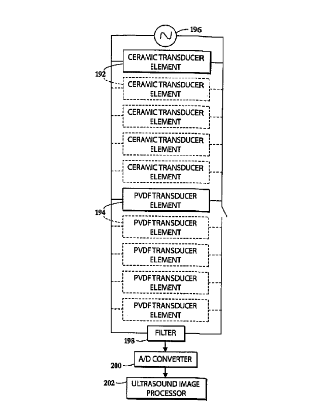
FIG. 9 is a circuit diagram applicable to any of the dual mode piezocomposite
transducers described herein. As shown in FIG. 9, one or more piezoelectric
ceramic
transducer elements 192 and one or more piezoelectric PVDF transducer elements
194 are
connected in parallel to a source of high-intensity alternating voltage 196
and to a directional
filter 198 having an output extending to an analog-to-digital converter 200
and from thence to
an ultrasonic signal processor 202.
A relatively low driving voltage applied by source 196 to ceramic transducer
elements
192 in a therapy mode does not engage PVDF transducer elements 194. PVDF
transducer
elements 194 have a substantially higher electrical impedance than the
impedance of ceramic
transducer elements 192 so that the total electrical impedance of the parallel
circuit of FIG. 9
quite similar to that of ceramic, so that the presence of PVDF elements 194 in
the circuit
consequently has little effect on electrical power transfer and produced
acoustic power. In an
imaging mode, the low acoustic impedance of the PVDF transducer elements 194
provide
larger amplitude broad band electrical signals to the ultrasound image
processor 202 in
response to received acoustic echoes due to the higher sensitivity of PVDF
material relative
to ceramic. The ceramic transducer elements 192 reflect most of the incoming
acoustic
energy due to high contrast in mechanical impedances between the ceramic and
water in an
absence of acoustic matching layers and produce much lower amplitude narrow
band electric
signals owing to the high power ceramic having a piezoelectric voltage
constant that is an
order-of-magnitude lower.
CA 02799717 2012-11-16
WO 2011/146138 PCT/US2011/000910
Ceramic transducer elements 192 and polymeric transducer elements 194 can
share
the same electrodes or be connected to different electrodes. The number of
individual
therapeutic ceramic transducer elements 192 and imaging polymeric elements
transducer
elements 194 depends on the application.
5 If a PVDF transducer element 194 is used to send and receive acoustic
signals as it is
done in a standard pulse-echo imaging systems, then there is a need to couple
that PVDF
transducer to both a high-voltage excitation pulse generator (not separately
shown) and the
sensitive receiving electronics, i.e., ultrasonic signal processor 202. A
transmit-receive (T/R)
switching circuit (not shown) that would close during the application of a
higher voltage
10 signal but open while the probe is receiving acoustic echoes can be used.
Alternatively, one
may use a circuit designed to send acoustic signals using one or more
piezoceramic
transducer elements 192 and receive echoes with PVDF transducer elements 194.
This is
feasible, because of close packed interpenetrant nature of piezocomposite
transducers
disclosed herein and consequent negligible differences in beam directivity
between ceramic
15 and polymer elements.
Piezocomposite ultrasound transducer devices 100, 106, 112, 123, and 136 are
provided with electrical contacts (not shown) enabling a connection of the
respective ceramic
transducer elements 102, 110, 118, 136, 142, and 368 in operative circuits for
generating, for
example, high-intensity focused ultrasound and enabling a connection of the
respective
20 polymeric transducer elements 104, 108, 114, 125, 140, 212, and 370 in
operative circuits for
scanning organic tissues to generate ultrasonic scan data for analysis and
processing into
images. Piezocomposite ultrasound transducer devices 100, 106, 112, 123, and
136 may be
further provided with mounting elements (not shown) for mechanically coupling
the
transducers exemplarily inside a probe or housing (not shown) and more
particularly inside a
liquid-filled bolus (not shown) that is contactable with a tissue surface to
enable ultrasonic
wave transmissions into and from organic tissues of a patient. Focusing lenses
exemplarily
in the form of acoustic Fresnel lenses (not shown) may be provided as
necessary, particularly
for piezocomposite ultrasound transducer devices 100, 106, and 112.
Dual-mode piezocomposite transducer devices 100, 123, 136, and 364 may be
activated by an alternative circuit configuration in which piezoelectric
ceramic transducer
elements 102, 126, 142, and 368 of the respective transducer devices are
electrically
independent from each other with at least a subset of the transducer elements
being operable
as a phased array while cross talk between members of the subset is minimized.
Likewise,
piezoelectric polymeric transducer elements 108, 125, 140, and 370 of
transducer devices
CA 02799717 2012-11-16
WO 2011/146138 PCT/US2011/000910
21
100, 123, 136, and 364 may be activated (energized and/or poled) by an
alternative circuit
configuration in which the piezoelectric polymeric transducer elements 108,
125, 140, and
370 are electrically independent from each other with at least a subset of the
transducer
elements being operable as a phased array while cross talk between members of
the subset is
minimized.
Electro-acoustic performance of a piezoelectric element with a given laminate
structure and materials can be simulated using the KLM model. (Krimholtz,
Leedom,
Matthaie 1970, New Equivalent Circuits for Elementary Piezoelectric
Transducers, Electron
Lett, 6, 13, 398-399.) This model can also be used to predict heat production
and electrical
power requirements. For example, calculated electrical impedance of 2 cm2 PZT
Navy Class
III element is resistive at 4 MHz resonance and presents a good electrical
match to a 50 Ohms
output impedance system. An electrical impedance of an equidimensional PVDF
element is
almost purely reactive and it is two orders of magnitude large in absolute
value. The electrical
impedance curves for ceramic (PZT) and polymeric (PVDF) elements are shown in
FIGS. 12
and 13.
Consistent with basic theory and development experience of therapeutic PVDF
phased arrays, the large reactive electrical impedance of PVDF requires a
significant driver
voltage of 1000 Volts peak-to-peak to achieve 3 - 5 W/cm2 acoustic power
output. The same
output acoustic power can be achieved in a ceramic driver only with 50 Volts
peak-to-peak.
Producing such and excessive drive voltage is technologically difficult,
limiting the
application of PVDF to a low power imaging applications.
One method of construction a piezocomposite transducer or array made of
ceramic
and polymeric elements will now be described. The idea is to encapsulate the
piezocomposite transducer made of a mix of ceramic and PVDF elements and
flexible filler
material between front and rear flex circuit layers. Ceramic elements provide
high-intensity
ultrasound in a therapeutic operating mode. The PVDF elements are used
exclusively for
imaging while ceramic elements are used predominantly for therapy and to
produce high
power acoustic imaging pulses. In a piezocomposite design, parasitic surface
vibrations are
dampened, leading to an improved therapeutic efficiency. Intramural area
consumed by
PVDF enables good-sensitivity medical imaging using a single or multiple
elements and
provides the potential for overall miniaturization of the piezocomposite
design. A shunt
inductor of the value 1/( woe Co), where coo is the radial frequency,
capacitance Co = ESA/t, es
is the clamped dielectric constant, A is the area of PVDF and t is the
thickness, can be used to
tune out the reactive component of PVDF elements. As shown in FIG. 10, each of
the
CA 02799717 2012-11-16
WO 2011/146138 PCT/US2011/000910
22
piezocomposite transducer devices described herein may have a final structure
comprising a
front flex-circuit layer 362, a piezocomposite layer 364 and a rear flex-
circuit layer 366.
Front electrode layer 362 consists of a piece of polyimide (Kapton ) flex-
circuit with a
single copper plane. This plane serves as the ground plane for the entire
device, including
ceramic and imaging elements. Piezocomposite layer 364 consists of individual
ceramic and
polymeric elements 368 and 370, which may be diced from one or several flat,
poled, and
plated pieces of PZT and PVDF. These elements 368 and 370 are bonded together
with an
adhesive to form a solid layer. Alternatively, ceramic elements 368 can be hot
pressed into a
polymeric PVDF substrate followed by subsequent poling. Rear flex-circuit
layer 366
consists of a four-layer flex circuit: (1) hot electrode pads for each
element; (2) a ground
plane (to minimize electrical coupling between elements); (3) a route layer
(to route the
individual signal lines to the edge of the array for contacts); and (4) a
ground plane (for RF
shielding and reduction of electrical coupling between elements).
A piezocomposite transducer with an arbitrary number of therapy and imaging
transducers may be constructed in a flat or concave shape as follows.
In outlined construction method one, first, forms a piezocomposite layer 364
by hot
pressing ceramic elements arranged in a desired geometrical pattern into a
polymeric
substrate at an elevated temperature not exceeding the Curie temperature of
ceramic. Second,
one immerses the hot-pressed piezocomposite layer 364 into an oil at about 100
C and
subjects it to an electric field of about 80 MV/m via external surface
electrodes in order to
produce or restore desired ferroelectric activity in a PVDF film surrounding
ceramic elements
368. The polarization of ceramic elements is not substantially affected below
its Curie
temperature, which is around 300 C -350 C for a typical high power ceramic.
Next, one
laminates the front flex-circuit layer 362 onto the cooled piezocomposite
layer 364 using a
low viscosity epoxy adhesive. The electrical contacts can also be established
by bonding thin
wires with a conductive epoxy, sputtering process, electrolytic deposition.
Optionally, using
a conventional acid bath and solvent sequence, one etches a thickness of PVDF
away to fill it
with low-durometer polymer and obtain a spatially distributed, movable design.
Additionally, the etched voids can be filled with metal powder to improve the
longevity and
thermal performance of piezocomposite in therapeutic high power mode. This
process can
also be performed using a precision milling machine or dicing saw to produce
grooves in a
piezoactive polymeric substrate that can later be filled with flexible passive
polymer. A non-
piezoelectric polymer (Kynar ) can also be press-bonded to the back of imaging
elements in
order to obtain a fundamental thickness resonance in PVDF of a half
wavelength. Next, one
CA 02799717 2012-11-16
WO 2011/146138 PCT/US2011/000910
23
heats the array slightly and wicks in an epoxy adhesive between the elements.
This is a
standard technique for making imaging arrays. Finally, one laminates the rear
flex-circuit
366 onto the back of the piezocomposite layer 264. A similar process can be
used when
starting with a solid piece of piezoceramic and creating a plurality of
recesses, grooves, holes,
cuts, dimples, or indentations on its surface that are later filled with
polymeric material,
which is later poled to thereby form a piezocomposite transducer element.
FIG. 11 depicts a dual mode transducer assembly 204 including a piezoceramic
therapy transducer element 206 and an acoustic Fresnel lens 208 spaced from
one another by
a liquid layer 210. Fresnel lens 208 is provided in a central region with a
piezoelectric
polymeric imaging transducer element 212. Transducer element 212 occupies a
through hole
214 in the lens. A backing layer 213 is paced by a liquid layer 215 from a
back side of
ceramic transducer element 206.
The volume fraction of piezoceramic to polymeric transducers may be tailored
for a
particular application need to enhance transmit, receive, or transmit and
receive response
rates, and the volume fraction is not uniform across a surface of a dual-mode
piezocomposite
transducer device as disclosed herein.
The calculated electrical impedances of Navy Class III piezoceramic and PVDF
piezopolymeric transducers with a surface area of 2 cm2 are shown in FIGS. 12
and 13
respectively, and are provided herein to illustrate significant differences in
the electro-
mechanical material properties of dual mode piezocomposite transducer
constituents.
As illustrated in FIG. 14, a dual-mode piezocomposite transducer module or
device
402 as described hereinabove with reference to FIGS. 1-4 and 10 may comprise
at least one
piezoelectric polymeric imaging transducer element 404 and at least one
piezoelectric
ceramic therapy transducer element 406 that include interleaved sections 404'
and 406',
respectively arranged in a planar array. Each transducer section or separate
transducer
element 404' and 406' extends from one face 408 of the dual-mode
piezocomposite transducer
module through to an opposite face 410 thereof.
As depicted in FIG. 15, a dual-mode piezocomposite transducer module or device
412
as described hereinabove with reference to FIGS. 1-4 and 10 may comprise at
least one
piezoelectric polymeric imaging transducer element 414 and at least one
piezoelectric
ceramic therapy transducer element 416 arranged in an overlapping array.
As shown in FIG. 16, a dual-mode piezocomposite transducer module or device
422
as described hereinabove with reference to FIGS. 1-4 and 10 may comprise at
least one
piezoelectric polymeric imaging transducer element 424 and at least one
piezoelectric
CA 02799717 2012-11-16
WO 2011/146138 PCT/US2011/000910
24
ceramic therapy transducer element 426 that are stacked one over the other,
with the
piezoelectric polymeric imaging transducer element 424 disposed on the
transmitting side of
the piezoelectric ceramic therapy transducer element 426, that is, between the
piezoelectric
ceramic therapy transducer element 426 and the target tissue. Transducer
elements 424 and
426 may be mounted in a holder so as to be spaced by a layer 428 of water or
other liquid.
As represented in FIG. 17, a dual-mode piezocomposite transducer module or
device
432 as described hereinabove with reference to FIGS. 1-4 and 10 may comprise
at least one
piezoelectric polymeric imaging transducer element 434 and at least one
piezoelectric
ceramic therapy transducer element 436 that include interleaved sections 434'
and 436',
respectively arranged in a planar array. Polymeric transducer element 434
extends from a
front face 438 of the dual-mode piezocomposite transducer module 432 through
to a back
face 440 thereof. Ceramic transducer sections or individual elements 436' are
disposed in
recesses, grooves, cuts, notches or indentations 442 and extend from back face
440 only
partway toward front face 438.
Where modules 402, 412, 422, and 432 have multiple separate piezoelectric
polymeric transducer elements 404', 414, 424, 434', those elements are
electrically
independent from each other and at least a subset of piezoelectric polymeric
transducer
elements 404', 414, 424, 434' may be operated as a receiving array (e.g., as a
phased array)
while acoustical and electrical cross talk between members of the subset is
minimized. To
that end each polymeric transducer element 404', 414, 424, 434' is provided
with a pair of
electrodes (not shown) separately connectable to an ultrasound signal
processor 202 (FIG. 9).
Where modules 402, 412, 422, and 432 have multiple separate piezoelectric
ceramic
transducer elements 406', 416, 426, 436', those elements are electrically
independent from
each other and at least a subset of piezoelectric ceramic transducer elements
406', 416, 426,
436' may be operated as a phased array, while acoustical and electrical cross
talk between
members of the subset is minimized. Each ceramic transducer element 406', 416,
426, 436' is
provided with a pair of electrodes (not shown) separately connectable to an
ultrasonic-
frequency waveform generator or voltage source 196 (FIG. 9).
The imaging and therapy elements of a dual-mode piezocomposite transducer
device
as disclosed herein may be mechanically held together by means of an open cell
metallic
foam structure that permits water flow and efficient cooling and characterized
by good
electrical conductivity. Such a foam structure is depicted schematically at
450 and 452 in
FIGS. 14 and 15.
CA 02799717 2012-11-16
WO 2011/146138 PCT/US2011/000910
FIG. 18 shows a portion of a dual mode piezocomposite transducer module having
polymeric imaging elements 502 and ceramic therapy elements 504 that are both
poled, as
indicated by arrows 506 and 508, in a direction normal to a surface 510
comprising individual
emitting and receiving element surfaces 512 and 514. Double headed arrows 516
indicate
the direction of vibration, that is, the direction along which alternating
compression and
rarefaction occur, while reference numerals 518 and 520 designate electrodes.
FIG. 19 shows a portion of a dual mode piezocomposite transducer module having
polymeric imaging elements 522 and ceramic therapy elements 524 that are
poled, as
indicated by arrows 526 and 528, in different directions to enable a
simultaneous emission
and reception of substantially different ultrasonic waves 530 and 532 to
simultaneously
monitor and induce lesion formation. Ultrasonic waves 530 and 532 are
represented by
double headed arrows that indicate the different directions of vibration,
while reference
numerals 538 and 540 designate electrodes.
FIG. 20 shows a portion of a dual mode piezocomposite transducer module having
polymeric imaging elements 542 and ceramic therapy elements 544 wherein the
latter, as
indicated by arrow 546, is poled in the thickness mode normal to a surface 548
comprising
individual emitting and receiving elements surfaces 550 and 552. Piezoelectric
polymeric
elements 542 are poled, as indicated by arrows 554, in a perpendicular
direction radial to the
center of said device in order to maximize the ability to perform multiwave
imaging. Double
headed arrows 556 and 558 indicate the different directions of vibration,
while reference
numerals 560 and 562 designate electrodes.
FIG. 21 depicts a dual-mode piezocomposite transducer device comprising
multiple
ceramic transducer elements 564 (only one shown) and multiple polymeric
transducer
elements 566 (only one shown) that are disposed in a laterally alternating or
interleaved
arrangement in a plane. Ceramic transducer elements 564 are energized by
alternating
voltage from a source 568 that is connected to the ceramic transducer elements
via one or
more ground electrodes 570 and one or more principal electrodes 572. Likewise,
one or more
polymeric transducer elements 566 may be energized by alternating voltage from
a source
574 that is connected to the polymeric transducer elements via one or more
ground electrodes
576 and one or more principal electrodes 578. In addition, polymeric
transducer elements
566 are scanned or sampled by an ultrasound imaging circuit (see FIG. 9).
FIG. 22 depicts a transducer device and circuit similar to that of FIG. 21,
except that
the ground electrodes 570 and 576 of the ceramic transducer elements 564 and
the polymeric
transducer elements 566 are commonly grounded.
CA 02799717 2012-11-16
WO 2011/146138 PCT/US2011/000910
26
As illustrated in FIG. 23, a dual-mode piezocomposite transducer device
comprising a
planar array of interleaved ceramic transducer elements 579 (only one shown)
and polymeric
transducer elements 580 has a common ground electrode 582 and a common main
electrode
584. This configuration is discussed above with reference to FIG. 9.
FIGS. 24-29 illustrate different electrode arrangements for a dual mode
piezocomposite transducer device wherein recesses, grooves, holes, cuts,
dimples, or
indentations 586 in a piezoelectric ceramic transducer matrix or base 588 are
filled with
piezoelectric polymeric material to form a plurality of imaging elements 590.
The same
electrode arrangements may be used in the reverse configurations (not shown)
where the
recesses, grooves, holes, cuts, dimples, or indentations 586 in provided in a
polymeric
transducer element matrix and are filled with piezoelectric ceramic material
to form a
plurality of therapy elements.
In FIG. 24, piezoelectric ceramic transducer element(s) 588 have ground
electrodes
592 and principal electrodes 594, while piezoelectric polymeric transducer
elements 590 have
separate ground electrodes 596 and principal electrodes 598. Ceramic element
electrodes 592
and 594 are connectable to a source 600 of waveform energy, while polymeric
element
electrodes 596 and 598 are connectable to an ultrasound imaging circuit (see
FIG. 9) and/or
source 602 of waveform energy.
In FIG. 25, ceramic transducer element(s) 588 and polymeric transducer
elements 590
have ground electrodes 604 and 606 that are interconnected and respective
principal
electrodes 608 and 610 that are electrically isolated by respective voltage
sources 612 and
614 or signal sampling and processing circuits (in the case of the imaging
elements 590).
FIG. 26 shows a different electrode arrangement wherein ground electrodes 616
and
618 are interconnected and main electrodes 620 and 622 are separate. FIG. 27
is the same as
FIG. 26 except for the inclusion of a switch 624.
FIG. 28 depicts an electrode configuration including a common ground electrode
625
and separate main electrodes 626 and 628 alternately energizable by a common
power supply
630 through the operation of a switch 632.
FIG. 29 illustrates an electrode configuration including a common ground
electrode
634 and separate main electrodes 636 and 638 energizable by respective power
supplies 640
and 642 (or poled by sampling and processing circuitry, in the case of
polymeric imaging
transducers 590). A switch 644 deactivates the polymeric imaging transducers
590.