Note: Descriptions are shown in the official language in which they were submitted.
CA 02803252 2014-12-17
WO 2012/003211 PCT/US2011/042354
MULTI-CHANNEL ENDORECTAL COILS AND INTERFACE DEVICES
THEREFOR
BACKGROUND OF THE INVENTION
Field of the Invention
[0002] The present invention generally relates to systems and methods of
obtaining images
and spectra of anatomical structures using magnetic resonance (MR) systems.
More
particularly, the present invention pertains to multiple embodiments of a
multichannel surface
coil array and associated interface devices capable of providing images and
spectroscopic
results from the MR signals obtained from the nuclei excited during MR
procedures.
Description of Related Art
[0003] The following background information is provided to assist the reader
to understand
the invention disclosed below and the environment in which it will typically
be used. The
terms used herein are not intended to be limited to any particular narrow
interpretation unless
clearly stated otherwise, either expressly or implied, in this document.
[0004] Magnetic resonance imaging (MRI) is a noninvasive method of producing
high
quality images of the interior of the human body. It allows medical personnel
to see inside the
human body without surgery or the use of ionizing radiation such as X-rays.
The images are
of such high resolution that cancer and other forms of pathology can often be
visually
distinguished from healthy tissue. Magnetic resonance techniques and systems
have also been
developed for performing spectroscopic analyses by which the chemical content
of body
tissue or other material can be ascertained.
[0005] MRI uses a powerful magnet, radio waves, and computer technology to
create
detailed images of the soft tissues, muscles, nerves, and bones in the body.
It does so by
taking advantage of a basic property of the hydrogen atom, an atom found in
abundance in all
cells within living organisms. In the absence of a magnetic field, the nuclei
of hydrogen
atoms spin like a top, or precess, randomly in every direction. When subject
to a strong
magnetic field, however, the spin-axes of the hydrogen nuclei align themselves
in the
direction of the field. This is because the nucleus of the hydrogen atom has
what is referred to
- 1 -
CA 02803252 2012-12-18
WO 2012/003211
PCT/US2011/042354
as a large magnetic moment, a strong inherent tendency to line up with the
direction of the
field. Collectively, the hydrogen nuclei of the area to be imaged create an
average vector of
magnetization that points parallel to the magnetic field.
[0006] A typical MRI system, or scanner, includes a main magnet, three
gradient coils, a
radio frequency (RF) antenna (often referred to as the whole body coil), and a
computer
station from which an operator can control the system. The chief component of
the MRI
system, however, is the main magnet. It is typically superconducting in nature
and cylindrical
in shape. Within its bore (an opening into which patients are placed during an
MRI
procedure), the main magnet generates a strong magnetic field, often referred
to as the BO
field, which is both uniform and static (non-varying). This BO magnetic field
is oriented
along the longitudinal axis of the bore, referred to as the z direction, which
compels the
magnetization vectors of the hydrogen nuclei in the body to align themselves
parallel to that
axis. In this alignment, the nuclei are prepared to receive RF energy of the
appropriate
frequency from the whole body coil. This frequency is known as the Larmor
frequency and is
governed by the equation co = y BO, where co is the Larmor frequency (at which
the hydrogen
atoms precess), y is the gyromagnetic constant, and BO is the strength of the
static magnetic
field.
[0007] The RF antenna, or whole body coil, is generally used both to transmit
pulses of RF
energy and to receive the resulting MR signals induced thereby in the hydrogen
nuclei.
Specifically, during its transmit cycle, the body coil broadcasts RF energy
into the cylindrical
bore. This RF energy creates a radio frequency magnetic field, also known as
the RF B1
field, whose magnetic field lines are directed in a line perpendicular to the
magnetization
vector of the hydrogen nuclei. The RF pulse causes the spin-axes of the
hydrogen nuclei to
tilt with respect to the main (BO) magnetic field, thus causing the net
magnetization vector to
deviate from the z direction by a known angle. The RF pulse, however, will
affect only those
hydrogen nuclei that are precessing about their axes at the frequency of the
RF pulse. In other
words, only the nuclei that "resonate" at that frequency will be affected, and
such resonance
is achieved in conjunction with the operation of the three gradient coils.
[0008] The gradient coils are electromagnetic coils. Each gradient coil is
used to generate a
linearly varying yet static magnetic field along one of the three spatial
directions (x,y,z)
within the cylindrical bore known as the gradient BI field. Positioned inside
the main
magnet, the gradient coils are able to alter the main magnetic field on a very
local level when
they are turned on and off very rapidly in a specific manner. Thus, in
conjunction with the
main magnet, the gradient coils can be operated according to various imaging
techniques so
- 2 -
CA 02803252 2012-12-18
WO 2012/003211
PCT/US2011/042354
that the hydrogen nuclei, at any given point or in any given strip, slice, or
unit of volume, will
be able to achieve resonance when an RF pulse of the appropriate frequency is
applied. In
response to the RF pulse, the precessing hydrogen atoms in the selected region
absorb the RF
energy being transmitted from the body coil, thus forcing the magnetization
vectors thereof to
tilt away from the direction of the main (BO) magnetic field. When the body
coil is turned off,
the hydrogen nuclei begin to release the RF energy in the form of the MR
signal, as explained
further below.
[0009] One well known technique that can be used to obtain images is referred
to as the
spin echo imaging technique. Operating according to this technique, the MRI
system first
activates one gradient coil to set up a magnetic field gradient along the z-
axis. This is called
the "slice select gradient", and it is set up when the RF pulse is applied and
it shuts off when
the RF pulse is turned off. It allows resonance to occur only within those
hydrogen nuclei
located within a slice of the area being imaged. No resonance will occur in
any tissue located
on either side of the plane of interest. Immediately after the RF pulse
ceases, all of the nuclei
in the activated slice are "in phase", i.e., their magnetization vectors all
point in the same
direction. Left to their own devices, the net magnetization vectors of all the
hydrogen nuclei
in the slice would relax, thus realigning with the z direction. Instead,
however, the second
gradient coil is briefly activated to create a magnetic field gradient along
the y-axis. This is
called the "phase encoding gradient". It causes the magnetization vectors of
the nuclei within
the slice to point, as one moves between the weakest and strongest ends of the
gradient, in
increasingly different directions. Next, after the RF pulse, slice select
gradient, and phase
encoding gradient have been turned off, the third gradient coil is briefly
activated to create a
gradient along the x-axis. This is called the "frequency encoding gradient" or
"read out
gradient", as it is only applied when the MR signal is ultimately measured. It
causes the
relaxing magnetization vectors to be differentially re-excited, so that the
nuclei near the low
end of the gradient begin to precess at a faster rate, and those at the high
end pick up even
more speed. When these nuclei relax again, the fastest ones (those which were
at the high end
of the gradient) will emit the highest frequency of radio waves.
[0010] Collectively, the gradient coils allow the MR signal to be spatially
encoded, so that
each portion of the area being imaged is uniquely defined by the frequency and
phase of its
resonance signal. In particular, as the hydrogen nuclei relax, each becomes a
miniature radio
transmitter giving out a characteristic pulse that changes over time,
depending on the local
microenvironment in which it resides. For example, hydrogen nuclei in fats
have a different
microenvironment than do those in water, and thus transmit different pulses.
Due to these
- 3 -
CA 02803252 2012-12-18
WO 2012/003211
PCT/US2011/042354
differences, in conjunction with the different water-to-fat ratios of
dissimilar tissues, different
tissues transmit radio signals of different frequencies. During its receive
cycle, the body coil
detects these miniature radio transmissions, which are often collectively
referred to as the MR
signal. From the body coil, these unique resonance signals are conveyed to the
receivers of
the MR system, where they are converted into mathematical data corresponding
thereto. The
entire procedure must be repeated multiple times to form an image with a good
signal-to-
noise ratio (SNR). Using multidimensional Fourier transformations, an MR
system can
convert the mathematical data into a two- or even a three-dimensional image.
[0011] When more detailed images of a specific part of the body are needed, a
local coil is
often used instead of the whole body coil. A local coil can take the form of a
volume coil or a
surface coil. A volume coil is used to surround or enclose the volume to be
imaged (e.g., a
head, an arm, a wrist, a leg, or a knee). A surface coil, however, is merely
placed upon the
surface of a patient so that the underlying region of interest (e.g., the
abdominal, thoracic,
and/or pelvic regions) can be imaged. In addition, a local coil can be
designed to operate
either as a receive-only coil or a transmit/receive (T/R) coil. The former is
only capable of
detecting the MR signals produced by the body in response to an MRI procedure,
as noted
above. A T/R coil, however, is capable of both receiving the MR signals as
well as
transmitting the RF pulses that produce the RF BI magnetic field, which is the
prerequisite
for inducing resonance in body tissue.
100121 It is well known in the field of MRI to use a single local coil,
whether surface or
volume, to detect the MR signals. According to the single coil approach, a
relatively large
local coil is used to cover or enclose the entire region of interest. Early
receiving coils were
just linear coils, meaning that they could detect only one of the two (i.e.,
vertical MX' and
horizontal MY') quadrature components of the MR signals produced by the region
of interest.
One example of a linear coil is the single loop coil shown in FIG. 1A. This
loop is only
capable of detecting magnetic fields (i.e., MR signals) that are oriented
perpendicular/vertical
to the plane of the loop as shown in FIG. 1B. Another example of a linear coil
is the butterfly
or saddle coil shown in FIG. 2A. Unlike the single loop, the butterfly coil is
only sensitive to
magnetic fields that are oriented parallel to the plane of the coil as shown
in FIG. 2B. This is
because a butterfly coil is constructed by twisting a loop in the middle to
form two identical
subloops about a midpoint. Because the currents flowing in the subloops are
the same but
flow in counter-rotating directions, the magnetic flux generated by the
current flowing
through one subloop of the symmetric structure is equal but opposite to the
flux due to the
current in the other subloop. Therefore, about the midpoint of the structure,
the vertical fields,
- 4 -
CA 02803252 2012-12-18
WO 2012/003211
PCT/US2011/042354
due to the counter-rotating currents, oppose and thus cancel each other. The
horizontal fields
generated by those currents, however, combine, yielding a magnetic field that
is oriented
parallel to the plane of the coil.
[0013] Accordingly, receiving coils employing quadrature mode detection,
meaning that
they could intercept both the vertical and horizontal components, have been
developed.
Compared to linear receiving coils, quadrature receiving coils enabled MRI
systems to
provide images for which the SNR was much improved, typically by as much as
41%. Even
with the improvement brought with quadrature mode detection, the single coil
approach still
provided images whose quality invited improvement. The disadvantage inherent
to the single
coil approach is attributable to just one coil structure being used to acquire
the MR signals
over the entire region of interest.
[0014] Phased array coils were also developed to overcome the shortcomings
with the
single coil approach. Instead of one large local coil, the phased array
approach uses a
plurality of smaller local coils, with each such coil covering or enclosing
only a portion of the
region of interest. In a system having two such coils, for example, each of
the coils would
cover or enclose approximately half of the region of interest, with the two
coils typically
being partially overlapped for purposes of magnetic isolation. The two coils
would acquire
the MR signals from their respective portions simultaneously, and they would
not interact
adversely due to the overlap. Because each coil covers only half of the region
of interest, each
such coil is able to receive the MR signals at a higher SNR ratio for that
portion of the region
of the interest within its coverage area. The smaller local coils of the
phased array thus
collectively provide the MRI system with the signal data necessary to generate
an image of
the entire region of interest that is higher in resolution than what can be
obtained from a
single large local coil.
[0015] One example of a phased array coil is the Gore torso array produced by
W.L.
Gore and Associates, Inc. The torso array contains four surface coils, two of
which are
disposed in an anterior paddle, and the other two are disposed in a posterior
paddle. The two
paddles are designed to be placed against the anterior and posterior surfaces,
respectively, of
the patient about the abdominal, thoracic, and pelvic regions. The torso array
is designed for
use with an MR system whose data acquisition system has multiple receivers.
The four leads
of the torso array, one each from the two anterior surface coils and the two
posterior surface
coils, can be connected to separate receivers, with each receiver amplifying
and digitizing the
signal it receives. The MR system then combines the digitized data from the
separate
receivers to form an image whose overall SNR is better than what could be
obtained from a
- 5 -
CA 02803252 2014-12-17
WO 2012/003211 PCT/US2011/042354
single local coil, or even two larger anterior and posterior local coils,
covering the entire
region of interest alone.
[0016] It is also well known to obtain images of internal bodily structures
through the use
of intracavity probes. An example of a prior art intracavity probe designed
primarily for use
with 1.01 and 1.5T MR systems can be found in United States Patent Nos.
5,476,095 ('095)
and 5,355,087 ('087), both of which are assigned to the assignee of the
present invention
The prior art probe disclosed is designed to be inserted into
bodily openings, such as the rectum, vagina, and mouth. These patents also
disclose interface
devices that are designed to interface the prior art intracavity probe with MR
imaging and
spectroscopy systems. A method of using the intracavity probe is disclosed in
United States
Patent No. 5,348,010, which is also assigned to the assignee of the present
invention
[0017] The prior art probe, operated in conjunction with its associated
interface unit,
allows an MR system to generate images of, and spectroscopic results for,
various internal
bodily structures, such as the prostate gland, colon, or cervix. Examples of
such prior art
probes include the BPX-15 prostate/endorectal coil (E-coil), the PCC-15
colorectal coil, and
the BCR-15 cervix coil, all of which are part of the eCoilTm line of
disposable coils produced
by MEDRAD, Inc. of Indianola, Pennsylvania. Examples of such interface units
include the
AID-II and the ATD-Torso units, also produced by MEDRAD, Inc.
[0018] The ATD-II unit is used to interface the prior art probe with one
receiver of an MR
system to provide images or spectra of the region of interest, namely, the
prostate gland,
colon, or cervix. The ATD-Torso unit is used to interface not only the prior
art probe but also
the Gore torso array with multiple receivers of the MR system. When connected
to such a
probe and the torso array, the AID-Torso unit allows the MR system to provide
images or
spectra not only of the prostate gland, colon, or cervix but also of the
surrounding anatomy,
i.e., the abdominal, thoracic, and pelvic regions.
[0019] U.S. Patent Nos. 7,747,310 and 7,885,704, both of which are assigned to
the
assignee of the present invention disclose several
intracavity probes, and associated interface devices, for use with MR systems
designed to
operate at higher field strengths than the prior art probes of the '087 and
'095 patents. For
example, the latter reference teaches a probe having a coil loop that includes
two drive
capacitors and a tuning capacitor, all of which are in series. Connected
across each drive
capacitor is an output cable having an electrical length of SL + n(A14). When
each output
- 6 -
CA 02803252 2014-12-17
WO 2012/003211 PCT/US2011/042354
cable is connected at its other end to the interface device, the coil loop is
thereby
interconnected through the interface device to the MR system.
[0020] With reference to FIG. 3, quadrature intracavity probes have been
developed. For
instance, International Patent Application Publication No. WO 2010/056911,
which is
assigned to the assignee of the present invention
discloses a single coil structure that is sensitive to both the vertical and
horizontal
components of the MR signal by virtue of a simple loop-type coil element and a
butterfly-
type coil element that share a center conductor. More specifically, the
quadrature coil,
generally designated 10, includes an outer loop 12, a center conductor 14
bisecting the outer
loop 12, and an output line, generally designated 16. The outer loop 12
includes a plurality of
capacitors including first and second drive capacitors 18 and 20 and first and
second tuning
capacitors 22 and 24. Of approximately equal values, the drive capacitors 18,
20 are serially
deployed within the outer loop 12 and at their junction node 26 form a virtual
ground for
electrically balancing and impedance matching the loop. Tuning capacitors 22,
24 are also
serially deployed within outer loop 12, with their common node 28 being
situated
diametrically opposite the junction node 26. Of approximately equal values,
the tuning
capacitors 22, 24 are selected to resonate the outer loop 12 at the operating
frequency of the
MR system. In that regard, outer loop 12 is shown in FIG. 3 as having four
inductors. The
values of those inductors merely represent the inductances inherent in the
conductive (e.g.,
copper) segments of the loop. The output line 16 includes two coaxial cables
30 and 32 with
the shield conductor of each connected to the junction node 26 of the coil 10.
The center
conductor 14 extends between and evenly bisects the junction and common nodes
26 and 28
of outer loop 12, and thus maintains the physical and electrical symmetry of
quadrature coil
10. FIG. 3 shows the center conductor 14 as having two inductors and a tuning
capacitor 34
symmetrically deployed along its length. Like outer loop 12, the values of
those inductors
merely represent the inductances inherent in the conductor. The value of the
tuning capacitor
34 has been selected so that its reactance at the operating frequency equals
the inductive
reactance of center conductor 14. This configuration permits the simple loop
and butterfly
elements of the coil to detect MR signals orthogonal and parallel,
respectively, to the plane of
the coil.
[0021] With reference to FIG. 4 and as disclosed in U.S. Patent No. 7,885,704,
a coil
having a phased array configuration for use as in an endorectal probe has been
developed.
The coil includes four coil loops 40, 41, 42, and 43 deployed in a phased
array configuration
in which each coil loop 40, 41, 42, and 43 is critically overlapped by its
neighbor. Each coil
- 7 -
CA 02803252 2012-12-18
WO 2012/003211
PCT/US2011/042354
loop 40, 41, 42, and 43 includes a drive capacitor 44, 45, 46, and 47 and a
tuning capacitor
48, 49, 50, and 51 arranged diametrically opposite to the drive capacitor 44,
45, 46, and 47. In
addition, each coil loop 40, 41, 42, and 43 includes an output line 52, 53,
54, and 55
connected across the respective drive capacitor 44, 45, 46, and 47.
Accordingly, a four
element, four channel configuration is provided. This arrangement provides a
demonstrably
higher signal-to-noise ratio (SNR) than the quadrature coil 10 described
hereinabove with
reference to FIG. 3; however, the coverage is less uniform due to the areas of
low signal in
the critically-coupled (i.e., overlapped conductor) areas. This non-uniformity
is undesirable
for use in an endorectal probe due to the higher amounts of non-uniformity
proximal to the
coil conductors.
[0022] Despite their widespread acceptance and good reputation in the
marketplace, these
prior art intracavity probes and interface devices nevertheless have a few
shortcomings. For
example, they offer limited coverage, exhibit lower signal-to-noise
performance, and
generally provide less overall flexibility as compared to the endorectal coil
technology
discussed hereinafter. It is therefore desirable to provide an endorectal coil
array and
associated interface device capable of providing greater overall flexibility
and higher quality
images and spectroscopic results from MR signals obtained from nuclei during
MR
procedures.
SUMMARY OF THE INVENTION
[0023] Therefore, it is an object of the present invention to provide a method
and system
that overcome some or all of the drawbacks and deficiencies evident in the
prior art. More
specifically, the endorectal coil array and associated interface devices of
the present invention
are capable of providing greater overall flexibility and higher quality images
and
spectroscopic results from MR signals obtained from nuclei during MR
procedures.
[0024] Accordingly, provided is a coil for use with a magnetic resonance
system for
obtaining images of a region of interest. The coil includes: (a) a pair of
coil loops arranged in
a phased array configuration each of which for receiving magnetic resonance
signals from the
region of interest corresponding thereto; and (b) a spacer material positioned
adjacent to an
anterior surface of the pair of the coil loops. Each of the coil loops has a
drive capacitor and a
tuning capacitor with the tuning capacitor having a value selected to resonate
the coil loop
corresponding thereto at an operating frequency of the magnetic resonance
system. The
spacer material enables a predetermined distance of between about 0.03 and
about 0.06
inches to exist between the pair of coil loops and the region of interest and
thereby: (i) reduce
- 8 -
CA 02803252 2012-12-18
WO 2012/003211
PCT/US2011/042354
intensity of the magnetic resonance signals in proximity of the coil loops;
(ii) maintain a
signal-to-noise ratio at a depth within the region of interest appropriate to
reconstruct the
images of the region of interest; and (iii) reduce artifacts in the images
inclusive of the Gibbs
artifact.
[0025] The coil may further include a pair of decoupling circuits each of
which connected
across the tuning capacitor of one of the coil loops. Each of the decoupling
circuits may be an
active decoupling circuit, a passive decoupling circuit, or both an active and
a passive
decoupling circuit. The coil may also further include a pair of output cables
each of which
connected at a first end thereof across the drive capacitor of one of the coil
loops such that
each of the drive capacitors is provided with a separate ground. An
intermediate conduit may
be provided that includes: (a) an input connector; (b) an output connector;
(c) pair of internal
cables for connecting at one end thereof, respectively, to the output cables
of the intracavity
probe via the input connector and approximate another end thereof to an
interface device for
the intracavity probe via the output connector; (d) a pair of baluns each of
which
interconnected between an end of one of the internal cables and at least one
of the input
connector and the output connector; and (e) at least one cable trap connected
thereabout.
100261 The phased array configuration may require the pair of coil loops to be
critically
overlapped, to share a common conductor, or to be arranged in a hybrid overlap
configuration
wherein at least a portion of each of the coil loops is overlapped and the
coil loops share a
common conductor.
[0027] A passive decoupling circuit may be provided at a second end of each of
the output
cables. Each of the passive decoupling circuits may include series connected
back-to-back
diodes and a reactance component. The reactance component may be at least one
of an
inductor and a capacitor.
[0028] The coil may be provided as part of an intracavity probe or may be a
surface coil.
The surface coil may be a head coil, a torso coil, a neck coil, a limb coil,
or any combination
thereof.
[0029] Also provided is an intracavity probe for use with a magnetic resonance
system for
obtaining images of a region of interest within a cavity of a patient. The
intracavity probe
includes: (a) a pair of coil loops arranged in a phased array configuration
each of which
receive magnetic resonance signals from the region of interest corresponding
thereto; (b) a
pair of decoupling circuits each of which connected across the tuning
capacitor of one of the
coil loops; (c) a pair of output cables each of which connected at a first end
thereof across the
drive capacitor of one of the coil loops such that each of the drive
capacitors is provided with
- 9 -
CA 02803252 2012-12-18
WO 2012/003211
PCT/US2011/042354
a separate ground; and (d) a spacer material positioned adjacent to an
anterior surface of the
pair of the coil loops. Each of the coil loops includes a drive capacitor and
a tuning capacitor
with the tuning capacitor having a value selected to resonate the coil loop
corresponding
thereto at an operating frequency of the magnetic resonance system. The spacer
material
enables a predetermined distance of between about 0.03 and about 0.06 inches
to exist
between the pair of coil loops and the region of interest and thereby: (i)
reduce intensity of
the magnetic resonance signals in proximity of the coil loops; (ii) maintain a
signal-to-noise
ratio at a depth within the region of interest appropriate to reconstruct the
images of the
region of interest; and (iii) reduce artifacts in the images or spectra
inclusive of the Gibbs
artifact when the intracavity probe is inserted into the cavity of the patient
during acquisition
of the images.
100301 Each of the decoupling circuits may be an active decoupling circuit, a
passive
decoupling circuit, or both an active and a passive decoupling circuit. An
intermediate
conduit may be provided that includes: (a) an input connector; (b) an output
connector; (c) a
pair of internal cables for connecting at one end thereof, respectively, to
the output cables of
the intracavity probe via the input connector and approximate another end
thereof to an
interface device for the intracavity probe via the output connector; (d) a
pair of baluns each of
which are interconnected between an end of one of the internal cables and at
least one of the
input connector and the output connector; and (e) at least one cable trap
connected
thereabout.
[0031.1 The phased array configuration may require the pair of coil loops to
be critically
overlapped, to share a common conductor, or to be arranged in a hybrid overlap
configuration
wherein at least a portion of each of the coil loops is overlapped and the
coil loops share a
common conductor.
100321 A passive decoupling circuit may be provided at a second end of each of
the output
cables. Each of the passive decoupling circuits may include series connected
back-to-back
diodes and a reactance component. The reactance component may be at least one
of an
inductor and a capacitor.
10033) In addition, provided is an interface device for interfacing a coil
comprising a pair
of coil loops arranged in a phased array configuration each of which receive
magnetic
resonance signals from a region of interest corresponding thereto with a
magnetic resonance
system. The interface device includes: (a) a first preamplifier for receiving
a signal from a
first coil loop of the pair of coil loops to produce a first amplified signal;
(b) a second
preamplifier for receiving a signal from a second coil loop of the pair of
coil loops to produce
- 10 -
CA 02803252 2012-12-18
WO 2012/003211
PCT/US2011/042354
a second amplified signal; (c) a first splitter operatively connected to the
first preamplifier for
dividing the first amplified signal into a right loop signal that is provided
to a first channel
output and a first composite signal; (d) a second splitter operatively
connected to the second
preamplifier for dividing the first amplified signal into a left loop signal
that is provided to a
second channel output and a second composite signal; (e) a third splitter
operatively
connected to the first splitter for dividing the first composite signal; (f) a
fourth splitter
operatively connected to the second splitter for dividing the second composite
signal; (g) a
zero degree combiner operatively connected to the third splitter and the
fourth splitter for
combining signals received therefrom to produce a saddle or butterfly signal
that is provided
to a third channel output; and (h) a 180 degree combiner operatively connected
to the third
splitter and the fourth splitter for combining signals received therefrom to
produce a whole
loop signal that is provided to a fourth channel output. The interface device
is configured to
selectively recognize each of the first, second, third, and fourth channel
outputs, thereby
allowing the magnetic resonance system coupled to the interface device to
produce images in
a plurality of different modes.
[00341 The first preamplifier and the second preamplifier may be provided with
a
predetermined reduced supply voltage as compared to a rated supply voltage of
the first
preamplifier and the second preamplifier. At least one attenuator may provide
an attenuation
nominally in the range of 3dB to 6dB. The at least one attenuator may be
positioned at at
least one of (a) between the first preamplifier and the first splitter; (b)
between the second
preamplifier and the second splitter; (c) after the first splitter; and (d)
after the second splitter.
The plurality of modes include, but are not limited to, Left Loop, Right Loop,
Whole Loop,
Whole Saddle, Right Loop and Left Loop (LL), Whole Loop and Whole Saddle, and
Right
Loop, Left Loop, Whole Loop, Whole Saddle (LLLS).
[0035] Also provided is a system for obtaining images of a region of interest.
The system
includes: (a) an intraeavity probe; and (b) an interface device for
interfacing the intracavity
probe with a magnetic resonance system. The intraeavity probe includes: (i) a
pair of coil
loops arranged in a phased array configuration each of which receive magnetic
resonance
signals from the region of interest corresponding thereto; (ii) a pair of
output cables each of
which connected at a first end thereof across the drive capacitor of one of
the coil loops such
that each of the drive capacitors is provided with a separate ground; and
(iii) a spacer material
positioned adjacent to an anterior surface of the pair of the coil loops. Each
of the coil loops
has a drive capacitor and a tuning capacitor with the tuning capacitor having
a value selected
to resonate the coil loop corresponding thereto at an operating frequency of
the magnetic
-11-
CA 02803252 2012-12-18
WO 2012/003211
PCT/US2011/042354
resonance system. The spacer material enables a predetermined distance of
between about
0.03 and about 0.06 inches to exist between the pair of coil loops and the
region of interest
and thereby reduce intensity of the magnetic resonance signals in proximity of
the coil loops,
maintain a signal-to-noise ratio at a depth within the region of interest
appropriate to
reconstruct the images of the region of interest, and reduce artifacts in the
images or spectra
inclusive of the Gibbs artifact when the intracavity probe is inserted into
the cavity of the
patient during acquisition of the images. The interface device includes: (i) a
first preamplifier
for receiving a signal from a first coil loop of the pair of coil loops to
produce a first
amplified signal; (ii) a second preamplifier for receiving a signal from a
second coil loop of
the pair of coil loops to produce a second amplified signal; (iii) a first
splitter operatively
connected to the first preamplifier for dividing the first amplified signal
into a right loop
signal and a first composite signal; (iv) a second splitter operatively
connected to the second
preamplifier for dividing the first amplified signal into a left loop signal
and a second
composite signal; (v) a third splitter operatively connected to the first
splitter for dividing the
first composite signal; (vi) a fourth splitter operatively connected to the
second splitter for
dividing the second composite signal; (vii) a zero degree combiner operatively
connected to
the third splitter and the fourth splitter for combining signals received
therefrom to produce a
saddle signal; and (viii) a 180 degree combiner operatively connected to the
third splitter and
the fourth splitter for combining signals received therefrom to produce a
whole loop signal.
100361 The first preamplifier and the second preamplifier are provided with a
predetermined reduced supply voltage as compared to a rated supply voltage of
the first
preamplifier and the second preamplifier. The interface device may further
include at least
one attenuator providing an attenuation nominally in the range of 3dB to 6dB.
The at least
one attcnuator may be positioned at at least one of: (a) between the first
preamplifier and the
first splitter; (b) between the second preamplifier and the second splitter;
(c) after the first
splitter; and (d) after the second splitter.
10037] The coil may further include a pair of decoupling circuits each of
which connected
across the tuning capacitor of one of the coil loops. Each of the decoupling
circuits may be an
active decoupling circuit, a passive decoupling circuit, or both an active and
a passive
decoupling circuit. An intermediate conduit may be provided that includes: (a)
an input
connector; (b) an output connector; (c) a pair of internal cables for
connecting at one end
thereof, respectively, to the output cables of the intracavity probe via the
input connector and
approximate another end thereof to an interface device for the intracavity
probe via the output
connector; (d) a pair of baluns each of which interconnected between an end of
one of the
- 12 -
CA 02803252 2012-12-18
WO 2012/003211
PCT/US2011/042354
internal cables and at least one of the input connector and the output
connector; and (e) at
least one cable trap connected thereabout.
[0038] The phased array configuration may require the pair of coil loops to be
critically
overlapped, to share a common conductor, or to be arranged in a hybrid overlap
configuration
wherein at least a portion of each of the coil loops is overlapped and the
coil loops share a
common conductor. A passive decoupling circuit may be provided at a second end
of each of
the output cables. Each of the passive decoupling circuits may include series
connected back-
to-back diodes and a reactance component. The reactance component may be at
least one of
an inductor and a capacitor.
[0039] In addition, provided is a coil for use with a magnetic resonance
system for
obtaining images of a region of interest. The coil includes: (a) a plurality
of coil loops
arranged in a phased array configuration each of which receive magnetic
resonance signals
from the region of interest corresponding thereto; (b) a plurality of output
cables each of
which connected at a first end thereof across the drive capacitor of one of
the coil loops; and
(c) at least one passive decoupling circuit provided at a second end of each
of the output
cables. Each of the coil loops has a drive capacitor and a tuning capacitor
with the tuning
capacitor having a value selected to resonate the coil loop corresponding
thereto at an
operating frequency of the magnetic resonance system.
[0040] A spacer material may be positioned adjacent to an anterior surface of
the pair of
the coil loops. The spacer material enables a predetermined distance of
between about 0.03
and about 0.06 inches to exist between the pair of coil loops and the region
of interest and
thereby reduce intensity of the magnetic resonance signals in proximity of the
coil loops,
maintain a signal-to-noise ratio at a depth within the region of interest
appropriate to
reconstruct the images of the region of interest, and reduce artifacts in the
images or spectra
inclusive of the Gibbs artifact.
[0041] Each of the passive decoupling circuits may include series connected
back-to-back
diodes and a reactance component. The reactance component may be at least one
of an
inductor and a capacitor.
[0042] These and other features and characteristics of the present invention,
as well as the
methods of operation and functions of the related elements of structures and
the combination
of parts and economies of manufacture, will become more apparent upon
consideration of the
following description and the appended claims with reference to the
accompanying drawings,
all of which form a part of this specification, wherein like reference
numerals designate
corresponding parts in the various figures. It is to be expressly understood,
however, that the
- 13-
CA 02803252 2012-12-18
WO 2012/003211
PCT/US2011/042354
drawings are for the purpose of illustration and description only and are not
intended as a
definition of the limits of the invention. As used in the specification and
the claims, the
singular form of "a", "an", and "the" include plural referents unless the
context clearly
dictates otherwise.
BRIEF DESCRIPTION OF THE DRAWINGS
[0043] FIG. 1A is a schematic diagram of a conventional single loop coil and
FIG. 1B is a
representation of the vertically oriented magnetic fields it is capable of
sensing;
[0044] FIG. 2A is a schematic diagram of a conventional butterfly coil and
FIG. 2B is a
representation of the horizontally oriented magnetic fields it is capable of
sensing;
[0045] FIG. 3 is a schematic diagram of a conventional quadrature endorectal
coil;
[0046] FIG. 4 is a schematic diagram of a conventional four channel phased
array
endorectal coil whose loops are partially overlapped;
[0047] FIG. 5 is a perspective view of an intracavity probe in accordance with
the present
invention;
[0048] FIG. 6 is a schematic diagram of a coil in accordance with a first
embodiment of
the present invention;
[0049] FIG. 7 is a schematic diagram of a coil in accordance with a second
embodiment of
the present invention;
[0050] FIG. 8 is an exemplary image produced by an MR system using the coil of
FIG. 6
illustrating the ghosting artifacts produced in the image;
[0051] FIG. 9 is a perspective exploded view of a coil in accordance with the
present
invention illustrating a spacer material used with the coil;
[0052] FIG. 10 is a cross-sectional assembled view of the coil of FIG. 9;
[0053] FIG. 11 is a schematic diagram of a coil in accordance with a third
embodiment of
the present invention;
[0054] FIG. 12 is a schematic diagram of a coil in accordance with a fourth
embodiment
of the present invention;
[0055] FIGS. 13A and 13B are block diagrams illustrating the coil of FIG. 12
connected
to an interface device in accordance with the present invention;
[0056] FIG. 14 is a block diagram of an interface device in accordance with
the present
invention;
[0057] FIG. 15 is a schematic diagram of the coil of FIG. 12 illustrating the
manner in
which current is induced therein when in Left Loop mode;
- 14 -
CA 02803252 2012-12-18
WO 2012/003211
PCT/US2011/042354
[0058] FIGS. 16A-16C form a schematic diagram of the interface device of FIG.
14
illustrating the manner in which the interface device operates when in Left
Loop mode;
[0059] FIG. 17 is a schematic diagram of the coil of FIG. 12 illustrating the
manner in
which current is induced therein when in Right Loop mode;
[0060] FIGS. 18A-18C form a schematic diagram of the interface device of FIG.
14
illustrating the manner in which the interface device operates when in Right
Loop mode;
[0061] FIG. 19 is a schematic diagram of the coil of FIG. 12 illustrating the
manner in
which current is induced therein when in Whole Loop mode;
[0062] FIGS. 20A-20C form a schematic diagram of the interface device of FIG.
14
illustrating the manner in which the interface device operates when in Whole
Loop mode;
[0063] FIG. 21 is a schematic diagram of the coil of FIG. 12 illustrating the
manner in
which current is induced therein when in Saddle Loop mode;
[0064] FIGS. 22A-22C form a schematic diagram of the interface device of FIG.
14
illustrating the manner in which the interface device operates when in Saddle
Loop mode;
and
[0065] FIG. 23 is a graph comparing the signal-to-noise ratios (SNR) of the
various coil
configurations.
DETAILED DESCRIPTION OF THE PREFERRED EMBODIMENTS
[0066] For purposes of the description hereinafter, the terms "upper", "up",
"lower",
"down", "right", "left", "vertical", "orthogonal", "horizontal", "top",
"bottom", "lateral",
"longitudinal", and derivatives thereof shall relate to the invention as it is
oriented in the
drawing figures and/or from the perspective of a patient during a procedure.
However, it is to
be understood that the invention may assume alternative variations and step
sequences,
except where expressly specified to the contrary. It is also to be understood
that the specific
devices and processes illustrated in the attached drawings, and described in
the following
specification, are simply exemplary embodiments of the invention. Hence,
specific
dimensions and other physical characteristics related to the embodiments
disclosed herein are
not to be considered as limiting.
[0067] In all of its embodiments and related aspects, the present invention
disclosed below
is ideally used with magnetic resonance (MR) systems designed to operate at
1.0, 1.5, or 3.0
Tesla or any field strength in between, though it is also applicable to those
operable at lower
or higher field strengths. The technology is also applicable to scanner
configurations with
horizontal or vertical bore magnets or other orientations and in closed or
open bore scanners.
- 15 -
CA 02803252 2012-12-18
WO 2012/003211
PCT/US2011/042354
100681 The coils discussed hereinafter may be incorporated into an intracavity
probe, such
as the endorectal probe 60 illustrated in FIG. 5. The intracavity probe 60
includes a flexible
shaft 62 and a balloon structure 64. The coils discussed hereinafter in
greater detail are
attached to an anterior surface of the balloon structure 64. The balloon
structure 64 is
configured to position the coil in operative proximity to a rectal prostatic
bulge of a patient
when the balloon structure 64 is inflated, which optimizes the coupling
between coil and the
target anatomy. The balloon structure 64 is preferably made of a medical-grade
latex or other
appropriate elastorneric material. Such material should, of course, be non-
paramagnetic and
exhibit low dielectric losses. The flexible shaft 62 defines two lumens (not
shown) therein.
Within its cylindrical wall near its distal end, the shaft 62 also defines a
hole (not shown) in
communication with one of the lumens. This lumen and hole together serve as a
passageway
for the fluid (e.g., gas or liquid) pumped into and expelled out of balloon
structure 64 when
inflated and deflated, respectively. Further away from its distal end, the
shaft 62 defines
another hole in its cylindrical wall. The other lumen and this hole act as the
conduit through
which the output cables are routed from the coil. The output cables may be
housed in a single
sheath 66 having a plug 68 at a proximal end thereof to connect the
intracavity probe 60 with
an appropriate interface device as discussed in greater detail hereinafter.
[00691 The intracavity probe 60 further includes an anti-migration disc 70 and
a handle 74.
Fixed to the proximal end of shaft 62, the handle 74 enables the probe 60 to
be easily
manipulated at its distal end and, inclusive of balloon structure 64 secured
thereon, is inserted
into the rectum and appropriately aligned within the cavity as described
below. The anti-
migration disc 70, composed of a semi-rigid plastic or other suitable polymer,
is desirably
semi-spherical in shape. As shown in FIG. 5, the disc 70 defines a slot 76.
This slot 76 allows
the disc 70 to be snapped onto the shaft 62. When affixed to the shaft 62
adjacent the anal
sphincter after the probe 60 has been inserted into the rectum, the anti-
migration disc 70
prevents the probe 60 from migrating superiorly due to the normal peristaltic
activity of the
colon.
100701 The intracavity probe 60 also includes a means for controlling
inflation of balloon
structure 64. The inflation control means desirably takes the form of a
syringe 78, a tube 80,
and a stop cock 82. The tube 80 connects the syringe 78 to the lumen for fluid
of the shaft 62
at the proximal end of the shaft 62. The stop cock 82 is connected in series
with the tube 80
and serves to control whether air is pumped to or released from the balloon
structure 64.
[0071] In operation, the distal end of the intracavity probe 60 is inserted
into the cavity via
the rectum while the balloon structure 64 is in the uninflated state. With the
distal end
- 16 -
CA 02803252 2012-12-18
WO 2012/003211
PCT/US2011/042354
inserted, the probe 60 is positioned both rotationally and longitudinally
within the cavity
adjacent the region of interest. Once the intracavity probe 60 is correctly
positioned, the anti-
migration disc 70 can then be snapped onto the shaft 62 adjacent the sphincter
to assure that
the intracavity probe 60 stays in position during the MR scanning procedure.
[0072] Before inflating the balloon structure 64, the stop cock 82 must be
switched to the
open state. By utilizing the syringe 78, the balloon structure 64 will inflate
via tube 80, stop
cock 82, and the lumen for fluid in the shaft 62. As the balloon structure 64
inflates, an outer
surface thereof is forced to abut against a wall of the cavity opposite the
region of interest,
thereby positioning the coil approximate the prostate gland for optimal
reception of the MR
signals therefrom during the MR scanning procedure. The stop cock 82 can then
be switched
to the closed position. The intracavity probe 60 can then be connected to the
appropriate
interface device via the plug 68 of the sheath 66.
[0073] When the scanning procedure is completed, the clinician need only
switch the stop
cock 82 to the open position to deflate the balloon structure 64. Whether or
not the anti-
migration disc 70 is removed from shaft 62, the distal end can then be removed
from the
rectum merely by gently pulling on the handle 74 of the intracavity probe 60.
[0074] Although the invention is described hereinabove and hereinafter in a
specific
implementation, i.e., as an endorectal coil array, which is capable of being
incorporated
within a suitable housing to form an intracavity probe insertable into the
rectum to obtain
images and/or spectra of the male prostate gland, it should be understood that
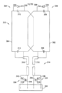
the invention is
equally capable of being adapted to obtain images of and/or spectra from other
regions of
interest, such as those accessible through the mouth, the vagina, or other
orifices penetrable
by an intracavity probe. It should also be apparent that the principles
presented herein may
also be applied to a wide variety of surface coil arrays, such as those
intended for imaging of
the head, neck, torso, limbs, and other structures of the body.
[0075] In general, the endorectal coils disclosed herein include a two element
layout that
has been configured to receive radio frequency (RF) currents from the whole
geometry and,
using appropriate splitters and combiners in an interface device discussed
hereinafter, turned
into a four channel output device.
[0076] With reference to FIG. 6, various aspects of a first embodiment of the
endorectal
coil array are illustrated. More specifically, FIG. 6 illustrates a schematic
diagram of a
prototype of the first embodiment of the endorectal coil array built for
operation with 1.5T
MR.! scanners.
- 17-
CA 02803252 2012-12-18
WO 2012/003211
PCT/US2011/042354
[0077] The endorectal coil, generally designated 100, includes an outer loop
102, a center
conductor 104 bisecting outer loop 102, and an output line, generally
designated 106. The
outer loop 102 includes a plurality of capacitors including first and second
drive capacitors
108 and 110 and first and second tuning capacitors 112 and 114. Of
approximately equal
values, the drive capacitors 108 and 110 are serially deployed within the
outer loop 102 and
at their junction node 116 form a virtual ground for electrically balancing
and impedance
matching the loop. Tuning capacitors 112 and 114 are also serially deployed
within outer
loop 102, with their common node 118 being situated diametrically opposite the
junction
node 116. Of approximately equal values, the tuning capacitors 112 and 114 are
selected to
resonate the outer loop 102 at the operating frequency of the MR system.
[0078] In this manner, the outer loop 102 of FIG. 6 has been tuned to detect
MR signals
emanating from the patient at the operating frequency of a 1.51 MR system. The
shape of
outer loop 102 dictates that the loop is capable of detecting only those MR
signals whose
field lines are oriented vertical to the plane of the loop. The aforementioned
tuning scheme,
however, also imposes a 180 degree phase shift upon the resulting voltage
signals output by
outer loop 102 representative of the vertically oriented MR signals it
detects. Specifically,
relative to the virtual ground at junction node 116, the phase of the voltage
signals detectable
across the first drive capacitor 108, i.e., at a first port, is 180 degrees
from the phase of the
voltage signals detectable across the second drive capacitor 110, i.e., at a
second port.
[0079] The center conductor 104 extends between and evenly bisects the
junction and
common nodes 116 and 118 of outer loop 102, and thus maintains the physical
and electrical
symmetry of the coil 100. FIG. 6 shows the center conductor 104 as having a
tuning
capacitor 120 deployed along its length. The value of the tuning capacitor 120
has been
selected so that its reactance at the operating frequency equals the inductive
reactance of
center conductor 104. This permits two modes of operation to occur
simultaneously. First, the
equal inductive and capacitive reactances enable center conductor 104 to act
as an open
circuit relative to outer loop 102. In such an instance, a first channel
output representative of
the whole loop (shown by arrow WL) is provided.
[0080] Beyond acting as an open circuit for outer loop 102 to enable detection
of the
vertical components of the MR signal, the center conductor 104 also operates
with outer loop
102 to emulate a butterfly-type or saddle-type coil for detecting MR signals
oriented parallel
to the plane of the coil 100. The tuning scheme of the present invention
creates not only a
simple loop current path for outer loop 102 but also an alternative current
path (involving
counter-rotating currents) for the outer loop 102 and the center conductor 104
combined.
- 18 -
CA 02803252 2012-12-18
WO 2012/003211
PCT/US2011/042354
Specifically, during the receive cycle and starting near junction node 116,
the current induced
by the horizontally-oriented MR signals flows across the second drive
capacitor 110 up to the
far end of outer loop 102 and into and down to center conductor 104. It then
crosses the
midpoint of the butterfly or saddle structure and flows across the first drive
capacitor 108 up
to the far end of outer loop 102 and into and down to center conductor 104 to
start the cycle
anew as long as the coil 100 is in position to detect MR signals during the
receive cycle of
operation. In such an instance, a second channel output representative of the
saddle/butterfly
mode (shown by arrow SL) is provided.
[00811 The output line 106 for the coil 100 can be implemented using various
mechanisms
such as coaxial cable, stripline, microstrip, or other transmission line
technologies. FIG. 6
shows two coaxial cables 122 and 124 with the shield conductor of each
connected to the
junction node 116 of the coil. The center conductor of cable 122 connects to
the other side of
the first drive capacitor 108, while the center conductor of cable 124
connects to the other
side of the second drive capacitor 110. The output line 106 should have an
electrical length of
SL + n(X14) for the reasons disclosed in U.S. Patent Application Publication
No.
2009/0076378. X. is the wavelength of the operating frequency of the MR system
and n is an
odd integer whose value will typically be (and is hereinafter treated as
being) equal to 1 as
the coil 100 will in practice always be reasonably close to the interface
device to which it will
connect. SL represents an additional length whose inductive reactance is of
the same
magnitude as the reactance of each of the first and second drive capacitors
108, 110 across
which the terminals of output line 106 connect. With a standard plug
accommodating the
conductors of both cables, for instance, the center and shield conductors of
each cable 122
and 124 connect to a suitable socket or other type connector for the interface
device.
[00821 In addition, based on an RF splitter configuration of the interface
device discussed
hereinafter, two channels may also be provided to obtain a left loop signal
(shown as arrow
LL in FIG. 6) and a right loop signal (shown as arrow RL in FIG. 6) with the
center
conductor 104 serving as a common conductor for both the loops.
[00831 During trial tests of the coil 100, it was determined that the signal-
to-noise ratio
(SNR), while superior to current endorectal coils, was not as high as desired.
In addition, the
images obtained using this coil 100 produced unsatisfactory ghosting artifacts
as will be
discussed in greater detail hereinafter.
[00841 Accordingly, a second embodiment of the coil was developed in an
attempt to
obtain a higher SNR. With reference to FIG. 7, this coil, generally designated
200, includes a
first coil loop 202 and a second coil loop 204. The pair of coil loops 202 and
204 is arranged
- 19 -
CA 02803252 2012-12-18
WO 2012/003211
PCT/HS2011/042354
in a phased array configuration each of which receive MR signals from the
region of interest
corresponding thereto. The first coil loop 202 includes a drive capacitor 206
and a tuning
capacitor 208. The tuning capacitor 208 has a value selected to resonate the
first coil loop 202
at the operating frequency of the MR system. The second coil loop 204 includes
a drive
capacitor 210 and a tuning capacitor 212. The tuning capacitor 212 has a value
selected to
resonate the second coil loop 204 at the operating frequency of the MR system.
[0085] The coil 200 also includes an output line 214 that includes two coaxial
cables 216
and 218. The first coaxial cable 216 is connected at a first end thereof
across the first drive
capacitor 206 and the second coaxial cable 218 is connected at a first end
thereof across the
second drive capacitor 210, such that each of the drive capacitors 206 and 210
share a
common ground. This configuration can be referred to as a hybrid overlap
configuration. A
standard plug accommodates the conductors of both cables at a second end
thereof, for
instance, the center and shield conductors of each cable 216 and 218, such
that the output line
214 can be connected to a suitable socket or other type of connector for the
interface device.
The output line 214 should also have an electrical length of SL + n(V4) for
the reasons
discussed hereinabove.
[0086] Accordingly, the second embodiment of coil 200 also includes two
elements
first coil loop 202 and second coil loop 204) and is configured to provide a
four channel
output. More specifically, coil 200 is configured to provide a first channel
output
representative of the whole loop, and a second channel output representative
of the
saddle/butterfly mode. In addition, based on an RF splitter configuration of
the interface
device discussed hereinafter, a third channel output may be provided to obtain
a left loop
signal, and a fourth channel output may be provided to obtain a right loop
signal.
[0087] However, during trial tests of the coil 200, while the SNR of this coil
configuration
was improved as compared to the first embodiment of coil 100, the images
obtained using
this coil 200 continued to produce unsatisfactory ghosting artifacts.
[0088] An unwanted byproduct of the endorectal coils illustrated in FIGS. 6
and 7 in
typical use is excessive signal intensity near the coil conductor, due to the
close proximity of
the coil conductor to the tissues of the rectal wall. This signal intensity
far exceeds typical
signal levels in the analog signal path, and can lead to undesirable effects,
including the
Gibbs artifact, which can manifest itself as "ghosting" of the image, even if
the subject is
motionless. This artifact differs between scanner manufacturers, due to
varying degrees of
post-processing employed, and tends to be more apparent on older scanners and
signal
- 20-
CA 02803252 2012-12-18
WO 2012/003211
PCT/US2011/042354
processing systems. Other effects include signal saturation, where the
contrast near the coil
conductor is minimal, and thus no clinically useful image detail is available.
[0089] With reference to FIG. 8, an exemplary image produced by an MR system
using
the coil of FIG. 6 or FIG. 7 illustrating the ghosting artifacts produced in
the image is
provided. These ghosting artifacts 250 appear as small, light rings emanating
from the
position where the coil is provided. Ghosting artifacts are also referred to
as "motion
artifacts" in literature. However, these artifacts are produced in images
provided using the
coils of FIGS. 6 and 7 even in the absence of motion. These artifacts can be
classified as
"Gibbs artifacts" or edge/transition/ringing artifacts since they are observed
due to the Gibbs
phenomenon when there is a sudden or abrupt shift/jump in a signal level at
the input stage of
the image processing.
[0090] The Gibbs phenomenon, named after the American physicist J. Willard
Gibbs, is
the peculiar manner in which the Fourier series of a piecewise continuously
differentiable
periodic function behaves at a jump discontinuity. The Gibbs phenomenon can be
seen as the
result of convolving a Heaviside step function (if periodicity is not
required) or a square wave
(if periodic) with a sinc function. The oscillations in the sine function
cause the ripples in the
output.
[0091] In MR imaging, the Gibbs phenomenon causes artifacts in the presence of
adjacent
regions of markedly differing signal intensity. Gibbs artifacts are bright or
dark lines that are
seen parallel and adjacent to borders of abrupt intensity change (see element
250 in FIG. 8).
These artifacts are related to the finite number of encoding steps used by the
Fourier
transform to reconstruct an image.
[0092] It has been verified that Gibbs artifacts increase with an increase in
a signal level
transition. Coils 100 and 200 each include a common conductor. The common
conductor
design has significantly higher SNR than previous coil designs. However, these
coils also
have a much enhanced transition of the signal level in comparison with the
current coil
design. The presence of these artifacts can be reduced by changing the
software and/or
hardware of the MR scanner of the MR system. For instance, an enhanced
filtering
mechanism, such as a low pass filter, can be provided at the scanner to reduce
the ripple after
a transition from a region of low signal intensity to a region of high signal
intensity. In
addition, the software of the MR scanner of the MR system could also be
reformulated to use
a compensation algorithm aimed to cancel out the Gibbs or ringing artifacts.
Both of these
solutions are undesirable because they require expensive redesigns of the MR
scanner. A
- 21 -
CA 02803252 2012-12-18
WO 2012/003211
PCT/US2011/042354
preferred solution is to reduce the Gibbs artifacts by altering the coil
design because the coils
are inexpensive, disposable units.
[0093] Therefore, various tests led to the discovery that changes could be
made to the coil
and interface device to drastically reduce the presence of Gibbs artifacts.
First, it was
discovered that spacing the coil away from the surface reduces the transition
level.
Accordingly and with reference to FIGS. 9 and 10, each of the coil designs
discussed herein
includes a spacer material positioned adjacent to an anterior surface of the
coil. For instance,
the spacer material may include three strips 220, 222, and 224. The spacer
material strips
220, 222, and 224 have a thickness to assure a predetermined distance of hi
and h2 between
the coil and the region of interest, such as a prostate, when the intracavity
probe including the
coil is inserted into the cavity, such as the rectum, of the patient. The
spacer material strip
222 provided over the overlap of the coil loops has a greater thickness than
the spacer
material strips 220 and 224 at the outside of the coil loops because the
artifacts produced in
this region are greater than the artifacts produced at the edges. The
predetermined distance
provided by the spacer material strips is typically about 0.03 inches to about
0.06 inches. The
spacer material may be any material that is not detected by an MR system, such
as a foam
material. While the use of strips of spacer material was described
hereinabove, a continuous
sheet of spacer material may also be utilized.
[0094] By spacing the coil away from the surface, the transition from a region
of low
signal intensity to a region of high signal intensity is reduced, thereby
reducing the Gibbs
artifacts. More specifically, the endorectal coil in its current form consists
of a pair of coil
loops on a substrate, supported by and enclosed in a biocompatible balloon.
This balloon is
designed to be inflated to press the coil loops against the rectal wall to
ensure consistent coil
positioning and close contact to enable the best imaging of the prostate gland
(in this use
case). The fact that the wall thickness of the balloon that covers the coil
element is very small
(0.010 inches or less) results in close proximity of the coil conductors to
the rectal wall.
[0095] It is a known phenomenon that an electromagnetic field (and thus the
resultant
signal intensity as seen by the interface and scanner signal path) follows the
"Inverse Square
Law," which, applied to this case, means that the signal intensity is
inversely proportional to
the square of the distance from the coil conductor. In practical terms, it
means that a doubling
of the distance of the coil conductor from the closest part of patient's
anatomy to the coil
conductor will result in a signal intensity of 1/4th of the previous level in
that anatomy, while
the reduction in signal will become less apparent further into the region of
interest at right
angles to the plane of the coil conductors.
- 22-
=
CA 02803252 2012-12-18
WO 2012/003211
PCT/US2011/042354
[0096] Thus, using an arbitrary coil conductor spacing of 0.010 inches, and
signal level of
36,000 units (measured as a pixel value of a small region of an imaging
phantom representing
the patient's anatomy closest to the endorectal coil conductor), doubling the
spacing to 0.020
inches, for instance, will result in a reduction of signal intensity in the
same region to 9,000
units. Hence, a pre-determined spacing provided on top of the coil conductor
reduces the
signal intensity jump at the proximal region of the imaging volume, and thus
works favorably
to reduce the artifacts including Gibbs artifacts.
[0097] In addition, it was discovered that the signal could be reduced
accompanied by a
greater reduction in the noise to increase the SNR while reducing artifacts by
making minor
changes in the interface device. First, the interface device includes a pair
of preamplifiers as
will be discussed in greater detail hereinafter. It has been found that
providing the
preamplifiers with a predetermined reduced supply voltage as compared to a
rated supply
voltage of the preamplifiers has the effect of reducing the signal produced by
the coil;
however, this reduction in signal is accompanied by a greater reduction in
noise.
Accordingly, the SNR is increased. For example, these preamplifiers are
typically provided
with a supply voltage of 1 OV. It has been found that decreasing the supply
voltage of the
preamplifier to 5V, and the positioning of an attenuator having an attenuation
of between 3dB
and 9dB after the preamplifiers, has the effect of reducing the signal
produced by the coil.
This reduction in signal, however, is accompanied by a greater reduction in
noise.
Accordingly, the SNR is increased.
[0098] Finally, while the combination of spacing the coil away from the
surface and
applying the preamplifier with a reduced supply voltage of 5V lowers the Gibbs
artifacts
produced in the images significantly, the artifacts produced in the images are
still greater than
in current coil designs. Accordingly, it was discovered that reduced signal
intensity
associated with reduced artifacts without compromising on SNR could be
achieved by
utilizing a coil having an overlapped two loop design where the two loops do
not include a
common conductor or a common ground.
[0099] More specifically, with reference to FIG. 11, a third embodiment of the
endorectal
coil, generally designated as 300, includes a first coil loop 302 and a second
coil loop 304.
The pair of coil loops 302 and 304 is arranged in a phased array configuration
each of which
receive MR signals from the region of interest corresponding thereto. The
first coil loop 302
includes a drive capacitor 306 and a tuning capacitor 308. The tuning
capacitor 308 has a
value selected to resonate the first coil loop 302 at the operating frequency
of the MR system.
The second coil loop 304 includes a drive capacitor 310 and a tuning capacitor
312, The
-23 -
CA 02803252 2012-12-18
WO 2012/003211
PCT/US2011/042354
tuning capacitor 312 has a value selected to resonate the second coil loop 304
at the operating
frequency of the MR system.
[00100] The coil 300 also includes an output line 314 that includes two
coaxial cables 316
and 318. The first coaxial cable 316 is connected at a first end thereof
across the first drive
capacitor 306 and the second coaxial cable 318 is connected at a first end
thereof across the
second drive capacitor 310 such that each of the drive capacitors 306 and 310
is provided
with a separate ground.
[00101] Accordingly, the third embodiment of coil 300 also includes two
elements (i.e.,
first coil loop 302 and second coil loop 304) and is configured to provide a
four channel
output. More specifically, coil 300 is configured to provide a first channel
output
representative of the whole loop, and a second channel output representative
of the
saddle/butterfly mode. In addition, based on an RF splitter configuration of
the interface
device discussed hereinafter, a third channel output may be provided to obtain
a left loop
signal and a fourth channel output may be provided to obtain a right loop
signal.
[00102] A standard plug 320 accommodates the conductors of both cables at a
second end
thereof, for instance, the center and shield conductors of each cable 316 and
318 such that the
output line 314 can be connected to a suitable socket or other type of
connector for the
interface device.
[00103] It was also discovered that the previously described embodiments of
the coil were
not designed to operate within safe SAR limits. Accordingly, additional
decoupling circuitry
is required to achieve these safe SAR limits. More specifically, a first
active decoupling
circuit 322 is connected across the tuning capacitor 308 of the first coil
loop 302, and a
second active decoupling circuit 324 is connected across the tuning capacitor
312 of the
second coil loop 304. Each of these decoupling circuits 322, 324 include a PIN
diode 326,
330 and an inductor 328, 332 provided in series. During the transmit cycle,
the interface
device is configured to bias the PIN diodes 326, 330 on, thereby opening the
coil due to the
parallel resonance. In addition, a first passive decoupling circuit 334 is
provided at the second
end of the first coaxial cable 316, and a second passive decoupling circuit
336 is provided at
the second end of the second coaxial cable 318. Each of these passive
decoupling circuits
334, 336 includes series connected back-to-back diodes 338, 342 and a
capacitor 340, 344.
The passive decoupling circuits 334, 336 are configured to conduct in response
to the higher
voltages induced by the RF excitation field. The use of these passive
decoupling circuits 334,
336 removes the necessity for the output line 314 to have an electrical length
of SL + n(X/4).
Accordingly, the output line 314 may have any practical electrical length.
-24 -
CA 02803252 2012-12-18
WO 2012/003211
PCT/US2011/042354
[00104] An alternative configuration of the endorectal coil in accordance with
a fourth
embodiment of the present invention is illustrated in FIGS. 12, 13A, and 13B.
The endorectal
coil, generally designated as 400, includes a first coil loop 402 and a second
coil loop 404.
The pair of coil loops 402 and 404 is arranged in a phased array
configuration, each of which
receive MR signals from the region of interest corresponding thereto. The
first coil loop 402
includes a drive capacitor 406 and a tuning capacitor 408. The tuning
capacitor 408 has a
value selected to resonate the first coil loop 402 at the operating frequency
of the MR system.
The second coil loop 404 includes a drive capacitor 410 and a tuning capacitor
412. The
tuning capacitor 412 has a value selected to resonate the second coil loop 404
at the operating
frequency of the MR system.
[00105] The coil 400 also includes an output line 414 that includes two
coaxial cables 416
and 418. The first coaxial cable 416 is connected at a first end thereof
across the first drive
capacitor 406, and the second coaxial cable 418 is connected at a first end
thereof across the
second drive capacitor 410 such that each of the drive capacitors 406 and 410
is provided
with a separate ground.
[00106] Accordingly, the fourth embodiment of coil 400 also includes two
elements (i.e.,
first coil loop 402 and second coil loop 404) and is configured to provide a
four channel
output. More specifically, coil 400 is configured to provide a first channel
output
representative of the whole loop, and a second channel output representative
of the
saddle/butterfly mode. In addition, based on an RF splitter configuration of
the interface
device discussed hereinafter, a third channel output may be provided to obtain
a left loop
signal and a fourth channel output may be provided to obtain a right loop
signal. Each of
these modes is discussed in greater detail hereinafter with reference to FIGS.
15-22.
1001071 The coil 400 also includes a first passive decoupling circuit 422
connected across
the tuning capacitor 408 of the first coil loop 402, and a second passive
decoupling circuit
424 connected across the tuning capacitor 412 of the second coil loop 404.
Each of these
decoupling circuits 422, 424 includes series connected back-to-back diodes
426, 430 and an
inductor 428, 432. The passive decoupling circuits 422, 424 are configured to
conduct in
response to the higher voltages induced by the RF excitation field.
Accordingly, the passive
decoupling circuits 422, 424 cause the coil to function as an open circuit
during the RF
transmit cycle. It should be noted that the diode combinations of the passive
decoupling
circuits 422, 424 could also provide the functionality of an active decoupling
diode.
Accordingly, although FIG. 12 only shows a pair of passive decoupling circuits
422, 424, it
- 25 -
CA 02803252 2012-12-18
WO 2012/003211
PCT/US2011/042354
should be apparent to one skilled in the art that these passive decoupling
circuits 422, 424
could be configured as both passive and active decoupling circuits.
[001081 With specific reference to FIGS. 13A and 13B and continued reference
to FIG.
12, a standard plug 434 accommodates the conductors of both cables 416 and 418
of output
line 414 at a second end thereof, for instance, the center and shield
conductors of each cable
416 and 418. The output line 414 may also be provided with a handle 436 to
allow for easier
handling of the intracavity probe. The requirement of the output line 414
having an electrical
length of SL + n(X/4) is eliminated in this embodiment with the addition of an
intermediate
conduit 438. The intermediate conduit 438 has an input connector 440
corresponding to and
for connection with the plug 434 of the output line 414, and an output
connector 442 for
connection to an interface device 500. The intermediate conduit 438 also
includes a pair of
internal cables for connecting at one end thereof, respectively, to the
coaxial cables 416 and
418 of the intracavity probe via the input connector 440 and approximate
another end thereof
to the interface device 500 via the output connector 442. A pair of baluns 444
is also
provided. Each of the baluns 444 is interconnected between an end of one of
the internal
cables and the input connector 440. It should also be noted that the pair of
baluns 444 could
also be connected between the end of one of the internal cables and the output
connector 442
at both the output and input ends of the intermediate conduit 438. The
intermediate conduit
438 further includes at least one cable trap 446, and desirably two cable
traps 446 as shown in
FIG. 13B, connected thereabout. The cable traps 446 prevent undesired current
from flowing
on the shield conductors of the pair of internal cables of the intermediate
conduits. The
interface device 500 is connected to the MR system via a cable 448 and a
connector 450. The
cable 448 may have a cable trap 452 positioned thereabout.
[00109] Due to the desire for a small, flexible, damage-resistant intracavity
probe, whose
design minimizes the likelihood of any internal componentry protruding through
the balloon-
type enclosure in which it is housed, each of the coils discussed hereinabove
may be
constructed of a thin, flexible dielectric material with copper patterns
applied to both sides to
form not only the conductive pathways but also all of the capacitors required
for tuning and
decoupling. Moreover, as each of the coils is intended to be offered as an
integral part of a
one-use disposable intracavity probe, such a fabrication technique will aid in
the goal of
realizing substantial savings during the manufacture of the probes. This is
because the
fabrication process for "preprinted" coils will involve significantly less
labor and less time to
inspect the finished product as compared to coils made from discrete
components.
- 26 -
CA 02803252 2012-12-18
WO 2012/003211
PCT/US2011/042354
[00110] The most effective passive decoupling scheme for the 2-loop endorectal
coil
disclosed herein is to use a non-magnetic, pre-packaged set of 4 anti-parallel
silicon PIN
diodes in series with the appropriate inductor to affect a blocking impedance
across each
loop's tuning capacitor as shown in FIG. 12. However, this is a very expensive
solution for
the endorectal coil, as it is intended to be a "single use" device, to be
disposed of after a
single scan procedure.
[00111] In an effort to provide a safe, yet cost effective method of passive
decoupling, an
alternate method was employed for the endorectal coil as shown in FIG. 11,
Passive
decoupling circuits 334, 336 comprising a chip capacitor in series with an
inexpensive anti-
parallel signal diode (in this case, for example, the part was a BAV-99) are
connected
between the center conductor and shield of each of the coil's coaxial cables,
on a small PCB
that also mounts the coil's RF plug 320. The value of the chip capacitor was
chosen so that in
combination with the coaxial cable stub length of 8.5 inches (24 degrees at
64M1-Iz), an
inductive reactance equal to the capacitive reactance of the loop's drive
capacitor would
result in a parallel-resonant blocking circuit in the coil loop upon diode
conduction.
100112] If the unlikely condition occurs that a patient is scanned using the
endorectal coil
without its dedicated interface device (which provides coil-type detection and
active
decoupling bias circuitry), the coil loops will couple to the scanner's body
transmit coil and
generate a small RF voltage which will propagate from the coil elements to the
signal diodes
through the coaxial cable and chip capacitor. As this voltage rises above the
conduction
threshold of the diode pair, they will start to conduct, thus acting like a
resistor in series with
the capacitor. This results in the blocking impedance increasing across the
loop's drive
capacitor, and as a consequence, the loop current will be diminished. It can
be deduced that if
incrementally more RF power is applied to the loop, that power will serve to
cause the diodes
to conduct further, until they reach full conduction. At this "equilibrium"
point, the loop
serves as a voltage source, and most of the absorbed power is distributed
between the loop's
drive capacitor, the coaxial cable (which serves as a lossy inductance), the
decoupling chip
capacitor, and the diode pair.
[00113] Careful testing and implementation of this method is required using
temperature
testing of the various components and the coil loop itself to ensure that the
equilibrium point
does not result in any excessive component heating or excess SAR generated by
the coil loop.
In any event, this method serves as a "second fault" safety measure, and is
anticipated to be a
rare occurrence.
- 27 -
CA 02803252 2012-12-18
WO 2012/003211
PCT/US2011/042354
[00114] This method also confers a cost advantage in that the diode pair and
the
decoupling capacitor are located far enough from the imaging region of
interest that
commercial components with slightly magnetic properties are acceptable for
use.
Accordingly, the coil configuration illustrated in FIG. 11 is the presently
preferred
embodiment.
[00115] With reference to FIG. 14 and with continuing reference to FIGS. 12,
13A, and
13B, an embodiment of an interface device, generally denoted as reference
numeral 500, for
interfacing the coil 400 with the appropriate input port(s) of a 1.5T MR
system is illustrated.
[00116] The interface device 500 includes preamplifier networks 502 and 504,
power
splitter networks 506, 508, 510, and 512, a 180 degree combining network 514,
and a zero
degree combining network 516. Attenuators (not shown) are provided to
nominally attenuate
the signal from about 3dB to about 9dB. The attenuators may be positioned at
at least one of
the following locations: (a) between the first preamplifier network 502 and
the first power
splitter network 506; (b) between the second preamplifier network 504 and the
second power
splitter network 508; (c) after the first power splitter network 506; and (d)
after the second
power splitter network 508. The power splitter and combining networks may be
implemented with standard Wilkinson style designs, and the preamplifier
networks may
ideally be realized with 28dB nominal gain. The preamplifier networks 502 and
504 may be
implemented using commercially available miniature low-noise, 28dB gain
shielded units
with a tuned input circuit designed to present a low impedance (11 real) at
the Larmor
frequency. Close proximity of the preamplifier networks 502 and 504 to their
respective PIN
diodes 518, 520 (see FIG. 16) in view of the low impedance of the former
allows some
measure of isolation during the receive cycle from other surface coils (or
arrays of same)
used in conjunction with the coil 400. Passive protection diode networks are
included in
interface device 500 to prevent excessive RF voltage from damaging the
preamplifier
networks 502 and 504 during the RF transmit pulse should interface device 500
be
disconnected while inside the bore of the MR system during the transmit cycle.
These diode
networks also provide some decoupling for the coil in the same scenario.
[00117] The power splitter networks 506 and 508 are used as 50 ohm/0 degree
splitters at
the outputs of the preamplifier networks 502 and 504. The power splitter
networks 510 and
512 are also used as 50 ohm/0 degree splitters at one output of each of the
power splitter
networks 506 and 508. The other output of each of the power splitter networks
506 and 508 is
sent directly to a cable trap 522 which directs the output to one of four
channels 524, 526,
528, and 530. The two combining networks 514 and 516 are also configured as 50
ohm
-28-
CA 02803252 2012-12-18
WO 2012/003211
PCT/US2011/042354
devices. As a result, these four networks may be interconnected as shown by
means of four
equal-phase length 50 ohm coaxial cable, PCB stripline, microstrip, or other
transmission line
media. In addition, the output of the two combining networks 514 and 516 is
provided to a
pair of switching networks 532 and 534 which directs the signal to the
appropriate channel or,
optionally, to a 90 degree hybrid combining network 536 if the scanner or MR
system is
configured to operate in a spectroscopic mode.
[00118] As should be apparent based on the above description of the present
invention, the
endorectal coils disclosed herein include a two element layout that has been
configured to
receive radio frequency (RF) currents from the whole geometry and, using
appropriate
splitters and combiners in the interface device, turned into a four channel
output device. More
specifically, the two element common conductor layout disclosed herein can
produce two
loops or a loop and saddle combination based on the combiner networks from the
same
feedpoints. In addition, both of these unique field patterns can be obtained
separately such
that four (4) unique channels which have unique radiation patterns associated
with the RF
current distribution can be obtained. It is apparent to one of ordinary skill
in the art that one
can extend this by creating various other phase combination networks and get
useful signal in
addition to the above mentioned as well. There are numerous possibilities to
get various
different field patterns based on the splitter-combiner networks and achieve
more than 2 (3, 4,
6, 8, etc.) individual channel outputs. It is also possible to extend this
theory to more than two
elements and achieve the same advantage of increased channel output, which has
been
demonstrated to yield superior SNR than merely the two element contribution.
[00119] It is also apparent, given the greater number of receivers available
in current MR
scanners, that coil layouts having a smaller number of imaging elements can be
built with the
disclosed technology to take advantage of those fewer imaging elements to
create imaging
systems having a greater number of channels, thereby leading to lower costs.
For example, a
current 16 element coil layout utilizes 16 preamplifiers in the interface
device even for an 8
channel multiplexed or combined output. By utilizing the theory of the present
invention, a
coil having a 16 element coil element involves utilizing only 8 preamplifiers
to obtain a 16
channel output. This would significantly contribute to less complex circuitry
because half the
number of preamplifiers and associated circuitry are required; better cable
management
because half the number of RF cables are required to be managed; less
expensive to make;
easier to fabricate and tune, yet with maximized performance for the region of
interest
selected. In addition, such coil layouts provide increased depth of
penetration over current
- 29 -
CA 02803252 2012-12-18
WO 2012/003211
PCT/US2011/042354
coils because it is well known in the art that larger loops lead to a greater
depth of
penetration.
[00120] Having set forth the components of the present invention, the
operation of coil 400
and interface device 500 in various modes will be discussed hereinafter with
reference to
FIGS. 13-19. Although the following description is based on the coil 400
illustrated in FIG.
12, this description is equally applicable to the embodiments of the coil
illustrated in FIGS.
6, 7, and 11 as well.
[00121] Left Loop mode
[00122] With reference to FIGS. 15 and 16A-16C, the operation of coil 400 and
interface
device 500 when in Left Loop mode will now be described. During transmit mode,
PIN diode
518 is turned on by a signal from a PIN diode bias 538 (see FIG. 16C). This
PIN diode 518
provides an RF short circuit to ground, which is reflected as an open circuit
at coaxial cable
416. Accordingly, first coil loop 402 is isolated and shown in phantom in FIG.
15.
[00123] As shown in FIG. 15, during the receive cycle, current will only be
seen as
flowing through the second coil loop 404 due to the PIN diode 518 acting as an
RF "open" to
help in isolating the first coil loop 402. Accordingly, current flows in the
second coil loop
404 as shown by arrow ILL, as shown in FIG. 15.
[00124] With reference to FIGS. 16A-16C, the manner in which interface device
500
processes the voltage signals received from coaxial cable 418 is now
described. The signal
from the drive capacitor 410 is first sent through preamplifier network 504,
which amplifies
the voltage signal and passes the resulting amplified version to power
splitter network 508.
One of the outputs of the power splitter 508 is provided to a second power
splitter network
512 and the other output is sent to the cable trap 522. The cable trap 522
sends this signal to
the fourth output channel 530 representing the left loop signal.
[00125] The flow of current through interface device 500 when in Left Loop
mode is
shown by arrow ILL in FIGS. 16A-16C.
[00126] Right Loop mode
[00127] With reference to FIGS. 17 and 18A-18C, the operation of coil 400 and
interface
device 500 when in Right Loop mode will now be described. During transmit
mode, PIN
diode 520 is turned on by a signal from a PIN diode bias 538 (see FIG. 18C).
This PIN diode
520 provides an RF short circuit to ground, which is reflected as an open
circuit at coaxial
cable 418. Accordingly, second coil loop 404 is isolated and shown in phantom
in FIG. 17.
[00128] As shown in FIG. 17, during the receive cycle, current will only be
seen as
flowing through the first coil loop 402 due to the PIN diode 520 acting as an
RF "open" to
-30-
CA 02803252 2012-12-18
WO 2012/003211
PCT/US2011/042354
help in isolating the second coil loop 404. Accordingly, current flows in the
first coil loop
402 as shown by arrow IRL, as shown in FIG. 17.
[00129] With reference to FIGS. 18A-18C, the manner in which interface device
500
processes the voltage signals received from coaxial cable 416 is now
described. The signal
from the drive capacitor 406 is first sent through preamplifier network 502,
which amplifies
the voltage signal and passes the resulting amplified version to power
splitter network 506.
One of the outputs of the power splitter 506 is provided to a second power
splitter network
510 and the other output is sent to the cable trap 522. The cable trap 522
sends this signal to
the third output channel 528 representing the right loop signal.
[00130] The flow of current through interface device 500 when in Right Loop
mode is
shown by arrow IRL in FIGS. 18A-18C.
[00131] Right Loop and Left Loop or LL mode
[00132] The operation of coil 400 and interface device 500 when in LL mode,
will now be
described. During the receive cycle, when operating in LL mode, coil 400
operates in the
same manner as described hereinabove for both the Right Loop and Left Loop
modes.
However, interface device 500 functions in a slightly different manner.
Instead of turning on
one of PIN diodes 518 and 520 by a signal from a PIN diode bias 538, both of
these PIN
diodes 518 and 520 remain off such that both a Right Loop signal and Left Loop
signal, as
described hereinabove, are provided to the third output channel 528 and the
fourth output
channel 530, respectively. Accordingly, a 2-channel signal is provided to the
host scanner.
[00133] Whole Loop mode
[00134] With reference to FIGS. 19 and 20A-20C, during the receive cycle, the
current
induced by the vertically oriented MR signals within an outer loop of coil 400
can be
represented by IwL, as it is the signal current shown flowing into and out of
the loop.
[00135] In this configuration, as the current IwL flows through the first coil
loop 402 and
the second coil loop 404, no current is seen flowing through the right edge of
the second coil
loop 404 and the left edge of the first coil loop 402. Accordingly, the right
edge of the second
coil loop 404 and the left edge of the first coil loop 402 act as an open
circuit relative to the
current flowing in the outer loop shown by arrows IwL. Therefore, the right
edge of the
second coil loop 404 and the left edge of the first coil loop 402 are
illustrated in FIG. 19 in
phantom.
[00136] Having completed discussion of the manner in which current flows
through coil
400 during a receive cycle when in Whole Loop mode, a description of the
operation of
interface device 500 during the receive cycle of the MR system when in Whole
Loop mode
-31 -
CA 02803252 2012-12-18
WO 2012/003211
PCT/US2011/042354
will now be described with specific reference to FIGS. 20A-20C. Coil 400
outputs voltage
signals representative of MR signals of both horizontal and vertical
orientation. For ease of
description, the voltage signals representative of the MR signals of
horizontal orientation are
referred to herein as "0 degree horizontal voltage signals" because they have
the same phase
at each port. The voltage signals representative of the MR signals of vertical
orientation are
referred to as "0 degree vertical voltage signals" for those output across the
drive capacitor
406 of the first coil loop 402, and "180 degree vertical voltage signals" for
those output from
the drive capacitor 410 of the second coil loop 404.
[00137] With continued reference to FIGS. 20A-20C, the manner in which
interface
device 500 processes the voltage signals received from output cables 416 and
418 is now
described. Preamplifier networks 502 and 504 each amplify the voltage signals
they receive
and pass the resulting amplified versions to first and second power splitter
networks 506 and
508, respectively. The signals from the first and second power splitter
networks 506 and 508
are then sent to third and fourth power splitter networks 510 and 512 such
that the signals
produced thereby are subsequently sent to the 0 degree combining network 516.
Because the
horizontal voltage signals received from power splitter networks 506 and 508
are in phase,
zero degree combining network 516 is able to constructively combine them.
Simultaneously,
zero degree combining network 516 also cancels the 90 degree vertical voltage
signals
received from power splitter network 506 with the -90 degree vertical voltage
signals
received from power splitter network 508. This yields a whole loop signal that
is fed through
switching network 534 to cable trap 522. Cable trap 522 directs the signal to
the second
output channel 526. The flow of current through interface device 500 when in
Whole Loop
mode is shown by arrows Ivn, in FIGS. 20A-20C.
[00138] Whole Saddle mode
[00139] With reference to FIGS. 21 and 22A-22C, the operation of coil 400 and
interface
device 500 when in Whole Saddle mode will now be described. As shown in FIG.
21, during
the receive cycle, coil 400 is also capable of emulating a butterfly-type or
saddle-type coil for
detecting MR signals oriented parallel to the plane of coil 400. The tuning
scheme of coil 400
allows for a simple loop current path for an outer loop as discussed
hereinabove with
reference to the Whole Loop mode but also alternative current paths, involving
counter-
rotating currents, for the outer loop, the right edge of the second coil loop
404, and the left
edge of the first coil loop 402 in various combinations. One of these
combinations is the
Whole Saddle mode as shown in FIG. 21. The flow of current through the coil in
Whole
Saddle mode is illustrated by arrows 'SL in FIG. 21.
-32-
CA 02803252 2012-12-18
WO 2012/003211
PCT/US2011/042354
[00140] Accordingly, although not physically formed by twisting a conductive
loop in the
middle to form two subloops about a midpoint as is the case with prior art
saddle coils, such
as the coil illustrated in FIG. 2A, the coil structure of the present
invention, nevertheless
electrically emulates the operation of a saddle coil.
[00141] The phase of the "vertical" voltage signals detectable across the
drive capacitor
406 of first coil loop 402 at the first coaxial cable 416 is 180 degrees out
of phase with the
"vertical" voltage signals detectable across the drive capacitor 410 of the
second coil loop
404 at the second coaxial cable 418. The significance of this 180 degree phase
differential
becomes apparent in connection with the operation of interface device 500
discussed below.
[00142] Having completed discussion of the manner in which current flows
through coil
400 during a receive cycle when in Whole Saddle mode, a description of the
operation of
interface device 500 during the receive cycle of the MR system when in
Whole Saddle
mode will now be described with specific reference to FIGS. 22A-22C. Coil 400
outputs
voltage signals representative of MR signals of both horizontal and vertical
orientation.
[00143] The manner in which interface device 500 processes the voltage signals
received
from the first and second coaxial cables 416 and 418 is now described. The
preamplifier
networks 502 and 504 each amplify the voltage signals they receive and pass
the resulting
amplified versions to the first and second power splitter networks 506 and
508, respectively.
The signals from the first and second power splitter networks 506 and 508 are
then sent to
third and fourth power splitter networks 510 and 512 such that the signals
produced thereby
are subsequently sent to the 180 degree combining network 514. Because the
vertical voltage
signals received from power splitter networks 510 and 512 are out of phase by
180 degrees,
180 degree combining network 514 is able to constructively combine them. 180
degree
combining network 514 cancels the horizontal voltage signals received from
power splitter
network 510 with the horizontal voltage signals received from power splitter
network 512
because they are received in phase. This yields a saddle loop signal that is
fed through
switching network 532 to cable trap 522. Cable trap 522 directs the signal to
the first output
channel 524. The flow of current through interface device 500 when in Saddle
mode is shown
by arrows 'SL in FIGS. 22A-22C.
[00144] Whole Loop and Whole Saddle mode
[00145] The operation of coil 400 and interface device 500 when in Whole Loop
and
Whole Saddle mode, will now be described. During the receive cycle, when
operating in
Whole Loop and Whole Saddle mode, coil 400 operates in the same manner as
described
hereinabove for both the Whole Loop and Whole Saddle modes. However, interface
device
-33-
CA 02803252 2012-12-18
WO 2012/003211
PCT/US2011/042354
500 functions in a slightly different manner. Instead of ignoring the output
of the first channel
524 as in the Whole Loop mode or ignoring the output of the second channel 526
as in the
Whole Saddle mode, a 2-channel signal is provided to the host scanner
representing both the
Whole Loop and the Whole Saddle signal, as described hereinabove, and is
provided to the
third output channel 528 and the fourth output channel 530, respectively.
Accordingly, a 2-
channel signal is provided to the host scanner.
[00146] Right Loop, Left Loop, Whole Loop, Whole Saddle or LLLS mode
[00147] The operation of coil 400 and interface device 500 when in Right Loop,
Left
Loop, Whole Loop, Whole Saddle, or LLLS mode, will now be described. During
the receive
cycle, when operating in LLLS mode, coil 400 operates in the same manner as
described
hereinabove for each of the Right Loop, Left Loop, Whole Loop, and Whole
Saddle modes.
In addition, the interface device 500 is configured to provide each of these
signals to the host
scanner such that a 4-channel output is provided to the host scanner of the MR
system.
[00148] Loon/Saddle Spectroscopy mode
[00149] The operation of coil 400 and interface device 500 when in Loop/Saddle
Spectroscopy mode, will now be described. During the receive cycle, when
operating in
Whole Loop and Whole Saddle modes, coil 400 operates in the same manner as
described
hereinabove for both the Whole Loop and Whole Saddle modes. However, interface
device
500 functions in a slightly different manner. The manner in which interface
device 500
processes the voltage signals received from output cables 416 and 418 when in
Loop/Saddle
Spectroscopy mode is now described. Preamplifier networks 502 and 504 each
amplify the
voltage signals they receive and pass the resulting amplified versions to
first and second
power splitter networks 506 and 508, respectively. The signals from the first
and second
power splitter networks 506 and 508 are then sent to third and fourth power
splitter networks
510 and 512 such that the signals produced thereby are subsequently sent to
the 0 degree
combining network 516 and the 180 degree combining network 514. The signals
from the
combining networks 514 and 516 are then fed through switching networks 532 and
534. The
switching networks 532 and 534 direct the signals to the 90 degree combining
network 536,
the output of which is sent to the cable trap 522. Cable trap 522 directs the
signal to the first
output channel 524.
[00150] While interface device 500 was described hereinabove as being used
with coil
400, interface device 500 could be used with any of coils 100, 200, and 300 to
yield the
above described modes.
- 34 -
CA 02803252 2012-12-18
WO 2012/003211
PCT/US2011/042354
[00151] Accordingly, the present invention allows for a two element coil
layout, such as
coils 100, 200, 300, and 400, that can produce two loops or a whole loop and
whole saddle
combination based on the power splitter and combiner networks provided in the
interface
device 500. In addition, these unique field patterns can be obtained
separately to provide four
unique channels which have unique radiation patterns. In addition, various
steps have been
taken to reduce ghosting artifacts while still achieving a high signal-to-
noise ratio. With
reference to FIG. 23, a graph illustrating the signal-to-noise ratio of
various coils discussed
hereinabove is provided. The current prior art coil, such as the coil
illustrated in FIG. 1A,
provides the lowest signal-to-noise ratio as shown by line 600. While the
common conductor
design of the coil 100 of FIG. 6 shows an improved signal-to-noise ratio as
shown by line
610, this increase was not as high as desired. Accordingly, the design for
coil 400 of FIG. 12
was reached and provided a much higher signal-to-noise ratio as shown by line
620.
However, ghosting artifacts were produced in images created by this coil.
Accordingly, it was
discovered that reducing the preamplifier power supply from 10V to 5V would
reduce the
ghosting artifacts. While this slightly reduces the signal-to-noise ratio
achieved by the coil, it
still has a higher signal-to-noise ratio than both the conventional coil and
the common
conductor coil 100 as shown by line 630.
[00152] Although the invention has been described in detail for the purpose of
illustration
based on what is currently considered to be the most practical and preferred
embodiments, it
is to be understood that such detail is solely for that purpose and that the
invention is not
limited to the disclosed embodiments, but, on the contrary, is intended to
cover modifications
and equivalent arrangements. For example, it is to be understood that the
present invention
contemplates that, to the extent possible, one or more features of any
embodiment can be
combined with one or more features of any other embodiment.
[00153] Accordingly, to promote the progress of science and the useful arts,
the inventor(s)
hereby secure by Letters Patent exclusive rights to all subject matter
embraced by the
following claims for the time prescribed by the Patent Act.
- 35 -