Note: Descriptions are shown in the official language in which they were submitted.
CA 02811450 2013-03-14
WO 2012/037257 PCT/US2011/051601
2240-308278
ROBOTIC SYSTEM TO AUGMENT ENDOSCOPES
CROSS-REFERENCE OF RELATED APPLICATION
[0001] This application claims priority to U.S. Provisional Application
No.
61/382,557 filed September 14, 2010, the entire contents of which are hereby
incorporated by
reference.
BACKGROUND
1. Field of Invention
[0002] The field of the currently claimed embodiments of this invention
relates to
robotic systems, and more particularly to robotic systems to augment
endoscopes.
2. Discussion of Related Art
[0003] Many different types of operations require the use of a clinical
endoscope,
including laparoscopic surgery, many GI tract surgeries, many sinus surgeries,
and trans-oral
laryngeal and tongue-based surgeries (Iro et al. Minimally Invasive Surgery in
Oto-Rhino-
Laryngology. European Archives of Oto-Rhino-Laryngology. Volume 250, Number 1,
1993;
Vaughan, Charles et al. Laryngeal Carcinoma: Transoral Treatment Utilizing the
CO2 Laser
The American Journal of Surgery, Volume 136, Issue 4, October 1978, pp 490-
493; Taylor,
Russell et al. Computer Integrated Surgery Technology and Applications. pp 603-
617.
Boston Massachusetts: The MIT Press, 1996). These surgeries utilize two main
types of
endoscopes: flexible and rigid. When more direct access to the target area is
possible, a rigid
endoscope is normally used, but when such access is not possible, such as in
GI tract surgery,
or deep trans-oral laryngeal surgery, a flexible endoscope must be used.
Robotic
manipulation of rigid endoscopes is quite well developed, particularly for
laparoscopic
surgery with systems such as the Automated Endoscope System for Optimal
Positioning
(AESOP) and the DaVinci surgical system. There are two main approaches for
robotically
controlling an endoscope. The most common approach in the literature is to
fully engineer a
completely robotic endoscope from scratch, which provides excellent control,
but is time
1
CA 02811450 2013-03-14
WO 2012/037257 PCT/US2011/051601
2240-308278
consuming and expensive. The second approach is to build a robot to control a
pre-existing
clinical endoscope. The DaVinci system uses a custom endoscopic camera as part
of its
system, whereas AESOP manipulates a pre-existing rigid clinical endoscope
(Taylor, Russell
et al. Computer Integrated Surgery Technology and Applications. pp 577-580.
Boston
Massachusetts: The MIT Press, 1996; Taylor, Russell etal. Computer Integrated
Surgery
Technology and Applications. pp 581-592. Boston Massachusetts: The MIT Press,
1996;
Horgan et al. Robots in Laparoscopic Surgery. Journal of Laparoendoscopic &
Advanced
Surgical Techniques; Volume 11, Number 6, 2001).
[0004] Robotic manipulation of flexible endoscopes, however, is far less
developed,
since they are inherently more difficult for a robot to control given their
flexibility. There
has been some work in robotic flexible endoscopes for GI tract surgery, but
this has mainly
involved complex custom engineered solutions rather than manipulation of
clinical
endoscopes (Taylor 1996 pp 577-580). One example of robotic manipulation of a
clinical
flexible endoscope is the pneumatic system proposed by Suzumori et al
(Suzumori et al. New
pneumatic rubber actuators to assist colonoscope insertion. Proceedings 2006
IEEE
International Conference on Robotics and Automation. ICRA 2006). This system
uses
pneumatic actuators to assist in the insertion of a clinical colonoscope. This
system is highly
adapted to colonoscopy however, relying on contact friction against the colon
walls to
generate force. It also does not manipulate the endoscope body or end
effector, since the
pneumatic actuators only act on the flexible part of the endoscope shaft.
Another approach
was taken by Shin et al. for laparoscopic surgery (Shin et al. Design of a
Dexterous and
Compact Laparoscopic Assistant Robot. SICE-ICASE International Joint
Conference 2006).
They made a custom laparoscope consisting of a rigid shaft and a rigid end
effector joined by
a cable operated flexure. The body and end effector of the endoscope were then
robotically
controlled (Shin 2006).
[0005] A hand-held flexible endoscope manipulator is also known from Eckl
et al.
(Ekcl R. et al. Comparison of manual Steering and Steering via Joystick of a
flexible Rhino
Endoscope. 32nd Annual International Conference of the IEEE EMBS Buenos Aires,
Argentina, August 31 - September 4, 2010). Their system manipulates a flexible
endoscope
using a hand-held pistol-grip manipulator that controls scope rotation and tip
angle, but not
2
CA 02811450 2013-03-14
WO 2012/037257 PCT/US2011/051601
2240-308278
translation. They have also shown the ability to attach this system to a
passive arm, and
control it with a joystick. In both cases, this system lacks a translational
motion degree of
freedom, which makes full robotic operation impossible. There thus remains a
need for
improved robotic systems to augment control over endoscopes.
SUMMARY
[0006] A robotic system for steerable tip endoscopes according to an
embodiment of
the current invention includes a support arm, an endoscope gripping assembly
rotatably
connected to the support arm by a rotation assembly, and a translation
assembly operatively
connected to the support arm. The endoscope gripping assembly is configured to
grip any
one of a plurality of differently structured endoscopes, the translation
assembly is configured
to move the support arm along a linear direction to thereby move an endoscope
when held by
the endoscope gripping assembly along an axial direction, and the rotation
assembly is
configured to rotate the endoscope along a longitudinal axis of rotation.
[0007] A robotically assisted or controllable flexible endoscope system
according to
an embodiment of the current invention includes a support arm, an endoscope
gripping
assembly rotatably connected to the support arm by a rotation assembly, a
steerable tip
endoscope held by a gripping mechanism of the endoscope gripping assembly, and
a
translation assembly operatively connected to the support arm. The translation
assembly is
configured to move the support arm along a linear direction to thereby move
the endoscope,
and the rotation assembly is configured to rotate the endoscope along a
longitudinal axis of
rotation.
BRIEF DESCRIPTION OF THE DRAWINGS
[0008] Further objectives and advantages will become apparent from a
consideration
of the description, drawings, and examples.
3
CA 02811450 2013-03-14
WO 2012/037257 PCT/US2011/051601
2240-308278
[0009] Figure 1 is an illustration of an example of a flexible endoscope
that can be
used with and/or incorporated as part of a robotic system according to
embodiments of the
current invention.
[0010] Figure 2 shows an example of a robotic system for steerable tip
endoscopes
according to an embodiment of the current invention.
[0011] Figure 3 shows another view a robotic system for steerable tip
endoscopes
according to an embodiment of the current invention.
[0012] Figure 4 shows another view a robotic system for steerable tip
endoscopes
according to an embodiment of the current invention.
[0013] Figure 5 shows another view a robotic system for steerable tip
endoscopes
according to an embodiment of the current invention.
[0014] Figure 6 shows another view a robotic system for steerable tip
endoscopes
according to another embodiment of the current invention.
[0015] Figure 7 shows a control unit that can be included in a robotic
system for
steerable tip endoscopes according to an embodiment of the current invention.
[0016] Figure 8 shows a view of a rotation assembly and an endoscope tip
control
assembly for a robotic system for steerable tip endoscopes according to an
embodiment of
the current invention.
[0017] Figure 9 shows water-tight covers for the rotation assembly and
the
endoscope tip control assembly of Figure 8.
[0018] Figure 10 shows a view of a translation assembly for a robotic
system for
steerable tip endoscopes according to an embodiment of the current invention.
[0019] Figure 11 shows a view of an electronics unit for a robotic system
for
steerable tip endoscopes according to an embodiment of the current invention.
4
CA 02811450 2013-03-14
WO 2012/037257 PCT/US2011/051601
2240-308278
[0020] Figure 12 shows a view of a translation assembly for a robotic
system for
steerable tip endoscopes according to another embodiment of the current
invention in which
additional motor components are also included.
[0021] Figure 13 shows a view of a rotation assembly for a robotic system
for
steerable tip endoscopes according to another embodiment of the current
invention.
[0022] Figure 14 shows a view of an endoscope tip control assembly for a
robotic
system for steerable tip endoscopes according to an embodiment of the current
invention.
DETAILED DESCRIPTION
[0023] Some embodiments of the current invention are discussed in detail
below. In
describing embodiments, specific terminology is employed for the sake of
clarity. However,
the invention is not intended to be limited to the specific terminology so
selected. A person
skilled in the relevant art will recognize that other equivalent components
can be employed
and other methods developed without departing from the broad concepts of the
current
invention. All references cited anywhere in this specification, including the
Background and
Detailed Description sections, are incorporated by reference as if each had
been individually
incorporated.
[0024] Many clinical applications require the use of an endoscope with a
flexible end
effector, such as ablation of laryngeal tumors. Such operations are typically
performed by
two surgeons, with one surgeon using both hands to manipulate the endoscope,
and another
using a surgical laser and a tissue manipulation instrument. This results in a
crowded
operating room environment, and a need for significant coordination between
two surgeons,
thus increasing the difficulty and overall cost of the operation. Some
embodiments of the
current invention can solve these problems by using a robotic system to
manipulate the
endoscope. Since modern clinical endoscopes have a working channel that a
laser fiber can
pass through, the laser can also be incorporated into the endoscope. The
robotic system can
allow single handed operation of the endoscope with the laser inside, allowing
one surgeon to
perform the entire operation using one hand to manipulate the endoscope/laser
and one to use
CA 02811450 2013-03-14
WO 2012/037257 PCT/US2011/051601
2240-308278
a tissue manipulation instrument. Since the weight of the endoscope and the
force needed to
manipulate the handle can both be handled by the robot, surgeon fatigue can be
reduced as
well.
[0025] The robot can hold the endoscope in a fixed position or precisely
move each
degree of freedom with virtually no tremor, thus improving surgical accuracy.
Since the
endoscope outputs a digital video signal, it is also possible for the robot to
utilize this to
provide more advanced features, such as image stabilization, 3D reconstruction
from
endoscopic images using endoscope motions to create a stereo baseline, image
overlay of
relevant data on the endoscope video feed, detailed recording of the endoscope
motions used
in a surgical procedure, which could later be used for training or position
recall, and virtual
fixtures for added safety.
[0026] Some embodiments of the current invention can provide a compact,
sterilizable, robust, accurate, robotic system for operating an unmodified
clinical flexible
endoscope with one hand. This can reduce the number of personnel needed to
perform many
operations, and also can keep the endoscope in position if the surgeon needs
to release it to
perform another task. The introduction of a robotic system between the surgeon
and the
endoscope can also increase accuracy, since hand tremor can be largely
eliminated. By
robotically supporting and manipulating the endoscope, surgeon fatigue can be
reduced as
well. Also a rigid endoscope rather than a flexible one can be used, if
desired.
[0027] Figure 1 is an illustration of an example of a flexible endoscope
100 that can
be used with or incorporated into embodiments of the current invention. The
flexible
endoscope 100 can be, but is not limited to, a conventional hand-held flexible
endoscope.
The flexible endoscope can be, for example, a laryngoscope, a colonoscope, a
bronchoscope,
or any of a variety of flexible endoscopes. The endoscope 100 has a hand piece
102 at a
proximal end and a flexible tip 104 at a distal end of the flexible endoscope
100. The
endoscope 100 also has a flexible shaft 106 and an eyepiece 108. The eyepiece
108 can be
used for direct viewing by an observer, or can be attached to an image pickup
device, such as
a video camera, for example. In this example, the flexible endoscope 100 has a
knob 110
that can be used manually to control the flexible tip 104.
6
CA 02811450 2013-03-14
WO 2012/037257 PCT/US2011/051601
2240-308278
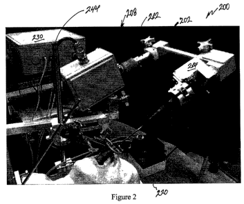
[0028] Figure 2 shows an embodiment of a robotic system 200 for steerable
tip
endoscopes according to an embodiment of the current invention. Figures 3-6
show
additional views of the robotic system 200. The robotic system 200 includes a
support arm
202, an endoscope gripping assembly 204 rotatably connected to the support arm
202 by a
rotation assembly 206, and a translation assembly 208 operatively connected to
the support
arm 202. The endoscope gripping assembly 204 is configured to grip any one of
a plurality
of differently structured endoscopes. The translation assembly 208 is
configured to move the
support atm 202 along a linear direction to thereby move an endoscope when
held by said
endoscope gripping assembly 204 along an axial direction. The rotation
assembly 206 is
configured to rotate the endoscope along a longitudinal axis of rotation.
[0029] In the embodiment of Figure 2, a bellows 210 encloses a moveable
section of
the translation assembly 208 to keep it water proof to facilitate cleaning and
sterilization.
The support arm 202 is articulated with a straight segment 212 that moves
linearly in
response to operation of the translation assembly 208.
[0030] The robotic system 200 also includes an endoscope tip control
assembly 214
adapted to be attached to the endoscope 216 to permit control of a flexible
tip of the
endoscope 216. Portions of the translation assembly 208 as well as electronics
are contained
within the waterproof box 218.
[0031] The robotic system 200 can also include a control unit 220 to
allow a user to
directly control at least one of the translation assembly 208, the rotation
assembly 206 or the
endoscope tip control assembly 214. Figure 7 shows a more detailed view of an
embodiment
of the control unit 220. In this example, the control unit 220 has an
emergency shut off
switch 222, a two-dimensional joystick 224 and a one-dimensional joystick 226.
[0032] Figures 2-6 of the robotic system 200 show the translation
assembly 208, the
rotation assembly 206 and the endoscope tip control assembly 214 contained
within water-
tight enclosures to facilitate cleaning and sterilization for surgical use. In
some
embodiments, components of the assemblies can be localized within a single
containment
structure, or they could have components distributed over containment
structures. Figure 8
shows an example in which the endoscope-tip control assembly 214 and the
rotation
7
CA 02811450 2013-03-14
WO 2012/037257 PCT/US2011/051601
2240-308278
assembly 206 have electric motors that are fully contained within the
respective structures.
The structures are open in the view of Figure 8 to show the interior
components. Figure 9
shows corresponding covers that can include o-rings for sealing the
containments structures
such that they are watertight. The endoscope tip control assembly 214 also
includes a spring
actuated clamp 228 to clamp on to the control knob for the flexible tip of the
endoscope so
that it can be turned by the endoscope tip control assembly 214. Figure 19
shows the interior
of the containment structure for the translation assembly 208 in which an
electric motor
drives a screw component. Figure 11 shows the interior of the electronics
container with the
top open.
[0033] Figure 12 shows an alternative embodiment of the interior of the
motor
enclosure 218 that contains translation assembly 208 as well as motors for the
rotation
assembly 206 and the endoscope tip control assembly 214. In this embodiment, a
motor 232
drives translation stage with a belt connected to the screw 234 for the linear
guide block and
rail assembly for translation stage 236. An Acme screw is suitable for screw
234 in some
embodiments. Motor 238 drives the rotation stage via a pulley and Bowden
cables. Motor
240 drives the distal tip control knob. A waterproof connector 242 is provided
for all
electrical connections. Figures 13 and 14 show the interiors of the rotation
assembly 206 and
endoscope tip control assembly 214 corresponding to the embodiment of Figure
12 in which
Bowden cables run through the support arm 202.
[0034] The robotic system 200 can also include an image pickup system
connected to
the endoscope gripping assembly 204 according to some embodiments such that
the image
pickup system can be rotated by the rotation assembly 206 along with the
endoscope 216.
The image pickup system can be, but is not limited to, a video camera.
[0035] The robotic system 200 can also include a support frame 244 in
some
embodiments that is adapted to hold the support arm 202. The support frame can
be a free-
standing support frame, or can be adapted to mount to another structure. In
the embodiment
of Figure 6, the support frame 244 has a bedrail mount 246 such that the
robotic system 200
can be attached to a bedrail 248. In this example, the control unit 220 is
also mounted to the
bedrail 248 with a bedrail mount 250.
8
CA 02811450 2013-03-14
WO 2012/037257 PCT/US2011/051601
2240-308278
[0036] In operation, the robotic system 200 can be fully autonomous,
remotely
operated, or locally operated, for example by the use of control unit 220. The
robotic system
can be placed into rough proximity to where it will be used. The translation
assembly 208
moves the section 212 of the support arm 202 back and/or forth in a linear
direction. This
translates the endoscope 216 back and forth along a linear direction to extend
more or less
along the path of interest. The rotation assembly 206 rotates the body of the
endoscope 216
similar to how one would rotate the body of an endoscope by hand. The
endoscope tip
control assembly 214, which is connected to the control knob of the endoscope
216, rotates
the control knob back and/or forward to effect motion of the flexible tip of
the endoscope.
[0037] The embodiments shown above have three degrees of control, i.e.,
translation
of the endoscope along a linear path, rotation of the endoscope about an axis
of the
endoscope handle, and control of the flexible tip of the endoscope. Other
embodiments could
include robotic and/or robot assisted control of additional degrees of
freedom, if desired.
EXAMPLE 1
[0038] A prototype was constructed using an old clinical laryngoscope and
the
Laparoscopic Assisted Robotic System (LARS) robot from the Laboratory for
Computational
Sensing and Robotics at Johns Hopkins University, augmented in the following
ways:
= The scope was attached to the LARS using a custom made adaptor
= The scope's handle was controlled using a custom made linkage and servo
motor
system attached to the adaptor
= Custom electrical systems were added to control this extra motor
= The program of the LARS was heavily modified to integrate these
additional
systems, and also to change its behavior to be appropriate for this
application
[0039] The prototype was successfully tested using a rubber airway
phantom.
9
CA 02811450 2013-03-14
WO 2012/037257 PCT/US2011/051601
2240-308278
[0040] Three degrees of freedom are often desired for the robotic control
of the
flexible laryngoscope; one to translate the endoscope in and out of the
airway, one to rotate
the endoscope along its axis through the airway, and one to control the tip of
the endoscope.
[0041] A plastic (delrin) adaptor was machined to securely hold the
endoscope and
interface it to the LARS. We chose a servo motor to control the scope handle,
and machined
aluminum brackets to attach the motor to the adaptor. To interface the motor
to the scope
handle we considered both a timing belt system and a 4 bar linkage, and chose
the latter for
simplicity and adjustability. The linkage was machined from aluminum. Since
the
endoscope requires an external camera, and the camera was not rotationally
fixed to the
scope, we designed and fabricated an aluminum camera holder to keep the camera
fixed with
respect to the scope. We also machined an aluminum bracket to hold the
connector for the
motor wiring to reduce strain on the motor wires.
[0042] An additional microprocessor was added to the LARS robot since it
was not
able to control an additional servo necessary for scope function. For power,
we tapped into
the 12V DC supply of the LARS, and used a switching voltage regulator to
achieve the 5V
needed for the servo. Since this type of servo does not have position feedback
we custom
modified it to provide one by tapping into its internal position feedback.
Since this signal is
very noisy due to sharing its ground with the servo motor's power ground, we
added a
buffered low-pass filter before passing the signal to the microprocessor's A/D
converter. The
microprocessor and associated electronics are all contained in their own
enclosure which is
not coupled to the scope.
[0043] The LARS robot software was modified to adapt it to the novel
task. We used
a 3D space mouse to control the LARS two 'degrees of freedom as well as the
scope tip
movement. The complicated dynamics of the scope tip motion relative to the
handle motion
can lead to problems. The tip motion is both highly nonlinear and exhibits
significant
hysteresis. Hysteresis compensation was added to the software to compensate
for this.
[0044] We tested the finished prototype with a phantom airway model. We
were able
to freely navigate the inside of the airway using the prototype. Notable
improvements in
stability and accuracy over using the scope by hand were achieved.
CA 02811450 2013-03-14
WO 2012/037257
PCT/US2011/051601
2240-308278
EXAMPLE 2
[0045] A fully functional robotically-controlled distal-tip
flexible laryngoscope that
meets the appropriate safety standards for operating room use was constructed.
This
embodiment includes some or all of the following features:
= Robot is fully enclosed and sealed, making it suitable for wash-down
applications.
= Robot is designed to mount easily to a Chung retractor for easy
attachment to a
surgical bed.
= Robot uses an easily changeable molded rubber adaptor to hold the
endoscope,
and an adjustable spring-loaded manipulator to control the endoscope handle,
making it easy to use different models of endoscope.
= No modifications to the endoscope are necessary since the endoscope
handle
manipulator simply cradles the endoscope handle.
= Robot has adjustable joints which allow the surgeon to configure it as
needed.
= Robot's main body is over the side of the bed, thus minimizing the amount
of
weight and bulk over the patient.
= Robot can include an adjustable malleable support for the flexible shaft
of the
endoscope to prevent it from drooping.
[0046] One embodiment used a Bowden cable mechanism to move the
scope handle
manipulator with the driving motor in the motor enclosure. In other
embodiments, these can
= be replaced by a motor and linkage placed directly in the endoscope
holder enclosure. The
rotation of the endoscope is achieved via a Bowden cable pulley system
actuated by a motor
in the motor enclosure.
11
CA 02811450 2013-03-14
WO 2012/037257 PCT/US2011/051601
2240-308278
Adjustable Malleable Endoscope Support:
[0047] The robot can also include an adjustable malleable support for the
un-actuated
flexible portion of the endoscope. This can allow the surgeon to bend the
endoscope roughly
into a desired configuration, and then manipulate it with the robot
essentially as though the
shaft were rigid. The support can include a bendable metal wire encased in
medical grade
rubber tubing, for example, which can be fixed to the endoscope shaft either
by wrapping it
around the shaft, or connecting it with surgical rubber loops. A surgical
rubber casing can
protect the patient from direct exposure to the aluminum support wires.
[0048] Further electrical and mechanical modifications to the system can
include:
= An extensive passive positioning system allowing the robot to be attached
to a
surgical bed rail and easily adjusted.
= Improved electronics including filters for all sensors, and a computer-
controlled
relay for emergency shut-off.
= A custom joystick system which also attaches to the bedrail.
= Friction collars on necessary passive joints to prevent them from moving
suddenly when unlocked.
= Motor upgrades to provide improved performance.
= Preliminary integrated computer vision guidance utilizing the video
stream from
the scope.
= Quick-release latch for scope holder.
[0049] The new custom passive positioning system not only allows the
surgeon to
adjust the position of the endoscope, but also to easily insert and remove the
endoscope. The
robot's insertion/extraction degree of freedom only has about 3.5 inches of
motion in this
example, so the 18 inch horizontal motion of the passive positioning arm
allows the surgeon
to coarsely insert the scope to the desired location, and then manipulate it
with precision
12
CA 02811450 2013-03-14
WO 2012/037257 PCT/US2011/051601
2240-308278
using the robotic degrees of freedom. All of the passive degrees of freedom
are also
lockable, to prevent undesired motion when the surgeon is operating. The two
passive
degrees of freedom that present a risk of moving independently under the force
of gravity
when unlocked have been fitted with friction collars to prevent any sudden
inadvertent
motion. All passive degrees of freedom can be locked and unlocked using a
knob, which
allows for quick adjustment. In addition, the robot's elbow joint can be used
to quickly raise
the scope away from the patient's head in case of emergency.
100501 The custom joystick system can be mounted directly to the bed
rail,
eliminating the need for extra tables or bed space for a conventional
joystick, and also
eliminating the chance slippage or of dropping a conventional joystick and
thus giving false
commands to the robot. The joystick enclosure's position can also be
adjustable, using a
lockable passive positioning arm. The joystick enclosure also incorporates an
emergency off
switch in this example, which physically cuts the power to all the motors, and
a USB
controlled relay, which allows the robot control computer to shut off the
motor power if any
faults are detected. The whole joystick assembly uses corrosion resistant, non-
toxic, water-
tight components, so it is wash-down compatible.
[0051] An embodiment of this invention is a three degree of freedom robot
as
described above which actuates both the body and flexible end effector of an
unmodified
clinical endoscope, with a malleable support for the scope shaft. It would
also be possible to
add extra degrees of freedom if desired, though three is all that is necessary
to achieve many
specific tasks with minimum complexity. Embodiments of the current invention
can be
useful for laryngeal surgery, for example. However, the broad concepts of the
current
invention are not limited to this example. Other embodiments can be applied
for a
colonoscope, a bronchoscope, or any of a variety of flexible or rigid
endoscopes.
[0052] Though the prototypes were constructed mostly from aluminum, other
materials can be used. For example, injection molded plastic parts may be
suitable in many
applications. It is also possible to mount motors directly at all of the
joints rather than using
cables to transmit mechanical forces. It is also be possible to mount the
robot independently
13
CA 02811450 2013-03-14
WO 2012/037257 PCT/US2011/051601
2240-308278
of the surgical bed, if desired. However, this is often not desirable because
relative motion
between the robot and the bed can degrade the endoscope image quality.
[0053] The system could also be implemented as a hand-held device that
can be
detachable from the translation stage. This would allow the surgeon to operate
the device
hand-held when convenient, and then attach the handheld component to the
translation stage
for more precise operation.
[0054] The embodiments illustrated and discussed in this specification
are intended
only to teach those skilled in the art how to make and use the invention. In
describing
embodiments of the invention, specific terminology is employed for the sake of
clarity.
However, the invention is not intended to be limited to the specific
terminology so selected.
The above-described embodiments of the invention may be modified or varied,
without
departing from the invention, as appreciated by those skilled in the art in
light of the above
teachings. It is therefore to be understood that, within the scope of the
claims and their
equivalents, the invention may be practiced otherwise than as specifically
described.
14