Note: Descriptions are shown in the official language in which they were submitted.
CA 02812097 2013-03-12
WO 2012/038958 PCT/1L2011/000745
MULTI-CAMERA ENDOSCOPE HAVING FLUID CHANNELS
FIELD OF THE INVENTION
Embodiments of the disclosure relate to a multi-camera endoscope
having fluid channels.
BACKGROUND
Endoscopes have attained great acceptance within the medical
community, since they provide a means for performing procedures with minimal
patient trauma, while enabling the physician to view the internal anatomy of
the
patient. Over the years, numerous endoscopes have been developed and
ro categorized according to specific applications, such as cystoscopy,
colonoscopy, laparoscopy, upper GI endoscopy and others. Endoscopes may
be inserted into the body's natural orifices or through an incision in the
skin.
An endoscope is usually an elongated tubular shaft, rigid or flexible,
having a video camera or a fiber optic lens assembly at its distal end. The
shaft
is connected to a handle, which sometimes includes an ocular for direct
viewing. Viewing is also usually possible via an external screen. Various
surgical tools may be inserted through a working channel in the endoscope for
performing different surgical procedures.
Endoscopes, such as colonoscopes, that are currently being used,
typically have a front camera for viewing the internal organ, such as the
colon,
an illuminator, a fluid injector for cleaning the camera lens and sometimes
also
the illuminator and a working channel for insertion of surgical tools, for
example,
for removing polyps found in the colon. Often, endoscopes also have fluid
injectors ("jet") for cleaning a body cavity, such as the colon, into which
they are
inserted. The illuminators commonly used are fiber optics which transmit
light,
generated remotely, to the endoscope tip section. The use of light-emitting
diodes (LEDs) for illumination is also known.
Among the disadvantages of such endoscopes, are their limited field of
view and their complicated packing of all the required elements, such as
1
CA 02812097 2013-03-12
WO 2012/038958 PCT/1L2011/000745
electronics and fiber optics together with fluid carrying elements in the
small
sized endoscope tip section.
There is thus a need in the art for endoscopes, such as colonoscopies,
that allow a broader field of view and also enable the function of all
necessary
elements in the tip section.
The foregoing examples of the related art and limitations related
therewith are intended to be illustrative and not exclusive. Other limitations
of
the related art will become apparent to those of skill in the art upon a
reading of
the specification and a study of the figures.
SUMMARY
The following embodiments and aspects thereof are described and
illustrated in conjunction with systems, tools and methods which are meant to
be exemplary and illustrative, not limiting in scope.
In accordance with some embodiments of the invention, there is provided
a tip section of a multi-camera endoscope, the tip section comprising: a
unitary
fluid channeling component adapted to channel fluid for insufflations and/or
irrigation (I/I), the unitary fluid channeling component comprising: a
proximal
opening adapted to receive a fluid tube, the proximal opening being in fluid
flow
connection with a front fluid (I/1) channel and a side fluid channel.
The fluid tube may include a gas tube and a liquid tube separated from
each other or combined into one tube. The front fluid channel may lead to a
front opening at a distal end in the unitary fluid channeling component; and
the
side fluid channel may lead to a left side opening and to a right side opening
in
the unitary fluid channeling component. The front fluid channel may extend
along the length of the unitary fluid channeling component. The side fluid
channel may be essentially perpendicular to the length of the unitary fluid
channeling component.
The front opening may be adapted to receive a fluid injector and wherein
the side openings are adapted to receive fluid injectors. The front channel,
the
2
CA 02812097 2013-03-12
WO 2012/038958 PCT/1L2011/000745
side channel or both may be drilled in the unitary fluid channeling component.
The front fluid channel, the side fluid channel or both may be partially
internal
and partially external to unitary fluid channeling component.
The unitary fluid channeling component may further include a working
channel adapted for the insertion of a medical tool. The unitary fluid
channeling
component may further include a jet fluid channel adapted to clean a body
cavity into which the endoscope is inserted. The unitary fluid channeling
component may further include a groove or a channel for guiding a cable.
In accordance with some embodiments of the invention, there is provided
a tip section of a multi-camera endoscope, the tip section comprising: a
unitary
fluid channeling component adapted to channel fluid for insufflations and/or
irrigation, the unitary fluid channeling component comprising: a first
proximal
opening adapted to receive a first fluid tube and connected to a front fluid
(I/1)
channel; and a second proximal opening adapted to receive a second fluid tube
and connected to a first side fluid (1/1) channel, wherein any of the first
and
second fluid tubes are adapted to transfer liquid, gas or a combination
thereof to
the tip section.
The front fluid channel may lead to a front opening at a distal end in the
unitary fluid channeling component; and the side fluid channel may lead to one
or more side opening in the unitary fluid channeling component. The front and
side openings may be adapted to receive fluid injectors. The front fluid
channel
may extend along the length of the unitary fluid channeling component. The
first
side fluid channel may lead to a left side opening and to a right side opening
in
the unitary fluid channeling component and may be essentially perpendicular to
the length of the unitary fluid channeling component.
The unitary fluid channeling component may further include a third
proximal opening adapted to receive a third fluid tube connected to a second
side fluid (1/1) channel.
Any of the side front channel and the one or more side channel may be
drilled in the unitary fluid channeling component. Any of the front fluid
channel
3
CA 02812097 2013-03-12
WO 2012/038958 PCT/1L2011/000745
and the one or more side fluid channels may be partially internal and
partially
external to unitary fluid channeling component.
The unitary fluid channeling component may further include a working
channel adapted for the insertion of a medical tool. The unitary fluid
channeling
component may further include a jet fluid channel adapted to clean a body
cavity into which the endoscope is inserted. The unitary fluid channeling
component may further include a groove or a channel for guiding a cable.
The tip section may have a diameter of about 17 mm or less. The tip
section may have a diameter of about 12 mm or less. The tip section may have
a diameter of about 10 mm or less.
In accordance with some embodiments of the invention, there is provided
an endoscope comprising a tip section as described herein.
In accordance with some embodiments of the invention, there is provided
a manifold for irrigation and/or insufflation (1/1) fluids, for providing gas
such as
CO2 or air for inflating the colon (or other body cavity) during diagnostic or
minimally invasive procedure, such as colonoscopy, and/or for providing
cleaning liquid, for example water or saline, for cleaning optical front
surfaces in
an endoscope having at least one forward looking camera and one or more side
looking cameras while maintaining small size of the tip section of the
endoscope.
According to a first exemplary embodiment of the current invention,
proximal opening for gas tube and liquid tube is directly opened to I/1
channel
manifold, located entirely within the tip section cylinder, the manifold
comprises:
According to the first exemplary embodiment of the current invention,
proximal opening for gas tube and liquid tube is directly opened to 1/1
channel
manifold, entirely within the cylinder which comprises:
a) a right I/1 opening, connected to the proximal opening, and into which
right I/1 injector is inserted;
4
CA 02812097 2013-03-12
WO 2012/038958 PCT/1L2011/000745
b) a front 1/1 channel connected to proximal opening, and leading to front
1/1 opening into which front I/1 injector is inserted; and
c) a cross 1/1 channel, connected to the proximal opening, and which is
opened to left 1/I opening a into which left I/1 injector is inserted.
According to a second exemplary embodiment of the current invention,
proximal opening for gas tube and liquid tube within a cylinder in the
endoscope's tip section is opened to I/1 channel manifold which comprises:
a) a right 1/1 opening into which right I/1 injector is inserted;
b) a front 1/1 channel within the cylinder, connected to front 1/1 opening
into which front 1/1 injector is inserted; and
c) a hole, connected to a groove on the surface of the cylinder which is
opened to left 1/1 a into which left 1/1 injector is inserted.
According to the third exemplary embodiment of the current invention
proximal opening for gas tube and liquid tube within a cylinder in the
endoscope's tip section is opened to right I/1 opening and through it to a 1/I
manifold which comprises:
a) a right 1/1 opening into which right 1/I injector is inserted;
b) a front 1/1 channel within the cylinder, connected to front 1/1 opening
into which front I/1 injector is inserted ; and
c) a groove on the surface of the cylinder, which receives cleaning fluids
from right 1/1 opening, and is opened to left 1/1 opening into which left 1/1
injector
is inserted.
According to a forth embodiment of the current invention, proximal
opening for gas tube and liquid tube within a cylinder in the endoscope's tip
section is opened to right 1/1 opening and through it to a 1/1 manifold which
comprises:
5
CA 02812097 2013-03-12
WO 2012/038958 PCT/1L2011/000745
a) a right I/1 opening into which right 1/1 injector is inserted;
b) a groove on the surface of the cylinder which receives cleaning fluids
from right 1/1 opening, and is opened to left 1/1 opening into which left 1/1
injector
is inserted; and
c) a front 1/1 groove on the surface of the cylinder, receiving 1/1 fluids
from
the groove, and connected to front I/1 opening into which front 1/1 injector
is
inserted.
According to a fifth embodiment of the current invention, proximal
opening for gas tube and liquid tube within a cylinder in the endoscope's tip
section is opened to a right I/1 opening and connected through hole to 1/1
manifold which comprises:
a) a right 1/1 opening into which right I/1 injector is inserted;
b) a groove on the surface of the cylinder which receives cleaning fluids
via a hole connected to the proximal opening, and is opened to left 1/1
opening
is into which left 1/1 injector is inserted; and
c) a front I/1 groove on the surface of the cylinder, receiving I/1 fluids
from
the hole, and connected to front 1/1 opening into which front I/1 injector is
inserted.
According to a sixth embodiment of the current invention, proximal
opening for gas tube and liquid tube within a cylinder in the endoscope's tip
section is opened to a hole and through it to a 1/1 manifold which comprises:
a) a grove on the surface of the cylinder which receives cleaning fluids
from proximal opening via the hole; and is connected to right 1/1 opening into
which right 1/1 injector is inserted;
b) the same groove is connected to left I/1 opening, to which left 1/1
injector is inserted; and
6
CA 02812097 2013-03-12
WO 2012/038958 PCT/1L2011/000745
c) a front Ill groove, receiving 1/1 fluids from the groove, and connected
to front I/1 opening into which front Ill injector is inserted.
According to an exemplary embodiment of the current invention, the
medical endoscope tip section is less than 17mm in diameter
In some embodiments the medical endoscope tip section is less than 12
mm in diameter
More details and features of the current invention and its embodiments
may be found in the description and the attached drawings.
Unless otherwise defined, all technical and scientific terms used herein
io have
the same meaning as commonly understood by one of ordinary skill in the
art to which this invention belongs. Although methods and materials similar or
equivalent to those described herein can be used in the practice or testing of
the
present invention, suitable methods and materials are described below. In case
of conflict, the patent specification, including definitions, will control. In
addition,
the materials, methods, and examples are illustrative only and not intended to
be limiting.
BRIEF DESCRIPTION OF THE FIGURES
Exemplary embodiments are illustrated in referenced figures.
Dimensions of components and features shown in the figures are generally
chosen for convenience and clarity of presentation and are not necessarily
shown to scale. It is intended that the embodiments and figures disclosed
herein are to be considered illustrative rather than restrictive. The figures
are
listed below:
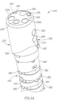
Figure la schematically depicts an external isometric view of an
endoscope having multiple fields of view according to an exemplary
embodiment of the current invention.
7
CA 02812097 2013-03-12
WO 2012/038958 PCT/1L2011/000745
Figure lb schematically depicts a front view of an endoscope having
multiple fields of view according to an exemplary embodiment of the current
invention.
Figure lc schematically depicts a side view of endoscope having multiple
fields of view according to an exemplary embodiment of the current invention.
Figure 2a schematically depicts an isometric cross section of an
endoscope having multiple fields of view, for use within bodily cavity
according
to an exemplary embodiment of the current invention.
Figure 2b schematically depicts a cross section of an endoscope tip
section having multiple fields of view showing some details of the tip section
according to an exemplary embodiment of the current invention.
Figure 2c schematically depicts an isometric proximal view of an inner
part of an endoscope tip section according to an exemplary embodiment of the
current invention.
Figure 3a schematically depicts a partially disassembled tip section of an
endoscope having I/1 channels manifold internal to a unitary fluid channeling
component, according to a first exemplary embodiment of the current invention.
Figure 3b schematically depicts an isometric cross section of an inner
part of a tip section, having 1/I channels manifold internal to a unitary
fluid
channeling component, according to a first exemplary embodiment of the
current invention.
Figure 3c schematically depicts an isometric cross section of a unitary
fluid channeling component of an inner part of a tip section having I/1
channels
manifold internal to the unitary fluid channeling component, according to a
first
exemplary embodiment of the current invention.
Figure 3d schematically depicts another isometric cross section of an
inner part of a tip section, showing the unitary fluid channeling component
8
CA 02812097 2013-03-12
WO 2012/038958 PCT/1L2011/000745
having Ill channels manifold internal to it, according to a first exemplary
embodiment of the current invention.
Figure 4a schematically depicts an isometric view of a partially
disassembled tip section of an endoscope having 1/1 channels manifold
partially
internal and partially external to the unitary fluid channeling component of
the tip
section, according to a second exemplary embodiment of the current invention.
Figure 4b schematically depicts an isometric view of an inner part of a tip
section having 1/1 channels manifold partially internal and partially external
to the
unitary fluid channeling component of the tip section, according to a second
ro exemplary embodiment of the current invention.
Figure 4c schematically depicts an isometric cross section of the inner
part, according to the second exemplary embodiment of the current invention.
Figure 5a schematically depicts an isometric view of a partially
disassembled tip section of an endoscope having 1/1 channels manifold
partially
internal and partially external to the unitary fluid channeling component of
the tip
section, according to a third exemplary embodiment of the current invention.
Figure 5b schematically depicts an isometric view of an inner part of a tip
section having 1/1 channels manifold partially internal and partially external
to a
unitary fluid channeling component of the inner part of the tip section,
according
to a third exemplary embodiment of the current invention.
Figure 5c schematically depicts an isometric cross section of the unitary
fluid channeling component, according to the third exemplary embodiment of
the current invention.
Figure 5d schematically depicts another isometric cross section of an
inner part of a tip section of an endoscope, according to the third exemplary
embodiment of the current invention.
Figure 6a schematically depicts an isometric cross section view of an
assembled tip section of an endoscope having I/1 channels manifold external to
9
CA 02812097 2013-03-12
WO 2012/038958 PCT/1L2011/000745
a unitary fluid channeling component of the inner part of the tip section,
according to a forth exemplary embodiment of the current invention.
Figure 6b schematically depicts an isometric view of an inner part of a tip
section having 1/1 channels manifold external to the unitary fluid channeling
component, according to the forth exemplary embodiment of the current
invention.
Figure 6c schematically depicts an isometric cross section of a unitary
fluid channeling component, according to the forth exemplary embodiment of
the current invention.
i o
Figure 7a schematically depicts an isometric view of an assembled tip
section of an endoscope having 1/1 channels manifold partially external to a
unitary fluid channeling component of an inner part of the tip section,
according
to a fifth exemplary embodiment of the current invention.
Figure 7b schematically depicts an isometric view of an inner part of a tip
section having I/1 channels manifold partially external to the unitary fluid
channeling component, according to the fifth exemplary embodiment of the
current invention.
Figure 7c schematically depicts another isometric view of an inner part of
a tip section having I/1 channels manifold partially external to the unitary
fluid
channeling component, according to a fifth exemplary embodiment of the
current invention.
Figure 7d schematically depicts an isometric cross section of an
endoscope tip section according to the fifth exemplary embodiment of the
current invention.
Figure 8a schematically depicts an isometric view of an assembled tip
section of an endoscope having I/1 channels manifold external to a unitary
fluid
channeling component of an inner part of the tip section, according to a sixth
exemplary embodiment of the current invention.
CA 02812097 2013-03-12
WO 2012/038958 PCT/1L2011/000745
Figure 8b schematically depicts an isometric view of a partially
disassembled tip section of an endoscope having I/1 channels manifold external
to the unitary fluid channeling component, according to a sixth exemplary
embodiment of the current invention.
Figure 9a schematically depicts an isometric proximal view of a main
section of an inner part of an endoscope tip section, according to an
exemplary
embodiment of the current invention.
Figure 9b schematically depicts an isometric cross section of the main
section of Figure 9a, according to an exemplary embodiment of the current
io invention.
Figure 9c schematically depicts an isometric proximal view of the main
section of Figure 9a, having liquid and gas tubes connected thereto, according
to an exemplary embodiment of the current invention.
DETAILED DESCRIPTION
While a number of exemplary aspects and embodiments have been
discussed above, those of skill in the art will recognize certain
modifications,
permutations, additions and sub-combinations thereof. It is therefore intended
that the following appended claims and claims hereafter introduced be
interpreted to include all such modifications, permutations, additions and sub-
combinations as are within their true spirit and scope.
In the description and claims of the application, each of the words
"comprise" "include" and "have", and forms thereof, are not necessarily
limited
to members in a list with which the words may be associated.
Figure la schematically depicts an external isometric view of an
endoscope 200 having multiple fields of view according to an exemplary
embodiment of the current invention.
According to an exemplary embodiment of the current invention, tip
section 230 of endoscope 200 comprises at least a forwards looking TV camera
11
CA 02812097 2013-03-12
WO 2012/038958 PCT/1L2011/000745
and at least one side looking TV camera. Tip section 230 is turnable by way of
flexible shaft 260 (which may also be referred to as a bending section, for
example a vertebra mechanism).
It is noted that the term "endoscope" as mentioned to herein may refer
particularly to a colonoscope, according to some embodiments, but is not
limited only to colonoscopes. The term "endoscope" may refer to any instrument
used to examine the interior of a hollow organ or cavity of the body.
Figure la shows front camera element 236 of forwards looking camera
116 (seen in figure 2b) on the front face 320 of tip section 230. Optical axis
of
io forwards looking camera 116 is substantially directed along the long
dimension
of the endoscope. However, since forwards looking camera 116 is typically a
wide angle camera, its Field Of View (FOV) may include viewing directions at
large angles to its optical axis. Additionally, optical windows 242a and 242b
of
LEDs 240a and 240b (seen for example in figure 2b) are also seen on front face
is 320 of tip section 230. It should be noted that number of LEDs used for
illumination of the FOV may vary. Distal opening 340 of working channel 262
(seen for example in figure 1 b) is preferably located on front face 320 of
tip
section 230, such that a surgical tool inserted through working channel tube
162, and through working channel 262 in the endoscope's tip section and
20 deployed beyond front face 320 may be viewed by forwards looking camera
116.
Distal opening 344 of a jet fluid channel is preferably also located on
front face 320 of tip section 230. Distal opening 344 of a jet fluid channel
may
be used for providing high pressure jet of fluid such as water or saline for
25 cleaning the walls of the body cavity.
1/I injector 346 having a nozzle 348 aimed at front camera element 236
may be used for injecting fluid (liquid and/or gas) to wash contaminants such
as
blood, feces and other debris from front camera element 236 of forwards
looking camera. Optionally the same injector is used for cleaning both front
30 camera element 236 and one or both optical windows 242a and 242b. I/1
12
CA 02812097 2013-03-12
WO 2012/038958 PCT/1L2011/000745
injector 346 may be fed by fluid such as water and/or gas which may be used
for cleaning and/or inflating a body cavity.
Visible on the side wall 362 of tip section 230 is the side camera (side
looking camera) element 256 of side looking camera 220 (two such cameras
are seen in figure 2a) and optical window 252 of LED 250. Optical axis of side
looking camera 220 is substantially directed perpendicular to the long
dimension of the endoscope. However, since side looking camera 220 is
typically a wide angle camera, its field of view may include viewing
directions at
large angles to its optical axis.
I/1 injector 366 having a nozzle 368 aimed at side camera element 256
may be used for injecting fluid to wash contaminants such as blood, feces and
other debris from side camera element 256 of side looking camera. The fluid
may include gas which may be used for inflating a body cavity. Optionally the
same injector is used for cleaning both side camera element 256 and optical
windows 252. It is noted that according to some embodiments, the tip may
include more than one window and LEDs, on the side and more than one
window and LEDs in the front (for example, two windows and two LEDs on the
side and three windows and three LEDs in the front). The 1/1 injectors are
configured to clean all or a part of these windows/LEDs). I/1 injectors 346
and
366 may be fed from same channel. An optional groove 370 helps directing the
cleaning fluid from nozzle 368 towards side camera element 256. Groove 370
may be beneficial when side wall 362 is near or pressed against the rectal
wall.
Optionally, I/1 injector 366 may be at least partially recessed in groove 370,
thus
reducing the maximum diameter of tip section 230 and reduce the risk of injury
to the rectal wall due to friction with I/1 injector 366.
In the depicted embodiment, flexible shaft 260 is constructed of a
plurality of links 382 connected to each other by pivots 384. Links 382 allows
pushing, pulling and rotating the endoscope while pivots 384 provide limited
flexibility. The shaft is preferably covered with an elastic sheath (removed
from
this figure for simplification purposes). The lumen in links 382 holds the
working
channel tube 162. Not seen in this figure are the jet channel connected to
distal
13
CA 02812097 2013-03-12
WO 2012/038958 PCT/1L2011/000745
opening 344, optional cleaning fluid channel and electrical cables supplying
power to the LEDs and cameras and transmitting video signals from the
camera. Generally, the shaft also comprises mechanical actuators (not seen),
for example cables attached to the links for directing and aiming the tip
section
during use.
It should be noted that while only one side looking camera is seen in
figure la, preferably at least two side looking cameras may be located within
tip
section 230. When two side looking cameras are used, the side looking
cameras are preferably installed such that their field of views are
substantially
opposing. However, different configurations and number of side looking
cameras are possible within the general scope of the current invention.
Figure lb schematically depicts a front view of tip section 230 of
endoscope 200 having multiple fields of view according to an exemplary
embodiment of the current invention.
According to an exemplary embodiment of the current invention, tip
section 230 of endoscope 200 comprises at least a forwards looking TV camera
and at least two side looking TV cameras. Figure lb shows a front camera
element 236 of forwards looking camera 116 on the front face 320 of tip
section
230. Additionally, optical windows 242a and 242b of LEDs 240a and 240b are
also seen on front face 320 of tip section 230. Distal opening 340 of working
channel and distal opening 344 of a jet channel are preferably also located on
front face 320 of tip section 230. I/1 injector 346 having a nozzle 348 is
also
visible in this view.
Additionally, I/1 injectors 366a and 366b aimed at side looking camera
element 256a and 256b respectively may be used for injecting fluid (the term
"fluid" may also include gas and/or liquid) to wash contaminants such as
blood,
feces and other debris from side camera elements 256a and 256b of side
looking cameras. According to some embodiments, the injectors may supply
liquid for cleaning any of the tip elements (such as any camera element,
windows, LEDs, and other elements).
14
CA 02812097 2013-03-12
WO 2012/038958 PCT/1L2011/000745
Figure 1c schematically depicts a side view of endoscope 200 having
multiple fields of view according to an exemplary embodiment of the current
invention.
Figure lc shows side camera element 256 of side looking camera 220,
optional groove 370 and optical window 252 on the side wall 362 of tip section
230. I/1 injectors 346 and 366 are also visible in this view.
Figure 2a schematically depicts a cross section isometric view of an
endoscope 400 having multiple fields of view according to another exemplary
embodiment of the current invention.
io
According to an exemplary embodiment of the current invention, tip
section 430 of endoscope 200 comprises at least a forwards looking TV camera
116 and two side looking cameras 220a and 220b.
Optical windows 242a and 242b of LEDs used for forward illumination
are also seen on front face of tip section 230.
Distal opening 340 of working channel is preferably located on front face
of tip section 230 such that a surgical tool inserted through the working
channel
262 and deployed beyond front face may be viewed by forwards looking camera
116.
Distal opening 344 of a jet channel is preferably also located on front
face of tip section 230.
1/I injector 346 having a nozzle aimed at front camera element of camera
116 may be used for injecting fluid (gas and/or water) to wash contaminants
such as blood, feces and other debris from front camera element of forwards
looking camera 116 and to inflate a body cavity (such as a colon) into which
the
endoscope (such as colonoscope) is inserted. Optionally the same injector is
used for cleaning the front camera element and one or both optical windows
242a and 242b. I/1 injector 346 may receive fluid from a fluid channel or may
be
fed by a dedicated cleaning fluid channel.
CA 02812097 2013-03-12
WO 2012/038958 PCT/1L2011/000745
Visible on right hand side of tip section 230 is side camera element 256b
of side looking camera 220b and optical window 252b of side illuminating LED.
Ill injector 366b having a nozzle aimed at side camera element 256b may
be used for injecting fluid to wash contaminants such as blood, feces and
other
Although not seen in this figure, it is understood that equivalent elements
366a, 370a, 256a and 252a are present on the left hand side of tip section
230.
Preferably, all the Ill injectors 346 and 366 are fed from same conduits.
In the depicted embodiment, flexible shaft (vertebra mechanism) 260 is
Figure 2b schematically depicts a cross section of an endoscope 200
having multiple fields of view showing some details of the tip section 230
According to embodiments of the current invention, tip section 230 of
endoscope 200 comprises at least a forwards looking TV camera 116 and two
side looking cameras 220a and 220b. Each of cameras 116 and 220 is provided
with an optical imaging system such as lens assemblies 132 and 232
16
CA 02812097 2013-03-12
WO 2012/038958 PCT/1L2011/000745
example, field of views 118 and 218 may be different. Additionally or
alternatively, other camera designs parameters such as: resolution, light
sensitivity, pixel size and pixel number, focal length, focal distance and
depth of
field may be selected to be same or different.
Light is provided by Light Emitting Diodes (LED) that illuminates the field
of views. Preferably, white light LEDs are used.
In the depicted embodiment, field of view 118 of forwards looking camera
116 is illuminated by two LEDs 240a and 240b located within the endoscope tip
section 230 and protected by optical window 242a and 242b respectively.
ro According to some embodiments, forwards looking camera 116 may be
illuminated by any other number of LEDs, for example, 1, 3, 4, 5 LEDs)
Similarly, in the depicted embodiment, field of views of side looking
camera 220 is illuminated by a single LED 250 located within the endoscope tip
section 230 and protected by optical window 252. According to some
embodiments, side looking camera 220 may be illuminated by any other
number of LEDs, for example, 2, 3, 4, 5 LEDs)
It should be noted that number of LED light sources and their position in
respect to the cameras may vary within the scope of the current invention. For
example few LEDs may be positioned behind the same protective window, a
camera and an LED or plurality of LED may be located behind the same
protective window, etc.
Tip section 230 of endoscope 200 is located at the distal end of a flexible
shaft 260. Similarly to shafts of the art, shaft 260 comprises a working
channel
262 for insertion of surgical tools. Additionally, shaft 260 may comprises
channels for irrigation, insufflation, suction and supplying liquid for
washing the
external elements of the cameras and optionally the light sources.
Figure 2c schematically depicts an isometric proximal view of an inner
part of a tip section of an endoscope according to an exemplary embodiment of
17
CA 02812097 2013-03-12
WO 2012/038958 PCT/1L2011/000745
the current invention showing the entrances of various channels in the inner
part of a tip section.
Inner part 100 of a tip section is located within the tip section and may be
used for holding in place the components of the endoscope's tip section such
as
Ill injectors 364, 366a and 366b; cameras, lenses and other elements. A cover
(not seen in this figure) is placed over inner part 100. Some elements, for
example Ill injectors 364 and 366 (and optionally side camera element 256b)
may be assembled after the cover was placed.
Inner part 100 of a tip section may comprise of several parts. In the
io depicted embodiment inner part 100 of the tip section comprises: unitary
fluid
channeling component 190, central section 192 and front section 194
(examples of which are seen in some of the following figures). Unitary fluid
channeling component 190 may be made of a metal or any other material, such
as a polymer, a composite material or any other appropriate material or
combination of materials. Unitary fluid channeling component 190, according to
some embodiments, may generally include two parts: a proximal fluid
channeling component section 190' and a distal fluid channeling component
section 190". Proximal fluid channeling component section 190' may have an
essentially cylindrical shape. Distal unitary channeling component section
190"
may partially continue the cylindrical shape of proximal fluid channeling
component section 190' and may have a shape of a partial cylinder (optionally
elongated partial cylinder), having only a fraction of the cylinder (along the
height axis of the cylinder), wherein another fraction of the cylinder (along
the
height axis of the cylinder) is missing. (Distal fluid channeling component
section 190", which may be integrally formed as a unitary block with proximal
fluid channeling component section 190'. The height of distal fluid channeling
component section 190" may by higher than that of proximal fluid channeling
component section 190'. In the case of distal fluid channeling component
section 190", the shape of the partial cylinder (for example, partial cylinder
having only a fraction of a cylinder shape along one side of the height axis)
may
provide a space to accommodate central section 192. Central section 192 may
18
CA 02812097 2013-03-12
WO 2012/038958 PCT/1L2011/000745
include electronics and optical components, such as light means (LEDs for
example), cameras (CCD or CMOS, for example) lenses, and other elements.
This configuration of inner part 100 of the tip section may thus be adapted to
separate the fluid channels and work channel, which are located in fluid
channeling component 190 from the sensitive electronic and optical parts which
are located in central section 192. This paragraph may apply to any one of
main
bodies 190a-190f.
On the proximal surface 191 of unitary fluid channeling component 190 is
proximal opening 144 of the jet fluid channel leading to distal opening 344 of
the
jet channel. Fluid tube (not shown in this figure for simplification purposes)
may
be inserted into, and affixed to distal opening 144 of the jet fluid channel.
The jet
fluid tube is threaded through flexible shaft 260 and is used for delivering
fluid to
the body cavity.
On the proximal surface 191 of unitary fluid channeling component 190 is
proximal opening 165 of the working channel 262 leading to distal opening 340
of the working channel. Working channel tube/tools may be inserted into, and
optionally affixed to proximal opening 165 of the working channel. The working
channel 162 is threaded through flexible shaft 260 and is used for delivering
surgical tools to the body cavity. Working channel 162 may also be used for
suction of fluid from the body cavity.
On the proximal surface 191 of unitary fluid channeling component 190 is
the electric cable opening 150 for electrical cable 396 (seen for example in
figure 2a). Electrical cable 396 is connected at its distal end to the
electronic
components such as cameras and light sources in the endoscope's tip section.
Electrical cable 396 is threaded through flexible shaft 260 and is used for
delivering electrical power and command signals to the tip section and
transmitting video signal from the cameras to be displayed to the user.
On the proximal surface 191 of unitary fluid channeling component 190 is
the Ill tubes proximal opening 110 for gas tube 114 and liquid tube 112 (seen
for example in figure 3a). Gas and fluid tubes may be inserted into, and
affixed
19
CA 02812097 2013-03-12
WO 2012/038958 PCT/1L2011/000745
to proximal opening 110 of I/1 channels manifold which delivers cleaning
fluids
to 1/1 injectors 364 and 366. The gas and liquid tubes (such as gas tube 114
and
liquid tube 112) may be threaded through flexible shaft 260 and are used for
delivering fluid (gas and/or liquid) to I/1 injectors 364 and 366 for cleaning
the
optical surfaces on the endoscope's tip section and for inflating a body
cavity.
The gas and liquid tubes (such as gas tube 114 and liquid tube 112) may also
be combined into one tube and connected to the tip section as one tube.
It should be realized that it is important to keep the dimensions of the tip
section of the endoscope small. Within the tight confines of the endoscope's
tip
to section
are the sensors, lenses, electric cables, at least one working channel,
and a plurality of fluid channels. In contrast to endoscopes of the art,
wherein
each of the fluid tubes was directed to its destination, embodiments of the
current invention provide I/1 channels manifold to supply cleaning liquid and
gas
to the plurality of I/1 injectors.
While figure 2c generically depicts the unitary fluid channeling
component 190, and shows its proximal surface 191, the following figures
depict
some specific exemplary embodiments of the I/1 channels manifolds and main
bodies (such as cylinders), according to embodiments within the general scope
of the current invention.
Figure 3a schematically depicts a partially disassembled tip section 230a
of an endoscope having I/1 channels manifold internal to unitary fluid
channeling
component 190a according to a first exemplary embodiment of the current
invention.
Cover 196a is designed to fit over inner part (of the tip section) 100a, and
to provide protection to the internal components in the inner part. Holes
164',
340', 344', 242a', 336', 242b', 256b', 252b' and 166b' in cover 196a are
aligned
with the corresponding components and channel openings 164, 165, 144, 242a,
336, 242b, 256b, 252b and 366b in inner part 100a respectively. Optional
groove 370b in cover 196a enable cleaning fluid from 1/1 injector 366b to
arrive,
and clean the front surface 252b of side looking camera. Not seen in this view
CA 02812097 2013-03-12
WO 2012/038958 PCT/1L2011/000745
are groove 370a, and holes 256a', 252a' and 166a' in cover 196a which are
aligned with the corresponding components and channel openings 256a, 252a
and 166a on the other side of inner part 100a respectively.
After fitting and attaching cover 196a over inner part 100a, 1/1 injectors
364, 366a and 366b may be inserted into the corresponding I/1 opening 164,
166a and 166b in unitary fluid channeling component 190a through the
corresponding holes 164', 166a' and 166b' in cover 196a. Preferably, 1/1
injectors 364, 366a and 366b may be removed from I/1 opening 164, 166a and
166b for cleaning the endoscope after use. Optionally, I/1 injectors 364, 366a
and 366b may be replaceable or disposable. Optionally, the nozzles, such as
nozzle 348, nozzle 368 or any other nozzle may be inserted into the unitary
fluid
channeling component, such as unitary fluid channeling components 190 or
190a, within an isolating (e.g., plastic) part into the opening to allows us
better
electric isolation particularly when the unitary fluid channeling component
and
the nozzles are made of metal.
In the first exemplary embodiment of the current invention, Ill opening
164, 166a and 166b are connected to proximal opening 110 for gas tube 114
and liquid tube 112 via I/1 manifold channels which are within unitary fluid
channeling component 190a. Distal opening 344' is the opening of a jet fluid
channel which may be used for providing high pressure jet of fluid such as
water or saline for cleaning the walls of the body cavity (such as the colon)
and
optionally for suction.
Figure 3b schematically depicts an isometric cross section of Inner part
100a having I/1 channels manifold internal to unitary fluid channeling
component
190a according to a first exemplary embodiment of the current invention.
In the depicted embodiment gas tube 114 and liquid tube 112 are
terminated in a plug 109 adapted to fit into proximal opening 110. It should
be
noted that although gas tube 112 appears above liquid tube 114, their order
may be reversed, they may be positioned side by side, or replaced with a
single
tube or the tubes may be joined to one tube before entering inner part 100a.
21
CA 02812097 2013-03-12
WO 2012/038958 PCT/1L2011/000745
Alternatively, each of gas tube 114 and liquid tube 112 is separately
connected
to unitary fluid channeling component 190, and their lumens open to a common
conduit.
Proximal opening 110 for gas tube 114 and liquid tube 112 is opened to
1/1 channel manifold. This cross section shows proximal opening 110 opened to
front 1/1 channel 171 leading to front 1/1 opening 164 into which front 1/1
injector
364 is inserted. According to some embodiments, front 1/1 channel 171(may also
be referred to as front fluid channel) may be drilled in unitary fluid
channeling
component 190a. It should be noted that unitary fluid channeling component
190a and other parts of inner part 100a may be machined or be made by
casting, sintering, injection or other manufacturing techniques.
Reference is now made to Figure 3c, which schematically depicts an
isometric cross section of unitary fluid channeling component 190a having 1/1
channels manifold internal to it according to a first exemplary embodiment of
the
current invention and to Figure 3d, which schematically depicts another
isometric cross section of inner part 110a, showing unitary fluid channeling
component 190a having I/1 channels manifold internal to it according to a
first
exemplary embodiment of the current invention.
Proximal opening 110 for gas tube 114 and liquid tube 112 is seen in this
figure opened to I/1 channel manifold. This cross section shows proximal
opening 110 opened to cross 1/1 channel 172 (may also be referred to as side
fluid channel or side 1/1 channel) leading to left I/1 opening 166a into which
left 1/1
injector 366a is inserted and to right 1/1 opening 166b into which right 1/1
injector
366b is inserted.
According to some embodiments, cross I/1 channel 172 may be drilled in
unitary fluid channeling component 190a.
According to the first exemplary embodiment of the current invention,
proximal opening 110 for gas tube 114 and liquid tube 112 is directly opened
to
1/1 channel manifold, within unitary fluid channeling component 190a which
comprises:
22
CA 02812097 2013-03-12
WO 2012/038958 PCT/1L2011/000745
a) a right Ill opening 166b, connected to proximal opening 110, and into
which right 1/1 injector 366b is inserted;
b) a front 1/I channel 171 connected to proximal opening 110, and leading
to front Ill opening 164 into which front 1/1 injector 364 is inserted; and
c) a cross I/1 channel 172, connected to the proximal opening, and which
is opened to left I/1 opening 166a into which left I/1 injector 366a is
inserted.
Figure 4a schematically depicts an isometric view of a partially
disassembled tip section 230b of an endoscope having Ill channels manifold
partially internal and partially external to unitary fluid channeling
component
190b according to a second exemplary embodiment of the current invention.
In contrast to the first embodiment depicted in figures 3, in the
embodiment depicted in figures 4, cleaning fluids are supplied to left Ill
injector
366a via a groove 472 in unitary fluid channeling component 190b. Groove 472
is connected in one side to proximal opening 110 by hole 474 and is opened to
left 1/1 opening 166a which can hardly be seen in this view.
Cover 196b is designed to fit over inner part 100b, and to provide
protection to the internal components of inner part 100b. Additionally, cover
196b is tightly fitted and preferably hermetically seals groove 472 to convert
it to
fluid tight conduit.
Figure 4b schematically depicts an isometric view of inner part 100b of
an endoscope tip section having Ill channels manifold partially internal and
partially external to unitary fluid channeling component 190b according to a
second exemplary embodiment of the current invention.
Figure 4c schematically depicts an isometric cross section of unitary fluid
channeling component 190b according to the second exemplary embodiment of
the current invention.
23
CA 02812097 2013-03-12
WO 2012/038958 PCT/1L2011/000745
According to the second exemplary embodiment of the current invention,
proximal opening 110 for gas tube 114 and liquid tube 112 is seen in this
figure
opened to I/1 channel manifold which comprises:
a) a right 1/1 opening166b, connected to proximal opening 110, into which
right I/1 injector 366b is inserted;
b) a front 1/1 channel 171 connected to front 1/1 opening 164 into which
front I/1 injector 364 is inserted; and
c) hole 474 connected to groove 472 which is opened to left 1/1 opening
166a (not seen here) into which left I/1 injector 366a (not seen here) is
inserted.
ro Figure
5a schematically depicts an isometric view of a partially
disassembled tip section 230c of an endoscope having I/1 channels manifold
partially internal and partially external to unitary fluid channeling
component
190c according to a third exemplary embodiment of the current invention.
In contrast to the first embodiment depicted in figures 3, in the
embodiment depicted in figures 5, fluids (liquid and/or gas) are supplied to
left
I/1 injector 366b via a groove 572 in unitary fluid channeling component 190c.
However, in contrast to the second embodiment, depicted in figures 4, groove
572 is connected in the right side to right I/1 opening 166b and opened on the
left to left I/1 opening 166a which can hardly be seen in this view.
Cover 196c is designed to fit over inner part 100c, and to provide
protection to the internal components of inner part 100c. Additionally, cover
196c is tightly fitted and preferably hermetically seals groove 572 to convert
it to
fluid tight conduit.
Figure 5b schematically depicts an isometric view of inner part 100c of
an endoscope tip section having 1/1 channels manifold partially internal and
partially external to unitary fluid channeling component 190c according to a
third
exemplary embodiment of the current invention.
24
CA 02812097 2013-03-12
WO 2012/038958 PCT/1L2011/000745
It should be noted that the location of groove 572 on surface of unitary
fluid channeling component 190c, and its depth and shape may be different.
Figure 5c schematically depicts an isometric cross section of unitary fluid
channeling component 190c according to the third exemplary embodiment of
the current invention.
Proximal opening 110 for gas tube 114 and liquid tube 112 is seen in this
figure opened to right 1/1 opening 166b and through it to groove 572 leading
to
left 1/1 opening 166a.
Figure 5d schematically depicts another isometric cross section of unitary
fluid channeling component 190c according to the third exemplary embodiment
of the current invention.
Proximal opening 110 for gas tube 114 and liquid tube 112 is seen in this
figure opened to right I/1 opening 166b and through it to 1/1 manifold which
comprises:
a) a right I/1 opening 166b, connected to proximal opening 110, into
which right I/1 injector 366b is inserted;
b) a front 1/1 channel 171, connected to proximal opening 110, ad leading
to front 1/1 opening 164 into which front 1/1 injector 364b is inserted ; and
c) a groove 572 which receives cleaning fluids from right 1/1 opening
166b, and is opened to left 1/1 opening 166a (not seen here) into which left
1/I
injector 366a is inserted.
Figure 6a schematically depicts an isometric cross section view of an
assembled tip section 230d of an endoscope having 1/1 channels manifold
external to unitary fluid channeling component 190d according to a forth
exemplary embodiment of the current invention.
CA 02812097 2013-03-12
WO 2012/038958 PCT/1L2011/000745
Similarly to third embodiment depicted in figures 5, groove 672 is
connected in the right side to right 1/1 opening 166b and opened on the left
to left
I/1 opening 166a.
However in contrast to the first, second and third embodiments depicted
in figures 3, 4, and 5, in the embodiment depicted in figures 6, fluids are
supplied to front 1/I injector 364 via a front groove 671 in unitary fluid
channeling
component 190d. Front groove 671 is opened in its proximal end to groove
672, and at its distal end to front 1/1 opening 164.
Cover 196d is designed to fit over inner part 100d, and to provide
io protection to the internal components of inner part 100d. Additionally,
cover
196d is tightly fitted and preferably hermetically seals grooves 671 and 672
to
convert them to fluid tight conduits.
Figure 6b schematically depicts an isometric view of inner part 100d of
an endoscope tip section having I/1 channels manifold external to unitary
fluid
is channeling component 190d according to a forth exemplary embodiment of
the
current invention.
It should be noted that the location of grooves 671 and 672 on surface of
unitary fluid channeling component 190d, and their depth and shape may be
different. For example, the location of any of the grooves may be completely
or
20 partially inside the cover, for example, within the walls of the cover.
Figure 6c schematically depicts an isometric cross section of unitary fluid
channeling component 190d according to the forth exemplary embodiment of
the current invention.
Proximal opening 110 for gas tube 114 and liquid tube 112 is seen in this
25 figure opened to right I/1 opening 166b and through it to groove 672
leading to
left I/1 opening 166a. Also seen in this figure is the intersection of grooves
672
and front groove 671
26
CA 02812097 2013-03-12
WO 2012/038958 PCT/1L2011/000745
According to the forth embodiment of the current invention, proximal
opening 110 for gas tube 114 and liquid tube 112 is opened to right 1/1
opening
166b and through it to an 1/1 manifold which comprises:
a) a right 1/1 opening 166b, connected to proximal opening 110, into
which right I/1 injector 366b is inserted;
b) groove 672 which receives cleaning fluids from right 1/1 opening 166b,
and is opened to left I/1 opening 166a into which left 1/1 injector 366a is
inserted;
and
c) front Ill groove 671, receiving 1/1 fluids from groove 672, and
m connected to front 1/1 opening 164 into which front I/1 injector 364 is
inserted.
Figure 7a schematically depicts an isometric view of an assembled tip
section 230e of an endoscope having 1/1 channels manifold partially external
to
unitary fluid channeling component 190e according to a fifth exemplary
embodiment of the current invention.
For clarity, cover 196d was drawn partially transparent to show inner part
100e.
Similarly to second embodiment depicted in figures 4, groove 772 is
proximal opening 110 by hole 774 and opened on the left to left 1/1 opening
166a
(not seen in this figure).
Similarly to the forth embodiment depicted in figures 5, cleaning fluids
are supplied to front Ill injector 364 via a front groove 771 in unitary fluid
channeling component 190e. Front groove 771 is opened in its proximal end to
groove 772, and at its distal end to front I/1 opening 164.
Cover 196e is designed to fit over inner part 100e, and to provide
protection to the internal components of inner part 100e. Additionally, cover
196e is tightly fitted and preferably hermetically seals grooves 771 and 772
to
convert them to fluid tight conduits.
27
CA 02812097 2013-03-12
WO 2012/038958 PCT/1L2011/000745
Figure 7b schematically depicts an isometric view of inner part 100e of
an endoscope tip section having Ill channels manifold partially external to
unitary fluid channeling component 190e according to a fifth exemplary
embodiment of the current invention.
It should be noted that the location of grooves 771 and 772 on surface of
unitary fluid channeling component 190d, and their depth and shape may be
different.
Figure 7c schematically depicts another isometric view of inner part 100e
of an endoscope tip section having Ill channels manifold partially external to
unitary fluid channeling component 190e according to a fifth exemplary
embodiment of the current invention.
This view depicts groove 772 connection to left I/1 opening 166a.
Figure 7d schematically depicts an isometric cross section of endoscope
tip section 230e according to the fifth exemplary embodiment of the current
invention.
Proximal opening 110 for gas tube 114 and liquid tube 112 is seen in this
figure opened to right Ill opening 166b. Also seen in this figure is hole 774
connecting proximal opening 110 to front groove 771 and the connection of
front groove 771 to front get opening 164.
According to the fifth embodiment of the current invention, proximal
opening 110 for gas tube 114 and liquid tube 112 is opened to right Ill
opening
166b and through hole 774 to Ill manifold which comprises:
a) a right I/1 opening 166b, connected to proximal opening 110, into
which right I/1 injector 366b is inserted;
b) groove 772 which receives fluids via hole 774 connected to proximal
opening 110, and is opened to left Ill opening 166a into which left Ill
injector
366a is inserted; and
28
CA 02812097 2013-03-12
WO 2012/038958 PCT/1L2011/000745
C) front I/1 groove 771, receiving 1/1 fluids from hole 774, and connected
to front I/1 opening 164 into which front I/1 injector 364b is inserted.
Figure 8a schematically depicts an isometric view of an assembled tip
section 230f of an endoscope having I/1 channels manifold external to unitary
fluid channeling component 190f in inner part 100f according to a sixth
exemplary embodiment of the current invention.
Similarly to forth embodiment depicted in figures 6, groove 872 in unitary
fluid channeling component 190f is connected in the right side to right I/1
opening 166b and opened on the left to left Ill opening 166a.
io
Similarly to forth embodiment depicted in figures 6, front groove 871 is
connected in its proximal end to groove 872.
However in contrast to the forth embodiment cleaning fluids are supplied
groove 871 and 872 via hole 874 connecting them to proximal opening 110.
Cover 196f is designed to fit over inner part 100f, and to provide
protection to the internal components of inner part 100f. Additionally, cover
196f
is tightly fitted and preferably hermetically seals grooves 871 and 872 to
convert
them to fluid tight conduits.
Figure 8b schematically depicts an isometric view of a partially
disassembled tip section 230f of an endoscope having Ill channels manifold
external to unitary fluid channeling component 190f in inner part 100f
according
to a sixth exemplary embodiment of the current invention.
It should be noted that the location of grooves 871 and 872 on surface of
unitary fluid channeling component 190d, and their depth and shape may be
different.
According to the sixth embodiment of the current invention, proximal
opening 110 for gas tube 114 and liquid tube 112 is opened hole 874 and
through it to an 1/1 manifold which comprises:
29
CA 02812097 2013-03-12
WO 2012/038958 PCT/1L2011/000745
a) grove 872 which receives cleaning fluids from proximal opening 110
via hole 874; connected to right I/1 opening 166b into which right I/1
injector 366b
is inserted;
b) same groove 872 connected to left I/1 opening, to which left I/1 injector
366a is inserted; and
c) front I/1 groove 871, receiving I/1 fluids from groove 872, and
connected to front I/1 opening into which front I/1 injector 364 is inserted.
It should be noted that optionally I/1 injectors 336a and 336b, and
optionally also 364 may be constructed as identical interchangeable inserts.
io Reference is now made to Figure 9a which schematically depicts an
isometric proximal view of a main section of an inner part of an endoscope tip
section, according to an exemplary embodiment of the current invention and to
Figure 9b, which schematically depicts an isometric cross section of the main
section of Figure 9a, according to an exemplary embodiment of the current
is invention.
Unitary fluid channeling component 990 of an inner part of a tip section of
an endoscope (such as colonoscope) is configured to be located within the tip
section and may be used for accommodating fluid channels, work channel and
optionally cable channel/recess and for holding in place the components such
20 as tubing/tubes, and injectors. Unitary fluid channeling component 990
may be
a part of the inner part of the tip section in a similar manner to that
described for
example in Figure 2c.
Unitary fluid channeling component 990, according to some
embodiments, may generally include two parts: a proximal fluid channeling
25 component section 990' and a distal fluid channeling component section
990".
Proximal fluid channeling component section 990' may have an essentially
cylindrical shape. Distal fluid channeling component section 990" may
partially
continue the cylindrical shape of proximal fluid channeling component section
990' and may have a shape of a partial cylinder (optionally elongated partial
CA 02812097 2013-03-12
WO 2012/038958 PCT/1L2011/000745
cylinder), having only a fraction of the cylinder (along the height axis of
the
cylinder), wherein another fraction of the cylinder (along the height axis of
the
cylinder) is missing. Distal fluid channeling component section 990" may be
integrally formed as a unitary block with proximal fluid channeling component
section 990'. The height of distal fluid channeling component section 990" may
by higher than that of proximal fluid channeling component section 990'. In
the
case of distal fluid channeling component section 990", the shape of the
partial
cylinder(for example, partial cylinder having only a fraction of a cylinder
shape
along one side of the height axis) may provide a space to accommodate a
io central section (not shown).
On proximal surface 991 of fluid channeling component 990 is proximal
opening 944 of the jet fluid channel leading to distal opening of a jet
channel
(not shown). A jet fluid tube may be inserted through a flexible shaft and may
be
used for delivering fluid to, and optionally suction of fluid from the body
cavity,
for cleaning purposes.
On proximal surface 991 of unitary fluid channeling component 990 is
proximal opening 965 of the working channel 262 leading to a distal opening of
the working channel (not shown).
Unitary fluid channeling component 990 includes groove 950 extending
from proximal surface 991along the length of proximal fluid channeling
component section 990'. Groove 950 is adapted to guide (and optionally hold in
place) an electric cable(s) which may be connected at its distal end to the
electronic components such as cameras and/or light sources in the
endoscope's tip section and deliver electrical power and/or command signals to
the tip section and/or transmitting video signal from the cameras to be
displayed
to the user. According to this embodiment the electrical cable(s) do not have
to
be threaded through proximal fluid channeling component section 990' (which
may be complicated) but can be simply placed in groove 950 and held by it.
On proximal surface 991 of unitary fluid channeling component 990 are
1/1 tubes proximal openings: front 1/1 proximal opening 910; right side I/1
proximal
31
CA 02812097 2013-03-12
WO 2012/038958 PCT/1L2011/000745
opening; 911 and left side 1/1 proximal opening 913. Front I/1 proximal
opening
910; right side 1/I proximal opening 911 (not shown) and left side I/1
proximal
opening 913 lead to front I/1 channel 970; right side I/1 channel 971 (not
shown);
and left side 1/1 channel 973, respectively. Front I/1 channel 970 extends
from
front 1/1 proximal opening 910, through proximal fluid channeling component
section 990' and distal fluid channeling component section 990" to front 1/1
opening 960. Left side I/1 channel 973 extends from right I/1 proximal opening
913, through proximal fluid channeling component section 990' to left 1/1
opening
963. Right side 1/I channel 971(not shown) extends from right I/1 proximal
lo opening 911(not shown), through proximal fluid channeling component
section
990' to right I/1 opening (not shown), similar to the left side arrangement.
Front I/1 channel 970 may include two parts: a proximal part 970'
(extending through proximal fluid channeling component section 990') and a
distal part 970" extending through distal fluid channeling component section
990"). Proximal part 970' of front 1/1 channel 970 is adapted to receive,
through
front I/1 proximal opening 910, tube 980 (shown in Figure 9c) which is adapted
to transfer fluid (liquid and/or gas) to front 1/1 channel 970. Tube 980 may
be
divided at any point along its length (for example at junction 981) into two
tubes,
one is adapted to transfer gas and the other is adapted to transfer liquid
(such
as water).
Left side I/1 channel 973 may be adapted to receive, at its proximal part,
through left side I/1 proximal opening 913, tube 982 (shown in Figure 9c)
which
is adapted to transfer fluid (liquid and/or gas) to left side I/1 channel 973.
Tube
982 may be divided at any point along its length (for example at junction 983)
into two tubes, one is adapted to transfer gas and the other is adapted to
transfer liquid (such as water).
Right side I/1 channel (not shown) may be adapted to receive, at its
proximal, through right side 1/I proximal opening 911, part tube 984 (shown in
Figure 9c) which is adapted to transfer fluid (liquid and/or gas) to right
side 1/1
channel. Tube 984 may be divided at any point along its length (for example at
32
CA 02812097 2013-03-12
WO 2012/038958 PCT/1L2011/000745
junction 985) into two tubes, one is adapted to transfer gas and the other is
adapted to transfer liquid (such as water).
The endoscope user can thus decide which fluid (gas, liquid or both) he
or she would like to pass through the Ill channel, which fluid, as mentioned
herein, may be used for cleaning and/or insufflation purposes.
Figure 9c schematically depicts an isometric proximal view of the main
section of Figure 9a, having liquid and gas tubes connected thereto, according
to an exemplary embodiment of the current invention.
Although the invention has been described in conjunction with specific
embodiments thereof, it is evident that many alternatives, modifications and
variations will be apparent to those skilled in the art. Accordingly, it is
intended
to embrace all such alternatives, modifications and variations that fall
within the
spirit and broad scope of the appended claims. All publications, patents and
patent applications mentioned in this specification are herein incorporated in
their entirety by reference into the specification, to the same extent as if
each
individual publication, patent or patent application was specifically and
individually indicated to be incorporated herein by reference. In addition,
citation
or identification of any reference in this application shall not be construed
as an
admission that such reference is available as prior art to the present
invention.
33