Note: Descriptions are shown in the official language in which they were submitted.
CA 02812532 2013-03-25
WO 2012/047408 PCT/US2011/049499
OTLS-P12-PCT
DEVICE AND METHOD FOR POSITIONING AN ELECTRODE IN TISSUE
CROSS-REFERENCE TO RELATED APPLICATIONS
[0001] This application claims the benefit of US Provisional Application
numbers
61/387,185 filed 28-SEP-2010 (entitled "Rhythm Support Device 2"), 61/412,992
filed
12-NOV-2010 (entitled "Pacing Device"), 61/420,060 filed 06-DEC-2010 (entitled
"Pacing Device"), 61/427,306 filed 27-DEC-2010 (entitled "Rhythm Support
Device 5"),
61/445,992 filed 23-FEB-2011 (entitled "Pacing Device"), and 61/501,450 filed
27-JUN-
2011 (entitled "Pacing Device"). This application also claims the benefit of
US Patent
Application number 13/219,874 (entitled "Device and method for positioning an
electrode in tissue) filed 29-AUG-2011. Each of these seven applications,
including six
provisional applications and one nonprovisional application, is incorporated
in its
entirety by this reference.
TECHNICAL FIELD
[0002] This invention relates generally to the electrode stimulation
device field,
and more specifically to a new and useful system and method for positioning an
electrode in tissue in the electrode stimulation device field.
BACKGROUND
[0003] Bradycardia (reduced heart rate) is a common condition affecting
millions
of patients annually. Although many such patients require implantation of
permanent
1
CA 02812532 2013-03-25
WO 2012/047408 PCT/US2011/049499
OTLS-P12-PCT
pacemaker devices to help regulate heart rate, other patients experience
bradycardia
with reversible causes that do not require permanent pacemaker implantation
and may
instead receive temporary bradycardia support, such as over a period of less
than one
week. One common treatment for temporary bradycardia support involves a system
including transvenous electrode pacing leads that are inserted directly into
the right
ventricle of the heart to stimulate and regulate cardiac function. However,
the
conventional versions of these systems have several drawbacks.
[0004] Thus, there is a need in the electrode stimulation device field to
create a
new and useful device and method for positioning an electrode in tissue in the
electrode
stimulation device field. This invention provides a new and useful device and
method for
positioning an electrode in tissue.
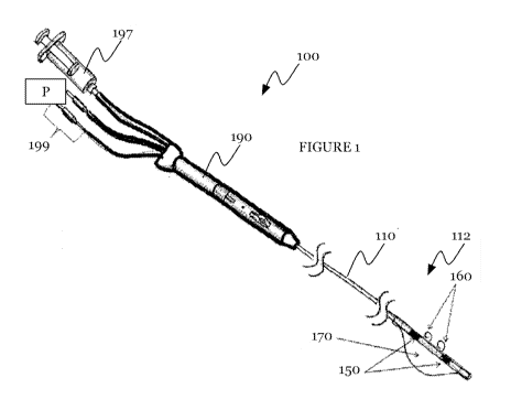
BRIEF DESCRIPTION OF THE FIGURES
[0005] FIGURES 1 and 2 are overall and detail views, respectively, of the
system
of a preferred embodiment;
[0006] FIGURES 3A-3H are detailed views of variations of the lead body of
the
system of a preferred embodiment;
[0007] FIGURES 4-7 are variations of an atraumatic tip of the lead body
of the
system of a preferred embodiment;
[0008] FIGURES 8 and 9A-9D are an exploded view and longitudinal cross-
sectional views of operation, respectively, of the handle in the system of a
preferred
embodiment;
2
CA 02812532 2013-03-25
WO 2012/047408 PCT/US2011/049499
OTLS-P12-PCT
[0009] FIGURES 10-13 are variations of the handle in the system of a
preferred
embodiment;
[0010] FIGURES 14A-14D are schematics of a process for assembling the
electrode array in the system of a preferred embodiment;
[0011] FIGURE 15 is a schematic of a variation of the electrode array;
[0012] FIGURES 16A and 16B are schematics of the anchoring elements in
the
first and second configurations, respectively, in the system of a preferred
embodiment;
[0013] FIGURES 17-20 are variations of the anchoring elements in the
system of
a preferred embodiment;
[0014] FIGURES 21-24 are variations of verifying anchoring element
fixation and
electrode array position in the system of a preferred embodiment;
[0015] FIGURES 25A and 25B are schematics of variations of the
displacement
mechanism arrangement in the system of a preferred embodiment;
[0016] FIGURE 26 and 27 are preferred and alternative variations of
assembling
the displacement mechanism;
[0017] FIGURES 28A and 28B are schematics of an example of the system of
a
preferred embodiment; and
[0018] FIGURES 29A-29F are schematics of the method of positioning an
electrode in tissue of a preferred embodiment.
3
CA 02812532 2013-03-25
WO 2012/047408 PCT/US2011/049499
OTLS-P12-PCT
DESCRIPTION OF THE PREFERRED EMBODIMENTS
[0019] The following description of preferred embodiments of the
invention is not
intended to limit the invention to these preferred embodiments, but rather to
enable any
person skilled in the art to make and use this invention.
1. Device for positioning an electrode in tissue
[0020] As shown in FIGURE 1, the device 100 of a preferred embodiment for
positioning an electrode in tissue includes: an elongate lead body no having a
distal
portion 112; an electrode array 150 coupled to the distal portion of the
elongate lead
body; an anchoring element 160 disposed within the elongate lead body and
having a
distal anchor tip 162, in which the anchoring element 160 is selectively
operable in a
first configuration 164 in which the anchor tip is substantially retracted
within the
elongate lead body and in a second configuration 166 in which the anchor tip
is at least
partially extended outside the elongate lead body and configured to fixate
within the
tissue 102; and a displacement mechanism 170, coupled to the distal portion
112 of the
elongate lead body 110, that is selectively expandable to bias the electrode
array 150
and/or the anchoring element 160 toward the tissue 102. The device 100 may
further
include an actuator 140 disposed within the lead body no and abuttingly
engaged with
or otherwise coupled to the anchoring element 160 to actuate the anchoring
element 160
between the first and second configurations. The device 100 may further
include a
handle 190 that is coupled to the elongate body and includes a slide coupled
to the
4
CA 02812532 2013-03-25
WO 2012/047408 PCT/US2011/049499
OTLS-P12-PCT
actuator with first and second slide positions corresponding to the first and
second
configurations of the anchoring element, respectively.
[0021] The device is preferably used to securely place a pacing electrode
lead in
cardiac tissue, such as for temporary bradycardia support. The device enables
reliable
implantation and maintenance of the position of the electrode lead. In
particular, as
shown in FIGURE 2, the elongate body is preferably navigable through the
cardiovascular system (e.g. veins, arteries) into the right ventricle of the
heart, such that
when the displacement mechanism 170 is expanded, the electrode array 150
and/or one
or more anchoring elements 160 are biased towards the intraventricular septum.
The
anchoring elements 160 are configured to fixate within tissue to secure the
electrode
array in contact with the intraventricular septum (tissue 102), which the
electrode array
may stimulate to help regulate heart rate. However, the device may
alternatively be used
to secure any suitable electrode array in any suitable tissue. For instance,
in one
variation (e.g. includes the electrode array, anchoring elements and a mode of
delivery
such as a catheter, without including a displacement mechanism), the device
may be
used in applications such as laparoscopic surgery, general surgery, spinal
surgery,
and/or other procedures for any suitable tissue.
1.1 Lead body
[0022] The elongate lead body no of the device functions to contain and
deliver
the electrode array 150, anchoring element 160, and displacement mechanism 170
to
target tissue within the body. The elongate lead body is preferably a
steerable lead or
CA 02812532 2013-03-25
WO 2012/047408 PCT/US2011/049499
OTLS-P12-PCT
other elongate body, such as a catheter with a stylet, preformed curve, or
other internal
steering system. Such steering systems are known by one ordinarily skilled in
the art,
although the elongate body or lead may include any suitable steering system
for
navigating in the cardiovascular system or other portion of the body. The lead
is
preferably approximately cylindrical, but may alternatively be substantially
flat or
planar, or have any suitable cross-section. The lead is preferably flexible
and made of a
biocompatible material such as polyurethane or polyimide, although at least
some
portions may be rigid.
[0023] As shown in FIGURE 3, the lead preferably includes a plurality of
lumens
that carry control elements (e.g. steering elements, electrical conductors 116
coupled to
the electrode array 150, anchoring elements 160, actuator 140 coupled to the
anchoring
elements, and/or fluid channel coupled to the displacement mechanism 170). At
least
some of these lumens may contain internal tubing within which a control
element is
telescopically disposed. The lumens may be arranged in groups such as a first
group
including at least one lumen 132 for the actuator 140 and anchoring element
160, a
second group including at least one lumen 134 for the conductors 116
(potentially
including a ground wire 118), and a third group including at least one lumen
136 for the
fluid or other actuator for the displacement mechanism. One or more of the
lumens
and/or internal tubing may be shaped with keys or other features to prevent
rotation of
control elements within the lead. For instance, as shown in FIGURE 3C, the
lumen 132
for the anchoring elements 160 may have an approximately rectangular cross-
section to
constrain alignment of the anchoring elements in a particular direction.
6
CA 02812532 2013-03-25
WO 2012/047408 PCT/US2011/049499
OTLS-P12-PCT
[0024] The first group of lumens passing along the lead preferably
includes a
lumen 132 for the actuator 140. The actuator 140 is preferably longitudinally
translatable within the elongate body and abuttingly engaged with the
anchoring
element 160 to actuate the anchoring element between the first and second
configurations. The actuator is preferably flexible, to help keep the overall
lead body no
flexible, such as for navigation through tissue and reduced tissue damage. In
one
variation, as shown in FIGURE 3E, the actuator 140 includes at least a
flexible portion
that includes a helical cut or groove path 142 passing longitudinally along
and
circumferentially around the actuator. For instance, the actuator may include
a wire
portion disposed within or otherwise coupled to a tube portion having a
helical cut 142.
However, the actuator may have a series of circumferential rings, include
pleats or zig-
zag cuts, or any suitable cuts and/or other features to contribute to
flexibility of the
actuator. In another variation, the actuator is additionally and/or
alternatively made of
mesh or flexible material.
[0025] The first group of lumens preferably further includes a lumen 132
for the
anchoring elements 160 extending from, and approximately concentric with, the
lumen
132 for the actuator 140. Between the lumens for the anchoring elements and
actuator,
the lead preferably further includes a sleeve 144 that functions to decouple
the
anchoring elements from rotation of the actuator and/or lead. In a preferred
variation,
as shown in FIGURE 3A, the sleeve is fixed to the anchoring elements 160 and
not fixed
to the actuator 140, such that the actuator 140 is free to rotate
independently from the
anchoring elements within the sleeve 144 and the actuator 140 is free to
translate to
7
CA 02812532 2013-03-25
WO 2012/047408 PCT/US2011/049499
OTLS-P12-PCT
abuttingly engage the anchoring elements 160. For instance, an abutting
cylinder 146
coupled to the distal end of the actuator 140 is preferably configured to push
and/or pull
the anchoring elements 160 within the lead body no. Alternatively, the
actuator 140
may be fastened to the sleeve and the anchoring element may be not fastened to
the
sleeve. In further alternative variations, both the actuator and the anchoring
elements
may be fastened (FIGURE 3D) or unfastened to the sleeve 144, or coupled to the
sleeve
in any suitable manner. In another alternative variation, the actuator 140 may
be
coupled directly to the anchoring elements 160 (e.g. crimping, welding) or be
integrally
formed from the same piece as the anchoring elements. The actuator 140 and/or
anchoring element 160 may be fastened to the sleeve by a snap lock such as a
ball joint,
crimping, fasteners or adhesive.
[0026] The second group of lumens preferably includes one or more lumens
134
for the conductors 116 that carry a current and/or a ground lead 118. The
conductors are
preferably wires made of an electrically conductive material, but may be
tubing or
another elongate shape. As shown in FIGURE 1, the proximal ends of the
conductors are
preferably coupled to generator electrodes 199 and a power source P that are
external to
the patient, and the distal end of the conductors are preferably coupled to
the electrode
array 150. In one variation, the device may include one current-carrying
conductive lead
per electrode in the electrode array such that each electrode can be
individually
controlled. In a second variation, at least a portion of the current-carrying
conductors
may be coupled to multiple electrodes. In a third variation, at least a
portion of the
current-carrying conductors may be coupled directly to an electrode, which is
in turn
8
CA 02812532 2013-03-25
WO 2012/047408 PCT/US2011/049499
OTLS-P12-PCT
coupled to one or more additional electrodes such that at least a portion of
the current-
carrying conductors is indirectly coupled to one or more electrodes. However,
the device
may include any suitable ratio of conductors to electrodes in the electrode
array, and in
any suitable arrangement.
[0027] The third group of lumens passing along the lead preferably
includes one
or more lumens 136 for an actuator of the displacement mechanism 170. In a
preferred
variation, the third group of lumens includes a lumen or channel that carries
a fluid
(preferably air) that may be used to expand the displacement mechanism. In
this
variation, as shown in FIGURE 1, the proximal end of the fluid-carrying lumen
may be
coupled to a syringe or other pump 197 that displaces fluid through the fluid-
carrying
lumen 136, and the distal end of the fluid-carrying lumen may be coupled to
the
displacement mechanism 170. In other variations, the third group of lumens may
carry
wires, rods, springs or any suitable actuator for the particular kind of
displacement
mechanism in the device.
[0028] In a preferred embodiment, as shown in FIGURES 3B-3C, the first
group
of lumens (including a lumen for housing the actuator 140 and anchoring
elements 160
is a central lumen 132 passing approximately axially along the lead body 110,
and the
second and third groups of lumens (housing the conductors and displacement
mechanism actuator) are peripheral lumens 134 and 136 that are
circumferentially
distributed around the central lumen 132. Alternatively, as shown in FIGURE 3F
the
lumen 132 for housing the actuator and anchoring elements may be off-center,
and the
second and/or third groups of lumens 134 and 136 may be arranged in other off-
center
9
CA 02812532 2013-03-25
WO 2012/047408 PCT/US2011/049499
OTLS-P12-PCT
lumens. However, any of the groups may be distributed, combined or otherwise
arranged in any suitable manner.
[0029] As shown in FIGURE 1, the lead preferably further includes a distal
portion 112 to which the electrode array 150, one or more anchoring elements
160, and
one or more displacement mechanisms are coupled. As shown in FIGURE 3G, the
distal
portion defines one or more apertures 133 through which the anchoring elements
160
deploy, preferably defining one aperture 133 per anchoring element but
alternatively the
ratio of apertures 133 to anchoring elements may be less than or greater than
1:1.
Similarly, the distal portion 112 preferably defines one or more apertures 135
through
which the conductors 116 extend to couple to the electrode array, with one
aperture 135
per conductive lead, or with the ratio of apertures to conductors less than or
greater
than 1:1. Alternatively, the distal portions of the conductors may remain
within the
elongate lead body no, and the electrode array 150 or other interconnects may
extend
through the apertures 135 into the elongate lead body to couple to the
conductors.
Similarly, the distal portion 112 preferably defines at least one aperture 137
through
which air or another fluid actuates the displacement mechanism. As shown in
FIGURE
3H, the distal portion 112 or other portions of the lead may also include
contrast
markers 124 made of a material that is visible under fluoroscopy, such as to
aid visual
confirmation of device position or placement.
[0030] The distal portion 112 of the lead preferably further includes an
atraumatic
tip 114, which functions to reduce or eliminate the likelihood of perforation
or other
damage to the tissue as the lead is navigated through tissue. The atraumatic
tip 114
CA 02812532 2013-03-25
WO 2012/047408 PCT/US2011/049499
OTLS-P12-PCT
absorbs at least substantially frontal forces (longitudinal force in a
proximal direction),
and more preferably forces in additional directions. The atraumatic tip 114
may include
softer, impact-absorbing material such as elastomer with a relatively low
durometer,
and/or may include geometry to help absorb forces. In a preferred embodiment,
as
shown in FIGURES 4A and 4B, the atraumatic tip 114 includes a hollow tubular
structure. The hollow tubular structure is preferably somewhat narrow and
elongate in a
free uncompressed mode (FIGURE 4A) relative to when in a compressed mode
(FIGURE 4B) such as when the atraumatic tip 114 encounters the right ventricle
or
other tissue). When the atraumatic tip is compressed, the hollow tubular
structure
preferably flares radially and increases surface are potentially in contact
the tissue,
thereby reducing risk of performation. The atraumatic tip 114 may additionally
and/or
alternatively include one or more features of several variations. In a first
class of
variations, the atraumatic tip 114 includes other versions of an expandable
tip, such as
an expandable cap ("mushroom" shape) as shown in FIGURES 5A and 5B, expandable
"umbrella" tines as shown in FIGURE 5C, "peeling" tines as shown in FIGURES 6A
and
6B or a distal balloon as shown in FIGURE 7C. In a second class of variations,
the
atraumatic tip deforms in a curled manner upon experiencing frontal forces. In
one
example, as shown in FIGURE 7A, the atraumatic tip may include a flexible tip
that
curls when experiencing frontal forces, and may further include an internal
stylet that
helps direct the curling of the flexible tip as the tip absorbs force. In
another example, as
shown in FIGURE 7B, the atraumatic tip may includes notches that bias the tip
to curl in
a particular direction to absorb force. In a third class of variations, as
shown in FIGURE
11
CA 02812532 2013-03-25
WO 2012/047408 PCT/US2011/049499
OTLS-P12-PCT
7D, the atraumatic tip may include a soft, compressible material and/or have a
rounded
(e.g. hemispherical) or smooth shape.
[0031] The device preferably further includes a handle 190 coupled to the
lead
body no. As shown in FIGURE 1, the handle is preferably coupled in-line to the
lead
body such that rotation of the handle corresponds to rotation of the lead
body, although
the handle may alternatively be coupled to the lead body no in any suitable
manner.
The handle 190 is preferably a tubular housing that contains the actuator, the
conductors, and/or actuator for the displacement mechanism 170. As shown in
FIGURES 8-9, the overall shape of the handle is preferably cylindrical (e.g.
somewhat
pen-shaped), with a tapered distal end from which the elongate lead body
extends, but
alternatively may be a bar-shaped handle, a cross-shape, somewhat planar, or
have any
suitable cross-section or overall shape. The handle 190 includes a slide 194
coupled to
the actuator 140 with first and second slide positions corresponding to the
first and
second configurations of the anchoring element, respectively. As shown in
FIGURE 9A,
the handle 190 further includes a trigger release 195 that selectively engages
the slide
194, such that when the trigger release is engaged with the slide, the slide
is constrained
in the first slide position, thereby keeping the anchoring element in the
second
configuration. In a preferred embodiment, the slide 194 is biased (such as
spring-loaded
with spring 193) towards the second slide position, such that when the trigger
release is
disengaged from the slide (FIGURE 9B), the slide is loaded to forcefully
travel from the
first slide position to the second slide position (FIGURE 9C), thereby
deploying the
anchoring element from the lead body. Alternatively, when the trigger release
is
12
CA 02812532 2013-03-25
WO 2012/047408 PCT/US2011/049499
OTLS-P12-PCT
disengaged from the slide, the slide may be freely movable by the user between
the first
and second slide positions. The trigger release 195 is preferably a button
that provides a
first stop to prevent slide movement from the first slide position to the
second position
(FIGURE 9A) and a second stop to prevent slide movement further distal than
the
second position (i.e. restrain the slide in the second position as in FIGURE
9D). The
handle 190 preferably further includes a reload switch 196 that retracts the
slide from
the second slide position to the first slide position, thereby retracting the
anchoring
elements into the lead body (FIGURE 9D).
[0032] In an alternative embodiment, as shown in FIGURES 10A-10E, the
handle
may be a cross-shaped handle 190' in which the reload switch 196 is decoupled
from the
slide 194 such that when the trigger release 195 is disengaged from the slide
194
(FIGURE 10B), the slide is loaded (e.g., with spring 193) to travel from the
first slide
position to the second slide position (FIGURE 10C), without effecting
corresponding
movement of the reload switch 196. Similar to the preferred embodiment of the
handle,
the reload switch 196 retracts the slide from the second slide position to the
first slide
position, thereby retracting the anchoring elements into the lead body (FIGURE
ioD)
and enabling the trigger release 195 to reengage with the slide for a repeated
deployment of the anchoring elements (FIGURE 10E). By default, the reload
switch is in
the "ready to reload" position shown in FIGURES loB and 10C.
[0033] The handle 190 may further include a septum 198 that reduces
likelihood
of blood and other fluids from entering the lead body no (e.g. through
apertures of the
anchoring elements 160) when the lead body is placed within the body. The
septum 198
13
CA 02812532 2013-03-25
WO 2012/047408 PCT/US2011/049499
OTLS-P12-PCT
prevents a pressure gradient between inside the lead body 110 and outside
environment
(e.g. right ventricle), such that fluids do not travel into the lead. As shown
in FIGURE 11,
in a preferred embodiment, the septum 198 is coupled to the tapered distal end
of the
handle and includes a thin membrane that the slide or actuator (e.g. wire
portion of the
actuator) can penetrate and travel through with little friction during
anchoring element
deployment and retraction. Alternatively, the septum 198 may be coupled to the
inside
of the lead at any location along the lead. The septum is preferably made of
an
elastomeric material. However, the septum may alternatively include any
suitable
structure and/or material that prevents a pressure difference between the
inside and
outside of the lead, thereby preventing fluid migration through the lumens of
the lead.
[0034] In some embodiments, the handle 190 is detachable from the lead.
For
instance, the handle may be detached after the anchoring elements are deployed
and the
electrode array 150 is fixated in the desired position. After the electrical
lead has served
its purpose and the anchoring elements are ready to be retracted from the
tissue, the
handle may be reattached to the lead. In these embodiments, the handle 190 may
be a
reusable tool that is sterilized and reused with multiple implantable lead
devices (which
may be disposable devices), although both the handle and lead may be
disposable. In
these embodiments, as shown in FIGURES 12A and 12B, the handle 190 may include
a
compartment accessible by a hinged cover and enables access to decouple
particular
mechanisms. In a first variation, as shown in FIGURE 12C, the actuator 140 is
decoupleable from the slide, thereby decoupling the lead no from the handle.
In a
second variation, the actuator 140 is decoupleable from the sleeve 144 and/or
anchoring
14
CA 02812532 2013-03-25
WO 2012/047408 PCT/US2011/049499
OTLS-P12-PCT
element 160, such that the handle and actuator may be pulled in a proximal
direction
away from the lead to decouple from the lead 110 as shown in FIGURE 12D,
thereby
decoupling the lead from the handle. However, the handle may be detachable
from the
lead in any suitable manner.
[0035] As shown in FIGURE 1, generator electrodes and the fluid pump (e.g.
syringe), coupled to the conductors and displacement mechanism actuator,
respectively,
are preferably located proximal to and aligned with the handle 190. However,
as shown
in FIGURE 13A, in an alternative embodiment the generator electrodes, fluid
pump 197
for the displacement mechanism, and/or fluid pump 197' for contrast fluid are
coupled
to the lead body 110 near the handle 190, such as at a junction with a Y-
connector or
other suitable connector. As shown in FIGURE 13B, the generator electrodes 199
and/or
fluid pumps 197 and 197' may be decoupled from the handle 190. For instance,
the
proximal end of the handle 190 may include ports that receive generator
electrode plugs
and fluid supply (e.g. luer lock coupling) for the displacement mechanism 170.
1.2 Electrode array
[0036] The electrode array 150 functions to provide a stimulation current
to
target tissue. As shown in FIGURE 14, the electrode array preferably includes
one or
more stimulation electrodes arranged on the distal portion 112 of the lead
body 110. In
particular, the electrodes may be pacing electrodes for temporary support of
bradycardia, but may additionally and/or alternatively be any suitable kind of
electrodes. In a preferred embodiment, the electrodes are ring or bands
arranged
CA 02812532 2013-03-25
WO 2012/047408 PCT/US2011/049499
OTLS-P12-PCT
serially along the length of the lead body 110, but may additionally and/or
alternatively
include any suitable electrodes of other shapes (e.g planar, circular,
elliptical) arranged
in any suitable manner. For instance, as shown in FIGURE 15, the electrode
array 150
may include an electrode on the distal tip of the lead. In one preferred
embodiment, the
electrode array 150 includes two ring electrodes 152 arranged on the distal
portion 112 of
the lead body 110.
[0037] During manufacture and assembly of the device, the electrodes are
placed
in electrical contact with the conductors 116 that carry current along the
lead body. As
shown in FIGURE 14A, the conductors are preferably passed along respective
lumens
within the lead body and extended outside the lead body through respective
apertures.
The extended ends of the conductors are wrapped circumferentially around the
lead
body, and the ring electrodes are slipped over the lead body 110 and over the
wrapped
conductors (FIGURE 14B). The electrodes may be secured over the wrapped
conductors
with epoxy or crimping, and then swaged or otherwise modified until the outer
diameter
of the ring electrodes is substantially equal to the diameter of the lead
body, such that
the electrodes lie flush with the lead body. However, the electrode array and
the lead
may be manufactured and assembled in any suitable method.
1.3 Anchoring element
[0038] The anchoring elements 160 of the device function to fixate with
tissue,
thereby securing the electrode array 150 adjacent to target tissue. The device
may
include one or multiple anchoring elements, each with an anchoring element
body and
16
CA 02812532 2013-03-25
WO 2012/047408 PCT/US2011/049499
OTLS-P12-PCT
an anchor tip 162. As shown in FIGURE 16, the anchoring elements 160
selectively
operate between a first and second configuration, where the anchoring elements
are
preferably operated in the first configuration 164 while the lead is navigated
in tissue to
the target tissue, and in the second configuration 166 when the lead is
adjacent to the
target tissue, although the anchoring elements may be operated in any suitable
manner.
In one embodiment, the first and second configurations are "deployed" and
"retracted"
modes, respectively, of the anchoring elements. In the first configuration 164
the
anchoring element is positioned at a first anchoring element position within
the lead
body 110, and the anchor tip 162 is substantially retracted within the lead
body
(FIGURE 16A). In the second configuration 166, the anchoring element is
positioned at
a second anchoring element position within the lead body 110 and the anchor
tip 162 is
at least partially extended outside the lead body and configured to fixate
within tissue
(FIGURE 16B). The second anchoring element position is preferably distal to
the first
anchoring element position, such that transition from the first configuration
to the
second configuration corresponds to a distal movement of the anchoring element
(and
the actuator 140, which is preferably coupled to the anchoring elements 160).
However,
in other variations the transition from the first configuration to the second
configuration
may correspond to any other suitable kinds of movement (e.g. proximal,
rotational)
movement of the anchoring element.
[0039] The device preferably includes a plurality of anchoring elements
160,
although in some embodiments the device may includes only one anchoring
element. In
a first variation, the anchoring elements are longitudinally aligned such that
the anchor
17
CA 02812532 2013-03-25
WO 2012/047408 PCT/US2011/049499
OTLS-P12-PCT
tips deploy in approximately the same direction (FIGURE 16). In a second
variation, the
anchoring elements are laterally aligned and circumferentially distributed
around the
lead body 110 (FIGURE 17A). In a third variation, the anchoring elements are
distributed both longitudinally along and circumferentially around the lead
body 110,
such as in a staggered arrangement (FIGURE 17B) or spiral arrangement (FIGURE
17C).
Furthermore, as shown in FIGURES 18 and 19, the plurality of anchoring
elements 160
may be located along the lead body 110 between electrodes, alternating with
electrodes,
and/or proximal and distal to electrodes (with electrodes between the
anchoring
elements). In a specific preferred embodiment, the device includes two
anchoring
elements longitudinally aligned with one another and located on the distal
portion 112 of
the lead body between the two ring electrodes. At least a portion of the
anchoring
elements may additionally and/or alternatively be coupled to a surface (e.g.
outer or
underside) of the displacement mechanism 170 (FIGURE 18E), or on the distal
tip of the
lead body. However, the device may include any suitable number of anchoring
elements
on any suitable portion of the lead body and/or displacement mechanism or
other
portion of the device, which may depend on the application of the device.
[0040] In an alternative embodiment, one or more anchoring elements 160
may
additionally and/or alternatively function as an electrode to replace or
supplement the
functionality of the electrode array 150. For example, an anchoring element
may include
an electrically conductive alloy or other material such as tantalum, where the
anchoring
element is wholly made of, embedded with, or coated with the electrically
conductive
material. This alternative embodiment of the device may include various
relative
18
CA 02812532 2013-03-25
WO 2012/047408 PCT/US2011/049499
OTLS-P12-PCT
positions of anchoring elements, electrodes, and the displacement mechanism.
For
example, as shown in FIGURE 19A, the lead body 110 may include anchoring
elements
160 (functioning as electrodes) proximal and/or distal to the displacement
mechanism
170, without additional, separate electrodes. As shown in FIGURES 19B and 19C,
the
lead body no may include both an anchoring element functioning as an
electrode, a
separate ring electrode, and/or an anchoring element on the distal tip of the
lead body.
[0041] The anchor tip 162 of an anchoring element is preferably uncurled
in the
first configuration, located within a lumen of the lead body no. As shown in
FIGURE
20A, the anchor tip 162 preferably has a biased cut 168 forming a sharpened
point, with
the bias cut angled such that when the anchor tip is retracted within the lead
body no,
the cut is substantially parallel to the wall of the lumen and may smoothly
slide along
the wall to reduce the friction between the anchor tip and the wall of the
surrounding
lumen and reduce force requirements for deployment. However, the bias cut may
be
angled at any suitable angle. In one embodiment, when transitioning to the
second
configuration, the anchor tip 162 preferably curls upon itself in at least a
partial loop
(e.g. partially circular loop such as "U"-shaped or "J"-shaped, or partial
loop of other
shapes such as triangle or square), such that after the sharpened point
pierces the tissue,
further deployment of the anchoring element results in the anchor tip
burrowing and
fixating in a curled manner. The curled shape or state helps reduce shifting
or
dislodgement of the anchor tip in that it is resistant to forces in many
directions. As
shown in FIGURE 20B, in the second configuration the anchor tip 162 is
preferably
curled in a circular loop having uniform radius of curvature, although as
shown in
19
CA 02812532 2013-03-25
WO 2012/047408 PCT/US2011/049499
OTLS-P12-PCT
FIGURE 20C, alternatively the radius of curvature may vary (e.g. spiral
inwards). The
anchor tip 162 may curl or bend in such a manner as to cross or overlap with
itself. The
anchor tip 162 may also bend in such a manner as to trace a path that returns
toward the
lead body, such as to contact the external portion of the lead body and/or re-
enter the
lead body. Retraction of the anchoring element after deployment withdraws the
curled
anchor tip in a reverse direction in the path that it burrowed during
deployment and
restores the anchor tip in a straightened shape within the lead body. In a
preferred
embodiment, uncurled refers to the relative configuration of the anchor tip
162 when
substantially retracted within the elongate body. Curled refers to the
relative
configuration of the anchor tip 162 when at least partially extended outside
the elongate
body. The shape of the anchor tip 162 in the first and second configurations
may be of
one or more of several variations, although it may be any suitable shape. In
alternative
variations, as shown in FIGURE 201), the anchor tip may additionally and/or
alternatively include hooks, barbs (e.g. acute bends) or other fixation
features in any
suitable shape. Furthermore, the anchor tip 162 may include bioresorbable
material
such that after a certain amount of time, the anchor tip dissolves and is
absorbed into
the body, and/or may include material that promotes or prevents tissue
adhesion. The
anchoring elements 160 are preferably made of nitinol wire or other
biocompatible
shape memory alloy, but may alternatively be formed wire and/or coated with
any
suitable biocompatible material. In an alternative variation, the anchoring
elements may
be of variable stiffness along the length, such as by allowing gel infusion
into selected
CA 02812532 2013-03-25
WO 2012/047408 PCT/US2011/049499
OTLS-P12-PCT
portions of the anchoring element or conducting an electrical signal to
electrically-
sensitive material to vary rigidity.
[0042] The anchoring elements 160 are preferably deployed and retracted,
as
described above, by spring-loaded or manually controlled longitudinal movement
of the
actuator within the lead body 110. However, in another variation, the anchor
tips may be
coupled to the displacement mechanism 170, such that when the displacement
mechanism expands, the anchor tips are deployed and fixated within the tissue.
In other
variations, the anchoring elements may be actuated with any suitable
mechanism, such
as cords. Furthermore, the deployment and retraction of the anchoring elements
may be
triggered by a manual action specifically for the anchoring elements (e.g.
button or slide
on the handle 190 and/or automatic means (e.g. triggered by expansion or
unexpansion
of the displacement mechanism or based on existence of electrical contact
between the
anchoring element and the electrode array).
[0043] The device preferably further includes one or more mechanisms for
verifying anchoring element deployment and fixation in tissue, which may
additionally
and/or alternatively be modified for verifying anchoring element retraction
and removal
from tissue (such as before removal of the lead from the patient).
Furthermore, the
anchor deployment verification mechanism may further function to verify the
position
of the electrode array 150 relative to tissue. As shown in FIGURE 21, in one
variation,
the anchor deployment verification mechanism includes a fluid injection port
in the lead
body that enables release of fluoroscopic contrast fluid under fluoroscopy.
For example,
the apertures from which the anchoring elements deploy may enable contrast
fluid 122
21
CA 02812532 2013-03-25
WO 2012/047408 PCT/US2011/049499
OTLS-P12-PCT
to flow out of the lead. When the anchoring elements 160 are not fixated in
tissue, the
released contrast fluid will at least initially tend to diffuse in
approximately the same
direction as the anchoring element deployment (FIGURE 21A). When the anchoring
elements 160 are fixated in tissue, the released contrast fluid flow will
initially be
blocked by the tissue and flow away from the tissue (FIGURE 22B). Monitoring
the flow
of contrast fluid and/or using the contrast fluid to visualize the target
environment (e.g.,
right ventricle) under fluoroscopy aids visual confirmation of anchor tip
fixation in
tissue 102. The contrast fluid injection may be manually controlled such as
with syringe
197' and/or automatically triggered by another action, such as deployment of
anchoring
elements.
[0044] Another variation of the anchor deployment verification mechanism
includes an electrical feedback circuit including one or more of the anchoring
elements
and one or more electrodes in the electrode array 150. As shown in FIGURE 22A,
when
a deployed anchor tip is not properly fixated in tissue, contact between the
deployed
anchor tip and a nearby electrode on the lead body triggers a switch on the
electrical
feedback circuit that is used to signal the error in anchor tip fixation. As
shown in
FIGURE 22B, when the deployed anchor tip is properly fixated in tissue, the
tissue
prevents contact between the deployed anchor tip and the electrode and leaves
the
switch open, which is used to signal correct anchor tip fixation.
Implementation of this
switch to an external electrical system is known and readily understood by one
ordinarily skilled in the art.
22
CA 02812532 2013-03-25
WO 2012/047408 PCT/US2011/049499
OTLS-P12-PCT
[0045] As shown in FIGURE 23, another variation of the anchor deployment
verification mechanism includes at least two sets of electrical pad pairs on
the lead body
110, including: a distal pad pair 128d near and on the same side as the
anchoring
elements 160 and electrodes and configured to contact the tissue, and a
proximal pad
pair 128p on an opposite side of the anchoring elements 160 and configured to
face away
from the tissue. The electrical pad pairs provide outputs of Vd (voltage
output across the
distal pad pair) and Vp (voltage output across the proximal pad pair). When
the anchor
tips are not properly fixated and the electrodes are not in contact with the
tissue, Vd =
Vp (approximately) because both electrical pad pairs are in contact with the
same
environment (e.g. blood, but not in contact with tissue). When the anchor tips
are
properly fixated and the electrodes are in contact with the tissue, Vd > Vp
due to the
impedance of the tissue in contact with the distal pad pair. The ratio between
Vd and Vp
(or the absolute or relative difference between Vd and Vp) can be displayed on
a real-
time graph, or other display such as an LED display or LCD screen, to the user
operating
the device in a patient, such that the change in the ratio results in a change
in the
displayed signal and, when the signal difference surpasses a particular
threshold, the
signal indicates tissue contact with the distal electrical pad pair 128d,
anchor tips 162,
and electrode array 150.
[0046] Another variation of the anchor deployment verification mechanism
includes pressure sensors. In one version, as shown in FIGURE 24, a pressure
sensor
126 is coupled to the lead body 110 and senses when the tissue is in contact
when the
lead body. In other versions, the pressure sensor may be coupled to the
anchoring
23
CA 02812532 2013-03-25
WO 2012/047408 PCT/US2011/049499
OTLS-P12-PCT
element, the electrode array 150, or any suitable portion of the device.
Furthermore,
although the anchor deployment verification mechanism is preferably one or
more of
these variations, the mechanism may additionally and/or alternatively be any
suitable
mechanism.
1.4 Displacement mechanism
[0047] The displacement mechanism 170 functions to bias the electrode
array 150
and/or anchoring elements, or other portion of the lead body, in a particular
direction,
preferably toward the tissue. As shown in FIGURES 21-24, the displacement
mechanism
is preferably coupled at least partially circumferentially around the distal
portion of the
lead body 110, and more preferably on at least a side of the lead body
opposite the
electrode array and/or anchoring elements. The displacement mechanism may
additionally and/or alternatively be coupled to the lead body on the same side
as the
anchoring elements (e.g. the anchoring elements 160 may be coupled to the
outer side of
the displacement mechanism 170), or circumferentially offset from the
anchoring
elements by approximately 90 degrees (FIGURE 25A) or any other suitable angle.
The
displacement mechanism 170 may be selectively unexpandable to reverse the bias
of the
electrode array 150 and/or anchoring elements 160, such as after the
deployment and
fixation of the anchoring elements in the tissue. The device preferably
includes one
displacement mechanism 170, but may include multiple displacement mechanisms
arranged on the lead body in any suitable arrangement; for example, as shown
in
FIGURE 25B, the device may include a proximal displacement mechanism 170
located
24
CA 02812532 2013-03-25
WO 2012/047408 PCT/US2011/049499
OTLS-P12-PCT
proximal to the electrode array and anchoring elements, and a distal
displacement
mechanism 170' located distal to the electrode array and anchoring elements.
[0048] The displacement mechanism 170 preferably includes a balloon 172
that is
selectively inflatable through a fluid channel in the lead body no. The
balloon is
preferably made of an elastomeric material such as silicon or polyurethane,
but may
alternatively be made of any suitable material. As shown in FIGURE 26, in a
preferred
embodiment the balloon may include circumferential bands 174 that slip over
the lead
and are sealed to couple the balloon to the lead. A preferred method of
manufacture of
the displacement mechanism includes cutting two partially circumferential
slits 178
along the length of a tube 176 (FIGURE 26A), folding the central portion
between the
slits inwards to form two circumferential bands 174 at each end of the tube
(FIGURE
26B), sliding the distal portion of the lead body no into the circumferential
band, and
sealing the edges of the tube to the lead body (FIGURE 26C). The lead body
preferably
includes an aperture 137, located underneath with a portion of the balloon,
that provides
air or other fluid to inflate the balloon 172 (FIGURE 26D). Alternatively, the
balloon
may be constructed from sheets. At least two sheets 180 may be sealed together
face-to-
face around their periphery to form an inflatable volume that is coupled to
the lead body
no and has an aperture aligned with an aperture in the lead body, such that
air or other
fluid in the fluid channel in the lead body passes through the apertures of
the lead body
and inflatable volume to expand the inflatable volume. In a first variation,
as shown in
FIGURE 27A, the inflatable volume is made from rectangular sheets and bonded
to a
tubular band that slips over and couples to the distal portion 112 of the lead
body, and
CA 02812532 2013-03-25
WO 2012/047408 PCT/US2011/049499
OTLS-P12-PCT
the tubular band 182 has an aperture that aligns with the other apertures to
enable
expansion of the inflatable volume. In a second variation, as shown in FIGURE
27B, the
inflatable volume is made of sheets with one or more extensions 184 (e.g. "I"-
shaped,
"H-shaped", "E"-shaped or "L"-shaped sheets) that are wrapped around the lead
body to
form the circumferential bands. In an alternative embodiment, the balloon is
an
approximately spherical or spheroidal volume, or another kind of inflatable
volume that
is coupled to the fluid channel of the lead body.
[0049] In alternative variations, the displacement mechanism 170 may be
other
expandable mechanisms, such as an expandable and retractable ring, scaffold,
or coil. In
some embodiments, the device may further include a mechanism for verifying
displacement mechanism expansion and/or retraction. For instance, the
displacement
mechanism may include contrast markers to visually aid confirmation of
displacement
mechanism expansion/retraction under fluoroscopy. In other variations, the
mechanism
for verifying displacement mechanism expansion/retraction may be similar to
the
anchor deployment verification mechanism.
[0050] Alternative embodiments of the device 100 may include any
combination
of the variations of the lead body, handle, electrode array, anchoring
element,
displacement device, and other mechanisms described above, and may include
additional suitable variations of such mechanisms and other suitable
modifications.
2. Example of an Embodiment of the Device
26
CA 02812532 2013-03-25
WO 2012/047408 PCT/US2011/049499
OTLS-P12-PCT
[0051] In an example of an embodiment of the device, the device includes
a
handle, an elongate tubular pellethane lead body distally coupled to the
handle and
having a distal portion that defines a plurality of apertures, including two
conductive
lead apertures, two anchoring element apertures, and an air aperture. As shown
in
FIGURES 28A and 28B, coupled to the distal portion of the lead body are: two
stimulating electrodes suitable for pacing cardiac rhythm, two nitinol
anchoring
elements with anchor tips that selectively deploy out of the lead body through
respective
anchoring element apertures, and an inflatable balloon coupled to the lead
body on a
side opposite the anchoring element apertures. The lead has an atraumatic tip
including
soft, compressible material. The two stimulating electrodes are platinum ring
electrodes
located approximately 10 mm apart. The two anchoring element apertures, which
are
approximately 1.5 mm long, are located between the electrodes, with the
proximal
anchoring element aperture located approximately 3.5 mm distal to the proximal
electrode, and the distal anchoring element aperture located approximately 3.5
mm
distal to the proximal anchoring element aperture. The inflatable balloon,
which is
approximately 16.5 mm long and selectively inflatable to bias the distal
portion of the
lead body in a particular direction toward tissue, is made from silicon tubing
and
includes two circumferential bands and a central portion between the bands.
The
proximal circumferential band of the balloon is approximately 3.5 mm long and
is
located approximately 1.25 mm proximal to the proximal electrode, and includes
an
aperture fluidically coupled to an air supply. The distal circumferential band
of the
balloon is approximately 3 mm long and located approximately 1.25 mm distal to
the
27
CA 02812532 2013-03-25
WO 2012/047408 PCT/US2011/049499
OTLS-P12-PCT
distal electrode. Each anchoring element is made of 0.008 in diameter nitinol
wire and
selectively operates in a retracted mode and in a deployed mode. The anchor
tips of the
anchoring elements are coated with a radio-opaque material visualizable under
fluoroscopy. In the retracted mode, each anchoring element is at a proximal
position in
the lead body and the anchor tip is uncurled and sheathed within the lead
body. In the
deployed mode, each anchoring element is at a distal position in the lead body
and the
anchor tip is in a curled loop configured to fixate within the tissue.
[0052] The lead body defines a plurality of lumens, including a lumen
receiving
an actuator (stainless steel push wire with an outside diameter of 0.018 in
and crimped
to a stainless steel cylindrical tube with an outside diameter of 0.025 in and
inside
diameter of 0.020") that pushes the anchoring elements in a distal direction
to deploy
the anchor tips, at least two lumens each for receiving a conductive lead that
extend
laterally outside the lead body and couple to respective ring electrodes, and
a lumen for
carrying fluid to inflate the balloon. The actuator includes a sleeve or
collar (stainless
steel tube with an outside diameter of 0.032 in and inside diameter of 0.029
in) that is
coupled to the anchoring elements, but decoupled from the actuator. Additional
dimensions of the lead body are shown in FIGURES 28A and 28B.
[0053] The handle is pen-shaped and includes a trigger release button
that is
coupled to a spring-loaded slide that, when released, transitions the
anchoring elements
from the retracted mode to the deployed mode. When the trigger release is
freed, the
slide slides from a proximal slide position to a distal slide position
corresponding to the
retracted mode and deployed modes, respectively, of the anchoring elements.
The
28
CA 02812532 2013-03-25
WO 2012/047408 PCT/US2011/049499
OTLS-P12-PCT
spring-loaded slide enables the actuator to push the anchoring elements from
the
proximal position to the distal position, thereby launching the anchor tips
into the
curled, deployed position. The handle further includes a reload switch that
retracts the
slide from the second slide position to the first slide position. The handle
is distally
removably coupled to an air supply (syringe) that provides air for inflating
the balloon,
and further distally removably coupled to generator electrodes that provide
current to
the electrodes.
3. Method for positioning an electrode in tissue
[0054] As shown in FIGURES 29A-29F, the method 200 for positioning an
electrode in tissue in a body includes: navigating, to a location adjacent to
the tissue, an
elongate lead body with an electrode array, at least one anchoring element
with a distal
anchor tip, and a displacement mechanism S210, biasing the electrode array
and/or at
least one anchoring element towards the tissue with the displacement mechanism
S220,
deploying at least one anchoring element S23o and allowing the anchor tip to
fixate
within the tissue S24o. The method may further include verifying position of
the
electrode array relative to the tissue S25o and verifying fixation of the
anchor tip within
the tissue S26o. In a preferred embodiment, the method 200 is used to provide
temporary pacing guidance from pacing electrodes in support of bradycardia,
although
the method may alternatively be used in any suitable electrode application. In
an
alternative embodiment, the method includes the step of biasing the electrode
array
29
CA 02812532 2013-03-25
WO 2012/047408 PCT/US2011/049499
OTLS-P12-PCT
and/or at least one anchoring element towards the tissue by deploying at least
one
anchoring element.
[0055] Navigating the lead body to the tissue S210 is a step known to one
ordinarily skilled in the art, and may include steps such as manipulating a
handle
coupled to the lead, manipulating a stylet, and activating steering wires.
However, any
suitable steps may be performed, depending on the exact design of the lead
body (e.g.
steerable lead, pre-formed curve, stylet) and/or applications of the lead in
varying
embodiments. Navigation may utilize fluoroscope, ultrasound, or other visual
modalities. In the preferred embodiment, as shown in FIGURE 29A, navigating
the lead
body includes navigating the lead body through blood vessels towards the
heart.
[0056] Biasing the electrode array and/or at least one anchoring element
towards
the tissue S220 functions to encourage direct contact between the electrode
array
and/or anchoring elements and the tissue, which improves fixation of the
anchoring
element within the tissue. As shown in FIGURE 29B, biasing the electrode array
preferably includes expanding the displacement mechanism S222. In a preferred
variation, expanding the displacement mechanism includes inflating a balloon,
such as
with a syringe, pump, or manual actuation. The balloon may be on a side of the
lead
body opposing the anchoring elements such that the balloon pushes against a
wall
opposing the tissue (e.g. wall of the right ventricle opposing the
interventricular septum)
to displace the lead body (along with the electrode array and anchoring
elements)
towards the tissue. In other words, expanding the displacement mechanism
includes
expanding the displacement mechanism substantially opposite the direection of
CA 02812532 2013-03-25
WO 2012/047408 PCT/US2011/049499
OTLS-P12-PCT
anchoring element deployment. In other alternative variations, biasing the
electrode
array and/or at least one anchoring element towards the tissue includes
expanding a
ring, scaffold, coil, or other suitable expanding mechanism in any suitable
direction.
[0057] In some embodiments, as shown in FIGURE 29C, biasing the electrode
array and/or at least one anchoring element towards the tissue additionally
and/or
alternatively includes biasing the tissue towards the lead body S224. For
instance,
biasing the tissue towards the lead body may include applying suction to pull
the tissue
towards the lead body (e.g. into the anchoring element apertures or other
apertures), or
providing pressure on the backside of the tissue (e.g. left ventricle side of
the
interventricular septum).
[0058] As shown in FIGURE 29D, deploying at least one anchoring element
S230
and allowing the anchor tip to fixate within the tissue S240 function to
secure the
electrode array in contact with the tissue. Deploying the anchoring element
preferably
includes freeing a trigger release, such as a button or slider, that releases
a spring-
loaded actuator to actuate the anchoring elements from the first configuration
to the
second configuration. However, the actuator may be actuated with a stylet,
cords, or any
suitable mechanism. Furthermore, in alternative variations, deploying the
anchoring
element may include any suitable actuation step that transitions the anchoring
element
from the first configuration to the second configuration. Allowing the anchor
tip to
fixate within the tissue preferably includes allowing the anchor tip to curl
into a loop
within the tissue, or additionally and/or alternatively includes allowing the
anchor tip to
engage barbs, hooks, or other fixation features within the tissue.
31
CA 02812532 2013-03-25
WO 2012/047408 PCT/US2011/049499
OTLS-P12-PCT
[0059] As shown in FIGURE 29E, verifying position of the electrode array
relative
to the tissue and verifying fixation of the anchor tip within the tissue
function to confirm
location of the electrode array (and potentially other portions of the lead
body). These
verifying steps may additionally and/or alternatively function to confirm
proper secure
deployment of the anchoring elements within the tissue. In a first variation,
verifying
steps S250 and S260 include monitoring location of contrast markers coupled to
at least
a portion of the lead body (lead, electrode array, anchoring elements or
displacement
mechanism) under fluoroscopy, such as monitoring a display that provides
visualization
of the contrast markers under fluoroscopy. In a second variation, as shown in
FIGURE
verifying steps S25o and S260 include releasing contrast fluid and monitoring
for
obstructed path of the contrast flow under fluoroscopy. In a third variation,
verifying
steps S25o and S260 include receiving an electrical signal that signifies when
the
anchoring element is in the second configuration and fixated in the tissue. In
a fourth
variation, verifying steps S25o and S260 include measuring a first electrical
measure
(e.g. voltage, impedance) across a first set of contact points intended to be
in contact
with the tissue, measuring a second electrical measure across second contact
points
intended to not be in contact with the tissue, and monitoring a comparison
between the
first and second electrical measures. However, any combination of these or
other
suitable verifying steps may be performed.
[0060] As shown in FIGURE 29F, the method may further include unexpanding
(e.g., retracting) the displacement mechanism S27o after allowing the anchor
tip to
fixate within the tissue. Unexpanding the displacement mechanism functions to
32
CA 02812532 2013-03-25
WO 2012/047408 PCT/US2011/049499
OTLS-P12-PCT
substantially restore normal operation of the tissue (e.g. reducing occlusion
in the right
ventricle while the electrode array is coupled to the interventricular septum)
and/or to
enable withdrawal of the lead body from the tissue (e.g. through the
cardiovascular
system). Unexpanding the displacement mechanism preferably includes deflating
the
balloon displacement mechanism. Deflating the balloon may include withdrawing
fluid
from the balloon by suction (e.g. withdrawal of the syringe, reverse pump or
manual
actuation), or otherwise releasing fluid from the balloon (e.g. allowing a
leak), although
other embodiments include any suitable reverse actuation performed in
expanding the
displacement mechanism.
[0061] As a person skilled in the art will recognize from the previous
detailed
description and from the figures and claims, modifications and changes can be
made to
the preferred embodiments of the invention without departing from the scope of
this
invention defined in the following claims.
33