Note: Descriptions are shown in the official language in which they were submitted.
CA 02816016 2013-04-25
WO 2012/061074
PCT/US2011/057519
METHODS FOR DETERMINING DIFFERENCES IN ALPHA-4 INTEGRIN
ACTIVITY BY CORRELATING DIFFERENCES IN sVCAM AND/OR
sMAdCAM LEVELS
FIELD
Described herein is a method of monitoring a change in alpha-4 integrin
activity in an individual by correlating the activity with the level of a
soluble
molecule, wherein the soluble molecule is vascular cell adhesion molecule
(sVCAM)
and/or soluble mucosal addressin cell adhesion molecule (sMAdCAM).
BACKGROUND
The inflammatory response of vascularized tissues to infection or injury is
affected by adhesion of leukocytes to the endothelial cells of blood vessels
and their
infiltration into the surrounding tissues. In a normal inflammatory response,
the
infiltrating leukocytes release toxic mediators, phagocytize debris and dead
cells, and
play a role in tissue repair and the immune response. However, in pathological
inflammation, infiltrating leukocytes are over-responsive and can cause
serious or
fatal damage. Integrins belong to a family of cell-surface glycoproteins
involved in
cell-adhesion, immune cell migration, and activation. Alpha-4 integrin is
expressed
by circulating leukocytes and forms heterodimeric receptors in conjunction
with either
the beta-1 or the beta-7 integrin subunit. Both alpha-4 beta-1 (a4131, or very
late
antigen-4 (VLA-4)) and alpha-4 beta-7 (a4P7) dimers play a role in the
migration of
leukocytes across the vascular endothelium and contribute to cell activation
and
survival within the parenchyma.
The alpha-4 beta-1 dimer binds to vascular cell adhesion molecule-1 (VCAM-
1), which is expressed by the vascular endothelium at many sites of chronic
inflammation. The alpha-4 beta-7 dimer interacts with mucosal addressin cell
adhesion molecule (MAdCAM-1), and mediates homing of lymphocytes to the gut.
Adhesion molecules such as alpha-4 integrins are potential targets for
treating
pathological and chronic inflammation. Alpha-4 integrin inhibitors have been
tested
for their anti-inflammatory potential both in vitro and in vivo in animal
models. The
in vitro experiments demonstrate that alpha-4 integrin inhibitors block
attachment of
1
CA 02816016 2013-04-25
WO 2012/061074
PCT/US2011/057519
lymphocytes to activated endothelial cells. Experiments testing the effect of
alpha-4
integrin inhibitors in animal models having an artificially induced condition
simulating multiple sclerosis (MS), experimental autoimmune encephalomyelitis
(EAE), have demonstrated that anti-alpha-4 integrin inhibitors prevent brain
inflammation and subsequent paralysis in the animals. Similarly, alpha-4
integrin
inhibitors have been shown to protect against intestinal inflammation in
animal
models of inflammatory bowel disease (IBD). Collectively, these experiments
identify alpha-4 integrin inhibitors as potentially useful therapeutic agents
for diseases
associated with pathological and chronic inflammation, such as MS and IBD.
However, there has been no efficient and reliable method to study the
pharmacokinetics and pharmacodynamics of agents that inhibit alpha-4 integrin.
The
currently available methods typically involve (1) measuring receptor
saturation and
receptor down-modulation in fresh blood samples by flow cytometry, or (2)
enumerating lymphocytes in freshly harvested blood samples. Both methods rely
on
the same-day analysis of fresh samples, which can be inconvenient when
analyzing
clinical samples. Additionally, these methods are not considered to be very
sensitive
measures of the functional inhibition of alpha-4 integrins. Recently, Millonig
et al., 1
Neuroimmunol. 227: 190-194 (2010) observed a statistically significant
decrease of
soluble VCAM-1 (sVCAM) in MS patients 4 weeks after administering Natalizumab.
Natalizumab is a humanized monoclonal antibody that specifically binds the a-
chain
of alpha-4 integrins. Millonig et al. suggested that the sVCAM level reached a
steady
state level of inhibition four weeks after the first Natalizumab application.
Although
Millonig et al. speculated that sVCAM might be a treatment efficacy monitoring
tool,
Millonig et al. admitted that both the clinical usefulness of the observed
correlation
and its biological significance remain to be elucidated.
Accordingly, there remains a need in the field to develop more efficient and
accurate methods, e.g., identifying and employing a reliable biomarker, to
evaluate
the pharmacokinetics and pharmacodynamics of a4 integrin inhibitors, which can
be
applied to treat various inflammatory and autoimmune diseases.
SUMMARY
The inhibition of alpha-4 integrin activity, whether by antibodies or small
molecules, correlates with a decrease in sVCAM and/or sMAdCAM level in bodily
2
CA 02816016 2013-04-25
WO 2012/061074
PCT/US2011/057519
fluids. The decrease of sVCAM and/or sMAdCAM levels is dose-dependent and can
be observed within days or even hours. Furthermore, the correlation between
the
inhibition of alpha-4 integrin and the decreased levels of sVCAM and/or
sMAdCAM
is seen in healthy individuals, as well as diseased individuals, thus is
independent of
the disease state. Accordingly, sVCAM and/or sMAdCAM can be used as a
pharmacodynamic biomarker for the biological activity of an agent such as an
antibody or drug that modulates alpha-4 integrin activity. Pharmacodynamic and
phatmacokinetic parameters of alpha-4 integrin modulators thus can be
determined
with respect to the in vivo biological activity of the modulator, without
potential
interference by inactive modulator metabolites, for example. Better
characterization
of these parameters will permit more accurate alpha-4 integrin modulator
dosing
regimens, for example, which can minimize potentially haii-nful side effects.
Accordingly, an in vitro method of detemiining a difference in alpha-4
integrin activity in an individual is provided comprising: a) measuring a
soluble
molecule in a first biological sample obtained from the individual immediately
before
administration of an alpha-4 integrin inhibitor; b) measuring the soluble
molecule in a
second biological sample, wherein the second biological sample has been
obtained
from the individual within thirty-one (31) days after administration of the
alpha-4
integrin inhibitor; and c) determining whether there is a decrease in the
levels of the
soluble molecule between the first and second biological samples, wherein the
decrease correlates with a decrease in alpha-4 integrin activity in the
individual, and
thereby determining whether there is a difference in alpha-4 integrin activity
in the
individual after administration of the alpha-4 integrin inhibitor compared
with before
administration of the alpha-4 integrin inhibitor, and wherein the soluble
molecule is
sVCAM and/or sMAdCAM. The second biological sample may be obtained, for
example, 1, 2, 3, 4, 5, 6, 7, 8, 9, 10, 11, 12, 13, 14, 15, 16, 17, 18, 19,
20, 21, 22, 23,
24, 25, 26, 27, 28, 29, 30, or 31 days after the individual is treated with
the alpha-4
integrin inhibitor.
The method may further comprise detecting a decrease in the levels of the
soluble molecule in the second biological sample compared with the first
biological
sample, and attributing said decrease to a decrease in alpha-4 integrin
activity in the
individual after administration of the alpha-4 integrin inhibitor compared
with before
3
CA 02816016 2013-04-25
WO 2012/061074
PCT/US2011/057519
administration of the alpha-4 integrin inhibitor. Additionally, the method may
further
comprise determining whether an adjustment in treatment of the individual is
required, wherein no decrease or a statistically insignificant decrease (p>
0.05) in the
levels of the soluble molecule between the first and second biological samples
indicates ineffective response to the alpha-4 integrin inhibitor requiring a
treatment
adjustment of the individual. Optionally, the method may further comprise
detecting
no decrease, or detecting a statistically insignificant decrease (p> 0.05), in
the level
of the soluble molecule in the second biological sample compared with the
first
biological sample, and concluding that a treatment adjustment of the
individual is
required. The treatment adjustment may comprise changing to a different alpha-
4
integrin inhibitor or increasing the dosage of the alpha-4 integrin inhibitor.
In one aspect, alpha-4 integrin activity may be alpha-4 beta-1 integrin
activity,
and the soluble molecule is sVCAM. In another aspect, alpha-4 integrin
activity is
alpha-4 beta-7 integrin activity, and wherein the soluble molecule is sMAdCAM.
In yet another aspect, the individual who has the administration of the alpha-
4
integrin inhibitor has a disease or disorder associated with a pathological or
chronic
inflammation. The disease or disorder may be selected from the group
consisting of
multiple sclerosis (MS), meningitis, encephalitis, inflammatory bowel disease,
rheumatoid arthritis (RA), asthma, acute juvenile onset diabetes, AIDS
dementia,
atherosclerosis, nephritis, retinitis, atopic dermatitis, psoriasis,
myocardial ischemia,
chronic prostatitis, complications from sickle cell anemia, lupus
erythematosus, and
acute leukocyte-mediated lung injury. The alpha-4 integrin inhibitor is an
antibody or
a small molecule.
In a further aspect, the first and/or the second biological sample is selected
from the group consisting of a tissue, a cell, and a body fluid. The first
and/or the
second biological sample may be in the form of frozen plasma or serum. A body
fluid
may be selected from the group consisting of blood, lymph, sera, plasma,
urine,
semen, synovial fluid, saliva, tears, bronchoalveolar lavage, and
cerebrospinal fluid.
The soluble molecule in the biological samples may be measured by a method
selected from the group consisting of enzyme-linked immunosorbent assays
(ELISA),
radioimmunoassay (RIA), Western blotting, and microbead-based protein
detection
assay.
4
CA 02816016 2013-04-25
WO 2012/061074
PCT/US2011/057519
Also provided is an in vitro use of sVCAM and/or sMAdCAM as a
pharmacodynamic biomarker for the activity of (i) alpha-4 integrin or (ii) a
modulator
of alpha-4 integrin activity. The alpha-4 integrin activity may be alpha-4
beta-1
integrin activity, and the pharmacodynamic biomarker may be sVCAM. The alpha-4
integrin activity may be alpha-4 beta-7 integrin activity, and the
pharmacodynamic
biomarker may be sMAdCAM. The modulator of alpha-4 integrin activity may be an
alpha-4 integrin inhibitor, for example, an antibody or a small molecule. The
in vitro
use of sVCAM and/or sMAdCAM as a pharmacodynamic biomarker for the activity
may be useful in an individual receiving treatment with a modulator of alpha-4
integrin activity. The individual may have a disease or disorder associated
with a
pathological or chronic inflammation. The disease or disorder associated with
a
pathological or chronic inflammation may be selected from the group consisting
of
multiple sclerosis (MS), meningitis, encephalitis, inflammatory bowel disease,
rheumatoid arthritis (RA), asthma, acute juvenile onset diabetes, AIDS
dementia,
atherosclerosis, nephritis, retinitis, atopic dermatitis, psoriasis,
myocardial ischemia,
chronic prostatitis, complications from sickle cell anemia, lupus
erythematosus, and
acute leukocyte-mediated lung injury.
BRIEF DESCRIPTION OF THE DRAWINGS
The accompanying drawings are incorporated into the specification and
provide non-limiting illustration of various embodiments. In the drawings:
FIG. 1 depicts exemplary alpha-4 integrin inhibitors (Compounds A-D) used
in the Examples.
FIG. 2 depicts decreased levels of sVCAM in various rat disease models
treated with small molecule alpha-4 integrin inhibitors. Experiments were
performed
as described in Example 1.
FIG. 3 depicts decreased levels of sVCAM in normal rats treated with small
molecule alpha-4 integrin inhibitors. Experiments were performed as described
in
Example 2.
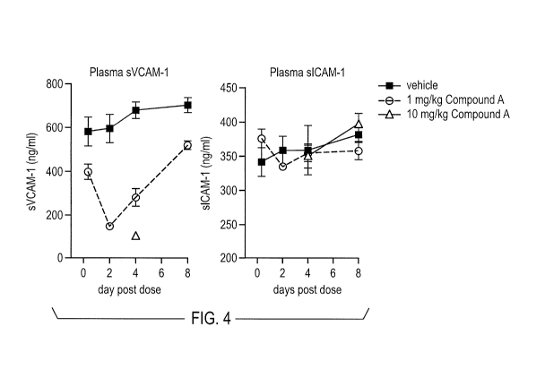
FIG. 4 depicts decreased levels of sVCAM in normal mice treated with small
molecule alpha-4 integrin inhibitors. The treatment of normal mice with alpha-
4
5
CA 02816016 2013-04-25
WO 2012/061074
PCT/US2011/057519
integrin inhibitors does not appear to affect soluble intracellular adhesion
molecule
(sICAM) level. Experiments were performed as described in Example 3.
FIG. 5 depicts that the effect of alpha-4 integrin inhibitors on sVCAM down-
regulation is dose-dependent and correlates with other markers of alpha-4
integrin
inhibition. Experiments were performed as described in Example 4.
FIG. 6 depicts decreased levels of sVCAM in mice treated with an antibody
inhibitor of alpha-4 integrin. Experiments were performed as described in
Example 5.
FIG. 7 depicts decreased levels of sVCAM in mice treated with a non-
pegylated small molecule inhibitor of alpha-4 integrin. Experiments were
perfonned
as described in Example 6.
FIG. 8 depicts that the effects of alpha-4 integrin inhibition on sVCAM levels
is dose-dependent and wears off as plasma levels of the alpha-4 integrin
inhibitor
declines. Experiments were performed as described in Example 7.
FIG. 9 depicts that alpha-4 integrin inhibition results in down-regulation of
sMAdCAM in several mouse models of colitis. Experiments were performed as
described in Example 8.
FIG. 10 depicts that alpha-4 integrin inhibition by a small molecule inhibitor
results in down-regulation of sMAdCAM in normal mice. Experiments were
performed as described in Example 9.
FIG. 11 depicts that alpha-4 integrin inhibition by an antibody inhibitor
results
in down-regulation of sMAdCAM in normal mice. Experiments were performed as
described in Example 10.
FIG. 12 depicts that down-regulation of sMAdCAM by alpha-4 integrin
inhibitors is dose-dependent, reversible, and correlates with in vitro
selectivity of the
alpha-4 integrin inhibitor for the alpha-4 beta-7 integrin heterodimer.
Experiments
were performed as described in Example 11.
FIG. 13 depicts selective down-regulation of sVCAM by an alpha-4 integrin
inhibitor selectively binding to the alpha-4 beta-1 integrin heterodimer.
Experiments
were performed as described in Example 11.
6
CA 02816016 2013-04-25
WO 2012/061074
PCT/US2011/057519
FIG. 14 depicts the correlation between the sVCAM / sMAdCAM levels and
the alpha-4 integrin antibody levels in mice. Experiments were performed as
described in Example 12.
DETAILED DESCRIPTION
1. Definitions
An "individual" as used herein may be any of mammalian animals (e.g.,
domesticated animals), including human, dog, cat, cattle, horse, goat, pig,
swine,
sheep, monkey, rat, and mouse. In one embodiment, the individual can be a
human.
The term "pathological and chronic inflammation" as used herein refers to an
inappropriate inflammation associated with disorders including, but not
limited to,
asthma, atherosclerosis, AIDS dementia, diabetes, inflammatory bowel disease,
rheumatoid arthritis, transplant rejection, graft versus host disease,
multiple sclerosis
(especially in MS involving further demyelination), for example, primary
progressive
multiple sclerosis (PPMS), secondary progressive multiple sclerosis (SPMS),
relapsing-remitting multiple sclerosis (RRMS), and progressive relapsing
multiple
sclerosis (PRMS), tumor metastasis, nephritis, atopic dermatitis, psoriasis,
myocardial
ischemia, chronic prostatitis, complications from sickle cell anemia, lupus
erythematosus, and acute leukocyte mediated lung injury. Such inflammation is
characterized by a heightened response of inflammatory cells, including
infiltrating
leukocytes. Over time, such pathological inflammation often results in damage
to
tissue in the region of inappropriate inflammation.
The term "alpha-4 integrin activity" as used herein refers to the accessible
amount of alpha-4 integrins, including both the alpha-4 beta-1 and alpha-4
beta-7
dimers, presented on the leukocyte cell surface. Alpha-4 integrin activity can
be
determined using any technique known in the art. For example, alpha-4 integrin
activity can be evaluated directly through cytometry using a florescently-
labeled
antibody specific to alpha-4 integrins. See, e.g., U.S. Patent No. 7,807,167.
Alternatively, alpha-4 integrin activity can be evaluated indirectly by
measuring
leukocyte infiltration in tissue samples. See, e.g., U.S. Patent No.
7,435,802; see also
Krumbholz et al., Neurology 71: 1350-1354 (2008).
The term "biological sample" as used herein refers to a biological material
from an individual. A biological sample may be, as non-limiting examples, a
tissue,
7
CA 02816016 2013-04-25
WO 2012/061074
PCT/US2011/057519
cell, whole blood, serum, body fluids, plasmic fluid, autoptical tissue sample
(e.g.,
brain, skin, lymph node, spinal cord), cultured cells or supernatants from
cultured
cells. The biological sample used will vary based on the assay format, the
detection
method, and the nature of the sample to be assayed. Methods for preparing
biological
samples are well known in the art and can be readily adapted in order to
obtain a
biological sample that is compatible with the method utilized.
The term "body fluid" used herein includes fluids that are found in
individuals. They include fluids that are excreted or secreted from the body,
as well
as fluids that nollually are not excreted or secreted. These fluids include,
as non-
limiting examples, aqueous humor, blood, serum, interstitial fluid, lymph,
mucus,
pleural fluid, saliva, plasma, urine, semen, tears, synovial fluid, wound
fluid, and/or
cerebrospinal fluid. Typically, blood including blood serum and blood plasma
are
used in the present embodiments.
The terms "specifically binds" or "binds specifically" as used herein means
that one member of a specific binding pair will not show any statistically
significant
binding to molecules other than its specific binding partner. A binding
partner may
show at least 1000 times the affinity of binding (measured as an apparent
association
constant) for its specific binding pair partner than a non-specific binding
partner. For
example, antibodies that bind to an alpha-4 integrin with a binding affinity
of 107
mole/L or more, typically 108 mole/1 or more, are said to bind specifically to
an alpha-
4 integrin.
The term "diagnostic kit" as used herein includes typically a detection system
with different packages of diagnostic antibodies and/or reagents that are
necessary for
the quantitative and/or qualitative evaluation of a biomarker. Kits generally
include
instructions for using the reagents and/or diagnostic antibodies. The
antibodies, as
well as any reagent, can be provided as a liquid, powder, tablet, or
suspension. The
antibodies and/or reagents may be provided in separate packages suitable for
application separately.
Unless defined otherwise, all technical and scientific temis used herein have
the same meaning as commonly understood by one of ordinary skill in the art.
It must
be noted that as used herein, the singular forms "a", "and", and "the" include
plural
referents unless the context clearly dictates otherwise. Thus, for example,
reference
8
CA 02816016 2013-04-25
WO 2012/061074
PCT/US2011/057519
to "an antibody" includes a plurality of such antibodies and reference to "the
dosage"
includes reference to one or more dosages and equivalents thereof known to
those
skilled in the art, and so forth.
2. Alpha-4 integrin inhibitors
Various types of alpha-4 integrin inhibitors having the ability to bind to and
inhibit alpha-4 integrin activity can be used in the present embodiments. Many
such
inhibitors have been identified and characterized, and representative examples
are
described below. Given the teachings disclosed herein, it is well within the
skill of
one in the art to identify other alpha-4 integrin inhibitors that will be able
to inhibit
the alpha-4-comprising integrin dimers in a manner that biologically mimics or
is
similar to the specifically described inhibitors. The present embodiments also
include
the chronic administration of such inhibitors and combinations thereof.
2.1. Antibodies or immunologically active fragments
In one embodiment, the alpha-4 integrin inhibitors are antibodies or
immunologically active fragments thereof that selectively bind to an alpha-4
integrin
or a dimer comprising alpha-4, such as alpha-4 beta-1 or alpha-4 beta-7.
Representative alpha-4 integrin antibodies are known in the art, including for
example
(1) Natalizumab, disclosed in U.S. Patent Nos. 5,168,062, 5,385,839,
5,730,978,
5,840,299, 6,033,665, and 6,602,503, (2) the CD49d antibodies manufactured by
Biolegend (San Diego, CA); and (3) PS/2 which is a rat anti-mouse alpha-4
integrin
antibody (the PS/2 hybridoma is available from the ATCC (Rockville, MD)). Non-
limiting example of alpha-4 integrin antibodies include those disclosed in
U.S. Patent
Nos. 5,565,332, 5,733,743, 5,837,242, 5,858,657, 5,871,734, 5,871,907,
5,872,215,
5,885,793, 5,888,507, 5,932,214, 5,969,108, 6,140,471, 6,172,197, 6,180,336,
6,225,447, and 7,176,184.
In one embodiment, the alpha-4 integrin inhibitor can be a monoclonal
antibody. In another embodiment, the antibody may be chemically modified,
e.g., by
pegylation. Additionally, other antibodies can be identified using techniques
available in the art. For example, antibodies capable of specifically binding
to alpha-
4 integrin can be produced using phage display technology. Antibody fragments
that
selectively bind to an alpha-4 integrin or a dimer comprising an alpha-4
integrin can
9
CA 02816016 2013-04-25
WO 2012/061074
PCT/US2011/057519
then be isolated. Exemplary methods for producing such antibodies via phage
display
are disclosed in U.S. Patent No. 6,225,447, for example.
Monoclonal antibodies can also be produced using the conventional
hybridoma methods. These methods have been widely applied to produce hybrid
cell
lines that secrete high levels of monoclonal antibodies against many specific
antigens,
and can also be used to produce monoclonal antibodies capable of specifically
binding
to alpha-4 integrins. For example, mice (e.g., Balb/c mice) can be immunized
with an
antigenic alpha-4 integrin epitope by intraperitoneal injection. After
sufficient time
has passed to allow for an immune response, the mice are sacrificed, and the
spleen
cells obtained and fused with myeloma cells, using techniques well known in
the art.
The resulting fused cells, hybridomas, are then grown in a selective medium,
and the
surviving cells grown in such medium using limiting dilution conditions. After
cloning and recloning, hybridomas can be isolated for secreting antibodies
(for
example, of the IgG or IgM class or IgG1 subclass) that selectively bind to
the target,
alpha-4 integrin or a dimer comprising an alpha-4 integrin. To produce agents
specific for human use, the isolated monoclonal can then be used to produce
chimeric
and humanized antibodies.
Antibodies that can be used as alpha-integrin inhibitors include, but are not
limited to, polyclonal, monoclonal, multispecific, human, humanized or
chimeric
antibodies, single chain antibodies (e.g., scFv), Fab fragments, F(ab')
fragments,
fragments produced by a Fab expression library, anti-idiotypic (anti-Id)
antibodies
(including, e.g., anti-Id antibodies to antibodies of the present
embodiments), and
epitope-binding fragments of any of the above. Typically, the antibodies are
human
antigen-binding antibody fragments, which include, but are not limited to,
Fab, Fab'
and F(ab')2, Fd, single-chain Fvs(scFv), single-chain antibodies, disulfide-
linked Fvs
(sdFv), and fragments comprising either a VL or VH domain. Antigen-binding
antibody fragments, including single-chain antibodies, may comprise the
variable
region(s) alone or in combination with the entirety or a portion of the
following: hinge
region, CH1, CH2, and CH3 domains. Also included are antigen-binding fragments
that can comprise any combination of variable region(s) with a hinge region,
CH1,
CH2, and CH3 domains. The antibodies may be from any animal origin including
birds and mammals. Typically, the antibodies are human, murine (e.g., mouse
and
rat), donkey, sheep, monkey, rabbit, goat, guinea pig, pig, camel, horse, or
chicken (or
other avian). As used herein, "human" antibodies include antibodies having the
CA 02816016 2013-04-25
WO 2012/061074
PCT/US2011/057519
amino acid sequence of a human immunoglobulin and include antibodies isolated
from human immunoglobulin libraries or from animals transgenic for one or more
human immunoglobulins and that do not express endogenous immunoglobulins, as
described, for example in, U.S. Patent No. 5,939,598.
Chimeric and humanized antibodies can be produced from non-human
antibodies, and can have the same or similar binding affinity as the antibody
from
which they are produced. Techniques for producing chimeric antibodies
(Morrison et
al., 1984 Proc. Nat'l. Acad. Sci. USA 81: 6851; Neuberger et al., 1984 Nature
312:
604; Takeda et al., 1985 Nature 314: 452) include splicing the genes from,
e.g., a
mouse antibody molecule of appropriate antigen specificity together with genes
from
a human antibody molecule of appropriate biological activity. For example, a
nucleic
acid encoding a variable (V) region of a mouse monoclonal antibody can be
joined to
a nucleic acid encoding a human constant (C) region, e.g., IgG1 or IgG4. The
resulting antibody is thus a species hybrid, generally with the antigen
binding domain
from the non-human antibody and the C or effector domain from a human or
primate
antibody.
Humanized antibodies are antibodies with variable regions that are primarily
from a human antibody (i.e., the acceptor antibody), but which have
complementarity
determining regions substantially from a non-human antibody (the donor
antibody).
See, e.g., Queen et al., Proc. Nat'l. Acad. Sci USA 86: 10029-10033 (1989); WO
90/07861, U.S. Patent Nos. 7,435,802, 6,054,297; 5,693,761; 5,585,089;
5,530,101;
and 5,224,539. The constant region or regions of these antibodies are
generally also
from a human antibody. The human variable domains are typically chosen from
human antibodies having sequences displaying a high homology with the desired
non-
human variable region binding domains. The heavy and light chain variable
residues
can be derived from the same antibody, or a different human antibody. In
addition,
the sequences can be chosen as a consensus of several human antibodies, such
as
described in WO 92/22653.
A PrimatizedTM antibody" is a recombinant antibody containing primate
variable sequences or antigen binding portions, and human constant domain
sequences. See, e.g., Newman, Bio/Technology, 1992, 10: 1455-60. Primatization
of
antibodies results in the generation of antibodies that contain monkey
variable
domains and human constant sequences. See, e.g.,U U.S. Patent No. 6,113,898.
This
technique modifies antibodies such that they are not rejected upon
administration in
11
CA 02816016 2013-04-25
WO 2012/061074
PCT/US2011/057519
humans because they are antigenic. This technique relies on immunization of
cynomolgus monkeys with human antigens or receptors. This technique was
developed to create high affinity monoclonal antibodies directed to human cell
surface antigens.
Specific amino acids within the human variable region are selected for
substitution based on the predicted conformation and antigen binding
properties. This
can be determined using techniques such as computer modeling, prediction of
the
behavior and binding properties of amino acids at certain locations within the
variable
region, and observation of effects of substitution. For example, when an amino
acid
differs between a non-human variable region and a human variable region, the
human
variable region can be altered to reflect the amino acid composition of the
non-human
variable region. In a specific embodiment, the antibodies used in the chronic
dosage
regime are humanized antibodies as disclosed in U.S. Patent No. 5,840,299. In
another embodiment, transgenic mice containing human antibody genes can be
immunized with an antigenic alpha-4 integrin structure and hybridoma
technology can
be used to generate human antibodies that selectively bind to alpha-4
integrin.
Chimeric, human, primatized, and/or humanized antibodies can be produced
by using recombinant expression, e.g., expression in human hybridomas (Cole et
al.,
Monoclonal Antibodies and Cancer Therapy, Alan R. Liss, p. 77 (1985)), in
myeloma
cells, or in Chinese hamster ovary (CHO) cells. Alternatively, antibody coding
sequences can be incorporated into transgenes for introduction into the genome
of a
transgenic animal and subsequent expression in the milk of the transgenic
animal.
See, e.g., U.S. Patent No. 6,197,946. Suitable transgenes include transgenes
having a
promoter and/or enhancer from a mammary gland specific gene, for example
casein
or p-lactoglobulin.
2.2. Small molecules
Small molecules for use in the present embodiments may encompass
compounds having a molecular weight of more than 50 and less than about 4,000
Daltons. Alternatively, these compounds may have covalently attached
polyethylene
glycol polymer chains (i.e., pegylation) to improve various properties of the
compounds, for example, extended half-life, improved tissue penetration, and
improved solubility. The pegylated conjugates thus may have a molecular weight
about 40 kilodaltons (kDa). Alpha-4 integrin inhibitors comprise functional
groups
12
CA 02816016 2013-04-25
WO 2012/061074
PCT/US2011/057519
necessary for structural interaction with proteins, particularly hydrogen
bonding, and
may include an amine, carbonyl, hydroxyl, or carboxyl group, typically at
least two of
functional chemical groups. The alpha-4 integrin inhibitors often comprise
cyclical
carbon or heterocyclic structures and/or aromatic or polyaromatic structures
substituted with one or more of the above-described functional groups. Alpha-4
integrin inhibitors may include, but not limited to: peptides, saccharides,
fatty acids,
steroids, purines, pyrimidines, derivatives, structural analogs, or
combinations thereof.
Non-limiting examples of alpha-4 integrin inhibitors are described, for
example, in
U.S. Patent Nos., 5,998,447 (heterocycles), 6,034,238 (heterocyclic
compounds),
6,331,552 (substituted imidazolidine), 6,399,643 (spiroimidazolidine
derivatives),
6,423,712 (2,4-substituted imidazolidine derivatives), 6,514,952 (hydantoin
derivatives), 6,521,654 (substituted imidazolidine derivatives), 6,667,331
(non-
peptidyl compounds), 6,667,334 (imidazolidine derivatives), 6,668,527 (non-
peptidyl
compounds), 6,680,333 (imidazolidine derivatives) , 6,756,378, 6,759,424
(imidazolidine derivatives), 6,838,439 (heterocytes), 6,903,128 (non-peptidyl
compounds), 6,962,937 (imidazolidine derivatives), 7,179,819, and 7,196,112.
Several representative small molecule alpha-4 integrin inhibitors are shown in
FIG. 1.
2.3. Anti-alpha-4 integrin peptides
The present embodiments also include any peptide that is capable of binding
to an alpha-4 integrin or a dimer comprising an alpha-4 subunit. Included are
peptides that are substantially homologous to a region of the extracellular
matrix or a
natural ligand of the specific alpha-4 integrin receptor or receptors
targeted. For
example, for the chronic inhibition of alpha-4 beta-1 receptor, peptides can
be used
that comprise at least a portion of the fibronectin IIICS region (e.g.,
peptides
comprising at least a portion of the CS-1 peptide sequence or a sequence
substantially
homologous to the CS-1 sequence) can be used to bind to a receptor and inhibit
the
activity of the alpha-4 comprising integrin. See, e.g., U.S. Patent No.
7,238,668.
3. Use of alpha-4 integrin inhibitors to treat diseases associated with
pathological or chronic inflammation
Alpha-4 integrin inhibitors can be used to treat various diseases associated
with pathological or chronic inflammation by blocking alpha-4-dependent
interactions. The alpha-4-dependent interaction with the VCAM-1 ligand on
endothelial cells is an early event in many inflammatory responses, including
those of
13
CA 02816016 2013-04-25
WO 2012/061074
PCT/US2011/057519
the central nervous system. Undesired diseases and conditions resulting from
inflammation and having acute and/or chronic clinical exacerbations include
multiple
sclerosis (Yednock et al., 1992 Nature 356: 63; Baron et al., 1993 J. Exp.
Med. 177:
57), meningitis, encephalitis, stroke, other cerebral traumas, inflammatory
bowel
disease (IBD) including ulcerative colitis and Crohn's disease (CD) (Hamann et
al.,
1994J. Immunol. 152: 3238; Podolsky et al., 1993 J. Clin. Invest. 92: 372),
rheumatoid arthritis (van Dinther-Janssen et al., 1991 J. Immunol. 147: 4207;
van
Dinther-Janssen et al., 1993 Annals Rheumatic Diseases 52: 672; Elices et al.,
1994 J.
Clin. Invest. 93: 405; Postigo et al., 1992 1 Clin. Invest. 89: 1445), asthma
(Mulligan
et al., 1993 J. Immunol. 150: 2407) and acute juvenile onset diabetes (Type 1)
(Yang
et al., 1993 Proc. Nat'l Acad. Sci. USA 90: 10494; Burkly et al., 1994
Diabetes 43:
529; Baron et al., 1994 J. Clin. Invest. 93: 1700), AIDS induced dementia
(Sasseville
et al., 1994 Am. J. Path. 144: 27); atherosclerosis (Cybulsky et al., 1991
Science 251:
788-91, Li et al., 1993 Arterioscler. Thromb. 13: 197), nephritis (Rabb et
al., 1995
Springer Semin. Immunopathol. 16: 417-25), retinitis, atopic dermatitis,
psoriasis,
myocardial ischemia, chronic prostatitis, complications from sickle cell
anemia, lupus
erythematosus, and acute leukocyte-mediated lung injury such as occurs in
adult
respiratory distress syndrome.
Inflammatory bowel disease is a collective term for two similar diseases
referred to as Crohn's disease (CD) and ulcerative colitis. CD is an
idiopathic,
chronic ulceroconstrictive inflammatory disease characterized by sharply
delimited
and typically transmural involvement of all layers of the bowel wall by a
granulomatous inflammatory reaction. Any segment of the gastrointestinal
tract, from
the mouth to the anus, may be involved, although the disease most commonly
affects
the terminal ileum and/or colon. Ulcerative colitis is an inflammatory
response
limited largely to the colonic mucosa and submucosa. Lymphocytes and
macrophages are numerous in lesions of inflammatory bowel disease and may
contribute to inflammatory injury.
Asthma is a disease characterized by increased responsiveness of the
tracheobronchial tree to various stimuli potentiating paroxysmal constriction
of the
bronchial airways. The stimuli cause release of various mediators of
inflammation
from IgE-coated mast cells including histamine, eosinophilic and neutrophilic
chemotactic factors, leukotrines, prostaglandin, and platelet activating
factor. Release
14
CA 02816016 2013-04-25
WO 2012/061074
PCT/US2011/057519
of these factors recruits basophils, eosinophils and neutrophils, which cause
inflammatory injury.
Atherosclerosis is a disease of arteries (e.g., coronary, carotid, aorta and
iliac).
The basic lesion, the atheroma, consists of a raised focal plaque within the
intima,
having a core of lipid and a covering fibrous cap. Atheromas compromise
arterial
blood flow and weaken affected arteries. Myocardial and cerebral infarcts are
a major
consequence of this disease. Macrophages and leukocytes are recruited to
atheromas
and contribute to inflammatory injury.
Rheumatoid arthritis is a chronic, relapsing inflammatory disease that
primarily causes impaiiiiient and destruction of joints. Rheumatoid arthritis
usually
first affects the small joints of the hands and feet but then may involve the
wrists,
elbows, ankles, and knees. The arthritis results from interaction of synovial
cells with
leukocytes that infiltrate from the circulation into the synovial lining of
joints. See,
e.g., Paul, Immunology 3rd ed., Raven Press, 1993.
Alpha-4 integrin inhibitors can be used in the treatment of organ or graft
rejection. Over recent years, there has been a considerable improvement in the
efficiency of surgical techniques for transplanting tissues and organs such as
skin,
kidney, liver, heart, lung, pancreas, and bone marrow. Perhaps the principal
outstanding problem is the lack of satisfactory agents for inducing
immunotolerance
in the recipient to the transplanted allograft or organ. When allogeneic cells
or organs
are transplanted into a host (i.e., the donor and donee are different
individuals from
the same species), the host immune system is likely to mount an immune
response to
foreign antigens in the transplant (host-versus-graft disease) leading to
destruction of
the transplanted tissue. CD8+ cells, CD4+ cells, and monocytes are all
involved in
the rejection of transplant tissues. Antibodies directed to alpha-4 integrin
are useful,
inter alia, to block alloantigen-induced immune responses in the donee thereby
preventing such cells from participating in the destruction of the
transplanted tissue or
organ. See, e.g., Paul et al., 1996 Transplant International 9: 420-425;
Georczynski
et al., 1996 Immunol. 87: 573-580); Georcyznski et al., 1995 Transplant.
Immunol. 3:
55-61; Yang et al., 1995 Transplantation 60: 71-76; and Anderson et al., 1994
APMIS
102: 23-27. A related use for the alpha-4 integrin inhibitors is modulating
the
immune response involved in "graft versus host" disease (GVHD). See, e.g.,
Schlegel
et al., J. Immunol. 155: 3856-3865 (1995). GVHD is a potentially fatal disease
that
occurs when immunologically competent cells are transferred to an allogeneic
CA 02816016 2013-04-25
WO 2012/061074
PCT/US2011/057519
recipient. In this situation, the donor's immunocompetent cells may attack
tissues in
the recipient. Tissues of the skin, gut epithelia, and liver are frequent
targets and may
be destroyed during the course of GVHD. The disease presents an especially
severe
problem when immune tissue is being transplanted, such as in bone marrow
transplantation; but less severe GVHD has also been reported in other cases as
well,
including heart and liver transplants. Alpha-4 integrin inhibitors are used,
inter alia,
to block activation of the donor T-cells thereby interfering with their
ability to lyse
target cells in the host.
Alpha-4 integrin inhibitors may be useful in inhibiting tumor metastasis.
Several tumor cells have been reported to express alpha-4 integrin and
antibodies to
alpha-4 integrin have been reported to block adhesion of such cells to
endothelial
cells. See, e.g., Steinback et al., 1995 Urol. Res. 23: 175-83; Orosz et al.,
1995 Int. J.
Cancer 60: 867-71; Freedman et al., 1994 Leuk Lymphoma 13: 47-52; and Okahara
et
al., 1994 Cancer Res. 54: 3233-6.
Alpha-4 integrin inhibitors may be useful in treating multiple sclerosis.
Multiple sclerosis (MS) is a progressive neurological autoimmune disease that
affects
an estimated 250,000 to 350,000 people in the United States. Multiple
sclerosis is
thought to be the result of a specific autoimmune reaction in which certain
leukocytes
attack and initiate the destruction of myelin, the insulating sheath covering
nerve
fibers. In an animal model for multiple sclerosis, murine monoclonal
antibodies
directed against alpha-4 beta-1 integrin have been shown to block the adhesion
of
leukocytes to the endothelium, and thus prevent inflammation of the central
nervous
system and subsequent paralysis in the animals. The onset of MS may be
dramatic or
so mild as to not cause a patient to seek medical attention. The most common
symptoms include weakness in one or more limbs, visual blurring due to optic
neuritis, sensory disturbances, diplopia, and ataxia. The course of disease
may be
stratified into three general categories: (1) relapsing MS, (2) chronic
progressive MS,
and (3) inactive MS. Relapsing MS is characterized by recurrent attacks of
neurologic dysfunction. MS attacks generally evolve over days to weeks and may
be
followed by complete, partial or no recovery. Recovery from attacks generally
occurs
within weeks to several months from the peak of symptoms, although rarely some
recovery may continue for 2 or more years. Chronic progressive MS results in
gradually progressive worsening without periods of stabilization or remission.
This
form develops in patients with a prior history of relapsing MS, although in
20% of
16
CA 02816016 2013-04-25
WO 2012/061074
PCT/US2011/057519
patients, no relapses can be recalled. Acute relapses also may occur during
the
progressive course. A third form is inactive MS. Inactive MS is characterized
by
fixed neurologic deficits of variable magnitude. Most patients with inactive
MS have
an earlier history of relapsing MS. The course of MS is also dependent on the
age of
the patient. For example, favorable prognostic factors include early onset
(excluding
childhood), a relapsing course and little residual disability 5 years after
onset. By
contrast, poor prognosis is associated with a late age of onset (i.e., age 40
or older)
and a progressive course. These variables are interdependent, since chronic
progressive MS tends to begin at a later age that relapsing MS. Disability
from
chronic progressive MS is usually due to progressive paraplegia or
quadriplegia in
individual patients.
Alpha-4 integrin inhibitors may be used with effective amounts of other
therapeutic agents against acute and chronic inflammation. Such agents include
other
antagonists of adhesion molecules (e.g., other integrins, selectins, and
immunoglobulin (Ig) super family members). See, e.g., Springer, 1990 Nature
346:
425-433; Osborn, 1990 Cell 62: 3; Hynes, 1992 Cell 9: 11. Other anti-
inflammatory
agents that can be used in combination with the alpha-4 integrin inhibitors
include
antibodies and other antagonists of cytokines, such as interleukins IL-1
through IL-13,
tumor necrosis factors a and 13, interferons cc, 13, and y, tumor growth
factor beta
(TGF-13), colony stimulating factor (CSF) and granulocyte monocyte colony
stimulating factor (GM-CSF). Other anti-inflammatory agents may also include
antibodies and other antagonists of chemokines such as MCP-1, MIP-lcc, MIP-
1(3,
RANTES, exotaxin, and IL-8. Other anti-inflammatory agents may further include
NSAIDS, steroids, and other small molecule inhibitors of inflammation.
4. Use of alpha-4 integrin inhibitors to treat autoimmune diseases
Alpha-4 integrin inhibitors also can be used to treat various autoimmune
diseases. An autoimmune disease herein is a disease or disorder arising from
and
directed against an individual's own tissues or a co-segregate or
manifestation thereof
or resulting condition therefrom. Examples of autoimmune diseases or disorders
include, but are not limited to arthritis (rheumatoid arthritis such as acute
arthritis,
chronic rheumatoid arthritis, gout or gouty arthritis, acute gouty arthritis,
acute
immunological arthritis, chronic inflammatory arthritis, degenerative
arthritis, type II
17
CA 02816016 2013-04-25
WO 2012/061074
PCT/US2011/057519
collagen-induced arthritis, infectious arthritis, Lyme arthritis,
proliferative arthritis,
psoriatic arthritis, Still's disease, vertebral arthritis, and juvenile-onset
rheumatoid
arthritis, osteoarthritis, arthritis chronica progrediente, arthritis
defonnans,
polyarthrifis chronica primaria, reactive arthritis, and ankylosing
spondylitis),
inflammatory hyperproliferative skin diseases, psoriasis such as plaque
psoriasis,
gutatte psoriasis, pustular psoriasis, and psoriasis of the nails, atopy
including atopic
diseases such as hay fever and Job's syndrome, dermatitis including contact
dermatitis, chronic contact dermatitis, exfoliative dermatitis, allergic
dermatitis,
allergic contact dermatitis, dermatitis herpetiformis, nummular den-natitis,
seborrheic
dermatitis, non-specific dermatitis, primary irritant contact dermatitis, and
atopic
den-natitis, x-linked hyper IgM syndrome, allergic intraocular inflammatory
diseases,
urticaria such as chronic allergic urticaria and chronic idiopathic urticaria,
including
chronic autoimmune urticaria, myositis, polymyositis/dermatomyositis, juvenile
dennatomyositis, toxic epidermal necrolysis, scleroden-na (including systemic
scleroderma), sclerosis such as systemic sclerosis, multiple sclerosis (MS)
such as
spino-optical MS, primary progressive MS (PPMS), and relapsing remitting MS
(RRMS), progressive systemic sclerosis, atherosclerosis, arteriosclerosis,
sclerosis
disseminata, ataxic sclerosis, neuromyelitis optica (NMO), inflammatory bowel
disease (IBD) (for example, Crohn's disease, autoimmune-mediated
gastrointestinal
diseases, colitis such as ulcerative colitis, colitis ulcerosa, microscopic
colitis,
collagenous colitis, colitis polyposa, necrotizing enterocolitis, and
transmural colitis,
and autoimmune inflammatory bowel disease), bowel inflammation, pyoderma
gangrenosum, erythema nodosum, primary sclerosing cholangitis, respiratory
distress
syndrome, including adult or acute respiratory distress syndrome (ARDS),
meningitis,
inflammation of all or part of the uvea, iritis, choroiditis, an autoimmune
hematological disorder, rheumatoid spondylitis, rheumatoid synovitis,
hereditary
angioedema, cranial nerve damage as in meningitis, herpes gestationis,
pemphigoid
gestationis, pruritis scroti, autoimmune premature ovarian failure, sudden
hearing loss
due to an autoimmune condition, IgE-mediated diseases such as anaphylaxis and
allergic and atopic rhinitis, encephalitis such as Rasmussen's encephalitis
and limbic
and/or brainstem encephalitis, uveitis, such as anterior uveitis, acute
anterior uveitis,
granulomatous uveitis, nongranulomatous uveitis, phacoantigenic uveitis,
posterior
uveitis, or autoimmune uveitis, glomerulonephritis (GN) with and without
nephrotic
syndrome such as chronic or acute glomerulonephritis such as primary GN,
immune-
18
CA 02816016 2013-04-25
WO 2012/061074
PCT/US2011/057519
mediated GN, membranous GN (membranous nephropathy), idiopathic membranous
GN or idiopathic membranous nephropathy, membrano- or membranous proliferative
GN (MPGN), including Type I and Type II, and rapidly progressive GN,
proliferative
nephritis, autoimmune polyglandular endocrine failure, balanitis including
balanitis
circumscripta plasmacellularis, balanoposthitis, erythema annulare
centrifugum,
erythema dyschromicum perstans, eythema multiform, granuloma annulare, lichen
nitidus, lichen sclerosus et atrophicus, lichen simplex chronicus, lichen
spinulosus,
lichen planus, lamellar ichthyosis, epiden-nolytic hyperkeratosis,
premalignant
keratosis, pyoderma gangrenosum, allergic conditions and responses, allergic
reaction, eczema including allergic or atopic eczema, asteatotic eczema,
dyshidrotic
eczema, and vesicular palmoplantar eczema, asthma such as asthma bronchiale,
bronchial asthma, and auto-immune asthma, conditions involving infiltration of
T
cells and chronic inflammatory responses, immune reactions against foreign
antigens
such as fetal A-B-0 blood groups during pregnancy, chronic pulmonary
inflammatory
disease, autoimmune myocarditis, leukocyte adhesion deficiency, lupus,
including
lupus nephritis, lupus cerebritis, pediatric lupus, non-renal lupus, extra-
renal lupus,
discoid lupus and discoid lupus erythematosus, alopecia lupus, systemic lupus
erythematosus (SLE) such as cutaneous SLE or subacute cutaneous SLE, neonatal
lupus syndrome (NLE), and lupus erythematosus disseminatus, juvenile onset
(Type
I) diabetes mellitus, including pediatric insulin-dependent diabetes mellitus
(IDDM),
adult onset diabetes mellitus (Type II diabetes), autoimmune diabetes,
idiopathic
diabetes insipidus, diabetic retinopathy, diabetic nephropathy, diabetic large-
artery
disorder, immune responses associated with acute and delayed hypersensitivity
mediated by cytokines and T-lymphocytes, tuberculosis, sarcoidosis,
granulomatosis
including lymphomatoid granulomatosis, Wegener's granulomatosis,
agranulocytosis,
vasculitides, including vasculitis, large-vessel vasculitis (including
polymyalgia
rheumatica and giant-cell (Takayasu's) arteritis), medium-vessel vasculitis
(including
Kawasaki's disease and polyarteritis nodosa/periarteritis nodosa), microscopic
polyarteritis, immunovasculitis, CNS vasculitis, cutaneous vasculitis,
hypersensitivity
vasculitis, necrotizing vasculitis such as systemic necrotizing vasculitis,
and ANCA-
associated vasculitis, such as Churg-Strauss vasculitis or syndrome (CSS) and
ANCA-associated small-vessel vasculitis, temporal arteritis, aplastic anemia,
autoimmune aplastic anemia, Coombs positive anemia, Diamond Blackfan anemia,
hemolytic anemia or immune hemolytic anemia including autoimmune hemolytic
19
CA 02816016 2013-04-25
WO 2012/061074
PCT/US2011/057519
anemia (AIHA), pernicious anemia (anemia perniciosa), Addison's disease, pure
red
cell anemia or aplasia (PRCA), Factor VIII deficiency, hemophilia A,
autoimmune
neutropenia, pancytopenia, leukopenia, diseases involving leukocyte
diapedesis, CNS
inflammatory disorders, multiple organ injury syndrome such as those secondary
to
septicemia, trauma or hemorrhage, antigen-antibody complex-mediated diseases,
anti-
glomerular basement membrane disease, anti-phospholipid antibody syndrome,
allergic neuritis, Behcet's disease/syndrome, Castleman's syndrome,
Goodpasture's
syndrome, Reynaud's syndrome, Sjogren's syndrome, Stevens-Johnson syndrome,
pemphigoid such as pemphigoid bullous and skin pemphigoid, pemphigus
(including
pemphigus vulgaris, pemphigus foliaceus, pemphigus mucus-membrane pemphigoid,
and pemphigus erythematosus), autoimmune polyendocrinopathies, Reiter's
disease
or syndrome, thermal injury, preeclampsia, an immune complex disorder such as
immune complex nephritis, antibody-mediated nephritis, polyneuropathies,
chronic
neuropathy such as IgM polyneuropathies or IgM-mediated neuropathy,
thrombocytopenia (as developed by myocardial infarction patients, for
example),
including thrombotic thrombocytopenic purpura (TTP), post-transfusion purpura
(PTP), heparin-induced thrombocytopenia, and autoimmune or immune-mediated
thrombocytopenia such as idiopathic thrombocytopenic purpura (ITP) including
chronic or acute ITP, scleritis such as idiopathic cerato-scleritis,
episcleritis,
autoimmune disease of the testis and ovary including autoimmune orchitis and
oophoritis, primary hypothyroidism, hypoparathyroidism, autoimmune endocrine
diseases including thyroiditis such as autoimmune thyroiditis, Hashimoto's
disease,
chronic thyroiditis (Hashimoto's thyroiditis), or subacute thyroiditis,
autoimmune
thyroid disease, idiopathic hypothyroidism, Grave's disease, polyglandular
syndromes
such as autoimmune polyglandular syndromes (or polyglandular endocrinopathy
syndromes), paraneoplastic syndromes, including neurologic paraneoplastic
syndromes such as Lambert-Eaton myasthenic syndrome or Eaton-Lambert
syndrome, stiff-man or stiff-person syndrome, encephalomyelitis such as
allergic
encephalomyelitis or encephalomyelitis allergica and experimental allergic
encephalomyelitis (EAE), myasthenia gravis such as thymoma-associated
myasthenia
gravis, cerebellar degeneration, neuromyotonia, opsoclonus or opsoclonus
myoclonus
syndrome (OMS), and sensory neuropathy, multifocal motor neuropathy, Sheehan's
syndrome, autoimmune hepatitis, chronic hepatitis, lupoid hepatitis, giant-
cell
hepatitis, chronic active hepatitis or autoimmune chronic active hepatitis,
lymphoid
CA 02816016 2013-04-25
WO 2012/061074
PCT/US2011/057519
interstitial pneumonitis (LIP), bronchiolitis obliterans (non-transplant) vs
NSIP,
Guillain-Barre syndrome, Berger's disease (IgA nephropathy), idiopathic IgA
nephropathy, linear IgA dennatosis, acute febrile neutrophilic dermatosis,
subcorneal
pustular dermatosis, transient acantholytic dermatosis, cirrhosis such as
primary
biliary cirrhosis and pneumonocirrhosis, autoimmune enteropathy syndrome,
Celiac
or Coeliac disease, celiac sprue (gluten enteropathy), refractory sprue,
idiopathic
sprue, cryoglobulinemia, amylotrophic lateral sclerosis (ALS; Lou Gehrig's
disease),
coronary artery disease, autoimmune ear disease such as autoimmune inner ear
disease (AIED), autoimmune hearing loss, polychondritis such as refractory or
relapsed or relapsing polychondritis, pulmonary alveolar proteinosis, Cogan's
syndrome/nonsyphilitic interstitial keratitis, Bell's palsy, Sweet's
disease/syndrome,
rosacea autoimmune, zoster-associated pain, amyloidosis, a non-cancerous
lymphocytosis, a primary lymphocytosis, which includes monoclonal B cell
lymphocytosis (e.g., benign monoclonal gammopathy and monoclonal gammopathy
of undetermined significance, MGUS), peripheral neuropathy, paraneoplastic
syndrome, channelopathies such as epilepsy, migraine, arrhythmia, muscular
disorders, deafness, blindness, periodic paralysis, and channelopathies of the
CNS,
autism, inflammatory myopathy, focal or segmental or focal segmental
glomerulosclerosis (FSGS), endocrine opthalmopathy, uveoretinitis,
chorioretinitis,
autoimmune hepatological disorder, fibromyalgia, multiple endocrine failure,
Schmidt's syndrome, adrenalitis, gastric atrophy, presenile dementia,
demyelinating
diseases such as autoimmune demyelinating diseases and chronic inflammatory
demyelinating polyneuropathy, Dressler's syndrome, alopecia areata, alopecia
totalis,
CREST syndrome (calcinosis, Raynaud's phenomenon, esophageal dysmotility,
sclerodactyly, and telangiectasia), male and female autoimmune infertility,
e.g., due to
anti-spermatozoan antibodies, mixed connective tissue disease, Chagas'
disease,
rheumatic fever, recurrent abortion, fanner's lung, erythema multiforme, post-
cardiotomy syndrome, Cushing's syndrome, bird-fancier's lung, allergic
granulomatous angiitis, benign lymphocytic angiitis, Alport's syndrome,
alveolitis
such as allergic alveolitis and fibrosing alveolitis, interstitial lung
disease, transfusion
reaction, leprosy, malaria, parasitic diseases such as leishmaniasis,
kypanosomiasis,
schistosomiasis, ascariasis, aspergillosis, Sampter's syndrome, Caplan's
syndrome,
dengue, endocarditis, endomyocardial fibrosis, diffuse interstitial pulmonary
fibrosis,
interstitial lung fibrosis, pulmonary fibrosis, idiopathic pulmonary fibrosis,
cystic
21
CA 02816016 2013-04-25
WO 2012/061074
PCT/US2011/057519
fibrosis, endophthalmitis, erythema elevatum et diutinum, erythroblastosis
fetalis,
eosinophilic faciitis, Shulman's syndrome, Felty's syndrome, flariasis,
cyclitis such as
chronic cyclitis, heterochronic cyclitis, iridocyclitis (acute or chronic), or
Fuch's
cyclitis, Henoch-Schonlein purpura, human immunodeficiency virus (HIV)
infection,
SCID, acquired immune deficiency syndrome (AIDS), echovirus infection, sepsis,
endotoxemia, pancreatitis, thyroxicosis, parvovirus infection, rubella virus
infection,
post-vaccination syndromes, congenital rubella infection, Epstein-Barr virus
infection,
mumps, Evan's syndrome, autoimmune gonadal failure, Sydenham's chorea, post-
streptococcal nephritis, thromboangitis ubiterans, thyrotoxicosis, tabes
dorsalis,
chorioiditis, giant-cell polymyalgia, chronic hypersensitivity pneumonitis,
keratoconjunctivitis sicca, epidemic keratoconjunctivitis, idiopathic
nephritic
syndrome, minimal change nephropathy, benign familial and ischemia-reperfusion
injury, transplant organ reperfusion, retinal autoimmunity, joint
inflammation,
bronchitis, chronic obstructive airway/pulmonary disease, silicosis, aphthae,
aphthous
stomatitis, arteriosclerotic disorders, aspenniogenese, autoimmune hemolysis,
Boeck's disease, cryoglobulinemia, Dupuytren's contracture, endophthalmia
phacoanaphylactica, enteritis allergica, erythema nodosum leprosum, idiopathic
facial
paralysis, chronic fatigue syndrome, febris rheumatica, Hamman-Rich's disease,
sensoneural hearing loss, haemoglobinuria paroxysmatica, hypogonadism, ileitis
regionalis, leucopenia, mononucleosis infectiosa, traverse myelitis, primary
idiopathic
myxedema, nephrosis, ophthalmia symphatica, orchitis granulomatosa,
pancreatitis,
polyradiculitis acuta, pyoderma gangrenosum, Quervain's thyreoiditis, acquired
spenic atrophy, non-malignant thymoma, vitiligo, toxic-shock syndrome, food
poisoning, conditions involving infiltration of T cells, leukocyte-adhesion
deficiency,
immune responses associated with acute and delayed hypersensitivity mediated
by
cytokines and T-lymphocytes, diseases involving leukocyte diapedesis, multiple
organ
injury syndrome, antigen-antibody complex-mediated diseases, antiglomerular
basement membrane disease, allergic neuritis, autoimmune polyendocrinopathies,
oophoritis, primary myxedema, autoimmune atrophic gastritis, sympathetic
ophthalmia, rheumatic diseases, mixed connective tissue disease, nephrotic
syndrome,
insulitis, polyendocrine failure, autoimmune polyglandular syndrome type I,
adult-
onset idiopathic hypoparathyroidism (AOIH), cardiomyopathy such as dilated
cardiomyopathy, epidermolisis bullosa acquisita (EBA), hemochromatosis,
myocarditis, nephrotic syndrome, primary sclerosing cholangitis, purulent or
22
CA 02816016 2013-04-25
WO 2012/061074
PCT/US2011/057519
nonpurulent sinusitis, acute or chronic sinusitis, ethmoid, frontal,
maxillary, or
sphenoid sinusitis, an eosinophil-related disorder such as eosinophilia,
pulmonary
infiltration eosinophilia, eosinophilia-myalgia syndrome, Loffler's syndrome,
chronic
eosinophilic pneumonia, tropical pulmonary eosinophilia, bronchopneumonic
aspergillosis, aspergilloma, or granulomas containing eosinophils,
anaphylaxis,
seronegative spondyloarthritides, polyendocrine autoimmune disease, sclerosing
cholangitis, sclera, episclera, chronic mucocutaneous candidiasis, Bruton's
syndrome,
transient hypogammaglobulinemia of infancy, Wiskott-Aldrich syndrome, ataxia
telangiectasia syndrome, angiectasis, autoimmune disorders associated with
collagen
disease, rheumatism, neurological disease, lymphadenitis, reduction in blood
pressure
response, vascular dysfunction, tissue injury, cardiovascular ischemia,
hyperalgesia,
renal ischemia, cerebral ischemia, and disease accompanying vascularization,
allergic
hypersensitivity disorders, glomerulonephritides, reperfusion injury, ischemic
re-
perfusion disorder, reperfusion injury of myocardial or other tissues,
lymphomatous
tracheobronchitis, inflammatory dermatoses, dermatoses with acute inflammatory
components, multiple organ failure, bullous diseases, renal cortical necrosis,
acute
purulent meningitis or other central nervous system inflammatory disorders,
ocular
and orbital inflammatory disorders, granulocyte transfusion-associated
syndromes,
cytokine-induced toxicity, narcolepsy, acute serious inflammation, chronic
intractable
inflammation, pyelitis, endarterial hyperplasia, peptic ulcer, valvulitis, and
endometriosis.
5. Use of alpha-4 integrin inhibitors to treat cancer
Alpha-4 integrin inhibitors also can be used to treat cancer. See, e.g.,U .S.
Published Patent Application No. 20090312353. The term cancer embraces a
collection of malignancies with each cancer of each organ consisting of
numerous
subsets. Typically, at the time of cancer diagnosis, "the cancer" consists in
fact of
multiple subpopulations of cells with diverse genetic, biochemical,
immunologic, and
biologic characteristics.
The types of cancers to be treated by an alpha-4 integrin inhibitor can be
those
that exhibit alpha-4 integrins or their ligands (for example, ligands of alpha-
4
integrins include VCAM-1 and/or MAdCAM-1). Representative cancers include, but
are not limited to, hematological malignancies, acute lymphoblastic leukemia
(ALL),
23
CA 02816016 2013-04-25
WO 2012/061074
PCT/US2011/057519
acute myelogenous leukemia (AML), chronic myelogenous leukemia (CML), chronic
lymphocytic leukemia (CLL), and multiple myeloma (MM). Leukemias may be
lymphoblastic or myelogenous. Lymphoblastic (or lymphocytic) leukemia affects
lymphocytes. Myelogenous leukemia affects myelocytes.
Lymphocytic neoplastic diseases may be characterized by a massive
expansion of a single B-cell clone, detectable by measuring the excessively-
produced
antibodies, measured in a serum protein electrophoresis test or peripheral
blood flow
cytometry. Such an expansion is said to be "monoclonal," and monoclonal
antibodies
produced by such a group of B-cells can cause illnesses such as amyloidosis
and
lupus, or can be indicative of an underlying malignancy. The concept of
clonality is
closely associated with malignancy, for example in diagnosing lymphomatoid
skin
lesions. The expansion of a particular clone of immune B-cells is usually
interpreted
by clinicians as evidence of unrestricted cell growth, the hallmark of cancer.
Lymphoid leukemia (or lymphocytic leukemia) is a type of leukemia affecting
lymphoid tissue. These leukemias are commonly divided by the stage of
maturation
at which the clonal (neoplastic) lymphoid population stopped maturing (i.e.,
acute
lymphoblastic leukemia or chronic lymphoblastic leukemia).
Acute lymphoblastic leukemia (ALL), also known as acute lymphocytic
leukemia, is a form of leukemia of the white blood cells. Malignant, immature
white
blood cells continuously multiply and are overproduced in the bone marrow. As
a
result, normal cells are crowded out of the bone marrow, and metastisize to
other
organs. "Acute" refers to the undifferentiated, immature state of the
circulating
lymphocytes, and to the rapid progression of disease, which can be fatal in
weeks to
months if left untreated.
Chronic lymphblastic leukemia (CLL; also known as chronic lymphoid
leukemia), affects B cells. B cells normally originate in the bone marrow and
develop
in the lymph nodes. In CLL, the DNA of B cells are damaged, so the cells no
longer
fight infection. However, the B cells continue to grow and crowd out the
healthy
blood cells. Thus, CLL is characterized by an abnormal neoplastic
proliferation of B
cells.
Most people are diagnosed without symptoms as the result of a routine blood
test that returns a high white blood cell count. However, as it advances, CLL
causes
24
CA 02816016 2013-04-25
WO 2012/061074
PCT/US2011/057519
swollen lymph nodes, spleen, and liver, and eventually anemia and infections.
Early
CLL is not treated, and late CLL is treated with chemotherapy and monoclonal
antibodies. Survival varies from 5 years to more than 25 years.
Acute myelogenous leukemia (AML), also known as acute myeloid leukemia,
is a cancer of the myeloid line of white blood cells, characterized by the
rapid
proliferation of abnormal cells which accumulate in the bone marrow and
interfere
with the production of normal blood cells. The symptoms of AML are caused by
replacement of namial bone marrow with leukemic cells, resulting in a drop in
red
blood cells, platelets, and normal white blood cells. These symptoms include
fatigue,
shortness of breath, easy bruising and bleeding, and increased risk of
infection. As an
acute leukemia, AML progresses rapidly and is typically fatal within weeks or
months
ifleft untreated.
Acute myelogenous leukemia (AML) is a potentially curable disease; but
generally only a minority of patients are cured with current therapy. AML can
be
treated initially with chemotherapy aimed at inducing a remission. Some
patients
may further receive a hematopoietic stem cell transplant.
Chronic myelogenous leukemia (CML) is a form of leukemia characterized by
the increased and unregulated growth of predominantly myeloid cells in the
bone
marrow and the accumulation of these cells in the blood. CML is a clonal bone
marrow stem cell disorder causing the proliferation of mature granulocytes
(neutrophils, eosinophils, and basophils) and their precursors. Historically,
it has
been treated with chemotherapy, interferon and bone marrow transplantation.
Multiple myeloma (MM) is a malignant proliferation of plasma cells that
typically originates in bone marrow and involves the skeleton. MM presents
clinical
features attributable to the particular sites of involvement and abnormalities
in
formation of plasma proteins. The condition is usually characterized by
numerous
diffuse foci or nodular accumulations of abnormal or malignant plasma cells in
the
marrow of various bones (especially the skull), causing palpable swellings of
the
bones, and occasionally in extraskeletal sites. Upon radiological exam, the
bone
lesions may have a characteristic "punched out" appearance.
The cells involved in the myeloma typically produce abnormal proteins and/or
abnoinial protein levels in the serum and urine. MM typically develops from
CA 02816016 2013-04-25
WO 2012/061074
PCT/US2011/057519
monoclonal gammopathy of undetermined significance (MGUS) to smoldering
multiple myeloma (SMM) to multiple myeloma (MM). Symptoms of these
conditions may include hypercalcemia, renal insufficiency, fatigue, anemia,
bone
pain, spontaneous fractures, increased frequency or duration of infection, or
abnon-nal
urine color or odor. An "M-spike" refers to a monoclonal peak that is
typically
visualized as a narrow band on electrophoretic gel, or an abnormal arc in
immunoelectrophoresis. It represents a proliferation of homogenous
immunoglobulin
produced by clone cells originating from a single common cell, e.g., a
monoclonal
immunoglobulin characterized by a heavy chain of a single class and subclass,
and
light chain of a single type (also referred to as M-protein, a monoclonal
protein, and
more broadly as a paraprotein).
6. VCAM-mediated diseases and diseases having elevated sVCAM levels
VCAM-mediated diseases include all diseases mediated by VCAM. See, e.g.,
WO 2010/053316. Non-limiting examples of VCAM-mediated diseases include
cancers, allergic responses, atherosclerosis, cardiovascular diseases, HIV
(human
immunodeficiency virus, AIDS) disease, arthritis, pneumonia,
hypercholesterolemina,
sepsis, dermatitis, psoriasis, Crohn's disease, cystic fibrosis, post
transplantation late
and chronic solid organ rejection, cell or islet transplantation rejection,
multiple
sclerosis, systemic lupus erythematosis, Graves' disease, thrombotic disease,
inflammatory bowel diseases, autoimmune diabetes, diabetic retinopathy,
rhinitis,
ischemia- reperfusion injury, post-angioplasty restenosis, osteomyelitis,
cold,
influenza virus disease, chronic obstructive pulmonary disease (COPD),
glomerulonephritis, Graves disease, gastrointestinal allergies, sickle cell
disease, and
conjunctivitis.
Additionally, sVCAM levels elevated in various diseases and disorders. See,
e.g., WO 2009/141786. Non-limiting examples of these diseases and disorders
having elevated sVCAM levels include sickle cell disease (SCD), multiple
myeloma,
cardiovascular disease (atherosclerosis), myocardial infarction, colorectal
cancer,
Hodgkin's disease, coronary artery disease, atherosclerotic aortic or thoracic
disease,
breast cancer, Dengue virus infection, hemorrhagic fever, idiopathic pulmonary
fibrosis, acute respiratory distress syndrome, renal function in patients with
sickle cell
disease (albuminuria), preeclampsia, eclampsia, allergic contact dermatitis,
26
CA 02816016 2013-04-25
WO 2012/061074
PCT/US2011/057519
myeloma,n on-Hodgkin's lymphoma, Hodgkin's lymphoma, ovarian cancer, renal
cancer, bladder cancer, gastrointestinal cancer, proliferative
vitreoretinopathy,
diabetic retinopathy, endometriosis, systemic lupus erythematosus (SLE), acute
myeloid leukemia, hypertriglyceridemia, heart transplant, pulmonary
sarcoidosis,
stroke, coronary artery disease, atherosclerosis, type II diabetes,
cardiopulmonary
bypass, sepsis, chronic renal failure, renal allograft, Graves' disease, deep
vein
thrombosis, and allergic rhinoconjunctivitis (allergic rhinitis).
7. MAdCAM as a target to treat inflammatory diseases
Mucosal addressin cell adhesion molecule (MAdCAM) is a member of the
immunoglobulin superfamily of cell adhesion receptors. While MAdCAM plays a
physiological role in gut immune surveillance, it appears to facilitate
excessive
lymphocyte extravasation in inflammatory bowel disease under conditions of
chronic
gastrointestinal tract inflammation. Antibodies that inhibit the binding of
az1137-
positive lymphocytes to MAdCAM have been shown to reduce lymphocyte
recruitment, tissue extravasation, inflammation, and disease severity in
animal
models. Anti-MAdCAM antibodies or composition containing thereof have been
suggested to be useful in treating various inflammatory diseases. See, e.g.,
U.S.
Published Patent Application No. 2009/0238820. Non-limiting inflammatory
diseases
that may be treated with an anti-MAdCAM antibody include Crohn's disease,
ulcerative colitis, diverticula disease, gastritis, liver disease, primary
biliary sclerosis,
sclerosing cholangitis, peritonitis, appendicitis, biliary tract disease,
acute transverse
myelitis, allergic dermatitis (e.g., allergic skin, allergic eczema, skin
atopy, atopic
eczema, atopic dermatitis, cutaneous inflammation, inflammatory eczema,
inflammatory dermatitis, flea skin, military dermatitis, military eczema,
house dust
mite skin), ankylosing spondylitis (Reiters syndrome), asthma, airway
inflammation,
atherosclerosis, arteriosclerosis, biliary atresia, bladder inflammation,
breast cancer,
cardiovascular inflammation (e.g., vasculitis, rheumatoid nail-fold infarcts,
leg ulcers,
polymyositis, chronic vascular inflammation, pericarditis, chronic obstructive
pulmonary disease), chronic pancreatitis, perineural inflammation, colitis
(including
amoebic colitis, infective colitis, bacterial colitis, Crohn's colitis,
ischemic colitis,
ulcerative colitis, idiopathic proctocolitis, inflammatory bowel disease,
psuodomembranouscolitis), collagen vascular disorders (rheumatoid arthritis,
systemic lupus erythematosus, progressive systemic sclerosis, mixed connective
tissue
27
CA 02816016 2013-04-25
WO 2012/061074
PCT/US2011/057519
disease, diabetes mellitus), Crohn's disease (regional enteritis,
granulomatous ileitis,
ileocolitis, digestive system inflammation), demyelinating disease (including
myelitis,
multiple sclerosis, disseminated sclerosis, acute disseminated
encephalomyelitis,
perivenous demyelination, vitamin B12 deficiency, Guilain-Barre syndrome, MS-
associated retrovirus), derniatomyositis, diverticulitis, exudative diarrheas,
gastritis,
granulomatous hepatitis, grenulomatous inflammation, cholecystitis, insulin-
dependent diabetes mellitus, liverinflammatory diseases (liver fibrosis
primary biliary
cirrhosis, hepatitis, sclerosing cholangitis), lung inflammation (idiopathic
pulmonary
fibrosis, eosinophilic granuloma of the lung, pulmonary histiocytosis X,
peribronchiolar inflammation, acute bronchitis), lymphogranuloma venereum,
malignant melanoma, mouth/tooth disease (including gingivitis, periodontal
disease),
mucositis, musculoskeletal system inflammation (myositis), nonalcoholic
steatohepatitis (nonalcoholic fatty liver disease), ocular & orbital
inflammation
(including uveitis, optic neuritis, peripheral rheumatoid ulceration,
peripheral corneal
inflammation), osteoarthritis, osteomyelitis, pharyngeal inflammation,
polyarthritis,
proctitis, psoriasis, radiation injury, sarcoidosis, sickle cell neuropathy,
superficial
thrombophlevitits, systemic inflammatory response syndrome, thuroiditis,
systemic
lupus erythematosus, graft versus host disease, acute burn injury, Behcet's
syndrome,
and Sjogrens syndrome.
8. Detection of sVCAM and/or sMAdCAM
sVCAM and/or sMAdCAM can be detected in biological samples. See, e.g.,
Leung et al., Immunol. Cell Biol. 82:400-409 (2004). The biological sample can
be
typically body fluid from an individual, for example, blood, serum, semen,
urine,
cerebrospinal fluid, or saliva. In some embodiments, the fluid can be a cell-
free
sample; however the inclusion of cells in a body fluid sample does not
preclude the
detection and/or quantification of sVCAM and/or sMAdCAM. In particular
examples, the fluid can be serum or plasma. sVCAM and/or sMAdCAM can be
detected using a diagnostic kit, for example.
Many techniques employing immunological techniques are known for the
detection and quantification of a protein or protein fragments. Examples of
methods
for the detection of protein antigens in biological samples, including methods
employing dip strips or other immobilized assay devices, are disclosed, for
instance in
the following patents: U.S. Patents Nos. 5,965, 356 (Herpes simplex virus type
28
CA 02816016 2013-04-25
WO 2012/061074
PCT/US2011/057519
seroassay); 6,114, 179 (Method and test kit for detection of antigens and/or
antibodies); and 6,057, 097 (Marker for pathologies comprising an autoimmune
reaction and/or inflammatory disease). These methods could readily be adapted
for
detection of sVCAM and/or sMAdCAM.
By way of example, Western blot analysis can be used to detect and quantify
sVCAM and/or sMAdCAM in a body fluid sample. In a typical Western blot,
proteins are electrophoretically separated on an acrylamide gel, then
transferred to a
membrane and detected with one or more antibodies. The antibody detection may
be
direct or indirect. For direct antibody visualization of the sVCAM or sMAdCAM
protein, the blot membrane is incubated with a labeled, sVCAM or sMAdCAM-
specific binding agent, for example a sVCAM or a sMAdCAM antibody conjugated
to alkaline phosphatase or horseradish peroxidase. For indirect antibody
visualization
of the sVCAM or sMAdCAM protein, the blot membrane is incubated first with an
unconjugated sVCAM-specific or sMAdCAM antibody (primary antibody), then with
a labeled antibody (secondary antibody) that recognizes the primary antibody.
For
instance, secondary antibodies for the indirect detection of primary
antibodies are
often conjugated with a detectable moiety, such as horseradish peroxidase,
alkaline
phosphatase, or radioactive or fluorescent tags.
Alternatively, a sandwich ELISA assay can be used to detect and quantify the
sVCAM and/or sMAdCAM. A typical sandwich ELISA format involves a specific
immobilized capture antibody, sample, a labeled detection antibody,
chromogens, and
stop solution. Antigen will bind to the immobilized capture antibody and thus
can be
detected with one or more antibodies. The antibody detection technique used
with an
ELISA may be direct or indirect. For direct antibody visualization of the
sVCAM or
sMAdCAM protein, anti-sVCAM or anti-sMAdCAM antibody is attached to a
substrate, the substrate is incubated with a body fluid sample, and the
substrate is then
incubated with another anti-sVCAM or anti-sMAdCAM antibody that has been
enzyme-conjugated, for example, an anti-sVCAM antibody or anti-sMAdCAM
antibody conjugated to alkaline phosphatase or horseradish peroxidase. For
indirect
antibody visualization of the sVCAM or sMAdCAM protein, an anti-sVCAM
antibody or anti-sMAdCAM antibody is attached to the substrate, and the
substrate is
incubated with a body fluid sample. The substrate is then incubated with an
unconjugated sVCAM-specific or sMAdCAM-specific antibody (primary antibody),
then with an enzyme-conjugated antibody (secondary antibody) that recognizes
the
29
CA 02816016 2013-04-25
WO 2012/061074
PCT/US2011/057519
primary antibody. Secondary antibodies for the indirect detection of primary
antibodies are often conjugated with horseradish peroxidase or alkaline
phosphatase.
A substrate solution is then added, acted upon by the enzyme, and effects a
color
change. The intensity of the color change is proportional to the amount of
antigen in
the original sample. Primary and secondary antibodies also can be coupled to
radioactive or fluorescent tags. The intensity of radioactive or fluorescent
labeling is
proportional to the amount of antigen present in the original sample.
Optionally, a microbead-based protein detection assay (also called
microsphere assay or flow-based bead assay) can be used to detect sVCAM and/or
sMAdCAM in biological samples, such as a serum sample from an individual. This
technology, as represented by systems developed by Luminex Corporation
(Austin,
TX) and other systems developed by Becton Dickinson (Franklin Lakes, NJ),
allows
one to process a very small amount of sample, typically 20 I, to detect a
protein, such
as sVCAM and/or sMAdCAM. One aspect of this assay is based on the coupling of
a
capture antibody to microspheres containing specific amounts of, for instance,
a red
dye and an infrared dye. After incubation of the microspheres with the sample,
a
secondary detection antibody coupled with biotin and streptavidin coupled with
phycoerythrin, the beads are analyzed with a flow cytometer or other flow-
based
fluorescence detection systems. One laser detects the beads and a second one
detects
the intensity of the phycoerythrin bound to those beads (technical notes are
available
from Luminex Corp., for instance at their website or through their catalog).
EXAMPLES
The following examples are provided in order to demonstrate and further
illustrate certain representative embodiments and aspect of the present
disclosure and
are not to be construed as limiting the scope thereof.
Materials and Methods
Alpha-4 integrin inhibitors
Exemplary alpha-4 integrin inhibitors (Compounds A-D) are shown in FIG. 1.
CA 02816016 2013-04-25
WO 2012/061074
PCT/US2011/057519
Quantification of plasma concentration of Compound A
Compound A was measured using a LC/MS/MS method. Following the
addition of an internal standard, plasma samples (anticoagulant: lithium
heparin) were
extracted using protein precipitation (acetonitrile with 0.1% formic acid).
After
evaporation of the filtered supernatant to dryness, and reconstitution, the
extracts were
analyzed by LC-API/MS/MS. The MRM (multiple reaction monitoring) transitions
for Compound A and the IS were m/z 257/114 and 270/91, respectively. The lower
limit of quantification was 20 ng/mL.
Blood lymphocyte count
Lymphocytes were quantified from whole blood samples collected in tubes
containing the anti-coagulant EDTA via a Cell-Dyn 3700 hematology analyzer
(Abbott Diagnostics).
Detection / quantification of Alpha-4 integrin expression
Whole blood was collected into tubes containing the anti-coagulant lithium
heparin. Samples were stained with AlexaFluor647-labeled anti-mouse CD49d
(alpha-4 integrin) antibody (Biolegend, San Diego, CA) for 30 minutes. Red
blood
cells were lysed (FACS lysing solution, BD Biosciences, San Jose, CA) and
samples
were washed twice in PBS containing 5% fetal bovine serum. Stained cells were
analyzed for shifts in the geometric mean fluorescence intensity using a BD
FACScan
flow cytometer.
Example 1 ¨ sVCAM is down-regulated during alpha-4 integrin inhibition in
various rat disease models (w/ small molecule inhibitors)
Alpha-4 integrin inhibition resulted in sVCAM down-regulation in three
models of inflammatory disease in rats. Compounds A and C are pegylated small-
molecule inhibitors of alpha-4 integrin. Compound B is a non-pegylated small
molecule inhibitor of alpha-4 integrin. All serum samples were analyzed by
Rules
Based Medicine, Inc (Austin, TX) with the RodentMAP assay, a multiplexed bead-
based immunoassay on a Luminex instrument (Luminex Corporation, Austin, TX) to
determine quantities of sVCAM in rat serum. Statistics were performed with one-
way
ANOVA.
31
CA 02816016 2013-04-25
WO 2012/061074
PCT/US2011/057519
The results are presented in FIG. 2. As shown in FIG. 2A, Lewis rats were
intradermally injected with guinea pig spinal cord and brain white matter
homogenate
in complete Freund's adjuvant and treated subcutaneously with cyclosporine A
(2
mg/kg every other day) for 20 days to induce chronic experimental autoimmune
encephalomyelitis. On day 30 post-induction, rats were treated with vehicle
(phosphate buffered saline, PBS) or 10 mg/kg Compound C every 3 days. On day
40
post-induction, serum samples were collected and analyzed for sVCAM content.
As shown in FIG. 2B, Sprague-Dawley rats were intrarectally instilled with
2,4,6-trinitrobenzene sulfonic acid (TNBS) to induce colitis or ethanol alone
as a
control. At days 1 and 4 post-TNBS instillation, rats were dosed
subcutaneously with
10 mg/kg Compound C. On day 5, serum samples were collected and analyzed for
sVCAM content.
As shown in FIG. 2C and 2D, rats carrying a human HLA.B27 transgene
spontaneously develop symptoms of inflammatory bowel disease as they age.
HLA.B27 transgenic rats were treated subcutaneously with Compound C (10 mg/kg
every 3 days), Compound A (10 mg/kg every 5 days), Compound B (100 mg/kg twice
a day), or vehicle (PBS) at 16-20 weeks of age. Serum was sampled after 20
(FIG.
2C) or 5 (FIG. 2D) days of treatment, and sVCAM levels were assessed. Alpha-4
integrin inhibition in each inflammatory disease model tested resulted in a
statistically
significant decrease in serum levels of sVCAM (*p < 0.05; **p < 0.01; ***p <
0.001).
Example 2 ¨ Alpha-4 integrin inhibition results in reduced sVCAM in normal
rats (w/ small molecule inhibitors)
In order to test whether alpha-4 integrin inhibition regulates sVCAM levels in
the absence of disease, normal (i.e., non-diseased) rats were injected with
alpha-4
inhibitors, and sVCAM levels were measured. Sprague Dawley rats were injected
subcutaneously with a single 10 mg/kg dose of Compound A (FIG. 3A), a single
10
mg/kg dose of Compound C (FIG. 3B), or a 100 mg/kg dose of Compound B twice
daily for four days (FIG. 3C). As shown in FIG. 3A and 3B, serum samples were
collected at 2 and 11 days post injection. As shown in FIG. 3C, serum samples
were
collected at 2 hours, 12 hours, and 11 days post last injection. All serum
samples
32
CA 02816016 2013-04-25
WO 2012/061074
PCT/US2011/057519
were analyzed via a multiplexed bead-based RodentMAP immunoassay on a Luminex
instrument to determine quantities of sVCAM in rat serum. Statistics were
performed
with one-way ANOVA. All three alpha-4 integrin inhibitors down-regulated sVCAM
in normal rats (*p < 0.05; **p ( 0.01; ***p < 0.001).
Example 3 ¨ Alpha-4 integrin inhibition specifically reduces sVCAM in normal
mice (w/ small molecule inhibitors)
To determine whether alpha-4 integrin inhibition resulted in the specific
down-regulation of the soluble form of its ligand (i.e., sVCAM), and not the
soluble
form of an adhesion molecule that is not an alpha-4 integrin ligand (i.e.,
ICAM-1),
normal mice were tested for modulation of both adhesion molecules after
treatment
with an alpha-4 integrin inhibitor. Balb/c mice were given a single
subcutaneous
injection of Compound A (1 mg/kg or 10 mg/kg) or vehicle (PBS). Plasma samples
were taken at 8 hours, 2 days, 4 days, and 8 days post-dose. Plasma samples
were
analyzed by ELISA for soluble VCAM-1 and soluble ICAM-1 using commercially
available kits (R&D Systems, Minneapolis, MN) (n= 4 mice/group/time point). As
shown in FIG. 4, the effect of alpha-4 integrin inhibition appeared to be
specific to the
soluble form of its ligand (sVCAM) and not the soluble form of an adhesion
molecule
that is not a ligand for alpha-4 integrin (sICAM).
Example 4 ¨ The effect of alpha-4 integrin inhibitors on sVCAM down-
regulation is dose-dependent and correlates with other markers of alpha-4
integrin inhibition
Alpha-4 integrin inhibition results both in an increase in the number of
circulating lymphocytes and down-regulation of alpha-4 integrin on the surface
of
circulating leukocytes. The correlation of sVCAM levels with alpha-4 integrin
expression and blood lymphocyte count after alpha-4 integrin inhibitor was
tested, as
well as the dose-dependency of each parameter. As shown in FIG. 5A-C, Balb/c
mice
were dosed subcutaneously with 0.1, 1, or 10 mg/kg Compound A or vehicle
(PBS).
Two days after dosing, animals were euthanized and blood was taken in order to
analyze sVCAM levels, alpha-4 integrin expression on the surface of
leukocytes, and
the number of lymphocytes in the blood. Plasma samples were analyzed for sVCAM
33
CA 02816016 2013-04-25
WO 2012/061074
PCT/US2011/057519
content using ELISA (R&D Systems, Minneapolis, MN) (FIG. 5A). From the same
animals, an aliquot of whole blood was stained with AlexaFluor647-labeled anti-
mouse CD49d (alpha-4 integrin) antibody (Biolegend, San Diego, CA), red blood
cells were lysed (FACS lysing solution, BD Biosciences, San Jose, CA), and
analyzed
for shifts in mean fluorescence intensity using a BD FACScan flow cytometer
(FIG.
5B). From the same animals, whole blood samples were analyzed for the number
of
lymphocytes using a Cell Dyn Hematology analyzer (Abbott Diagnostics,
Illinois)
(FIG. 5C). One-way ANOVA was used to determine statistical significance. As
shown in FIG. 5D-F, C57BL/6 mice were dosed subcutaneously with 0.5, 1, or 3
mg/kg Compound C or vehicle. Blood was taken at 4 hrs and 1, 2, 3, 4, 7, 10,
14, and
21 days post-dose. Plasma soluble VCAM levels (FIG. 5D) and alpha-4 integrin
expression on blood leukocytes (FIG. 5E) was analyzed as described above.
Respective levels in vehicle (PBS)-treated animals sampled at on day 2 post
dose are
indicated by dotted lines. FIG. 5F shows the correlation between sVCAM and
alpha-
4 integrin expression on a per-animal basis from the day 1-21 time points (n=4
mice/group/time point). sVCAM down-regulation by alpha-4 integrin inhibitors
proved to be dose-dependent and correlated well with both alpha-4 integrin
expression on the surface of leukocytes as well as blood lymphocyte counts (*
p <
0.05; ** p < 0.01; *** p < 0.001).
=
Example 5 ¨ Soluble VCAM is also reduced using an antibody inhibitor of alpha-
4 integrin
To demonstrate the effect of alpha-4 integrin inhibition on sVCAM levels is
not unique to small molecule inhibitors of alpha-4 integrin, an antibody
inhibitor of
alpha-4 integrin was tested for its ability to modulate sVCAM levels. As shown
in
FIG. 6A, Balb/c mice were given a single 10 mg/kg intraperitoneal dose of a
rat anti-
mouse alpha-4 integrin antibody (clone PS/2) or a rat IgG2b isotype control
antibody.
Blood was sampled prior to dosing (naive) and on days 2, 4, and 7 post-dose,
and
analyzed for soluble VCAM by ELISA. A 10 mg/kg dose of PS/2, but not isotype
control, elicited a sustained down-regulation of sVCAM in plasma. As shown in
FIG.
6B, a follow-on study was performed to assess dose and time-dependency of
sVCAM
down-regulation by an antibody inhibitor of alpha-4 integrin. C57BL/6 mice
were
treated intraperitoneally with a 0.5, 1, 3, or 10 mg/kg dose of PS/2 or a 10
mg/kg dose
34
CA 02816016 2013-04-25
WO 2012/061074
PCT/US2011/057519
of a rat IgG2b isotype control antibody. Plasma samples were collected at 4
hours
and 1, 2, 4, 7, 10, 14, and 21 days post-dose and analyzed for sVCAM levels.
The
data as shown in FIG. 6 indicate that sVCAM down-regulation is dependent on
the
dose of PS/2, and sVCAM levels recover over time. Dotted lines indicate
individual
levels of sVCAM on day 2 in mice treated with isotype control antibody (n=4
mice/group/time point).
Example 6 ¨ sVCAM levels are down-regulated by a non-pegylated small
molecule inhibitor of alpha-4 integrin. sVCAM levels are not affected by PEG
alone
To demonstrate the effect of alpha-4 integrin inhibition on sVCAM levels is
not limited to pegylated small molecule inhibitors, nor is elicited by PEG
itself,
noimal mice were dosed with Compound D (a non-pegylated alpha-4 integrin
inhibitor) and the PEG backbone on which Compound A is built. Balb/c mice were
given a single subcutaneous dose of vehicle (PBS), a single subcutaneous dose
of
Compound A (10 mg/kg), a single subcutaneous dose of the PEG backbone on which
Compound A is built (10 mg/kg), or 5 subcutaneous doses of Compound D (50
mg/kg) every 12 hours. Two days after Compound A and PEG injection, and 4
hours
after the final Compound D injection, blood was sampled. sVCAM was measured by
ELISA (R&D Systems) (FIG. 7A) and blood lymphocytes were quantitated using a
Cell-Dyn Hematology analyzer (Abbott Diagnostics) (FIG. 7B). Statistics were
performed using one-way ANOVA and statistically significant differences
compared
to vehicle treated animals are denoted. Both Compound A and Compound D, but
not
PEG, were able to elicit increased blood lymphocytes and down-regulate sVCAM
in
plasma (*p < 0.05; **p < 0.01; ***p < 0.001).
Example 7 ¨ The effects of alpha-4 integrin inhibition on soluble VCAM levels
is
dose dependent and wears off as plasma levels of alpha-4 integrin inhibitor
decline.
To deteimine whether the regulation of sVCAM by alpha-4 integrin inhibitors
is related to circulating drug level, and whether the effect on sVCAM is
reversible,
these parameters were measured over a three week period after dosing nolinal
mice.
CA 02816016 2013-04-25
WO 2012/061074
PCT/US2011/057519
C57BL/6 mice were dosed subcutaneously with a single 0.5, 1, or 3 mg/kg dose
of
Compound A or vehicle (PBS). Blood was sampled at four hours and 1, 2, 3, 4,
10,
14, and 21 days post-dose and analyzed for sVCAM levels in plasma by ELISA
(dotted lines indicate vehicle control levels at day 2) (FIG. 8A) and Compound
A
levels in plasma using an LC/MS/MS method (FIG. 8B). The limit of detection
for
Compound A using this method is 10 ng/ml. sVCAM and Compound A levels from
days 1-21 were plotted on a per-mouse basis to demonstrate correlation (FIG.
8C). In
samples where Compound A was undetectable, a value of 10 ng/ml was assigned
(n=4
mice/group/time point). sVCAM levels correlated well with circulating drug
level,
and returned to baseline as drug levels became undetectable in plasma (* p <
0.05; **
p < 0.01; *** p < 0.001).
Example 8 ¨ sMAdCAM is down-regulated when alpha-4 integrin is inhibited in
mouse models of colitis
Compound C is a pegylated small molecule inhibitor of both alpha-4 beta-1
integrin and alpha-4 beta-7 integrin. PS/2 is a rat anti-mouse alpha-4
integrin
blocking antibody. Both these alpha-4 integrin inhibitors were administered to
mice
with induced forms of colitis. Serum samples were tested for sMAdCAM levels by
ELISA (R&D Systems, Minneapolis, MN). In the first mouse model of colitis,
CD45RBhi CD4+ cells were isolated from Balb/c spleens via magnetic bead
activated
cell sorting for CD4+ cells (untouched CD4+ cell isolation kit, Miltenyi
Biotec)
followed by fluorescence-activated cell sorting for CD45RBhi cells. CD4+
CD45RBhi cells were injected intraperitoneally into SCID mice. Symptoms of
colitis
began to appear one week post cell transfer. Eight weeks after transfer,
animals were
treated with either vehicle (PBS) or Compound C (10mg/kg) every 3 days for a
period
of 15 days. At this time, animals were sacrificed and serum samples were
analyzed
for sMAdCAM. The results are presented in FIG. 9A. Statistics shown are in
comparison to the CD45RBhi transfer + vehicle group. Transfer of cells
significantly
increased the amount of circulating sMAdCAM. The data shown in FIG. 9A
indicate
that treatment with Compound C statistically significantly reduced sMAdCAM
levels.
In a second mouse model, chronic colitis was induced in Balb/c mice via
administering 4% Dextran Sulphate Sodium (DSS) in their drinking water for 7
days
followed by 7 days of tap water. This cycle was repeated four times. Mice
displayed
36
CA 02816016 2013-04-25
WO 2012/061074
PCT/US2011/057519
symptoms of colitis during each DSS cycle. On day 56, mice entered a chronic
disease state and began treatment with either vehicle (PBS) or Compound C (10
mg/kg) every 3 days for 15 days. At this time, animals were sacrificed and
serum
samples were analyzed for sMAdCAM. The results are presented in FIG. 9B.
Statistics shown are in comparison to sMAdCAM levels in naïve mice. The data
shown in FIG. 9B indicate that (1) DSS induced a statistically significant
increase in
the amount of sMAdCAM in the serum, and (2) treatment with Compound C
statistically significantly reduced sMAdCAM levels.
In a third mouse model, Balb/c mice were administered 3% DSS in their
drinking water for 5 days to induce acute colitis. On day 6, water was
switched to tap
water and animals were dosed with Compound C (10 mg/kg every 3 days) or PS/2
(10mg/kg every 5 days). Serum was collected on day 14 and samples were
analyzed
for sMAdCAM. The results were presented in FIG. 9C. In both treatment groups,
the
level of sMAdCAM was below the quantitation limit (BQL) of the assay (FIG.
9C).
These experiments demonstrate that alpha-4 integrin inhibition in mouse models
of
colitis results in statistically significant down-regulation of sMAdCAM levels
(* p <
0.05; ** p ( 0.01; *** p < 0 .001).
Example 9 ¨ sMAdCAM is down-regulated when alpha-4 integrin is inhibited in
normal mice with a small molecule inhibitor
Compound D is a small molecule inhibitor of alpha-4 integrin and was tested
for its ability to down-regulate sMAdCAM in the plasma of normal mice. Balb/c
mice were administered subcutaneously 50 mg/kg Compound D or vehicle (PBS)
every 12 hours. Four hours after the 5th dose, plasma was sampled and analyzed
for
sMAdCAM by ELISA. The results as shown in FIG. 10 indicate that Compound D
treatment results in statistically significant down-regulation of sMAdCAM in
plasma
(*p < 0.05; ** p < 0.01; ***p < 0.001).
Example 10 ¨ An antibody inhibitor of alpha-4 integrins also results in
sMAdCAM down-modulation in normal mice
To test whether an antibody inhibitor of alpha-4 integrin can modulate
sMAdCAM levels, to test dose-dependency, and to measure the ability of sMAdCAM
37
CA 02816016 2013-04-25
WO 2012/061074
PCT/US2011/057519
levels to recover after alpha-4 integrin inhibition, PS/2 was administered
intraperitoneally to C57BL/6 mice at 0.5, 1, 3, and 10 mg/kg and plasma was
sampled
at four hours and 1, 2, 4, 7, 10, 14, and 21 days post dose. As a control, a
rat IgG2b
isotype control antibody was dosed at 10 mg/kg intraperitoneally and plasma
was
sampled at day 2. sMAdCAM levels in plasma samples were measured by ELISA
(FIG. 11). Dotted lines indicate the sMAdCAM levels present in the 4 isotype
control
treated mice at day two (n=4 mice/group/timepoint; LLOQ = lower limit of
quantitation of the ELISA assay). The data as shown in FIG. 4 demonstrate that
an
antibody inhibitor of alpha-4 integrin dose-dependently modulates sMAdCAM
levels.
Example 11 ¨ Down-regulation of sMAdCAM by alpha-4 integrin inhibitors is
dose dependent, reversible, and correlates with in vitro selectivity of the
alpha-4
integrin inhibitor for the a4b7 integrin heterodimer
Alpha-4 integrin forms heterodimers with either beta-1 or beta-7 integrins.
MAdCAM is a ligand for alpha-4 beta-7 (a4137) while VCAM is a ligand for alpha-
4
beta-1 (a4131). Alpha-4 integrin inhibitors can display different selectivity
for a4f37
and a4 1. To test the whether the in vitro selectivity of alpha-4 inhibitors
for a4f37
correlates with in vivo down-regulation of sMAdCAM, experiments were conducted
using two alpha-4 integrin inhibitors that display different selectivity for
a4137.
Compound C and Compound A are both pegylated small molecule inhibitors of
alpha-
4 integrin.
FIG. 12A depicts the in vitro selectivity of these compounds for a4131 and
a4137. The induction of a4[31 and a4137-specific epitopes by the compounds was
measured using the following assay. Lymphocytes isolated from human blood by
Ficoll gradient were incubated with a titration of Compound A or Compound C in
PBS with 5% FBS and either 10 mg/ml 2G3 (ligand induced anti-beta-7 antibody)
or
15/7 (ligand induced anti-beta-1 antibody). After incubation with a PE-
conjugated
anti-mouse IgG secondary antibody, epitope induction was measured by flow
cytometry. Data are expressed as % binding. The data as shown in FIG. 12A
indicate
that (1) Compound C binds to both a4131 and a4f37 integrin with equal potency;
and
(2) Compound A is 100-fold more selective in binding to a4131 over a4f37.
38
CA 02816016 2013-04-25
WO 2012/061074
PCT/US2011/057519
To investigate whether in vitro selectivity translated to differential down-
regulation of sMAdCAM in vivo, Compound A and Compound C were administered
subcutaneously to C57BL/6 at 0.1, 0.3, 0.5, 1, and 3 mg/kg. At 48 hours post
dose,
plasma was collected and sMAdCAM was quantitated. The results are presented in
FIG. 12B. Compound C appears more potent than Compound A in down-regulating
sMAdCAM, suggesting that selectivity for a4í37 is mediating the effect on the
soluble
form of its ligand. Significance was calculated via one-way ANOVA and compared
to vehicle control (* p < 0.05; ** p < 0.01; *** p < 0.001, n=4 mice/group,
ND=not
done).
In order to measure the dose/time relationship of sMAdCAM down-regulation
by a4í37 inhibition, both Compound A (FIG. 12C) and Compound C (FIG. 12D) were
dosed subcutaneously to C57BL/6 mice at 0.5, 1, and 3 mg/kg, and plasma was
collected at 4 hrs andl, 2, 3, 4, 7, 10, 14, and 21 days post-dose. Dotted
lines indicate
the sMAdCAM levels in vehicle treated animals at day 2 (n=4 mice/group/time
point). As shown in FIG. 12D, Compound C, the pan-alpha-4 integrin inhibitor,
down-regulated sMAdCAM to a greater extent than Compound A that is a selective
a4P1 inhibitor. This suggests a4í37 inhibition is necessary to evoke sMAdCAM
down-regulation. Additionally, MAdCAM levels recover to baseline levels over
time.
As shown in FIGs. 12E and 12F, sVCAM was measured in samples taken
from the same animals as above via ELISA (R&D Systems). Dotted lines indicate
the
sVCAM levels in vehicle treated animals at day 2 (n=4 mice/group/time point).
Both
Compound A and Compound C were similarly potent in down-regulating sVCAM in
plasma samples, evoking the similar selectivity of these compounds for a4í31,
the
VCAM ligand. Overall, these data indicate that in vitro selectivity for a4í37
or a4P1
is mirrored in vivo via down-regulation of sMAdCAM or sVCAM, respectively.
The selectivity of Compound A as discussed above was further verified in
human subjects. For this, forty-one individuals were administered orally with
Compound A at 0.5 mg/kg. Whole blood was collected at various time points post
dose (up to 28 days post dose). Both sVCAM and sMAdCAM levels were
quantitated by ELISA as described above. Compound A was known to block the
binding of 9F10 to alpha-4 integrins. The expression levels of a4P1 and a4í37
were
determined by measuring mean fluorochrome intensity (MFI) of white blood cells
39
CA 02816016 2013-04-25
WO 2012/061074
PCT/US2011/057519
incubated with a fluorescent-labeled 9F10 (a mouse anti-human alpha-4 integrin
antibody). It was also known that Compound A, upon its binding to integrin
receptors, induces the expression of specific ligand-induced binding site
epitopes on
J31 and P7 subunits, which are recognized by mouse monoclonal antibodies 15/7
and
2G3 (as described above). The saturation levels of a4131 and a4f37 were
determined
using fluorescence-labled 15/7 and 2G3 antibodies, and calculated as follows:
MFI _of _test sample¨MFI_ _background
%saturation = ___________________________________________________
MFI of _saturated _control ¨ MFI _background
The data are presented in FIG. 13. FIG. 13A indicates that administering
Compound
A in human subjects results in a marked decrease of a4f31 expression levels
from as
early as 1 day post dose to at least 14 days post dose. The a4f31 levels
return to the
base level about 7 days post dose. FIG. 13B shows that oc4f31 becomes
saturated
about two days after administering Compound A, and the saturation lasts at
least
another 13 days (15 days post dose). The saturation levels of oc4f37, however,
drops
significantly 8 days after administration. As shown in FIG. 13C, the sVCAM
levels
decrease significantly 1 day after administration, and start returning to the
base line
(prior to administration) 14 days after administration. The sMAdCAM levels,
however, remain close to the base level even 28 days after administration.
These data
are consistent with the above in vitro observation that Compound A is more
selective
in binding to a4131 over a4f37.
Example 12 ¨ Correlation between the alpha-4 integrin inhibitor levels and the
sVCAM / sMAdCAM levels in mice
Thirty-eight (38) mice (C57BL/6) were administered intraperitoneally with
various amounts of PS/2 (an anti-alpha-4 integrin antibody). Plasma samples
were
collected at various time points post-dose. The sVCAM levels, the sMAdCAM
levels, and PS/2 levels in the plasma samples were analyzed by ELISA methods
as
described in the above examples. The sVCAM and sMAdCAM levels (% average
vehicle) are plotted against the PS/2 concentrations, as shown in FIG. 14. The
results
indicate strong negative linear correlations for both sVCAM (r = -0.61; p <
0.0001)
and sMAdCAM (r = -0.42;p < 0.0041), namely, the higher concentration of the
anti-
CA 02816016 2013-04-25
WO 2012/061074
PCT/US2011/057519
alpha-4 integrin antibody (corresponding to a higher level of inhibition of
alpha-4
integrins), the lower level of sVCAM or sMAdCAM.
Various modifications and variations of the described methods and system of
the disclosure will be apparent to those skilled in the art without departing
from the
scope and spirit of the disclosure. Although the disclosure has been described
in
connection with specific representative embodiments, it should be understood
that the
subject matters as claimed should not be unduly limited to such specific
embodiments. Indeed, various modifications of the described modes for carrying
out
the disclosure that are obvious to those skilled in the art are intended to be
within the
scope of the following claims.
Embodiments of the present disclosure provide a method of monitoring the
change of the alpha-4 integrin activities in an individual by correlating with
the
soluble vascular cell adhesion molecule (sVCAM) and/or soluble mucosal
addressin
cell adhesion molecule (sMAdCAM) levels.
For example, embodiments of the present disclosure (1) provide an in vitro
method of determining a difference in alpha-4 integrin activity in an
individual,
comprising: a) measuring a soluble molecule in a first biological sample
obtained
from the individual immediately before administration of an alpha-4 integrin
inhibitor; b) measuring the soluble molecule in a second biological sample,
wherein
the second biological sample has been obtained from the individual within
thirty-one
days after administration of the alpha-4 integrin inhibitor; and c)
determining whether
there is a decrease in the levels of the soluble molecule between the first
and second
biological samples, wherein the decrease correlates with a decrease in alpha-4
integrin
activity in the individual, and thereby deteimining whether there is a
difference in
alpha-4 integrin activity in the individual after administration of the alpha-
4 integrin
inhibitor compared with before administration of the alpha-4 integrin
inhibitor, and
wherein the soluble molecule is sVCAM and/or sMAdCAM.
(2) Embodiments of the present disclosure also provide a method of the above
(1), further comprising detecting a decrease in the levels of the soluble
molecule in
the second biological sample compared with the first biological sample, and
attributing said decrease to a decrease in alpha-4 integrin activity in the
individual
41
CA 02816016 2013-04-25
WO 2012/061074
PCT/US2011/057519
after administration of the alpha-4 integrin inhibitor compared with before
administration of the alpha-4 integrin inhibitor.
(3) Embodiments of the present disclosure also provide a method of the above
(1) or (2), wherein alpha-4 integrin activity is alpha-4 beta-1 integrin
activity, and
wherein the soluble molecule is sVCAM.
(4) Embodiments of the present disclosure also provide a method of the above
(1) or (2), wherein alpha-4 integrin activity is alpha-4 beta-7 integrin
activity, and
wherein the soluble molecule is sMAdCAM.
(5) Embodiments of the present disclosure also provide a method of any of the
above (1)-(4), wherein the individual has a disease or disorder associated
with a
pathological or chronic inflammation.
(6) Embodiments of the present disclosure also provide a method of the above
(5), wherein the disease or disorder is selected from the group consisting of
multiple
sclerosis (MS), meningitis, encephalitis, inflammatory bowel disease,
rheumatoid
arthritis (RA), asthma, acute juvenile onset diabetes, AIDS dementia,
atherosclerosis,
nephritis, retinitis, atopic dermatitis, psoriasis, myocardial ischemia,
chronic
prostatitis, complications from sickle cell anemia, lupus erythematosus, and
acute
leukocyte-mediated lung injury.
(7) Embodiments of the present disclosure also provide a method of any of the
above (1)-(6), wherein the alpha-4 integrin inhibitor is an antibody.
(8) Embodiments of the present disclosure also provide a method of any of the
above (1)-(7), wherein the first and/or the second biological sample is
selected from
the group consisting of a tissue, a cell, and a body fluid.
(9) Embodiments of the present disclosure also provide a method of the above
(8), wherein the first and/or the second biological sample is a body fluid
selected from
the group consisting of blood, lymph, sera, plasma, urine, semen, synovial
fluid,
saliva, tears, bronchoalveolar lavage, and cerebrospinal fluid.
(10) Embodiments of the present disclosure also provide a method of the
above (8), wherein the first and/or the second biological sample is in the
form of
frozen plasma or serum.
(11) Embodiments of the present disclosure also provide a method of any of
the above (1)-(10), wherein the second biological sample is obtained from the
individual one day after administration of the alpha-4 integrin inhibitor.
42
CA 02816016 2013-04-25
WO 2012/061074
PCT/US2011/057519
(12) Embodiments of the present disclosure also provide a method of any of
the above (1)-(11), wherein the soluble molecule is measured by a method
selected
from the group consisting of enzyme-linked immunosorbent assays (ELISA),
radioimmunoassay (RIA), Western blotting, and microbead-based protein
detection
assay.
(13) Embodiments of the present disclosure also provide a method of any of
the above (1)-(12), further comprising determining whether an adjustment in
treatment of the individual is required, wherein no decrease or a
statistically
insignificant decrease (p> 0.05) in the levels of the soluble molecule between
the first
and second biological samples indicates ineffective response to the alpha-4
integrin
inhibitor requiring a treatment adjustment of the individual.
(14) Embodiments of the present disclosure also provide a method of the
above (13), further comprising detecting no decrease, or detecting a
statistically
insignificant decrease (p> 0.05), in the level of the soluble molecule in the
second
biological sample compared with the first biological sample, and concluding
that a
treatment adjustment of the individual is required.
(15) Embodiments of the present disclosure also provide a method of the
above (13) or (14), wherein the treatment adjustment comprises changing to a
different alpha-4 integrin inhibitor or increasing the dosage of the alpha-4
integrin
inhibitor.
(16) Embodiments of the present disclosure also provide an in vitro use of
sVCAM and/or sMAdCAM as a pharmacodynamic biomarker for the activity of (i)
alpha-4 integrin or (ii) a modulator of alpha-4 integrin activity.
(17) Embodiments of the present disclosure also provide a use of the above
(16), comprising in vitro use of sVCAM and/or sMAdCAM as a pharmacodynamic
biomarker for said activity in an individual receiving treatment with a
modulator of
alpha-4 integrin activity.
(18) Embodiments of the present disclosure also provide a us of the above
(17), wherein the modulator is an alpha-4 integrin inhibitor.
(19) Embodiments of the present disclosure also provide a use of the above
(17), wherein the individual has a disease or disorder associated with a
pathological or
chronic inflammation, optionally selected from the group consisting of
multiple
sclerosis (MS), meningitis, encephalitis, inflammatory bowel disease,
rheumatoid
arthritis (RA), asthma, acute juvenile onset diabetes, AIDS dementia,
atherosclerosis,
43
CA 02816016 2013-04-25
WO 2012/061074
PCT/US2011/057519
nephritis, retinitis, atopic dermatitis, psoriasis, myocardial ischemia,
chronic
prostatitis, complications from sickle cell anemia, lupus erythematosus, and
acute
leukocyte-mediated lung injury.
(20) Embodiments of the present disclosure also provide a use of any of the
above (16)-(19), wherein the alpha-4 integrin activity is alpha-4 beta-1
integrin
activity, and wherein the pharmacodynamic biomarker is sVCAM.
(21) Embodiments of the present disclosure also provide a use of any of the
above (16)-(19), wherein the alpha-4 integrin activity is alpha-4 beta-7
integrin
activity, and wherein the pharmacodynamic biomarker is sMAdCAM.
44