Note: Descriptions are shown in the official language in which they were submitted.
SYSTEM OF PREOPERATIVE PLANNING AND
PROVISION OF PAT1ENT-SPECIFIC SURGICAL AIDS
Related Application
This application claims priority from U.S. Provisional Application
No. 61/408,392, filed October 29, 2010.
Technical Field
The present invention relates to a preoperative planning system and, more
particularly, to a system of preoperative planning and provision of patient-
specific
surgical aids.
Backeround of the Invention
The scapula, commonly known as the "shoulder blade", is a flat, triangular
bone that lies over the back of the upper ribs. A right scapula 100 is
depicted in
posterior, anterior, and right side views in Figs. 1A, 1B, and 1C,
respectively. The
posterior surface of the scapula 100 can be readily felt through a patient's
skin.
The scapula 100 serves as an attachment point for some of the muscles and
tendons
of the arm, neck, chest, and back, and aids in the movements of the arm and
shoulder. The scapula 100 is also well padded with muscle, so that it may be
difficult to palpate boney landmarks, The rear surface of each scapula 100 is
divided into unequal portions by a spine 102. This spine 102 leads to a head
104,
which ends in the acromion process 106. A coracoid process 108 forms a
prominence of the shoulder that curves forward and down below the clavicle
(collarbone, not shown). The acromion process 106 joins the clavicle and
provides
attachments for muscles of the arm and chest muscles. The acromion process 106
is a bony prominence at the top of the scapula 100. On the head 104 of the
scapula 100, between the acromion and coracoid processes 106 and 108, is a
depression or cavity called the glenoid vault 110, shown partially in dashed
line in
the Figures. The glenoid vault 110 joins with the head of the upper arm bone
(humerus, not shown) in a ball-and-socket manner to enable articulation of the
shoulder joint thereby formed. Similarly, though not shown, an acetabulum of
the
CA 2816339 2018-02-26
CA 028163392013-04-26
WO 2012/058355
PCT/US2011/057957
-2-
hip joint mates with a head of an upper leg bone (femur) to form an analogous
ball-
and-socket manner for hip joint articulation.
For treatment of various problems with the shoulder, hip, or other body
joint or bone (such as degenerative arthritis and/or traumatic injury), one
method of
providing relief to a patient is to replace the articulating surfaces with an
artificial or prosthetic joint. In the case of a shoulder, the humerus and
glenoid
vault 110 articulating surfaces are replaced. In the case of a hip, the femur
and
acetabulum articulating surfaces can be replaced. Both of these examples are
of
ball-and-socket type joints. hinge-type joints, such as the knee or elbow, and
static/fixed skeletal components, such as the long bones of the arm or leg, as
well
as interfaces such as those between spinal vertebrae and intervertebral discs,
could
also be subject to replacement and/or repair by the implantation of artificial
or
prosthetic components or other fixation devices related to the treatment of
fractures, the sequelae of trauma, congenital pathology, or other issues
causing a
lack of ideal function. For clarity of description, the subject application
will be
hereafter described as the rehabilitation and/or replacement of a patient's
shoulder
joint.
In such surgical procedures, pain relief, increased motion, and/or anatomic
reconstruction of the joint are goals of the orthopedic surgeon. With multiple
variations in human anatomy, prosthetic systems must be carefully designed,
chosen, and implanted to accurately replicate the joints that they replace or
the
bone structures that they aim to change (in any manner).
A shoulder replacement procedure may involve a partial shoulder
replacement (not shown) or the total shoulder replacement shown in Fig. 2. In
a
total shoulder replacement procedure, a humeral component 212 having a head
portion is utilized to replace the natural head portion of the upper arm bone,
or
humerus 214. The humeral component 212 typically has an elongated stem which
is utilized to secure the humeral component to the patient's humerus 214, as
depicted. In such a total shoulder replacement procedure, the natural bearing
surface of the glenoid vault 110 is resurfaced, lined, or otherwise
supplemented
with a cup-shaped glenoid component 216 that provides a bearing surface for
the
head portion of the humeral component 212. The depicted total shoulder
CA 028163392013-04-26
WO 2012/058355
PCT/US2011/057957
-3-
replacement of Fig. 2 is an "anatomical" shoulder replacement. A "reverse"
shoulder replacement is also known in the art.
Standard prosthetic glenoid components 216 are available in a number of
different sizes and configurations. However, most are designed for use in an
scapula having minimal bone loss or deformity. When the scapula has bone loss
and/or significant pathology due to disease or trauma, the standard glenoid
component 216 may be difficult to implant and/or may not enable desired
shoulder
function, if it cannot be implanted in a preferred manner. The surgeon may
thus
need to substantially modify the patient's glenoid vault 110 during surgery in
an
attempt to make the standard glenoid component 216 fit into the glenoid vault.
Pre-surgical planning tools are available to help the surgeon anticipate the
changes
which will be needed to reform the patient's pathological anatomy. However,
the
surgeon cannot always readily determine whether even a remodeled glenoid
vault 110 will fit as desired with a standard prosthesis because the surgeon
does
not know how a "normal" glenoid vault 110 (for which the standard prosthesis
is
designed) should be shaped for that patient.
It is known to use computer aided design ("CAD") software to design
custom prostheses based upon imported data obtained from a computerized
tomography ("CT") scan of a patient's body. For example, mirror-imaged CT data
of a patient's contralateral "noinial- joint could be used, if the
contralateral joint
does not also display a pathological anatomy. However, using a unique
prosthesis
design for each patient can result in future biomechanical problems resulting
from
a non-proven design and takes away the familiarity that the surgeon will
likely
have with standardized prosthesis designs. Thus, prosthesis designs that are
entirely customized are considered sub-optimal solutions.
Further, detailed preoperative planning, using two- or three-dimensional
images of the shoulder joint, often assists the surgeon in compensating for
the
patient's anatomical limitations. During the surgery, for example, an
elongated pin
may be inserted into the surface of the patient's bone, at a predetermined
trajectory
and location, to act as a passive landmark or active guiding structure in
carrying
out the preoperatively planned implantation. This "guide pin" may remain as a
portion of the implanted prosthetic joint or may be removed before the surgery
is
CA 028163392013-04-26
WO 2012/058355
PCT/US2011/057957
-4-
concluded. This type of pin-guided installation is common in any joint
replacement procedure--indeed, in any type of surgical procedure in which a
surgeon-placed fixed landmark is desirable.
In addition, and again in any type of surgical procedure, modern minimally
invasive surgical techniques may dictate that only a small portion of the bone
or
other tissue surface being operated upon is visible to the surgeon. Depending
upon
the patient's particular anatomy, the surgeon may not be able to precisely
determine the location of the exposed area relative to the remaining, obscured
portions of the bone through mere visual observation. For example, in a
shoulder
surgery, the scapula 100 is mobile along the chest wall and it therefore may
be
difficult to define the fixed relationship of the glenoid vault 110 to the
body of the
scapula 100 (i.e., using the plane of the scapula as a reference to the
glenoid vault)
and/or the body of the scapula to an external coordinate system in the
operating
room. These factors, particularly in a minimally invasive surgical procedure,
may
make it difficult for the surgeon to orient the glenoid vault during surgery.
Again,
a guide pin may be temporarily or permanently placed into the exposed bone
surface to help orient the surgeon and thereby enhance the accuracy and
efficiency
of the surgical procedure.
One goal of shoulder surgery may be to modify the pathologic bone to
correct pathologic version to be within the normal range or the normal version
of
the patient's native anatomy before the bone loss occurred. During surgery,
and
particularly minimally invasive procedures, the plane of the scapula may be
difficult or impossible to detefinine by direct visual inspection, resulting
in the
need for assistive devices or methods to define both the pathologic version
present
at the time of surgery and the intended correction angle.
It is generally believed that there is a preferred orientation for the glenoid
component 216 to provide a full range of motion and to minimize the risk of
dislocation. Some example orientations of the glenoid component 216 relative
to
the glenoid face are about 50 of anteversion to about 15 of retroversion;
average
version is about 1-2 of retroversion. This broadly replicates the natural
angle of
the glenoid. however, the specific angular orientation of the glenoid portion
varies
from patient to patient.
CA 028163392013-04-26
WO 2012/058355
PCT/US2011/057957
-5-
With a view to overcoming these and other disadvantages, some
arrangements have been recently suggested in which a three-dimensional
intraoperative surgical navigation system is used to render a model of the
patient's
bone structure. This model is displayed on a computer screen and the user is
provided with intraoperative three-dimensional information as to the desired
positioning of the instruments and the glenoid component 216 of the prosthetic
implant. However, surgical navigation arrangements of this type are not wholly
satisfactory since they generally use only a low number of measured landmark
points to register the patient's anatomy and to specify the angle of the
prosthetic
implant component (e.g., a glenoid component 216), which may not provide the
desired level of accuracy. Further, the information provided by such systems
may
be difficult to interpret and may even provide the user with a false sense of
security. Moreover, these systems are generally expensive to install and
operate
and also have high user training costs.
Various proposals for trial prosthetic joint components have been made in
an attempt to overcome the problems associated with accurately locating the
glenoid component 216 of the prosthetic implant. While these trial systems may
help with checking whether the selected position is correct, they are not well-
suited
to specify the correct position initially, and thus there still is user desire
for a
system which may assist a user in placement of prosthetic implant component in
a
prepared native tissue site.
Finally, due to factors such as the high cost of operating room time and the
patient detriment sometimes posed by lengthy surgeries, the surgeon or other
user
may wish to simulate a surgical procedure during preoperative planning, in
order to
become familiar with the tasks that will be required and possibly reduce the
time
and/or actions needed to perform the surgery.
In summary, preoperative planning and/or simulation, regardless of the
planning tasks undertaken or the nature of the changes to be made to the
patient's
native tissue, will generally reduce the need for intraoperative imaging in
most
surgical procedures and should result in decreased operative time and
increased
positional accuracy, all of which are desirable in striving toward a positive
patient
outcome.
CA 028163392013-04-26
WO 2012/058355
PCT/US2011/057957
-6-
Summary of the Invention
In an embodiment of the present invention, a method of preoperative
planning and provision of patient-specific surgical aids is described. A
virtual
model of a native patient tissue is created. A virtual device is placed into a
predetet mined device on
relative to the virtual model of the native patient
tissue. At least one predetermined landmark orientation is specified for
placement
of at least one virtual landmark relative to the native patient tissue. A
virtual
patient-specific template containing the predetermined landmark orientation
and having a landmark guiding feature is generated. At least one virtual
patient-specific placement guide configured to interact simultaneously with at
least
one previously placed virtual landmark and the virtual device when the virtual
device is in the predetermined device orientation is generated. A physical
patient-specific template is created as a tangible representation of the
virtual
patient-specific template. A physical patient-specific placement guide is
created as
a tangible representation of the virtual patient-specific placement guide.
In an embodiment of the present invention, a method of preoperative
planning and provision of patient-specific surgical aids is described. A
device for
placement in engagement with a native patient tissue is chosen. A
predetermined
device orientation for the device with respect to the native patient tissue is
virtually
specified. At least one landmark is virtually placed in a predetermined
landmark
orientation with respect to the predetermined device orientation. A patient-
specific
placement guide is virtually modeled, the patient-specific placement guide
being
simultaneously mated with the device and registered with at least one landmark
when the device is in the predetermined device orientation. A patient-specific
template is virtually modeled, the patient-specific template being configured
to
mate with the native patient tissue, the patient-specific template having a
landmark
guiding feature configured to place the landmark in the predetermined landmark
orientation when the patient-specific template is mated with the native
patient
tissue. A physical version of the patient-specific placement guide is created.
A
physical version of the patient-specific template is created.
CA 028163392013-04-26
WO 2012/058355
PCT/US2011/057957
-7-
In an embodiment of the present invention, a computer readable medium is
described. The computer readable medium has computer executable instructions
for receiving scanned image data based on an imaging scan of a native patient
tissue. An image of the native patient tissue based on the received scanned
image
data is displayed. Placement of an image of a selected device is displayed
over the
image of the native patient tissue. The image of the selected device over the
image
of the native patient tissue is reoriented into a predetermined device
orientation.
Placement of an image of at least one selected landmark is displayed in a
predetermined landmark orientation over the image of the native patient
tissue.
Placement of an image of a selected guide blank is displayed in a
predetermined
guide orientation over the image of the native patient tissue and the image of
the
selected device, when the image of the selected device is in the predetermined
device orientation. The selected guide blank is provided with at least one
orienting
feature, the provided orienting feature being registered with at least one
selected
landmark when the image of a selected guide blank is in the predetermined
guide
orientation and the image of the selected device is in the predetermined
device
orientation. A physical guide is fabricated from the selected guide blank
having
the provided orienting feature.
In an embodiment of the present invention, a method of preoperative
planning and provision of at least one patient-specific surgical aid is
described. A
virtual model of a native patient tissue is created. A physical model of the
native
patient tissue as a tangible representation of the virtual model of the native
patient
tissue is created. The physical model of the native patient tissue includes at
least
one information feature providing clinically useful information to the user.
Brief Description of the Drawings
For a better understanding of the invention, reference may be made to the
accompanying drawings, in which:
Fig. lA is an anterior view of a right scapula;
Fig. 1B is a posterior view of the scapula of Fig. 1A;
Fig. 1C is a side view of the scapula of Fig. 1A;
CA 028163392013-04-26
WO 2012/058355
PCT/US2011/057957
-8-
Fig. 2 is a partial sectional anterior view of a prosthetic shoulder joint in
a
patient;
Fig. 3 is a flowchart describing one embodiment of the present invention;
Figs. 4-10 are example user views of a program for generating the
embodiment of Fig. 3;
Figs. 11A-11B are schematic views depicting a use environment for the
embodiment of Fig. 3;
Figs. 12A-12C are schematic views depicting placement options for one
element of the embodiment of Fig. 3 in a first configuration;
Figs. 13A-13C are schematic views depicting placement options for one
element of the embodiment of Fig. 3 in a second configuration;
Figs. 14A-14B are schematic views depicting options for one element of
the embodiment of Fig. 3 in the first configuration; and
Fig. 15 is a schematic view of a computer system that can be employed to
implement systems and methods described herein, such as based on computer
executable instructions running on the computer system.
Description of Embodiments
The patient tissue is shown and described herein at least as a scapula 100
and the prosthetic implant component is shown and described herein at least as
a
glenoid component 216, but the patient tissue and corresponding prosthetic
implant
component could be any desired types such as, but not limited to, hip joints,
shoulder joints, knee joints, ankle joints, phalangeal joints, metatarsal
joints, spinal
structures, long bones (e.g., fracture sites), or any other suitable patient
tissue use
environment for the present invention. For example, the prosthetic implant
component could be an internal fixation device (e.g., a bone plate), a
structure of a
replacement/prosthetic joint, or any other suitable artificial device to
replace or
augment a missing or impaired part of the body.
The term "lateral'. is used herein to refer to a direction indicated by
directional arrow 118 in Fig. 1C; the lateral direction in Fig. 1C lies
substantially
within the plane of the drawing and includes all of the superior, inferior,
anterior,
and posterior directions. The term "longitudinal" is used herein to refer to a
CA 028163392013-04-26
WO 2012/058355
PCT/US2011/057957
-9-
direction defined perpendicular to the plane created by directional arrow 118,
with
the longitudinal direction being substantially into and out of the plane of
the
drawing in Fig. 1C and representing the proximal (toward the medial line of
the
body) and distal (out from the body) directions, respectively.
In accordance with the present invention, Fig. 3 is a flowchart depicting
one example series of steps of a method of preoperative planning and provision
of
patient-specific surgical aids. In first action block 320, a virtual three-
dimensional
model of a native patient tissue is created. A "native" patient tissue herein
is used
to reference the status of the actual, physical patient tissue at the time
that the
surgery is being planned. For example, the native patient tissue may have been
in
the "native" state from birth, or may instead be subject to a congenital or
acquired
deficiency and accordingly be in an altered state as compared to the patient
tissue
originally present in the patient. Regardless of the mechanism by which the
patient
tissue came into the "native" condition, the "native" patient tissue is used
herein to
reference the expected state of the patient tissue at the time of the
operation--when
the user cuts into the patient's body, the native patient tissue is what will
be found
at the surgical site.
The virtual model of the native patient tissue may be based upon, for
example, scanned image data taken from an imaging scan of the native patient
tissue. The term "model" is used herein to indicate a replica or copy of a
physical
item, at any relative scale and represented in any medium, physical or
virtual. The
patient tissue model may be a total or partial model of a subject patient
tissue, and
may be created in any suitable manner. For example, and as presumed in the
below description, the patient tissue model may be based upon computer
tomography ("CT-) data imported into a computer aided drafting ("CAD") system.
Additionally or alternatively, the native patient tissue model may be based
upon
digital or analog radiography, magnetic resonance imaging, or any other
suitable
imaging means. The patient tissue model will generally be displayed for the
user
to review and manipulate preoperatively, such as through the use of a computer
or
other graphical workstation interface. While this description presumes a
three-dimensional model, one of ordinary skill in the art could use a
two-dimensional model in a similar manner to that shown and described herein,
CA 028163392013-04-26
WO 2012/058355
PCT/US2011/057957
-10-
without harm to the present invention. An example of a virtual model of the
native
patient tissue is the native patient tissue model 422 shown in Figs. 4-10.
Figs. 4-10 pictorially depict the preoperative planning procedure described
in the Fig. 3 flowchart. Figs. 4-10 are example user views of a computer
program
and/or system for implementing a method of using the present invention, with a
perspective view on the left side of each Figure and coronal, sagittal
(looking
distally from underneath the perspective view, as shown), and transverse
views,
respectively, from top to bottom on the right side of each Figure.
During preoperative planning with a system such as that described, the user
can view the native patient tissue model 422 and, based upon knowledge of
other
patient characteristics (such as, but not limited to, height, weight, age, and
activity
level), choose a desired device, described hereafter as a stock device 424,
for use in
the surgical procedure. This use may include placement in engagement with a
native patient tissue model 422, as shown in second action block 326 of Fig.
3.
Visually, such as in the user view of Fig. 4, an image of the selected desired
stock
device 424 may be placed over the native patient tissue model image.
A desired device could be the depicted stock prosthetic implant, a custom
prosthetic implant, a stock or custom instrument (not shown), or any other
desired
item. Because three-dimensional image models are available of many instruments
and prosthetic implants, whether stock or custom, the user may be able to
"install"
the instrument or prosthetic implant virtually in the native patient tissue
model 422
via the preoperative computer simulation described herein. During such a
simulation, the user can automatically and/or manually adjust or reorient the
position of the virtual stock device 424 with respect to the virtual native
patient
tissue model 422, even to the extent of simulating the dynamic interaction
between
the two, as may be helpful to refine the selection, placement, and orientation
of the
stock device for a desired patient outcome. The stock device 422 may be chosen
from a library of available stock devices, with the choice based upon any
factor or
characteristic desired.
The term "stock" is used herein to indicate that the component indicated is
not custom-manufactured or -configured for the patient, but is instead
provided as
a standard inventory item by a manufacturer. A particular stock component may
CA 028163392013-04-26
WO 2012/058355
PCT/US2011/057957
-11-
be selected automatically by the system and/or manually by the user from a
product line range (e.g., the aforementioned library) of available components,
optionally with the user specifying a desired configuration, general or
particular
size (e.g., small, medium, large, or a specific measurement), material, or any
other
characteristic of the component. Indeed, the stock component could be
manufactured only after the user has selected the desired options from the
range of
choices available. However, the stock component is differentiated from a
custom-manufactured or bespoke component in that the stock component is
agnostic and indifferent regarding a particular patient anatomy during the
design
and manufacturing processes for an instrument, prosthetic implant, or other
component intended for that patient, while the patient anatomy is an input
into at
least one design and/or manufacturing process for a custom-manufactured
component. The following description presumes the use of a stock prosthetic
implant and stock instrument, though one of ordinary skill in the art will be
able to
provide for the use of the present invention with a custom-manufactured
prosthetic
implant or instrument, instead.
At third action block 328 of Fig. 3, the stock device 424 is placed, or
reoriented, into a predetermined device orientation relative to the native
patient
tissue model 422, to achieve the position shown in Fig. 4. An orientation of a
structure, as used herein, includes both the absolute location of the
structure upon
or with respect to another structure and the arrangement or positioning in
space of
the structure (e.g., rotation, pitch, yaw, camber, or any other placement-
related
variable of the structure).
The system may place the stock device 424 into the predetermined device
orientation automatically by the system and/or manually by the user, based
upon
any suitable criteria. For example, the system may provide at least two
optional
device orientations and compare the optional device orientations to each other
based upon any desired device property(ies), in a weighted or unweighted
manner.
Device properties that could factor into the comparison include at least one
of
device size, device shape, device material, number of fasteners to be used,
type of
fasteners, size of fasteners, shape of fasteners, amount of patient tissue
alteration,
type of patient tissue alteration, orientation of the stock device relative to
an other
CA 028163392013-04-26
WO 2012/058355
PCT/US2011/057957
-12-
stock device (e.g., orientation of one part of a prosthetic joint relative to
another
part of the prosthetic joint which has already been [virtually] placed with
respect to
the native patient tissue model), and physical quality of the native patient
tissue. A
plurality of optional device orientations could be compared to one another
based
on these or any other suitable factors, in any suitable manner (e.g., using a
decision
algorithm or comparison scheme). It is contemplated that certain device
properties
may be more important than others, and that the comparisons will be made
automatically by the system and/or manually by the user to allow for
compromises--if needed--on certain device properties in order to strive for a
better
overall outcome.
Once the comparison(s) is (are) made, the user and/or system chooses an
optional device orientation based upon the comparison and designates the
chosen
optional device orientation as the predeteimined device orientation. The
predetermined device orientation of the stock device 424 with respect to the
native
patient tissue model 422 is shown in the Fig. 4 view. As is especially
apparent in
the coronal (top right) and transverse (bottom right) portions of Fig. 4,
there may
be some overlap or superposition between the stock device 424 and the native
patient tissue model 422. This superposition is permissible in the virtual
environment of the described system and may helps to indicate areas of the
native
patient tissue model 422 which could be targeted for alteration during
placement of
the stock device 424.
Once a chosen stock device 424 has been virtually placed in a desired
orientation with respect to the native patient tissue model 422 (it will be
understood that some mechanical modification might need to be made to the
actual
native patient tissue to accomplish this implant placement in situ), the
placement of
any fasteners or other penetrating structures 430 (e.g., a drill, guide pin,
or other
surgical tool), when present, may also be planned through the use of the
computer
simulation. Consideration of the location, amount, and pathology of the
patient
tissue, any of the above device properties, or any other desired factors, may
be
taken into account in this optional penetrating structure 430 planning. The
penetrating structure(s) 430 may be chosen from a library of available
penetrating
structures.
CA 028163392013-04-26
WO 2012/058355
PCT/US2011/057957
-13-
Manually and/or with automatic computer assistance, the user can
experiment with various fastener sizes, placements, and orientations for
securing
the stock prosthetic implant to the patient tissue, and/or with various other
types of
penetrating structure 430 insertions into the native patient tissue model 422
similarly to the previously described device placement, until reaching at
least one
predetefinined penetration orientation (such as that shown in Fig. 4) for at
least one
penetrating structure(s) 430 to be used with the surgical procedure being
planned,
as described in fourth action block 332 of the Fig. 3 flowchart. When the
penetrating structure 430 positioning has been finalized, with the stock
device 424
virtually positioned in a predetermined device orientation with respect to the
patient tissue, a location and target trajectory 434 may be defined for each
of the
penetrating structures 430 present (if any) to follow during installation. The
term
"trajectory" is used herein to indicate an invisible line along which an
elongate
body will travel to reach the predetermined penetration orientation.
Once the predetermined device orientation and any desired predetermined
penetration orientation(s), when present, are known, the displayed images of
the
selected stock device 424 and/or of any included penetrating structures 430
may be
removed from the displayed image of the native patient tissue model 422, for
greater clarity in following portion(s) of the preoperative planning system.
The
displayed images of the selected stock device 424 and/or of any included
penetrating structures 430 may be reinstated and re-removed, as desired,
during
any phase of the below operations.
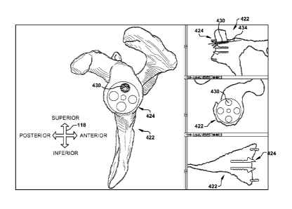
As shown in fifth action block 336 of Fig. 3, at least one landmark 538
(shown in Fig. 5) may be placed in at least one predetermined landmark
orientation
relative to the native patient tissue model 422. The landmark(s) 538, when
present,
represent a chosen point in space and/or indicate a chosen
direction/orientation
relative to the native patient tissue model 422 and are used to convey
positional
information to the user during a surgical procedure. For example, a guide pin
is
displayed as a three-dimensional landmark 538a spaced apart from the stock
device 424 over the image of the native patient tissue model 422 in Fig. 5,
while an
aperture or cavity formed in the native patient tissue model is shown as a
two-dimensional landmark 538b (i.e., represented by a cross mark when seen
from
CA 028163392013-04-26
WO 2012/058355
PCT/US2011/057957
-14-
above or below) corresponding to a central portion of the stock device in Fig.
5. In
fact, the "negative" aperture-type landmark 538b of Fig. 5 is configured to
receive
a device shaft 540 of the stock device 424, which helps to locate and
stabilize the
stock device with respect to the native patient tissue model 422. One of
ordinary
skill in the art would readily be able to instead provide a "positive" pin- or
shaft-
type landmark (not shown) protruding from the native patient tissue model 422
and
adapted to be received in a cavity (not shown) of another type of device, in
an
axle-type manner.
Regardless of the number, location, type, or any other characteristics of the
provided landmark(s) 538, it is contemplated that the user will want to
transfer the
landmarked infoimation to the actual patient tissue during the surgical
procedure.
To that end, a patient-specific template may be created using the system
described
herein. The landmark 538 could also or instead be placed during the surgical
procedure using a robotic surgical aid, adjustable reusable (e.g., "dial-in")
tools,
intraoperative imaging, or any other suitable placement aid.
As shown at sixth action block 342 of Fig. 3, a patient-specific template is
generated, which may be accomplished by the system with steps represented in
user views such as the sequence of Figs. 6-7. As shown in Fig. 6, a template
blank 644 is placed into a desired (final) predeteimined template orientation
with
respect to the native patient tissue model 422. The template blank 644 may be
selected, automatically and/or manually, from a library of available template
blanks and may be placed, again automatically and/or manually, into the
predetermined template orientation based upon any of the above device
properties
or any other desired factors.
As is particularly apparent in the coronal (top right) and transverse
(bottom right) portions of Fig. 6, at least a portion of the native patient
tissue
model 422 and at least a portion of the template blank 644 (virtually) overlap
to
create a superposed volume 646 of space which is occupied by both the native
patient tissue model and the template blank. Since this superposed volume 646
is
impracticable during the actual physical surgical procedure, the superposed
volume 646 is (again, virtually) removed from the template blank 644 to create
a
mating surface 748 of the template blank adjacent the native patient tissue
-15-
model 422. In other words, the system adjusts the dimensions of the bottom
template surface 748 to mate with a surface of the native patient tissue model
422.
The term "mate" is used herein to indicate a relationship in which the
contours of
two structures are at least partially matched or coordinated in at least two
dimensions.
The mating surface 748 may be seen in particularly the coronal (top right)
and transverse (bottom right) portions of Fig. 7. The patient-specific
template 750
may be, for example, the type disclosed in co-pending U.S. Patent Application
No. to be determined, filed October 27, 2011, titled "System and Method for
Association of a Guiding Aid with a Patient Tissue" and claiming priority to
U.S.
Provisional Patent Application No. 61/408,359, liled October 29, 2010 and
titled
"System and Method for Association of a Guiding Aid with a Patient Tissue".
Regardless of its nature, the patient-specific template 750 virtually contains
or embodies at least one predetermined landmark orientation and has at least
one
landmark guiding feature 752 configured to place a landmark 538 in the
predetermined landmark orientation when the patient-specific template 750 is
mated with the native tissue model 422. As shown in Fig. 7, at least one
landmark
guiding feature 752 is an aperture through the patient-specific template 750
which
is configured to guide a penetrating structure, such as a guide pin or drill
bit, into
the native patient tissue model 422 at a predetermined penetration location
and
with a specified target trajectory 434.
When the landmark 538 is a two-dimensional landmark such as a marking
on the surface of the native patient tissue, the target trajectory 434 of the
landmark
guiding feature 752 will likely he of little to no import. In contrast, when
the
landmark 538 is a three-dimensional landmark such as a drilled hole or an
elongate
guide pin, the target trajectory 434 of the landmark may bear some
significance. In
Fig. 7, the depicted target trajectory 434 corresponds to a desired drilling
trajectory
for an aperture which receives a device shaft 540 at a later stage of the
surgical
procedure. In this sense, therefore, at least one of the landmark guiding
features 752 shown in Fig. 7 may also serve as a penetration-guiding feature.
CA 2816339 2018-02-26
-16-
Once the landmark(s) 538 have been virtually placed into the
predetermined landmark orientation(s) at fifth action block 336 of Fig. 3 and
the
patient-specific template 750 created at sixth action block 342, the stock
device
424 may be (virtually) re-placed upon the native patient tissue model 422 and
at
least one patient-specific placement guide 958 may be generated at seventh
action
block 356 of Fig. 3. The patient-specific placement guide 958 may be
configured
to interact simultaneously with at least one previously placed landmark (here,
at
least guide pin-type landmark 538a) and with the stock device 424 when the
stock
device is in the predetermined device orientation.
The patient-specific placement guide 958 may be, for example, similar to
any of those disclosed in co-pending U.S. Patent Application No, to be
determined,
filed October 27, 2011, titled "System and Method for Assisting with
Attachment
of a Stock Implant to a Patient Tissue" and claiming priority to U.S.
Provisional
Patent Application No. 61/408,324, filed October 29, 2010 and titled "System
and
Method for Assisting with Attachment of a Stock Implant to a Patient Tissue",
or
in co-pending U.S. Patent Application No. to be determined, filed Oct. 27,
2011,
titled "System and Method for Assisting with Attachment of a Stock Instrument
to a Patient Tissue" and claiming priority to U.S. Provisional Patent
Application
No. 61/408,376, filed Oct. 29, 2010 and titled "System and Method for
Assisting with Attachment of a Stock Instrument to a Patient Tissue".
Regardless of the type of patient-specific placement guide 958
provided, the patient-specific placement guide may be generated similarly to
the
patient-specific template 750. Namely, a placement guide blank 854, shown in
Fig. 8, may be automatically or manually selected, optionally from a library
of
available placement guide blanks. It is contemplated that the placement guide
blank 356 may be selected responsive to the selection of the stock device 424,
because in many applications of the present invention, the patient-specific
placement guide 958wi11 nest into or mate with some physical feature of the
stock
device. For example, and as shown in particularly the transverse view of Fig.
8,
the placement guide blank 356 may nest with a portion of the stock device 424
CA 2816339 2018-02-26
CA 028163392013-04-26
WO 2012/058355
PCT/US2011/057957
-17-
substantially collinear with the device shaft 540 to help positively locate
the
patient-specific placement guide with respect to the stock device.
The placement guide blank 854, once selected by any suitable procedure,
may then be (virtually) altered to register with at least one landmark 538, as
shown
in Fig. 9 when the patient-specific placement guide 958 is mated with the
stock
device 424 and the stock device is in the predetermined device orientation.
Registration of the patient-specific placement guide 958 with a chosen
landmark 538 helps to indicate that the stock device 424 has achieved the
predetermined device orientation when the patient-specific placement guide 958
is
mated or nested with the stock device and the stock device is in contact with
the
native patient tissue model. The term "register" or "registration" is used
herein to
indicate a predetermined condition of correct alignment or proper relative
position
between a landmark 538 (of any type) and some feature of the structure (here,
the
patient-specific placement guide 958) being registered. For example, when the
landmark 538 is a two-dimensional marking on the native patient tissue model
422,
the registration might occur when an inscribed mark on the patient-specific
placement guide 958 aligns with the two-dimensional landmark.
As another example, and as shown in Fig. 9, the landmark 538a might be a
three-dimensional landmark such as a guide pin. In this instance, the patient-
specific placement guide 958 includes at least one orienting feature 960
(having
previously been provided to the guide blank) which will register with the
selected
landmark 538a by contact with the guide pin embodying that landmark when the
patient-specific placement guide 958 is mated or nested with the stock device
424
(as shown in Fig. 9) and the stock device is in contact with the native
patient tissue
model in the predetermined device orientation. In the view of Fig. 9, the
stock
device 424 is not yet in the predetermined device orientation as indicated by
the
separation of the orienting feature 960 and the landmark 538a, though the
patient-specific placement guide 958 is mated with the stock device, as can be
seen
in particularly the coronal and transverse views of Fig. 9.
In addition to the guiding/orienting function provided by the
patient-specific placement guide 958, at least one penetration-guiding feature
962
(four shown in Fig. 9) may be provided by the patient-specific placement
guide.
CA 028163392013-04-26
WO 2012/058355
PCT/US2011/057957
-18-
Here, the target trajectory 434a indicates a target trajectory and associated
penetration location associated with a landmark 538b, whereas the target
trajectories marked 434b (shown in dashed line in the coronal and transverse
views
since not strictly present in those sections) and the associated penetration
locations
in Fig. 9 are associated with one or more penetrating structures (not shown in
Fig. 9), such as fasteners, drill bits, other surgical tools, or any other
components
used in the surgical procedure which the user wishes to guide with the
assistance of
the patient-specific placement guide 958.
Fig. 10 is similar to Fig. 9, though the stock device 424 has been reoriented
into the predetemiined device orientation, as indicated by the registration of
the
orienting feature 960 and the landmark 538a. As can also been seen in Fig. 10,
the
body of the patient-specific placement guide 958 has been rotated sufficiently
to
bring the penetration-guiding features 962 into a different rotational
orientation
with respect to the native patient tissue model 422 than that of Fig. 9. The
target
trajectories 434b of the penetration-guiding features 962 should be in the
desired
penetration locations with respect to the native patient tissue model 422 when
the
stock device 424 has been brought into the predetermined device orientation.
Once the patient-specific template 750 and/or the patient-specific
placement guide 958 have been generated as desired, including any desired
features as described above, a physical version of the patient-specific
template is
created at eighth action block 364 of Fig. 3 and a physical version of the
patient-specific placement guide is created at ninth action block 366 of Fig.
3.
These physical versions of the patient-specific template 750 and/or the
patient-specific placement guide 958 are tangible (e.g., material and
palpable)
representations of the virtual versions of the corresponding items as
manipulated,
adjusted, and otherwise created using a system similar to that shown via the
user
views of Figs. 4-10.
Optionally, and as shown in tenth action block 368 of Fig. 3, a physical
three-dimensional version of the native patient tissue model 422 may be
fabricated
as a tangible (e.g., material and palpable) representation of the virtual
version of
the native patient tissue model. This physical native patient tissue model
(not shown) may be useful in preoperative planning, visualization, and
-19-
consideration of the surgical procedure by the user (e.g., for assisting with
performing the surgery using patient specific templates and/or adjustable
surgical
instruments). To that end, the physical native patient tissue model may
include at
least one information feature providing clinically useful information to the
user,
"Clinically useful" information is used herein to indicate any information,
other
than the structure of the native patient tissue itself, that assists one of
ordinary skill
in the art with some pre- and/or intra-operative task. An "information
feature" is
any physical feature or characteristic of the physical native patient tissue
model
which signifies or communicates the clinically useful information to the user,
optionally in combination with a preoperative plan. For example, only a
portion of
the scapula 100 may be fabricated as a physical native patient tissue model,
with
planar faces bounding the omitted portions of the scapula. Those planar faces
may
be chosen at predetermined distances from, and/or with predetermined
orientations
with respect to, a structure of interest on the physical native tissue model.
As
another example, an information feature may be a physical characteristic that
facilitates transfer of information from the native patient tissue model 422
to the
actual patient anatomy, perhaps by facilitating the setting of an adjustable,
reusable
tool such as that disclosed in co-pending U.S. Patent Application No.
12/854,362,
filed August 11, 2010 and titled "Method and Apparatus for Insertion of an
Elongate Pin into a Surface".
In one example embodiment of a physical native tissue model giving spatial
information, for instance, a planar face bounding a lower portion of the
physical
native tissue model may be substantially parallel to a transverse plane of the
scapula 100. Often the patient is oriented during surgery such that the plane
of the
scapula 100 is not identifiable with reference to the orientation of the
glenoid
vault 110 in the surgical field. Accordingly, by placing the physical native
tissue
model with an information feature in a known position (e.g, by placing a lower
face of the physical native tissue model flat on a table), one of ordinary
skill in the
art can readily envision obscured portions of the patient's native tissue
anatomy
through reference to the physical native tissue model, which may be configured
to
provide the user with a visualization of the native patient tissue in the same
CA 2816339 2018-02-26
CA 028163392013-04-26
WO 2012/058355
PCT/US2011/057957
-20-
orientation as in the patient's body but without the surrounding tissue that
prevents
the user from directly seeing structures such as, but not limited to, the
acromion
process 106, the coracoid process 108, or any other structure of the scapula
100.
This may be particularly useful when the physical native tissue model is
fabricated
at a 1:1 scale with the native patient anatomy, but also will have utility
when the
model is scaled up or down from the patient's actual tissue.
As another example embodiment of a physical native tissue model giving
spatial information, a pin-receiving aperture may be provided in the physical
native
tissue model, to receive a guide pin and thus demonstrate a certain direction
or axis
to the user with respect to the native tissue. As a corollary to this example,
an axis-
, direction-, or plane-indicating structure may extend from the physical
native
tissue model to serve as a user visualization aid or reference.
The physical native tissue model could he used to interact with an implant
or instrument before or during the surgical procedure, as well. For example, a
user
could rehearse certain interactions of an implant or instrument with the
physical
native tissue model to gain familiarity with the way that the implant or
instrument
is likely to intraoperatively interact with the patient's native tissue.
Physical native tissue models with information features or specific
landmarks related to the preoperatively developed surgical plan are not
currently
provided or used as references during surgical procedures. The availability of
a
physical native tissue model to use as a reference in this manner may
supplement
or even supplant the need for intraoperative imaging, which is likely to
reduce cost,
operating room clutter, and time required for the surgical procedure.
The patient's name, identification number, surgeon's name, and/or any
other desired identifier may be molded into, printed on, attached to, or
otherwise
associated with the physical version(s) of the patient-specific template 750,
the patient-specific placement guide 958, and/or the native patient tissue
model 422 in a legible manner. The tangible representations of the patient-
specific
template 750, the patient-specific placement guide 958, and/or the native
patient
tissue model 422 may be made by any suitable method such as, but not limited
to,
selective laser sintering ("SLS"), fused deposition modeling ("FDM"),
stereolithography ("S LA"), laminated object manufacturing ("LOM"), electron
-21-
beam melting ("EBM"), 3-dimensional printing ("3DP"), contour milling from a
suitable material, computer numeric control ("CNC"), other rapid prototyping
methods, or any other desired manufacturing process.
Once the physical versions of the patient-specific template 750,
the patient-specific placement guide 958, and/or the native patient
tissue model 422 have been manufactured and prepared for use (e.g.,
mechanically
or chemically cleaned, cured, sterilized, or the like) using any suitable
process(es),
they are available for use during surgical procedures.
The preoperative planning system disclosed herein allows the user to
experiment with different placements and selections of stock devices 424
and/or
custom or patient-specific components in an effort to produce positive patient
outcomes. Figs. 11A-14B depict various examples of steps, alternate options,
and
considerations that one of ordinary skill in the art may find useful in
preoperative
planning, particularly with respect to selection of the stock device 424 and
of the
predetermined device orientation.
Figs. 11A-11B depict a transverse view of a native patient tissue model 422
of a typical clinical case of a patient with osteoarthritis, having moderate
bone loss.
The scapular plane 1170 is perpendicular to reference plane 1172. The
reference
plane also represents the 0 reference from which glenoid version is measured.
The lower portion of Figs. 11A-11B is posterior and the top portion of these
Figures is anterior, as shown by direction arrow 118'. The diagonal dashed
line
labeled 1174 represents the native glenoid plane of the patient. In this case,
the
native glenoid plane 1174 exhibits a retroversion angle of approximately 26'
from
the reference plane 1172. Glenoid version in the normal population is reported
to
commonly be between 5 of anteversion and 15 of retroversion. The average
normal glenoid version is approximately 1-2 of reqoversion.
The goal of arthroplasty surgery is to correct pathologic anatomy and
restore as best as possible normal anatomy and function, Corrective options
range
between placing an implant component at the standard ideal of perpendicular to
the
plane of the scapula (0 ) up to the pathologic version (in this case, 26 of
retroversion). Common practice today is to correct version with an attempt to
place
CA 2816339 2018-02-26
-77-
a stock device 424 approximately perpendicular to the scapular plane 1170
(i.e., lying along the reference plane 1172 at about 0 of version). For
clarity of
description, the "angle" of the stock device 424 is referenced hereafter as
being the
angle measured from a top face of the stock device, the top face being
foremost in
the perspective view of Fig. 4.
There normally will be a secondary surgical goals to minimize removal of
patient tissue needed to accommodate the stock device 424, seat the entire
stock
device on the prepared patient tissue surface, and minimize unwanted
perforation
of the outer walls of the glenoid vault 110 or other patient tissue by the
device
shaft 540 or another penetrating structure 430 used in the surgical procedure
or
remaining in the patient tissue postoperatively. When formulating a
preoperative
plan, typical items of concern include the bone (or other patient tissue) loss
in the
patient, the position and orientation of the normal joint line, and where the
stock
device 424 or other component should be placed to aim toward a positive
patient
outcome.
The present inventors have found that an average patient tissue model 1176
(e.g., a "vault model") may be useful in tailoring a surgical procedure to fit
the
needs of an individual patient. A suitable average patient tissue model 1176
is
described in co-pending U.S. Patent Application Serial No. 12/043,634, filed
March 6, 2008, and titled "Method and Apparatus for Preparing for a Surgical
Procedure".
In a similar manner, the shape of an average acetabular vault may be used
as a suitable average patient tissue model and have some clinical relevance
when
defining the normal anatomic relationships from the pathologic anatomy in a
hip
use environment. The average patient tissue model 1176 of a glenoid vault 110
is
shown superimposed on the native patient tissue model 422 in Fig. 11B.
Although
this is an "average" view, the contours of the average patient tissue model
1176
can be seen to substantially mirror the contours of the native glenoid vault
110 of
even the depicted pathologic scapula 100.
Fig. 1111 is similar to Fig. 11A, with the addition of an average patient
tissue model 1176. The average patient tissue model 1176 helps to define the
location of the normal joint line and the version of the normal glenoid fossa
1178
CA 2816339 2018-02-26
CA 028163392013-04-26
WO 2012/058355
PCT/US2011/057957
-23-
in a patient-specific manner. The average patient tissue model 1176 may help
define reconstruction goals in pathologic cases, and may assist with selection
of
position and type of a stock device 424 or a custom device (not shown).
Selection
of version for the stock device 424 may be at least partially dependent upon
the
version of the average patient tissue model 1176 which defines patient-
specific
normal anatomy. In the patient of Figs. 11A-14B, normal patient version, based
upon the average patient tissue model 1176, may be seen to be approximately 12
of retroversion, as shown by the angle of the rightmost face (in the
orientation of
the Figures) of the average patient tissue model 1176 with respect to
reference
plane 1172.
When planning a surgical procedure using preoperative imaging, the user
may specify at least one structural change to the native patient tissue to
facilitate
placement of a stock device in a predetei mined device orientation. For
example,
native patient tissue could be drilled, planed, reamed or otherwise removed,
or the
native patient tissue could be built up using bone grafts or other substances,
with
the latter being much more difficult to do than the former during a standard
surgical procedure. Using the system described above, a (virtual) altered
patient
tissue model (not shown) can be generated and viewed or otherwise used in the
preoperative planning. Optionally, a physical three-dimensional version of the
altered patient tissue model may be fabricated as a tangible representation of
the
virtual version of the altered patient tissue model. When provided, the
physical
altered patient tissue model may also include at least one information feature
providing clinically useful information to the user. For example, a landmark
538
(e.g., a cavity or aperture) may be present in the physical altered patient
tissue
model and may therefore be made palpable to the user during the surgical
procedure. The physical altered patient tissue model, when present, may be
used
and referenced similarly to the aforementioned physical native patient tissue
model.
Figs. 12A-14B are partial transverse cross-sectional schematic views of a
scapula which depict a comparison of the likely surgical outcomes for various
preoperative planning options. Figs. 12A-14B depict various ways in which the
native patient tissue model 422 can be compared to a reference patient tissue
model
CA 028163392013-04-26
WO 2012/058355
PCT/US2011/057957
-24-
(regardless of whether any alterations are made to the native patient tissue
model),
and the effect of that comparison on the predetermined device orientation. The
predeteimined device orientation can be adjusted, automatically by the system
and/or manually by the user, responsive to the comparison of the native
patient
tissue model 422 to the reference patient tissue model. The reference patient
tissue
may be at least one of a (mirrored) image of a contralateral patient tissue of
the
same or a different patient, a value taken from a standard reference patient
tissue, a
value range taken from a standard reference patient tissue, and the
aforementioned
average patient tissue model 1176. In Figs. 12A-14B, the reference patient
tissue
is shown and described as being the average patient tissue model 1176. In
Fig. 12A, a stock device 424 has been superimposed upon the native patient
tissue
model 422 of Figs. 11A-11B in a version of 00 from the corona' plane (shown in
Figs. 11A-13C as scapular plane 1170), with the bottom portion (in the
orientation
of Figs. 11A-13C) of the stock device being located on an outer surface of the
native patient tissue. Since Figs. 12A-13C show the scapula 100 having
portions
of the native tissue removed to accommodate each stock device 424, the patient
tissue shown can be described as an altered patient tissue model 1280. The
excision of fairly large amounts of native patient tissue is likely to
adversely affect
the dynamics within the shoulder joint. Additionally, the glenoid vault 110
may be
shaved down enough that the device shaft 540 is in danger of breaching the
glenoid
vault wall, which is generally undesirable and can cause patient discomfort
and
possibly result in undesirable reoperation. Accordingly, one goal of a pre-
surgical
planning process using the average patient tissue model 1176 is to attempt to
replicate the total volume (or area, as depicted in the cross-sectional views
of
Figs. 12A-14B) of the average patient tissue model 1176 with a combination of
the
total volume (or area) of the altered patient tissue model 1280 and the stock
device 424.
It is apparent from Fig. 12A that a substantial amount of the native patient
tissue will have to be removed from the native patient tissue model 422 to
allow
the stock device 424 to seat firmly and maintain the 00 version with the stock
device 424 substantially centered, posteriorly to anteriorly, upon the glenoid
CA 028163392013-04-26
WO 2012/058355
PCT/US2011/057957
-25-
fossa 1178. 'the device shaft 540 in Fig. 12A is in danger of breaching the
glenoid
vault 110 wall, which should be avoided.
Fig. 12B also shows an altered patient tissue model 1280 with a relatively
large volume of native patient tissue removed, though less removed than in
Fig. 12A. In Fig. 12B, the version is still corrected to 0' from the reference
plane 1172, but the stock device 424 has been moved upward (in the orientation
of
the Figures) to distance the device shaft 540 from the glenoid vault 110 wall.
This
shifting of the stock device 424 can be seen to have a different adverse
effect,
however--namely, the stock device now substantially overhangs the anterior
edge
of the glenoid fossa 1178.
This problematic 0 version correction is an example of a value taken from
a standard reference patient tissue--many users will routinely correct version
in all
such cases to 0 as shown. As an example of a value range taken from a
standard
reference patient tissue, the version may be corrected to a value taken from
the
range of -5 to +5 , with the user's experience and intuition leading to the
selection
of one value from that range. Another example, in a hip standard reference
patient
tissue, might prescribe a range of 10-30' of anteversion and 30-55' of
abduction
for an acetabular prosthetic implantation. However, a seemingly reasonable
value
based upon a standard reference patient tissue--whether for a shoulder, hip,
or any
other type of surgery--may markedly depart from a value which leads to an
acceptable result for a particular patient.
As a result, users will sometimes employ a mirror image of a contralateral
native patient tissue (from that patient or another patient) to use as a
reference
patient tissue. However, even if there is a contralateral native patient
tissue to
consult (e.g., the patient is not an amputee in that respect), the
contralateral native
patient tissue may be pathologically or congenitally asymmetrical from even
the
original state of the native patient tissue which is being surgically
corrected. Thus,
there is a need for another reference patient tissue for comparison to the
native
patient tissue model 422.
In the aforementioned co-pending "Method and Apparatus for Preparing for
a Surgical Procedure" U.S. Patent Application, the average patient tissue
model 1176 (i.e., the "vault model") is proposed as providing an appropriate
CA 028163392013-04-26
WO 2012/058355
PCT/US2011/057957
-26-
reference patient tissue for a wide range of patients. The average patient
tissue
model 1176 is shown in Figs. 12A-13C superimposed over the altered patient
tissue model 1280. Accordingly, one of ordinary skill in the art, with
reference to
the average patient tissue model 1176, will be motivated to preserve more of
the
native patient tissue by altering the native tissue model 422, and placing the
stock
device 424 with reference to the average patient tissue model 1176.
In the situation of Fig. 12C, the average patient tissue model 1176 helps
define the native patient joint line and the native version for that
particular patient.
Accordingly, the average patient tissue model 1176 helps direct the selection
of the
stock device 424 to restore the native joint line and the patient's native
version,
thereby reducing the risk of excessive bone removal or perforation of the
native
patient tissue during or after the stock device is installed. Fig. 12C depicts
an
altered patient tissue model 1280 with the average patient tissue model 1176
superposed thereupon and the stock device 424 placed according to the average
patient tissue model (here, rotated clockwise, in the orientation of the
Figures.). It
can be seen that placement of the stock device 424 in a patient-specific
version
(informed by the average patient tissue model 1176) will center the device
shaft 420 (posteriorly to anteriorly) in the glenoid vault 110, provide more
thorough patient tissue contact for the stock device, and result in less
patient tissue
removal and greater centering of the stock device on the glenoid fossa 1178 as
compared to the OD versions of Figs. 12A and 12B. Accordingly, the stock
device 424 placement in Fig. 12C would seem to provide a preferred
predetermined device orientation compared to the orientations shown in Figs.
12A
and 12B.
Figs. 13A-13C depict a similar orientation comparison sequence to that of
Figs. 12A-12C, but including a different stock device 424a than that shown in
Figs. 12A-12C. The stock device 424a includes a thickened leftmost section
(in the orientation of the Figures) which helps to compensate for the
pathologic
state of the native patient tissue. This selection of this stock device 424a,
having a
second configuration as compared to the first configuration of the stock
device 424
of Figs. 12A-12C allows for the combination of the native glenoid vault 110
plus
the stock device 424a to have a similar, and similarly arranged, volume of
material
CA 028163392013-04-26
WO 2012/058355
PCT/US2011/057957
as that of the average patient tissue model 1176. The arrangements of
Figs. 13A-13C are analogous to those of Figs. 12A-12C, excepting the
differences
in the stock devices 424 and 424a, and therefore the description of Figs. 12A-
12C
will not be repeated with respect to 13A-13C.
The views of the combination of the altered glenoid vault 110 plus the
stock device 424a of Figs. 13A-13C may be favorably contrasted with the
analogous views of Figs. 12A-12C, wherein the combination of the altered
glenoid
vault 110 plus the stock device 424 has a substantially smaller volume in the
latter
when compared to the average patient tissue model 1176, and thus the latter
will
have less strength and ability to mechanically perform for the patient as
needed for
a suitably long time after the surgical procedure. Accordingly, the stock
device 424a selection and placement of Fig. 13C appears to meet the goal of
preserving native tissue the best of all of the options shown in Figs. 12A-
13C.
Figs. 14A-14B show the effects of device orientation upon the native
patient tissue model 422. In Fig. 14A, the version has been corrected to 00.
That
is, the target trajectory 534 of the patient-specific template 750 is
substantially
parallel to the scapular plane 1170. As is apparent in Fig. 14A, the device
shaft 540 is cutting markedly into the coronal bone of the scapula 100 in an
undesirable manner, and a relatively large volume of native patient tissue
will need
to be removed (near the top of Fig. 14A) to accept the stock device 424. In
Fig. 14B, the version has been corrected to a value chosen by the user with
consideration of the native patient tissue model 422--the version in Fig. 14B
is
approximately 12 . As can be seen, by simply tilting the stock device 424 in
Fig. 14B as suggested by the average patient tissue model 1176 or by a chosen
value out of a value range taken from a standard reference patient tissue, the
stock
device 424 is seated more securely in the glenoid vault 110, with less removal
of
native patient tissue required.
Fig. 15 illustrates a computer system 1582 that can be employed to
implement systems and methods described herein, such as based on computer
executable instructions running on the computer system. The user may be
permitted to preoperatively simulate the planned surgical procedure using the
computer system 1582 as desired. The computer system 1582 can be
CA 028163392013-04-26
WO 2012/058355
PCT/US2011/057957
implemented on one or more general purpose networked computer systems,
embedded computer systems, routers, switches, server devices, client devices,
various intermediate devices/nodes and/or stand alone computer systems.
Additionally, the computer system 1582 can be implemented as part of the
computer-aided engineering (CAE) tool running computer executable instructions
to perform a method as described herein.
The computer system 1582 includes a processor 1584 and a system
memory 1586. Dual microprocessors and other multi-processor architectures can
also be utilized as the processor 1584. The processor 1584 and system
memory 1586 can be coupled by any of several types of bus structures,
including
a memory bus or memory controller, a peripheral bus, and a local bus using any
of
a variety of bus architectures. The system memory 1586 includes read only
memory (ROM) 1588 and random access memory (RAM) 1590. A basic
input/output system (BIOS) can reside in the ROM 1588, generally containing
the
basic routines that help to transfer information between elements within the
computer system 1582, such as a reset or power-up.
The computer system 1582 can include one or more types of long-term
data storage 1592, including a hard disk drive, a magnetic disk drive, (e.g.,
to read
from or write to a removable disk), and an optical disk drive, (e.g., for
reading a
CD-ROM or DVD disk or to read from or write to other optical media). The
long-term data storage 1592 can be connected to the processor 1584 by a drive
interface 1594. The long-term data storage 1592 components provide nonvolatile
storage of data, data structures, and computer-executable instructions for the
computer system 1582. A number of program modules may also be stored in one
or more of the drives as well as in the RAM 1590, including an operating
system,
one or more application programs, other program modules, and program data.
A user may enter commands and information into the computer
system 1582 through one or more input devices 1596, such as a keyboard or a
pointing device (e.g., a mouse). These and other input devices are often
connected to the processor 1584 through a device interface 1598. For example,
the input devices can be connected to the system bus by one or more a parallel
port, a serial port or a universal serial bus (USB). One or more output
CA 028163392013-04-26
WO 2012/058355
PCT/US2011/057957
-29-
device(s) 15100, such as a visual display device or printer, can also be
connected
to the processor 1584 via the device interface 1598.
The computer system 1582 may operate in a networked environment using
logical connections (e.g., a local area network (LAN) or wide area network
(WAN)
to one or more remote computers 15102. A given remote computer 15102 may be
a workstation, a computer system, a router, a peer device or other common
network node, and typically includes many or all of the elements described
relative
to the computer system 1582. The computer system 1582 can communicate with
the remote computers 15102 via a network interface 15104, such as a wired or
wireless network interface card or modem. In a networked environment,
application programs and program data depicted relative to the computer
system 1582, or portions thereof, may be stored in memory associated with the
remote computers 15102.
It is contemplated that multiple versions of the patient-specific
template 750 and/or the patient-specific placement guide 958 could be created
during preoperative planning and fabricated as options for the user to select
from
during the surgical procedure. For example, the user may not be able to clear
away
surrounding (e.g., soft) tissue from the native patient tissue as well as
expected. In
this situation, it may be useful to have a patient-specific template 750 with
a
smaller footprint for easier insertion into the surgical wound and
manipulation at
the surgical site, even though the smaller footprint means that there is less
mating
surface 748 to mate with the native patient tissue and provide positive
location
assistance for the patient-specific template 750.
While aspects of the present invention have been particularly shown and
described with reference to the preferred embodiment above, it will be
understood
by those of ordinary skill in the art that various additional embodiments may
be
contemplated without departing from the spirit and scope of the present
invention.
For example, the specific methods described above for using the described
system
are merely illustrative; one of ordinary skill in the art could readily
determine any
number of tools, sequences of steps, or other means/options for virtually or
actually placing the above-described apparatus, or components thereof, into
positions substantially similar to those shown and described herein. Any of
the
CA 028163392013-04-26
WO 2012/058355
PCT/US2011/057957
-30-
described structures and components could be integrally formed as a single
piece
or made up of separate sub-components, with either of these formations
involving
any suitable stock or bespoke components and/or any suitable material or
combinations of materials: however, the chosen material(s) should be
biocompatible for most applications of the present invention. The mating
relationships formed between the described structures need not keep the
entirety of
each of the "mating" surfaces in direct contact with each other but could
include
spacers or holdaways for partial direct contact, a liner or other intermediate
member for indirect contact, or could even be approximated with intervening
space
remaining therebetween and no contact. Though certain components described
herein are shown as having specific geometric shapes, all structures of the
present
invention may have any suitable shapes, sizes, configurations, relative
relationships, cross-sectional areas, or any other physical characteristics as
desirable for a particular application of the present invention. An adhesive
(such as, but not limited to, bone cement) could be used in conjunction with
the
system and method described herein. The patient-specific template 750 and/or
the
patient-specific placement guide 958 may include a plurality of structures
cooperatively forming the base body and temporarily or permanently attached
together in such a manner as to permit relative motion (e.g., pivoting,
sliding, or
any other motion) therebetween. The patient-specific placement guide 958 may
not actually be patient-specific but could instead be a stock item in
situations
where the landmark(s) 538 are placed to "standardize" a particular native
patient
tissue model with a standard frame of reference. Any structures or features
described with reference to one embodiment or configuration of the present
invention could be provided, singly or in combination with other structures or
features, to any other embodiment or configuration, as it would be impractical
to
describe each of the embodiments and configurations discussed herein as having
all of the options discussed with respect to all of the other embodiments and
configurations. Any of the components described herein could have a surface
treatment (e.g., texturization, notching, etc.), material choice, and/or other
characteristic chosen to provide the component with a desired interaction
property
(e.g., tissue ingrowth, eluting of a therapeutic material, etc.) with the
surrounding
CA 028163392013-04-26
WO 2012/058355
PCT/US2011/057957
-31-
tissue. The system is described herein as being used to plan and/or simulate a
surgical procedure of implanting one or more prosthetic structures into a
patient's
body, but also or instead could be used to plan and/or simulate any surgical
procedure, regardless of whether a non-native component is left in the
patient's
body after the procedure. A device or method incorporating any of these
features
should be understood to fall under the scope of the present invention as
determined
based upon the claims below and any equivalents thereof.
Other aspects, objects, and advantages of the present invention can be
obtained from a study of the drawings, the disclosure, and the appended
claims.