Note: Descriptions are shown in the official language in which they were submitted.
CA 02825265 2013-07-19
WO 2011/091185
PCT/US2011/021947
TISSUE CLOSURE DEVICE AND METHOD
CROSS-REFERENCE TO RELATED APPLICATIONS
This application claims the benefit of U.S. Provisional Patent Application
Serial No.
61/296,868, filed on January 20, 2010, which is hereby incorporated herein in
its entirety by
reference thereto.
Further, each of the following is hereby incorporated in its entirety by
reference
thereto: U.S. Patent Application Serial No. ______ , Attorney Docket No.
14895/3,
filed on January 20, 2011, U.S. Patent Application Serial No. ___ , Attorney
Docket No. 14895/4, filed on January 20, 2011; and U.S. Patent Application
Serial No.
______________ , Attorney Docket No. 14895/5, filed on January 20, 2011.
FIELD OF THE INVENTION
The present invention relates to a tissue closure device and method.
BACKGROUND INFORMATION
Some surgical interventions involve piercing or cutting a hole into a tissue
wall. For
example, thoracoscopic procedures typically involve piercing one or more
tissue walls with a
trocar or other sharp device and insertion of a cannula to maintain an opening
in the tissue.
Surgical instruments may be inserted through the cannula in order to access a
surgical site
disposed beyond the tissue. For example, a thoracoscopic cardiac procedure may
involve a
trans-apical valve repair. This procedure requires access to the outer wall of
patient's heart,
e.g., via a small intercostal hole in the patient. This procedure further
involves piercing the
outer wall of the heart to form an access hole, and insertion of a cannula to
maintain a desired
diameter of the access hole and to protect the pierced heart tissue during
subsequent insertion
and/or removal of thoracoscopic tools through the cannula. Thoracoscopic
surgical tools may
then be inserted through the cannula and into one or more chambers of the
heart in order to
repair defects or damaged portions of the heart.
Further, some pericardiocentesis procedures involve inserting a needle, via an
intercostal opening in the patient, into the pericardial sac, guiding a
flexible guide wire
through the needle, and subsequent removal of the needle with the guide wire
left in place.
After removal of the needle, a tapered dilator may be advanced over the guide
wire to dilate
the opening in the pericardium tissue. The dilated opening, or tract, allows
room for a
CA 02825265 2013-07-19
WO 2011/091185
PCT/US2011/021947
catheter. After the dilation, the catheter is guided over the guide wire into
the pericardial sac
to drain fluid from the pericardium.
Thoracoscopic surgical procedures are generally less intrusive than more
traditional
forms of surgery, since they generally require relatively small entry
openings. However,
these small openings may be difficult to close, especially where the closure
location is inside
the patient's body. For example, referring the procedures described above,
after removal of
the cannula and any surgical instruments extending therethrough, the aperture
formed in the
tissue, e.g., the heart or pericardium tissue, is closed within the patient's
body. Since these
exemplary thoracoscopic procedures involve accessing the *patient's thorax
through a small
intercostal aperture through the patient's skin and other underlying tissues
(e.g., fat and/or
fascia), closure methods such as suturing are more complicated than with non-
thoracoscopic
surgical procedures. In particular, applying sutures with remotely operated
thoracoscopic
instruments is more difficult and complicated than directly manipulating a
suture needle by
hand at the surgical site. This difficulty can result in defective closures
and/or closures that
require more time than necessary.
Defective closures may expose the patient to increased risk of complications
such as
internal bleeding and/or infection. Even where defective closures are
recognized and
addressed prior to completion of the surgical procedure, the correction of
defective closures
increases the time required to effect the closure and may expose the tissue to
additional
trauma. It is generally desirable to minimize the amount of time for a
surgical procedure in
order to reduce the possibility of complications and unnecessary trauma to the
patient.
Thus, there is a need for a closure mechanism and method that is simple to
operate,
reliable, and requires a small amount of time in which to form an effective
closure.
SUMMARY
In accordance with example embodiments of the present invention, a device
includes:
a plurality of anchors; at least one elastic closure element coupled to the
anchors and
configured to urge the anchors toward each other; and a driver configured to
drive the
anchors, with the closure element coupled to the anchors, into tissue; wherein
the closure
element has an elasticity sufficient to urge the anchors, driven into the
tissue, toward each
other to close an aperture in the tissue located between the anchors driven
into the tissue and
to resist opposing forces exerted on the anchors that urge the anchors apart.
The opposing forces may be exerted on the anchors by at least one of (a) the
tissue,
(b) a fluid flow, (c) pneumatic pressure, (d) hydraulic pressure, and (e)
external forces.
2
CA 02825265 2013-07-19
WO 2011/091185
PCT/US2011/021947
The device may further include a safety release mechanism including a
plurality of
spring-loaded members, each spring-loaded member independently movable between
an
engagement position and a disengagement position, the safety release mechanism
adapted to
prevent the driver from driving the anchors unless all of the spring-loaded
members are in the
engagement position.
The anchors may each include an elongated body having a distal tip configured
to
pierce the tissue when the respective anchor is distally driven into the
tissue.
The anchors may each include an anchoring projection configured to resist
proximal
movement of the anchor after the anchor is driven into the tissue.
The anchoring projection is a wing extending proximally and radially from a
connection between the wing and the elongated body to a free end.
The wing may include a plurality of proximally extending cutting projections
at the
free end of the wing.
The wing may be formed by a cut progressing radially inwardly and distally
into the
elongated body.
The elongated body and the wing may include a plurality of longitudinally
extending
corrugations, the corrugations providing a plurality of proximally extending
cutting
projections at the free end of the wing.
The anchors may each include first and second anchoring projections configured
to
resist proximal movement of the anchor after the anchor is driven into the
tissue, the first and
second anchoring projections being disposed at respective positions that are
offset from each
other along the length of the elongated body.
The first and second anchoring projections may be first and second wings
formed
respectively by first and second cuts progressing radially inwardly and
distally into the
elongated body and ending at respective locations that are offset from each
other along the
length of the elongated body.
The closure element may include at least one of a band, an elastomeric band,
and a
band formed of silicon.
The anchors may each include a hooked projection configured to receive the
band.
The hooked projection may be configured to maintain engagement between the
band
and the anchor by preventing the band from moving off the proximal end of the
anchor.
The device may include a plurality of closure elements.
Each of the plurality of closure elements may contact two or more of the
anchors.
The closure elements may form a pattern of two overlapping V-shaped
configurations.
3
CA 02825265 2013-07-19
WO 2011/091185
PCT/US2011/021947
The plurality of closure elements may contact three or more of the anchors.
The at least one closure element may include a monolithic V-shaped element
coupling
three of the anchors.
The device may include two monolithic V-shaped closure elements each
configured
to contact three of the anchors. The two V-shaped closure elements may overlap
to form a
diamond-shaped operational window.
The device may further include a centering element configured to receive a
guide
wire. The centering element may be a tubular shaft.
The anchors may be disposed along a ring-shaped circumference in the first
configuration.
The closure element may be prevented from extending within the ring-shaped
circumference by one or more tubes.
The driver may configured to simultaneously drive the plurality of anchors.
The driver may comprise a spring-loaded element configured to impact and
impart a
distally directed momentum to the anchors.
The device may further include a trigger configured to release the spring-
loaded
element from a preloaded position in order to drive the plurality of anchors.
The device may further include a handle, the trigger being disposed in handle.
The handle, the trigger, and the driver may be detachable from the cannula,
the outer
working tube, the plurality of anchors, and the closure element.
The plurality of anchors and the closure element may be formed of
bioabsorbable
materials.
In accordance with example embodiments of the present invention, a device
includes:
a plurality of anchors; and at least one elastic closure element coupled to
the anchors and
configured to urge the anchors toward each other; wherein the closure element
has an
elasticity sufficient to urge the anchors, driven into the tissue, toward each
other to close an
aperture in the tissue located between the anchors driven into the tissue and
to resist opposing
forces exerted on the anchors that urge the anchors apart.
The opposing forces may be exerted on the anchors by at least one of (a) the
tissue,
(b) a fluid flow, (c) pneumatic pressure, (d) hydraulic pressure, and (e)
external forces.
In accordance with example embodiments of the present invention, a method
includes: implanting a plurality of anchors into tissue; and urging the
implanted anchors
towards each other by at least one elastic closure element coupled to the
anchors with
sufficient force to (a) close an aperture in the tissue located between the
implanted anchors
4
CA 02825265 2013-07-19
WO 2011/091185
PCT/US2011/021947
and (b) resist opposing forces exerted on the implanted anchors that urge the
anchors apart
and the aperture open.
The opposing forces may be exerted on the anchors by at least one of (a) the
tissue,
(b) a fluid flow, (c) pneumatic pressure, (d) hydraulic pressure, and (e)
external forces.
In accordance with example embodiments of the present invention, a method
includes: implanting a plurality of anchors into tissue; urging the implanted
anchors towards
each other by at least one elastic closure element coupled to the anchors;
forming an aperture
in the tissue between the implanted anchors, the elastic closure element
urging the implanted
anchors towards each other and towards the aperture with sufficient force to
(a) maintain the
aperture in the tissue in a closed position and (b) resist opposing forces
exerted on the
implanted anchors that urge the anchors apart and urges the aperture open;
inserting an
instrument through the aperture; and after removing the instrument from the
aperture, again
urging the implanted anchors towards each other and towards the aperture by
the elastic
closure element with sufficient force to (a) maintain the aperture in the
tissue in the closed
position and (b) resist opposing forces exerted on the implanted anchors that
urge the anchors
apart and the aperture open.
The opposing forces may be exerted on the anchors by at least one of (a) the
tissue,
(b) a fluid flow, (c) pneumatic pressure, (d) hydraulic pressure, and (e)
external forces.
In accordance with example embodiments of the present invention, a method
includes: forming an aperture in tissue; inserting a centering device through
the aperture;
implanting a plurality of anchors into the tissue using the centering device
to center the
anchors about the aperture; urging the implanted anchors towards each other
and towards the
aperture by at least one elastic closure element coupled to the anchors;
inserting an
instrument through the aperture; and after removing the instrument from the
aperture, again
urging the implanted anchors towards each other and towards the aperture by
the elastic
closure element with sufficient force to (a) maintain the aperture in the
tissue in the closed
position and (b) resist opposing forces exerted on the implanted anchors that
urge the anchors
apart and the aperture open.
The opposing forces may be exerted on the anchors by at least one of (a) the
tissue,
(b) a fluid flow, (c) pneumatic pressure, (d) hydraulic pressure, and (e)
external forces.
In accordance with example embodiments of the present invention, a surgical
device
comprises two or more anchors, a driver configured to drive the anchors into a
tissue, and at
least one elastic closure element extending between the anchors and configured
to urge the
anchors from a first configuration in which the anchors are a first distance
from each other,
5
CA 02825265 2013-07-19
WO 2011/091185
PCT/US2011/021947
toward a second configuration in which the anchors are a second distance from
each other,
the second distance being less than the first distance, wherein the surgical
device is
configured to maintain the driven anchors in the first configuration and to
selectably release
the driven anchors to allow the anchors to be moved by the at least one
closure element
toward the second configuration.
The anchors may each include an elongated body having a distal tip configured
to
pierce the tissue when the respective anchor is distally driven into the
tissue.
The anchors may each include an anchoring projection configured to resist
proximal
movement of the anchor after the anchor is driven into the tissue.
The anchoring projection may be a wing extending proximally and radially from
a
connection between the wing and the elongated body to a free end.
The wing may include a plurality of proximally extending cutting projections
at the
free end of the wing.
The wing may be formed by a cut progressing radially inwardly and distally
into the
elongated body.
The elongated body and the wing may include a plurality of longitudinally
extending
corrugations, the corrugations providing a plurality of proximally extending
cutting
projections at the free end of the wing.
The anchors may each include first and second anchoring projections configured
to
resist proximal movement of the anchor after the anchor is driven into the
tissue, the first and
second anchoring projections being disposed at respective positions that are
offset from each
other along the length of the elongated body.
The first and second anchoring projections may be first and second wings
formed
respectively by first and second cuts progressing radially inwardly and
distally into the
elongated body and ending at respective locations that are offset from each
other along the
length of the elongated body.
The closure element may be a band. The band may form a continuous loop. The
band may be elastomeric. The band may be formed of silicon.
The anchors may each include a hooked projection configured to receive the
band.
The hooked projection may be configured to maintain engagement between the
band
and the anchor by preventing the band from moving off the proximal end of the
anchor.
The device may include a two or more closure elements. Each of the plurality
of
closure elements may contact only two of the anchors. For example, the two or
more closure
elements may include four closure elements or may include six anchors, two of
the six
6
CA 02825265 2013-07-19
WO 2011/091185
PCT/US2011/021947
anchors being connected to only two of four closure elements, and four of the
six anchors
being connected to only a respective one of the four closure elements. The
closure elements
may form a pattern of two or more overlapping V-shaped configurations.
The surgical plurality of closure elements may contact three or more of the
anchors.
The at least one closure element may include a monolithic V-shaped element
configured to contact three of the anchors.
The at least one closure element may include two or more monolithic V-shaped
elements each configured to contact three of the anchors. For example, the V-
shaped
elements may overlap to form a diamond-shaped operational window.
The device may further comprise a centering element configured to receive a
guide
wire. The device of claim 25, wherein the centering element is a tubular
shaft. The centering
element may have a proximal portion configured to allow the centering
mechanism to be
retracted from the remainder of the surgical device.
The device may further comprise at least one pressure sensor configured to
indicate
whether the device is adequately contacting the tissue prior to driving the
anchors.
The at least one pressure sensor may include at least one contact element
extending
distally from a distal end of the device. The at least one contact element may
be depressible
when a distal end of the device is pressed against the tissue.
The device may further comprise a key plate and at least one key member, the
at least
one key member having a first position in which the at least key member is
engaged with the
key plate and a second position in which the at least one key member is
disengaged with the
key plate, wherein depression of the contact element causes the at least one
key member to
move from the first position to the second position.
The key plate may prevent driving of the anchors when the at least one key
member is
engaged with the key plate.
The at least one key member includes a plurality of key members each being
independently movable by a respective contact element. The key plate may
prevent driving
of the anchors if any one of the key members is engaged with the key plate.
The anchors may be disposed along a ring-shaped circumference in the first
configuration.
The closure element may be prevented from extending within the ring-shaped
circumference when the anchors are maintained in the first configuration.
7
CA 02825265 2013-07-19
WO 2011/091185
PCT/US2011/021947
The surgical device may further comprise a cannula configured to provide
access to a
surgical site disposed between the anchors when the anchors are maintained in
the first
configuration.
The cannula may be configured to maintain the anchors in the first
configuration.
The anchors and closure element may be disposed at a position radially
exterior to the
cannula.
The surgical device may further comprise an outer working tube, the cannula
extending within the outer working tube.
At least one of the cannula and the outer working tube may have an outer
surface
configured to prevent the anchor and the closure element from extending to any
radial
position corresponding to an interior of the cannula.
The surgical device may include a plurality of closure elements prevented from
extending to any radial position corresponding to the interior channel of the
cannula.
The cannula may include a distal portion having a flanged orientation in which
the
distal portion forms a radially extending flange configured to prevent the
closure elements
from moving distally beyond the distal end of the cannula. The flange may
extend radially
beyond an outer surface of the outer working tube.
The distal portion of the cannula may be actuatable to a second orientation,
in which
the distal portion of the inner working channel does not prevent the closure
elements from
moving distally beyond the distal end of the cannula.
The flange may extend distally when the distal portion of the cannula is in
the second
orientation.
The distal portion of the cannula may be actuatable from the flanged
orientation to the
second orientation by proximally sliding the cannula with respect to the outer
working tube.
The depth to which the anchors are driven by the driver may be limited by
contact
between the closure element and the radially extending flanges.
The driver may be configured to simultaneously drive the plurality of anchors.
The driver may comprise a spring-loaded element configured to impact and
impart a
distally directed momentum to the anchors.
The surgical device may further comprise a trigger configured to release the
spring-
loaded element from a preloaded position in order to drive the plurality of
anchors.
The surgical device may further comprise a handle, the trigger being disposed
in
handle.
8
CA 02825265 2013-07-19
WO 2011/091185
PCT/US2011/021947
The surgical device may further comprise a safety element configured to
prevent the
trigger from releasing the spring-loaded element when the safety element is in
a safety
position.
The handle, the trigger, and the driver may be detachable from the cannula,
the outer
working tube, the plurality of anchors, and the closure element.
The plurality of anchors and/or the closure element may be formed of
bioabsorbable
materials.
In accordance with example embodiments of the present invention, a method
comprises: implanting two or more anchors into a tissue; maintaining the
implanted anchors
in a first configuration in which the anchors are a first distance from each
other; urging the
anchors from the first configuration toward a second configuration in which
the anchors are a
second distance from each other, the second distance being less than the first
distance;
forming an aperture in the tissue in an area between the two or more anchors;
and constricting
the aperture by allowing the anchors to move from the first configuration to
the second
configuration..
The aperture may be formed while the implanted anchors are maintained in the
first
configuration.
The aperture may be formed with a trocar.
The method may further comprise performing a thoracoscopic surgical procedure
through the aperture.
The closure device may include a cannula configured to maintain the closure
device
in the preloaded state, the thoracoscopic surgical procedure being performed
through the
cannula.
The tissue may be a heart tissue.
The thoracoscopic surgical procedure may be a trans-apical valve repair.
In accordance with example embodiments of the present invention, a surgical
device
comprises a plurality of anchors configured to be driven into a tissue, and at
least one closure
element extending between the anchors and configured to urge the anchors from
a first
configuration in which the anchors are a first distance from each other,
toward a second
configuration in which the anchors are a second distance from each other, the
second distance
being less than the first distance, wherein the surgical device is configured
to maintain the
anchors in the first configuration during a surgical procedure and to
subsequently allow the
anchors to be moved by the closure element toward the second configuration.
9
CA 02825265 2013-07-19
WO 2011/091185
PCT/US2011/021947
In accordance with example embodiments of the present invention, a surgical
device
comprises a driver configured to drive a plurality of anchors into a tissue in
a first anchor
configuration in which the anchors are a first distance from each other,
wherein the device is
configured to maintain the driven anchors in the first anchor configuration
and to selectably
release the driven anchors to allow the anchors to be moved by at least one
closure element
toward a second anchor configuration in which the anchors are closer to each
other than when
the anchors are in the first anchor configuration.
The driver may be configured to drive each anchor by striking, e.g., a) the
respective
anchor or b) a pin configured to transfer momentum from the driver to the
anchor.
The driver may be configured to be actuated from a proximal position to a
distal
position in which the driver imparts momentum to each respective anchor by
striking a) the
respective anchor or b) a pin configured to transfer momentum from the driver
to the
respective anchor. The driver may be configured to be actuated by a spring.
Further features and aspects of example embodiments of the present invention
are
described in more detail below with reference to the appended Figures.
BRIEF DESCRIPTION OF THE DRAWINGS
Figures 1 and 2 show a surgical closure device and a detailed view of a distal
tip of
the surgical device in accordance with an example embodiment of the present
invention.
Figure 3 is a front view with an inset partial front view of the surgical
closure device
of Figure 1.
Figure 4 shows an anchor of the self-acting closure arrangement of the device
of
Figure 1.
Figure 5A shows a subassembly of the surgical closure device of Figure 1.
Figure 5B is a partial view of the subassembly of Figure 4.
Figure 5C is a partial sectional view of the device of Figure 1 taken through
a plane
containing the longitudinal axis of the device and bisecting two opposed
anchors.
Figure 6A is a partial cross-sectional view of the subassembly of Figure 5A
with a
safety mechanism engaged.
Figure 6B is a partial cross-sectional view of the subassembly of Figure 5A
with the
safety mechanism disengaged.
Figure 6C is a partial cross-sectional view of the subassembly of Figure 5A
when a
trigger is in a depressed state.
CA 02825265 2013-07-19
WO 2011/091185
PCT/US2011/021947
Figure 7 is a partial view of the working tube and a self-acting closure
arrangement of
the surgical closure device of Figure 1.
Figure 8A is a cross-sectional view according to plane A of Figure 7.
Figure 8B is a cross-sectional view according to plane A of Figure 7 when a
cannula
is disposed in the outer working tube.
Figures 8C to 8D sequentially and schematically illustrate the refraction of
the
cannula of Figure 8B with respect to the outer working tube and the release of
the closure
elements.
Figure 9A is a partial view of the outer working tube of the device of Figure
1 with
the self-acting closure arrangement inserted into a tissue.
Figure 9B is a partial view of the outer working tube and a cannula with the
self-
acting closure arrangement of the device of Figure 1 inserted into a tissue.
Figure 10A shows the self-acting closure arrangement of the device of Figure 1
inserted in the tissue after removal of the cannula and working tube.
Figures 10B and 10C schematically illustrate the forces exerted by the anchors
of
Figure 10A.
Figures 10D and 10E illustrate the anchors of Figure 10A when drawn to their
closed
or approximated positions to close a hole in a tissue.
Figure 11 shows a closure element with a V-shaped configuration in accordance
with
an example embodiment of the present invention.
Figure 12 shows another V-shaped closure element in accordance with an example
embodiment of the present invention.
Figure 13 shows an anchor in accordance with an example embodiment of the
present
invention.
Figure 14 shows a plurality of anchors of Figure 13 and closure elements of
Figure 12
when closing a hole in a tissue.
Figure 15 shows a surgical closure device in accordance with an example
embodiment
of the present invention.
Figure 16 shows a front perspective view of a distal end portion of the
surgical
closure device of Figure 15 with anchors and closure elements.
Figure 17A is a partial view of a subassembly of the device of Figure 15.
Figure 17B is a side view of the trigger of the device of Figure 15.
Figure 17C is a top view of the trigger of the device of Figure 15.
Figure 17D is a bottom view of the trigger of the device of Figure 15.
11
CA 02825265 2013-07-19
WO 2011/091185
PCT/US2011/021947
Figure 18A is a partial view of a trigger subassembly of the device of Figure
15 with
the trigger in an initial state.
Figure 18B is a partial view of the trigger subassembly of Figure 18 with the
trigger
depressed.
Figure 18C is a front cross-sectional view of a subassembly of the device of
Figure 15
showing the key plate in an engaged state and in a first position.
Figure 18D is a front cross-sectional view of the subassembly of Figure 18C
showing
the key plate in a disengaged state and in the first position.
Figure 18E is a front cross-sectional view of the subassembly of Figure 18C
showing
the key plate in a disengaged state and in a second position.
Figure 19A is a schematic illustration showing the engagement of the trigger
bar of
the device of Figure 15 with a hammer sleeve.
Figure 19B is a schematic illustration showing the trigger bar of the device
of Figure
disengaged with the hammer sleeve.
15 Figure 19C is a schematic front view of the latch member and safety
switch of the
device of Figure 15 with the safety switch in an engaged state.
Figure 19D is a schematic front view of the latch member and safety switch of
the
device of Figure 15 with the safety switch in a disengaged state.
Figure 20A shows the anchors driven into a tissue without closure elements.
Figure 20B shows the tissue of Figure 20A punctured at a location within the
periphery defined by the anchors.
Figure 20C shows the anchors disposed around the puncture formed in Figure
20B.
Figure 20D shows the puncture of Figures 20B and 20C closed by the anchors and
closure elements.
Figure 20E shows the anchors surrounding the punctured tissue.
DETAILED DESCRIPTION
As set forth in greater detail below, example embodiments of the present
invention
allow for the reliable and effective closure of an opening in tissue (e.g., a
pericardial window)
that limits the possibility of human error, e.g., by eliminating the need for
suturing. In some
examples, a surgical device anchors a plurality of anchors, which are
connected to each other
by one or more elastic closure elements, into the tissue. The anchors are
driven into the
tissue in a spaced-apart configuration in which the elastic closure elements
are tensioned
between the anchors. The anchors are held in the spaced-apart arrangement
while a surgical
12
CA 02825265 2013-07-19
WO 2011/091185
PCT/US2011/021947
procedure is performed through a tissue opening formed between the anchored
locations of
the anchors. In order to close the opening, the device simply releases the
anchors from the
spaced-apart arrangement such that the tensioned elastic closure elements draw
the anchors,
as well as the tissue in which the anchors are anchored, toward the tissue
opening. Thereby,
the tissue opening is held closed. The tension remaining in the elastic
closure elements
offsets the opposing forces that may be entered on the anchors by at least one
of (a) the
tissue, (b) the fluid flow, (c) pneumatic pressure, (d) hydraulic pressure,
and (e) external
forces.
Referring, for example, to Figures 1 to 10E, a surgical procedure involves
positioning
a surgical closure device 5 at a surgical entry location, e.g., a location on
the wall of a heart
where access to the interior of the heart is desired. The surgical closure
device 5 is then
actuated, e.g., via a trigger, to drive a plurality of anchors 200 into the
tissue at predetermined
locations spaced around the surgical entry location. The anchors 200 are
preloaded toward
the entry location by pre-tensioned closure elements 300 in the form of
elastic bands. The
anchors 200 are maintained in their outward positions by a cannula 400 and/or
an outer
working tube 100. After the anchors 200 are driven, the portions of the
surgical device other
than the cannula 400, outer working tubes 100, the anchors 200, and the
closure elements 300
are removed.
The cannula 400 then provides a working channel through which the surgical
procedure may be performed. For example, a trocar may be extended through the
channel of
the cannula 400 to pierce the tissue 900. Other surgical instruments may then
be inserted
through the working channel in accordance with any suitable thoracoscopic
procedure. To
conclude the procedure, any thoracoscopic surgical tools extending through the
working
channel are withdrawn and the cannula 400 and working tube 100 are proximally
withdrawn
from the surgical entry location. The withdrawal of the cannula 400 and
working tube 100
causes the pre-tensioned closure elements 300 to draw the anchors 200 toward
the surgical
entry site. Since the anchors 200 are anchored in the tissue surrounding the
surgical entry
location, this results in the tissue surrounding the surgical entry location
being drawn
together, thereby closing the surgical entry hole. In contrast to conventional
procedures, no
sutures are required.
Although a cannula 400 is provided separately from the outer working tube 100,
it
should be understood that example embodiments may include only a single tube.
For
example, if the cannula 400 is not provided in the device 5, the working tube
100 functions as
the cannula.
13
CA 02825265 2013-07-19
WO 2011/091185
PCT/US2011/021947
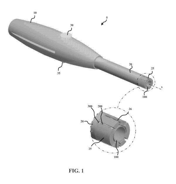
Figures 1 and 2 illustrate an example surgical closure device 5. The surgical
closure
device 5 includes a handle 10 configured to be held by an operator, e.g., a
surgeon, to operate
the surgical closure device 5 during a surgical procedure. A shaft 20 extends
distally from
the handle 10 and includes a distal end portion 25. An outer working tube 100
is disposed in
a bore of the shaft 20 and extends concentrically along the longitudinal axis
x of the shaft 20.
The outer working tube 100 is distally exposed through an opening in the shaft
20. The outer
working tube 100 has an outer diameter that is smaller than an inner diameter
of the shaft 20,
thus allowing the outer working tube 100 to be slidable along the longitudinal
axis x.
Although each of the outer working tube 100 and the shaft 20 are configured as
right circular
cylinders with concentric through bores, it should be understood that the
outer working tube
100 and/or the shaft 20 may be provided with any appropriate geometry, e.g., a
cross-section
that is oval, polygonal, etc. and/or a cross-section that varies along the
longitudinal axis x.
Further, the geometry of the bore may differ substantially from the outer
geometry for the
outer working tube 100 and/or the shaft 20.
Referring to the inset partial view in Figure 1, the distal end portion 25 of
the shaft 20
includes six notches or slots 26, which extend from the distal tip of the
shaft 20 a proximal
distance along the longitudinal axis x. The slots 26 may be formed in any
suitable manner,
e.g., making three cuts in the distal end portion 25, each cut forming two of
the slots 26 on
opposed sides of the axis x. The dimensions of the slots 26 are selected to
allow six
respective anchors 200 to be disposed in the slots 26. In this regard, the
wall thickness of the
shaft (i.e., the distance between the bore and the outer surface) and the
width of each slot 26
may be selected to be slightly greater than a respective lateral dimension of
the anchor 200.
Where the anchor 200 has a radial projection, the width of the slot 26 may be
less than a
diameter of the anchor through the projection. Thus, the geometry of the slot
26 may require
that the anchor 200 be oriented such that the radial projection is at least
approximately
aligned with the longitudinal axis x of the shaft 20, since the anchor 200
would not otherwise
fit into the slot 26.
Figure 3 is a front view of the surgical closure device 5. The slots 26, with
respective
anchors 200, are non-uniformly spaced apart along the circumferential
periphery of the shaft
20. In particular, two groups of slots 26 are provided, one on the opposite
side of the axis x
from the other. Each of the two groups includes three slots 26 equally spaced
apart. The
circumferential spacing between the groups is greater than the circumferential
spacing
between the individual slots 26 in each group.
14
CA 02825265 2013-07-19
WO 2011/091185
PCT/US2011/021947
Referring to the inset partial view in Figure 3, the slots 26 include side
walls with
opposed, longitudinally extending cylindrical grooves 27 for receiving the
body 201 of the
anchor 200. Further, the closure element 300 attached to the anchor 200 is
able to pass along
the cylindrical grooves 27. The slots 26 are also elongated in the radial
direction to
accommodate wings 207 and 208, which are described in greater detail below
with regard to
Figure 4. Further, there is a gap between the outer working tube 100 and the
end portion 25
of the shaft 20 to allow the closure elements 300 to be disposed therebetween
as illustrated in
Figure 3.
Figure 4 shows an anchor or implant 200 which is configured to be driven into
a
tissue. The anchor 200 includes a corrugated body 201. The body 201 includes
grooves 203
that extend axially along the length of the body 201. Thus, extending
circumferentially
around the body 201, a plurality of grooves 203 alternate with a plurality of
ridges 205.
Further, the anchor body 201 includes a pair of wings or split portions 207
and 208. The split
portions 207 and 208 are formed by respective splits or cuts 209 into the body
201. In this
regard, the splits 209 may be formed by making a cut radially into the body
201 and
extending in an axial direction. Thus, the two split portions 207 and 208 are
attached to the
remainder of the body 201 at a distal position and extend proximally to free
ends. The free
ends include a plurality of sharp protrusions along a curved surface. These
points are formed
due to the corrugations. In particular, the ridges 205 form the sharp
protrusions, as illustrated
in the inset partial side view in Figure 4, which are advantageous for
gripping tissue and
preventing distal sliding of the anchor 200. Although each split portion 207
and 208 includes
three such protrusions as illustrated, it should be understood that the anchor
200 may be
designed such that one or more of the split portions has any other number of
protrusions,
including a single sharp protrusion. For example, if a larger number of sharp
protrusions are
desired, the body 201 could be more densely corrugated (i.e., a greater number
of alternating
grooves 203 and ridges 205 could be provided) and/or the angle of the cut or
slice could be
adjusted. Further, the length of proximal extension of the projections may be
adjusted by
varying the depth of the grooves 203 with respect to the ridges 205.
The split portions 207 and 208 do not substantially impede distal insertion
into tissue
but resist proximal movement from an insertion location by engaging the
tissue. It has been
discovered that the combination of the pointed and/or sharp-edged proximal
ends of the split
portions 207 and 208 with the alternating ridges on the proximal end of the
split portions
creates improved performance.
CA 02825265 2013-07-19
WO 2011/091185
PCT/US2011/021947
Further, the split portions or wings 207 and 208 are axially offset from each
other.
For example, split 207 is axially located at position a along axis xx and
split 208 is axially
located at position b along axis xx. This allows for greater structural
strength of the other
portions of the body 201 as compared to a non-offset configuration. In
particular, since the
cuts progress continually radially inward as they progress distally, a non-
offset portion would
have a substantially smaller amount of material in cross-section at the distal
end of the cut.
This would lead to a mechanically weak point or region along the axis of the
body and could
lead to mechanical failure, especially in anchors of small dimensions.
Although the anchors 200 utilize a pair of wings 207 and 208 to anchor the
anchors
200 against proximal retraction from a tissue, it should be appreciated that
any number of
wings may be provided, and that as an alternative or in addition to the wings
207 and 208,
any other appropriate anchoring structure(s), e.g., anchoring filaments, may
be provided.
The distal tip of the anchor 200 is pyramidal, with a sharp point, and a
plurality of
surfaces separated by edges that converge at the sharp point. Although four
planar surfaces
are provided, it should be appreciated that any appropriate suitable number of
surfaces may
be provided and that one or more or all of the surfaces may be non-planar.
The anchor 200 also includes a hooked end portion 210. The hooked portion 210
is
configured to receive one or more closure elements 300. On the side of the
anchor 200
opposite the hooked portion 210 is an alignment projection 220 configured to
rotationally
align the anchor 200 about its longitudinal axis xx. Although the anchors 200
in the
illustrated examples are aligned with the alignment projection 220 and the
split portions 207
and 208 being intersected by and aligned along a plane containing the
longitudinal axis x of
the shaft 20 and the longitudinal axis xx of the anchor 200, it should be
understood that the
alignment projection 220 and the split portions 207 and 208 may be intersected
by and
aligned along a plane that contains the longitudinal axis xx of the anchor 200
and is
transverse, e.g., perpendicular, to the plane containing the longitudinal axis
x of the shaft 20
and the longitudinal axis xx of the device 20. Further, the alignment
projection may be
provided at any appropriate location around the circumference of the anchor
200 relative to
the split portions 207 and 208 and that any appropriate number of alignment
projections 220
may be provided for a particular anchor 200.
Although the anchor 200 is shown in the exemplary illustrations with closure
elements 300, it should be understood that the anchor 200 may be used in
connection with
any other closure elements, including, e.g., closure elements 1300, 2300
described in greater
detail below.
16
CA 02825265 2013-07-19
WO 2011/091185
PCT/US2011/021947
The anchor 200 may be produced by first forming the body 201 with the
corrugations,
e.g., by injection molding or extrusion, and subsequently forming split
portions 207 and 208,
e.g., by cutting radially into the side of the body 201. As illustrated, the
cut is curved, with
an angle (at the proximal entry point), relative to the longitudinal axis xx
of the body 201,
that gradually decreases from the proximal initial cutting location toward the
distal end of the
anchor 200 and eventually becoming linear. Although the split or cut of the
illustrated
example is made with a curved or varying angle with respect to the
longitudinal axis xx of the
body 201, it should be understood that any appropriate cut, including a linear
cut, may be
made.
Although the anchor 200 includes two wings or split portions spaced equally
around
the radial periphery of the body 201, it should be appreciated that any number
of split
portions, including a single split portion may be provided and at any
appropriate spacing
around the radial periphery of the anchor 200.
Modern manufacturing processes allow for near nano technology applications.
This
allows the anchors 200 to be manufactured in a size and complexity that may
not have been
possible in years past. The anchor 200 may be injection molded of either
absorbable or non
absorbable polymers and then processed (e.g., by cutting) to add the features
of the wings 207
and 208. Although the anchors 200 are formed of polymer, it should be
appreciated that any
appropriate material may used, e.g., metal or a composite material. The
anchors 200 may
have a diameter of, e.g., one millimeter, or approximately one millimeter, and
a length that is
in a range from, e.g., 5 millimeters to 10 millimeters. According to some
example
embodiments, the diameter is less than one millimeter. According to some
example
embodiments, the diameter is in a range from 0.8 millimeters to 1.2
millimeters. It should be
understood, however, that other dimensions may be provided.
Figure 5 shows a subassembly of the surgical closure device 5. The subassembly
includes the trigger 30, the safety slide 35, a safety slide bias spring 40, a
hammer sleeve 500,
a driving spring 550, anvil pins 600, the outer working sleeve 100, and
anchors 200. In the
state illustrated in Figure 5, the surgical closure device 5 is loaded and
ready to be actuated in
order to drive the anchors 200. In this regard, a proximal end of the hammer
sleeve 500
contacts a distal end of the driving spring 550, which is in a compressed
state as illustrated in
Figure 5. To maintain the hammer sleeve 500 in its proximal position while the
compressed
driving spring 550 applies a distally directed force, the hammer sleeve 500
latches with a
trigger plate 32 of the trigger 30, as schematically illustrated in Figure 6A.
In Figures 6A to
6C, the hammer sleeve 500 and the trigger plate 32 are shown in cross-section
to facilitate
17
CA 02825265 2013-07-19
WO 2011/091185
PCT/US2011/021947
illustration. To latch the hammer sleeve 500, the hammer sleeve 500 is pushed
proximally,
while the trigger 30 is in a depressed state (such as illustrated in Figure
6C) until a lip or step
proximally clears the proximal side of the trigger plate 32. The trigger 32 is
then moved
(e.g., via a spring bias force and/or manually) to a non-depressed position,
as illustrated in
Figure 6A. The trigger moves in a transverse direction between the depressed
and non-
depressed positions by sliding within lateral channels in the housing of the
handle 10.
However, any appropriate guiding mechanism may be provided.
To maintain the trigger 32 in the non-depressed position in order to prevent
or reduce
the likelihood of accidental driving of the anchors 200 (e.g., due to user
error, during
shipping, storage, etc.), the safety slide includes a safety rib or bar 38
which, as illustrated in
Figure 6A, is positioned adjacent the trigger plate 32 to form a positive or
hard stop, thereby
obstructing movement of the trigger 30 from the non-depressed position of
Figure 6A to the
depressed position of Figure 6C. As illustrated, e.g., in Figure 6A, the
safety slide 35
includes a pair of lateral projections 36 configured to longitudinally slide
within a
corresponding channel in the housing of the handle 10. It should be
understood, however,
that any appropriate guide mechanism may be provided. The safety slide 35 also
includes a
knob portion 37 to facilitate sliding of the safety slide 35 using, e.g., one
of the operator's
fingers.
When the operator desires to drive the anchors 200, the operator must first
move the
safety slide 35 into a driving position in which the safety bar 38 does not
obstruct movement
of the trigger plate 32. Referring to Figure 5, the safety slide is urged or
biased toward the
proximal safety position by a compression spring 40. Thus, the operator must
continuously
apply a force to the knob 37 until the bottom of the trigger plate 32 moves to
a position that
prevents or blocks the safety bar 38 from returning to the safety position.
This may provide
for even greater safety, since the operator must generally coordinate the
holding of the safety
slide 35 in the driving position while depressing the trigger 30. It should be
understood,
however, that the safety slide 35 may be configured to remain in the driving
position without
continuous application of force. Further, it should be understood that the
device 5 may be
provided without any safety mechanism.
Figure 6B shows safety slide 35 in the driving position. Although the safety
slide is
moved distally, i.e., in the direction of the arrow shown in Figure 6B, it
should be understood
that the safety switch may be configured to move in any suitable direction to
move between
safety and firing positions. After the safety slide 35 is moved to the driving
position shown
in Figure 6B, the operator depresses the trigger 30, e.g., with one of the
operator's fingers,
18
CA 02825265 2013-07-19
WO 2011/091185
PCT/US2011/021947
until the lower portion of the trigger plate 32 clears the step 505 of the
hammer sleeve 500,
thereby releasing the hammer sleeve 500 for distal movement actuated by the
compressed
driving spring 550.
Referring, e.g., to the partial sectional view of Figure 6B, the hammer sleeve
500 is
spaced apart from the anvil pins 600 prior to depressing the trigger. The
anvil pins 600 are
slidable along the longitudinal axis x of the shaft 20 within respective bores
of the shaft 20
corresponding to respective anchors 200. As the hammer sleeve 500 moves
forward, it gains
speed and momentum. Upon contact with the proximal ends of the anvil pins 600,
the
hammer sleeve 600 imparts a momentum to the anchors 200, since the distal ends
of the anvil
pins 600 are in alignment with the proximal ends of the anchors 200. In this
manner, the
anchors 200 are driven at a substantial speed, which facilitates driving of
the anchors 200 into
soft tissue.
The anchors are preferably driven at a speed greater than 50 meters per
second, more
preferably in a range of 50 to 350 meters per second, and most preferably at
350 meters per
second. However, it should be understood that the anchors 200 may be driven at
any suitable
speed sufficient for the anchors to puncture tissue.
Further, the anchors 200 may be driven into a single layer or multiple layers
of tissue
and that the speed may be selected based on the structural properties,
dimensions, and relative
locations of the one or more tissues into which the anchors are driven.
In order to accurately penetrate soft tissues that are not held or secured on
a distal
side, a rapid penetration of each layer of tissue may be required in order to
effect penetration
of the tissue layer or layers. If an anchor 200 is applied slowly, the tissue
or tissues may be
pushed distally away by the anchor 200 without adequate penetration. Thus,
some example
delivery mechanisms eject each implant at a relatively high speed, as set
forth above.
Although the example device 5 utilizes a spring-loaded mechanical driving
mechanism, it
should be understood that other drivers may be provided. In some examples,
saline is used to
pressurize a channel within a catheter, needle, or other tube at such a rate
that a plunger will
eject the anchor at the precise speed. Further example embodiments push the
anchors using
long push rods which run the length of a catheter or other tube. The ejection
modality may
be computer-controlled and/or operator-controlled. For example, as with the
spring loaded
mechanical system of the illustrated examples, an ejection force may be
predetermined and
repeatable by an operator's actuation of a trigger 30.
Moreover, the driver may be configured to drive the anchors 200 to a
predetermined
depth. Although the illustrated examples control the depth by contact between
closure
19
CA 02825265 2013-07-19
WO 2011/091185
PCT/US2011/021947
elements 300 (described in greater detail below), which are coupled to the
anchors 200, and
flanges or flared portions 405, any other depth-controlling mechanism may
additionally or
alternatively be provided. For example, the precision of the depth may be
accomplished by a
precise hydraulic driving force, engagement with other stops, or a suture that
tautens to limit
the depth. Further, the depth may be monitored using fluoroscopy or any other
appropriate
imaging mechanism. The driving mechanism may include pressurized saline or
other
hydraulic fluid that is pressurized through the thoracoscopic catheter shaft.
Thus, very
precise control may be accomplished.
Figure 6 is an enlarged partial view of the subassembly of Figure 4. As
illustrated, a
plurality of closure elements 300 are coupled to the hook portions 210 of the
anchors 200.
There are four closure elements 300, each of which is coupled to the hook
portions 210 of
exactly two anchors 200. Thus, as illustrated, e.g., in Figure 10, two anchors
200 are attached
to exactly two different closure elements 300 and four anchors 200 are
attached to exactly
one closure element 300. It should be understood, however, that other
arrangements may be
provided.
Figure 7 is a partial view of the working tube 100, the anchors 200, and the
closure
elements 300. As illustrated in Figure 7, the anchors 200 have been driven,
e.g., into tissue.
The anchors 200 and the closure elements 300 form a self-acting closure
arrangement of the
surgical closure device 5. During driving of the anchors 200, the closure
elements 300 are
also driven an analogous distance due to the engagement of the closure
elements 300 with the
anchors 200.
Referring to the cross-sectional view of Figure 8A, the closure elements 300
are
layered and are held along the periphery of the outer working tube 100,
thereby preventing
the closure elements 300 from pulling the anchors 200 toward each other.
Figure 8B is the same as Figure 8A, except that a cannula 400 is disposed
within the
outer working tube 100. The elements shown in Figure 8B may be separated from
the
remainder of the surgical device 5 to allow a surgical procedure to be
conducted. For
example, a trocar may be inserted longitudinally through the interior of the
cannula 400 to
pierce the tissue at a location encircled by the anchors 200 that are anchored
into the tissue.
The piercing of the tissue may provide access to the opposed side of the
tissue (e.g., the
interior of a viscus such as the heart, etc.) by thoracoscopic or other
surgical instruments.
The cannula 400 includes six radially extending flared portions or flats 405.
The
cannula 400 extends concentrically within the outer working tube 100. The
cannula 400
extends distally beyond the distal end of the outer working tube 100 such that
the flats 404
CA 02825265 2013-07-19
WO 2011/091185
PCT/US2011/021947
fold over the distal end of the outer working tube 100. The radial extension
of the flats 405
beyond the circumferential periphery of the outer working tube 100 allows the
flats 405 to
form positive or hard stops that prevent or resist the closure elements 300
from inadvertently
sliding off the end of the outer working tube 100, e.g., during thoracoscopic
procedures being
performed with access through the cannula 400.
When the procedure no longer requires access through the cannula 400, any
surgical
instruments may be refracted via the cannula 400 from the viscus being
operated upon. At
this stage, the hole in the tissue formed by the trocar should be closed. In
order to do so, the
cannula 400 is moved relative to the outer working tube 100, as illustrated
sequentially in
Figures 8C and 8D. In doing so, the flats 405, which are formed as leaf
springs, rotate to a
longitudinal orientation and are retracted. Thus, the flats 405 no longer form
stops against
distal sliding of the closure elements 300 along the outer working tube 100.
This orientation
is illustrated in Figure 8D. The flats 405 may be formed of any suitable
material, e.g., a
shape memory material such as nitinol, spring steel, etc.
The flats 405 may be bistable, with two rest orientation: one corresponding to
the
radially flared orientation, and the other corresponding to the longitudinal
orientation.
After the flats are retracted, the cannula 400 and the outer working tube 100
are
proximally refracted from the surgical entry site. Since the closure elements
300 are engaged
with the hooked portions 210 of the anchors 200, which are anchored into the
tissue against
proximal retraction, the closure elements remain adjacent the surgical closure
site. Thus, the
proximal retraction of the cannula 400 and the outer working tube 100 causes
the outer
working tube 100 to slide distally with respect to the closure elements 300.
Further distal
retraction of the cannula 400 and outer working tube 100 causes the closure
elements 300 to
slip off of the distal end of the outer working tube 100, thereby entirely
disengaging the
closure tubes 300, as well as the anchors 200, from the cannula 400 and
working tube 100.
Since the closure elements 300 are pre-tensioned, they pull the anchors 200
toward the hole
formed at the surgical entry location. Since the anchors 200 are anchored into
the tissue
surrounding the hole, the pulling of the anchors into approximation causes the
surrounding
tissue to be pulled toward the hole. Thus, the hole is squeezed shut, with the
closure elements
300 maintaining a closure force to keep the hole closed.
Figure 9A is a partial view of the outer working tube 100 with the anchors 200
inserted into the tissue 900. Figure 9B is the same as Figure 9B but
schematically shows the
flats 405, which extend between the outer working tube 100 and the closure
elements 300 to
prevent the closure elements 300 from causing premature or inadvertent closure
of entry
21
CA 02825265 2013-07-19
WO 2011/091185
PCT/US2011/021947
opening in the tissue. Figure 9B may be a working arrangement, whereby the
portions of the
surgical device 5 other than the cannula 400, the outer working tube 100, the
anchors 200,
and the closure elements 300 are removed. Thus, various other surgical
instruments, e.g.,
thoracoscopic surgical devices, may be maneuvered through the interior of the
cannula 400
and the working tube 100.
Figure 10 shows the self-acting closure arrangement, in this case the anchors
200 and
the closure elements 300, inserted in the tissue after removal of the cannula
400 and working
tube 100. For illustration purposes, the anchors 200 are shown in their
initial driven positions
in the tissue 900. In other words, for ease of illustration, the arrangement
is illustrated as
though the anchors 200 are being prevented from being pulled together by the
closure
elements 300. The anchors 200 are disposed around a surgical entry opening
905, such as
formed, e.g., by a trocar.
The anchors 200 are arranged in two opposed groups of anchors. To facilitate
the
description of the arrangement shown in Figure 10A, the anchors 200 are
provided individual
reference numbers 200a, 200b, 200c, 200d, 200e, and 200f. The first group
includes anchors
200a, 200b, and 200c, and the second group includes anchors 200d, 200e, and
200f. Each of
the anchors in each group is connected by a closure element 300 directly to at
least one
anchor of the other group. Further, no two anchors within either group are
directly connected
to each other by a closure element. That is, each closure element 300 is
connected at one end
to an anchor of the first group 200a, 200b, 200c and at the other end to an
anchor of the
second group 200d, 200e, 200f. Thus, the forces exerted by the elements 300
are primarily
directed in a direction from one group toward the other group.
It is further seen from Figure 10A that the anchor/closure element arrangement
is
configured as two opposed and overlapping V-shaped groups. The first V-shaped
group is
formed of anchors 200a, 200e, 200c and closure elements 301, 304. The second V-
shaped
group is formed of anchors 200d, 200b, 200f and closure elements 302, 303.
Since each closure element is wrapped around two anchors and forms a single
complete loop, the force exerted by the respective closure element at each
anchor is equal to
the sum of the tension forces in the two band portions extending between the
two anchors to
which the closure element is connected. Moreover, the force is exerted along a
line
extending between the two anchors to which the closure element is connected.
In this regard,
the forces exerted at the locations of the anchors 200a, 200b, 200c, 200d,
200e, 200f are
illustrated in Figure 10B by arrows F301a, F301e, F302b, F302d, F303b, F303f,
F304c, and
F304e which represent respective force vectors. In particular, F301a
represents the force
22
CA 02825265 2013-07-19
WO 2011/091185
PCT/US2011/021947
exerted by closure element 301 at the anchored location of anchor 200a, F301e
represents the
force exerted by closure element 301 at the anchored location of anchor 200e,
F302b
represents the force exerted by closure element 302 at the anchored location
of anchor 200b,
F302d represents the force exerted by closure element 302 at the anchored
location of anchor
200d, F303b represents the force exerted by closure element 303 at the
anchored location of
anchor 200b, F303f represents the force exerted by closure element 303 at the
anchored
location of anchor 200f, F304c represents the force exerted by closure element
304 at the
anchored location of anchor 200c, and F304e represents the force exerted by
closure element
304 at the anchored location of anchor 200e. Further, the forces form three
pairs of
complementary forces that are equal and opposite to each other. In particular,
a first pair
F301a, F301e, a second pair F302b, F302d, a third pair F303b, F303f, and a
fourth pair
F304c, F304e. Each pair corresponds to a single closure element 301, 302, 303,
304,
respectively and are directed in opposite directions along the extension of
the respective
closure element 301, 302, 303, 304 between the two anchors 200 to which the
respective
closure element 301, 302, 303, 304 is connected.
Since anchors 200a, 200c, 200d, 200f are each connected to a single closure
element
301, 304, 302, 303, respectively, only a single force vector F301a, F304c,
F302d, F303f,
respectively, is shown in Figure 10B. Since anchors 200b and 200e are each
connected to
two closure elements, two force vectors are associated with each of anchors
200b and 200e in
Figure 10B. That is, anchor 200b, which is connected to closure elements 302
and 303, has
two force vectors F302b and F303b acting through the anchored location of
anchor 200b, and
anchor 200e, which is connected to closure elements 301 and 304, has two force
vectors
F301e, F304e acting through the anchored location of anchor 200b.
Since the forces represented by vectors F302b and F303b both act through the
same
location, i.e., the anchored location of the anchor 200b, the resultant force
through the
anchored location of anchor 200b may be determined as the sum of the two
vectors F302b
and F303b. Likewise, since the forces represented by vectors F301e and F304e
both act
through the anchored location of the anchor 200e, the resultant force through
the anchored
location of anchor 200b may be determined as the sum of the two vectors F302b
and F303b.
Accordingly, Figure 10C schematically illustrates the total forces exerted by
the closure
elements on each anchor, with the force exerted through anchor 200b
represented by the
resultant vector F302f + F303f and the force exerted through anchor 200e
represented by the
resultant vector F30 1 e + F304e.
23
CA 02825265 2013-07-19
WO 2011/091185
PCT/US2011/021947
Due to the positioning of the anchors 200a, 200b, 200c, 200d, 200e, 200f and
the
arrangement of the closure elements 301, 302, 303, 304, a greater amount of
compressive
force is exerted in the direction of a y axis than a z axis. The z axis
corresponds to a line that
extends between the first group of anchors 200a, 200b, 200c and the second
group of anchors
200d, 200e, 200f and is at least approximately equidistant from the first
group of anchors
200a, 200b, 200c and the second group of anchors 200d, 200e, 200f. The y axis
is
perpendicular to the z axis, and both the x axis and the y axis extend along
the surface of the
tissue 900.
Since compressive force is greater in directions parallel to the x axis than
in directions
parallel to the z axis, the self-acting closure formed by the anchors 200a,
200b, 200c, 200d,
200e, 200f and the closure elements 301, 302, 303, 304 tends to close the
opening 905 such
that the opening 905 is flattened or elongated along the z axis, as
illustrated in closure of
Figure 10D. This may be desirable to maintain a more reliable closure that is
more resistant
to leaking.
As schematically illustrated in Figure 10D, the anchors 200a, 200b, 200c,
200d, 200e,
200f have been drawn into their closed or approximated positions, thereby
pulling the tissue,
to which they are anchored, toward the opening 905, thereby closing the
opening 905 as
illustrated. To facilitate illustration, the closure elements 301, 302, 303,
304 are not shown in
Figure 10D. However, Figure 10E shows the closure of 10D with the closure
elements 301,
302, 303, 304. The forces being exerted by the closure elements 301, 302, 303,
304 on the
anchors 200a, 200b, 200c, 200d, 200e, 200f are analogous to those illustrated
in Figures 10B
and 10C. However, since the exemplary closure elements 301, 302, 303, 304 are
have a
spring-like elasticity, the force exerted by the closure elements 301, 302,
303, 304 may be
reduced as the anchors 200a, 200b, 200c, 200d, 200e, 200f are drawn into
approximation.
In the resting closure position (i.e., the position at which the anchors 200a
, 200b,
200c, 200d, 200e, 200f settle after transient movement from the orientation
around the
working tube 100) illustrated in Figures 10D and 10E, the force exerted by the
closure
elements 301, 302, 303, 304 through each anchor 200a, 200b, 200c, 200d, 200e,
200f is equal
to an oppositely directed resistance force exerted onto the anchors 200a,
200b, 200c, 200d,
200e, 200f by the tissue at the respective location of each anchor 200a, 200b,
200c, 200d,
200e, 200f.
Figure 11 shows another closure element 1300. The closure element 1300
includes
three anchor-receiving portions 1310, 1320, 1330 arranged in a V-shaped
configuration with
portion 1320 being disposed at the vertex. Arm 1340 spans directly from anchor-
receiving
24
CA 02825265 2013-07-19
WO 2011/091185
PCT/US2011/021947
portion 1310 to anchor-receiving portion 1320, and arm 1350 spans directly
from anchor-
receiving portion 1320 to anchor-receiving portion 1330. The anchor-receiving
portions
1310, 1320, 1330 each have a respective aperture 1312, 1322, 1332 for
receiving a respective
anchor, e.g., the anchor 200 described above or the anchor 1200 described in
greater detail
below with respect to Figure 13. The anchor-receiving portions 1310, 1320,
1330 are each
toroidal in shape and have a greater material thickness than the arms 1340 and
1350. It
should be understood, however, that any appropriate geometry may be provided
and that any
appropriate material thickness may be provided. The toroidal shape of the
anchor-receiving
portions 1310, 1320, 1330 couple with the anchors 200, 1200 in a manner
analogous to the
band-shaped closure elements 300 described above with regard to anchor 200.
The closure element 1300 functions in the same manner described above with
regard
to the closure elements 300, but differs in that only two closure elements are
required to
generate the same forces illustrated in Figures 10B and 10C. In particular,
the closure
element 1300 performs the same function as the two closure elements 301, 304,
or the two
closure elements 302, 303 of the second V-shaped groups described above with
respect to
Figure 10A. Further, the closure element 1300 differs in that a single
structural element, i.e.,
each of arms 1320, extends between opposed anchors.
Figure 12 shows another closure element 2300, which includes three anchor-
receiving
portions 2310, 2320, 2330 arranged in a V-shaped configuration with portion
2320 being
disposed at the vertex. Arm 2340 spans directly from anchor-receiving portion
2310 to
anchor-receiving portion 2320, and arm 2350 spans directly from anchor-
receiving portion
2320 to anchor-receiving portion 2330. The anchor-receiving portions 2310,
2320, 2330
each have a respective aperture 2312, 2322, 2332 for receiving a respective
anchor. The
anchor 2300 includes all of the features described above with respect to
anchor 1300, but
differs only in that the Arms 2340, 2350 have are widened to be substantially
the same width
as the outer diameter of each of the anchor-receiving portions 2310, 2320,
2330. This may be
advantageous to provide additional strength and tension force when the arms
2340, 2350 are
stretched.
Figure 13 shows an anchor 1200. Anchor 1200 is identical to anchor 200
described
above except that a proximal end portion 1250 includes a circumferential
channel 1255
formed as a continuous radial recess extending around the entire circumference
of the anchor
1200. The channel opens in the radial direction and includes a distally
directed first surface
1260 and an opposed proximally directed second surface 1265. Extending between
the first
and second surfaces 1260 and 1265 is a surface 1270 corresponding to a reduced-
diameter
CA 02825265 2013-07-19
WO 2011/091185
PCT/US2011/021947
portion 1280 of the anchor 1200. Although the reduced-diameter portion 1280 is
cylindrical
and concentric with the longitudinal axis xx' of the anchor 1200, it should be
understood that
any appropriate geometry and orientation may be provided. For example, the
reduced-
diameter portion 1280 may be frustoconical and/or have a cross section that is
curved when
viewed in a direction perpendicular to the longitudinal axis xx' of the anchor
1200. Further,
the surface 1270 of the reduced-diameter portion 1280 may vary along the
circumference of
the anchor 1200.
The circumferential channel 1255 axially separates a proximal head portion
1285
from the distal remainder of the body of the anchor 1200.
When one or more closure elements 300, 1300, 2300 is coupled to the anchor
1200,
the first surface 1260 restrains the one ore more closure elements 300, 1300,
2300 from
proximally sliding beyond the channel 1255 and off the end of the anchor 1200.
Likewise, the
second surface 1265 restrains the one or more closure elements 300, 1300, 2300
from sliding
distally beyond the channel 1255. In this regard, the dimensions of the
channel 1265, e.g.,
the width and depth of the channel 1265, may be selected to accommodate a
particular
number of closure elements 300, 1300, 2300, or a single closure element 300,
1300, 2300.
A particular closure element 300, 1300, 2300 is mated to the anchor 1200 by
mating
placing the anchor 300, 1300, 2300 around the reduced-diameter portion 1280 of
the anchor
1200. For example, an anchor-receiving portion 1310, 1320, 1330 of anchor 1300
and/or an
anchor-receiving portion 2310, 2320, 2330 of anchor 2300 may be mated to the
anchor 1200
stretching the respective anchor-receiving portion 1310, 1320, 1330, 2310,
2320, 2330 over
the proximal head portion 1285 and onto the reduced-diameter portion 1280 of
the anchor
1200. When mated in this manner, the reduced-diameter portion 1280 extends
through the
respective aperture 1312, 1322, 1332, 2312, 2322, 2332, with the anchor-
receiving portion
1310, 1320, 1330, 2310, 2320, 2330 constrained between the first and second
walls or
surfaces 1260 and 1265 of the channel 1255. In this regard, the apertures
1312, 1322, 1332,
2312, 2322, 2332 may have resting diameters that are the same, larger, or
smaller than the
diameter of the reduced-diameter portion 1280. It may be advantageous,
however, to provide
a resting diameter that is less than the outer diameter of the first surface
1260, the second
surface 1265, and/or the proximal head portion 1285 in order to resist
inadvertent
disengagement of the closure element 1300, 2300 from the anchor 1200.
The channel 1255 performs a function analogous to that of the hooked portion
210
described above with respect to Although the anchor 1200 does not include a
hooked portion
26
CA 02825265 2013-07-19
WO 2011/091185
PCT/US2011/021947
such as hooked portion 210 of anchor 200, it should be understood that one or
more hooked
portions may be provided in combination with the channel arrangement of anchor
1200.
Figure 14 shows a plurality of anchors 1200 of Figure 13 and closure
elements2300 of
Figure 12 when closing an hole 1905 in a tissue 1900. As with the example
described above
regarding anchors 200, individual instances of the anchor 1200 are denoted
with lower-case
letters. In this regard, anchors 1200a, 1200b, 1200c, 1200d, 1200e, and 1200f
are arranged in
the same configuration as described above with respect to anchors 200a, 1200b,
1200c,
1200d, 1200e, and 1200f and exert the same forces respectively. Axes yy and zz
in Figure 14
correspond to axes y and z described above.
In Figure 14, there are first and second instances of closure element 2300,
with the
second instance being distinguished by like reference characters being
followed with the
character '(prime). In comparison to the overlapping V-shaped arrangements
shown in
Figure 10A, arm 2350 of Figure 14 performs a function analogous to the closure
element 301,
arm 2340' performs a function analogous to the closure element 302, arm 2350'
performs a
function analogous to the closure element 303, and arm 2340 performs a
function analogous
to the closure element 304. Further, as with the arrangement of Figure 10A,
the two V-
shaped arrangements are both overlapping and interlocking. That is, when
viewed along a
line normal to the surface of the tissue 900, 1900, each V-shaped arrangement
of each
configuration has a first extension that intersects on a proximal side of the
respective opposed
V-shaped configuration and a second extension that intersects on a distal side
of the
respective V-shaped configuration. Thus, referring to Figure 10A, closure
element 302
overlaps closure element 301 and closure element 304 overlaps closure element
303, with
respect to the surface of the tissue 900. Likewise, referring to Figure 14,
arm 2340' overlaps
arm 2350 and arm 2340 overlaps arm 2350'. It should be understood, however,
that other
configurations may be provided.
Figure 15 shows a surgical closure device 1005 according to an example
embodiment
of the present invention. Except as indicated otherwise, the surgical closure
device 1005
includes features that are the same or analogous to all of the features of the
surgical device 5
described in greater detail above. Further, the features described with
respect to surgical
closure device 1005 may be provided in combination with any feature of
surgical closure
device 5.
The surgical closure device 1005 includes a handle 1010 including a pistol
grip 1015
configured to be held by an operator, e.g., a surgeon, to operate the surgical
closure device
1005 during a surgical procedure. A shaft 1020 extends distally from the
handle 10 and
27
CA 02825265 2013-07-19
WO 2011/091185
PCT/US2011/021947
includes a distal end portion 1025. Unlike the surgical closure device 5, the
surgical closure
device 1005 does not, at least initially, include an outer working tube or a
cannula extending
therewithin. Instead, the surgical closure device 1005 includes a centering
mechanism 1800
in the form an elongated tubular shaft with a distal portion 1805 that tapers
to have a reduced
diameter at the a distal end of the centering mechanism 1800. An inner guide
bore 1810
extends along the longitudinal axis of the centering mechanism 1800 from the
distal end 1815
to the proximal end 1825 of the centering mechanism 1800. The longitudinal
axis of the
centering mechanism 1800 corresponds to the longitudinal axis x' of the shaft
1020 when the
device is assemble in the state illustrated in Figure 15.
The centering mechanism 1800 may be especially advantageous during "over the
wire" surgical procedures such as pericardiocentesis. Some pericardiocentesis
procedures
involve inserting a needle, via an intercostal opening into the patient's
thorax, into the
pericardial sac, guiding a guide wire through the needle, and subsequent
removal of the
needle with the guide wire left in place. After removal the needle, a tapered
dilator may be
advanced over the guide wire to dilate the opening in the pericardium tissue.
The dilated
opening, or tract, allows room for a catheter. After the dilation, the
catheter is guided over
the guide wire into the pericardial sac to drain fluid from the pericardium.
Referring the device 1005, after the flexible guide wire is placed at the
desired
location in the pericardial sac and needle has been withdrawn, the free
proximal end of the
guide wire is introduced into the distal opening of the guide bore 1810 and
extended entirely
through the guide bore 1810 until the guide wire extends from the proximal end
portion 1820.
The device 1005 is then guided into the patient's body to the location of the
pericardial tissue
by distally sliding along the guide wire extending through the guide bore
1810. Once
positioned such that the distal end portion 1025 of the shaft 1020 abuts the
tissue, six anchors
1200 are driven into the tissue in the same general manner described above
with regard to the
anchors 200.
Referring to Figure 16, the anchors 1200 are mated with two overlapping
closure
elements 1300 in the same manner described above. In contrast to the closure
elements 300
of the device 5, the closure elements 1300 are not held radially outwardly on
the surface of
any tube or other structure during driving of the anchors 1200. Rather, the
closure elements
1300 form an operational window 1060 via the overlapping V-shaped structure of
the closure
elements 1300, which is described in greater detail above with regard to
closure elements
300, 2300.
28
CA 02825265 2013-07-19
WO 2011/091185
PCT/US2011/021947
Since the centering mechanism 1800, including the guide bore 1810, extends
through
the operational window 1060 when the guide wire is threaded through the guide
bore 1810, it
is ensured that the guide wire 1810, as well as any instruments passing over
the guide wire
1810, extend through the operation window 1060 after the anchors are driven.
As illustrated in Figure 16, the tension on the elastomeric closure elements
1300
causes the anchor-receiving portions 1310, 1320, 1330 to stretch and
elastically deform.
Thus, the apexes of the V-shaped portions have moved closure to each other.
Further, the
displacement of the vertices causes the anchors 1300 to each have a Y-shaped
configuration
as illustrated in Figure 16.
After the anchors are driven into the tissue, the centering mechanism 1800 is
separated from the remainder of the device 1005 and distally retracted by
sliding along the
longitudinal axis x' and along the guide wire away from the surgical site. The
centering
mechanism 1050 may be removed by the operator by proximally pulling a proximal
knob
1057 that projects proximally from the handle 1010.
Upon removal of the guide mechanism 1050, the guide wire exits the guide bore
1810. The proximal free end of the guide wire may then be threaded into a
tapered dilator,
which may be guided along the guide wire and through the shaft 1020 to the
operational
window 1060. The dilator may then further progress in order to contact and
dilate the tract of
tissue through which the guide wire extends. After dilation, the dilator may
be proximally
retracted and disengaged from the guide wire, at which stage a catheter may be
threaded and
progressed along the wire, through the shaft 1020 and the operational window
1060. The
catheter is further progressed through the dilated tissue opening and into the
pericardium. At
this stage, the guide wire may be retracted and pericardial fluid allowed to
drain through the
catheter.
Upon completion of the draining, the catheter may be proximally withdrawn from
the
surgical site and through the shaft 1020, at which stage there are no surgical
components
extending through the dilated opening. At this stage, the device 1005 may be
proximally
retracted from the tissue. The pulling the distal end of the shaft 1020 from
the tissue causes
disengagement, or release, of the anchors 1200, allowing the closure elements
1300 to pull
the anchors 1200 together in the same manner schematically illustrated in
Figure 14, thereby
closing the opening in the same manner the opening 1905 is closed in Figure
14.
Referring to the inset partial view in Figure 15, the distal end portion 1025
of the shaft
1020 includes six slots 1026 analogous to the slots 26 described above with
regard to device
29
CA 02825265 2013-07-19
WO 2011/091185
PCT/US2011/021947
5. In the inset partial view, the anchors 1200 are shown schematically to
facilitate illustration
of the other components of the device 1005.
Referring to Figure 16, the slots 1026 of the device 1005 have a cross-
sectional shape
analogous to the slots 26 of the device 5, including circular bulges
corresponding to
cylindrical grooves 1027 and dimensioned to allow a small clearance between
the diameter of
the main body of the anchor 1200.
Narrowed portions 1028 extend from opposite sides of the enlarged region 1029
created by the cylindrical grooves 1027. The narrowed portions 1028 are
configured to
receive the split portions 1207, 1208 of the anchor 1200 but are more narrow
than the
diameter of the body 1201 of the anchor 1200, thereby ensuring that the anchor
1200 is
constrained in the enlarged region 1029 of the cylindrical grooves 1027. Thus,
when
received in the slots 1026, the anchors 1200 are retained in their axial
alignment such that the
longitudinal axis xx' is aligned with the longitudinal axis x' of the shaft
1020.
The end portion 1025 is as a separate piece that is attached to the remainder
of the
shaft 1020. In this regard, the end portion 1025 may be replaced with a like
end portion 1025
or an end portion 1025 with a different configuration, e.g., an end portion
that holds the
anchors in a different pattern. Further, the end portion 1025, together with
the anchors and
closure elements, may form a cartridge that is used once and discarded, with a
new cartridge
attached for additional procedures. Moreover, it should be understood that the
end portion
1025 may be integrally formed as a single monolithic piece with the remainder
of the shaft
1020.
Although the surgical closure device 1005 uses a driving mechanism analogous
to the
driving mechanism of device 5, including a hammer sleeve and anvil pins
(obstructed from
view by the shaft 1020 in Figure 15), the device 1005 includes a different
trigger and safety
mechanism.
Referring to Figure 15, the device 1005 includes a trigger 1030 extending
below the
housing 1010 in the same general direction as the pistol grip 1015 such that
when the
operator, e.g., a surgeon, grasps the pistol grip 1015, the trigger 1030 is
actuatable with the
operators fingers, e.g., the index and/or middle fingers, by proximally
pulling a gripping
portion 1031, which is exposed from the housing 1010, to pivot the trigger
1030 as set forth
in greater detail below.
Referring to Figures 15 and 17 to 19, the trigger 1030 is pivotable with
respect to the
handle 1010 about a pivot axis p which corresponds to the longitudinal axis
defined by a
pivot pin 1040 on which the trigger 1030 is mounted. In particular, the pivot
pin 1040
CA 02825265 2013-07-19
WO 2011/091185
PCT/US2011/021947
extends within corresponding bore 1032 of the trigger 1030, which is
illustrated, e.g., in
Figure 18C. The axial ends of the pivot pin 1040 are mounted in corresponding
recesses in
the handle 1010.
The trigger 1030 includes a pair of planar faces 1033 that face away from each
other
in opposite directions along the pivot axis p. The planar faces 1033 extend
along in the
regions of the trigger around the bore 1032 and extending proximally along a
proximal arm
1033.
The proximal arm 1033 extends proximally with respect to the pivot axis p and
has a
curved upper surface 1034. Extending from each lateral side of the proximal
arm are lateral
projections 1036, which project outwardly away from respective planar faces
1031 and
generally extend parallel to the pivot axis p. The lateral projections 1036
each have a curved
upper surface 1037.
A latch member 1045 includes a distally disposed transverse portion 1050 that
extends generally along the pivot axis p and transverse with respect to the
longitudinal axis x'
of the shaft 1020 when the device is assembled. A pair of parallel arms 1055
extends
proximally from the transverse portion 1050. Each of the parallel arms 1055
includes a bore
1056 configured to receive the pivot pin 1040 and a pair of opposed faces 1057
configured to
receive the trigger 1030 therebetween such that each of the outwardly directed
faces 1033 of
the trigger 1030 faces a respective one of the inwardly directed faces 1057 of
the arms 1055
when the device 1005 is in the assembled state. When the trigger 1030 is
received between
the arms 1055 of the latch element 1045 in the assembled state of the device
1005, the bores
1056 are concentric with the bore 1032 of the trigger 1030, with the pivot pin
1040 extending
through each of the two bores 1056 of the arms 1055 and the bore 1032 of the
trigger 1030,
thereby provided a mechanism about which the trigger 1030 and the latch
element 1045 are
pivotable about their common pivot axis p. Thus, the latch member 1045 engages
the trigger
1030 at the pivot pin 1040 in a manner analogous to a clevis. Although the
trigger 1030 and
the latch element 1045 pivot about a single common axis p, it should be
understood that the
trigger 1030 and the latch element 1045 may pivot about separate axes.
The portions of arms 1055 extending proximally from the pivot axis p include
lower
surfaces 1058 configured to engage with the upper surface 1035 of the proximal
arm 1034 of
the trigger 1030. Thus, when the trigger is pulled proximally, the trigger
pivots about the
pivot axis p in a first rotational direction CW that is clockwise when viewed
from the side
shown in Figure 17B.
31
CA 02825265 2013-07-19
WO 2011/091185
PCT/US2011/021947
The transverse portion 1050 of the latch member 1045 also includes a latching
projection 1052 that projects upwardly beyond the adjacent structure of the
latch member
1045.
Referring to Figure 19A, a driver in the form of a hammer sleeve 1500 is in
its
preloaded proximal position and being urged or biased distally by a driving
spring 1550
(shown in Figure 17A) in the same manner as the hammer sleeve 500 of the
device 5. The
driving spring 1550 is mounted concentrically with respect to the hammer
sleeve 1500 and
the shaft 1020 and exerts the distally directed force on the hammer sleeve
1500 via a force
transfer flange 1560 extending circumferentially around the hammer sleeve
1500. Although
the driving springs 550 and 1550 described in connection with devices 5 and
1005 are
configured as compression springs, it should be understood that tension
springs or other drive
mechanisms may be provided.
The hammer sleeve 1500 includes a latching channel 1510 that is configured to
receive the latching projection 1052 to thereby restrain the hammer sleeve
1500 by forming a
positive stop between the latching projection 1052 and the latching channel
1510. In order to
release the hammer sleeve to drive the anchors 1200 in the same manner
described above
with regard to the device 5, the trigger is pulled distally to pivot the
trigger in the first
rotational direction CW about the pivot axis p. This pivoted orientation is
illustrated in
Figure 19B.
As illustrated in Figure 19B, the rotation of the trigger 1030 causes the
lateral
projections 1036 to contact and push against the lower surface 1058 of the
arms 1055,
thereby rotating the latch member 1045 into its triggered position, i.e., the
position shown in
Figure 19B. In the triggered orientation of the latch member 1045, the
rotation of the latch
member 1045 has caused the distally located latching projection 1052 to
disengage the
latching channel 1510 of the hammer sleeve, thereby allowing the hammer sleeve
to be
driven along the longitudinal axis x' of the shaft 1020 in the distal
direction D to drive the
anchors 1020.
As illustrated in Figure 1, there are two safety mechanisms that prevent the
release of
the hammer sleeve 1500 by the latch member 1045. Both of these safety
mechanisms must
be simultaneously disengaged, or changed from a locked state to an unlocked
state, in order
for device to drive the anchors 1200.
The first safety mechanism includes a pressure sensing mechanism including
spring-
loaded contact elements 1100, illustrated, e.g., in the inset portion of
Figure 15. The contact
elements 1100 are configured as rectangular blocks that slide along the
longitudinal axis x' of
32
CA 02825265 2013-07-19
WO 2011/091185
PCT/US2011/021947
the shaft 1020 between an extended position as illustrated in the inset
portion of Figure 15,
wherein the contact elements 1100 extend distance beyond the distal end
surface of the shaft
1020, and a proximal position in which the contact elements 1100 are pushed
proximally with
respect to the shaft 1020, e.g., until the contact elements 1100 are flush
with the distal ends of
the shaft 1020. The safety release mechanism may include a plurality of spring-
loaded
members, each spring-loaded member independently movable between an engagement
position and a disengagement position, the safety release mechanism adapted to
prevent the
driver from driving the anchors unless all of the spring-loaded members are in
the
engagement position.
Each contact element 1100 is axially slidable within a respective
correspondingly
dimensioned slot 1080, illustrated, e.g., in Figure 16. Although the
illustrated example
includes four rectangular contact elements that are evenly spaced at
approximately 90-degree
increments about the longitudinal axis x' of the shaft 1020, it should be
understood than any
appropriate number (including one) of contact elements 1100 having any
suitable geometry
and disposed at any suitable location(s) may be provided.
Each contact element 1100 is supported on a respective pressure transfer shaft
1120
that extends and is axially slidable within a respective bore 1085 that
extends parallel to the
longitudinal axis x' of the shaft 1020. Each pressure transfer shaft 1120 is
proximally
coupled to a key member 1140, which as illustrated in Figure 17A, extends into
and engages
a key plate 1160. One or more springs exerts a spring force on the key members
1140 to urge
or bias the contact elements 1100 toward their distally extended positions.
When the distal end of the shaft 1020 is pressed against a tissue through
which the
anchors 1200 are desired to be driven, the tissue exerts a proximally directed
pressure on the
contact elements 1100, which are initially in their distally extended
positions due to the
spring loading. The contact elements are pushed proximally with respect to the
shaft 1020
when the pressure exerted by the tissue exceeds the bias or urging force of
the spring(s). This
proximal movement within each slot 1080 is mechanically transferred via the
respective
pressure transfer shaft 1120 to the key element 1140, thereby moving the key
member
proximally beyond the key plate 1160. In this regard, the there is a
substantially 1:1
relationship between the axial movement of each contact element 1100 and the
respective key
member 1140. It should be understood, however, that the device may be
configured to
provide a relationship between axial movement of the key member 1140 and the
axial
movement of the respective contact element 1100 that is other than 1:1.
Further, although the
example device 1005 utilizes sliding shafts 1120 to mechanically couple and
transfer force
33
CA 02825265 2013-07-19
WO 2011/091185
PCT/US2011/021947
from the contact elements 1100 to the respective key members 1140, the contact
elements
may be mechanically coupled to the key members 1140 by other mechanisms, e.g.,
hydraulic
and/or pneumatic systems.
The key plate 1160 is slidable within the handle 1010 along an axis transverse
to the
longitudinal axis x' of the shaft 1020 and the pivot axis p defined by the
pivot pin 1040. In
this regard, the key plate 1160 is slidable between a first position,
illustrated in Figures 17A,
18A, and 19A, and a second position, illustrated in Figures 18B and 19A. The
movement of
the key plate 1160 between the first and second positions is along a path that
is substantially
within a plane perpendicular to the pivot axis p. Referring the Figure 19B,
the key plate 1160
moves from the first position to the second position by moving in the
direction U. Although
the path the key plate 1160 travels between the first and second positions is
linear, it should
be appreciated that the path may be non-linear, e.g., curved. Further, a plane
that includes the
pivot axis p and intersects a bottom surface 1161 of the key plate 1160
rotates in the first
rotational direction CW when the key plate 1160 moves from the first position
to the second
position. Likewise, the plane rotates in a second rotational direction
opposite the first
direction CW when the key plate moves from the second position to the first
position.
The key plate 1160 is slidably supported by a proximal support block 1090 that
is
fixedly mounted in the handle 1010 of the device 1005. In the illustrated
example, the key
plate 1140 is supported by a pair of parallel guide ribs 1092 of the support
block 1090 so that
the key plate 1160 is slidable between the first and second positions. The
support block 1090
also supports each of the key members 1140 so that each of the key members
1140 are
slidable along the longitudinal axes f, g, h, i of the respective shaft 1140
to which the key
member 1140 is attached. Thus, the key members 1140 are permitted to slide
axially along
axes f, g, h, i, but are constrained from moving with respect to the handle
1010, shaft 1020,
and other fixed components of the housing of the device 1005.
The geometry of the key plate 1160 is selected such that the key plate 1160 is
prevented from moving to the second position if any one of the key members
1140 is still
engaged with the plate, which would indicate that one of the contact elements
1100 at the
distal end of the shaft 1020 is not fully proximally depressed.
The geometry of the key plate 1160 is such that each of the pressure transfer
shafts are
allowed to pass through the key plate 1160 when the key plate is either of the
first and second
positions. However, the geometry of the key plate 1160 does not allow any of
the key
members 1140 to extend axially into any recess defined by the key plate 1160
when the key
plate 1160 is in the second position. In the illustrated example, this is
achieved due to the
34
CA 02825265 2013-07-19
WO 2011/091185
PCT/US2011/021947
fact that each key member 1140 has a diameter, when viewed along a line
parallel to the
direction of movement of the plate 1160, that is greater than a diameter of
the respective
pressure transfer shaft 1120 to which it is coupled.
Referring to Figures 18A to 18E, the key plate 1160 has a complex cutout
geometry
including enlarged regions 1165 configured to axially receive respective key
members 1140.
Referring to Figure 18A, when the key plate 1160 is in the first position, the
clearance
between the structure of the key plate 1160 and the respective enlarged
regions 1165 where
the longitudinal axes f, g, h, and i of the four respective pressure transfer
shafts 1120 pass
through the key plate 1160 is sufficient to axially receive the key member
1140. Referring to
Figure 18B, when the key plate 1160 is in the second position, the clearance
between the
structure of the key plate 1160 and the respective regions 1170 where the
longitudinal axes of
the pressure transfer shafts 1120 pass through the key plate 1160 is
insufficient to axially
receive the key member 1140, but great enough to allow the pressure transfer
shafts 1120 to
pass through.
As illustrated in Figure 18C the geometry of each key member 1140 is received
in a
closely fitting corresponding recess of the key plate 1160 such that the key
plate 1160 is not
able to move in the direction U from the first position (illustrated, e.g., in
Figures 18C and
18D) to the second position (illustrated in Figure 18E). Referring to Figure
18D, all four of
the key members 1140 have been proximally depressed via the proximal
depression of the
corresponding contact elements 1100 at the distal end of the shaft 1020,
thereby resulting in
the key plate being in a disengaged state with respect to the key members
1140. As
illustrated in Figure 18D, the key members 1140 are have proximally cleared
the structure of
the key plate 1160 while the key plate 1160 is in the first position. At this
stage, the regions
1170 of the key plate 1140 are able to receive the shafts 1120, which have
reduced diameters
with respect to the respective key members 1140 to which the shafts 1120 are
attached. Thus,
the key plate 1140 is in an unlocked state since it is able to be moved in the
direction U from
the first position illustrated in Figure 18D to the second position
illustrated in Figure 18E. As
previously indicated, this movement is achieved by contact and application of
force between
the upper surface 1035 of the proximal extension 1034 of the trigger 1030
Since the key members 1140 are radially constrained in the handle 1010, the
key plate
1160 is prevented from moving to the second position when any one or more of
the key
members 1160 are extended into the cutout geometry of the key plate 1160.
Thus, the first
safety mechanism is in a locked state when any one of the contact elements
1100 is not fully
CA 02825265 2013-07-19
WO 2011/091185
PCT/US2011/021947
depressed, leading to engagement between at least one of the key members 1140
and the key
plate 1160.
Referring again to Figure 19A, since the key plate is not allowed to move from
the
illustrated first position in the locked state, contact between the upper
surface 1035 of the
proximal arm 1034 of the trigger 1030 and the lower surface 1161 as the
trigger 1030 would
form a positive stop to prevent the trigger 1030 from adequately rotating to
disengage the
latch member 1045 from the hammer sleeve 1500. Thus, all four contact elements
1100 must
be depressed in order for the device 1005 to drive the anchors 1200. This
safety mechanism
is advantageous because it requires that the distal end of the shaft 1020 be
properly seated
against the tissue before driving the anchors 1200, thereby reducing the
possibility of
inadvertent or improper driving of the anchors 1200.
As illustrated in Figure 17A, the key plate 1160 is urged toward the first
position by a
spring 1162. Since the operator may need to reposition the distal end of the
shaft 1020 before
driving the anchors 1200, the spring urging or biasing of the contact elements
1100 toward
the first position ensures that the contact elements 1100 will spring back to
their extending
positions. For example, the operator may press the distal end of the shaft
1020 against a first
portion of tissue such that all four of the contact elements 1100 are
sufficiently depressed,
thereby causing all four of the key members 1140 to move proximally from the
key plate
1160. At this stage, the first safety mechanism is in a disengaged state, in
order to allow
firing if the operator pulls the trigger 1030. Thus, the key plate 1160 is
slidable between the
first and second positions. If there were no urging of the key plate 1160
toward the first
position, the key plate 1160 could inadvertently slide to a position (e.g.,
the second position
or a position between the first and second positions) that would prevent the
key members
1140 from re-engaging the key plate 1160. Thus, even if the operator pulls the
distal end of
the shaft 1020 away from the first portion of tissue, e.g., to reposition the
device 1005, first
safety mechanism would remain in the disengaged state and the contact elements
1100 would
not be returned to their distally extended positions via the bias spring
force. Thus, the first
safety mechanism would not be effective at this stage. Since the spring 1162
acts to urge the
key plate 1160 toward its first position, it serves to ensure that the distal
end of the shaft 1020
may be repositioned multiple times without rendering the first safety
mechanism ineffective.
The housing 1010 includes a window 1013 that provides a visual indication to
the
operator regarding the state of the contact elements 1100. For example, there
may be four
discrete indicators that corresponding to respective contact elements 1100.
Thus, the operator
would be able to see that less than all of the four contact elements 1100 are
depressed and
36
CA 02825265 2013-07-19
WO 2011/091185
PCT/US2011/021947
would therefore know to continue maneuvering the device until all four contact
elements
1100 are depressed. Further, the indicators may allow the operator to know
which specific
contact element 100 is not depressed, so that that the operator may maneuver
the device 1005
accordingly.
Although the pressure sensing of device 1005 is purely mechanical, it should
be
understood that other pressure sensing arrangements may be provided. For
example,
electronic pressure sensors may be provided.
The second safety mechanism includes the safety switch 1060. As illustrated in
Figures 19A and 19C, the safety switch 1060 is in a first position in which a
first surface
1062 of the safety switch 1060 forms a positive stop against the bottom
surface of the latch
member 1045 to prevent the latch member 1045 from rotating about the pivot
axis p into the
disengaged position illustrated, e.g., in Figure 19B.
The safety switch 1060 is slidably mounted within a corresponding bore of the
handle
1010. The safety switch 1060 is slidable about its longitudinal axis s between
the first
position with respect to the latch member 1045 and the second position with
respect to the
latch member 1045, illustrated in Figures 19B and 19D. In this regard, a first
axial end 1066
is exposed from a first side of the housing 1010 and an opposite axial end
1068 is exposed
from a second side of the housing 1010. The operator may move the safety
switch from the
first position to the second position by pressing the first axial end 1066
along the axis s.
Likewise, the operator may move the safety switch from the second position to
the first
position by pressing the second axial end 1068 along the axis s.
In the second position, the first surface 1062 has moved along the axis s to a
position
that does not impede the rotation of the latch member 1045. Thus, the latch
member 1045 is
freed to rotate to the second position to thereby release the hammer sleeve
1500 and drive the
anchors 1200. Accordingly, the second safety mechanism is engaged when the
safety switch
is in the first position and disengaged when the safety switch is in the
second position.
A second surface 1064 forms a positive stop to prevent the latch member 1045
from
rotating in the direction CW beyond the second position.
As indicated above, both safety mechanisms must be disengaged in order to
drive the
anchors 1200 from the device 1005. The first safety mechanism ensures that the
distal end of
the shaft 1020 is properly seated against the tissue and the second safety
mechanism prevents
unintended firing due to inadvertent pulling of the trigger 1030. In this
regard, the operator
may wish to keep the second safety mechanism engaged until satisfied with the
placement of
the distal end of the shaft 1020.
37
CA 02825265 2013-07-19
WO 2011/091185
PCT/US2011/021947
Although the first and second safety mechanisms in the illustrated examples
are
entirely mechanical, it should be understood that other mechanisms may be
provided. For
example, electronic elements may be incorporated into the system and/or
specific force or
pressure values at the locations of the contact elements may be interpreted by
a processor and
a decision made, e.g., according to an algorithm, whether or not to allow
driving of the
anchors 1200.
Referring to Figure 17A the handle 1010 is formed as two corresponding
injection
molded halves, one of which is illustrated in Figure 17A. Each half of the
handle 1010
includes various structures configured to receive and support other components
within the
handle 1010. For example, a plurality of support ribs 1012 mate with a
corresponding pair of
respective support slots 1012 in the shaft 1020 to secure the shaft 1020 to
the handle 1010
when the device is assembled. In the assembled state, the first and second
halves are
connected by anchors 1011, which are screws in the illustrated example.
Although an
injection molded handle with two joined halves is provided, it should be
appreciated that the
handle 1010 may be formed in any appropriate manner.
The closure elements 300, 1300, 2300 disclosed herein may be elastomeric,
e.g.,
silicon. It should be understood, however, that the closure elements 300,
1300, 2300 may be
formed of any appropriate material, e.g., a bio-absorbable material. Further,
where the
anchors 200, 1200 are also formed of bio-absorbable material, the entire self-
acting closure
assembly including anchors 200 and/or 1200 as well as closure elements 300,
1300, and/or
1400 (which is typically left in the patient after completion of the
procedure) may be
absorbable into the patient's body. Although a plurality of elastomeric
closure elements 300,
1300, 2300 are described in connection with the exemplary embodiments, it
should be a
single continuous closure element may be provided (e.g., a single monolithic
piece that
extends among the various anchors 200, 1200). Further, as an alternative or in
addition to the
one or more elastomeric closure elements, any other urging mechanism, e.g.,
springs, may be
provided as a closure element. Further, it should be understood that the
pattern according to
which the anchors 200, 1200 and closure elements 300, 1300, 2300 are oriented
may vary
from the exemplary embodiments described herein.
Although the described use of the example device 5 includes driving of the
anchors
200, 1200 prior to forming a surgical access aperture, it should be understood
that the anchors
200, 1200 may be driven after forming the aperture. Similarly, it is feasible
to drive the
anchors 200, 1200 from the device 1005 prior to dilating the hole. However,
driving the
anchors after forming the aperture or dilating the hole may be less
advantageous because the
38
CA 02825265 2013-07-19
WO 2011/091185
PCT/US2011/021947
formation of the aperture in the former procedure and the dilation in the
latter presses tissue
away from the hole and any subsequently driven anchors would therefore be at a
location
closer to the aperture when the tissue is in a relaxed state. Thus, the amount
of tissue
between the anchors 200, 1200 would be less, likely resulting in less
compressive force being
exerted to the tissue in comparison to anchors driven prior to forming the
surgical access
aperture.
Further, it should be understood that the closure devices 5, 1005 may be
provided in
connection with any appropriate surgical device, e.g., a flexible
thoracoscopic shaft.
Moreover, any appropriate driving mechanism for driving the anchors 200 may be
provided.
Although the closure elements 300, 1300, 2300 are each formed as a single
monolithic piece, it should be understood that any closure element described
herein may be
comprised of multiple component pieces.
Moreover, although the examples described herein are describes as firing a
plurality
of anchors 200, 1200 that are each identical to each other, it should be
understood that a
driven set of anchors may include one or more anchors that differ from the
other anchors of
the set. For example, situations with non-uniform tissue properties and/or
dimensions may be
addressed by firing, e.g., simultaneously, different types of anchors at
different locations. In
this regard, the device 5, 1005 may be adapted to receive different types of
anchors in the
same slot and/or have interchangeable housing portions to receive the various
anchors.
Further, the anchors 200, 1200 may include any of the features of the
fasteners or
other analogous implants disclosed in U.S. Provisional Patent Application
Serial No.
61/296,868, filed on January 20, 2010 and in U.S. Patent Application Serial
No.
______________ , Attorney Docket No. 14895/3, filed on January 20, 2011, and
may be driven
using any mechanism disclosed therein.
Further, any of the implantable elements described herein, e.g., anchors 200,
1200
and/or closure elements 300, 1300, 2300 may be formed wholly or partly of a
material
absorbable into the patient's body, or of a non-absorbable material, depending
on, e.g., the
specific application. For example, these elements may be formed of
polyglycolic acid
(PGA), or a PGA copolymer. These elements may also, or alternatively, be
formed of
copolymers of polyester and/or nylon and/or other polymer(s). Moreover, these
elements may
contain one or more shape-memory alloys, e.g., nitinol and/or spring-loaded
steel.
Absorbable materials may be advantageous where there is a potential for
misfiring or
improper locating of the various implants. For example, in a situation where
the driver drives
an anchor 200, 1200 at an unintended location, or where the tissue does not
properly receive
39
CA 02825265 2013-07-19
WO 2011/091185 PCT/US2011/021947
the anchor 200, 1200, the anchor 200, 1200 even where not needed, would be
relatively
harmless, as it would eventually absorb into the patient's body.
Although particular example surgical applications have been described above,
the
devices 5, 1005 are in no way limited to these examples.
Although the present invention has been described with reference to particular
examples and exemplary embodiments, it should be understood that the foregoing
description
is in no manner limiting. Moreover, the features described herein may be used
in any
combination.