Note: Descriptions are shown in the official language in which they were submitted.
CA 02829352 2013-09-06
WO 2012/122162
PCT/US2012/027857
MICRONEEDLE DEVICES AND METHODS
This application claims the benefit of U.S. Provisional Application No.
61/449,993, filed March 7, 2011, which is incorporated herein by reference in
its entirety.
BACKGROUND
Transdermal delivery of a therapeutic agent such as a drug through the skin to
the
local tissue or systemic circulatory system without piercing the skin, such as
with a
transdermal patch, has been used successfully with certain agents. Passive
delivery of this
type involves the agent diffusing across at least the stratum corneum, where
the rate of
diffusion through the stratum corneum can be rate limiting.
In some instances, active delivery of a therapeutic agent has been conducted
in
order to increase agent flux through the stratum corneum. Here an external
energy source,
such as an electrical potential, ultrasound, or heat, is applied, thereby
aiding the transport
of the agent through the stratum corneum or through the skin.
Mechanically penetrating or disrupting the outermost skin layers in order to
enhance the amount of agent being transdermally delivered has also been done.
For
example, during or after scratching the skin, such as with a scarifier,
vaccines have been
applied either topically of via the scarifier tines. In this case, the
delivered amount is not
critical, as only a very small amount of vaccine is needed to effectively
immunize a
patient.
Delivery of a desired amount of an agent by mechanically penetrating the
stratum
corneum can be compromised because of the mechanical and surface properties of
skin.
For example, skin can deflect and resist puncturing by very small piercing
elements,
causing non-uniform penetration of the skin. In addition, a coating on the
piercing
elements can be at least partially wiped from the element during penetration,
and thereby
fail to be deposited beneath the stratum corneum.
Use of an agent reservoir in communication with channels running through the
piercing elements has been employed. The reservoir is pressurized in order to
force the
- 1 -
CA 02829352 2013-09-06
WO 2012/122162
PCT/US2012/027857
agent in fluid form through the small channels. Such systems are significantly
more
expensive to manufacture.
Microneedle devices having a dried coating on the surface of a microneedle
array
have desirable features compared to fluid reservoir devices. The devices are
generally
simpler and may directly introduce a therapeutic substance into the skin
without the need
for providing reliable control of fluid flow through very fine channels in the
microneedle
device.
Even with these developments, there continues to be an interest in and need
for
more predictable and controlled delivery of agents across the stratum corneum.
SUMMARY
Microneedle devices, comprising solid microneedles coated with or containing
certain local anesthetics in solid form, have now been made, which provide a
controlled,
immediate, and sustained dose of the local anesthetic to tissue underlying the
stratum
corneum, such as the epidermis.
Clinical procedures, including, for example, venepuncture, intravenous
catheterization, and dermatological procedures, may cause pain or discomfort.
In some
instances, this has been addressed using topical anesthesia, such as EMLATm
cream, a
eutectic mixture of 2.5% lidocaine and 2.5% prilocaine. However, minimum
application
time for these materials is on the order of 60 minutes.
It has now been found that lidocaine and/or prilocaine tissue levels after
only 1
minute exposure to the presently provided array of microneedles can be higher
than the
total level of lidocaine and prilocaine in tissue after a 60 minute
application of EMLATm
cream. Moreover, unexpectedly, it has now been found that microneedles, coated
with or
containing lidocaine and/or prilocaine in combination with an alpha adrenergic
agonist,
can provide higher tissue levels of lidocaine and/or prilocaine for an
extended period of
time compared with lidocaine and/or prilocaine used without the alpha
adrenergic agonist.
The local anesthetic dose is, therefore, found to be extended, or in other
words, sustained
at a higher level for a longer period of time.
In addition, the dose is limited to the amount of local anesthetic coated on
or
contained in the microneedles. After penetrating the stratum corneum, the
local anesthetic
in the coating on the microneedles dissolves in the tissue underlying the
stratum corneum.
- 2 -
CA 02829352 2013-09-06
WO 2012/122162
PCT/US2012/027857
In the case of dissolvable microneedles, the local anesthetic in the
microneedles dissolves
in the tissue. As such, the dose may be controlled without fear of delivering
more than is
needed or toxic levels.
Accordingly, in one embodiment there is provided a medical device, comprising:
an array of microneedles, and
a coating disposed on the microneedles,
wherein the coating comprises:
a local anesthetic selected from the group consisting of lidocaine,
prilocaine, and a combination thereof; and
a local anesthetic dose-extending component selected from the group
consisting of alpha 1 adrenergic agonists, alpha 2 adrenergic agonists, and a
combination thereof;
wherein the local anesthetic is present in an amount of at least 1 wt-%
based upon total weight of solids in the coating, and
wherein the dose-extending component/local anesthetic weight ratio is at
least 0.0001.
In another embodiment, there is provided a method of extending a topically
delivered local anesthetic dose in mammalian tissue, the method comprising:
contacting the tissue with a local anesthetic-coated microneedle device,
wherein the device comprises:
an array of microneedles, and
a coating disposed on the microneedles,
wherein the coating comprises:
a local anesthetic selected from the group consisting of lidocaine,
prilocaine, and a combination thereof; and
a local anesthetic dose-extending component selected from the
group consisting of alpha 1 adrenergic agonists, alpha 2 adrenergic
agonists, and a combination thereof;
wherein the local anesthetic is present in an amount of at least 1
wt-% based upon total weight of solids in the coating, and
wherein the dose-extending component/local anesthetic weight ratio
is at least 0.0001.
- 3 -
CA 02829352 2013-09-06
WO 2012/122162
PCT/US2012/027857
In another embodiment, there is provided a method of making a local anesthetic-
coated microneedle device comprising:
providing an array of microneedles,
providing a composition comprising:
a local anesthetic selected from the group consisting of lidocaine,
prilocaine, and a combination thereof;
a local anesthetic dose-extending component selected from the
group consisting of alpha 1 adrenergic agonists, alpha 2 adrenergic
agonists, and a combination thereof; and
a volatilizable carrier;
wherein the dose-extending component/local anesthetic weight ratio
is at least 0.0001;
contacting the microneedles with the composition, and
volatilizing at least a portion of the carrier to provide a coating disposed
on the
microneedles;
wherein the coating comprises the local anesthetic in an amount of at least 1
wt-%
based upon total weight of solids in the coating; and
wherein the device comprises the array of microneedles with the coating
disposed
on the microneedles.
In another embodiment, there is provided a medical device, comprising an array
of
dissolvable microneedles, the microneedles comprising:
a dissolvable matrix material;
at least 1 wt-% of a local anesthetic selected from the group consisting of
lidocaine, prilocaine, and a combination thereof; and
a local anesthetic dose-extending component selected from the group consisting
of
alpha 1 adrenergic agonists, alpha 2 adrenergic agonists, and a combination
thereof;
wherein the dose-extending component/local anesthetic weight ratio is at least
0.0001, and
wherein wt-% is based upon total weight of solids in all portions of the
dissolvable
microneedles which contain the local anesthetic.
In another embodiment, there is provided a method of extending a topically
delivered local anesthetic dose in mammalian tissue, the method comprising:
- 4 -
CA 02829352 2013-09-06
WO 2012/122162
PCT/US2012/027857
contacting the tissue with a local anesthetic-containing dissolvable
microneedle
device, wherein the device comprises an array of dissolvable microneedles
comprising:
a dissolvable matrix material;
at least 1 wt-% of a local anesthetic selected from the group consisting of
lidocaine, prilocaine, and a combination thereof; and
a local anesthetic dose-extending component selected from the group consisting
of
alpha 1 adrenergic agonists, alpha 2 adrenergic agonists, and a combination
thereof;
wherein the dose-extending component/local anesthetic weight ratio is at least
0.0001, and
wherein wt-% is based upon total weight of solids in all portions of the
dissolvable
microneedles which contain the local anesthetic.
In another embodiment, there is provided a method of making a local anesthetic-
containing dissolvable microneedle device, the method comprising:
providing a composition comprising a local anesthetic selected from the group
consisting of lidocaine, prilocaine, and a combination thereof, a local
anesthetic dose-
extending component selected from the group consisting of alpha 1 adrenergic
agonists,
alpha 2 adrenergic agonists, and a combination thereof, and a volatilizable
carrier;
providing a mold having an array of microstructured cavities;
loading the composition into the mold;
volatilizing at least a portion of the volatilizable carrier;
providing a composition comprising a dissolvable matrix material and a
volitilizable carrier;
loading the composition comprising the dissolvable matrix material into the
mold;
volatilizing at least a portion of the volatilizable carrier; and
removing a solid dissolvable microneedle array comprising dissolvable
microneedles from the mold;
wherein the dissolvable microneedles comprise at least 10 wt-% dissolvable
matrix
material, at least 1 wt-% local anesthetic, and the local anesthetic dose-
extending
component, wherein the dose-extending component/local anesthetic weight ratio
is at least
0.0001, and wherein wt-% is based upon total weight of solids in all portions
of the
dissolvable microneedles which contain the local anesthetic; and
wherein the device comprises the solid dissolvable microneedle array.
- 5 -
CA 02829352 2013-09-06
WO 2012/122162
PCT/US2012/027857
In another embodiment, there is provided a method of making a local anesthetic-
containing dissolvable microneedle device, the method comprising:
providing a composition comprising a dissolvable matrix material, a local
anesthetic selected from the group consisting of lidocaine, prilocaine, and a
combination
thereof, a local anesthetic dose-extending component selected from the group
consisting of
alpha 1 adrenergic agonists, alpha 2 adrenergic agonists, and a combination
thereof, and a
volatilizable carrier;
providing a mold having an array of microstructured cavities;
loading the composition into the mold;
volatilizing at least a portion of the volatilizable carrier; and
removing a solid dissolvable microneedle array comprising dissolvable
microneedles from the mold;
wherein the dissolvable microneedles comprise at least 10 wt-% dissolvable
matrix
material, at least 1 wt-% local anesthetic, and the local anesthetic dose-
extending
component, wherein the dose-extending component/local anesthetic weight ratio
is at least
0.0001, and wherein wt-% is based upon total weight of solids in all portions
of the
dissolvable microneedles which contain the local anesthetic; and
wherein the device comprises the solid dissolvable microneedle array.
DEFINITIONS
The following terms are used herein according to the following definitions.
The term "wt-%" means weight percent. In embodiments where wt-% is based upon
total weight of solids, solids are those ingredients which are not volatile.
For example, the
total weight of solids does not include the volatilizable carrier.
The term "volatilizable carrier" refers to materials which can be volatilized
and in
which the local anesthetic and dose-extending component may be dissolved or
dispersed.
Such materials include, for example, water and/or volatile organic solvents,
such as, for
example, short chain alcohols, short chain ethers, short chain ketones, and
short chain
esters (e.g., C1_4 monohydroxy alcohols, C1_4 ethers, C1_4 ketones, C1_4
esters, and the like).
Material which can be volatilized are those wherein at least 50 percent of the
material volatilizes from a coating on the microneedles at an ambient
temperature and
duration at which less than 1 percent of the local anesthetic and dose-
extending component
- 6 -
CA 02829352 2013-09-06
WO 2012/122162
PCT/US2012/027857
degrade. For certain embodiments, the volatilizable carrier has a boiling
point of at most
120 C, preferably at most 100 C.
"Subject" and "patient" include humans, sheep, horses, cattle, pigs, dogs,
cats, rats,
mice, or other mammals.
The terms "comprises" and variations thereof do not have a limiting meaning
where
these terms appear in the description and claims.
The above summary of the present invention is not intended to describe each
disclosed embodiment or every implementation of the present invention. The
description
that follows more particularly exemplifies illustrative embodiments. In the
application,
guidance is provided through lists of examples, which examples can be used in
various
combinations. In each instance, the recited list serves only as a
representative group and
should not be interpreted as an exclusive list.
BRIEF DESCRIPTION OF THE DRAWINGS
The disclosure may be more completely understood in consideration of the
following detailed description of various embodiments of the disclosure in
connection
with the accompanying drawings, in which:
FIG. 1 is a schematic cross-sectional view of an uncoated microneedle array.
FIG. 2 is a schematic perspective view of a microneedle device in the form of
a
patch.
FIG. 3 is a schematic cross-sectional view of a coated microneedle array.
FIG. 4 is a schematic cross-sectional view of a dissolvable microneedle array.
FIG. 5 is an optical micrograph of uncoated microneedles in a microneedle
array.
FIG. 6 is an optical micrograph of coated microneedles in a microneedle array.
FIG. 7 is an optical micrograph of coated microneedles in a microneedle array
after 1 minute in tissue.
The figures are not necessarily to scale. Like numbers used in the figures
refer to
like components. However, it will be understood that the use of a number to
refer to a
component in a given figure is not intended to limit the component in another
figure
labeled with the same number.
- 7 -
CA 02829352 2013-09-06
WO 2012/122162
PCT/US2012/027857
DETAILED DESCRIPTION OF ILLUSTRATIVE EMBODIMENTS
In the following description, reference is made to the accompanying drawings
that
form a part hereof, and in which are shown by way of illustration several
specific
embodiments. It is to be understood that other embodiments are contemplated
and may be
made without departing from the scope or spirit of the present disclosure. The
following
detailed description, therefore, is not to be taken in a limiting sense.
All scientific and technical terms used herein have meanings commonly used in
the
art unless otherwise specified. The definitions provided herein are to
facilitate
understanding of certain terms used frequently herein and are not meant to
limit the scope
of the present disclosure.
Unless otherwise indicated, all numbers expressing feature sizes, amounts, and
physical properties used in the specification and claims are to be understood
as being
modified in all instances by the term "about." Accordingly, unless indicated
to the
contrary, the numerical parameters set forth in the foregoing specification
and attached
claims are approximations that can vary depending upon the desired properties
sought to
be obtained by those skilled in the art utilizing the teachings disclosed
herein.
The recitation of numerical ranges by endpoints includes all numbers subsumed
within that range (e.g., 1 to 5 includes 1, 1.5, 2, 2.75, 3, 3.80, 4, and 5)
and any range
within that range.
As used in this specification and the appended claims, the singular forms "a",
"an",
and "the" encompass embodiments having plural referents, unless the content
clearly
dictates otherwise. As used in this specification and the appended claims, the
term "or" is
generally employed in its sense including "and/or" unless the content clearly
dictates
otherwise.
As indicated above, provided herein are medical devices which can deliver a
local
anesthetic to tissue of a subject, methods of using the devices, and methods
of making the
devices. The devices include an array of microneedles which are either coated
with the
local anesthetic or which are dissolvable and contain the local anesthetic.
The local anesthetic, which can be lidocaine, prilocaine, or a combination
thereof,
is combined with a local anesthetic dose-extending component selected from the
group
consisting of alpha 1 adrenergic agonists, alpha 2 adrenergic agonists, and a
combination
thereof The dose-extending component/local anesthetic weight ratio is at least
0.0001.
- 8 -
CA 02829352 2013-09-06
WO 2012/122162
PCT/US2012/027857
As used herein, the local anesthetic and the dose-extending component include
the
free base, a pharmaceutically acceptable salt thereof, and/or a combination of
free base
and pharmaceutically acceptable salt. The local anesthetic weight and the dose-
extending
component weight as well as the dose-extending component/local anesthetic
weight ratio
may be calculated based upon the weight of local anesthetic and the weight of
dose-
extending component used or, alternatively, upon the weights of the free base
forms of the
local anesthetic and dose-extending component used. In this alternative case,
for example,
if a salt is used, the weight of the anion portion is subtracted out to give
the weight of the
free base form.
For certain embodiments, including any one of the above embodiments, the dose-
extending component/local anesthetic weight ratio is preferably at least
0.0005, more
preferably at least 0.001, even more preferably at least 0.003, 0.005, or
0.01. For certain
of these embodiments, the weight ratio is preferably at most 0.3, more
preferably at most
0.15, even more preferably at most 0.1, 0.05, or 0.01. For certain
embodiments, including
any one of the above embodiments, the dose-extending component/local
anesthetic weight
ratio is 0.0005 to 0.1 or 0.003 to 0.1. For certain of these embodiments, for
example,
wherein a dose-extending component such as epinephrine is used, preferably the
dose-
extending component/local anesthetic weight ratio is 0.0005 to 0.01.
For certain embodiments, including any one of the above embodiments,
preferably
the local anesthetic is present in an amount of at least 1 wt-% based upon
total weight of
solids in the coating, more preferably at least 3 wt-%, more preferably at
least 5 wt-%,
more preferably at least 10 wt-%, most preferably at least 20 wt-%. For
certain of these
embodiments, the local anesthetic is present in an amount of at most 99.99 wt-
% based
upon total weight of solids in the coating, preferably at most 99.9 wt-%, more
preferably
at most 99.7 wt-%, more preferably at most 98 wt-%, more preferably at most 95
wt-%,
most preferably at most 90 wt-%, 70 wt-%, or 50 wt-%. For certain embodiments,
including any one of the above embodiments, the local anesthetic is present in
an amount
of 20 wt-% to 90 wt-%, based upon total weight of solids in the coating.
For certain embodiments, including any one of the above embodiments, the local
anesthetic dose-extending component is present in an amount of at least 0.01
wt-% based
upon the total weight of solids in the coating. For certain of these
embodiments,
preferably the dose extending-component is present in an amount of at least
0.015 wt-%,
- 9 -
CA 02829352 2013-09-06
WO 2012/122162
PCT/US2012/027857
more preferably at least 0.03 wt-%, most preferably at least 0.06 wt-% or 0.1
wt-%. For
certain of these embodiments, the dose extending-component is present in an
amount of at
most 25 wt-%, preferably at most 15 wt-%, more preferably at most 10 wt-%,
most
preferably at most 9 wt-%, 7 wt-%, or 5 wt-%. For certain embodiments,
including any
one of the above embodiments, the local dose-extending component is present in
an
amount of 0.06 wt-% to 9 wt-%, based upon total weight of solids in the
coating.
For certain embodiments, including any one of the above embodiments, the local
dose-extending component is selected from the group consisting of clonidine,
apraclonidine, brimonidine, detomidine, dexmedetomidine, fadolmidine,
guanfacine,
guanabenz, guanoxabenz, amitraz, guanethidine, lofexidine, methyldopa,
medetomidine,
romifidine, tizanidine, tolonidine, xylazine, cirazoline, etilefrine,
metaraminol,
methoxamine, methylnorepinephrine, midodrine, modafinil, noradrenaline,
phenylephrine,
tetrahydrozoline, xylometazoline, oxymetazoline, amidephrine, anisodamine,
epinephrine,
ergotamine, indanidine, mivazerol, naphazoline, octopamine, rilmenidine,
synephrine,
talipexole, and a combination thereof.
For certain embodiments, including any one of the above embodiments, the local
dose-extending component is selected from the group consisting of clonidine,
apraclonidine, brimonidine, detomidine, dexmedetomidine, guanfacine,
guanabenz,
amitraz, guanethidine, lofexidine, methyldopa, tizanidine, etilefrine,
metaraminol,
methoxamine, methylnorepinephrine, midodrine, modafinil, noradrenaline,
phenylephrine,
tetrahydrozoline, xylometazoline, oxymetazoline, amidephrine, anisodamine,
epinephrine,
ergotamine, indanidine, mivazerol, naphazoline, octopamine, rilmenidine,
talipexole, and a
combination thereof
It has now been found that using the presently provided devices and methods,
certain alpha 1 adrenergic agonists, which are vasoconstrictors, cause
discoloration of the
tissue in which the local anesthetic/dose-extending component are
administered. In one
example, at certain levels epinephrine has been found to cause discoloration
of the tissue,
resulting in a blue or bruised appearance. By comparison, clonidine,
apraclonidine, and
guanfacine, which are considered to be primarily alpha 2 adrenergic agonists,
were found
to extend the local anesthetic dose without any appreciable tissue
discoloration.
Accordingly, for certain embodiments, including any one of the above
embodiments,
preferably the local anesthetic dose-extending component is an alpha 2
adrenergic agonist.
- 10 -
CA 02829352 2013-09-06
WO 2012/122162
PCT/US2012/027857
For certain embodiments, including any one of the above embodiments,
preferably the
local anesthetic dose-extending component is clonidine, aparaclonidine,
guanfacine or a
combination thereof For certain of these embodiments, the dose-extending
component is
clonidine.
The present coatings and dissolvable microneedles may also include at least
one
excipient. An excipient can function to maintain the active nature of the
local anesthetic
and dose-extending component, to facilitate the performance of a coating
formulation
when depositing a coating on the microneedles, to resist disruption of the
coating or the
microneedle structure itself when penetrating the stratum corneum or other
tissue, or a
combination thereof Accordingly, for certain embodiments, including any one of
the
above embodiments which includes a coating deposited on microneedles or the
microneedle itself comprising the local anesthetic, the coating or microneedle
itself further
comprises at least one excipient.
The amount of the at least one excipient in the coating, and therefore in the
coating
formulation used for depositing the coating can vary depending on the identity
of the
components in the coating formulation, the amount of local anesthetic and dose-
extending
component desired on the microneedle array, the type of microneedle array
being coated,
the shape and location of the coating on the microneedle, other considerations
not
discussed herein, or some combination thereof.
For certain embodiments, including any one of the above embodiments which
includes an excipient, preferably the excipient is present in the coating in
an amount of at
least 2 wt-% based upon the total weight of solids in the coating, more
preferably at least 5
wt-%, most preferably at least 10 wt-%. For certain of these embodiments,
preferably the
exipient is present in the coating in an amount of at most 98 wt-%, more
preferably at
most 90 wt-%, most preferably at most 75 wt-% or 50 wt-%. For certain of these
embodiments, preferably the coating comprises 10 to 75 wt-% or 10 to 50 wt-%
of the at
least one excipient, wherein wt-% is based upon total solids content of the
coating.
Exemplary excipients can include, for example, buffers, carbohydrates,
polymers,
amino acids, peptides, surfactants, proteins, non-volatile non-aqueous
solvents, acids,
bases, antioxidants and saccharin.
At least one buffer may be used for at least a portion of the at least one
excipient.
The buffer can generally function to stabilize the pH of a coating formulation
used for
- 11 -
CA 02829352 2013-09-06
WO 2012/122162
PCT/US2012/027857
depositing the coating on the microneedles. The particular buffer to be
utilized can
depend at least in part on the particular local anesthetic and dose-extending
component
that are included in the coating. The pH of the formulation can, for example,
help to
maintain the solubility of the local anesthetic and dose-extending component
at a desired
level. Generally, commonly utilized buffers can be used in the coating
formulations.
Exemplary buffers can include for example, histidine, phosphate buffers,
acetate
buffers, citrate buffers, glycine buffers, ammonium acetate buffers, succinate
buffers,
pyrophosphate buffers, Tris acetate (TA) buffers, and Tris buffers. Buffered
saline
solutions can also be utilized as buffers. Exemplary buffered saline solutions
include, for
example, phosphate buffered saline (PBS), Tris buffered saline (TBS), saline-
sodium
acetate buffer (SSA), saline-sodium citrate buffer (SSC).
At least one carbohydrate, including mixtures of carbohydrates, may be used
for at
least a portion of the at least one excipient. The carbohydrate can be a
saccharide,
including mono-, di-, and polysaccharides, and may include, for example, non-
reducing
sugars such as raffinose, stachyose, sucrose, and trehalose; and reducing
sugars such as
monosaccharides and disaccharides. Exemplary monosacharides can include
apiose,
arabinose, digitoxose, fucose, fructose, galactose, glucose, gulose,
hamamelose, idose,
lyxose, mannose, ribose, tagatose, sorbitol, xylitol, and xylose. Exemplary
disaccharides
can include for example sucrose, trehalose, cellobiose, gentiobiose, lactose,
lactulose,
maltose, melibiose, primeverose, rutinose, scillabiose, sophorose, turanose,
and vicianose.
In embodiments, sucrose, trehalose, fructose, maltose, or combinations thereof
can be
utilized. All optical isomers of exemplified sugars (D, L, and racemic
mixtures) are also
included herein.
Polysaccharides can include for example starches such as hydroxyethyl starch,
pregelatinized corn starch, pentastarch, dextrin, dextran or dextran sulfate,
gamma-
cyclodextrin, alpha-cyclodextrin, beta-cyclodextrin, glucosyl-alpha-
cyclodextrin,
maltosyl-alpha-cyclodextrin, glucosyl-beta-cyclodextrin, maltosyl-beta-
cyclodextrin, 2-
hydroxy-beta-cyclodextrin, 2-hydroxypropyl-beta-cyclodextrin, 2-hydroxypropyl-
gamma-
cyclodextrin, hydroxyethyl-beta-cyclodextrin, methyl-beta-cyclodextrin,
sulfobutylether-
alpha-cyclodextrin, sulfobutylether-beta-cyclodextrin, and sulfobutylether-
gamma-
cyclodextrin. In embodiments, hydroxyethyl starch, dextrin, dextran, gamma-
- 12 -
CA 02829352 2013-09-06
WO 2012/122162
PCT/US2012/027857
cyclodextrin, beta-cyclodextrin, or combinations thereof can be utilized. In
embodiments,
dextrans having an average molecular mass of 35,000 to 76,000 can be utilized.
The at least one carbohydrate can be a cellulose. Suitable celluloses can
include
for example hydroxyethyl cellulose (HEC), methyl cellulose (MC),
microcrystalline
cellulose, hydroxypropyl methyl cellulose (HPMC), hydroxyethylmethyl cellulose
(HEMC), hydroxypropyl cellulose (HPC), and mixtures thereof
At least one polymer may be used for at least a portion of the at least one
excipient.
Suitable polymers include, for example, polyvinyl pyrrolidone (PVP),
polyethylene glycol
(PEG), polyvinyl alcohol (PVA), and polyethylene glycol sorbitan isostearate.
In
embodiments, polyvinyl pyrrolidones (PVP) having an average molecular weight
of
10,000 can be utilized. In embodiments, polyvinyl pyrrolidones (PVP) having an
average
molecular weight of 5,000 to 1.5 million can be utilized. In embodiments,
polyethylene
glycols having an average molecular weight of 300 to 8,000 can be utilized.
At least one amino acid may be used for at least a portion of the at least one
excipient. Suitable amino acids can include for example lysine, histidine,
cysteine,
glutamate, lysine acetate, sarcosine, proline, threonine, asparagine, aspartic
acid, glutamic
acid, glutamine, isoleucine, leucine, methionine, phenylalanine, serum
tryptophan,
tyrosine, valine, alanine, arginine, and glycine. In many cases the salt form
of the amino
acids can be used to increase the aqueous solubility of the amino acid in an
aqueous media
or formulation.
At least one peptide may be used for at least a portion of the at least one
excipient.
The amino acids making up the peptide may be the same or at least some may be
different
from each other. Suitable polyamino acids (the same amino acids) can include
for
example polyhistidine, polyaspartic acid, and polylysine.
At least one protein may be used for at least a portion of the at least one
excipient.
Suitable proteins can include for example human serum albumin and
bioengineered human
albumin.
At least one saccharin may be used for at least a portion of the at least one
excipient. In one example, the saccharin is saccharin sodium dihydrate.
At least one lipid may be used for at least a portion of the at least one
excipient. In
one example, the lipid may be dipalmitoylphosphatidylcholine (DPPC).
- 13 -
CA 02829352 2013-09-06
WO 2012/122162
PCT/US2012/027857
At least one acid and/or base may be used for at least a portion of the at
least one
excipient. For example, at least one weak acid, weak base, strong acid, strong
base, or
some combination thereof may be used. Acids and bases can serve the purpose of
solubilizing or stabilizing the local anesthetic and/or the dose-extending
component.
These acids and bases can be referred to as counterions. These acids and bases
can be
organic or inorganic. Exemplary weak acids include for example acetic acid,
propionic
acid, pentanoic acid, citric acid, succinic acid, glycolic acid, gluconic
acid, glucuronic
acid, lactic acid, malic acid, pyruvic acid, tartaric acid, tartronic acid,
fumaric acid,
glutamic acid, aspartic acid, malonic acid, butyric acid, crotonic acid,
digylcolic acid, and
glutaric acid. Exemplary strong acids include for example hydrochloric acid,
hydrobromic
acid, nitric acid, sulfonic acid, sulfuric acid, maleic acid, phosphoric acid,
benzene
sulfonic acid, and methane sulfonic acid. Exemplary weak bases include for
example
ammonia, morpholine, histidine, lysine, arginine, monoethanolamine,
diethanolamine,
triethanolamine, tromethamine, methylglucamine, and glucosamine. Exemplary
strong
bases include for example sodium hydroxide, potassium hydroxide, calcium
hydroxide,
and magnesium hydroxide.
At least one surfactant may be used for at least a portion of the at least one
excipient. The at least one surfactant can be amphoteric, cationic, anionic,
or nonionic.
Suitable surfactants can include for example lecithin, polysorbates (such as
polysorbate
20, polysorbate 40, and polysorbate 80 for example), glycerol, sodium
lauroamphoacetate,
sodium dodecyl sulfate, cetylpyridinium chloride (CPC), dodecyltrimethyl
ammonium
chloride (DoTAC), sodium desoxycholate, benzalkonium chloride, sorbitan
laurate, and
alkoxylated alcohols (such as laureth-4).
At least one inorganic salt may be used for at least a portion of the at least
one
excipient. Suitable inorganic salts can include for example sodium chloride,
and
potassium chloride.
A non-volatile, non-aqueous solvent may also be used for at least a portion of
the
at least one excipient. Examples may include propylene glycol,
dimethylsulfoxide,
glycerin, 1-methy1-2-pyrrolidinone, N,N-dimethylformamide, and the like.
At least one antioxidant may be used for at least a portion of the at least
one
excipient. Suitable antioxidants can include for example sodium citrate,
citric acid,
ascorbic acid, methionine, sodium ascorbate, and combinations thereof
- 14 -
CA 02829352 2013-09-06
WO 2012/122162
PCT/US2012/027857
For certain embodiments, including any one of the above embodiments which
includes an excipient, the at least one excipient is selected from the group
consisting of
sucrose, dextrins, dextrans, hyroxyethyl cellulose (HEC), polyvinyl
pyrrolidone (PVP),
polyethylene glycols, amino acids, peptides, polysorbates, human serum
albumin,
saccharin sodium dihydrate, and a combination thereof
For certain embodiments, including any one of the above embodiments which
includes an excipient, the at least one excipient is a saccharide. For certain
of these
embodiments, the saccharide is selected from the group consisting of dextran,
sucrose,
trehalose, and a combination thereof
As indicated above, in the method of making a local anesthetic-coated
microneedle
device provided herein, a composition is provided which includes a local
anesthetic
selected from the group consisting of lidocaine, prilocaine, and a combination
thereof; a
local anesthetic dose-extending component selected from the group consisting
of alpha 1
adrenergic agonists, alpha 2 adrenergic agonists, and a combination thereof;
and a
volatilizable carrier; wherein the dose-extending component/local anesthetic
weight ratio
is at least 0.0001. The amounts of these ingredients in the composition are
chosen in order
to achieve the above described amounts of the solid, non-volatile ingredients
in the
resulting coating deposited on the microneedles. This composition is also
referred to
herein as a coating formulation and may further include any of the excipients
described
above and amounts thereof in order to achieve the amounts in the deposited
coating as
described above. The coating is deposited on the microneedles by contacting
the
microneedles with the composition.
Coating formulations used for depositing the coating on the microneedles
generally
include water as a solvent, which is a volatilizable carrier. Generally, the
solvent in the
coating formulation is selected such that it may dissolve or disperse the
local anesthetic,
the dose-extending component, and any excipients, if present. The coating
formulation
can also include at least one co-solvent (which may also be a volatilizable
carrier) in
addition to water. Examples of volatile co-solvents (also volatilizable
carriers) which may
be used include ethanol, isopropanol, methanol, propanol, and butanol.
Examples of non-
volatile co-solvents, also referred to above as excipients, include propylene
glycol,
dimethysulfoxide, glycerin, 1-methy1-2-pryrrolidinone, and N,N-
dimethylformamide.
- 15 -
CA 02829352 2013-09-06
WO 2012/122162
PCT/US2012/027857
For certain embodiments, preferably the coating formulations can have an
overall
solids content from 5% to 80% by weight; from 10% to 70% by weight; or from
50% to
70% by weight.
Coating formulations used for depositing the coating on the microneedles can
be
further described by various properties of the formulations. Exemplary
properties that can
be utilized to further describe the coating formulations include for example,
the viscosity
of the coating formulation, the surface tension of the coating formulation,
the contact
angle of the coating formulation on the material comprising the microneedles,
or some
combination thereof
Generally, viscosity is a measurement of the resistance of a fluid which is
being
deformed by either shear stress or tensile stress. Coating formulations can be
characterized by their resistance to being deformed by a shear stress, which
can also be
referred to as the shear viscosity of the aqueous formulation. Various
instruments can be
used for viscosity testing, including rheometers, for example rheometers from
TA
Instruments (New Castle, DE).
When a coating formulation is too viscous, the coating formulation will be
difficult
to utilize in manufacturing methods, can produce non-reproducible coatings
(and therefore
non-reproducible amounts of local anesthetic and dose-extending component that
will be
administered by the microneedle array upon use), and can result in an overall
reduction in
the coating weight. If a coating formulation is not viscous enough, the
coating
formulation will not be able to effectively coat the microneedle surfaces
(which could
therefore require more dips of the microneedle in the coating formulation,
thereby
increasing the manufacturing costs), and in some cases capillary forces can
cause the
formulation to coat the microneedle and the microneedle substrate, which is
sometimes
referred to as "capillary jump". The desired viscosity of a coating
formulation can depend
at least in part on the geometry of the microneedles, the particular coating
method being
utilized, the desired number of coating steps, other considerations not
discussed herein, or
some combination thereof.
For certain embodiments, including any one of the above method embodiments
which includes contacting the microneedles with the composition, preferably
the coating
formulation can have a viscosity (or shear viscosity) of from 500 to 30,000
centipoise
- 16 -
CA 02829352 2013-09-06
WO 2012/122162
PCT/US2012/027857
(cps) when measured at a shear rate of 100s-1 at a temperature of 25 C, more
preferably
500 to 10,000 cps, even more preferably from 500 to 8,000 cps.
Various methods can be utilized to measure surface tension of the coating
formulation. An exemplary type of surface tension measurement is based on the
pendant
drop method. In a pendant drop method of measuring surface tension, a drop of
liquid is
suspended from the end of a tube by surface tension. The force due to surface
tension is
proportional to the length of the boundary between the liquid and the tube.
Various
instruments that encompass optical systems for measuring the relevant
parameters of the
drop and software packages for calculating the surface tension based on the
measured
parameters can be utilized herein. An exemplary instrument includes the Drop
Shape
Analysis System (Model DSA 100S) available from Kriiss (Hamburg, Germany).
Generally, if a coating formulation has too high a surface tension, the
coating
formulation may not be able to effectively coat the microneedle surfaces
(which could
therefore require more dips of the microneedle in the coating formulation
thereby
increasing the manufacturing costs), it may be difficult to get the coating
formulation to
effectively coat the microneedle, or a combination thereof If a coating
formulation has
too low a surface tension, the coating formulation may spread excessively
along the needle
due to "favorable wetting of the surface", in which case it not only coats the
tip of the
microneedle but it extends further down the microneedle toward the microneedle
substrate, and may in some cases actually coat the microneedle substrate. The
desired
surface tension of a coating formulation can depend at least in part on the
geometry of the
microneedles, the particular coating method being utilized, the desired number
of coating
steps, other considerations not discussed herein, or some combination thereof.
For certain embodiments, including any one of the above method embodiments
which includes contacting the microneedles with the composition (coating
formulation),
preferably the composition can have a surface tension (measured at ambient, or
room
temperature conditions) that is not greater than 60 dynes/cm, more preferably
not greater
than 55 dynes/cm. For certain of these embodiments, preferably the coating
formulation
has a surface tension from 40 dynes/cm to 55 dynes/cm.
A coating formulation can be further characterized by its contact angle with
the
material comprising the microneedles (also referred to as the "microneedle
material"). It
should be noted that the contact angle of the coating formulation with respect
to the
- 17 -
CA 02829352 2013-09-06
WO 2012/122162
PCT/US2012/027857
microneedle material is measured on a horizontal substrate made of the
microneedle
material.
The microneedle material can be (or include) silicon or a metal such as
stainless
steel, titanium, or nickel titanium alloy. The microneedle material can also
be (or include)
a medical grade polymeric material. Generally, the contact angle of a coating
formulation
with the microneedle material is an indication of the affinity of the coating
formulation for
the microneedle material. The lower the contact angle is, the stronger the
attraction of the
coating formulation for the microneedle material, resulting in increased
wetting of the
microneedle surface. The contact angle of the coating formulation on the
microneedle
material can be measured using various methods, for example, using the sessile
drop
method. Generally, a goniometer (or an instrument that employs a goniometer)
can be
utilized to measure contact angles, an example of such an instrument is the
Drop Shape
Analysis System (Model DSA 100S) available from Kriiss (Hamburg, Germany). In
embodiments, the contact angle can be measured within 5 seconds of the
transfer of the
coating formulation onto the substrate (microneedle material).
Generally, if a coating formulation has a contact angle that is too low (the
coating
formulation is strongly attracted to the microneedle material), the coating
formulation can
produce inconsistent coatings (and therefore inconsistent amounts of local
anesthetic and
dose-extending component on the microneedle array), or the coating formulation
may
spread excessively along the needle due to "favorable wetting of the surface",
in which
case it not only coats the tip of the microneedle but it extends further down
the
microneedle toward the microneedle substrate and may in some cases actually
coat the
microneedle substrate. A contact angle that is too low can also increase the
chances of
capillary jump, particularly in a coating formulation having a low viscosity.
If a coating
formulation has a contact angle that is too high (the coating formulation is
not strongly
attracted or even repelled from the microneedle material), it may be difficult
to get the
coating formulation to effectively coat the microneedle. The desired contact
angle of a
coating formulation on the microneedle material can depend at least in part on
the
composition of the microneedles, the geometry of the microneedles, the
particular coating
method being utilized, the desired number of coating steps, other
considerations not
discussed herein, or some combination thereof.
- 18 -
CA 02829352 2013-09-06
WO 2012/122162
PCT/US2012/027857
For certain embodiments, including any one of the above method embodiments
which includes contacting the microneedles with the composition, preferably
the
composition (coating formulation) can have a contact angle (measured at
ambient, or room
temperature conditions) with the microneedle material of 50 degrees or
greater, 55 degrees
or greater, or 65 degrees or greater.
For certain embodiments, including any one of the above embodiments,
microneedle material can be a medical grade polymeric material. Exemplary
types of
medical grade polymeric materials include for example, polycarbonate, and
liquid
crystalline polymer (referred to herein as "LCP").
As indicated above, the method of making a local anesthetic-coated microneedle
device provided herein includes a step of providing an array of microneedles.
The step of
providing the microneedle array can be accomplished by manufacturing the
microneedle
array, obtaining a microneedle array (for example by purchasing the
microneedle array),
or by some combination thereof
Generally, an "array" refers to medical devices described herein that include
more
than one (in embodiments, a plurality) structure capable of piercing the
stratum corneum
to facilitate the transdermal delivery of the local anesthetic and dose-
extending component
to the skin. The terms "microstructure", or "microneedle" refer to the
structures
associated with an array that are capable of piercing the stratum corneum to
facilitate the
transdermal delivery of the local anesthetic and dose-extending component to
the skin. By
way of example, microstructures can include needle or needle-like structures
as well as
other structures capable of piercing the stratum corneum. The term
"microneedle array"
or "array of microneedles" therefore can refer to a plurality of structures
that are capable
of piercing the stratum corneum to facilitate the transdermal delivery of the
local
anesthetic and dose-extending component to the skin.
Microneedle arrays useful in disclosed embodiments may include any of a
variety
of configurations, such as those described in the following patents and patent
applications,
the disclosures of which are incorporated herein by reference thereto. One
embodiment
for the microneedle arrays includes the structures disclosed in U. S. Patent
Application
Publication No. 2005/0261631 (the disclosure of which is incorporated herein
by reference
thereto), which describes microneedles having a truncated tapered shape and a
controlled
aspect ratio. A further embodiment for the microneedle arrays includes the
structures
- 19 -
CA 02829352 2013-09-06
WO 2012/122162
PCT/US2012/027857
disclosed in U.S. Patent No. 6,881,203 (the disclosure of which is
incorporated herein by
reference thereto), which describes tapered microneedles with at least one
channel formed
on the outside surface. Another embodiment for the microneedle arrays includes
the
structures disclosed in U.S. Provisional Patent Application 61/168,268 (the
disclosure of
which is incorporated herein by reference thereto) and U.S. Provisional Patent
Application
61/115,840 (the disclosure of which is incorporated herein by reference
thereto), which
both describe hollow microneedles. For certain embodiments, including any one
of the
embodiments described herein, preferably the microneedles are solid
microneedles. Solid
microneedles are solid throughout.
Generally, a microneedle array includes a plurality of microneedles. FIG. 1
shows
a portion of a microneedle array 100 that includes four microneedles 110 (of
which two
are referenced in FIG. 1) positioned on a microneedle substrate 120. Each
microneedle
110 has a height h, which is the length from the tip of the microneedle 110 to
the
microneedle base at substrate 120. Either the height of a single microneedle
or the
average height of all microneedles on the microneedle array can be referred to
as the
height of the microneedle, h. For certain embodiments, including any one of
the
embodiments described herein, each of the plurality of microneedles (or the
average of all
of the plurality of microneedles) have a height of about 100 to 1200
micrometers (pm),
preferably about 200 to 1000 [tm, more preferably about 200 to 750 pm. For
certain
embodiments, including any one of the embodiments described herein, the array
of
microneedles contains 200 to 1500 microneedles per cm2 of the array of
microneedles.
A single microneedle or the plurality of microneedles in a microneedle array
can
also be characterized by their aspect ratio. The aspect ratio of a microneedle
is the ratio of
the height of the microneedle, h, to the width (at the base of the
microneedle), w (as seen
in FIG. 1). The aspect ratio can be presented as h:w. For certain embodiments,
including
any one of the embodiments described herein, each of the plurality of
microneedles (or the
average of all of the plurality of microneedles) has (have) an aspect ratio in
the range of
2:1 to 5:1. For certain of these embodiments, each of the plurality of
microneedles (or the
average of all of the plurality of microneedles) has (have) an aspect ratio of
at least 3:1.
A microneedle or the plurality of microneedles in a microneedle array can also
be
characterized by shape. For certain embodiments, including any one of the
embodiments
described herein, each of the plurality of microneedles can have a square
pyramidal shape
- 20 -
CA 02829352 2013-09-06
WO 2012/122162
PCT/US2012/027857
or the shape of a hypodermic needle. For certain of these embodiments,
preferably the
shape is square pyramidal.
For certain embodiments, including any one of the embodiments described
herein,
the device may be in the form of a patch. One example of such an embodiment is
shown in
more detail in FIG. 2. FIG. 2 illustrates a device comprising a patch 20 in
the form of a
combination of a microneedle array 22, pressure sensitive adhesive 24 and
backing 26.
Such a patch 20, or a device including multiple microneedle arrays or multiple
patches 20
can be referred to as a delivery device. The microneedle array 22 is
illustrated with
microneedles 10 protruding from a microneedle substrate 14. The microneedles
10 may
be arranged in any desired pattern or distributed over the microneedle
substrate 14
randomly. As shown, the microneedles 10 are arranged in uniformly spaced rows.
For
certain embodiments, including any one of the embodiments described herein,
microneedle arrays can have a distal-facing surface area of more than about
0.1 cm2 and
less than about 20 cm2. For certain of these embodiments, preferably the
microneedle
array area is more than about 0.5 cm2 and less than about 5 cm2. In one
embodiment (not
shown), a portion of the substrate 14 of the patch 20 is non-patterned. In one
embodiment
the non-patterned surface has an area of more than about 1 percent and less
than about 75
percent of the total area of the device surface that faces a skin surface of a
patient. In one
embodiment the non-patterned surface has an area of more than about 0.10
square inch
(0.65 cm2) to less than about 1 square inch (6.5 cm2). In another embodiment
(shown in
FIG. 2), the microneedles are disposed over substantially the entire surface
area of the
array 22, such that there is essentially no non-patterned area.
In the method of making a local anesthetic-coated microneedle device described
herein, the step of contacting the microneedles with the composition (also
referred to
herein as the coating formulation) can be carried out by dip coating the
microneedles.
Such methods are described, for example, in copending U.S. Provisional Patent
Application 61/349,317 filed May 28, 2010 (the disclosure of which is
incorporated herein
by reference), particularly with reference to FIGS. 3A, 3B, and 3C therein.
When dip coating, wasting local anesthetic and dose-extending component is
avoided by contacting only a portion of the microneedle height with the
coating
formulation and avoiding contact with the microneedle substrate. FIG. 3
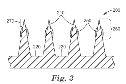
illustrates, in
cross-section, a portion of a microneedle array 200 that includes four
microneedles 210 (of
-21 -
CA 02829352 2013-09-06
WO 2012/122162
PCT/US2012/027857
which two are referenced in FIG. 3) positioned on a microneedle substrate 220.
Coating
250 is disposed on microneedles 210 no more than distance 260 from the tip of
the
microneedles. This is accomplished by contacting not more than a portion of
the
microneedle height with the coating formulation. Accordingly, for certain
embodiments,
including any one of the method embodiments described herein that includes the
step of
contacting the microneedles with the composition (also referred to herein as
the coating
formulation) the microneedles each have a tip and a base, the tip extending a
distance (h)
from the base, and contacting is carried out by contacting the tips of the
microneedles and
a portion of the microneedles extending not more than 90 percent of the
distance (0.9h)
from the tips to the bases with the composition, preferably not more than 70
percent of the
distance (0.7h), more preferably not more than 50 percent of the distance
(0.5h). It is to
be understood that the distance can apply to a single microneedle or to an
average of the
microneedles in an array. For certain embodiments, including any one of the
embodiments described herein which includes a coating disposed on the
microneedles, at
least 50 % of the microneedles have the coating present on the microneedles
near the tip
and extending not more than 90 percent of the distance toward the base,
preferably not
more than 70 percent of the distance, more preferably not more than 50 percent
of the
distance.
When the microneedles are contacted with the coating formulation, the
microneedles are facing downward into the coating formulation. For certain
embodiments, preferably after the microneedles are contacted with the coating
formulation, contacting is terminated and the microneedles are positioned
facing upward
prior to and/or during volatilizing at least a portion of the volatilizable
carrier. In this
position, a portion of the coating formulation remaining on the microneedles
may flow
toward the base, leaving the tips of the microneedles exposed or with only a
small amount
of coating formulation on the tips. The degree to which flow occurs can depend
upon
factors such as the viscosity, contact angle, and surface tension as described
above.
After removing the microneedles from the coating formulation, some of the
coating formulation remains on the microneedles, the amount depending upon the
coating
formulation properties and surface properties of the microneedle material as
described
above. At least a portion of the volatilizable carrier is removed from the
coating
formulation adhering to the microneedles, leaving the coating disposed on the
- 22 -
CA 02829352 2013-09-06
WO 2012/122162
PCT/US2012/027857
microneedles. One or more additional contacting steps may be used. The shape
of the
coating, average coating thickness, and amount of the surface of the
microneedle covered
by the coating depends upon the factors discussed above as well as the number
of times
the contacting step is repeated.
FIG. 3 illustrates one embodiment with the coating disposed on the
microneedles,
wherein the tips of the microneedles are essentially exposed (no coating or a
relatively
small amount of coating) a distance 270 from the tip. For certain embodiments,
including
any one of the embodiments described herein which includes a coating disposed
on the
microneedles, the tips of the microneedles are exposed or only as small amount
of coating
is on the tips. For certain of these embodiments distance 270 is at least 1
percent (0.1h), 3
percent (0.03h) or 6 percent (0.06h) of the distance from the tip to the base.
For certain of
these embodiments, distance 270 is at most 10 percent (0.1h) of the distance
from the tip
to the base.
FIG. 5 is an optical micrograph illustrating four microneedles of a
microneedle
array prior to contacting the microneedles with the composition (coating
formulation).
For certain embodiments, including any one of the embodiments described herein
which includes a coating disposed on the microneedles, the coating is present
on the
microneedles in an average amount of 0.01 to 2 micrograms per microneedle.
Coating
weight can be determined by weighing the microneedle array before and after
the coating
is disposed on the microneedles and dividing the difference by the number of
microneedles in the array. Preferably, the coated microneedle array has come
to a
constant weight, indicating that the volatilizable carrier has been removed,
before taking
the weight after the coating is disposed. Alternatively, the total amount of a
solid
component (such as the local anesthetic) in the coating on all the
microneedles of the
entire array can be determined analytically and then the total weight of
solids calculated
based upon the know weight of all solid components used in the coating
formulation.
Volatilizing the carrier can be performed using various means including for
example, drying at ambient conditions; drying at conditions other than ambient
conditions
(such as temperatures other than room temperature or a humidity other than an
average
humidity); drying for various times; drying with heat, lyophilization, freeze
drying; other
similar techniques; or combinations thereof.
- 23 -
CA 02829352 2013-09-06
WO 2012/122162
PCT/US2012/027857
FIG. 6 is an optical micrograph illustrating four microneedles of a
microneedle
array after contacting the microneedles with the composition (coating
formulation) and
volatilizing the carrier.
Once at least a portion of the carrier (which may be a portion or all of the
solvent)
in the coating formulation has evaporated (either after a single contacting
step or multiple
contacting steps), the coating formulation on the microneedle array can be
referred to as
the "coating" as described above.
Methods of coating microneedle arrays can be used to form coated microneedle
arrays. A coating disposed on the microneedles or the coated microneedle array
can
include a coating on at least a portion of the plurality of microneedles.
As indicated above, a medical device, comprising an array of dissolvable
microneedles, a method of extending a topically delivered local anesthetic
dose in
mammalian tissue using the array of dissolvable microneedles, and a method of
making a
local anesthetic-containing dissolvable microneedle device are also provided
herein. The
dissolvable microneedles may contain the same components in the various
amounts
described above for the coatings disposed on the microneedles.
FIG. 4 illustrates, in cross-section, a portion of a microneedle array 300
that
includes four microneedles 310 (of which two are referenced in FIG. 4)
positioned on a
microneedle substrate 320. Dissolvable microneedle portion 360 includes the
local
anesthetic and dose-extending component and may optionally further contain any
of the
excipients as described above. The remaining portion of the dissolvable
microneedle and
substrate 320 comprise a dissolvable matrix material. In order to avoid
wasting the local
anesthetic and dose-extending component, these materials are preferably
located only in
portion 360. However, the local anesthetic and dose-extending component can be
included in the entire volume of the microneedles or throughout the entire
microneedle
array 300, including the substrate 320. Preferably, the dissolvable matrix
material is
included in portion 360 as well as all other portions of the microneedles.
The wt-% of the local anesthetic and dose-extending component in the
dissolvable
microneedles is based upon the total weight of solids in all portions of the
microneedle
array that contain these materials. For example, in FIG. 4, the total weight
of solids in
portion 360 is the basis for the wt-% values.
- 24 -
CA 02829352 2013-09-06
WO 2012/122162
PCT/US2012/027857
The dissolvable matrix material may be any solid material which dissolves
sufficiently in the tissue underlying the stratum corneum to allow the local
anesthetic and
dose-extending component to be released into the tissue, preferably within 10
minutes,
more preferably within 1 minute. For certain embodiments, including any one of
the
above embodiments which includes dissolvable microneedles, the dissolvable
matrix
material is selected from the group consisting of hyaluronic acid,
carboxymethylcellulose,
hydroxpropylmethylcellulose, methylcellulose, polyvinyl alcohol, polyvinyl
pyrrolidone,
sucrose, glucose, dextran, trehalose, maltodextrin, and a combination thereof
Dissolvable microneedle arrays may be fabricated by casting and drying a
solution
containing volatilizable carrier and dissolvable matrix material (preferably
water soluble)
in a mold containing the microstructured cavities. The internal shape of the
microstructured cavities corresponds to the external shape of the dissolvable
microneedles.
The mold can be comprised of materials such as polydimethylsiloxane (PDMS) or
other
plastics that do not permanently bind to or that have low adhesion to
materials used to
make the dissolvable microneedles.
The local anesthetic and dose-extending component can be incorporated into
dissolvable microneedles by first loading a solution of these components with
a
volatilizable carrier (preferably also including the dissolvable matrix
material) into the
mold containing microstructured cavities. After at least partially drying
(volatilizing at
least a portion of the volatilizable carrier), the mold is filled with a
solution of dissolvable
matrix material (without the anesthetic and dose-extending component),
followed by
drying. Alternatively, in a one-step process, the local anesthetic and dose-
extending
component can be combined with the dissolvable matrix material in a solution
with the
volatilizable carrier and the mold filled with this solution, followed by
drying. The same
volatilizable carriers described above in the coating formulations may be used
here.
Drying can be carried out using methods such as lyophilization,
centrifugation,
vacuum, and/or heating. After drying, the solid dissolvable microneedle array
is removed
from the mold and is ready for use. These solutions may be made using water
and/or
organic solvents, such as ethanol, as described above to assure solubilization
of all
materials used in the microneedle array.
- 25 -
CA 02829352 2013-09-06
WO 2012/122162
PCT/US2012/027857
Microneedle devices provided herein may be used for immediate delivery, for
example, application and immediate removal of the device from the application
site.
Immediate removal may be within 10 minutes or less, preferably within 1 minute
or less.
FIG. 7 is an optical micrograph illustrating coated microneedles provided
herein
after 1 minute in tissue. It can be clearly seen that most, if not all, of the
coating was
removed and remained in the tissue.
Application of the microneedle device may be carried out by contacting the
tissue
of a subject with the microneedles and applying hand pressure to force the
microneedles
into the tissue. Alternatively, an application device may be used which
applies the
pressure, forcing the microneedles into the tissue. This can provide a more
even
distribution of pressure and force the microneedles into the tissue at an
optimum velocity
so that essentially all of the microneedles can release the local anesthetic
and dose-
extending component into the tissue. For certain embodiments, including any
one of the
above embodiments of the method of extending a topically delivered local
anesthetic dose
in mammalian tissue, contacting the tissue with a microneedle device is
carried out at a
microneedle velocity of 5 to 10 meters/second.
The following examples are provided to more particularly illustrate various
embodiments of the present invention, but the particular materials and amounts
thereof
recited in these examples, as well as other conditions and details are in no
way intended to
limit this invention.
EXAMPLE S
All formulations used to coat the microneedle arrays in the following examples
were prepared on a weight percent basis (w/w %) and were prepared in water.
For
example, a formulation comprised of 30% dextran, 30% lidocaine hydrochloride,
and
0.3% clonidine hydrochloride included 39.7% water.
- 26 -
CA 02829352 2013-09-06
WO 2012/122162
PCT/US2012/027857
Example 1
Formulation Containing Lidocaine with Clonidine
The microneedle arrays were injection molded (3M, St. Paul, MN) from Class VI,
medical grade liquid crystalline polymer (LCP) (Vectra 0 MT1300, Ticona
Plastics,
Auburn Hills, Michigan) with a surface area of approximately 1.27cm2. Each
microneedle
array featured 316 four-sided pyramidal-shaped microneedles arranged in an
octagonal
pattern, with microneedle heights of nominally 500 microns, an aspect ratio of
approximately 3:1, and a tip-to-tip distance between neighboring microneedles
of
nominally 550 microns.
Lidocaine was coated onto the microneedle arrays using a dip-coating process
with
a formulation comprised of 30% dextran (from Pharmacosmos, Holbaek, Denmark),
30%
lidocaine hydrochloride (Sigma, St. Louis, MO) and 0.3% clonidine
hydrochloride
(Spectrum Chemical & Laboratory Products, New Brunswick, NJ). Prior to
coating, the
microneedle arrays were cleaned with 70% isopropyl alcohol (BDH, West Chester,
PA)
and dried in a 35 C oven for 1 hr. Microneedle arrays were then dipped into
the coating
solution once. The coated microneedles were allowed to dry for 1 hr at 35 C.
For in vivo
application, each array was attached to a 5 cm2 adhesive patch with 1513
double-sided
medical adhesive (3M Company, St. Paul, MN). The arrays were stored in a light
and
moisture proof foil pouch (Oilver-Tolas Healthcare Packaging, Feasterville,
PA) at room
temperature prior to in vivo application.
The determination of lidocaine content in the formulation coated on the
microneedles of an array was conducted using an Agilent 1100 HPLC (Agilent
Technologies, Wilmington, DE) equipped with a quaternary pump, well-plated
thermostatted autosampler, thermostatted column compartment, and diode array
UV
detector. The formulation coated on the microneedles of an array was desorbed
into an
appropriate volume of diluent, (0.1% trifluoroacetic acid (TFA, J T. Baker,
Phillpsburg,
NJ) in water), and injected into the HPLC system. The results were quantified
against an
external standard of lidocaine (free base) at a similar concentration to the
coating amount.
A Zorbax SB-C18 column, 3.5 m particle size, 150 x 3.0mm I.D. (Agilent
Technologies,
Wilmington, DE) was used for the separation. The mobile phase consisted of two
eluents:
eluent A was 100% water with 0.1% TFA and eluent B was 100% acetonitrile
(Spectrum
-27 -
CA 02829352 2013-09-06
WO 2012/122162
PCT/US2012/027857
Chemical & Laboratory Products, New Brunswick, NJ) with 0.1% TFA. A linear
gradient
from 80/20 to 0/100 (A/B) was applied over 5 min. The flow rate was 0.5 mL/min
and the
UV detection wavelength was 230 nm. The total run time was 8 minutes. A total
of 5
replicates were conducted. The results from the individual replicates were
averaged to
provide a measured lidocaine loading amount of 94.1 3.0 mcg/array.
The in vivo delivery of lidocaine to tissue using the coated microneedle array
described above was determined using naïve young adult female mixed breed
agricultural
swine (Yorkshire X from Midwest Research Swine, Gibbon, MN). Swine with
minimal
skin pigmentation and weighing 10-40 kg were selected for the study. The
animals were
initially sedated with ketamine (10 mg/kg) and glycopyrro late (0.011 mg/kg)
was
intramuscularly administered to reduce salivary, tracheobronchial, and
pharyngeal
secretions. Hair and dirt on pig skin at the intended application sites were
removed prior
to application of the microneedle array to minimize complications. Skin test
sites were
selected based on lack of skin pigmentation and skin damage. The hair was
first clipped
using an electric shaver followed by shaving with a wet multi-blade disposable
razor
(Schick Xtreme3) and shaving cream (Gillette Foamy Regular) while the animal
was
under anesthesia.
A light surgical plane of anesthesia was achieved by administering 1.5-5%
isoflurane in 1.5-4 L of oxygen by mask. Anesthetized animals were placed in
lateral
recumbency on insulated table pads. During the experiment, the animals were
placed on a
heated table to control body temperature at approximately 38 C. Animals were
observed
continuously until normal recovery was attained. A microneedle array was
applied to the
swine rib with a spring-loaded applicator that provided an impact velocity of
approximately 8 m/s, held in place with the applicator for 5 seconds before
removing the
applicator, and remained in contact with the skin for 1 minute. The applicator
was
previously described in International Publication No. WO 2005/123173 Al. The
patch
was removed and the application site was swabbed with a cotton ball moistened
with
phosphate buffered saline (PBS) (EMD chemicals Inc., Gibbstown, NJ) to remove
any
residual lidocaine remaining on the skin surface. Following this swabbing, a
dry cotton
ball was used to remove any residual PBS. A 4 mm skin biopsy (Disposable
Biopsy Punch
from Miltex Inc., York, PA) was collected from the microneedle array
application site
- 28 -
CA 02829352 2013-09-06
WO 2012/122162
PCT/US2012/027857
following removal of the array at time points of 0, 5, 15, 30, 60, 90, and 120
minutes. The
biopsy punch samples were stored at -20 C until analyzed.
The animal facility used was accredited by the Association for Assessment and
Accreditation of Laboratory Animal Care (AAALAC, Frederick, Maryland) and all
procedures were in accordance with an approved Institutional Animal Care and
Use
Committee (IACUC) protocol.
Lidocaine was extracted from each swine skin tissue biopsy punch using
enzymatic
digestion. The skin tissue was weighed into a glass vial, then tissue
digestion buffer
containing 0.1 U proteinase K (EMD Chemicals, San Diego, CA) per mg of skin
tissue
was added to the vial. The tissue was digested at 55 C for 5 hours. The
digestion process
produced a homogenous sample solution.
Protein precipitation was used to prepare the digested tissue samples for
analysis
by LC/MS/MS. Protein was removed from the digested tissue samples by adding 2
volumes of methanol, containing mepivacaine as the internal standard, followed
by
centrifugation at 14,000 RPM for 10 minutes. The resulting sample was
quantitatively
analyzed using a Sciex API3000 triple quadrupole mass spectrometer (Applied
Biosystems, Foster City, CA) running in positive ion mode using Turbo IonSpray
interface
to monitor the product ions resulting from the m/z transitions: 235 ¨> 86.2
for lidocaine
and 247 ¨> 97.5 for mepivacaine. The linear range for lidocaine was 50.0 to
20,000 ng/mL
evaluated using 1/x2 curve weighting.
A total of 3 replicates were conducted. The results from the individual
replicates
were averaged and are presented in Table 1.
Table 1. Tissue Concentration of Lidocaine
0 min 5 min 15 min 30 min 60 min 90 min 120 min
Lidocaine Tissue
Concentration
255.0 216.0 186.3 111.9 107.6 61.7 27.6
(ng/mg)
Standard Deviation 67.7 88.4 33.3 32.6 11.2 21.8 0.9
(ng/mg)
- 29 -
CA 02829352 2013-09-06
WO 2012/122162
PCT/US2012/027857
Example 2
Formulation Containing Lidocaine with Epinephrine
The microneedle arrays were injection molded (3M, St. Paul, MN) from Class VI,
medical grade liquid crystalline polymer (LCP) (Vectra 0 MT1300, Ticona
Plastics,
Auburn Hills, Michigan) with a surface area of approximately 1.27cm2. Each
microneedle
array featured 316 four-sided pyramidal-shaped microneedles arranged in an
octagonal
pattern, with microneedle heights of nominally 500 microns, an aspect ratio of
approximately 3:1, and a tip-to-tip distance between neighboring microneedles
of
nominally 550 microns.
Lidocaine was coated onto the microneedle arrays using a dip-coating process
with
a formulation comprised of 30% dextran (from Pharmacosmos, Holbaek, Denmark),
30%
lidocaine hydrochloride (Sigma, St. Louis, MO) and 0.03% epinephrine
bitartrate (Sigma,
St. Louis, MO). Prior to coating, the microneedle arrays were cleaned with 70%
isopropyl
alcohol (BDH, West Chester, PA) and dried in a 35 C oven for 1 hr. Microneedle
arrays
were then dipped into the coating solution once. The coated microneedles were
allowed to
dry for 1 hr at 35 C. For in vivo application, each array was attached to a 5
cm2 adhesive
patch with 1513 double-sided medical adhesive (3M Company, St. Paul, MN). The
arrays
were stored in a light and moisture proof foil pouch (Oliver-Tolas Healthcare
Packaging,
Feasterville, PA) at room temperature prior to in vivo application.
The determination of lidocaine content in the formulation coated on the
microneedles of an array was conducted using an Agilent 1100 HPLC (Agilent
Technologies, Wilmington, DE) equipped with a quaternary pump, well-plated
thermostatted autosampler, thermostatted column compartment, and diode array
UV
detector. The formulation coated on the microneedles of an array was desorbed
into an
appropriate volume of diluent, (0.1% trifluoroacetic acid (TFA, J T. Baker,
Phillpsburg,
NJ) in water), and injected into the HPLC system. The results were quantified
against an
external standard of lidocaine (free base) at a similar concentration to the
coating amount.
A Zorbax SB-C18 column, 3.5 m particle size, 150 x 3.0mm I.D. (Agilent
Technologies,
Wilmington, DE) was used for the separation. The mobile phase consisted of two
eluents:
eluent A was 100% water with 0.1% TFA and eluent B was 100% acetonitrile
(Spectrum
Chemical & Laboratory Products, New Brunswick, NJ) with 0.1% TFA. A linear
gradient
- 30 -
CA 02829352 2013-09-06
WO 2012/122162
PCT/US2012/027857
from 80/20 to 0/100 (A/B) was applied over 5 min. The flow rate was 0.5 mL/min
and the
UV detection wavelength was 230 nm. The total run time was 8 minutes. A total
of 5
replicates were conducted. The results from the individual replicates were
averaged to
provide a measured lidocaine loading amount of 96.3 3.4 mcg/array.
The in vivo delivery of lidocaine to tissue using the coated microneedle array
described above was determined using naïve young adult female mixed breed
agricultural
swine (Yorkshire X from Midwest Research Swine, Gibbon, MN). Swine with
minimal
skin pigmentation and weighing 10-40 kg were selected for the study. The
animals were
initially sedated with ketamine (10 mg/kg) and glycopyrro late (0.011 mg/kg)
was
intramuscularly administered to reduce salivary, tracheobronchial, and
pharyngeal
secretions. Hair and dirt on pig skin at the intended application sites were
removed prior
to application of the microneedle array to minimize complications. Skin test
sites were
selected based on lack of skin pigmentation and skin damage. The hair was
first clipped
using an electric shaver followed by shaving with a wet multi-blade disposable
razor
(Schick Xtreme3) and shaving cream (Gillette Foamy Regular) while the animal
was
under anesthesia.
A light surgical plane of anesthesia was achieved by administering 1.5-5%
isoflurane in 1.5-4 L of oxygen by mask. Anesthetized animals were placed in
lateral
recumbency on insulated table pads. During the experiment, the animals were
placed on a
heated table to control body temperature at approximately 38 C. Animals were
observed
continuously until normal recovery was attained. A microneedle array was
applied to the
swine rib with a spring-loaded applicator that provided an impact velocity of
approximately 8 m/s, held in place with the applicator for 5 seconds before
removing the
applicator, and remained in contact with the skin for 1 minute. The applicator
was
previously described in International Publication No. WO 2005/123173 Al. The
patch
was removed and the application site was swabbed with a cotton ball moistened
with
phosphate buffered saline (PBS) (EMD chemicals Inc., Gibbstown, NJ) to remove
any
residual lidocaine remaining on the skin surface. Following this swabbing, a
dry cotton
ball was used to remove any residual PBS. A 4 mm skin biopsy (Disposable
Biopsy Punch
from Miltex Inc., York, PA) was collected from the microneedle array
application site
following removal of the array at time points of 0, 5, 15, 30, 60, 90, and 120
minutes. The
biopsy punch samples were stored at -20 C until analyzed.
-31 -
CA 02829352 2013-09-06
WO 2012/122162
PCT/US2012/027857
The animal facility used was accredited by the Association for Assessment and
Accreditation of Laboratory Animal Care (AAALAC, Frederick, Maryland) and all
procedures were in accordance with an approved Institutional Animal Care and
Use
Committee (IACUC) protocol.
Lidocaine was extracted from each swine skin tissue biopsy punch using
enzymatic
digestion. The skin tissue was weighed into a glass vial, then tissue
digestion buffer
containing 0.1 U proteinase K (EMD Chemicals, San Diego, CA) per mg of skin
tissue
was added to the vial. The tissue was digested at 55 C for 5 hours. The
digestion process
produced a homogenous sample solution.
Protein precipitation was used to prepare the digested tissue samples for
analysis
by LC/MS/MS. Protein was removed from the digested tissue samples by adding 2
volumes of methanol, containing mepivacaine as the internal standard, followed
by
centrifugation at 14,000 RPM for 10 minutes. The resulting sample was
quantitatively
analyzed using a Sciex API3000 triple quadrupole mass spectrometer (Applied
Biosystems, Foster City, CA) running in positive ion mode using Turbo IonSpray
interface
to monitor the product ions resulting from the m/z transitions: 235 ¨> 86.2
for lidocaine
and 247 ¨> 97.5 for mepivacaine. The linear range for lidocaine was 50.0 to
20,000 ng/mL
evaluated using 1/x2 curve weighting.
A total of 3 replicates were conducted. The results from the individual
replicates
were averaged and are presented in Table 2.
Table 2. Tissue Concentration of Lidocaine
0 min 5 min 15 min 30 min 60 min 90 min 120 min
Lidocaine Tissue
Concentration
343.0 356.7 388.7 124.3 175.7 146.3 20.0
(ng/mg)
Standard Deviation 134.9 22.8 83.7 52.7 54.6 31.9 6.4
(ng/mg)
Example 3
Formulation Containing Prilocaine with Clonidine
The microneedle arrays were injection molded (3M, St. Paul, MN) from Class VI,
medical grade liquid crystalline polymer (LCP) (Vectra 0 MT1300, Ticona
Plastics,
Auburn Hills, Michigan) with a surface area of approximately 1.27cm2. Each
microneedle
- 32 -
CA 02829352 2013-09-06
WO 2012/122162
PCT/US2012/027857
array featured 316 four-sided pyramidal-shaped microneedles arranged in an
octagonal
pattern, with microneedle heights of nominally 500 microns, an aspect ratio of
approximately 3:1, and a tip-to-tip distance between neighboring microneedles
of
nominally 550 microns.
Prilocaine was coated onto the microneedle arrays using a dip-coating process
with
a formulation comprised of 30% dextran (from Pharmacosmos, Holbaek, Denmark),
15%
prilocaine hydrochloride (Spectrum Chemical & Laboratory Products, New
Brunswick,
NJ) and 0.15% clonidine hydrochloride (Spectrum Chemical & Laboratory
Products, New
Brunswick, NJ). Prior to coating, the microneedle arrays were cleaned with 70%
isopropyl
alcohol (BDH, West Chester, PA) and dried in a 35 C oven for 1 hr. Microneedle
arrays
were then dipped into the coating solution once. The coated microneedles were
allowed to
dry for 1 hr at 35 C. For in vivo application, each array was attached to a 5
cm2 adhesive
patch with 1513 double-sided medical adhesive (3M Company, St. Paul, MN). The
arrays
were stored in a light and moisture proof foil pouch (Oliver-Tolas Healthcare
Packaging,
Feasterville, PA) at room temperature prior to in vivo application.
The determination of prilocaine content in the formulation coated on the
microneedles of an array was conducted using an Agilent 1100 HPLC (Agilent
Technologies, Wilmington, DE) equipped with a quaternary pump, well-plated
thermostatted autosampler, thermostatted column compartment, and diode array
UV
detector. The formulation coated on the microneedles of an array was desorbed
into an
appropriate volume of diluent, (0.1% trifluoroacetic acid (TFA, J T. Baker,
Phillpsburg,
NJ) in water), and injected into the HPLC system. The results were quantified
against an
external standard of prilocaine (free base) at a similar concentration to the
coating amount.
A Zorbax SB-C18 column, 3.5 m particle size, 150 x 3.0mm I.D. (Agilent
Technologies,
Wilmington, DE) was used for the separation. The mobile phase consisted of two
eluents:
eluent A was 100% water with 0.1% TFA and eluent B was 100% acetonitrile
(Spectrum
Chemical & Laboratory Products, New Brunswick, NJ) with 0.1% TFA. A linear
gradient
from 80/20 to 0/100 (A/B) was applied over 5 min. The flow rate was 0.5 mL/min
and the
UV detection wavelength was 230 nm. The total run time was 8 minutes. A total
of 5
replicates were conducted. The results from the individual replicates were
averaged to
provide a measured prilocaine loading amount of 45.6 1.2 mcg/array.
- 33 -
CA 02829352 2013-09-06
WO 2012/122162
PCT/US2012/027857
The in vivo delivery of prilocaine to tissue using the coated microneedle
array
described above was determined using naïve young adult female mixed breed
agricultural
swine (Yorkshire X from Midwest Research Swine, Gibbon, MN). Swine with
minimal
skin pigmentation and weighing 10-40 kg were selected for the study. The
animals were
initially sedated with ketamine (10 mg/kg) and glycopyrro late (0.011 mg/kg)
was
intramuscularly administered to reduce salivary, tracheobronchial, and
pharyngeal
secretions. Hair and dirt on pig skin at the intended application sites were
removed prior
to application of the microneedle array to minimize complications. Skin test
sites were
selected based on lack of skin pigmentation and skin damage. The hair was
first clipped
using an electric shaver followed by shaving with a wet multi-blade disposable
razor
(Schick Xtreme3) and shaving cream (Gillette Foamy Regular) while the animal
was
under anesthesia.
A light surgical plane of anesthesia was achieved by administering 1.5-5%
isoflurane in 1.5-4 L of oxygen by mask. Anesthetized animals were placed in
lateral
recumbency on insulated table pads. During the experiment, the animals were
placed on a
heated table to control body temperature at approximately 38 C. Animals were
observed
continuously until normal recovery was attained. A microneedle array was
applied to the
swine rib with a spring-loaded applicator that provided an impact velocity of
approximately 8 m/s, held in place with the applicator for 5 seconds before
removing the
applicator, and remained in contact with the skin for 1 minute. The applicator
was
previously described in International Publication No. WO 2005/123173 Al. The
patch
was removed and the application site was swabbed with a cotton ball moistened
with
phosphate buffered saline (PBS) (EMD chemicals Inc., Gibbstown, NJ) to remove
any
residual prilocaine remaining on the skin surface. Following this swabbing, a
dry cotton
ball was used to remove any residual PBS. A 4 mm skin biopsy (Disposable
Biopsy Punch
from Miltex Inc., York, PA) was collected from the microneedle array
application site
following removal of the array at time points of 0, 5, 15, 30, 60, 90, and 120
minutes. The
biopsy punch samples were stored at -20 C until analyzed.
The animal facility used was accredited by the Association for Assessment and
Accreditation of Laboratory Animal Care (AAALAC, Frederick, Maryland) and all
procedures were in accordance with an approved Institutional Animal Care and
Use
Committee (IACUC) protocol.
- 34 -
CA 02829352 2013-09-06
WO 2012/122162
PCT/US2012/027857
Prilocaine was extracted from each swine skin tissue biopsy punch using
enzymatic digestion. The skin tissue was weighed into a glass vial, then
tissue digestion
buffer containing 0.1 U proteinase K (EMD Chemicals, San Diego, CA) per mg of
skin
tissue was added to the vial. The tissue was digested at 55 C for 5 hours. The
digestion
process produced a homogenous sample solution.
Protein precipitation was used to prepare the digested tissue samples for
analysis
by LC/MS/MS. Protein was removed from the digested tissue samples by adding 2
volumes of methanol, containing mepivacaine as the internal standard, followed
by
centrifugation at 14,000 RPM for 10 minutes. The resulting sample was
quantitatively
analyzed using a Sciex API3000 triple quadrupole mass spectrometer (Applied
Biosystems, Foster City, CA) running in positive ion mode using Turbo IonSpray
interface
to monitor the product ions resulting from the m/z transitions: 221.1 ¨> 86.1
for prilocaine
and 247 ¨> 97.5 for mepivacaine. The linear range for prilocaine was 50.0 to
20,000
ng/mL evaluated using 1/x2 curve weighting.
A total of 3 replicates were conducted. The results from the individual
replicates
were averaged and are presented in Table 3.
Table 3. Tissue Concentration of Prilocaine
0 min 5 min 15 min 30 min 60 min 90 min 120 min
Prilocaine Tissue
Concentration 119.7 75.0 78.2 56.8 27.3 14.9
8.3
(ng/mg)
Standard Deviation 21.6 25.0 6.9 9.8 11.8 7.0 0.8
(ng/mg)
Example 4
Formulation Containing Lidocaine with Guanfacine
The microneedle arrays were injection molded (3M, St. Paul, MN) from Class VI,
medical grade liquid crystalline polymer (LCP) (Vectra 0 MT1300, Ticona
Plastics,
Auburn Hills, Michigan) with a surface area of approximately 1.27 cm2. Each
microneedle array featured 316 four-sided pyramidal-shaped microneedles
arranged in an
octagonal pattern, with microneedle heights of nominally 500 microns, an
aspect ratio of
approximately 3:1, and a tip-to-tip distance between neighboring microneedles
of
nominally 550 microns.
- 35 -
CA 02829352 2013-09-06
WO 2012/122162
PCT/US2012/027857
Lidocaine was coated onto the microneedle arrays using a dip-coating process
with
a formulation comprised of 30% dextran (from Pharmacosmos, Holbaek, Denmark),
30%
lidocaine hydrochloride (Sigma, St. Louis, MO) and 0.3% guanfacine
hydrochloride
(Sigma, St. Louis, MO). Prior to coating, the microneedle arrays were cleaned
with 70%
isopropyl alcohol (BDH, West Chester, PA) and dried in a 35 C oven for 1 hr.
Microneedle arrays were then dipped into the coating solution once. The coated
microneedles were allowed to dry for 1 hr at 35 C. For in vivo application,
each array was
attached to a 5 cm2 adhesive patch with 1513 double-sided medical adhesive (3M
Company, St. Paul, MN). The arrays were stored in a light and moisture proof
foil pouch
(Oliver-Tolas Healthcare Packaging, Feasterville, PA) at room temperature
prior to in vivo
application.
The determination of lidocaine content in the formulation coated on the
microneedles of an array was conducted using an Agilent 1100 HPLC (Agilent
Technologies, Wilmington, DE) equipped with a quaternary pump, well-plated
thermostatted autosampler, thermostatted column compartment, and diode array
UV
detector. The formulation coated on the microneedles of an array was desorbed
into an
appropriate volume of diluent, (0.1% trifluoroacetic acid (TFA, J T. Baker,
Phillpsburg,
NJ) in water), and injected into the HPLC system. The results were quantified
against an
external standard of lidocaine (free base) at a similar concentration to the
coating amount.
A Zorbax SB-C18 column, 3.5 m particle size, 150 x 3.0mm I.D. (Agilent
Technologies,
Wilmington, DE) was used for the separation. The mobile phase consisted of two
eluents:
eluent A was 100% water with 0.1% TFA and eluent B was 100% acetonitrile
(Spectrum
Chemical & Laboratory Products, New Brunswick, NJ) with 0.1% TFA. A linear
gradient
from 80/20 to 0/100 (A/B) was applied over 5 min. The flow rate was 0.5 mL/min
and the
UV detection wavelength was 230 nm. The total run time was 8 minutes. A total
of 5
replicates were conducted. The results from the individual replicates were
averaged to
provide a measured lidocaine loading amount of 89.9 2.5 mcg/array.
The in vivo delivery of lidocaine to tissue using the coated microneedle array
described above was determined using naïve young adult female mixed breed
agricultural
swine (Yorkshire X from Midwest Research Swine, Gibbon, MN). Swine with
minimal
skin pigmentation and weighing 10-40 kg were selected for the study. The
animals were
initially sedated with ketamine (10 mg/kg) and glycopyrro late (0.011 mg/kg)
was
- 36 -
CA 02829352 2013-09-06
WO 2012/122162
PCT/US2012/027857
intramuscularly administered to reduce salivary, tracheobronchial, and
pharyngeal
secretions. Hair and dirt on pig skin at the intended application sites were
removed prior
to application of the microneedle array to minimize complications. Skin test
sites were
selected based on lack of skin pigmentation and skin damage. The hair was
first clipped
using an electric shaver followed by shaving with a wet multi-blade disposable
razor
(Schick Xtreme3) and shaving cream (Gillette Foamy Regular) while the animal
was
under anesthesia.
A light surgical plane of anesthesia was achieved by administering 1.5-5%
isoflurane in 1.5-4 L of oxygen by mask. Anesthetized animals were placed in
lateral
recumbency on insulated table pads. During the experiment, the animals were
placed on a
heated table to control body temperature at approximately 38 C. Animals were
observed
continuously until normal recovery was attained. A microneedle array was
applied to the
swine rib with a spring-loaded applicator that provided an impact velocity of
approximately 8 m/s, held in place with the applicator for 5 seconds before
removing the
applicator, and remained in contact with the skin for 1 minute. The applicator
was
previously described in International Publication No. WO 2005/123173 Al. The
patch
was removed and the application site was swabbed with a cotton ball moistened
with
phosphate buffered saline (PBS) (EMD chemicals Inc., Gibbstown, NJ) to remove
any
residual lidocaine remaining on the skin surface. Following this swabbing, a
dry cotton
ball was used to remove any residual PBS. A 4 mm skin biopsy (Disposable
Biopsy Punch
from Miltex Inc., York, PA) was collected from the microneedle array
application site
following removal of the array at time points of 0, 5, 15, 30, 60, 90, and 120
minutes. The
biopsy punch samples were stored at -20 C until analyzed.
The animal facility used was accredited by the Association for Assessment and
Accreditation of Laboratory Animal Care (AAALAC, Frederick, Maryland) and all
procedures were in accordance with an approved Institutional Animal Care and
Use
Committee (IACUC) protocol.
Lidocaine was extracted from each swine skin tissue biopsy punch using
enzymatic
digestion. The skin tissue was weighed into a glass vial, then tissue
digestion buffer
containing 0.1 U proteinase K (EMD Chemicals, San Diego, CA) per mg of skin
tissue
was added to the vial. The tissue was digested at 55 C for 5 hours. The
digestion process
produced a homogenous sample solution.
-37 -
CA 02829352 2013-09-06
WO 2012/122162
PCT/US2012/027857
Protein precipitation was used to prepare the digested tissue samples for
analysis
by LC/MS/MS. Protein was removed from the digested tissue samples by adding 2
volumes of methanol, containing mepivacaine as the internal standard, followed
by
centrifugation at 14,000 RPM for 10 minutes. The resulting sample was
quantitatively
analyzed using a Sciex API3000 triple quadrupole mass spectrometer (Applied
Biosystems, Foster City, CA) running in positive ion mode using Turbo IonSpray
interface
to monitor the product ions resulting from the m/z transitions: 235 ¨> 86.2
for lidocaine
and 247 ¨> 97.5 for mepivacaine. The linear range for lidocaine was 50.0 to
20,000 ng/mL
evaluated using 1/x2 curve weighting.
A total of 3 replicates were conducted. The results from the individual
replicates
were averaged and are presented in Table 4.
Table 4. Tissue Concentration of Lidocaine
0 min 5 min 15 min 30 min 60 min 90 min 120 min
Lidocaine Tissue
Concentration
362.3 204.0 170.3 168.3 93.0 92.1 72.9
(ng/mg)
Standard Deviation 86.7 24.6 42.1 6.4 26.4 10.9
27.8
(ng/mg)
Example 5
Formulation Containing Lidocaine with Apraclonidine
The microneedle arrays were injection molded (3M, St. Paul, MN) from Class VI,
medical grade liquid crystalline polymer (LCP) (Vectra 0 MT1300, Ticona
Plastics,
Auburn Hills, Michigan) with a surface area of approximately 1.27 cm2. Each
microneedle array featured 316 four-sided pyramidal-shaped microneedles
arranged in an
octagonal pattern, with microneedle heights of nominally 500 microns, an
aspect ratio of
approximately 3:1, and a tip-to-tip distance between neighboring microneedles
of
nominally 550 microns.
Lidocaine was coated onto the microneedle arrays using a dip-coating process
with
a formulation comprised of 30% dextran (from Pharmacosmos, Holbaek, Denmark),
30%
lidocaine hydrochloride (Sigma, St. Louis, MO) and 0.3% apraclonidine
hydrochloride
(Sigma, St. Louis, MO). Prior to coating, the microneedle arrays were cleaned
with 70%
isopropyl alcohol (BDH, West Chester, PA) and dried in a 35 C oven for 1 hr.
- 38 -
CA 02829352 2013-09-06
WO 2012/122162
PCT/US2012/027857
Microneedle arrays were then dipped into the coating solution once. The coated
microneedles were allowed to dry for 1 hr at 35 C. For in vivo application,
each array was
attached to a 5 cm2 adhesive patch with 1513 double-sided medical adhesive (3M
Company, St. Paul, MN). The arrays were stored in a light and moisture proof
foil pouch
(Oliver-Tolas Healthcare Packaging, Feasterville, PA) at room temperature
prior to in vivo
application.
The determination of lidocaine content in the formulation coated on the
microneedles of an array was conducted using an Agilent 1100 HPLC (Agilent
Technologies, Wilmington, DE) equipped with a quaternary pump, well-plated
thermostatted autosampler, thermostatted column compartment, and diode array
UV
detector. The formulation coated on the microneedles of an array was desorbed
into an
appropriate volume of diluent, (0.1% trifluoroacetic acid (TFA, J T. Baker,
Phillpsburg,
NJ) in water), and injected into the HPLC system. The results were quantified
against an
external standard of lidocaine (free base) at a similar concentration to the
coating amount.
A Zorbax SB-C18 column, 3.5 m particle size, 150 x 3.0mm I.D. (Agilent
Technologies,
Wilmington, DE) was used for the separation. The mobile phase consisted of two
eluents:
eluent A was 100% water with 0.1% TFA and eluent B was 100% acetonitrile
(Spectrum
Chemical & Laboratory Products, New Brunswick, NJ) with 0.1% TFA. A linear
gradient
from 80/20 to 0/100 (A/B) was applied over 5 min. The flow rate was 0.5 mL/min
and the
UV detection wavelength was 230 nm. The total run time was 8 minutes. A total
of 5
replicates were conducted. The results from the individual replicates were
averaged to
provide a measured lidocaine loading amount of 84.3 4.4 mcg/array.
The in vivo delivery of lidocaine to tissue using the coated microneedle array
described above was determined using naïve young adult female mixed breed
agricultural
swine (Yorkshire X from Midwest Research Swine, Gibbon, MN). Swine with
minimal
skin pigmentation and weighing 10-40 kg were selected for the study. The
animals were
initially sedated with ketamine (10 mg/kg) and glycopyrro late (0.011 mg/kg)
was
intramuscularly administered to reduce salivary, tracheobronchial, and
pharyngeal
secretions. Hair and dirt on pig skin at the intended application sites were
removed prior
to application of the microneedle array to minimize complications. Skin test
sites were
selected based on lack of skin pigmentation and skin damage. The hair was
first clipped
using an electric shaver followed by shaving with a wet multi-blade disposable
razor
- 39 -
CA 02829352 2013-09-06
WO 2012/122162
PCT/US2012/027857
(Schick Xtreme3) and shaving cream (Gillette Foamy Regular) while the animal
was
under anesthesia.
A light surgical plane of anesthesia was achieved by administering 1.5-5%
isoflurane in 1.5-4 L of oxygen by mask. Anesthetized animals were placed in
lateral
recumbency on insulated table pads. During the experiment, the animals were
placed on a
heated table to control body temperature at approximately 38 C. Animals were
observed
continuously until normal recovery was attained. A microneedle array was
applied to the
swine rib with a spring-loaded applicator that provided an impact velocity of
approximately 8 m/s, held in place with the applicator for 5 seconds before
removing the
applicator, and remained in contact with the skin for 1 minute. The applicator
was
previously described in International Publication No. WO 2005/123173 Al. The
patch
was removed and the application site was swabbed with a cotton ball moistened
with
phosphate buffered saline (PBS) (EMD chemicals Inc., Gibbstown, NJ) to remove
any
residual lidocaine remaining on the skin surface. Following this swabbing, a
dry cotton
ball was used to remove any residual PBS. A 4 mm skin biopsy (Disposable
Biopsy Punch
from Miltex Inc., York, PA) was collected from the microneedle array
application site
following removal of the array at time points of 0, 5, 15, 30, 60, 90, and 120
minutes. The
biopsy punch samples were stored at -20 C until analyzed.
The animal facility used was accredited by the Association for Assessment and
Accreditation of Laboratory Animal Care (AAALAC, Frederick, Maryland) and all
procedures were in accordance with an approved Institutional Animal Care and
Use
Committee (IACUC) protocol.
Lidocaine was extracted from each swine skin tissue biopsy punch using
enzymatic
digestion. The skin tissue was weighed into a glass vial, then tissue
digestion buffer
containing 0.1 U proteinase K (EMD Chemicals, San Diego, CA) per mg of skin
tissue
was added to the vial. The tissue was digested at 55 C for 5 hours. The
digestion process
produced a homogenous sample solution.
Protein precipitation was used to prepare the digested tissue samples for
analysis
by LC/MS/MS. Protein was removed from the digested tissue samples by adding 2
volumes of methanol, containing mepivacaine as the internal standard, followed
by
centrifugation at 14,000 RPM for 10 minutes. The resulting sample was
quantitatively
analyzed using a Sciex API3000 triple quadrupole mass spectrometer (Applied
- 40 -
CA 02829352 2013-09-06
WO 2012/122162
PCT/US2012/027857
Biosystems, Foster City, CA) running in positive ion mode using Turbo IonSpray
interface
to monitor the product ions resulting from the m/z transitions: 235 ¨> 86.2
for lidocaine
and 247 ¨> 97.5 for mepivacaine. The linear range for lidocaine was 50.0 to
20,000 ng/mL
evaluated using 1/x2 curve weighting.
A total of 3 replicates were conducted. The results from the individual
replicates
were averaged and are presented in Table 5.
Table 5. Tissue Concentration of Lidocaine
0 min 5 min 15 min 30 min 60 min 90 min 120 min
Lidocaine Tissue
Concentration
437.3 244.7 196.3 268.7 201.7 169.7 96.7
(ng/mg)
Standard Deviation 52.6 56.5 55.1 43.8 37.8 30.1
55.3
(ng/mg)
Comparative Example 1
Formulation Containing Lidocaine without a Dose-Extending Component
The microneedle arrays were injection molded (3M, St. Paul, MN) from Class VI,
medical grade liquid crystalline polymer (LCP) (Vectra 0 MT1300, Ticona
Plastics,
Auburn Hills, Michigan) with a surface area of approximately 1.27cm2. Each
microneedle
array featured 316 four-sided pyramidal-shaped microneedles arranged in an
octagonal
pattern, with microneedle heights of nominally 500 microns, an aspect ratio of
approximately 3:1, and a tip-to-tip distance between neighboring microneedles
of
nominally 550 microns.
Lidocaine was coated onto the microneedle arrays using a dip-coating process
with
a formulation comprised of 30% dextran (from Pharmacosmos, Holbaek, Denmark),
30%
lidocaine hydrochloride (Sigma, St. Louis, MO). Prior to coating, the
microneedle arrays
were cleaned with 70% isopropyl alcohol (BDH, West Chester, PA) and dried in a
35 C
oven for 1 hr. Microneedle arrays were then dipped into the coating solution
once. The
coated microneedles were allowed to dry for 1 hr at 35 C. For in vivo
application, each
array was attached to a 5 cm2 adhesive patch with 1513 double-sided medical
adhesive
(3M Company, St. Paul, MN). The arrays were stored in a light and moisture
proof foil
pouch (Oliver-Tolas Healthcare Packaging, Feasterville, PA) at room
temperature prior to
in vivo application.
-41 -
CA 02829352 2013-09-06
WO 2012/122162
PCT/US2012/027857
The determination of lidocaine content in the formulation coated on the
microneedles of an array was conducted using an Agilent 1100 HPLC (Agilent
Technologies, Wilmington, DE) equipped with a quaternary pump, well-plated
thermostatted autosampler, thermostatted column compartment, and diode array
UV
detector. The formulation coated on the microneedles of an array was desorbed
into an
appropriate volume of diluent, (0.1% trifluoroacetic acid (TFA, J T. Baker,
Phillpsburg,
NJ) in water), and injected into the HPLC system. The results were quantified
against an
external standard of lidocaine (free base) at a similar concentration to the
coating amount.
A Zorbax SB-C18 column, 3.5 m particle size, 150 x 3.0mm I.D. (Agilent
Technologies,
Wilmington, DE) was used for the separation. The mobile phase consisted of two
eluents:
eluent A was 100% water with 0.1% TFA and eluent B was 100% acetonitrile
(Spectrum
Chemical & Laboratory Products, New Brunswick, NJ) with 0.1% TFA. A linear
gradient
from 80/20 to 0/100 (A/B) was applied over 5 min. The flow rate was 0.5 mL/min
and the
UV detection wavelength was 230 nm. The total run time was 8 minutes. A total
of 5
replicates were conducted. The results from the individual replicates were
averaged to
provide a measured lidocaine loading amount of 94.0 9.0 mcg/array.
The in vivo delivery of lidocaine to tissue using the coated microneedle array
described above was determined using naïve young adult female mixed breed
agricultural
swine (Yorkshire X from Midwest Research Swine, Gibbon, MN). Swine with
minimal
skin pigmentation and weighing 10-40 kg were selected for the study. The
animals were
initially sedated with ketamine (10 mg/kg) and glycopyrro late (0.011 mg/kg)
was
intramuscularly administered to reduce salivary, tracheobronchial, and
pharyngeal
secretions. Hair and dirt on pig skin at the intended application sites were
removed prior
to application of the microneedle array to minimize complications. Skin test
sites were
selected based on lack of skin pigmentation and skin damage. The hair was
first clipped
using an electric shaver followed by shaving with a wet multi-blade disposable
razor
(Schick Xtreme3) and shaving cream (Gillette Foamy Regular) while the animal
was
under anesthesia.
A light surgical plane of anesthesia was achieved by administering 1.5-5%
isoflurane in 1.5-4 L of oxygen by mask. Anesthetized animals were placed in
lateral
recumbency on insulated table pads. During the experiment, the animals were
placed on a
heated table to control body temperature at approximately 38 C. Animals were
observed
- 42 -
CA 02829352 2013-09-06
WO 2012/122162
PCT/US2012/027857
continuously until normal recovery was attained. A microneedle array was
applied to the
swine rib with a spring-loaded applicator that provided an impact velocity of
approximately 8 m/s, held in place with the applicator for 5 seconds before
removing the
applicator, and remained in contact with the skin for 1 minute. The applicator
was
previously described in International Publication No. WO 2005/123173 Al. The
patch
was removed and the application site was swabbed with a cotton ball moistened
with
phosphate buffered saline (PBS) (EMD chemicals Inc., Gibbstown, NJ) to remove
any
residual lidocaine remaining on the skin surface. Following this swabbing, a
dry cotton
ball was used to remove any residual PBS. A 4 mm skin biopsy (Disposable
Biopsy Punch
from Miltex Inc., York, PA) was collected from the microneedle array
application site
following removal of the array at time points of 0, 5, 15, 30, 60, 90, and 120
minutes. The
biopsy punch samples were stored at -20 C until analyzed.
The animal facility used was accredited by the Association for Assessment and
Accreditation of Laboratory Animal Care (AAALAC, Frederick, Maryland) and all
procedures were in accordance with an approved Institutional Animal Care and
Use
Committee (IACUC) protocol.
Lidocaine was extracted from each swine skin tissue biopsy punch using
enzymatic
digestion. The skin tissue was weighed into a glass vial, then tissue
digestion buffer
containing 0.1 U proteinase K (EMD Chemicals, San Diego, CA) per mg of skin
tissue
was added to the vial. The tissue was digested at 55 C for 5 hours. The
digestion process
produced a homogenous sample solution.
Protein precipitation was used to prepare the digested tissue samples for
analysis
by LC/MS/MS. Protein was removed from the digested tissue samples by adding 2
volumes of methanol, containing mepivacaine as the internal standard, followed
by
centrifugation at 14,000 RPM for 10 minutes. The resulting sample was
quantitatively
analyzed using a Sciex API3000 triple quadrupole mass spectrometer (Applied
Biosystems, Foster City, CA) running in positive ion mode using Turbo IonSpray
interface
to monitor the product ions resulting from the m/z transitions: 235 ¨> 86.2
for lidocaine
and 247 ¨> 97.5 for mepivacaine. The linear range for lidocaine was 50.0 to
20,000 ng/mL
evaluated using 1/x2 curve weighting.
A total of 3 replicates were conducted. The results from the individual
replicates
were averaged and are presented in Table 6.
- 43 -
CA 02829352 2013-09-06
WO 2012/122162
PCT/US2012/027857
Table 6. Tissue Concentration of Lidocaine
0 min 5 min 15 min 30 min 60 min 90 min 120 min
Lidocaine Tissue
Concentration 129.7 59.5 45.6 21.5 15.7 12.6
5.0
(ng/mg)
Standard Deviation 24.7 19.2 8.8 4.9 7.6 2.9 2.0
(ng/mg)
Comparative Example 2
Formulation Containing Prilocaine without a Dose-Extending Component
The microneedle arrays were injection molded (3M, St. Paul, MN) from Class VI,
medical grade liquid crystalline polymer (LCP) (Vectra 0 MT1300, Ticona
Plastics,
Auburn Hills, Michigan) with a surface area of approximately 1.27cm2. Each
microneedle
array featured 316 four-sided pyramidal-shaped microneedles arranged in an
octagonal
pattern, with microneedle heights of nominally 500 microns, an aspect ratio of
approximately 3:1, and a tip-to-tip distance between neighboring microneedles
of
nominally 550 microns.
Prilocaine was coated onto the microneedle arrays using a dip-coating process
with
a formulation comprised of 30% dextran (from Pharmacosmos, Holbaek, Denmark),
and
15% prilocaine hydrochloride (Spectrum Chemical & Laboratory Products, New
Brunswick, NJ). Prior to coating, the microneedle arrays were cleaned with 70%
isopropyl
alcohol (BDH, West Chester, PA) and dried in a 35 C oven for 1 hr. Microneedle
arrays
were then dipped into the coating solution once. The coated microneedles were
allowed to
dry for 1 hr at 35 C. For in vivo application, each array was attached to a 5
cm2 adhesive
patch with 1513 double-sided medical adhesive (3M Company, St. Paul, MN). The
arrays
were stored in a light and moisture proof foil pouch (Oliver-Tolas Healthcare
Packaging,
Feasterville, PA) at room temperature prior to in vivo application.
The determination of prilocaine content in the formulation coated on the
microneedles of an array was conducted using an Agilent 1100 HPLC (Agilent
Technologies, Wilmington, DE) equipped with a quaternary pump, well-plated
thermostatted autosampler, thermostatted column compartment, and diode array
UV
detector. The formulation coated on the microneedles of an array was desorbed
into an
appropriate volume of diluent, (0.1% trifluoroacetic acid (TFA, J T. Baker,
Phillpsburg,
- 44 -
CA 02829352 2013-09-06
WO 2012/122162
PCT/US2012/027857
NJ) in water), and injected into the HPLC system. The results were quantified
against an
external standard of prilocaine (free base) at a similar concentration to the
coating amount.
A Zorbax SB-C18 column, 3.5 m particle size, 150 x 3.0mm I.D. (Agilent
Technologies,
Wilmington, DE) was used for the separation. The mobile phase consisted of two
eluents:
eluent A was 100% water with 0.1% TFA and eluent B was 100% acetonitrile
(Spectrum
Chemical & Laboratory Products, New Brunswick, NJ) with 0.1% TFA. A linear
gradient
from 80/20 to 0/100 (A/B) was applied over 5 min. The flow rate was 0.5 mL/min
and the
UV detection wavelength was 230 nm. The total run time was 8 minutes. A total
of 5
replicates were conducted. The results from the individual replicates were
averaged to
provide a measured prilocaine loading amount of 51.3 1.6 mcg/array.
The in vivo delivery of prilocaine to tissue using the coated microneedle
array
described above was determined using naïve young adult female mixed breed
agricultural
swine (Yorkshire X from Midwest Research Swine, Gibbon, MN). Swine with
minimal
skin pigmentation and weighing 10-40 kg were selected for the study. The
animals were
initially sedated with ketamine (10 mg/kg) and glycopyrro late (0.011 mg/kg)
was
intramuscularly administered to reduce salivary, tracheobronchial, and
pharyngeal
secretions. Hair and dirt on pig skin at the intended application sites were
removed prior
to application of the microneedle array to minimize complications. Skin test
sites were
selected based on lack of skin pigmentation and skin damage. The hair was
first clipped
using an electric shaver followed by shaving with a wet multi-blade disposable
razor
(Schick Xtreme3) and shaving cream (Gillette Foamy Regular) while the animal
was
under anesthesia.
A light surgical plane of anesthesia was achieved by administering 1.5-5%
isoflurane in 1.5-4 L of oxygen by mask. Anesthetized animals were placed in
lateral
recumbency on insulated table pads. During the experiment, the animals were
placed on a
heated table to control body temperature at approximately 38 C. Animals were
observed
continuously until normal recovery was attained. A microneedle array was
applied to the
swine rib with a spring-loaded applicator that provided an impact velocity of
approximately 8 m/s, held in place with the applicator for 5 seconds before
removing the
applicator, and remained in contact with the skin for 1 minute. The applicator
was
previously described in International Publication No. WO 2005/123173 Al. The
patch
was removed and the application site was swabbed with a cotton ball moistened
with
- 45 -
CA 02829352 2013-09-06
WO 2012/122162
PCT/US2012/027857
phosphate buffered saline (PBS) (EMD chemicals Inc., Gibbstown, NJ) to remove
any
residual prilocaine remaining on the skin surface. Following this swabbing, a
dry cotton
ball was used to remove any residual PBS. A 4 mm skin biopsy (Disposable
Biopsy Punch
from Miltex Inc., York, PA) was collected from the microneedle array
application site
following removal of the array at time points of 0, 5, 15, 30, 60, 90, and 120
minutes. The
biopsy punch samples were stored at -20 C until analyzed.
The animal facility used was accredited by the Association for Assessment and
Accreditation of Laboratory Animal Care (AAALAC, Frederick, Maryland) and all
procedures were in accordance with an approved Institutional Animal Care and
Use
Committee (IACUC) protocol.
Prilocaine was extracted from each swine skin biopsy punch using enzymatic
digestion. The skin tissue was weighed into a glass vial, then tissue
digestion buffer
containing 0.1 U proteinase K (EMD Chemicals, San Diego, CA) per mg skin was
added
to the vial. The tissue was digested at 55 C for 5 hours. The digestion
process produced a
homogenous sample solution.
Protein precipitation was used to prepare the digested tissue samples for
analysis
by LC/MS/MS. Protein was removed from the digested tissue samples by adding 2
volumes of methanol, containing mepivacaine as the internal standard, followed
by
centrifugation at 14,000 RPM for 10 minutes. The resulting sample was
quantitatively
analyzed using a Sciex API3000 triple quadrupole mass spectrometer (Applied
Biosystems, Foster City, CA) running in positive ion mode using Turbo IonSpray
interface
to monitor the product ions resulting from the m/z transitions: 221.1 ¨> 86.1
for prilocaine
and 247 ¨> 97.5 for mepivacaine. The linear range for prilocaine was 50.0 to
20,000
ng/mL evaluated using 1/x2 curve weighting.
A total of 3 replicates were conducted. The results from the individual
replicates
were averaged and are presented in Table 7.
Table 7. Tissue Concentration of Prilocaine
0 min 5 min 15 min 30 min 60 min 90 min 120 min
Prilocaine Tissue
Concentration 79.0 48.9 41.1 16.3 7.2 6.3 2.8
(ng/mg)
Standard Deviation 17.5 9.0 29.5 5.6 2.5 0.8 1.0
(ng/mg)
- 46 -
CA 02829352 2013-09-06
WO 2012/122162
PCT/US2012/027857
Example 6
Formulations Containing Lidocaine with Clonidine Coated
onto Microneedle Arrays by Dip Coating
The microneedle arrays were injection molded (3M, St. Paul, MN) from Class VI,
medical grade liquid crystalline polymer (LCP) (Vectra 0 MT1300, Ticona
Plastics,
Auburn Hills, Michigan) with a surface area of approximately 1.27cm2. Each
microneedle
array featured 316 four-sided pyramidal-shaped microneedles arranged in an
octagonal
pattern, with microneedle heights of nominally 500 microns, an aspect ratio of
approximately 3:1, and a tip-to-tip distance between neighboring microneedles
of
nominally 550 microns.
Prior to coating, the microneedle arrays were cleaned with 70% isopropyl
alcohol
(BDH, West Chester, PA) and dried in a 35 C oven for 30 to 60 minutes. All
coating
formulations were prepared on a weight percent basis (w/w %) and were prepared
in
water. The materials used to prepare lidocaine formulations were received from
the
following sources. Dextran 60 was purchased from Pharmacosmos (Hollbaek,
Denmark).
Hydroxyethyl cellulose (HEC) 100cP; sucrose; and clonidine hydrochloride were
USP or
NF grade and were purchased from Spectrum Chemical & Laboratory Products (New
Brunswick, NJ). Lidocaine hydrochloride was received from Sigma (St. Louis,
MO).
Lidocaine formulations were prepared by adding the solutes directly to water
and mixing
them until all solutes were dissolved. The formulations were then dip coated
onto the
microneedle arrays.
The determination of lidocaine content in the formulation coated on an array
was
conducted using an Agilent 1100 HPLC (Agilent Technologies, Wilmington, DE)
equipped with a quaternary pump, well-plated thermostatted autosampler,
thermostatted
column compartment, and diode array UV detector. The formulation coated on the
microneedles of an array was desorbed into an appropriate volume of diluent,
(0.1%
trifluoroacetic acid (TFA, J T. Baker, Phillpsburg, NJ) in water), and
injected into the
HPLC system. A Zorbax SB-C18 column, 3.5 m particle size, 150 x 3.0mm I.D.
(Agilent
Technologies, Wilmington, DE) was used for the separation. The mobile phase
consisted
of two eluents: eluent A was 100% water with 0.1% TFA and eluent B was 100%
acetonitrile (Spectrum Chemical & Laboratory Products, New Brunswick, NJ) with
0.1%
-47 -
CA 02829352 2013-09-06
WO 2012/122162
PCT/US2012/027857
TFA. A linear gradient from 80/20 to 0/100 (A/B) was applied over 5 min. The
flow rate
was 0.5 mL/min and the UV detection wavelength was 230 nm. The total run time
was 8
minutes. The amount of lidocaine coated on the microneedles of an array using
the dip
coating method described above is presented in Table 8 for six different
formulations. The
amount of lidocaine coated on the microneedles of an array is reported as both
mcg/array
and weight percent (w-%).
Table 8.
Lidocaine coated Lidocaine Excipients
on microneedles coated on coated on
Formulation of an array microneedles
microneedles of
(mcg/array) of an array an array (w-
%)
(w-%)
30% dextran, 0.5% lidocaine- 3.8 2% 98%
HC1, 0.005% clonidine-HC1
3% HEC, 30% sucrose, 1%
lidocaine-HC1, 0.01% clonidine 5.6 3% 97%
HC1
45% dextran, 5% lidocaine-HC1, 12.9 10% 90%
0.05% clonidine-HC1
30% dextran, 30% lidocaine- 94.1 50% 50%
HC1, 0.3% clonidine-HC1
1% HEC, 50% lidocaine-HC1, 61 98% 2%
0.05% clonidine-HC1
50% lidocaine-HC1, 0.05% 53.4 100% 0%
clonidine-HC1
The complete disclosures of the patents, patent documents, and publications
cited
herein are incorporated by reference in their entirety as if each were
individually
incorporated. Various modifications and alterations to this invention will
become
apparent to those skilled in the art without departing from the scope and
spirit of this
invention. It should be understood that this invention is not intended to be
unduly limited
by the illustrative embodiments and examples set forth herein and that such
examples and
embodiments are presented by way of example only with the scope of the
invention
intended to be limited only by the claims that follow.
- 48 -