Note: Descriptions are shown in the official language in which they were submitted.
SELF-RETAINING VARIABI,F, LOOP SUTURES
[0001] This application claims priority to United States Provisional
Patent
Application No. 61/466,924, filed March 23, 2011.
FIELD OF THE INVENTION
[0002] The present invention relates generally to sutures, including
self-
retaining sutures and unidirectional self-retaining sutures, methods of
manufacturing the
sutures, and their uses in wound repair and surgical procedures.
BACKGROUND OF INVENTION
[0003] Wound closure devices such as sutures, staples and tacks have
been
widely used in superficial and deep surgical procedures in humans and animals
for
closing wounds, repairing traumatic injuries or defects, joining tissues
together
(bringing severed tissues into approximation, closing an anatomical space,
affixing
single or multiple tissue layers together, creating an anastomosis between two
hollow/luminal structures, adjoining tissues, attaching or reattaching tissues
to their
proper anatomical location), attaching foreign elements to tissues (affixing
medical
implants, devices, prostheses and other functional or supportive devices), and
for
repositioning tissues to new anatomical locations (repairs, tissue elevations,
tissue
grafting and related procedures) to name but a few examples.
[0004] Sutures are often used as wound closure devices. Sutures
typically
consist of a filamentous suture thread attached to a needle with a sharp
point. Suture
threads can be made from a wide variety of materials including bioabsorbable
(that
break down completely in the body over time), or non-absorbable (permanent;
non-
degradable) materials. Absorbable sutures have been found to be particularly
useful in
situations where suture removal might jeopardize the repair or where the
natural healing
process renders the support provided by the suture material unnecessary after
wound
healing has been completed; as in, for example, completing an uncomplicated
skin
closure. Non-degradable (non-absorbable) sutures are used in wounds where
healing
may be expected to be protracted or where the suture material is needed to
provide
can_dms. \1116719641
CA 2830961 2018-05-09
CA 02830961 2013-09-20
WO 2012/129534 PCMJS2012/030441
physical support to the wound for long periods of time; as in, for example,
deep tissue
repairs, high tension wounds, many orthopedic repairs and some types of
surgical
anastomosis. Also, a wide variety of surgical needles are available; the shape
and size
of the needle body and the configuration of the needle tip is typically
selected based
upon the needs of the particular application.
100051 To use an ordinary suture, a suture needle is advanced through
the
desired tissue on one side of the wound and then through the adjacent side of
the
wound. The ends of the suture arc then brought into proximity to one another
and then
held together, e.g., by tying a knot in the suture to hold the wound closed.
Knot tying
takes time and causes a range of complications, including, but not limited to
(i) spitting,
a condition where the suture, usually a knot, pushes through the skin after a
subcutaneous closure), (ii) infection (bacteria are often able to attach and
grow in the
spaces created by a knot), (iii) bulk/mass (a significant amount of suture
material left in
a wound is the portion that comprises the knot), (iv) slippage (knots can slip
or come
untied), and (v) irritation (knots serve as a bulk "foreign body" in a wound).
Suture
loops associated with knot tying may lead to ischemia (knots can create
tension points
that can strangulate tissue and limit blood flow to the region) and increased
risk of
dehiscence or rupture at the surgical wound. Knot tying is also labor
intensive and can
comprise a significant percentage of the time spent closing a surgical wound.
Additional operative procedure time is not only bad for the patient
(complication rates
rise with time spent under anesthesia), but it also adds to the overall cost
of the
operation (many surgical procedures are estimated to cost between $15 and $30
per
minute of operating time).
100061 Self-retaining sutures (including barbed sutures) differ from
conventional sutures in that self-retaining sutures possess numerous tissue
retainers
(such as barbs) which anchor the self-retaining suture into the tissue
following
deployment and resist movement of the suture in a direction opposite to that
in which
the retainers face, thereby eliminating the need to tie knots to affix
adjacent tissues
together (a "knotless" closure). Knotless tissue-approximating devices having
barbs
have been previously described in, for example, U.S. Pat. No. 5,374,268,
disclosing
armed anchors having barb-like projections, while suture assemblies having
barbed
lateral members have been described in U.S. Pat. Nos. 5,584,859 and 6,264,675.
Sutures having a plurality of barbs positioned along a greater portion of the
suture are
2
described in U.S. Pat No. 5,931,855, which discloses a unidirectional barbed
suture, and
U.S. Pat. No. 6,241,747, which discloses a bidirectional barbed suture.
Methods and
apparatuses for forming barbs on sutures have been described in, for example,
U.S. Pat.
Nos. 6,848,152. Self-retaining systems for wound closure also result in better
approximation of the wound edges, evenly distribute the tension along the
length of the
wound (reducing areas of tension that can break or lead to ischemia), decrease
the bulk
of suture material remaining in the wound (by eliminating knots) and reduce
spitting
(the extrusion of suture material ¨ typically knots - through the surface of
the skin). All
of these features are thought to reduce scarring, improve cosmesis, and
increase wound
strength relative to wound closures using plain sutures or staples. Thus, self-
retaining
sutures, because such sutures avoid knot tying, allow patients to experience
an
improved clinical outcome, and also save time and costs associated with
extended
surgeries and follow-up treatments.
[0007] The ability
of self-retaining sutures to anchor and hold tissues in place
even in the absence of tension applied to the suture by a knot is a feature
that also
provides superiority over plain sutures. When closing a wound that is under
tension,
this advantage manifests itself in several ways: (i) self-retaining sutures
have a
multiplicity of retainers which can dissipate tension along the entire length
of the suture
(providing hundreds of "anchor" points that produce a superior cosmetic result
and
lessens the chance that the suture will "slip" or pull through) as opposed to
knotted
interrupted sutures which concentrate the tension at discrete points; (ii)
complicated
wound geometries can be closed (circles, arcs, jagged edges) in a uniform
manner with
more precision and accuracy than can be achieved with interrupted sutures;
(iii) self-
retaining sutures eliminate the need for a "third hand" which is often
required for
maintaining tension across the wound during traditional suturing and knot
tying (to
prevent "slippage" when tension is momentarily released during tying); (iv)
self-
retaining sutures are superior in procedures where knot tying is technically
difficult,
such as in deep wounds or laparoscopiciendoscopic procedures; and (v) self-
retaining
sutures can be used to approximate and hold the wound prior to definitive
closure. As a
result, self-retaining sutures provide easier handling in anatomically tight
or deep places
(such as the pelvis, abdomen and thorax) and make it easier to approximate
tissues in
can_dms \ 111671964 \ 1
3
CA 2830961 2018-05-09
CA 02830961 2013-09-20
WO 2012/129534 PCT/US2012/030441
laparoscopic/endoscopic and minimally invasive procedures; all without having
to
secure the closure via a knot. Greater accuracy allows self-retaining sutures
to be used
for more complex closures (such as those with diameter mismatches, larger
defects or
purse string suturing) than can be accomplished with plain sutures.
100081 A self-retaining suture may be unidirectional, having one or more
retainers oriented in one direction along the length of the suture thread; or
bidirectional,
typically having one or more retainers oriented in one direction along a
portion of the
thread, followed by one or more retainers oriented in another (often opposite)
direction
over a different portion of the thread (as described with barbed retainers in
U.S. Pat.
Nos. 5,931,855 and. 6,241,747). Although any number of sequential or
intermittent
configurations of retainers are possible, a common form of self-retaining
suture
involves a needle at one end of a suture thread which has barbs having tips
projecting
"away" from the needle. Projecting "away" from the needle means that the tip
of the
retainer is further away from the needle and the portion of suture comprising
the suture
may be pulled more easily through tissue in the direction of the needle than
in the
opposite direction. Examples of various retainer configurations are described,
for
example, in U.S. Patent Application Publication Nos. 20040060409, 20040060410,
20080255611, and 20100087855. In addition, self-retaining sutures having high-
density retainer configurations arc described in U.S. Patent Application
Serial No.
61/329,436.
100091 Unidirectional self-retaining sutures and their uses have been
described
in various publications as mentioned above. Various unidirectional sutures
with
anchors, included anchors having loop elements, have been described in, for
example,
U.S. Patent Application Publication Nos. 20050267531, 20040060410,
20080255611,
and 20100063540.
SUMMARY
100101 It is desirable in some applications to use unidirectional
sutures which, at
their trailing ends, have anchors configured to more effectively resist
tensions and
effectively preclude movement when the suture is deployed in tissue. It is
also
desirable in some applications to provide unidirectional sutures with anchors
which,
when deployed in tissue, have a minimal amount of anchor material entering the
tissue
as well as a minimal amount of anchor material remaining outside the tissue.
Thus, it is
4
desirable to provide improved unidirectional self-retaining sutures which have
enhanced
ability to anchor into the surrounding tissue, enhanced tissue holding
capabilities, enhanced
maximum load, and enhanced clinical performance.
[0011] The present invention provides improved unidirectional self-
retaining sutures
which have enhanced ability to anchor into the surrounding tissue, enhanced
tissue holding
capabilities, enhanced maximum load, and enhanced clinical performance.
[0012] In some embodiments of the present invention there is provided a
self-
retaining suture having a first end for penetrating tissue, an elongated
suture body having a
periphery, a first plurality of retainers on the periphery of the elongated
body and oriented
towards the first end, the first plurality of retainers yielding toward the
suture body during
movement of the suture through tissue in a direction of deployment of the
first end, and
resisting movement of the suture, when in tissue, in a direction substantially
opposite the
direction of deployment of the first end, and a second end having a variable
loop of variable
circumference. A fixed loop slidably engages the elongated body so that the
circumference
of the variable loop may be changed by sliding the fixed loop along the
elongated body, and
the first end may pass through the variable loop to secure tissue as an
anchor, the anchor
preventing movement of the suture in the direction of deployment of the first
end.
[0012A] In one embodiment, there is provided a self-retaining suture
system
comprising a self-retaining suture as just described and a needle, wherein the
diameter of the
needle is greater than the diameter of the fixed loop.
[0013] In some of these embodiments, at least one of the retainers of
the first
plurality may differ in configuration from other retainers of the first
plurality.
[0014] In some of these embodiments, the cross section of the elongated
suture body
may be non-circular. In some embodiments in which the elongated suture body
has a non-
circular cross sections the cross section may be polygonal.
[0015] In some of these embodiments, the first end is adapted to
penetrate tissue,
while in other of these embodiments the first end is attached to a needle.
[0016] In some of these embodiments, the suture may have a surface
feature on at
least some of the periphery of the elongated body between the fixed loop and
the first
plurality of retainers, wherein the surface feature resists the sliding of the
fixed loop over the
surface feature. In some embodiments including surface features, the surface
feature is
disposed at least in the circumference of the variable loop.
[0017] In some embodiments having surface features, the suture feature
may
include roughening, dimpling, corrugations, ridges, or other textures, while
in other
can_dms \ 111671964 k1
CA 2830961 2018-05-09
CA 02830961 2013-09-20
WO 2012/129534 PCT/US2012/030441
such embodiments, the surface feature may include a second plurality of
retainers
which are oriented away from the first end and thus provide resistance to the
sliding of
the fixed loop over them. In some of those embodiments in which the surface
features
include a second plurality of retainers, at least some of the retainers of the
second
plurality may differ in configuration from retainers of the first plurality.
100181 In some embodiments of the invention, the fixed loop has an inner
transverse length which is at least about the same as the transverse length of
the suture
cross section, and may be up to ten times the transverse length of the suture
cross
section. In some of these embodiments, the inner transverse length of the
fixed loop
may be up to four times the transverse length of the suture cross section,
while in other
of these embodiments it may be up to three times the transverse length of the
suture
cross section. In yet other of these embodiments, the inner transverse length
of the
fixed loop may be about one-and-a-half times the transverse length of the
suture cross
section to about ten times transverse length of the suture cross section,
while in others it
may be about one-and-a-half times the transverse length of the suture cross
section to
about four times transverse length of the suture cross section. In yet others,
it may be
about twice the transverse length of the suture cross section to about three
times the
transverse length of the suture cross section.
100191 In some embodiments of the invention, the fixed loop may include
a
grasp engagement element, or a visible or tactile marking.
100201 In some embodiments of the present invention there is provided a
self-
retaining suture having a first end for penetrating tissue; an elongated
suture body
having a periphery and a cross section, the cross section having a transverse
length; a
first plurality of retainers on the periphery of the elongated body which are
oriented to
the first end, the first plurality of retainers yielding toward the suture
body during
movement of the suture through tissue in a direction of deployment of the
first end, and
resisting movement of the suture, when in tissue, in a direction substantially
opposite
the direction of deployment of the first end; a second end having a variable
loop of
variable circumference. The variable loop includes a fixed loop slidably
engaging the
elongated body so that the circumference of the variable loop may be changed
by
sliding the fixed loop along the elongated body, and the first end may pass
through the
variable loop to secure tissue as a third, anchoring loop in tissue, the
anchoring loop
preventing movement of the suture in the direction of deployment of the first
end.
6
CA 02830961 2013-09-20
WO 2012/129534 PCT/US2012/030441
[0021] In some embodiments of the present invention there is provided a
self-
retaining suture having a first end for penetrating tissue; an elongated
suture body
having a periphery and a cross section, the cross section having a transverse
length; a
first plurality of retainers on the periphery of the elongated body which are
oriented to
the first end, the first plurality of retainers yielding toward the suture
body during
movement of the suture through tissue in a direction of deployment of the
first end, and
resisting movement of the suture, when in tissue, in a direction substantially
opposite
the direction of deployment of the first end: a second end having a slip knot,
the slip
knot including a loop of variable circumference so that the circumference of
the loop
may be changed by sliding the slip knot, and the first end may pass through
the loop to
secure tissue as an anchor for preventing movement of the suture in the
direction of
deployment of the first end.
[0022] In some embodiments of the present invention there is provided a
self-
retaining suture including a first end for penetrating tissue; an elongated
suture body
having a periphery and a cross section, the cross section having a transverse
length; a
first plurality of retainers on the periphery of the elongated body and
oriented to the
first end, the first plurality of retainers yielding toward the suture body
during
movement of the suture through tissue in a direction of deployment of the
first end, and
resisting movement of the suture, when in tissue, in a direction substantially
opposite
the direction of deployment of the first end: and a second end having a slip
knot, the
slip knot including a loop of variable circumference. Sliding the slip knot
causes the
circumference of the loop to change, and the first end may pass through the
loop to
secure tissue, thereby creating an anchoring loop in the tissue for preventing
movement
of the suture in the direction of deployment of the first end.
[0023] In some embodiments of the present invention there is provided a
self-
retaining suture that includes a first end for penetrating tissue; an
elongated suture body
having a periphery and a cross section, the cross section having a transverse
length (t1);
a first plurality of retainers on the periphery of the elongated body and
oriented to the
first end, the first plurality of retainers yielding toward the suture body
during
movement of the suture through tissue in a direction of deployment of the
first end, and
resisting movement of the suture, when in tissue, in a direction substantially
opposite
the direction of deployment of the first end; and a second end having a
variable loop of
variable circumference, wherein the variable loop includes a fixed loop having
an inner
7
CA 02830961 2013-09-20
WO 2012/129534 PCT/US2012/030441
transverse length (TL) and slidably engaging the elongated body, so that the
circumference of the loop may be changed by sliding the slip knot. The ratio
of TL:tl is
about 1:1 to about 10:1. The first end may pass through the variable loop to
secure
tissue as an anchor for preventing movement of the suture in the direction of
deployment of the first end.
100241 In any embodiments of the self-retaining suture of the invention,
the
suture may additionally include a therapeutic agent.
100251 The present invention yet further provides clinical methods and
procedures enabled by such improved self-retaining sutures of small diameter.
100261 In one embodiment there is provided a method of suturing tissue,
the
method comprising (a) providing a suture thread attached to a suture needle, a
portion
of the suture thread forming a loop having an adjustable circumference; (b)
threading
the needle through the loop; and (c) deploying the needle through tissue of a
patient and
approximating the tissue with the suture thread. Optionally, one or more of
the
following statements may further described this embodiment: the loop comprises
suture thread and a fixed loop, the fixed loop having an opening through which
the
suture thread passes to thereby form the loop having an adjustable
circumference; the
fixed loop and any means by which the fixed loop is formed or attached to the
suture
thread, all lie on a surface of the tissue after the tissue has been
completely
approximated; the circumference of the loop is adjusted to a desired value
prior to
threading the needle through the loop; the circumference of the loop is
adjusted to a
desired value after threading the needle through the loop; the circumference
of the loop
is adjusted to a desired value in the range of 0.5 to 3 inches; the
circumference of the
loop is reduced to a desired value; the needle is passed into and then out of
tissue at first
and second locations, respectively, prior to being threaded through the loop;
the suture
thread comprises tissue retainers; the suture thread comprises cuts in the
suture thread,
the cuts forming the tissue retainers where optionally a cut lies in a single
plane, or in
two planes; a cut into the suture thread provides a barb where the barb is a
tissue
retainer, and there are a plurality of cuts in the suture thread; tissue
retainers are present
on a portion of the suture thread that forms the loop having an adjustable
circumference; tissue retainers are absent from a portion of the loop having
an
adjustable circumference.
8
[0027] In another embodiment there is provided a method of anchoring
a suture
at a location on tissue of a patient, the method comprising: (a) providing a
suture thread
with an eyelet, the suture thread attached to a suture needle at a deployment
end of the
suture thread; (b) deploying the suture needle into tissue at the location,
and then
withdrawing the suture needle from tissue at an exit point; (c) passing the
needle
through a loop comprising suture thread, the loop having a variable
circumference; (d)
applying tension to the suture thread by pulling on the deployment end of the
suture
thread; (c) thereby providing an anchor on top of the tissue, the anchor
comprising the
eyelet, the loop and a portion of the suture thread, the anchor resisting
movement of the
suture thread in the direction of the deployment end of the suture thread.
[0027A] In one embodiment, there is provided a method of making a
self-
retaining suture system, the method comprising: providing a suture thread, the
suture
thread comprising a first end for penetrating tissue and either comprising or
being
attached to a fixed loop, the fixed loop having a diameter; forming a
plurality of cuts in
the suture thread to provide a plurality of tissue retainers; threading the
first end of the
suture thread through the fixed loop to thereby form a variable loop of
variable
circumference; threading the first end of the suture through the variable loop
to provide
a suture ready for packaging; placing the suture ready for packaging into a
package
suitable for storing the suture and suitable for allowing a clinician to
readily access the
suture ready for packaging; and attaching a needle to the first end of the
suture thread,
wherein the diameter of the needle is greater than the diameter of the fixed
loop.
[0028] The details of one or more embodiments are set forth in the
description
below. Other features, objects and advantages will be apparent from the
description,
the drawings, and the claims.
BRIEF DESCRIPTION OF THE DRAWINGS
[0029] Features of the invention, and the nature and various
advantages thereof
will be apparent from the accompanying drawings and the following detailed
description of various embodiments of the invention.
can_dms \ 111671964 \ 1
9
CA 2830961 2018-05-09
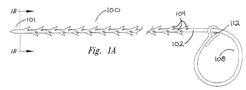
[0030] Fig. 1A and 1C are views of a self-retaining variable loop
suture in
accordance with an embodiment of the present invention.
[0031] Fig. 1B is a cross-sectional view of the suture in Fig. 1A,
taken along the
line in Fig. lA that is labeled "lB".
[0032] Fig. 2 is a view of a self-retaining variable loop suture in
accordance
with another embodiment of the present invention.
[0033] Fig. 3 is a view of a self-retaining variable loop suture in
accordance
with another embodiment of the present invention, having a needle at its
deployment
end.
[0034] Figs. 4A and 4B is a method of using a self-retaining
variable loop
suture in accordance with an embodiment of the present invention.
[0035] Fig. 5A is a view of the variable loop portion of a self-
retaining suture in
accordance with an embodiment of the invention, illustrating a visible
demarcation of
the fixed loop.
can_dms: M1671964\1
9a
CA 2830961 2018-05-09
CA 02830961 2013-09-20
WO 2012/129534 PCT/US2012/030441
[0036] Fig. 5B is a view of the variable loop portion of a self-
retaining suture in
accordance with an embodiment of the invention, illustrating a surface feature
of
embodiment.
100371 Fig. 5C is a view of the variable loop portion of a self-
retaining suture in
accordance with an embodiment of the invention, illustrating a surface feature
of that
embodiment.
100381 Fig. 5D is a view of the variable loop portion of a self-
retaining suture in
accordance with an embodiment of the invention, illustrating a surface feature
of that
embodiment.
[0039] Fig. 6A is a view of the variable loop portion of a self-
retaining suture in
accordance with an embodiment of the invention, illustrating a visible
demarcation of
the fixed loop of that embodiment.
[0040] Fig. 6B is a view of the variable loop portion of a self-
retaining suture in
accordance with another embodiment of the invention, illustrating a grasp
engagement
element of that embodiment.
[0041] Fig. 6C is a view of the variable loop portion of a self-
retaining suture in
accordance with yet another embodiment of the invention, illustrating a grasp
engagement element of that embodiment.
100421 Fig. 7 is a view of the variable loop portion of is a view of the
variable
loop portion of a self-retaining suture in accordance with a further
embodiment of the
invention, illustrating a configuration of a fixed loop of that embodiment.
[0043] Fig. 8 is a view of the variable loop portion of is a view of the
variable
loop portion of a self-retaining suture in accordance with another embodiment
of the
invention, illustrating a configuration of a fixed loop of that embodiment.
[0044] Fig. 9 is a view of the variable loop portion of is a view of the
variable
loop portion of a self-retaining suture in accordance with yet another
embodiment of the
invention, illustrating a configuration of a fixed loop in of that embodiment.
DETAILED DESCRIPTION
Definitions
[0045] Definitions of certain terms that may be used herein include the
following.
CA 02830961 2013-09-20
WO 2012/129534 PCT/US2012/030441
[0046] "Armed suture" refers to a suture having a suture needle at the
suture
deployment end.
[0047] "Braided suture" refers to a suture comprising a multifilamentary
suture
thread. The filaments in such suture threads are typically braided, twisted,
or woven
together.
[0048] "Degradable (also referred to as "biodegradable" or
"bioabsorbable")
suture" refers to a suture which, after introduction into a tissue is broken
down and
absorbed by the body. Typically, the degradation process is at least partially
mediated
by, or performed in, a biological system. "Degradation" refers to a chain
scission
process by which a polymer chain is cleaved into bloomers and monomers. Chain
scission may occur through various mechanisms, including, for example, by
chemical
reaction (e.g., hydrolysis, oxidation/reduction, enzymatic mechanisms or a
combination
or these) or by a thermal or photolytic process. Polymer degradation may be
characterized, for example, using gel permeation chromatography (GPC), which
monitors the polymer molecular mass changes during erosion and breakdown.
Degradable suture material may include polymers such as polyglycolic acid,
copolymers of glycolide and lactide, copolymers of trimethylene carbonate and
glycolide with diethylene glycol (e.g., MAXONTM, Tyco Healthcare Group),
terpolymer composed of glycolide, trimethylene carbonate, and dioxanone (e.g.,
BIOSYNTM [glycolide (60%), trimethylene carbonate (26%), and dioxanone (14%)],
Tyco Healthcare Group), copolymers of glycolide, caprolactone, trimethylene
carbonate, and lactide (e.g., CAPROSYNTM, Tyco Healthcare Group). These
sutures
can be in either a braided multifilament form or a monofilatnent form. The
polymers
used in the present invention can be linear polymers, branched polymers or
multi-axial
polymers. Examples of multi-axial polymers used in sutures are described in
U.S.
Patent Application Publication Nos. 20020161168, 20040024169, and 20040116620.
Sutures made from degradable suture material lose tensile strength as the
material
degrades.
[0049] Medical device" or "implant" refers to any object placed in the
body for
the purpose of restoring physiological function, reducing/alleviating symptoms
associated with disease, and/or repairing/replacing damaged or diseased organs
and
tissues. While normally composed of biologically compatible synthetic
materials (e.g.,
medical-grade stainless steel, titanium and other metals: polymers such as
polyurethane,
11
CA 02830961 2013-09-20
WO 2012/129534 PCT/US2012/030441
silicon, PLA, PLGA and other materials) that are exogenous, some medical
devices and
implants include materials derived from animals (e.g., "xenografts" such as
whole
animal organs; animal tissues such as heart valves; naturally occurring or
chemically-
modified molecules such as collagen, hyaluronic acid, proteins, carbohydrates
and
others), human donors (e.g., "allografts" such as whole organs; tissues such
as bone
grafts, skin grafts and others), or from the patients themselves (e.g.,
"autografts" such as
saphenous vein grafts, skin grafts, tendon/ligament/muscle transplants).
Medical
devices that can be used in procedures in conjunction with the present
invention
include, but are not restricted to, orthopaedic implants (artificial joints,
ligaments and
tendons; screws, plates, and other implantable hardware), dental implants,
intravascular
implants (arterial and venous vascular bypass grafts, hemodialysis access
grafts; both
autologous and synthetic), skin grafts (autologous, synthetic), tubes, drains,
implantable
tissue bulking agents, pumps, shunts, sealants, surgical meshes (e.g., hernia
repair
meshes, tissue scaffolds), fistula treatments, spinal implants (e.g.,
artificial
intervertebral discs, spinal fusion devices, etc.).
[0050] "Monofilament suture" refers to a suture comprising a
monofilamentary
suture thread.
[0051] "Needle attachment" refers to the attachment of a needle to a
suture
requiring same for deployment into tissue, and can include methods such as
crimping,
swaging, using adhesives, and so forth. The point of attachment of the suture
to the
needle is known as the swage.
[0052] "Needle diameter" refers to the diameter of a suture deployment
needle
at the widest point of that needle. While the term "diameter" is often
associated with a
circular periphery, it is to be understood herein to indicate a cross-
sectional dimension
associated with a periphery of any shape. The dimension is the longest
dimension
between two points on the periphery of the shape, i.e., the distance between
the two
points on the periphery that are the furthest from one another.
100531 "Non-degradable (also referred to as "non-absorbable") suture"
refers to
a suture comprising material that is not degraded by chain scission such as
chemical
reaction processes (e.g., hydrolysis, oxidation/reduction, enzymatic
mechanisms or a
combination of these) or by a thermal or photolytic process. Non-degradable
suture
material includes polyamide (also known as nylon, such as nylon 6 and nylon
6.6),
polyester (e.g., polyethylene terephthlate), polytetrafluoroethylene (e.g.,
expanded
12
CA 02830961 2013-09-20
WO 2012/129534 PCT/US2012/030441
polytetrafluoroethylene), polyether-ester such as polybutester (block
copolymer of
butylene terephthalate and polytetra methylene ether glycol), polyurethane,
metal
alloys, metal (e.g., stainless steel wire), polypropylene, polyethelene, silk,
and cotton.
Sutures made of non-degradable suture material are particularly suitable for
applications in which the suture is meant to remain permanently or is meant to
be
physically removed from the body.
[0054] "Retainer configurations" refers to configurations of tissue
retainers and
can include features such as size, shape, surface characteristics, and so
forth. These are
sometimes also referred to as "barb configurations".
[0055] "Self-retaining suture" refers to a suture that does not require
a knot or
anchor on at least one of its ends in order to maintain its position into
which it is
deployed during a surgical procedure. These may be monofilament sutures or
braided
sutures, and are positioned in tissue in two stages, namely deployment and
affixation,
and include at least one tissue retainer.
[0056] "Self-retaining system" refers to a self-retaining suture
together with
means for deploying the suture into tissue. Such deployment means include,
without
limitation, suture needles and other deployment devices as well as
sufficiently rigid and
sharp ends on the suture itself to penetrate tissue.
[0057] "Suture deployment end" refers to an end of the suture to be
deployed
into tissue. A deployment means such as a suture needle may be located at the
suture
deployment end, or the suture thread may be formed into a sufficiently sharp
and rigid
structure so as to penetrate tissue on its own, where this sharp and rigid
structure is
located at the suture deployment end of the suture.
[0058] "Suture diameter" refers to the diameter of the body of the
suture when
viewed in cross-section. While the term "diameter" is often associated with a
circular
periphery, it is to be understood herein to indicate a cross-sectional
dimension (or
distance, or length) associated with a periphery of any shape. For a non-
circular shape,
the diameter is the longest distance between any two points on the periphery
of the
cross section, which may also be referred to as the cross-sectional distance.
The cross-
sectional shape of the suture body or thread is viewed at a location along the
suture
where there are either no barbs, or the barbs that are present are pushed
against the
suture body so that they are flush with the surface of the suture body. In one
embodiment, the suture body or thread has a generally circular cross-sectional
shape.
13
CA 02830961 2013-09-20
WO 2012/129534 PCT/US2012/030441
While the suture body may have a circular or generally circular cross-
sectional shape,
the cross-sectional shape may be non-circular, e.g., it may be polygonal,
e.g., 3-
(triangular), 4-, 5- or 6-sided (hexagonal) sided. The cross section of the
suture body
may have an oval, an ellipsoid, an oblong, or a semi-circular appearance.
Suture sizing
is based upon diameter. United States Pharmacopeia ("USP") designation of
suture size
runs from 0 to 7 in the larger range and 1-0 to 11-0 in the smaller range; in
the smaller
range, the higher the value preceding the hyphenated zero, the smaller the
suture
diameter. Under the USP nomenclature system, the actual diameter of a suture
will
depend on the suture material, so that, by way of example, a suture of size 5-
0 and made
of collagen will have a diameter of 0.15 mm, while sutures having the same USP
size
designation but made of a synthetic absorbable material or a non-absorbable
material
will each have a diameter of 0.1 mm. The selection of suture size for a
particular
purpose depends upon factors such as the nature of the tissue to be sutured
and the
importance of cosmetic concerns; while smaller sutures may be more easily
manipulated through tight surgical sites and are associated with less
scarring, the tensile
strength of a suture manufactured from a given material tends to decrease with
decreasing size. It is to be understood that the sutures and methods of
manufacturing
sutures disclosed herein are suited to a variety of diameters, including
without
limitation 7, 6, 5, 4, 3, 2, 1, 0, 1-0, 2-0, 3-0, 4-0, 5-0, 6-0, 7-0, 8-0, 9-
0, 10-0 and 11-0.
It is to be understood that a variety of suture lengths may be used with the
sutures
described herein.
100591 "Suture needle" refers to needles used to deploy sutures into
tissue,
which come in many different shapes, forms and compositions. There are two
main
types of needles, traumatic needles and atraumatic needles. Traumatic needles
have
channels or drilled ends (that is, holes or eyes) and are supplied separate
from the suture
thread and are threaded on site. Atraumatic needles are eyeless and are
attached to the
suture at the factory by swaging whereby the suture material is inserted into
a channel
at the blunt end of the needle which is then deformed to a final shape to hold
the suture
and needle together. As such, atraumatic needles do not require extra time on
site for
threading and the suture end at the needle attachment site is smaller than the
needle
body. In the traumatic needle the thread comes out of the needle's hole on
both sides
and often the suture rips the tissues to a certain extent as it passes
through. Most
modern sutures are swaged atraumatic needles. Atraumatic needles may be
14
CA 02830961 2013-09-20
WO 2012/129534 PCT/US2012/030441
permanently swaged to the suture or may be designed to come off the suture
with a
sharp straight tug. These "pop-offs" are commonly used for interrupted
sutures, where
each suture is only passed once and then tied. For barbed sutures that are
uninterrupted,
these atraumatic needles would be ideal. Suture needles may also be classified
according to their point geometry. For example, needles may be (i) "tapered"
whereby
the needle body is round and tapers smoothly to a point; (ii) "cutting"
whereby the
needle body is triangular and has sharpened cutting edge on the inside; (iii)
"reverse
cutting" whereby the cutting edge is on the outside; (iv) "trocar point" or
"tapercut"
whereby the needle body is round and tapered, but ends in a small triangular
cutting
point; (v) "blunt" points for sewing friable tissues; (vi) "side cutting" or
"spatula
points" whereby the needle is flat on top and bottom with a cutting edge along
the front
to one side (these are typically used for eye surgery). Suture needles may
also be of
several shapes including, (i) straight, (ii) half curved or ski, (iii) 1/4
circle, (iv) 3/8
circle, (v) 1/2 circle, (vi) 5/8 circle, (v) and compound curve. Suturing
needles are
described, for example, in US Patent Nos. 6,322,581 and 6,214,030 (Mani, Inc.,
Japan);
and 5,464,422 (W.L. Gore, Newark, DE); and 5,941,899; 5,425,746; 5,306,288 and
5,156,615 (US Surgical Corp., Norwalk, CT); and 5,312,422 (Linvatec Corp.,
Largo,
FL); and 7,063,716 (Tyco Healthcare, North Haven, CT). Other suturing needles
are
described, for example, in US Patent Nos. 6,129,741; 5,897,572; 5,676,675; and
5,693,072. The sutures described herein may be deployed with a variety of
needle types
(including without limitation curved, straight, long, short, micro, and so
forth), needle
cutting surfaces (including without limitation, cutting, tapered, and so
forth), and needle
attachment techniques (including without limitation, drilled end, crimped, and
so forth).
Moreover, the sutures described herein may themselves include sufficiently
rigid and
sharp ends so as to dispense with the requirement for deployment needles
altogether.
100601 "Suture thread" refers to the filamentary body component of the
suture,
and, for sutures requiring needle deployment, does not include the suture
needle. The
suture thread may be monofilamentary, i.e., formed of a single filament, or
multifilamentary, i.e., formed from a combination of two or more filaments,
e.g., three
filaments arranged in a braided fashion. The terms "filament" and
"filamentary" are
used in their ordinary sense, to refer to a long thin structure, and therefore
in many
instances herein the suture thread is also identified as the elongated body or
elongated
suture body, where these terms are interchangeable. The filamentous suture
thread has
CA 02830961 2013-09-20
WO 2012/129534 PCT/US2012/030441
a length that is many times its diameter, and in various embodiments the
suture thread
has a length that is at least 5 times, or at least 10 times, or at least 20
times, or at least
30 times, or at least 40 times, or at least 50 times the diameter of the
thread. Indeed, the
length of the suture thread may even be at least 100 times the diameter of the
thread. In
addition to being filamentous, the suture thread is highly flexible. In other
words, the
thread will bend in any direction as the surgeon moves the suture through the
tissue of
the patient. The thread may have some memory of its storage condition, for
example, if
the thread has been stored for a long period of time in a wound-up circular
form, it may
tend to return to that form even after it has been released from its storage
container and
unwound. However, the thread is nevertheless going to follow the needle to
which it is
attached along any path which the needle makes through and around tissue or a
wound.
The thread can therefore be described as flexible, or pliable. Stated another
way, any
two adjacent segments of suture thread may be placed, relative to one another,
at any
angle from essentially or very near to 0 (where the two segments are folded
back upon
one another) to 180 degrees (where the two segments follow in tandem along a
single
straight line). The suture thread has a length, where that length is typically
at least 5
inches, or at least 10 inches, or at least 15 inchers, or at least 20 inches.
The suture
thread will typically have two ends, which may be described as a deployment
end
and/or a trailing end. In such a case, the deployment end of the suture thread
is that end
which will first enter tissue, usually being adjacent to a needle, while the
trailing end of
a suture thread would be that end of the thread which is not the deployment
end.
100611 "Tissue elevation procedure" refers to a surgical procedure for
repositioning tissue from a lower elevation to a higher elevation (i.e. moving
the tissue
in a direction opposite to the direction of gravity). The retaining ligaments
of the face
support facial soft tissue in the normal anatomic position. However, with age,
gravitational effects achieve a downward pull on this tissue and the
underlying
ligaments, and fat descends into the plane between the superficial and deep
facial
fascia, thus allowing facial tissue to sag. Face-lift procedures are designed
to lift these
sagging tissues, and are one example of a more general class of medical
procedure
known as a tissue elevation procedure. More generally, a tissue elevation
procedure
reverses the appearance change that results from gravitation effects over
time, and other
temporal effects that cause tissue to sag, such as genetic effects. It should
be noted that
tissue can also be repositioned without elevation; in some procedures tissues
are
16
CA 02830961 2013-09-20
WO 2012/129534 PCT/US2012/030441
repositioned laterally (away from the midline), medially (towards the midline)
or
inferiorly (lowered) in order to restore symmetry (i.e. repositioned such that
the left and
right sides of the body "match").
100621 "Tissue
retainer", or simply "retainer", refers to a suture element having
a retainer body projecting from the suture body and a retainer end adapted to
penetrate
tissue; an example of a tissue retainer is a barb. Each retainer is adapted to
resist
movement of the suture in a direction other than the direction in which the
suture is
deployed into the tissue by the clinician, by being oriented substantially to
the
deployment direction (that is, they lie flat when pulled in the deployment
direction, and
open or "fan out" when pulled in a direction contrary to the deployment
direction). As
the tissue-penetrating end of each retainer points away from the deployment
direction
when moving through tissue during deployment, the tissue retainers should not
catch or
grab tissue during this phase. Once the self-retaining suture has been
deployed, a force
exerted in another direction (often substantially opposite to the deployment
direction)
causes the retainers to be displaced from their deployment positions (that is,
yielding
toward or resting substantially along the suture body), forces the retainer
ends to open
(or "fan out") from the suture body in a manner that catches and penetrates
into the
surrounding tissue, and results in tissue being caught between the retainer
and the suture
body, thereby "anchoring" or affixing the self retaining suture in place.
100631
"Unidirectional suture" refers to a suture having a deployment end, a
trailing end, and retainers oriented to the deployment end. The trailing end
may be used
to prevent the suture from moving out of the tissue in the direction of
deployment,
either by having a knot tied in it or by being provided with an anchoring
element that
remains outside the point in the tissue into which the deployment end of the
suture was
initially inserted. (In
contrast, a bidirectional suture has retainers oriented in one
direction at one end and retainers oriented in the other direction at the
other end. A
bidirectional suture is typically armed with a needle at each end of the
suture thread.
The bidirectional suture may have a retainer-free transitional segment located
between
the two retainer orientations.
100641 "Wound
closure" refers to a surgical procedure for closing of a wound.
An injury, especially one in which the skin or another external or internal
surface is cut,
torn, pierced, or otherwise broken is known as a wound. A wound commonly
occurs
when the integrity of any tissue is compromised (e.g., skin breaks or burns,
muscle
17
CA 02830961 2013-09-20
WO 2012/129534 PCT/US2012/030441
tears, or bone fractures). A wound may be caused by an act, such as a gunshot,
fall, or
surgical procedure; by an infectious disease; or by an underlying medical
condition. Surgical wound closure facilitates the biological event of healing
by joining,
or closely approximating, the edges of those wounds where the tissue has been
torn, cut,
or otherwise separated. Surgical wound closure directly apposes or
approximates the
tissue layers, which serves to minimize the volume of new tissue formation
required to
bridge the gap between the two edges of the wound. Closure can serve both
functional
and aesthetic purposes. These purposes include elimination of dead space by
approximating the subcutaneous tissues, minimization of scar formation by
careful
epidermal alignment, and avoidance of a depressed scar by precise eversion of
skin
edges.
Unidirectional Self-Retaining Sutures
100651 Self-retaining sutures (including barbed sutures) differ from
conventional sutures in that they possess numerous tiny tissue retainers (such
as barbs)
which anchor into the tissue following deployment and resist movement of the
suture in
a direction opposite to that in which the retainers face, thereby eliminating
the knots
that would otherwise have to be tied, around the deployment end of the suture,
to affix
adjacent tissues together (a "knotless" closure) at the site where the suture
deployment
end exits from the tissue. By eliminating knot tying, associated complications
are
eliminated, including, but not limited to (i) spitting (a condition where the
suture,
usually a knot) pushes through the skin after a subcutaneous closure), (ii)
infection
(bacteria are often able to attach and grow in the spaces created by a knot),
(iii)
bulk/mass (a significant amount of suture material left in a wound is the
portion that
comprises the knot), (iv) slippage (knots can slip or come untied), and (v)
irritation
(knots serve as a bulk "foreign body" in a wound). Suture loops in the tissue
that are
created by knots tied during a surgical procedure may lead to ischemia (they
create
tension points that can strangulate tissue and limit blood flow to the region)
and
increased risk of dehiscence or rupture at the surgical wound. Knot tying is
also labor
intensive and can comprise a significant percentage of the time spent closing
a surgical
wound. Additional operative procedure time is not only bad for the patient
(complication rates rise with time spent under anesthesia), but it also adds
to the overall
cost of the operation (many surgical procedures are estimated to cost between
$15 and
18
CA 02830961 2013-09-20
WO 2012/129534 PCT/US2012/030441
$30 per minute of operating time). Thus, knotless sutures not only allow
patients to
experience an improved clinical outcome, but they also save time and costs
associated
with extended surgeries and follow-up treatments.
[0066] Self-retaining sutures for wound closure also result in better
approximation of the wound edges, evenly distribute the tension along the
length of the
wound (reducing areas of tension that can break or lead to ischemia), decrease
the bulk
of suture material remaining in the wound (by eliminating knots tied during
procedures)
and reduce spitting (the extrusion of suture material ¨ typically knots -
through the
surface of the skin. All of these features are thought to reduce scarring,
improve
cosmesis, and increase wound strength relative to wound closures effected with
plain
sutures or staples.
[0067] Self-retaining sutures also lend themselves to a variety of
specialized
indications; for example, they are suitable for tissue elevation procedures
where tissue
is moved from its previous location and repositioned into a new anatomical
location
(this is typically performed in cosmetic procedures where "drooping" tissue is
elevated
and fixed in a more "youthful" position; or where "out-of-position" tissue is
moved
back to its correct anatomical location). Such procedures include facelifts,
brow lifts,
breast lifts, buttocks lifts, and so forth.
[0068] Unidirectional self-retaining sutures and their uses have been
described
in various publications mentioned above. While the segment of suture thread
adjacent
to the deployment end of a unidirectional self-retaining suture is provided
with tissue
retainers for preventing slippage of the suture in a direction substantially
opposite the
direction of deployment, the trailing end may be provided with an anchor to
prevent
slippage in the deployment direction (and in order to avoid the undesirable
potential
effects of requiring a knot to be tied during a surgical procedure in the
trailing end of a
unidirectional suture). Various unidirectional sutures with anchors, included
anchors
having loop elements, have been described in, for example, U.S. Patent
Application
Publication Nos. 20050267531, 20040060410, 20080255611, and 20100063540.
[0069] Several problems common to existing unidirectional self-retaining
sutures having loop anchors can be addressed by the embodiments of this
invention.
For example, unidirectional sutures featuring fixed loop anchors, such as
those
described in some of the aforementioned publications, have several
disadvantages, the
first of which is that the size of the fixed loop should typically be fairly
small (that is,
19
CA 02830961 2013-09-20
WO 2012/129534 PCT/US2012/030441
not much bigger than the size of the first stitch that the clinician would
wish to make
with it), which requires the clinician to make some effort (and therefore
expend some
valuable surgical time) in finding the loop and running the deployment end of
the suture
through it. Because the suture of the present invention includes a variable
loop anchor,
the clinician is presented with a large loop through which he or she can
easily pass the
deployment end of the suture; this is of particular benefit in laparoscopic
procedures.
Then, when such a suture is pulled through tissue, if the first stitch taken
is larger than
the longest interior dimension of the fixed loop after the suture body has
been drawn
through it and tensioned, then the base of the loop (that is, where the loop
joins the
suture body) can be pulled into the tissue, resulting in potential issues such
as those
described above in connection with knot-tying. On the other hand, if the first
stitch
taken is smaller than the longest interior dimension of the fixed loop after
the suture
body has been drawn through it and tensioned, then excess loop material
remains at the
tissue site, an axiomatically undesirable condition which could also cause
surgical
instruments to get caught on the excess material during the procedure. In the
case of
the present invention, the adjustable nature of the variable loop anchor
allows the
clinician to avoid these difficulties.
100701 In addition, there are physical issues of loop integrity
associated with a
fixed loop anchor. For example, issues of fixed loop attachment are avoided by
the
variable loop suture of the present invention. Where the loop of a fixed loop
suture is
welded or otherwise attached to the suture body, either as a separate
structure joined at
its base to the suture or as an end of the suture turned back onto and
attached to the
suture to create a looped portion, the base of the loop (where it joins the
suture body) is
the attachment region and is also where the suture is pulled into the tissue.
As such, it
is subject to tissue drag and the potential for breakage or peeling at the
attachment
region. While this may be dealt with by increasing the length of the
attachment region
and/or providing a taper or chamfer, it is avoided entirely by sutures of the
present
invention as the eyelet of the variable loop will sit superficial to the
tissue being
approximated, will not need to pass into the tissue, and is not subject to
tissue drag. In
addition, for sutures of the present invention, the main load when tensioning
the tissue
is taken by the variable loop as opposed to the eyelet. As a result, the
eyelet does not
hold the primary tension when seating the first stitch, and the weld length
can be
CA 02830961 2013-09-20
WO 2012/129534 PCT/US2012/030441
shortened thereby reducing local biomaterial effects (inflammation and/or risk
of
infection) on wound healing.
[0071] Unidirectional self-retaining sutures of the present invention
are
provided with a variable length loop configuration at one end and a deployment
end at
the other. Wound closure is achieved by starting at one end of the wound
containing
tissue to be approximated, passing the deployment end through both edges of
the tissue,
pulling the end of the suture containing the needle through the tissue until
the loop
segment is near the first edge of tissue, and passing the end with the needle
back
through the variable loop portion of the device. Tension is pulled until the
loop seats on
the tissue and the desired hold is achieved. The deployment end is now passed
repeatedly through the tissue in a pattern determined by the clinician to best
facilitate
wound closure starting at the end just seated moving in one direction toward
the other
terminus of the tissue to be approximated. A "J stitch" can used to complete
the process
and the needle is removed akin to the procedure used with bi-directional
configurations.
[0072] Turning now to FIGS 1A, 1B and 1C, there is illustrated a suture
100
having a deployment end 101 on an elongated body 102 which is alternatively
referred
to herein as the suture thread, which body (or thread) has a transverse cross-
sectional
length (the longest transverse dimension on the cross section). This
transverse cross-
sectional length is illustrated in FIG. 1B, where Fig. 1B is a cross-sectional
view of the
suture of Fig. 1A, taken along the line in Fig. 1A that is labeled "lB" where
this
transverse cross-sectional length is denoted by "tl" in FIG. lb, and FIG 1B
also shows
the cross section of the suture body 102 and three retainers each identified
as 104 which
are shown with different darkening to make the point that they not are the
same distance
away from the viewer located at position 1B. It is to be understood that the
cross-
sectional shape of the suture is not limited to circular, but may be non-
circular as well
(such as an ellipse, a triangle, a square, other polygons, etc.).
[0073] Continuing with FIGS. lA to 1C, the body 102 bears a plurality of
retainers 104 oriented toward the deployment end 101, and an eyelet 112
through which
the suture body passes, thus forming the variable loop 108. The eyelet is in
essence a
fixed loop, but one that, in tissue, sits outside the tissue into which the
suture is
deployed. The presence of the variable loop as part of the anchoring structure
assures
that all of the force exerted on the anchor is not solely exerted on the fixed
loop. This is
advantageous because the force is therefore distributed over a broader
structure, and the
21
CA 02830961 2013-09-20
WO 2012/129534 PCT/US2012/030441
fixed loop (eyelet) or portions thereof, e.g., the attachment region as
discussed later
herein, are not drawn into the tissue. One benefit is that the anchor can more
readily be
accessed and then cut away from the suture thread, allowing for greater ease
in
removing the suture thread after it is deemed that the healing process no
longer require
the presence of the suture. Absent the variable loop, the anchor would be
composed
solely of the eyelet or the eyelet in combination with a portion of the suture
thread that
passes through the eyelet but does not form a variable loop. An anchor that is
formed
solely of a fixed loop, or is formed from a fixed loop in combination with a
suture
thread passing through the fixed loop but not forming a variable loop, is
observed to
pinch the tissue at the anchoring point and may lead to undesirable side
effects, for
example, tissue necrosis. An anchor formed from a fixed loop (eyelet) and a
variable
loop and a portion of the elongated body (suture thread) provides for less
pinching of
the tissue and thus less opportunity for undesired tissue necrosis.
100741 As shown in FIG. 1C, the deployment end 101 can be passed through
the
variable loop 108. As the deployment end 101 continues to be drawn through the
variable loop 108, and tension is applied to the suture thread 102 from the
direction of
the deployment end 101, more and more of the suture thread 102 will be drawn
or
threaded through the variable loop. In practice, the deployment end 101 will
pass
through a patient's tissue before it passes through the variable loop 108, and
therefore
as the suture thread 102 passes through the variable loop 108, the eyelet 112
will be
pulled toward the surface of the patient's tissue and will eventually be held
firmly on
that tissue by the tension or force exerted on the deployment end 101. With
continued
pulling or force or tension, the circumference of the variable loop will tend
to decrease,
until such time as the clinician determines that the variable loop has a
desired
circumference, at which time the clinician will stop pulling on the deployment
end and
the anchor is thus formed. Thereafter, the clinician will return to sewing the
patient's
tissue with the now-anchored suture.
100751 As shown in FIG. 1A and 1C, the region of the suture body along
which
the plurality of retainers is provided may be greater than the region of the
suture body
that is used in forming an eyelet. Furthermore, the eyelet does not
necessarily contain
any retainers, although retainers of a sort may be present in order to assist
in gripping
the suture body within the eyelet. Retainers may be absent from the variable
loop
22
CA 02830961 2013-09-20
WO 2012/129534 PCT/US2012/030441
portion of the suture, as shown in FIG. lA and 1C, or retainers may be present
in this
portion of the suture as shown in later figures provided herein.
[0076] As can be seen in FIG. 2, suture 200 has retainers 204 on suture
body
202, which run along most of the length of the suture body 202 including the
variable
loop 208 formed from the suture body 202, to approach eyelet 212. As retainers
204
are oriented toward the deployment end 201 of the suture 200, eyelet 212
passes easily
over the suture thread 202 and retainers 204 located thereon when the suture
is pulled
through eyelet 212 (or eyelet 212 is pulled over suture body 202) in the
direction of
deployment to decrease the circumference of variable loop 208.
[0077] As illustrated by comparison of FIG. 2 and FIG. 3, the deployment
end
of a suture 200 and 300, respectively, may be pointed. As shown in FIG. 2, the
deployment end 201 may be pointed by converting the end of the suture body 202
into a
sharp and rigid structure. Or, as illustrated in FIG. 3, the deployment end
301 may
become pointed due to the attachment of a needle 303 to a terminus of the
suture thread
302. FIG. 3 shows needle 303 at the deployment end 301 of variable loop suture
300.
[0078] In one embodiment, the invention provides a self-retaining system
comprising a self-retaining suture as described herein including a deployment
means.
The self-retaining suture comprises a suture thread with a plurality of tissue
retainers
and one or more (usually only one is necessary) eyelets. The eyelet may be
formed into
a circular or generally circular shape, and in this shape the diameter of the
eyelet can be
measured in the usual way as the distance between any two opposing points (two
points
on opposite sides of the circle) on the inside of the eyelet. The needle
diameter may be
selected in view of the eyelet diameter. For example, the needle diameter may
be larger
than the eyelet diameter, for example, the needle diameter may be at least 5%
greater,
or at least 10% greater, or at least 15% greater or at least 20% greater than
the eyelet
diameter. In this example, a fixed loop is formed when the deployment end of
the
suture body passes through the eyelet and then the deployment end of the
suture is
attached to a suture needle. Since the suture needle has a diameter that is
greater than
the eyelet diameter, the deployment end of the suture cannot be taken back
through the
eyelet without breaking the eyelet and/or the needle, unless the eyelet is
made of a
flexible material which can stretch. The suture body of the invention
typically does not
stretch to any appreciable extent. Thus, the loop may be seen as being a fixed
loop. In
another example, the needle diameter is approximately the same as the eyelet
diameter,
23
CA 02830961 2013-09-20
WO 2012/129534 PCT/US2012/030441
in other words, the needle diameter is plus/minus 5% of the eyelet diameter,
or in
another embodiment, plus/minus 10% of the eyelet diameter. In this case, the
needle
diameter and the eyelet diameter are approximately the same, and it will be
difficult or
impossible to pull the deployment end of the suture back through the eyelet,
after the
deployment end has been attached to a needle. In another example, the needle
diameter
is chosen to be less than the eyelet diameter, such as where the needle
diameter is less
than 90% of the eyelet diameter, or less than 80%, or less than 70% or less
than 60% or
less than 50% of the eyelet diameter. In this case, the deployment end may be
attached
to a needle, and then the needle may be threaded through the eyelet. This
option
provides greater flexibility in forming the variable looped suture.
[0079] The needle diameter is typically chosen to be at least the same
as the
suture diameter, and in various embodiments the needle diameter is at least
110%, or at
least 120%, or at least 130%, or at least 140%, or at least 150% of the suture
thread
diameter.
[0080] Use of self-retaining variable loop sutures of the present
invention is
illustrated in FIGS. 4A and 4B. In those drawings, suture 400 is drawn in a
first stitch
through tissue (indicated as a hatched region, "T"), and then elongated body
402 is
drawn through the variable loop 408. When suture 400 is then pulled in the
direction of
the deployment end 401 (indicated with an arrow), suture body 402 continues to
pass
through loop 408, tensioning the variable loop and decreasing its size as it
passes
through eyelet 412. The suture is thus anchored and ready for continued
deployment
through tissue.
[0081] In one embodiment, the invention provides a method of suturing,
where
this method comprises: (a) providing a self-retaining system comprising a
suture needle
attached at an end of a self-retaining suture, the self-retaining suture
comprising a
suture thread having a thread diameter, a plurality of tissue retainers and an
eyelet,
where the suture thread passes through the eyelet to form a variable loop
having an
original diameter; (b) inserting the needle into the tissue of a patient at a
first tissue
location; (c) withdrawing the needle from the tissue of the patient at a
second tissue
location; (d) passing the needle and at least some of the suture thread
through the
variable loop; and (e) inserting the needle into the tissue of the patient at
a third tissue
location. Optionally, one or more of the following statements may be used in
combination with a statement providing a method of suturing as provided
herein: the
24
CA 02830961 2013-09-20
WO 2012/129534 PCT/US2012/030441
suture thread is passed through the variable loop while simultaneously the
diameter of
the variable loop is decreased where optionally the decrease is greater than
50% of the
original variable loop diameter; the diameter of the variable loop is
decreased to
provide a variable loop diameter that is less than 10 times the thread
diameter; the
diameter of the variable loop is decreased until the variable loop fits snugly
around the
suture thread; the suture thread is passed through the variable loop until the
eyelet, the
variable loop and the suture body together form an anchor on the tissue, and
where
further movement of the suture thread in the direction of the suture needle is
resisted by
the anchor.
100821 In another embodiment, the invention provides a method of
suturing
tissue, the method comprising: (a) providing a suture thread attached to a
suture needle,
a portion of the suture thread forming a loop having an adjustable
circumference; (b)
threading the needle through the loop; and (c) deploying the needle through
tissue of a
patient and approximating the tissue with the suture thread. Optionally, one
or more of
the following statements may be used in combination with a statement providing
a
method of suturing as provided herein: the loop comprises suture thread and a
fixed
loop (also referred to as an eyelet), the fixed loop having an opening through
which the
suture thread passes to thereby form the loop having an adjustable
circumference; the
fixed loop (also referred to herein as the eyelet) and any means by which the
fixed loop
is formed or attached to the suture thread, all lie on a surface of the tissue
after the
tissue has been completely approximated; the circumference of the variable
loop is
adjusted to a desired value prior to threading the deployment end or needle
through the
variable loop; the circumference of the variable loop is adjusted to a desired
value after
threading the deployment end or needle through the variable loop; the
circumference of
the loop is adjusted to a desired value in the range, where that desired range
may be 0.5
to 3 inches or 0.5 to 2 inches, or 0.5 to 1 inch, depending on the custom of
the clinician
and the nature of the wound that is being sewn; the circumference of the loop
is reduced
to a desired value, i.e., a value desired by the clinician as appropriate for
his or her
comfort and the wound being sewn: the needle is passed into and then out of
tissue at
first and second locations, respectively, prior to being threaded through the
variable
loop; the suture thread comprises tissue retainers; the suture thread
comprises cuts in
the suture thread, the cuts forming the tissue retainers, in other words, the
cut provides a
separation between suture thread material on either side of the cut, where the
portion of
CA 02830961 2013-09-20
WO 2012/129534 PCT/US2012/030441
suture thread material nearer the periphery of the suture thread may be pulled
up and
away from the suture thread on the other side of the cut, to thereby form a
structure
which is a tissue retainer; a cut made in the suture thread lies in a single
plane, or in two
planes such as where the angle of the cut is changed during the process of
forming the
cut in the suture thread, e.g., the first cut into the suture is relatively
deep while the cut
after the first cut is not (or not very) deep; a cut is made into the suture
thread so as to
provide a barb; tissue retainers are present on that portion of the suture
thread that
forms the loop having an adjustable circumference; tissue retainers are absent
from a
portion of the loop having an adjustable circumference.
100831 In another embodiment, the invention provide a method of suturing
that
includes forming an anchor at a location on tissue of a patient, the method
comprising:
(a) providing a suture thread with an eyelet, the suture thread attached to a
suture needle
at a deployment end of the suture thread; (b) deploying the suture needle into
tissue at
the location, and then withdrawing the suture needle from tissue at an exit
point; (c)
passing the needle through a loop comprising suture thread, the loop having a
variable
circumference; (d) applying tension to the suture thread by pulling on the
deployment
end of the suture thread; (e) to thereby provide an anchor on top of the
tissue, the
anchor comprising the eyelet, the loop and a portion of the suture thread, the
anchor
resisting movement of the suture thread in the direction of the deployment end
of the
suture thread. Optionally, one or more of the following statements may be used
in
combination with a statement providing a method of suturing as provided
herein: the
loop comprises suture thread and a fixed loop (also referred to as an eyelet),
the fixed
loop having an opening through which the suture thread passes to thereby form
the loop
having an adjustable circumference; the fixed loop (also referred to herein as
the eyelet)
and any means by which the fixed loop is formed or attached to the suture
thread, all lie
on a surface of the tissue after the tissue has been completely approximated;
the
circumference of the variable loop is adjusted to a desired value prior to
threading the
deployment end or needle through the variable loop; the circumference of the
variable
loop is adjusted to a desired value after threading the deployment end or
needle through
the variable loop; the circumference of the loop is adjusted to a desired
value in the
range, where that desired range may be 0.5 to 3 inches or 0.5 to 2 inches, or
0.5 to 1
inch, or at least 0.5 inches, or at least 1 inch, or at least 1.5 inches,
depending on the
custom of the clinician and the nature of the wound that is being sewn; the
26
CA 02830961 2013-09-20
WO 2012/129534 PCT/US2012/030441
circumference of the loop is reduced to a desired value, i.e., a value desired
by the
clinician as appropriate for his or her comfort and the wound being sewn; the
needle is
passed into and then out of tissue at first and second locations,
respectively, prior to
being threaded through the variable loop; the suture thread comprises tissue
retainers;
the suture thread comprises cuts in the suture thread, the cuts forming the
tissue
retainers, in other words, the cut provides a separation between suture thread
material
on either side of the cut, where the portion of suture thread material nearer
the periphery
of the suture thread may be pulled up and away from the suture thread on the
other side
of the cut, to thereby form a structure which is a tissue retainer; a cut made
in the suture
thread lies in a single plane, or in two planes such as where the angle of the
cut is
changed during the process of forming the cut in the suture thread, e.g., the
first cut into
the suture is relatively deep while the cut after the first cut is not (or not
very) deep; a
cut is made into the suture thread so as to provide a barb; tissue retainers
are present on
that portion of the suture thread that forms the loop having an adjustable
circumference;
tissue retainers are absent from a portion of the loop having an adjustable
circumference.
[0084] To serve the purpose of allowing a clinician to identify and
differentiate
the eyelet, a market may be placed in the vicinity of the eyelet. The marker
should be
readily recognized and distinguished by the physician under the conditions in
which the
suture is to be used. For example, in microsurgery applications, markers may
be used
that are visible under the microscope, but not necessarily visible to the
naked eye.
Likewise in endoscopic applications, markers should used be that are visible
through
the endoscope and associated display system. If the suture will be used with
fluoroscopic visualization then the markers may include radiopaque markers. If
the
suture will be used with ultrasound visualization then the markers may include
echogenic markers. Thus, different markers and different types of markers may
be
appropriate under different circumstances depending upon the circumstances of
the
procedure and the scanning/imaging/visualization technology utilized in the
procedure.
100851 Markers can include different colors such as red, green, orange,
yellow,
green, blue etc. In some cases it may be desirable to use a color for markers
that is
uncommon in the operative environment. For example, it may be desirable to use
green
markers because green is not common in the human body. In endoscopic
applications
using green is advantageous because the video system can be programmed to
emphasize
27
CA 02830961 2013-09-20
WO 2012/129534 PCT/US2012/030441
green and enhance marker visualization without interfering with the remainder
of the
image.
[0086] The markers can be formed by various conventional methods. For
example, the markers can be coated, sprayed, glued, dyed, stained, or
otherwise affixed
to the self-retaining suture systems or components thereof. Traditional
colourant
application processes include, without limitation, dipping, spraying (by, for
example, an
ink jet), painting, printing, applying and/or coating colourants on the suture
section of
interest. Critical fluid extraction (such as carbon oxide) may also be used to
add
colourant locally to all or part of the section desired to be marked.
Alternatively,
colourant(s) for the suture section of interest may be included in a portion
of the suture
material that is used to form the suture body, wherein that portion is in the
section of
interest of the manufactured suture.
[0087] Additionally, the suture section of interest can be demarcated by
using
an energy-activated colourant. For example, when a laser-activated colourant
(that is, a
pigment or dye which permanently changes colour after being exposed to laser
energy)
is used to colour the suture, then the eyelet or other suture section of
interest can be
demarcated by using laser energy to permanently change the suture coating in
the suture
section of interest. This also applies to using other energy activated
colourants which
are activated by other energy sources such as, but not limited to, heat,
chemicals,
microwaves, ultraviolet light, or x-rays. For example, bleaching chemicals
such as
sodium hypochlorite or hydrogen peroxide will permanently change the
colourant's
colour which allows for the demarcation of the eyelet or other region of the
suture.
[0088] Additionally, the colourant(s) employed for demarcating the
suture
section of interest may be included on a plastic biocotnpatible material which
is applied
on the suture at the section of interest. Such a layer may be absorbable, such
as a
polyglycolide coating which has a colourant to mark the suture section of
interest, or it
may be a non-absorbable material, such silicone. The coloured material may be
synthetic or may be derived from a natural source (whether the material be
modified or
unmodified), such as collagen. The plastic biocompatible material may be
applied to
the suture before or after the retainers are formed on the suture body.
[0089] Alternatively, the eyelet or other suture region may be reverse-
marked,
such that where the suture body is already visibly coloured, the colourant may
be absent
from all or part of the suture section of interest such that at least a
portion of the section
28
CA 02830961 2013-09-20
WO 2012/129534 PCT/US2012/030441
of interest is optically distinguishable by the surgeon from the rest of the
suture. Such a
suture may manufactured by including a colourant-free portion of suture
material in the
suture section of interest area during the manufacture of the suture body (for
example,
by extrusion) or by removal of colourant from the suture section of interest
after the
suture body has been manufactured, whether before or after retainers have been
formed
on the suture body. Colourant may be removed locally by, for example, critical
fluid
extraction such as (e.g., carbon oxide). It is not necessary to remove all of
the colourant
from the section of interest of the suture as long as there is a difference
detectible by a
surgeon between the section of interest and the rest of the suture.
[0090] Another example of a reverse-marked suture is one that lacks a
coloured
layer that is present on the rest of the suture body. A plastic biocompatible
material
bearing a colourant may be applied on the other sections of the suture, and at
least
where the other sections border the section of interest. Examples of such
materials are
discussed above. As in the foregoing examples, demarcating the suture section
of
interest may be effected in the suture manufacturing process either before or
after
forming retainers.
[0091] Another example of a reverse-marked suture is one having a
coaxial
structure wherein each coaxial layer having a different colour, and a portion
of the
outermost layer(s) is removed to visually expose a layer below. For example, a
dual-
layer monofilament polypropylene suture can be produced with a white inner
core
(intercoaxial layer) with a blue outer coaxial layer, and portions of the
outer layer can
be removed to visually expose the white inner monofilament to mark the suture
section
of interest.
[0092] Yet another example of a reverse-marked suture is one in which an
external coating is removed (or partially removed) from the suture in the
suture section
of interest, and where either the coating or base suture has a contrasting
colour
difference. This technique of removing (or partially removing) material in the
suture
section of interest may also create a tactile demarcation of the suture
section of interest.
[0093] The marking may include a radio-detectable compound or magnetic
resonance imaging detectable compound. For example the suture section of
interest
provided with barium sulfate (BaSO4), such as by impregnating the suture with
barium
sulfate or adding a coating containing barium sulfate, will be detectable by
electromagnetic energy. In the case of x-ray detection, the barium sulfate
marked
29
CA 02830961 2013-09-20
WO 2012/129534 PCT/US2012/030441
section of interest would be radiopaque. Likewise, computed tomography (CT)
scans
or computed axial tomography (CAT) scans can be used to detect the radio
detectable
section of interest. The use of electromagnetic energy for radio detection of
the
transition section is not limited to using x-ray wavelengths as other radio
frequencies
may be used. Likewise, gadolinium (Gd) or gadolinium compounds can be used for
the
marking of the suture section of interest especially when the detection will
be done by
using magnetic resonance imaging (MRI). The use of radio detectable or
magnetic
resonance imaging detectable marking may be useful to the surgeon during
laparoscopic surgical procedures.
100941 The region of the variable loop at the base of the eyelet may be
marked
to increase its visibility to the clinician, either for the purpose of
indicating where it is
during suture deployment, or to allow the clinician to identify where the
anchor is in
order to sever it for the purpose of removing the deployed suture from the
tissue. FIGS.
5A, 5B, 5C, and 5D illustrate the trailing ends of variable loop sutures
having such
markings. In FIG. 5A, variable loop 508 of suture 500 includes eyelet 512,
which is
marked at its base 514. Suture 520 of FIG. 5B includes a variable loop 528
having
demarcation 534 near eyelet 532 in addition to a surface feature 536 adjacent
to the
eyelet base 534 where feature 536 provides a different tactile feeling to the
suture 520
compared to other locations on the suture, while suture 540 of FIG. 5C has a
variable
loop 548 having demarcation 554 near eyelet 552, and suture 560 of FIG. 5D
shows a
variable loop 568 having two demarcations 534 near eyelet 572 and demarcation
576
located elsewhere on the variable loop 568.
100951 In some embodiments, the suture may additionally be provided with
a
surface feature for some portion of the suture body adjacent to the fixed
loop, in order
to provide some resistance, perceptible to the clinician, to the variable loop
being pulled
all the way to the fixed loop. While in some of these embodiments it is not
necessary
for the surface feature to prevent the variable loop from being tightened all
the way to
the fixed loop, in yet other embodiments the surface feature may function to
do just that.
Some examples, without limitation, of suitable surface features are dimpling,
rippling,
corrugation, roughening, serrations, ridges, filaments, so forth. In this
connection, FIG.
5B includes surface feature 536 near eyelet 532. Of course, these surface
features may
also take the form of a short segment retainers oriented away from the suture
deployment end, which would resist the movement of the suture body through the
CA 02830961 2013-09-20
WO 2012/129534 PCT/US2012/030441
eyelet, The latter may be of the same pattern as the balance of the suture or
may be
different, i.e., a short segment of retainers having the same or different
configuration as
the plurality of barbs oriented to the deployment end.
100961 In FIG 5C, retainers 544 oriented toward the deployment end of
the
suture pass easily through eyelet 552, while retainers 558 are oriented in the
opposite
direction and so somewhat resist having the eyelet drawn over them. Similarly,
suture
560 of FIG. 5D is provided with retainers 564 oriented toward the deployment
and
retainers 578, which are oppositely oriented. Suture 560 is also provided with
an
additional marking 576 in between the two sets of retainers, in order to allow
the
clinician to identify the region between the two sets.
100971 Referring now to FIG 6A, this provides a view of the variable
loop
portion 608 of a self-retaining suture 600 in accordance with an embodiment of
the
invention, illustrating a visible demarcation 614 of the fixed loop of that
embodiment.
In other words, the fixed loop (alternatively referred to as the eyelet) has a
different
surface appearance or a different surface tactile feel in comparison to the
adjoining
suture thread. In FIG. 6A, the eyelet 612 is constructed from a darker colored
material
than the material that forms the adjoining suture thread 602. Alternatively,
the eyelet
could be formed from a lighter colored material, or a totally different
colored material
than the adjoining suture thread. The eyelet might be formed of material
having a matt
finish while the adjoining suture thread had a natural or shiny appearance.
The eyelet
may also, or alternatively, be formed of materials that contain grooves or
other
indentations or ridges that are not present on the adjoining suture thread,
which
provides a tactile distinction between the eyelet region and the adjoining
suture thread.
With this visible demarcation between the eyelet and the adjoining suture
thread, the
clinician who uses the suture, or perhaps a robot that handles the suture, can
more
readily see the eyelet and distinguish the eyelet from the adjoining suture
thread.
100981 Referring now to FIGS. 6B and 6C, the eyelet may be provided with
or
be in combination with a grasp engagement element, to facilitate the removal
of the
anchor from the suture if it is decided that the suture is to be removed from
the tissue.
As shown in FIG 6B, suture 620 having variable loop 628 with an eyelet 632 and
an
attachment region 634 has a tab 636 as the grasp engagement element, while
suture 640
of FIG. 6C includes a length of unbarbed suture material 656 as a grasp
engagement
element located on or as part of the integrally-formed eyelet 652 of variable
loop 648.
31
CA 02830961 2013-09-20
WO 2012/129534 PCT/US2012/030441
Grasp engagement elements 636 and 656 illustrate two options for a feature
that is part
of, or is attached to the eyelet and is of a size and orientation which can
readily be
grasped or grabbed or gripped or held by the health care professional as a
means of
lifting the eyelet and associated anchor away from the tissue. A grasp
engagement
element is an optional feature of each of the variable loop sutures described
herein.
100991 The grasp engagement element can be made from either or both
absorbable or non-absorbable materials. For example, a non-absorbable grasp
engagement element made from polyester felt or polytetrafluoroethylene felt
can be
used to allow the surgeon to find and gently pull the eyelet to facilitate
removal of the
anchor from the suture body if and when it is desired to remove the suture
from tissue
(by detaching the anchor and then pulling the suture out from its deployment
end).
Examples of absorbable materials include glycolide and glycolide-lactide
polymers.
The use of an absorbable grasp engagement element can be especially useful for
deep
cavity tissue closures where the surgeon may wish to have a choice of whether
or not to
leave the grasp engagement element inside the body. Additionally, the grasp
engagement element can be coloured (as shown by the shading of tab 636 of FIG.
6B)
to improve the visibility of the element. This includes, but is not limited
to, using
fluorescent colourants, radio detectable compounds, or magnetic resonance
imaging
detectable compounds.
Manufacturing and Materials
[00100] Suture threads described herein may be produced by any suitable
method, including without limitation injection moulding, stamping, cutting,
laser,
extrusion, and so forth. With respect to cutting, polymeric thread or
filaments may be
manufactured or purchased for the suture body, and the retainers can be
subsequently
cut onto or into the suture body; they may be hand-cut, laser-cut, or
mechanically
machine-cut using blades, cutting wheels, grinding wheels, and so forth. The
sutures
may be made of any suitable biocompatible material, and may be further treated
with
any suitable biocompatible material, whether to enhance the sutures' strength,
resilience, longevity, or other qualities, or to equip the sutures to fulfill
additional
functions besides joining tissues together, repositioning tissues, or
attaching foreign
elements to tissues. As are appropriate to the indication or use of the suture
in question,
the sutures may be provided with retainers of various configurations,
arrangements,
32
CA 02830961 2013-09-20
WO 2012/129534 PCT/US2012/030441
densities, and so forth, such as those taught in publications referenced
herein.
[00101] Variable loops for sutures of the present invention may be formed
in
several ways. Referring to the suture 700 shown in FIG. 7, variable loop 708
includes
eyelet 712 through which suture body 702 passes. Eyelet 712 is formed by
passing
what would otherwise be the non-deployment terminus 791 of suture body 702
around
and back upon itself and attaching it externally to the suture body 702 at
attachment
region 714. In this case, the attachment region 714 is adjacent to the eyelet
712. In
contrast, and referring to the suture 800 shown in FIG. 8 which illustrates
eyelet 812 of
variable loop 808 formed from the trailing end (alternatively referred to as
the non-
deployment end) 891 of suture body 802 on an internal attachment, that is, the
attachment region 814 is inside and is part of the structure that forms the
eyelet. The
attachments mentioned in reference to these figures may be made to have a
permanent
nature by welding, gluing, and so forth.
[00102] Noteworthy is that the non-deployment end of the suture thread
need not
have any particular shape or appearance or function when it is located in the
attachment
region. Since neither the eyelet nor the adjacent attachment region of the
variable loop
sutures of the present invention are intended to enter tissue, there is no
need to facilitate
the entry of, e.g., the attachment region by providing any particular shape
that would
facilitate such entry. For example, the end or tip of the non-deployment end
of the
sutures of the present invention may be squared off compared to the sides of
the
adjacent suture thread, rather than having an angle or slant that would
facilitate entry
into tissue. The deployment end may, in fact, be designed or adapted to retard
entry of
the attachment region into tissue. This is particularly relevant when the
eyelet is
formed by having the attachment region located adjacent to the eyelet. This
same issue
is not relevant if the attachment region is part of the material that forms
the eyelet.
[00103] Alternatively, the eyelet 912 may be integral to the suture body
902 as is
shown by the suture 900 illustrated in FIG. 9, in which eyelet 912 of variable
loop 908
is continuous with suture body 902. Thus, in this embodiment, there is no
attachment
region.
[00104] As a further alternative embodiment, the eyelet may be formed
independently from the suture thread, and then the eyelet is joined to the
suture thread
at a suitable location. For example, the non-deployment end of the suture
thread may
be tied to an eyelet, in the same manner as the end of a rope can be tied to a
ring. In
33
CA 02830961 2013-09-20
WO 2012/129534 PCT/US2012/030441
this alternative embodiment, it is desirable that the joining together of the
suture thread
and the eyelet be done in a secure manner, so that the eyelet does not become
separated
from the suture thread at an inopportune time. A secure joining of the eyelet
and the
suture thread may be accomplished, for example, by welding the knot which is
used to
tie the eyelet to the suture thread. As another example, the eyelet may take
the form of
a ring (or polygon or other suitable shape defining a hole through which the
suture
thread may pass or be threaded) where the ring has a hole through which the
suture
thread may pass in order to secure the ring to the suture thread. For
instance, after
passing through this hole, the non-deployment end of the suture thread may be
formed
into a knot of such a size that it cannot easily pass through the hole. In
this way, the
suture thread is secured to an eyelet. As a final example, the eyelet may be
securely
joined to a feature where the feature is readily secured to the suture thread.
For
instance, two rings joined in the shape of a figure 8, the two rings not
necessarily being
of the same diameter, provides a structure where the suture thread may be tied
to one
ring, while the adjoining ring is available to serve as the eyelet. Rather
than having the
eyelet joined to a ring, the eyelet may be joined to a tab which can be folded
and
crimped around the suture thread. The eyelet might alternatively be joined to
a hollow
cylinder, where the non-deployment end of the suture thread may be inserted
into the
cylinder, and then the cylinder is crimped to secure the suture thread within
the cylinder
(in a like manner to the well known way in which the deployment end of a
suture is
swaged to a needle, but in this case the non-deployment end of a suture thread
would be
swaged to an eyelet). Having the eyelet formed separately from the suture
thread
provides for greater flexibility (i.e., independence) in selecting materials
from which to
form the suture thread and the eyelet.
[00105] The dimensions of the eyelet may be varied; for example, the
inner
transverse length of the eyelet (that is, the longest inner dimension across
the eyelet)
may be as small as about the transverse length of the suture body cross
section (that is,
the longest dimension across the suture body cross section, regardless of the
shape of
the cross section) and as large as about four times or even ten times the
transverse
length of the suture body cross section. Other ranges for the inner transverse
length that
may be suitable are one-and-a-half times the transverse length of the suture
cross
section to about ten times transverse length of the suture cross section, one-
and-a-half
times the transverse length of the suture cross section to about four times
transverse
34
CA 02830961 2013-09-20
WO 2012/129534 PCT/US2012/030441
length of the suture cross section, or about twice the transverse length of
the suture
cross section to about three times the transverse length of the suture cross
section.
[00106] After preparation, the self-retaining suture system may be
packaged for
ease of storage, handling and use. Suitable packaging systems are known in the
art,
where exemplary suture packages are described in U.S Patent Publication Nos.
20110056859 and 20100230300. Before, but preferably after the packaging
process,
the suture may be sterilized by, e.g., radiation.
[00107] In one embodiment there is provided a method of making a self-
retaining
suture system, the method comprising (a) providing a suture thread, the suture
thread
comprising a deployment end and either comprising or being attached to an
eyelet, (b)
forming a plurality of cuts in the suture thread to provide a plurality of
tissue retainers;
(c) threading the deployment end of the suture thread through the eyelet to
thereby form
a loop of variable circumference; (d) threading the deployment end of the
suture
through the loop of variable circumference to provide a suture ready for
packaging; (e)
placing the suture ready for packaging into a package suitable for storing the
suture and
suitable for allowing a health care worker to readily access the suture ready
for
packaging. One or more of the following statements may be used in combination
with
the description of a method of making a self-retaining suture system, to
further describe
and state the invention: the method further comprises attaching a suture
needle to an
end of the suture thread, where optionally the needle is attached to the
suture thread
after the end of the suture thread has been threaded through the eyelet or the
needle is
attached to the suture thread after the end of the suture thread has been
threaded through
the loop of variable circumference; the suture thread is sterilized; the
eyelet is integrally
formed with the suture thread; the eyelet is attached to the suture thread;
the eyelet is
formed by (a) folding a non-deployment end of the suture thread, also referred
to as the
trailing end of the suture thread, back upon a portion of the suture thread to
provide for
an attachment region, wherein the non-deployment end of the suture is in
contact with
the portion of the suture thread in the attachment region; and (b) adhering
the non-
deployment end and the portion of the suture thread in the attachment region
to one
another, where in such a case, the adhering may be achieved by welding
together the
non-deployment end and the portion of the suture thread in the attachment
region or it
may be achieved by gluing together the non-deployment end and the portion of
the
suture thread in the attachment region; the eyelet comprises the attachment
region
CA 02830961 2013-09-20
WO 2012/129534 PCT/US2012/030441
which will occur in the case where the deployment end is folded back onto a
portion of
the suture thread that forms part of the eyelet; the eyelet is adjacent to the
attachment
region which will occur in the case where the deployment end is folded back
onto a
portion of the suture thread that does not form part of the eyelet but which
is adjacent to
the eyelet at the base of the eyelet.
[00108] Self-retaining sutures described herein may also incorporate
materials
that further promote tissue engagement. In addition to tissue engagement at
the
retainers, use of tissue engagement-promoting materials in at least part of
the suture
bodies (whether or not such materials also make up all or part of the
retainers) can
enhance the ability of the sutures to stay in place. One such class of tissue
engagement-
promoting materials are porous polymers that can be extruded to form suture
bodies,
including both microporous polymers and polymers that can be extruded with
bubbles
(whether bioabsorbable or nonbioabsorbable). A suture synthesized with such
materials
can have a three-dimensional lattice structure that increases tissue
engagement surface
area and permits tissue infiltration into the suture body itself, thus having
a primary
structure that promotes successful suture use. Moreover, by optimizing pore
size,
fibroblast ingrowth can be encouraged, further facilitating the suture to be
anchored in
the tissue.
100109] One such microporous polymer is cPTFE (expanded polytetra-
fluoroethylene). Self-retaining incorporating ePTFE (and related microporous
materials) are well-suited to uses requiring a strong and permanent lift (such
as breast
lifts, face lifts, and other tissue repositioning procedures), as tissue
infiltration of the
suture results in improved fixation and engraftment of the suture and the
surrounding
tissue thus providing superior hold and greater longevity of the lift.
[00110] Additionally, self-retaining sutures described herein may be
provided
with compositions to promote healing and prevent undesirable effects such as
scar
formation, infection, pain, and so forth. This can be accomplished in a
variety of
manners, including for example: (a) by directly affixing to the suture a
formulation
(e.g., by either spraying the suture with a polymer/drug film, or by dipping
the suture
into a polymer/drug solution), (b) by coating the suture with a substance such
as a
hydrogel which will in turn absorb the composition, (c) by interweaving
formulation-
coated thread (or the polymer itself formed into a thread) into the suture
structure in the
case of multi-filamentary sutures, (d) by inserting the suture into a sleeve
or mesh
36
CA 02830961 2013-09-20
WO 2012/129534 PCT/US2012/030441
which is comprised of, or coated with, a formulation, or (e) constructing the
suture itself
with a composition. Such compositions may include without limitation anti-
proliferative agents, anti-angiogcnic agents, anti-infective agents, fibrosis-
inducing
agents, anti-scarring agents, lubricious agents, echogenic agents, anti-
inflammatory
agents, cell cycle inhibitors, analgesics, and anti-microtubule agents. For
example, a
composition can be applied to the suture before the retainers are formed, so
that when
the retainers engage, the engaging surface is substantially free of the
coating. In this
way, tissue being sutured contacts a coated surface of the suture as the
suture is
introduced, but when the retainer engages, a non-coated surface of the
retainer contacts
the tissue. Alternatively, the suture may be coated after or during formation
of retainers
on the suture if, for example, a fully-coated rather than selectively-coated
suture is
desired. In yet another alternative, a suture may be selectively coated either
during or
after formation of retainers by exposing only selected portions of the suture
to the
coating. The particular purpose to which the suture is to be put or the
composition may
determine whether a fully-coated or selectively-coated suture is appropriate;
for
example, with lubricious coatings, it may be desirable to selectively coat the
suture,
leaving, for instance, the tissue-engaging surfaces of the sutures uncoated in
order to
prevent the tissue engagement function of those surfaces from being impaired.
On the
other hand, coatings such as those comprising such compounds as anti-infective
agents
may suitably be applied to the entire suture, while coatings such as those
comprising
fibrosing agents may suitably be applied to all or part of the suture (such as
the tissue-
engaging surfaces). The purpose of the suture may also determine the sort of
coating
that is applied to the suture; for example, self-retaining sutures having anti-
proliferative
coatings may be used in closing tumour excision sites, while self-retaining
sutures with
fibrosing coatings may be used in tissue repositioning procedures and those
having anti-
scarring coatings may be used for wound closure on the skin. As well, the
structure of
the suture may influence the choice and extent of coating; for example,
sutures having
an expanded segment may include a fibrosis-inducing composition on the
expanded
segment to further secure the segment in position in the tissue. Coatings may
also
include a plurality of compositions either together or on different portions
of the suture,
where the multiple compositions can be selected either for different purposes
(such as
combinations of analgesics, anti-infective and anti-scarring agents) or for
their
synergistic effects.
37
CA 02830961 2013-09-20
WO 2012/129534 PCT/US2012/030441
Clinical Uses
[00111] In addition to the general wound closure and soft tissue repair
applications described in the preceding sections, self retaining sutures can
be used in a
variety of other indications.
[00112] Self-retaining sutures described herein may be used in various
dental
procedures, i.e., oral and maxillofacial surgical procedures. The above-
mentioned
procedures include, but are not limited to, oral surgery (e.g., removal of
impacted or
broken teeth), surgery to provide bone augmentation, surgery to repair
dentofacial
deformities, repair following trauma (e.g., facial bone fractures and
injuries), surgical
treatment of odontogenic and non-odontogenic tumors, reconstructive surgeries,
repair
of cleft lip or cleft palate, congenital craniofacial deformities, and
esthetic facial
surgery. Self-retaining dental sutures may be degradable or non-degradable,
and may
typically range in size from USP 2-0 to USP 6-0.
[00113] Self-retaining sutures described herein may also be used in
tissue
repositioning surgical procedures. Such surgical procedures include, without
limitation,
face lifts, neck lifts, brow lifts, thigh lifts, and breast lifts. Self-
retaining sutures used in
tissue repositioning procedures may vary depending on the tissue being
repositioned;
for example, sutures with larger and further spaced-apart retainers may be
suitably
employed with relatively soft tissues such as fatty tissues.
[00114] Self-retaining sutures described herein may also be used in
microsurgical
procedures that are performed under a surgical microscope (and thus may be
referred to
as "self-retaining microsutures"). Such surgical procedures include, but are
not limited
to, reattachment and repair of peripheral nerves, spinal microsurgery,
microsurgery of
the hand, various plastic microsurgical procedures (e.g., facial
reconstruction),
microsurgery of the male or female reproductive systems, and various types of
reconstructive microsurgery. Microsurgical reconstruction is used for complex
reconstructive surgery problems when other options such as primary closure,
healing by
secondary intention, skin grafting, local flap transfer, and distant flap
transfer are not
adequate. Self-retaining microsutures have a very small caliber, often as
small as USP
9-0 or USP 10-0, and may have an attached needle of corresponding size. They
may be
degradable or non-degradable.
[00115] Self-retaining sutures as described herein may be used in
similarly small
38
CA 02830961 2013-09-20
WO 2012/129534 PCT/US2012/030441
caliber ranges for ophthalmic surgical procedures and thus may be referred to
as
"ophthalmic self-retaining sutures". Such procedures include but are not
limited to
keratoplasty, cataract, and vitreous retinal microsurgical procedures.
Ophthalmic self-
retaining sutures may be degradable or non-degradable, and have an attached
needle of
correspondingly-small caliber.
[00116] Self retaining sutures can be used in a variety of veterinary
applications
for a wide number of surgical and traumatic purposes in animal health.
[00117] Although the present invention has been shown and described in
detail
with regard to only a few exemplary embodiments of the invention, it should be
understood by those skilled in the art that it is not intended to limit the
invention to the
specific embodiments disclosed. Various modifications, omissions, and
additions may
be made to the disclosed embodiments without materially departing from the
novel
teachings and advantages of the invention, particularly in light of the
foregoing
teachings. Accordingly, it is intended to cover all such modifications,
omissions,
additions, and equivalents as may be included within the spirit and scope of
the
invention as defined by the following claims. Some specific embodiments of the
invention are:
100118] A self-retaining suture comprising: a first end for penetrating
tissue; an
elongated suture body (which may alternatively be referred to as a suture
thread),
having a periphery: a first plurality of retainers on the periphery of the
elongated body
(which may alternatively be referred to as a suture thread or suture body) and
oriented
to the first end, the first plurality of retainers yielding toward the suture
body during
movement of the suture through tissue in a direction of deployment of the
first end, and
resisting movement of the suture, when in tissue, in a direction substantially
opposite
the direction of deployment of the first end; a second end having a variable
loop of
variable circumference, wherein the variable loop includes a fixed loop (which
may
alternatively be referred to as an eyelet) slidably engaging the elongated
body for
slidingly varying the circumference of the variable loop, and wherein the
first end may
pass through the variable loop to secure tissue as an anchor, the anchor
preventing
movement of the suture in the direction of deployment of the first end.
Optionally, one
or more of the following statements may be used to further describe a self-
retaining
suture provided herein: the first end is adapted to penetrate tissue; the
first end is
attached to a needle; the suture further comprises a surface feature on at
least some of
39
CA 02830961 2013-09-20
WO 2012/129534 PCT/US2012/030441
the periphery of the elongated body between the fixed loop (eyelet) and the
first
plurality of retainers, the surface feature resisting sliding of the fixed
loop over the
surface feature, where optionally the surface feature comprises a second
plurality of
retainers, the second plurality of retainers being oriented away from the
first end and/or
the surface feature is disposed at least in part in the circumference of the
variable loop;
the elongated body has a cross section having a transverse length (tl) and the
fixed loop
has an inner transverse length (TL) and wherein the ratio of TL:tl is about
1:1 to about
10:1 or the ratio of TL:tl is about 1:1 to about 4:1 or the ratio of TL:tl is
about 1:1 to
about 3:1 or the ratio of TL:tl is about 1.5:1 to about 10:1 or the ratio of
TL:tl is about
1.5:1 to about 4:1 or the ratio of TL:tl is about 2:1 to about 3:1; the fixed
loop is
attached to or further comprises a grasp engagement element; at least one of
the
retainers of the first plurality differs in configuration from other retainers
of the first
plurality; retainers of the second plurality differ in configuration from
retainers of the
first plurality; the fixed loop further comprises a visible marking; the cross
section of
the elongated suture body (alternatively called suture thread) is non-
circular; the cross
section of the elongated suture body is polygonal; the suture thread further
comprises a
therapeutic agent.
[00119] A self-retaining suture comprising: a first end for penetrating
tissue; an
elongated suture body (alternatively called a suture thread) having a
periphery and a
cross section, the cross section having a transverse length (t1); a first
plurality of
retainers on the periphery of the elongated body (alternatively called the
elongated
suture body or suture thread) and oriented to the first end, the first
plurality of retainers
yielding toward the suture body (alternatively called a suture thread) during
movement
of the suture through tissue in a direction of deployment of the first end,
and resisting
movement of the suture, when in tissue, in a direction substantially opposite
the
direction of deployment of the first end; a second end having a variable loop
of variable
circumference, wherein the variable loop includes a fixed loop (alternatively
identified
as an eyelet) slidably engaging the elongated body for slidingly varying the
circumference of the variable loop, and wherein the first end may pass through
the
variable loop to secure tissue as a third, anchoring loop in tissue for
preventing
movement of the suture in the direction of deployment of the first end.
[00120] A self-retaining suture comprising: a first end for penetrating
tissue; an
elongated suture body having a periphery and a cross section, the cross
section having a
CA 02830961 2013-09-20
WO 2012/129534 PCT/US2012/030441
transverse length (t1); a first plurality of retainers on the periphery of the
elongated body
and oriented to the first end, the first plurality of retainers yielding
toward the suture
body during movement of the suture through tissue in a direction of deployment
of the
first end, and resisting movement of the suture, when in tissue, in a
direction
substantially opposite the direction of deployment of the first end; a second
end having
a slip knot, the slip knot comprising a loop of variable circumference through
which the
first end may pass to secure tissue as an anchor for preventing movement of
the suture
in the direction of deployment of the first end.
[00121] A self-retaining suture comprising: a first end for penetrating
tissue; an
elongated suture body having a periphery and a cross section, the cross
section having a
transverse length (t1); a first plurality of retainers on the periphery of the
elongated body
and oriented to the first end, the first plurality of retainers yielding
toward the suture
body during movement of the suture through tissue in a direction of deployment
of the
first end, and resisting movement of the suture, when in tissue, in a
direction
substantially opposite the direction of deployment of the first end; a second
end having
a slip knot, the slip knot comprising a loop of variable circumference through
which the
first end may pass to secure tissue as an anchoring loop in tissue for
preventing
movement of the suture in the direction of deployment of the first end.
100122] A self-retaining suture comprising: a first end for penetrating
tissue; an
elongated suture body having a periphery and a cross section, the cross
section having a
transverse length (t1); a first plurality of retainers on the periphery of the
elongated body
and oriented to the first end, the first plurality of retainers yielding
toward the suture
body during movement of the suture through tissue in a direction of deployment
of the
first end, and resisting movement of the suture, when in tissue, in a
direction
substantially opposite the direction of deployment of the first end; a second
end having
a variable loop of variable circumference, wherein the variable loop includes
a fixed
loop having an inner transverse length (TL) and slidably engaging the
elongated body
for slidingly varying the circumference of the variable loop, wherein the
ratio of TL:tl is
about 1:1 to about 10:1; wherein the first end may pass through the variable
loop to
secure tissue as an anchor for preventing movement of the suture in the
direction of
deployment of the first end.
41