Note: Descriptions are shown in the official language in which they were submitted.
CA 02852031 2014-04-11
WO 2013/055983 PCT/US2012/059858
Segmented, epsilon-Caprolactone-Rich, Poly(epsilon-
Caprolactone-co-p-Dioxanone) Copolymers for Medical
Applications and Devices Therefrom
FIELD OF THE INVENTION
This invention relates to novel semi-crystalline, epsilon-caprolactone-rich
block
copolymers of epsilon-caprolactone and p-dioxanone for long term absorbable
medical
applications, in particular, surgical sutures and hernia meshes. This
invention also relates
to tissue engineered blood vessels for treatment of vascular disease.
BACKGROUND OF THE INVENTION
Synthetic absorbable polyesters are well known. The open and patent literature
particularly describe polymers and copolymers made from glycolide, L(-)-
lactide, D(+)-
lactide, meso-lactide, epsilon-caprolactone,p-dioxanone, and trimethylene
carbonate.
One very important application of absorbable polyesters is their use as
surgical
sutures. Absorbable sutures generally come in two basic forms, multifilament
braids and
monofilament fibers. For a polymer to function as a monofilament, it must
generally
possess a glass transition temperature, Tg, below room temperature. A low Tg
helps to
insure a low Young's modulus which in turn leads to filaments that are soft
and pliable. A
high Tg material would result in a wire-like fiber that would lead to
relatively difficult
handling sutures; in this art such sutures would be referred to or described
as having a poor
"hand". If a polymer possesses a high Tg, and it is to be made into a suture,
it invariably
must be a construction based on multifilament yarns; a good example of this is
a braid
construction. It is known that monofilament sutures may have advantages versus
multifilament sutures. Advantages of monofilament structures include a lower
surface
area, with less tissue drag during insertion into the tissue, with possibly
less tissue reaction.
1
CA 02852031 2014-04-11
WO 2013/055983 PCT/US2012/059858
Other advantages include no wicking into interstices between filaments in
which
bacteria can move and locate. There is some thought that infectious fluids
might more
easily move along the length of a multifilament construction through the
interstices; this of
course cannot happen in monofilaments. Monofilament fiber is generally easier
to
manufacture as there are no braiding steps usually associated with
multifilament yarns.
Absorbable monofilaments sutures have been made from poly(p-dioxanone) and
other low Tg polymers. A very important aspect of any bioabsorbable medical
device is
the length of time that its mechanical properties are retained. For example,
in some
surgical applications it is important to retain strength for a considerable
length of time to
allow the body the time necessary to heal while performing its desired
function. Slowly
healing situations include, for example, diabetic patients or bodily areas
having poor blood
supply. Absorbable long term sutures have been made from conventional
polymers,
primarily made from lactide. Examples include a braided suture made from a
high-lactide,
and lactide/glycolide copolymer. In this art, those skilled in the art will
appreciate that it is
clear that monofilament and multifilament bioabsorbable sutures exist and that
short term
and long term bioabsorbable sutures exist. What does not presently exist is a
bioabsorbable polymer that can be made into a suture that is soft enough to be
made into a
monofilament and maintain its properties post-implantation to function long
term. There
then remains a problem of providing such a polymer, and there is a need not
only for such
a polymer, but also a need for a suture made from such a polymer. It is to be
understood
that these polymers would also be useful in the construction of fabrics such
as surgical
meshes.
Besides opportunities in long term sutures and meshes, there exists
opportunities
for such polymers in devices that must be made from a deformable resin,
ideally fabricated
by known and conventional methods including as injection molding.
Crystalline block copolymers of epsilon-caprolactone and p-dioxanone are
disclosed in US 5,047,048. The copolymers covered in the patent range from
about 5 to
about 40 weight percent epsilon-caprolactone and the absorption profile is
similar to
2
CA 02852031 2014-04-11
WO 2013/055983 PCT/US2012/059858
poly(p-dioxanone). The absorbable surgical filaments have a tensile strength
similar to
poly(p-dioxanone) with better pliability than poly(p-dioxanone) and a lower
Young's
modulus of elasticity. The described copolymers are random copolymers. It is
expected
that fibers made from these epsilon-caprolactone/p-dioxanone copolymers, rich
in p-
dioxanone, will retain their mechanical properties post-implantation similar
to p-dioxanone
homopolymer. There then remains a need for a material that could retain
mechanical
properties significantly longer than that exhibited by the copolymers of '048
and that
would possess Young's moduli low enough to allow fabrication into soft
monofilament
fibers useful as suture or mesh components. With regard to mechanical
properties, US
5,047,048 teaches away from epsilon-caprolactone/p-dioxanone block copolymers
having
a polymerized epsilon-caprolactone level greater than about 40 percent. They
state a more
preferred range between about 5 to about 30 percent, with a most preferred
range being
between about 5 and about 20 percent.
US 4,791,929 and US 4,788,979, both entitled, "Bioabsorbable Coating for a
Surgical Article", describe bioabsorbable coatings for a surgical article. The
coatings
comprise a copolymer manufactured from the monomer caprolactone and at least
one other
copolymerizable monomer. The former patent describes random copolymers while
the
later patent describes lower molecular weight block copolymers consistent with
coating
applications. The inherent viscosity of the block copolymer ranges from about
0.1 to 1.0
dl/g as measured at a concentration of 0.5 g/dl CHC13 at a temperature of 30
C. An
aliphatic polyester of this inherent viscosity range is believed to be
generally unsuitable to
make strong fiber, so it appears that the inventors did not direct their
invention to surgical
articles in which strength is a factor.
US 5,531,998, entitled "Polycarbonate-based Block Copolymers and Devices",
describes block copolymers based on lactones including caprolactone, but
require a hard
segment.
3
CA 02852031 2014-04-11
WO 2013/055983 PCT/US2012/059858
US 5,314,989, entitled "Absorbable Composition", describes a block copolymer
for
use in the fabrication of bioabsorbable articles such as monofilament surgical
sutures. The
copolymer is prepared by copolymerizing one or more hard phase forming
monomers and
1,4-dioxan-2-one, and then polymerizing one or more hard phase forming
monomers with
the dioxanone-containing copolymer. The materials of this invention require a
hard phase.
Similarly, US 5,522,841, entitled "Absorbable Block Copolymers and Surgical
Articles Fabricated Therefrom", describes absorbable surgical articles formed
from a block
copolymer having one of the blocks made from hard phase forming monomers and
another
of the blocks made from random copolymers of soft phase forming monomers. Hard
phase
forming monomers are said to include glycolide and lactide while soft phase
forming
monomers include 1,4-dioxane-2-one and 1,3-dioxane-2-one and caprolactone.
US 5,705 181, entitled "Method of Making Absorbable Polymer Blends of
Polylactides, Polycaprolactone and Polydioxanone", describes absorbable binary
and
tertiary blends of homopolymers and copolymers of poly(lactide),
poly(glycolide), poly(8-
caprolactone), and poly(p-dioxanone). These materials are blends and not
copolymers.
US 5,133,739 describes block copolymers prepared from caprolactone and
glycolide having a hard phase. US 2009/0264040A1 describes melt blown nonwoven
materials prepared from caprolactone/glycolide copolymers. Although both of
these are
directed towards absorbable materials containing polymerized caprolactone,
they absorb
rather quickly and thus are not useful for long term implants.
Another area of concern is cardiovascular-related disorders. Cardiovascular-
related
disorders are a leading cause of death in developed countries. In the US
alone, one
cardiovascular death occurs every 34 seconds and cardiovascular disease-
related costs are
approximately $250 billion. Current methods for treatment of vascular disease
include
chemotherapeutic regimens, angioplasty, insertion of stents, reconstructive
surgery, bypass
grafts, resection of affected tissues, or amputation. Unfortunately, for many
patients, such
4
CA 02852031 2014-04-11
WO 2013/055983 PCT/US2012/059858
interventions show only limited success, and many patients experience a
worsening of the
conditions or symptoms.
These diseases often require reconstruction and replacement of blood vessels.
Currently, the most popular source of replacement vessels is autologous
arteries and veins.
Such autologous vessels, however, are in short supply or are not suitable
especially in
patients who have had vessel disease or previous surgeries.
Synthetic grafts made of materials such as polytetrafluoroethylene (PTFE) and
Dacron are popular vascular substitutes. Despite their popularity, synthetic
materials are
not suitable for small diameter grafts or in areas of low blood flow. Material-
related
problems such as stenosis, thromboembolization, calcium deposition, and
infection have
also been demonstrated.
Therefore, there is a clinical need for biocompatible and biodegradable
structural
matrices that facilitate tissue infiltration to repair/regenerate diseased or
damaged tissue.
In general, the clinical approaches to repair damaged or diseased blood vessel
tissue do not
substantially restore their original function. Thus, there remains a strong
need for
alternative approaches for tissue repair/regeneration that avoid the common
problems
associated with current clinical approaches.
The emergence of tissue engineering may offer alternative approaches to repair
and
regenerate damaged/diseased tissue. Tissue engineering strategies have
explored the use of
biomaterials in combination with cells, growth factors, bioactives, and
bioreactor processes
to develop biological substitutes that ultimately can restore or improve
tissue function.
The use of colonizable and remodelable scaffolding materials has been studied
extensively
as tissue templates, conduits, barriers, and reservoirs. In particular,
synthetic and natural
materials in the form of foams and textiles have been used in vitro and in
vivo to
reconstruct/regenerate biological tissue, as well as deliver agents for
inducing tissue
growth.
5
CA 02852031 2014-04-11
WO 2013/055983
PCT/US2012/059858
Such tissue-engineered blood vessels (TEBVs) have been successfully fabricated
in
vitro and have been used in animal models. However, there has been very
limited clinical
success.
Regardless of the composition of the scaffold and the targeted tissue, the
template
must possess some fundamental characteristics. The scaffold must be
biocompatible,
possess sufficient mechanical properties to resist the physical forces applied
at the time of
surgery, porous enough to allow cell invasion, or growth, easily sterilized,
able to be
remodeled by invading tissue, and degradable as the new tissue is being
formed.
Furthermore, the scaffold may be fixed to the surrounding tissue via
mechanical means,
fixation devices, or adhesives. So far, conventional materials, alone or in
combination,
lack one or more of the above criteria. Accordingly, there is a need for
scaffolds that can
resolve the potential pitfalls of conventional materials.
There is a need in this art for novel, long term bioabsorbable sutures that
have good
handling characteristics and strength retention. There is a further need in
this art for novel
bioabsorbable polymer compositions for manufacturing such sutures and other
bioabsorbable medical devices.
6
CA 02852031 2014-04-11
WO 2013/055983 PCT/US2012/059858
SUMMARY OF THE INVENTION
Novel semi-crystalline, epsilon-caprolactone-rich block copolymers of epsilon-
caprolactone and p-dioxanone for long term absorbable medical applications are
disclosed.
The novel segmented, semicrystalline, synthetic, absorbable copolymers of the
present
invention consist of lactone monomers selected from the group consisting ofp-
dioxanone
and epsilon-caprolactone, wherein the epsilon-caprolactone is a major
component.
Another aspect of the present invention is a long term bioabsorbable suture
made
from the above-described copolymer.
Yet another aspect of the present invention is a bioabsorbable medical device
made
from the above described suture.
Still yet another aspect of the present invention is a method of manufacturing
a
medical device from said novel copolymers.
A further aspect of the present invention is a method of performing a surgical
procedure wherein a medical device made from the novel copolymers of the
present
invention is implanted in tissue in a patient.
The invention also relates to a tissue engineered blood vessel (TEBV)
comprising a
scaffold having an inner braided mesh tube having an inner surface and an
outer surface, a
melt blown sheet on the outer surface of the inner braided mesh tube, and an
outer braided
mesh tube on the melt blown sheet. Furthermore, the scaffold of the TEBV may
be
combined with one or more of cells, cell sheets, cell lysate, minced tissue,
and cultured
with or without a bioreactor process. Such tissue engineered blood vessels may
be used to
repair or replace a native blood vessel that has been damaged or diseased.
These and other aspects and advantages of the present invention will become
more
apparent from the following description and accompanying drawings.
7
CA 02852031 2014-04-11
WO 2013/055983 PCT/US2012/059858
BRIEF DESCRIPTION OF THE DRAWINGS
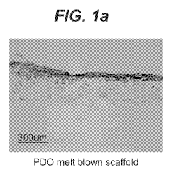
FIG. la Histology of Hematoxylin/Eosin (H&E) stained images after 7 days of
culturing
FIG. lb Histology of Hematoxylin/Eosin (H&E) stained images after 7 days of
culturing
Rat smooth muscle cells (SMC) on 75/25 poly(glycolide-co-caprolactone)
(PGA/PCL)
melt blown scaffolds.
FIG. 2 DNA contents of Human Umbilical Tissue cells (hUTC) on collagen coated
PDO
melt blown scaffolds and PDO melt blown scaffolds.
FIG. 3 DNA contents in three scaffolds (p-dioxanone) (PDO) melt blown
scaffold, 90/10
FIG. 4a H&E stained image of iMA cells seeded on a 65/35 PGA/PCL foam at 1
day.
FIG. 4c H&E stained image of iMA cells seeded on a 90/10 PGA/PLA needle
punched
scaffold at 1 day.
scaffold at 7 days.
FIG. 4e H&E stained image of iMA cells seeded on a PDO melt blown scaffold at
1 day.
8
CA 02852031 2014-04-11
WO 2013/055983 PCT/US2012/059858
FIG. 5 Procedures for generating a braided mesh/rolled melt blown 9/91
Cap/PDO/Braided mesh scaffold.
FIG. 6 SEM of a braided mesh/rolled melt blown 9/91 Cap/PDO/Braided mesh
scaffold.
FIG. 7 Cross-sectional SEM view of a braided mesh/rolled melt blown 9/9
Cap/PDO/Braided mesh scaffold.
FIG. 8a H&E stained image of a scaffold of a braided mesh/a rolled melt blown
(PDO/PCL)/a braided mesh with hUTC cultured in bioreactor cassette for 7 days.
FIG. 8b H&E stained image of a scaffold of a braided mesh/a rolled melt blown
(PDO/PCL)/a braided mesh with hUTC cultured in bioreactor cassette for 7 days.
FIG. 8c H&E stained image of a scaffold of a braided mesh/a rolled melt blown
(PDO/PCL)/a braided mesh with hUTC cultured in bioreactor cassette for 7 days.
FIG. 8d H&E stained image of a scaffold of a braided mesh/a rolled melt blown
(PDO/PCL)/a braided mesh with hUTC cultured in bioreactor cassette for 7 days.
DETAILED DESCRIPTION OF INVENTION
Poly(epsi/on-caprolactone) is a low Tg (-60 C) semi-crystalline polyester.
Although this material has a low elastic modulus it does not absorb quickly
enough for
many key surgical applications, i.e., it lasts too long in vivo. It has been
found, however,
that certain epsilon-caprolactone-rich copolymers are particularly useful for
the present
application. For instance, a 91/9 mol/mol poly(epsi/on-caprolactone-co-p-
dioxanone)
copolymer [91/9 Cap/PDO] was prepared in a sequential addition type of
polymerization
starting with a first stage charge of epsilon-caprolactone followed by a
subsequent second
stage ofp-dioxanone. The total initial charge was 75/25 mol/mol epsilon-
caprolactone/p-
dioxanone. Due to incomplete conversion of monomer-to-polymer and difference
in
9
CA 02852031 2014-04-11
WO 2013/055983 PCT/US2012/059858
reactivity, it is not uncommon to have the final (co)polymer composition
differ from the
feed composition. The final composition of the copolymer was found to be 91/9
mol/mol
epsilon-caprolactone/p-dioxanone. See EXAMPLE 3 for the details of this
copolymerization.
The present invention is directed towards copolymers of epsilon-caprolactone
and
p-dioxanone. More specifically, this class of copolymers rich in epsilon-
caprolactone and
made to have a blocky sequence distribution, that is non-random. In epsilon-
caprolactone/p-dioxanone copolymers in which the majority of the material is
based on p-
dioxanone, there is present a breakdown rate which is too fast to be useful in
long term
applications. The compositions must be rich in epsilon-caprolactone, e.g.,
having a
polymerized epsilon-caprolactone content of 50 percent or greater.
Dimensional stability in a fiber used to manufacture a surgical suture is very
important to prevent shrinkage, both in the sterile package before use, as
well as in the
patient after surgical implantation. Dimensional stability in low Tg material
can be
achieved by crystallization of the formed article. Regarding the phenomena of
crystallization of copolymers, a number of factors play important roles. These
factors
include overall chemical composition and the sequence distribution.
Although the overall level of crystallinity (and the Tg of the material) plays
a role
in dimensional stability, it is important to realize that the rate of
crystallization is critical to
processing. If a low Tg material is processed and it rate of crystallization
is very slow, it is
very difficult to maintain dimensional tolerances since shrinkage and warpage
easily occur.
Fast crystallization is thus an advantage. To increase the rate of
crystallization of a
copolymer of given overall chemical composition, a block structure would be
preferable
over a random sequence distribution. However, achieving this with the two
lactone
monomers, epsilon-caprolactone and p-dioxanone is known to be very difficult.
10
CA 02852031 2014-04-11
WO 2013/055983 PCT/US2012/059858
Poly(p-dioxanone) has a low ceiling temperature, accordingly at elevated
temperatures it tends to exist with a high fraction of monomer at equilibrium.
When
starting with fully polymerized material at elevated temperatures, it
"depolymerizes"
thereby resulting in a combination of polymer and regenerated monomer.
Regenerated
equilibrium monomer levels for poly(p-dioxanone) can be rather high,
approaching 30 to
50 percent at reaction temperatures of 110 to 160 C.
On the other hand, it is quite difficult to polymerize epsilon-caprolactone at
temperatures lower than about 160 C. There then exists a problem as to how to
achieve
polymerization of these two co-monomers to produce a block structure with high
enough
molecular weight so as to result in products having good mechanical
properties.
The novel copolymers of the present invention are prepared by first
polymerizing
the epsilon-caprolactone monomer at temperatures between about 170 C and about
240 C.
Temperatures between about 185 and about 195 C are particularly advantageous.
Although a monofunctional alcohol such as dodecanol might be used for
initiation, a diol
such as diethylene glycol has been found to work well. Combinations of mono-
functional
and di-functional, or multifunctional conventional initiators may also be
used. Reaction
times can vary with catalyst level. Suitable catalysts include conventional
catalysts such as
stannous octoate. The catalyst may be used at a monomer/catalyst level ranging
from
about 10,000/1 to about 300,000/1, with a preferred level of about 25,000/1 to
about
100,000/1. After the completion of this first stage of the polymerization, the
temperature is
lowered substantially, but still above a temperature of 60 C. Once the
temperature is
lowered, for example to 150 C, p-dioxanone monomer can be added to the
reactor; this can
be conveniently done by pre-melting this second monomer and adding it in a
molten form.
Once the p-dioxanone monomer is added, the temperature is brought to about 110
C to
complete the co-polymerization.
Alternately, once the p-dioxanone monomer is added, the temperature can be
brought to about 110 C, maintained at this temperature for some period of time
(e.g. 3 to 4
hours), followed by polymer discharge into suitable containers for subsequent
low
11
CA 02852031 2014-04-11
WO 2013/055983 PCT/US2012/059858
temperature polymerization (e.g., 80 C) for an extended period of time to
complete the co-
polymerization. Higher monomer-to-polymer conversions may be possible
utilizing this
alternate low temperature finishing approach.
It will be clear to one skilled in the art that various alternate
polymerization
approaches are possible and still produce the copolymer of the subject
invention.
One might then consider a process in which the reaction temperature after the
initial stage of polymerizing the epsilon-caprolactone is dropped immediately
to
110 C prior to the addition of the p-dioxanone monomer. Again, one skilled in
the
art can provide a variety of alternate polymerization schemes.
Poly(epsi/on-caprolactone-co-p-dioxanone) copolymers rich in polymerized
epsilon-caprolactone having levels of incorporated p-dioxanone greater than
about 40
mole percent are unsuitable for copolymers of the present invention because of
crystallization difficulties. Poly(epsi/on-caprolactone-co-p-dioxanone)
copolymers
comprising a polymerized epsilon-caprolactone having a molar level between 60
to 95
percent and a polymerized p-dioxanone molar level between 5 to 40 percent are
useful in
the practice of the present invention. This class of copolymers, the
poly(epsilon-
caprolactone-co-p-dioxanone) family rich epsilon-caprolactone, should ideally
contain
about 10 to about 30 mole percent of polymerized p-dioxanone.
The copolymers of the subject invention are semicrystalline in nature, having
a
crystallinity level ranging from about 10 to about 50 percent. They will have
a molecular
weight sufficiently high to allow the medical devices formed therefrom to
effectively have
the mechanical properties needed to perform their intended function. For melt
blown
nonwoven structure the molecular weight may be a little lower, and for
extruded fibers,
they may be a little higher. Typically, for example, the molecular weight of
the
copolymers of the subject invention will be such so as to exhibit inherent
viscosities as
measured in hexafluoroisopropanol (HFIP, or hexafluoro-2-propanol) at 25 C and
at a
concentration of 0.1 g/dL between about 0.5 to about 2.5 dL/g. The surgical
suture made
from the novel copolymers of the present invention preferably is a
monofilament with a
12
CA 02852031 2014-04-11
WO 2013/055983 PCT/US2012/059858
Young's modulus of less than about 150,000 psi. In one embodiment, the
copolymer has a
glass transition temperature below about 25 C. The novel copolymers of the
present
invention will preferably have an absorption time between about 6 and about 24
months.
In one embodiment, the medical devices made of the copolymers of the present
invention may contain conventional active ingredients, such as antimicrobials,
antibiotics,
therapeutic agents, hemostatic agents, radio-opaque materials, tissue growth
factors, and
combinations thereof In one embodiment the antimicrobial is Triclosan, PHMB,
silver
and silver derivatives or any other bio-active agent.
The copolymers of the subject invention can be melt extruded by a variety of
conventional means. Monofilament fiber formation can be accomplished by melt
extrusion followed by extrudate drawing with or without annealing.
Multifilament fiber
formation is possible by conventional means. Methods of manufacturing
monofilament
and multifilament braided sutures are disclosed in U.S. Patent No. 5133739,
entitled
"Segmented Copolymers of epsilon-Caprolactone andGlycolide" and U.S. Patent
No.
6712838 entitled "Braided Suture with Improved Knot Strength and Process to
Produce
Same", are incorporated by reference herein in their entirety.
The copolymers of the present invention may be used to manufacture
conventional
medical devices in addition to sutures using conventional processes. For
example,
injection molding might be accomplished after allowing the copolymer to
crystallize in the
mold; alternately biocompatible nucleating agents might be added to the
copolymer to
reduce cycle time. The medical devices may include, in addition to meshes, the
following
conventional devices meshes, tissue repair fabrics, suture anchors, stents,
orthopedic
implants, staples, tacks, fasteners, suture clips, etc.
Sutures made from the copolymers of the present invention may be used in
conventional surgical procedures to approximate tissue or affix tissue to
medical devices.
Typically, after a patient is prepared for surgery in a conventional matter,
including
swabbing the outer skin with antimicrobial solutions and anesthetizing the
patient, the
13
CA 02852031 2014-04-11
WO 2013/055983 PCT/US2012/059858
surgeon will make the required incisions, and, after performing the required
procedure
proceed to approximate tissue using the long-term absorbable sutures of the
present
invention (in particular monofilament sutures) made from the novel copolymers
of the
present invention. In addition to tissue approximation, the sutures may be
used to affix
implanted medical devices to tissue in a conventional manner. After the
incisions are
approximated, and the procedure is completed, the patient is then moved to a
recovery
area. The long-term absorbable sutures of the present invention in the patient
retain their
strength in vivo for the required time to allow effective healing and
recovery.
Also disclosed herein as an invention is a tissue engineered blood vessel
(TEBV)
comprised of an inner braided mesh tube having an inner surface and an outer
surface, a
melt blown sheet disposed on the outer surface of the inner braided mesh tube,
and an
outer braided mesh tube disposed on the melt blown sheet. Furthermore, the
TEBV may
be combined with one or more of cells, cell sheets, cell lysate, minced
tissue, and cultured
with or without a bioreactor process. Such tissue engineered blood vessels may
be used to
repair or replace a native blood vessel that has been damaged or diseased. In
tissue
engineering, the rate of resorption of the scaffold by the body preferably
approximates the
rate of replacement of the scaffold by tissue. That is to say, the rate of
resorption of the
scaffold relative to the rate of replacement of the scaffold by tissue must be
such that the
structural integrity, e.g. strength, required of the scaffold is maintained
for the required
period of time. If the scaffold degrades and is absorbed unacceptably faster
than the
scaffold is replaced by tissue growing therein, the scaffold may exhibit a
loss of strength
and failure of the device may occur. Additional surgery then may be required
to remove
the failed scaffold and to repair damaged tissue. The TEBV described herein
advantageously balances the properties of biodegradability, resorption,
structural integrity
over time, and the ability to facilitate tissue in-growth, each of which is
desirable, useful,
or necessary in tissue regeneration or repair.
The braided mesh tubes and the melt blown sheet are prepared from
biocompatible,
biodegradable polymers. The biodegradable polymers readily break down into
small
segments when exposed to moist body tissue. The segments then are either
absorbed by or
14
CA 02852031 2014-04-11
WO 2013/055983 PCT/US2012/059858
passed from the body. More particularly, the biodegraded segments do not
elicit
permanent chronic foreign body reaction, because they are absorbed by the body
or passed
from the body such that no permanent trace or residual of the segment is
retained by the
body. For the purposes of this invention the terms bioabsorbable and
biodegradable are
used interchangeably.
The biocompatible, biodegradable polymers may be natural, modified natural, or
synthetic biodegradable polymers, including homopolymers, copolymers, and
block
polymers, linear or branched, segmented or random, as well as combinations
thereof.
Particularly well suited synthetic biodegradable polymers are aliphatic
polyesters which
include but are not limited to homopolymers and copolymers of lactide (which
includes
D(-)-lactic acid, L(+)-lactic acid, L(-)-lactide, D(+)-lactide, and meso-
lactide), glycolide
(including glycolic acid), epsilon-caprolactone, p-dioxanone (1,4- dioxan-2-
one), and
trimethylene carbonate (1,3- dioxan-2-one).
For a tubular structure to fulfill the requirements set out for a successful
TEBV (or
similar tubular device or sheet stock scaffold), it must possess certain key
properties. The
structure as a whole must exhibit an ability to allow radial expansion in a
pulsatile manner
similar to what is seen in human arteries. This means, in part, to match the
elastic modulus
of arteries. An elastic modulus of 1 to 5 MPa would be appropriate, and an
elastic
modulus lower than that exhibited by poly(p-dioxanone) is sought.
Moreover, the retention time of mechanical properties, post-implantation, must
be
sufficient for the intended use. If the device is to be pre-seeded with cells
and the cells
allowed to propagate prior to implantation of the device, then the pre-seeded
device must
withstand the rigors of surgical implantation, including fixation at both
ends. If the device
is to be implanted without being pre-seeded with cells, the device must
possess sufficient
retention of mechanical properties to allow appropriate cellular in-growth to
be functional.
In general, a retention time of mechanical properties greater than that
exhibited by poly(p-
dioxanone) is sought. It is to be understood that a successful material must
still absorb in
an appropriate time frame, i.e. 6 to 18 months, and typically not more than
about 24
CA 02852031 2014-04-11
WO 2013/055983 PCT/US2012/059858
months. One material that may come under the consideration of some researchers
is
poly(epsi/on-caprolactone). This material, although having a low elastic
modulus, does
not absorb quickly enough to meet requirements.
Dimensional stability of a low modulus polymeric fiber that is not cross-
linked as
in rubber fibers is generally achieved by inducing some measure of
crystallinity. It is to be
understood that the rate at which a polymer crystallizes is also very
important during the
process of melt blowing the nonwoven fabric itself If it crystallizes too
slowly, the low
modulus nature of the material cannot support the structure and the fabric
collapses onto
itself resulting into a film-like structure. In one embodiment, a polymer has
a glass
transition temperature below 25 C.
In some instances, it may be desirable to have the fibers making up the
nonwoven
fabric quite small in diameter; i.e. 2 to 6 microns in diameter or lower. To
achieve this, it
may be necessary to limit the molecular weight of the resin. In one
embodiment, a
polymer exhibits an inherent viscosity between 0.5 and 2.0 dL/g.
Existing materials are deficient in meeting the new challenges presented. Two
copolymer systems that meet the challenging requirements set forth above have
unexpectedly been discovered. These systems are both based on the lactone
monomers p-
dioxanone and epsilon-caprolactone. In one case, the monomer ratio favors p-
dioxanone;
that is, p-dioxanone-rich poly(epsi/on-caprolactone-co-p-dioxanone). In the
other case, the
monomer ratio favors epsilon-caprolactone; that is, epsilon-caprolactone -rich
poly(epsi/on-caprolactone-co-p-dioxanone).
Copolymer I: Segmented, p-dioxanone-Rich, Poly(epsi/on-caprolactone-co-p-
dioxanone) Copolymers [PDO-Rich Cap/PD0].
Poly(p-dioxanone) is a low Tg (-11 C) semi-crystalline polyester finding
extensive
utility as a suture material and as injection molded implantable medical
devices. It will be
understood by one having ordinary skill in the art that the level of
crystallinity needed to
16
CA 02852031 2014-04-11
WO 2013/055983 PCT/US2012/059858
achieve dimensional stability in the resulting fabric will depend on the glass
transition
temperature of the (co)polymer. That is, to avoid fabric shrinkage, warpage,
buckling, and
other consequences of dimensional instability, it is important to provide some
level of
crystallinity to counteract the phenomena. The level of crystallinity that is
needed for a
particular material of given glass transition temperature with given molecular
orientation
can be experimentally determined by one having ordinary skill in the art. The
level for
crystallinity required to achieve dimensional stability in melt blown nonwoven
fabrics may
be a minimum of about 20 percent in polymeric materials possessing glass
transition
temperatures of about minus 20 C.
Besides the level of crystallinity, the rate of crystallization is very
important in the
melt blown nonwoven process. If a material crystallizes too slowly, especially
if it
possesses a glass transition temperature below room temperature, the resulting
nonwoven
product may have a collapsed architecture, closer to a film than a fabric. A
slow-to-
crystallize (co)polymer will be quite difficult to process into desired
structures.
It would be advantageous to have a material exhibiting a greater reversible
extensibility (i.e. elasticity) and a lower modulus than poly(p-dioxanone).
Certain p-
dioxanone-rich copolymers are particularly useful for this application.
Specifically, a 9/91
mol/mol poly(epsi/on-caprolactone-co-p-dioxanone) copolymer [9/91 Cap/PDO] was
prepared in a sequential addition type of polymerization starting with a first
stage charge of
epsilon-caprolactone followed by a subsequent second stage ofp-dioxanone. The
total
initial charge was 7.5/92.5 mol/mol epsilon-caprolactone/p-dioxanone. See
EXAMPLE 2
for the details of this copolymerization.
Poly(epsi/on-caprolactone-co-p-dioxanone) copolymers rich in polymerized p-
dioxanone having levels of incorporated epsilon-caprolactone greater than
about 15 mole
percent are unsuitable for the present application, because it is difficult to
prepare melt
blown nonwoven fabrics from such copolymers. It is speculated that this may be
because
p-dioxanone-rich poly(epsi/on-caprolactone-co-p-dioxanone) copolymers having
greater
than about 15 mole percent of incorporated epsilon-caprolactone exhibit too
high an elastic
17
CA 02852031 2014-04-11
WO 2013/055983 PCT/US2012/059858
modulus resulting in "snap-back" of extruded fibers leading to very lumpy
unsuitable
fabric. See EXAMPLES 1 and 5 for the synthesis and processing details,
respectively.
Copolymer II: Segmented, epsilon-caprolactone-Rich, Poly(epsilon-
caprolactone-co-p-dioxanone) Copolymers [Cap-Rich Cap/PD0].
Poly(epsi/on-caprolactone) is also a low Tg (-60 C) semi-crystalline
polyester. As
previously discussed, this material, although having a low elastic modulus,
does not absorb
quickly enough to meet requirements. It has been found, however, that certain
epsilon-
caprolactone-rich copolymers are particularly useful for the present
application.
Specifically, a 91/9 mol/mol poly(epsi/on-caprolactone-co-p-dioxanone)
copolymer [91/9
Cap/PDO] was prepared in a sequential addition type of polymerization starting
with a first
stage charge of epsilon-caprolactone followed by a subsequent second stage ofp-
dioxanone. The total initial charge was 75/25 mol/mol epsilon-caprolactone/p-
dioxanone.
Due to incomplete conversion of monomer-to-polymer and difference in
reactivity, it is not
uncommon to have the final (co)polymer composition differ from the feed
composition.
The final composition of the copolymer was found to be 91/9 mol/mol epsilon-
caprolactone/p-dioxanone. See EXAMPLE 3 for the details of this
copolymerization.
Poly(epsi/on-caprolactone-co-p-dioxanone) copolymers rich in polymerized
epsilon-caprolactone having levels of incorporated p-dioxanone greater than
about 20 mole
percent are unsuitable for the present application, because it is difficult to
prepare melt
blown nonwoven fabrics from such copolymers. It is speculated that this may be
because
epsilon-caprolactone-rich poly(epsi/on-caprolactone-co-p-dioxanone) copolymers
having
levels of incorporated p-dioxanone greater than about 20 mole percent do not
crystallize
quickly enough leading to unsuitable fabric.
As discussed herein, suitable synthetic bioabsorbable polymers for the present
invention include poly(p-dioxanone) homopolymer (PDO) and p-dioxanone/epsi/on-
caprolactone segmented copolymers rich in p-dioxanone. The latter class of
polymers, the
18
CA 02852031 2014-04-11
WO 2013/055983 PCT/US2012/059858
poly(p-dioxanone-co-epsilon-caprolactone) family rich in p-dioxanone should
ideally
contain up to about 15 mole percent of polymerized epsilon-caprolactone.
Additionally, p-dioxanone/epsi/on-caprolactone segmented copolymers rich in
epsilon-caprolactone are useful in practicing the present invention. This
class of polymers,
the poly(p-dioxanone-co-epsilon-caprolactone) family rich epsilon-
caprolactone, should
ideally contain up to about 20 mole percent of polymerized p-dioxanone.
Other polymer systems that may be advantageously employed include the
poly(lactide-co-epsilon-caprolactone) family of materials. Within this class,
the
copolymers rich in polymerized lactide having about 99 to about 65 mole
percent
polymerized lactide and the copolymers rich in polymerized epsilon-
caprolactone having
about 99 to about 85 mole percent polymerized epsilon-caprolactone are useful.
Other polymer systems that may be employed include the poly(lactide-co-p-
dioxanone) family of materials. Within this class, the copolymers rich in
polymerized
lactide having about 99 to about 85 mole percent polymerized lactide and the
copolymers
rich in polymerized p-dioxanone having about 99 to about 80 mole percent
polymerized p-
dioxanone are useful. It is to be understood that the copolymers in this
poly(lactide-co-p-
dioxanone) family of materials rich in polymerized lactide maybe more useful
where a
stiffer material is required.
Other polymer systems that may be employed include the poly(lactide-co-
glycolide) family of materials. Within this class, the copolymers rich in
polymerized
lactide having about 99 to about 85 mole percent polymerized lactide and the
copolymers
rich in polymerized glycolide having about 99 to about 80 mole percent
polymerized
glycolide are useful. It is to be understood that the copolymers in this
poly(lactide-co-
glycolide) family of materials rich in polymerized lactide maybe more useful
where a
stiffer material is required. Likewise, the copolymers in this poly(lactide-co-
glycolide)
family of materials rich in polymerized glycolide maybe more useful when a
faster
absorption time is required.
19
CA 02852031 2014-04-11
WO 2013/055983 PCT/US2012/059858
Another polymer class that may be employed includes the poly(glycolide-co-
epsilon-caprolactone) family of materials. Within this class, the copolymers
rich in
polymerized glycolide having about 99 to about 70 mole percent polymerized
glycolide
and the copolymers rich in polymerized epsilon-caprolactone having about 99 to
about 85
mole percent polymerized epsilon-caprolactone are useful. It is to be
understood that the
copolymers in this poly(glycolide-co-epsilon-caprolactone) family of materials
rich in
polymerized glycolide maybe more useful when a faster absorption time is
required.
Likewise, the copolymers in this poly(glycolide-co-epsilon-caprolactone)
family of
materials, rich in polymerized epsilon-caprolactone, maybe more useful when a
softer
material is required.
Suitable natural polymers include, but are not limited to collagen,
atelocollagen,
elastic, and fibrin and combinations thereof. In one embodiment, the natural
polymer is
collagen. In yet another embodiment, the combination of natural polymer is an
acellular
omental matrix.
In accordance herewith, a melt blown nonwoven process having utility herein
will
now be described. A typical system for use in a melt blown nonwoven process
consists of
the following elements: an extruder, a transfer line, a die assembly, hot air
generator, a web
formation system, and a winding system.
As is well known to those skilled in the art, an extruder consists of a heated
barrel
with a rotating screw positioned within the barrel. The main function of the
extruder is to
melt the copolymer pellets or granules and feed them to the next element. The
forward
movement of the pellets in the extruder is along the hot walls of the barrel
between the
flights of the screw. The melting of the pellets in the extruder results from
the heat and
friction of the viscous flow and the mechanical action between the screw and
the walls of
the barrel. The transfer line will move molten polymer toward the die
assembly. The
transfer line may include a metering pump in some designs. The metering pump
may be a
positive-displacement, constant-volume device for uniform melt delivery to the
die
assembly.
CA 02852031 2014-04-11
WO 2013/055983 PCT/US2012/059858
The die assembly is a critical element of the melt blown process. It has three
distinct components: a copolymer feed distribution system, spinnerets
(capillary holes),
and an air distribution system. The copolymer feed distribution introduces the
molten
copolymer from the transfer line to distribution channels/plates to feed each
individual
capillary hole uniformly and is thermal controlled. From the feed distribution
channel the
copolymer melt goes directly to the die capillary. The copolymer melt is
extruded from
these holes to form filament strands which are subsequently attenuated by hot
air to form
fine fibers. During processing, the entire die assembly is heated section-wise
using
external heaters to attain the desired processing temperatures. In one
embodiment, a die
temperature of about 210 to 280 C for CAP/GLY 25/75 copolymer, about 110 to
210 C
for PDO/CAP 92.5/7.5 copolymer, and 120 to 220 C for PDS homopolymer is
useful. In
another embodiment, a die temperature range is from about 210 C to about 260 C
for
CAP/GLY 25/75 copolymer, about 150 C to about 200 C for PDO/CAP 92.5/7.5
copolymer, and about 160 C to about 210 C for PDS homopolymer. In another
embodiment, a die pressure of about 100 to 2,000 psi is useful. In another
embodiment, a
die pressure range is from about 100 to about 1200 psi.
The air distribution system supplies the high velocity hot air. The high
velocity air
is generated using an air compressor. The compressed air is passed through a
heat
exchange unit, such as an electrical or gas heated furnace, to heat the air to
desired
processing temperatures. In one embodiment, an air temperature of about 200 C
to 350 C
for CAP/GLY 25/75 copolymer, about 180 to 300 C for PDO/CAP 92.5/7.5
copolymer,
and about 180 to 300 C for PDS homopolymer is useful. In another embodiment,
an air
temperatures range is from about 220 C to about 300 C for CAP/GLY 25/75
copolymer,
about 200 C to about 270 C for PDO/CAP 92.5/7.5 copolymer, and about 200 to
about
270 C for PDS homopolymer. In another embodiment, an air pressure of about 5
to 50 psi
is useful, and in another embodiment an air pressure range is from about 5 to
about 30 psi.
It should be recognized that the air temperature and the air pressure may be
somewhat
equipment dependent, but can be determined through appropriate experiment.
21
CA 02852031 2014-04-11
WO 2013/055983 PCT/US2012/059858
As soon as the molten copolymer is extruded from the die holes, high velocity
hot
air streams attenuate the copolymer streams to form microfibers. With the
equipment
employed, a screw speed of about 1 to 100 RPM is adequate. As the hot air
stream
containing the microfibers progresses toward the collector screen, it draws in
a large
amount of surrounding air that cools and solidifies the fibers. The solidified
fibers
subsequently get laid randomly onto the collecting screen, forming a self-
bonded web.
The collector speed and the collector distance from the die nosepiece can be
varied to
produce a variety of melt blown webs. With the equipment employed, a collector
speed of
about 0.1 to 100 m/min is adequate. Typically, a vacuum is applied to the
inside of the
collector screen to withdraw the hot air and enhance the fiber laying process.
The melt blown web is typically wound onto a tubular core and may be processed
further according to the end-use requirement. In one embodiment, the nonwoven
construct
formed by the melt blown extrusion of the aforementioned copolymer is
comprised of
microfibers having a fiber diameter ranging from about 1 to 8 mircometres. In
another
embodiment, the microfibers have a fiber diameter ranging from about 1 to 6
micrometres.
The melt blown process used to synthesize the TEBVs of the present invention
is
advantageous with respect to other processes, including electrostatic
spinning, for various
reasons. For example, the melt blown process may be better for the environment
than
other processes because it does not need a solvent to dissolve a polymer.
Another
advantage is that the melt blown process is a one-step process wherein the
molten polymer
resin is blown by high speed air onto a collector such as a conveyor belt or a
take-up
machine to form a nonwoven fabric. Moreover, the diameters of melt blown
fibers are in
the range of 0.1 micron to 50 microns. A combination of the broad range fibers
provides a
scaffold having large pores and porosity. Furthermore, composite scaffolds
having
micro/nano scale fibers can be produced using a combination of a melt blown
and an
electrospun scaffold. The electrospun scaffold may be used as a barrier, as it
possesses
much smaller pore sizes which can impede transport from one side to the other.
Another
advantage is that the rolling process does not require glue for the graft to
keep its tubular
shape, and the rolling process does not need sutures to reinforce the strength
of the graft.
22
CA 02852031 2014-04-11
WO 2013/055983 PCT/US2012/059858
The TEBV has overall dimensions that reflect desired ranges that, in
combination
with the one or more of cells, cell sheets, cell lysate, minced tissue, and a
bioreactor
process, will replace a small diameter, damaged or diseased vein or artery
blood vessel.
Desirable dimensions include but are not limited to: internal diameter (3-7mm
preferable,
Internal Wall Length Compliance Burst Suture Tensile
Diameter Thickness (cm) (%)
Pressure retention (peak
(mm) (mm)
(mm (gmf) stress)
Hg)
PDO 2 & 5 0.5 1-20 0.5-1 1500- 310 5
MPa
2500
Vessel 2 & 5 0.5-0.7 1-20 0.2-10 1500- 100-500 2-
20
4500
MPa
The TEBV has physical properties that reflect desired ranges that, in
conjunction
with one or more of cells, cell sheets, cell lysate, minced tissue, and a
bioreactor process,
will replace a small diameter, damaged or diseased vein or artery blood
vessel. Desirable
physical properties include but are not limited to: compliance (0.2-10 percent
preferable,
0.7-7 percent most preferable); suture retention strength (100gm-4Kg
preferable, 100-
23
CA 02852031 2014-04-11
WO 2013/055983 PCT/US2012/059858
preferable; 2-20 most preferable) and orthogonal/radial (0.5-15n preferable, 1-
10 most
preferable) and random (0.5-15 preferable, 1-10 most preferable) and wet/long
(1-30
preferable; 2-20 most preferable); failure strain (%) of longitudinal/axial (1-
200 preferable;
5-75 most preferable) and orthogonal/radial (5-400 preferable, 10-300 most
preferable) and
random (5-400 preferable, 10-300 most preferable) and wet/long (1-200
preferable; 20-100
most preferable).
The TEBV has morphology that reflects desired ranges that, in conjunction with
one or more of cells, cell sheets, cell lysate, minced tissue, and a
bioreactor process, will
replace a small diameter, damaged or diseased vein or artery blood vessel.
Desirable
morphology includes but is not limited to: pore size (1-200um preferable, most
preferable
less than 100um); porosity (40-98 percent preferable, most preferable 60-95
percent);
surface area/vol (0.1-7 m2/cm3 preferable, most preferable 0.3-5.5 m2/cm3);
water
permeability (1-10m1 cm2/min @80-120mm Hg preferable, most preferable <5m1
cm2/min
@l2OmmHg); and orientation of polymer/fibers (allows proper cell seeding,
adherence,
growth, and ECM formation). Polymer/fiber orientation will also allow proper
cell
migration, and is important for the minced tissue fragments such that cells
will migrate out
of the fragments and populate the TEBV.
The TEBV has biocompatibility that reflects desired properties for a TEBV
that, in
conjunction with one or more of cells, cell sheets, cell lysate, minced
tissue, and a
bioreactor process, will replace a small diameter, damaged or diseased vein or
artery blood
vessel. Desirable biocompatibility includes but is not limited: absorption (6-
24 months
preferable to allow greatest vol. of TEBV to be occupied by cells and ECM);
tissue
reaction (minimal); cell compatibility (adherence, viability, growth,
migration and
differentiation not negatively impacted by TEBV); residual solvent (minimal);
residual
Et0 (minimal); and hemocompatible (non-thrombogenic).
24
CA 02852031 2014-04-11
WO 2013/055983 PCT/US2012/059858
The tissue engineered blood vessel scaffold is prepared by the following
method:
A first braided mesh tube having an inner surface and an outer surface is
provided
as described above and placed on a mandrel. Then, a melt blown sheet is
provided as
described above and rolled onto the outer surface of the first braided mesh
tube. Next, a
second braided mesh tube is positioned over the rolled melt blown sheet.
In one embodiment, the tissue engineered blood vessel further comprises cells.
Suitable cells that may be combined with the TEBV include, but are not limited
to: stem
cells such as multipotent or pluripotent stem cells; progenitor cells, such as
smooth muscle
progenitor cells and vascular endothelium progenitor cells; embryonic stem
cells;
postpartum tissue derived cells such as, placental tissue derived cells and
umbilical tissue
derived cells; endothelial cells, such as vascular endothelial cells; smooth
muscle cells,
such as vascular smooth muscle cells; precursor cells derived from adipose
tissue; and
arterial cells, such as cells derived from the radial artery and the left and
right internal
mammary artery (IMA), also known as the internal thoracic artery.
In one embodiment, the cells are human umbilical tissue derived cells (hUTCs).
The methods for isolating and collecting human umbilical tissue-derived cells
(hUTCs)
(also referred to as umbilical-derived cells (UDCs)) are described in United
States Patent
No. 7,510,873, incorporated herein by reference in its entirety. In another
embodiment, the
TEBV further comprises human umbilical tissue derived cells (hUTCs) and one or
more
other cells. The one or more other cells includes, but is not limited to
vascular smooth
muscle cells (SMCs), vascular smooth muscle progenitor cells, vascular
endothelial cells
(ECs), or vascular endothelium progenitor cells, and/or other multipotent or
pluripotent
stem cells. hUTCs in combination with one or more other cells on the TEBV may
enhance
the seeding, attachment, and proliferation of, for example, ECs and SMCs on
the TEBV.
hUTCs may also promote the differentiation of the EC or SMC or progenitor
cells in the
TEBV construct. This may promote the maturation of TEBVs during the in vitro
culture
as well as the engraftment during the in vivo implantation. hUTCs may provide
trophic
support or provide and enhance the expression of ECM proteins. The trophic
effects of the
CA 02852031 2014-04-11
WO 2013/055983 PCT/US2012/059858
cells, including hUTCs, can lead to proliferation of the vascular smooth
muscle or vascular
endothelium of the patient. The trophic effects of the cells, including hUTCs,
may induce
migration of vascular smooth muscle cells, vascular endothelial cells,
skeletal muscle
progenitor cells, vascular smooth muscle progenitor cells, or vascular
endothelium
progenitor cells to the site or sites of the regenerated blood vessel.
Cells can be harvested from a patient (before or during surgery to repair the
tissue)
and the cells can be processed under sterile conditions to provide a specific
cell type. One
of skill in the art is aware of conventional methods for harvesting and
providing the cells
as described above such as described in Osteoarthritis Cartilage 2007
Feb;15(2):226-31
and incorporated herein by reference in their entirety. In another embodiment
the cells are
genetically modified to express genes of interest responsible for pro-
angiogenic activity,
anti-inflammatory activity, cell survival, cell proliferation or
differentiation or
immunomodulation.
The cells can be seeded on the TEBV for a short period of time, e.g. less than
one
day, just prior to implantation, or cultured for longer a period, e.g. greater
than one day, to
allow for cell proliferation and extracellular matrix synthesis within the
seeded TEBV prior
to implantation. In one embodiment, a single cell type is seeded on the TEBV.
In another
embodiment, one or more cell types are seeded on the TEBV. Various cellular
strategies
could be used with these scaffolds (i.e., autologous, allogenic, xenogeneic
cells etc.). In
one embodiment, smooth muscle cells can be seeded on the outer lumen of the
TEBV and
in another embodiment, endothelial cells can be seeded in the inner lumen of
the TEBV.
The cells are seeded in an amount sufficient to provide a confluent cell
layer. Preferably,
cell seeding density is about 2 x 10⁵/cm2.
In another embodiment the tissue engineered blood vessel further comprises
cell
sheets. Cell sheets may be made of hUTCs or other cell types. Methods of
making cell
sheets are described in U.S. Application No. 11/304,091, published on July 13,
2006 as
U.S. Patent Publication No. US 2006-0153815 Al and incorporated herein by
reference in
its entirety. The cell sheet is generated using thermoresponsive polymer
coated dishes that
26
CA 02852031 2014-04-11
WO 2013/055983 PCT/US2012/059858
allow harvesting intact cell sheets with the decrease of the temperature.
Alternatively,
other methods of making cell sheets include, but are not limited to growing
cells in a form
of cell sheets on a polymer film. Selected cells may be cultured on a surface
of glass,
ceramic or a surface-treated synthetic polymer. For example, polystyrene that
has been
subjected to a surface treatment, like gamma-ray irradiation or silicon
coating, may be used
as a surface for cell culture. Cells grown to over 85 percent confluence form
cell sheet
layer on cell growth support device. Cell sheet layer may be separated from
cell growth
support device using proteolysis enzymes, such as trypsin or dispase. Non-
enzymatic cell
dissociation could also be used. A non-limiting example includes a mixture of
chelators
sold under the trade name CELLSTRIPPER (Mediatech, Inc., Herndon, Va.), a non-
enzymatic cell dissociation solution designed to gently dislodge adherent
cells in culture
while reducing the risk of damage associated with enzymatic treatments.
Alternatively, the surface of the cell growth support device, from which
cultured
cells are collected, may be a bed made of a material from which cells detach
without a
proteolysis enzyme or chemical material. The bed material may comprise a
support and a
coating thereon, wherein the coating is formed from a polymer or copolymer
which has a
critical solution temperature to water within the range of 0 C to 80 C.
In one embodiment, one or more cells sheets are combined with the TEBV as
described herein above by layering the cell sheets on the melt blown sheet and
then rolling
the sheet on the tube. The one or more cell sheets may be of the same cell
type or of
different cell types as described herein above. In one embodiment, multiple
cell sheets
could be combined to form a robust vascular construct. For example, cell
sheets made of
endothelial cells and smooth muscle cells could be combined with the scaffold
to form
TEBVs. Alternatively, other cell types such as hUTC cell sheets could be
combined with
endothelial cell sheets and the scaffold to form TEBVs. Furthermore, cell
sheets made of
hUTCs can be wrapped around a pre-formed TEBV composed of a scaffold, ECs, and
SMCs to provide trophic factors supporting maturation of the construct.
27
CA 02852031 2014-04-11
WO 2013/055983 PCT/US2012/059858
Cell sheets may be grown on the melt blown sheet to provide reinforcement and
mechanical properties to the cell sheets. Reinforced cell sheets can be formed
by placing
biodegradable or non-biodegradable reinforcing members at the bottom of
support device
prior to seeding support device with cells. Reinforcing members are as
described herein
above. Cell sheet layer that results will have incorporated the reinforcing
scaffold
providing additional strength to the cell sheet layer, which can be
manipulated without the
requirement for a backing layer. A preferred reinforcing scaffold is a mesh
comprised of
poly(p-dioxanone). The mesh can be placed at the bottom of a Corning Ultra
low
attachment dish. Cells can then be seeded on to the dishes such that they will
form cell-
cell interactions but also bind to the mesh when they interact with the mesh.
This will give
rise to reinforced cell sheets with better strength and handling
characteristics. Such
reinforced cell sheets may be rolled into a TEBV or the reinforced cell sheet
layer may be
disposed on a scaffold (as described above).
In another embodiment, the cell sheet is genetically engineered. The
genetically
engineered cell sheet comprises a population of cells wherein at least one
cell of the
population of cells is transfected with an exogenous polynucleotide such that
the
exogenous polynucleotide expresses express diagnostic and/or therapeutic
product (e. g., a
polypeptide or polynucleotide) to assist in tissue healing, replacement,
maintenance and
diagnosis. Examples of "proteins of interest" (and the genes encoding same)
that may be
employed herein include, without limitation, cytokines, growth factors,
chemokines,
chemotactic peptides, tissue inhibitors of metalloproteinases, hormones,
angiogenesis
modulators either stimulatory or inhibitory, immune modulatory proteins,
neuroprotective
and neuroregenerative proteins and apoptosis inhibitors. More specifically,
preferred
proteins include, without limitation, erythropoietin (EPO), EGF, VEGF, FGF,
PDGF, IGF,
KGF, IFN-a, IFN-6, MSH, TGF-a, TGF-13, TNF-a, IL-1, BDNF, GDF-5, BMP-7 and IL-
6.
In another embodiment the tissue engineered blood vessel further comprises
cell
lysate. Cell lysates may be obtained from cells including, but not limited to
stem cells
such as multipotent or pluripotent stem cells; progenitor cells, such as
smooth muscle
progenitor cells and vascular endothelium progenitor cells; embryonic stem
cells;
28
CA 02852031 2014-04-11
WO 2013/055983 PCT/US2012/059858
postpartum tissue derived cells such as, placental tissue derived cells and
umbilical tissue
derived cells, endothelial cells, such as vascular endothelial cells; smooth
muscle cells,
such as vascular smooth muscle cells; precursor cells derived from adipose
tissue; and
arterial cells such as cells derived from the radial artery and the left and
right internal
mammary artery (IMA), also known as the internal thoracic artery. The cell
lysates and
cell soluble fractions may be stimulated to differentiate along a vascular
smooth muscle or
vascular endothelium pathway. Such lysates and fractions thereof have many
utilities.
Use of lysate soluble fractions (i.e., substantially free of membranes) in
vivo, for example,
allows the beneficial intracellular milieu to be used allogeneically in a
patient without
introducing an appreciable amount of the cell surface proteins most likely to
trigger
rejection or other adverse immunological responses.
Methods of lysing cells are well-known in the art and include various means of
mechanical disruption, enzymatic disruption, chemical disruption, or
combinations thereof
Such cell lysates may be prepared from cells directly in their growth medium
and thus
containing secreted growth factors and the like, or may be prepared from cells
washed free
of medium in, for example, PBS or other solution. The cell lysate can be used
to create a
TEBV according to the present invention by placing a TEBV into a cell culture
plate and
adding cell lysate supernatant onto the TEBV. The lysate loaded TEBV can then
be placed
into a lyophilizer for lyophilization.
In yet another embodiment the tissue engineered blood vessel further comprises
minced tissue. Minced tissue has at least one viable cell that can migrate
from the tissue
fragments onto the TEBV. More preferably, the minced tissue contains an
effective
amount of cells that can migrate from the tissue fragments and begin
populating the TEBV.
Minced tissue may be obtained from one or more tissue sources or may be
obtained from
one source. Minced tissue sources include, but are not limited to muscle
tissue, such as
skeletal muscle tissue and smooth muscle tissue; vascular tissue, such as
venous tissue and
arterial tissue; skin tissue, such as endothelial tissue; and fat tissue.
29
CA 02852031 2014-04-11
WO 2013/055983 PCT/US2012/059858
The minced tissue is prepared by first obtaining a tissue sample from a donor
(autologous, allogenic, or xenogeneic) using appropriate harvesting tools. The
tissue
sample is then finely minced and divided into small fragments either as the
tissue is
collected, or alternatively, the tissue sample can be minced after it is
harvested and
collected outside the body. In embodiments where the tissue sample is minced
after it is
harvested, the tissue samples can be washed three times in phosphate buffered
saline. The
tissue can then be minced into small fragments in the presence of a small
quantity, for
example, about 1 ml, of a physiological buffering solution, such as, phosphate
buffered
saline, or a matrix digesting enzyme, such as 0.2 percent collagenase in Ham's
F12
medium. The tissue is minced into fragments of approximately 0.1 to 1 mm 3 in
size.
Mincing the tissue can be accomplished by a variety of methods. In one
embodiment, the
mincing is accomplished with two sterile scalpels cutting in parallel and
opposing
directions, and in another embodiment, the tissue can be minced by a
processing tool that
automatically divides the tissue into particles of a desired size. In one
embodiment, the
minced tissue can be separated from the physiological fluid and concentrated
using any of
a variety of methods known to those having ordinary skill in the art, such as,
for example,
sieving, sedimenting or centrifuging. In embodiments where the minced tissue
is filtered
and concentrated, the suspension of minced tissue preferably retains a small
quantity of
fluid in the suspension to prevent the tissue from drying out.
The suspension of minced living tissue can be used to create a TEBV according
to
the present invention by depositing the suspension of living tissue upon a
biocompatible
TEBV, such that the tissue and the TEBV become associated. Preferably, the
tissue is
associated with at least a portion of the TEBV. The TEBV can be implanted in a
subject
immediately, or alternatively, the construct can be incubated under sterile
conditions that
are effective to maintain the viability of the tissue sample.
In another aspect of the invention, the minced tissue could consist of the
application of two distinct minced tissue sources (e.g., one surface could be
loaded with
minced endothelial tissue and the other surface could be loaded with minced
smooth
muscle tissue).
CA 02852031 2014-04-11
WO 2013/055983 PCT/US2012/059858
In one embodiment, the tissue engineered blood vessels and one or more of
cells,
cell sheets, cell lysate, or minced tissue is enhanced by combining with
bioactive agents.
Suitable bioactive agents include, but are not limited to an antithrombogenic
agent, an anti-
inflammatory agent, an immunosuppressive agent, an immunomodulatory agent, pro-
angiogenic, an antiapoptotic agent, antioxidants, growth factors, angiogenic
factors,
myoregenerative or myoprotective drugs, conditioned medium, extracellular
matrix
proteins, such as, collagen, atelocollagen, laminin, fibronectin, vitronectin,
tenascin,
integrins, glycosaminoglycans (hyaluronic acid, chondroitin sulfate, dermatan
sulfate,
heparan sulfate, heparin, keratan sulfate and the like), elastin and fibrin;
growth factors
and/or cytokines, such as vascular endothelial cell growth factors, platelet
derived growth
factors, epidermal growth factors, fibroblast growth factors, hepatocyte
growth factors,
insulin-like growth factors, and transforming growth factors.
Conditioned medium from cells as described previously herein allows the
beneficial trophic factors secreted by the cells to be used allogeneically in
a patient without
introducing intact cells that could trigger rejection, or other adverse
immunological
responses. Conditioned medium is prepared by culturing cells in a culture
medium, then
removing the cells from the medium. Conditioned medium prepared from
populations of
cells, including hUTCs, may be used as is, further concentrated, for example,
by
ultrafiltration or lyophilization, or even dried, partially purified, combined
with
pharmaceutically-acceptable carriers or diluents as are known in the art, or
combined with
other bioactive agents. Conditioned medium may be used in vitro or in vivo,
alone or
combined with autologous or allogenic live cells, for example. The conditioned
medium,
if introduced in vivo, may be introduced locally at a site of treatment, or
remotely to
provide needed cellular growth or trophic factors to a patient. This same
medium may also
be used for the maturation of the TEBVs. Alternatively, hUTC or other cells
conditioned
medium may also be lyophilized onto the TEBVs prior to seeding with both ECs
and
SMCs.
31
CA 02852031 2014-04-11
WO 2013/055983 PCT/US2012/059858
From a manufacturing perspective, hUTCs or other cells or conditioned medium
may shorten the time for the in vitro culture or fabrication of TEBVs. This
will also result
in the use of less starting cells making autologous sources of ECs and SMCs a
more viable
option.
In one embodiment, the tissue engineered blood vessels further comprising
cells,
cell sheets, cell lysate, or minced tissue is enhanced by combining with a
bioreactor
process. These tissue engineered blood vessels may be cultured with or without
a
bioreactor process. The TEBV may be cultured using various cell culture
bioreactors,
including but not limited to a spinner flask, a rotating wall vessel (RWV)
bioreactor, a
perfusion-based bioreactor or combination thereof. In one embodiment the cell
culture
bioreactor is a rotating wall vessel (RWV) bioreactor or a perfusion-based
bioreactor. The
perfusion-based bioreactor will consist of a device for securing the TEBV and
allow
culture medium to flow through the lumen of the TEBV, and may also allow for
seeding
and culturing of cells on both the inner (lumen) and outer surfaces of the
TEBV. The
perfusion bioreactors may also have the capability of generating pulsatile
flow and various
pressures for conditioning of the cell-seeded TEBV prior to implantation.
Pulsatile flow
stress during bioreactor process is preferably 1-25 dynes/cm2 over lday-lyr,
and more
preferably a gradual increase from 1-25 dynes/cm2 over 2-4wks.
The TEBV having cells, cell sheets, cell lysate, or minced tissue and
optionally
bioactive agents may be cultured for longer a period, e.g. greater than one
day, to allow for
cell proliferation and matrix synthesis within the TEBV prior to implantation.
Cell sheets,
cell lysate, or minced tissue are applied to the TEBV as described herein
above and
transferred to the bioreactor for longer term culture, or more preferably,
seeded and
cultured within the bioreactor. Multiple bioreactors may be also used
sequentially, e.g. one
for initial seeding of cells, and another for long-term culture.
The process of seeding and culturing cells on the TEBV using a bioreactor may
be
repeated with multiple cell types sequentially, e.g. smooth muscle cells are
seeded and
cultured for a period of time, followed by seeding and culture of endothelial
cells, or
32
CA 02852031 2014-04-11
WO 2013/055983 PCT/US2012/059858
simultaneously (e.g. smooth muscle cells on the outer surface, and endothelial
cells with
on the inner surface (lumen) of the scaffolds). The TEBV may or may not be
cultured for
a period of time to promote maturation. The bioreactor conditions can be
controlled as to
promote proper maturation of the construct. Following the culture period, the
construct
can be removed and implanted into a vascular site in an animal or human.
General cell culture conditions include temperatures of 37 C and 5 percent
CO2.
The cell seeded constructs will be cultured in a physiological buffered salt
solution
maintained at or near physiological pH. Culture media can be supplemented with
oxygen
to support metabolic respiration. The culture media may be standard
formulations or
modified to optimally support cell growth and maturation in the construct. The
culture
media may contain a buffer, salts, amino acids, glucose, vitamins and other
cellular
nutrients. The media may also contain growth factors selected to establish
endothelial and
smooth muscle cells within the construct. Examples of these may include VEGF,
FGF2,
angiostatin, endostatin, thrombin and angiotensin II. The culture media may
also be
perfused within the construct to promote maturation of the construct. This may
include
flow within the lumen of the vessel at pressures and flow rates that may be at
or near
values that the construct may be exposed to upon implant.
The media is specific for the cell type being cultured (i.e., endothelial
medium for
endothelial cells, and smooth muscle cell medium for SMCs). For the perfusion
bioreactor
especially, there are other considerations taken into account such as but not
limited to shear
stress (related to flow rate), oxygen tension, and pressure.
The TEBVs can be also be electrically stimulated to enhance the attachment or
proliferation of the different cell types. The electrical stimulation can be
performed during
the culture and expansion of the cells prior to the fabrication of the TEBV,
during the
maturation phase of the TEBV, or during implantation. Cells, including hUTCs
may also
be electrically stimulated during the production of conditioned medium.
33
CA 02852031 2014-04-11
WO 2013/055983 PCT/US2012/059858
The present invention also provides a method for the repair or regeneration of
tissue inserting the TEBV described above at a location on the blood vessel in
need of
repair. These TEBV structures are particularly useful for the regeneration of
tissue
between two or more different types of tissues. For a multi-cellular system in
the simplest
case, one cell type could be present on one side of the scaffold and a second
cell type on
the other side of the scaffold. Examples of such regeneration can be vascular
tissue with
smooth muscle on the outside and endothelial cells on the inside to regenerate
vascular
structures. This process can be achieved by culturing different cell types on
either side of
the melt blown sheet at the same time or in a step wise fashion.
The invention also relates to methods of treating tissue using the TEBV
prepared
by the methods described herein. The TEBV can be used in arteriovenous
grafting,
coronary artery grafting or peripheral artery grafting. For example, in a
typical
arteriovenous (AV) surgical procedure used for the treatment of end-stage
renal failure
patients, the surgeon makes an incision through the skin and muscle of the
forearm. An
artery and a vein are selected (usually the radial artery and the cephalic
vein) and an
incision is made into each. The TEBV is then used to anastomos the ends of the
artery and
the vein. The muscle and skin are then closed. After the graft has properly
healed (4-6
weeks), the successful by-pass can be used to treat the patient's blood.
In a coronary by-pass (CABG) procedure, a TEBV would be used for patients
suffering from arteriosclerosis, a common arterial disorder characterized by
arterial walls
that have thickened, have lost elasticity, and have calcified. This leads to a
decrease in
blood supply which can lead to damage to the heart, stroke and heart attacks.
In a typical
CABG procedure, the surgeon opens the chest via a sternotomy. The heart's
functions are
taken over by a Heart and Lung machine. The diseased artery is located and one
end of the
TEBV is sewn onto the coronary arteries beyond the blockages and the other end
is
attached to the aorta. The heart is restarted, the sternum is wired together
and the incisions
are sutured closed. Within a few weeks, the successful by-pass procedure is
fully healed
and the patient is functioning normally.
34
CA 02852031 2014-04-11
WO 2013/055983 PCT/US2012/059858
The following examples are illustrative of the principles and practice of this
invention, although not limited thereto. Numerous additional embodiments
within the
scope and spirit of the invention will become apparent to those skilled in the
art once
having the benefit of this disclosure.
EXAMPLE 1: Synthesis of Segmentedp-Dioxanone-Rich Poly(epsi/on-caprolactone-
co-p-dioxanone) Triblock Copolymer at 17/83 by Mole
Using a 10-gallon stainless steel oil jacketed reactor equipped with
agitation, 4,123
grams of epsilon-caprolactone was added along with 63.9 grams of diethylene
glycol and
16.6 mL of a 0.33M solution of stannous octoate in toluene. After the initial
charge, a
purging cycle with agitation at a rotational speed of 6 RPM in an upward
direction was
conducted. The reactor was evacuated to pressures less than 550 mTorr followed
by the
introduction of nitrogen gas. The cycle was repeated once again to ensure a
dry
atmosphere. At the end of the final nitrogen purge, the pressure was adjusted
to be slightly
above one atmosphere. The vessel was heated by setting the oil controller at
195 C at a
rate of 180 C per hour. The reaction continued for 6 hours and 10 minutes from
the time
the oil temperature reached 195 C.
In the next stage, the oil controller set point was decreased to 120 C, and
20,877
grams of molten p-dioxanone monomer was added from a melt tank with the
agitator speed
of 7 RPM in an upward direction for 70 minutes. At the end of the reaction,
the agitator
speed was reduced to 5 RPM, and the polymer was discharged from the vessel
into suitable
containers. The containers were placed into a nitrogen oven set at 80 C for a
period of 4
days. During this solid state polymerization step, the constant nitrogen flow
was
maintained in the oven to reduce possible moisture-induced degradation.
The crystallized polymer was then removed from the containers and placed into
a
freezer set at approximately -20 C for a minimum of 24 hours. The polymer was
then
removed from the freezer and placed into a Cumberland granulator fitted with a
sizing
screen to reduce the polymer granules to approximately 3/16 inches in size.
The granules
were then sieved to remove any "fines" and weighed. The net weight of the
ground and
CA 02852031 2014-04-11
WO 2013/055983 PCT/US2012/059858
sieved polymer was 19.2 kg, which was next placed into a 3 cubic foot
Patterson¨Kelley
tumble dryer to remove any residual monomer. The dryer was closed and the
pressure was
reduced to less than 200 mTorr. Once the pressure was below 200 mTorr, dryer
rotation
was activated at a rotational speed of 5-10 RPM with no heat for 10 hours.
After 10 hours,
the oil temperature was set to 80 C at a heat up rate of 120 C per hour. The
oil
temperature was maintained at approximately 80 C for a period of 32 hours. At
the end of
the heating period, the batch was allowed to cool for a period of 3 hours
while maintaining
rotation and vacuum. The polymer was discharged from the dryer by pressurizing
the
vessel with nitrogen, opening the discharge valve, and allowing the polymer
granules to
descend into waiting vessels for long term storage.
The long term storage vessels were air tight and outfitted with valves
allowing for
evacuation so that the resin was stored under vacuum. The dried resin
exhibited an
inherent viscosity of 1.1 dL/g, as measured in hexafluoroisopropanol at 25 C
and at a
concentration of 0.10 g/dL. Gel permeation chromatography analysis showed a
weight
average molecular weight of approximately 43,100 Daltons. Nuclear magnetic
resonance
analysis confirmed that the resin contained 83.0 mole percent poly(p-
dioxanone) and 16.2
mole percent poly(epsdon-caprolactone) with a residual monomer content of less
than 1.0
percent.
EXAMPLE 2: Synthesis of Segmentedp-Dioxanone-Rich Poly(epsi/on-caprolactone-
co-p-dioxanone) Triblock Copolymer at 9/91 by Mole (PDO-Rich Cap/PDO
copolymer)
Using a 10-gallon stainless steel oil jacketed reactor equipped with
agitation, 2,911
grams of epsilon-caprolactone was added along with 90.2 grams of diethylene
glycol and
23.4 mL of a 0.33M solution of stannous octoate in toluene. The reaction
conditions in the
first stage were closely matched those in Example 1.
In the second, copolymerization stage, the oil controller set point was
decreased to
120 C, and 32,089 grams of molten p-dioxanone monomer was added from a melt
tank
with the agitator rotating at 7.5 RPM in a downward direction for 40 minutes.
The oil
36
CA 02852031 2014-04-11
WO 2013/055983 PCT/US2012/059858
controller was then set to 115 C for 20 minutes, then to 104 C for one hour
and 45
minutes, and finally to 115 C 15 minutes prior to the discharge. The post
curing stage
(80 C/4 days) and grounding and sieving procedure were conducted according to
Example
1. The net weight of the ground and sieved polymer was 31.9 kg, which was then
placed
into a 3 cubic foot Patterson¨Kelley tumble dryer for monomer removal
following
conditions described in the Example 1.
The dried resin exhibited an inherent viscosity of 0.97 dL/g, as measured in
hexafluoroisopropanol at 25 C and at a concentration of 0.10 g/dL. Gel
permeation
chromatography analysis showed a weight average molecular weight of
approximately
33,000 Daltons. Nuclear magnetic resonance analysis confirmed that the resin
contained
90.4 mole percent poly(p-dioxanone) and 8.7 mole percent poly(epsdon-
caprolactone) with
a residual monomer content of less than 1.0 percent.
EXAMPLE 3: Synthesis of Segmented epsilon-caprolactone-Rich Poly(epsilon-
caprolactone-co-p-dioxanone) Triblock Copolymer at 91/9 by Mole (Cap-Rich
Cap/PDO copolymer) [Initial Feed Charge of 75/25 Cap/PDO]
Using a 10-gallon stainless steel oil jacketed reactor equipped with
agitation,
18,492 grams of epsilon-caprolactone was added along with 19.1 grams of
diethylene glycol
and 26.2 mL of a 0.33M solution of stannous octoate in toluene. After the
initial charge, a
purging cycle with agitation at a rotational speed of 10 RPM in a downward
direction was
initiated. The reactor was evacuated to pressures less than 500 mTorr followed
by the
introduction of nitrogen gas. The cycle was repeated once again to ensure a
dry
atmosphere. At the end of the final nitrogen purge, the pressure was adjusted
to be slightly
above one atmosphere. The rotational speed of the agitator was reduced to 7
RPM in a
downward direction. The vessel was heated by setting the oil controller at 195
C at a rate
of 180 C per hour. The reaction continued for 4 hours from the time the oil
temperature
reached 195 C. After this period, the reaction was continued for an additional
1/2 hour
under vacuum to remove the unreacted epsilon-caprolactone monomer.
37
CA 02852031 2014-04-11
WO 2013/055983 PCT/US2012/059858
In the second, copolymerization stage, the oil controller set point was
decreased to
180 C, and 5,508 grams of molten p-dioxanone monomer was added from a melt
taffl( with
the agitator speed of 10 RPM in a downward direction for 15 minutes. The
agitator speed
was then reduced to 7.5 RPM in the downward direction. The oil controller was
then set
up to 150 C for 30 minutes, then to 115 C for one hour and 15 minutes, then to
110 C for
20 minutes, and finally to 112 C for 30 minutes 15 minutes prior to the
discharge.
At the end of the final reaction period, the agitator speed was reduced to 2
RPM in
the downward direction, and the polymer was discharged from the vessel into
suitable
containers. Upon cooling, the polymer was removed from the containers and
placed into a
freezer set at approximately -20 C for a minimum of 24 hours. The polymer was
then
removed from the freezer and placed into a Cumberland granulator fitted with a
sizing
screen to reduce the polymer granules to approximately 3/16 inches in size.
The granules
were sieved to remove any "fines" and weighed. The net weight of the ground
and sieved
polymer was 17.5 kg, which was then placed into a 3 cubic foot
Patterson¨Kelley tumble
dryer to remove any residual monomer.
The dryer was closed, and the pressure was reduced to less than 200 mTorr.
Once
the pressure was below 200 mTorr, dryer rotation was activated at a rotational
speed of 5-
10 RPM with no heat for 10 hours. After the 10 hour period, the oil
temperature was set to
40 C at a heat up rate of 120 C per hour. The oil temperature was maintained
at 40 C for
a period of 32 hours. At the end of this heating period, the batch was allowed
to cool for a
period of 4 hours while maintaining rotation and vacuum. The polymer was
discharged
from the dryer by pressurizing the vessel with nitrogen, opening the discharge
valve, and
allowing the polymer granules to descend into waiting vessels for long term
storage.
The long term storage vessels were air tight and outfitted with valves
allowing for
evacuation so that the resin was stored under vacuum. The dried resin
exhibited an
inherent viscosity of 2.01 dL/g, as measured in hexafluoroisopropanol at 25 C
and at a
concentration of 0.10 g/dL. Gel permeation chromatography analysis showed a
weight
average molecular weight of approximately 71,000 Daltons. Nuclear magnetic
resonance
38
CA 02852031 2014-04-11
WO 2013/055983 PCT/US2012/059858
analysis confirmed that the resin contained 8.61 mole percent poly(p-
dioxanone) and 90.88
mole percent poly(epsdon-caprolactone) with a residual monomer content of less
than 1.0
percent.
EXAMPLE 4: Melt Blown Nonwoven Made from 9/91 Cap/PDO Copolymer
On a six-inch melt blown nonwoven line of the type described hereinabove
equipped with single screw extruder, a copolymer of 9/91 Cap/PDO (prepared as
described
in Example 2) with 33,000 Daltons weight-average molecular weight was extruded
into
melt blown nonwovens. This process involved feeding the solid polymer pellets
into a
feeding hopper on an extruder. The extruder had a 1-1/4" single screw with
three heating
zones which gradually melt the polymer and extruded the molten polymer through
a
connector or transfer line. Finally, the molten polymer was pushed into a die
assembly
containing many capillary holes of which emerged small diameter fibers. The
fiber
diameter was attenuated at the die exit as the fiber emerged using high
velocity hot air.
About 6 inches from the die exit was a rotating collection drum on which the
fibrous web
was deposited and conveyed to a wind up spool. The melt blown line was of
standard
design as described by Buntin, Keller and Harding in U.S. Patent No.
3,978,185, the
contents of which are hereby incorporated by reference in their entirety. The
die used had
210 capillary holes with a diameter of 0.018 inch per hole. The processing
conditions and
resulting properties of melt blown nonwovens are listed in the following table
which
follows:
Experimental conditions for Melt-Blown processing of 9/91 Cap/PDO copolymer
Samples 1 2 3
Processing Conditions:
Die Temperature ( C) 184 183 182
Die Pressure (psi) 400 400 400
Air Temperature ( C) 255 255 255
Air Pressure (psi) 16 16 16
Metering Pump Speed (rpm) 2.3 2.3 2.3
Throughput (grams/hole/minute) 0.161 0.161
0.161
Collector Speed (meters/minute) 2.70 5.49
10.98
Nonwoven Properties:
Base Weight (gsm) 40 20 10
Fiber Diameter (micrometres) 3.0 ¨ 6.0 3.0 ¨ 6.0 3.0 ¨ 6.0
Average Pore Size (micrometres) 26.5 35.7 44.1
39
CA 02852031 2014-04-11
WO 2013/055983 PCT/US2012/059858
EXAMPLE 5: Melt Blown Nonwoven Made from 17/83 Cap/PDO Copolymer
On a six-inch melt blown nonwoven line of the type described hereinabove,
equipped with single screw extruder, a copolymer of Cap/PDO 17/83 (prepared as
described in Example 1) with 43,100 Daltons weight-average molecular weight
was
extruded into melt blown nonwovens. This process involved feeding the solid
polymer
pellets into a feeding hopper on an extruder. The extruder had a 1-1/4" single
screw with
three heating zones which gradually melt the polymer and extruded the molten
polymer
through a connector or transfer line. Finally, the molten polymer was pushed
into a die
assembly containing many capillary holes of which emerged small diameter
fibers. The
fiber diameter was attenuated at the die exit as the fiber emerges using high
velocity hot
air. About 6 inches from the die exit was a rotating collection drum on which
the fibrous
web was deposited and conveyed to a wind up spool. The melt blown line was of
standard
design as described by Buntin, Keller and Harding in U.S. Patent No.
3,978,185, the
contents of which are hereby incorporated by reference in their entirety. The
die used had
210 capillary holes with a diameter of 0.018 inch per hole. Similar processing
conditions
as in the previous example of Cap/PDO 10/90 were used to make the nonwoven.
Cap/PDO 17/83, however, was too elastic and stretchy. In addition, Cap/PDO
17/83
solidified too slowly to form fibrous shapes for melt blown nonwovens. It
either formed
very big size of fibers and/or granulated particles. Thus, the experiment
indicated
Cap/PDO 17/83 is not suitable for making melt blown nonwovens.
EXAMPLE 6A: Melt Blown Nonwoven Made from 25/75 epsilon-
Caprolactone/Glycolide Copolymer
This example illustrates the processing of an epsilon-caprolactone/glycolide
25/75
copolymer (final mole composition) into melt blown nonwoven constructs. The
copolymer used in this example can be made by the method outlined in the paper
entitled,
"Monocry10 suture, a new ultra-pliable absorbable monofilament suture"
Biomaterials,
Volume 16, Issue 15, October 1995, Pages 1141-1148.
40
CA 02852031 2014-04-11
WO 2013/055983 PCT/US2012/059858
On a six-inch melt blown nonwoven line equipped with single screw extruder,
the
epsilon-caprolactone/glycolide copolymer having a composition of 25 mole
percent
polymerized epsilon-caprolactone and 75 mole percent of polymerized glycolide,
and
having an inherent viscosity (IV) of 1.38 dL/g, was extruded into melt blown
nonwoven
constructs. The melt blown line was of standard design as described by Buntin,
Keller and
Harding in US 3,978,185.
The process employed involved feeding the solid polymer pellets into a feeding
hopper on extruder. The extruder was equipped with a 1-1/4" diameter single
screw with
three heating zones. The extruder gradually rendered the polymer molten and
conveyed
the melt through a connector or transfer line. Finally, the molten polymer was
pushed into
a die assembly containing many capillary holes (arranged in the traditional
linear fashion)
through which emerged small diameter fibers. The fiber diameter was attenuated
using
high velocity hot air at the die exit as the fibers emerged. The fibrous web
ensuing from
the die assembly was deposited on a rotating collection drum positioned about
6 inches
from the die exit. The web then conveyed onto a wind up spool. The die used
had 210
capillary holes with a diameter of 0.014 inch per hole. The processing
conditions and
resulted properties of the melt blown nonwoven constructs are listed in the
following Table
1.
25
41
CA 02852031 2014-04-11
WO 2013/055983 PCT/US2012/059858
Table 1. Processing Conditions and Resulted Melt Blown Nonwoven
Properties.
Samples 1 2
Processing Conditions:
Die Temperature ( C) 237 236
Die Pressure (psi) 350 350
Air Temperature ( C) 270 270
Air Pressure (psi) 17 17
Extruder Speed (rpm) 8.1 8.1
Throughput
0.188 0.188
(grams/hole/minute)
Collector Speed
4.2 8.0
(meters/minute)
Nonwoven Properties:
Base Weight (gsm) 38 20
Fiber Diameter
2.5 ¨ 6.0 2.5 ¨ 6.0
(micrometres)
Average Pore Size
19.9 30.5
(micrometres)
EXAMPLE 6B: Melt Blown Nonwoven Made from Poly(p-Dioxanone) Homopolymer
The Poly(p-Dioxanone) homopolymer used in this example can be made by the
methods outlined in the literature. These include the descriptions provide in
the book
entitled, "Handbook of biodegradable polymers", Abraham J. Domb, Joseph Kost,
David
M. Wiseman, eds. (CRC Press, 1997), especially Chapter 2 "Poly(p-Dioxanone)
and Its
Copolymers" authored by R.S. Bezwada, D.D. Jamiolkowski, and K. Cooper.
On a six-inch melt blown nonwoven line of the type described hereinabove,
equipped with single screw extruder, a poly(p-dioxanone) homopolymer with
70,000
grams/mole weight-average molecular weight was extruded into melt blown
nonwovens.
This process involved feeding the solid polymer pellets into a feeding hopper
on an
extruder. The extruder had a 1-1/4" single screw with three heating zones
which gradually
melt the polymer and extruded the molten polymer through a connector or
transfer line.
Finally, the molten polymer was pushed into a die assembly containing many
capillary
holes of which emerged small diameter fibers. The fiber diameter was
attenuated at the die
42
CA 02852031 2014-04-11
WO 2013/055983 PCT/US2012/059858
exit as the fiber emerges using high velocity hot air. About 6 inches from the
die exit was
a rotating collection drum on which the fibrous web was deposited and conveyed
to a wind
up spool. The melt blown line was of standard design as described by Buntin,
Keller and
Harding in U.S. Patent No. 3,978,185, the contents of which are hereby
incorporated by
reference in their entirety. The die used had 210 capillary holes with a
diameter of 0.018
inch per hole. The processing conditions and resulted properties of melt blown
nonwovens
are listed in the following Table 2.
Table 2. Processing Conditions and Resulted Melt Blown Nonwoven Properties.
Samples 1 2 3
Processing Conditions:
Die Temperature ( C) 194 194 195
Die Pressure (psi) 600 600 600
Air Temperature ( C) 250 250 250
Air Pressure (psi) 22 22 22
Extruder Speed (rpm) 2.3 2.3 2.3
Throughput (grams/hole/minute) 0.079 0.079 0.079
Collector Speed (meters/minute) 1.52 3.00 5.80
Nonwoven Properties:
Base Weight (gsm) 35 18 10
Fiber Diameter (micrometres) 3.0 ¨ 6.0 3.0 ¨ 6.0
3.0 ¨ 6.0
Average Pore Size (micrometres) 13.0 31.5 41.8
EXAMPLE 7: Synthesis of a 65/35 PGA/PCL Foam Scaffold
A 5 percent wt./wt. polymer solution was prepared by dissolving 5 part 35/65
PCL/PGA with 95 parts of solvent 1,4-dioxane. The solution was prepared in a
flask with
a magnetic stir bar. To dissolve the copolymer completely, the mixture was
gently heated
to 60 C and continuously stirred overnight. A clear homogeneous solution was
then
obtained by filtering the solution through an extra coarse porosity filter
(Pyrex brand
extraction thimble with fritted disc).
A lyophilizer (Dura-StopTM, FTS system) was used. The freeze dryer was
powered up and the shelf chamber was maintained at ¨17 C for approximately 30
minutes.
Thermocouples to monitor the shelf temperature were attached for monitoring.
The
43
CA 02852031 2014-04-11
WO 2013/055983 PCT/US2012/059858
homogeneous polymer solution was poured into an aluminum mold. The mold was
placed
into a lyophilizer maintained at -17 C (pre-cooling). The lyophilization cycle
was started
and the shelf temperature was held at -17 C for 15 minutes and then held at -
15 C for 120
minutes. A vacuum was applied to initiate drying of the dioxane by
sublimation. The
Example 8: Attachment and growth of Rat smooth muscle cells on Poly(p-
dioxanone)
melt blown scaffolds and 75/25 PGA/PCL melt blown scaffolds
15 PDO melt blown scaffolds and 75/25 PGA/PCL melt blown scaffolds,
prepared as
described in Examples 6A and 6B above were evaluated for the growth of the Rat
smooth
muscle cells. Rat smooth muscle cells (SMC, Lonza Walkersville, Inc, Cat#: R-
ASM-580)
were suspended in SmGM-2 bulletkit (Lonza, cat#CC-3182) and then seeded onto
PDO
and 75/25 PGA/PCL melt blown scaffolds (5mm diameter punches) at a density of
0.5 X
44
CA 02852031 2014-04-11
WO 2013/055983 PCT/US2012/059858
Example 9: Attachment and growth of Human Umbilical Tissue cells (hUTC- on
PDO melt blown scaffolds and collagen coated PDO melt blown scaffolds
PDO melt blown scaffolds (prepared as described in Example 6B) and collagen
coated PDO melt blown scaffolds were evaluated for supporting human umbilical
tissue
cells growth. These scaffolds were punched into 5mm diameter disks, and some
of the
scaffolds were coated with 25-50 ul of rat tail type 1 collagen at
concentration of 5Oug/m1
in 0.02N acetic acid (BD cat#354236). The coated scaffolds were incubated at
room
temperature for one hour and washed with PBS 3 times. The collagen coated
scaffolds
were allowed to air dry for half hour. Then hUTC cells, isolated and collected
as described
in United States Patent No. 7,510,873, were seeded onto 5mm scaffolds at a
density of 0.5
X 106 /scaffold and cultured with cell culture growth medium (DMEM/low
glucose, 15
percent fetal bovine serum, glutamax solution).
The scaffolds were harvested at 1 day and 7 days. The scaffolds with hUTC were
washed with PBS once and evaluated with LIVE/DEAD staining (Molecular Probes:
catalog number L-3224) and DNA measurement (CyQuant assay). The Live/Dead
images
and DNA results indicated that melt blown scaffolds support hUTC attachment
and
proliferation (Fig.2). Some cells attached to the scaffolds at day 1, and
cross section
images of the scaffolds showed an increased density of cells within the
scaffolds from day
1 to day 7.
Example 10: Preparation of Human Internal Mammary Arterial cells
Human internal mammary artery was obtained from the National Disease Research
Interchange (NDRI, Philadelphia, PA). To remove blood and debris, the artery
was
trimmed and washed in Dulbecco's modified Eagles medium or phosphate buffered
saline
(PBS, Invitrogen, Carlsbad, California). The entire artery was then
transferred to a 50
milliliter conical tube. The tissue was then digested in an enzyme mixture
containing 0.25
Units/milliliter collagenase (Serva Electrophoresis, Heidelberg, Germany) and
2.5
Units/milliliter dispase (Roche Diagnostics Corporation, Indianapolis, IN).
The enzyme
mixture was then combined with iMAC growth medium (Advanced DMEM/F12 (Gibco),
L-glutamine (Gibco), Pen/Strep. (Gibco) containing 10 percent fetal bovine
serum (FBS).
CA 02852031 2014-04-11
WO 2013/055983 PCT/US2012/059858
The tissue was incubated at 37 C for two hours. The digested artery was
removed from the
50 ml conical tube and discarded. The resulting digest was then centrifuged at
150g for 5
minutes, and the supernatant was aspirated. The cell pellet resulting digest
was re-
suspended in 20 milliliter growth medium and filtered through a 70-micron
nylon BD
Falcon Cell Strainer (BD Biosciences, San Jose, CA). The cell suspension was
centrifuged
at 150g for 5 minutes. The supernatant was aspirated and the cells were re-
suspended in
fresh iMAC growth medium and plated into tissue culture flask. The cells were
then
cultured at 37 C and 5 percent CO2 incubator.
Example 11: Attachment and growth of Human Internal Mammary Arterial cells
(iMAC) on PDO melt blown scaffolds, a 65/35 PGA/PCL foam scaffold, and a 90/10
PGA/PLA needle punched scaffold
Three PDO melt blown scaffolds (prepared as described in Example 6B), a 65/35
PGA/PCL foam scaffold (prepared as described in Example 7), and a 90/10
PGA/PLA
needle punched scaffold were evaluated for supporting human Internal Mammary
Arterial
cells (iMAC). The 90/10 PGA/PLA needle punched scaffold was produced by
Concordia
Manufacturing, LLC (Coventry, RI), and the thickness and density of the
scaffold were 1.5
mm and 100mg/cc.
Primary iMAC cells as prepared in Example 10 were seeded onto the 65/35
PGA/PCL foam, the 90/10 PGA/PLA needle punched scaffold, and the PDO melt
blown
scaffolds. All the scaffolds were punched into a 5mm diameter scaffold and
seeded with
iMA cells at a density of 0.5 X 10⁶/scaffold and supplemented with media
containing
Advanced DMEM/F12 (Invitrogen Cat# 12634-010), 10 percent FBS (Gamma
irradiated:
Hyclone cat# 5H30070.03), and Penstrep. The scaffolds were cultured for 1 day
and 7days
at 37 C and 5 percent CO2 incubator. To determine cell ingrowths, CyQuant
assay (DNA
content) (Figure 3) and histology (Figures 4a-f) were used to measure cell
adhesion and
proliferation. DNA results indicated that melt blown scaffolds supported iMAC
attachment and proliferation compared with the 65/35 PGA/PCL foam and the
90/10
PGA/PLA needle punched scaffolds. Histology results showed more iMAC migration
into
46
CA 02852031 2014-04-11
WO 2013/055983 PCT/US2012/059858
the PDO melt blown scaffold than the 65/35 PGA/PCL foam and the 90/10 PGA/PLA
melt
blown scaffolds at day 7.
Example 12: Synthesis of a braided mesh/rolled melt blown Cap/PDO/braided mesh
scaffold
For the present invention, two sizes (2mm, 3mm) of PDO mesh tubes were
fabricated at Secant Medical (Perkasie, PA) to form the inner and outer
braided mesh
tubes. Hundred micron PDO monofilament was wound onto 24 individual braiding
spools
and setup on one of Secant Medical's braiding machines. The 24 ends of 100
micron PDO
monofilament was braided onto a 2mm or a 3mm mandrel having 18" in length in a
lx1
pattern at approximately a 90 braid angle. The mandrel was then put on a rack
and heat-
set in an inert atmosphere oven at 85C for 15 mins.
To prepare the rolled melt-blown 9/91 poly (epsilon-caprolactone-co-p-
dioxanone)
(9/91 Cap/PDO) sheet-mesh scaffold, a braided mesh (2mm inner diameter, 24
ends of 100
micron polydioxanone monofilament, Secant Medical) was first compressed and
placed
onto a mandrel (2mm Teflon coated rod). The braided mesh was then allowed to
relax to
regain its original diameter. The 9/91 Cap/PDO melt blown sheet (3 cm x 3 cm
sheets)
was then placed onto the braided mesh and rolled. A second braided mesh (3mm
inner
diameter, 24 ends of 100 micron polydioxanone monofilament, Secant Medical)
was
compressed and slid across the rolled melt blown tube. The second braided mesh
was
allowed to relax so that the mesh tightly wrapped around the rolled tube. The
inner lumen
mesh-rolled melt blown-outer mesh scaffold was then removed from the mandrel.
Figure
5 shows the procedure of the rolling process. Figures 6 and 7 show SEM images
of a
braided mesh/rolled melt blown Cap/PDO/braided mesh scaffold.
Example 13: Burst Strength Tests of rolled scaffolds of a braided mesh/a
rolled 9/91
Cap/PDO melt blown tube/ a rolled braided mesh
Rolled scaffolds of a braided mesh/a rolled 9/91 Cap/PDO melt blown tube/ a
rolled braided mesh prepared as described in Example 12 above were used for
testing burst
47
CA 02852031 2014-04-11
WO 2013/055983 PCT/US2012/059858
strength (n=3). Three grafts were placed in complete media (DMEM low glucose
supplemented with 15 percent FBS, 1 percent P/S) for a period of 1 hour before
undergoing burst strength testing. For burst strength testing, grafts had thin
latex water
balloons inserted through the center and tied down to the burst device with 2-
0 silk suture.
Air was permitted to flow into the graft at a rate of 10 mmHg/min until
rupture occurred,
and pressure was recorded using mmHg. The burst strength results were shown in
Table 3,
below. All three scaffolds showed burst strength greater than 3000 mmHg.
Table 3. Burst strength of mesh/rolled degraded polymer grafts after 0 day (1
hour).
Sample Burst Pressure (mmHg)
Mesh/Rolled melt blown tube/Mesh- 1 3367
Mesh/Rolled melt blown tube/Mesh- 2 3363
Mesh/Rolled melt blown tube/Mesh- 3 3380
Example 14: Synthesis of a polycaprolactone (PCL) electrospun sheet
Solutions of 150 mg/mL of PCL (Lakeshore Biomaterials, Mw:125kDa, lot
no.:LP563) in 1,1,1,3,3,3-hexafluoro-2-propanol (HFP, TCI America Inc.)
solvent were
prepared. Solutions were left in a box (dark environment) overnight on a
shaker plate to
ensure that all PCL had dissolved and formed a homogenous solution. 4 mL of
polymer
solution was then drawn into a plastic Beckton Dickinson syringe (5m1) and
placed in a
KD Scientific syringe pump (Model 100) to be dispensed at a rate of 5.5 ml/hr.
A high
voltage power supply (Spellman CZE1000R; Spellman High Voltage Electronics
Corporation) was used to apply a voltage of +22 kV to a blunt tip 18 gauge
needle fixed to
the solution containing syringe. Solutions were electrospun onto a 2.5 cm
diameter
cylindrical grounded mandrel placed 20cm from the needle tip and rotating at a
rate of
¨400 rpm to produce a scaffold of randomly oriented fibers.
Immediately after electrospinning, the mandrel and the scaffold were quickly
dunked in an ethanol bath, and the scaffold was carefully slid off the
mandrel. The tube
(inner diameter 2.5 cm, thickness:-60 to 100 microns, length: 10cm) was then
placed in a
fume hood for 30 minutes to allow for the evaporation of any residual ethanol.
The tube
was cut to make a 10 cm X 10 cm sheet.
48
CA 02852031 2014-04-11
WO 2013/055983 PCT/US2012/059858
Example 15: Synthesis of a scaffold of a braided mesh/Rolled Melt blown 9/91
Cap/PDO sheet /Electrostatic spun PCL sheet/ Braided Mesh Scaffold
For the present invention, two sizes (2mm, 3mm) of PDO mesh tubes were
fabricated at Secant Medical (Perkasie, PA) to form the inner and outer
braided mesh
tubes. Hundred micron PDO monofilament was wound onto 24 individual braiding
spools
and setup on one of Secant Medical's braiding machines. The 24 ends of 100
micron PDO
monofilament was braided onto a 2mm or a 3mm mandrel having 18" in length in a
lx1
pattern at approximately a 90 braid angle. The mandrel was then put on a rack
and heat-
set in an inert atmosphere oven at 85C for 15 mins.
As described in Example 9, Human Umbilical Tissue cells (cell density of 1.75
x
106/cm2 /scaffold) were seeded onto 9/91 Cap/PDO melt blown nonwoven scaffolds
(3X3
cm2) (prepared as described in Example 4) and poly(caprolactone) (PCL)
electrospun
scaffolds (2.5X3 cm2) (prepared as described in Example 14). Cell seeded
scaffolds were
cultured with low glucose DMEM (Gibco), 15 percent fetal bovine serum
(HyClone),
GlutaMax (Gibco) and 1 percent Pen Strep (Gibco). Culture medium was changed
every
2-3 days, and samples were maintained in culture dishes for up to 1 week.
After one week of static culturing, the cell seeded melt blown nonwoven
scaffold
sheet was rolled onto a braided mesh (2mm inner diameter, 24 ends of 100
micron
polydioxanone monofilament, Secant Medical (Perkasie, PA), which was placed
onto a
mandrel (2mm Teflon coated rod). On top of the rolled melt blown scaffold, the
cell
seeded electrospun (PCL) sheet was rolled onto the melt blown scaffold. A
second braided
mesh (3mm inner diameter, 24 ends of 100micron polydioxanone monofilament,
Secant
Medical) was placed onto the rolled melt blown/electrospun tubular scaffold.
The scaffold
was placed into bioreactor cassette and cultured for an additional week. At
the end of
culturing, the cell seeded scaffolds were fixed in 10 percent formalin and a
cross section
was stained with H&E. Histology results showed cellular infiltration within
the tubular
scaffold in Figures 8a-d.
49
CA 02852031 2014-04-11
WO 2013/055983 PCT/US2012/059858
EXAMPLE 16: Synthesis of Segmented epsilon-caprolactone-Rich Poly(epsilon-
caprolactone-co-p-dioxanone) Triblock Copolymer at 82/18 by Mole (Cap-Rich
Cap/PDO copolymer) [Initial Feed Charge of 70/30 Cap/PDO]
The synthesis of this copolymer was done by following the procedure of Example
3
with the first stage charge of 18,078 grams of epsilon-caprolactone and 20.1
grams of
diethylene glycol and 27.4 mL of a 0.33M solution of stannous octoate in
toluene. In the
second, copolymerization stage, 6,923 grams of molten p-dioxanone monomer was
added
from a melt tank.
At the end of copolymerization step, the copolymer was discharged into Teflon
coated discharge containers and placed into nitrogen curing oven set at 80 C
for 22 hours
for the additional solid state polymerization step. After the completion of
the reaction, the
trays were removed from oven, place in freezer until ready for grinding and
drying steps as
described in Example 3.
The dried resin exhibited an inherent viscosity of 1.74 dL/g, as measured in
hexafluoroisopropanol at 25 C and at a concentration of 0.10 g/dL. Gel
permeation
chromatography analysis showed a weight average molecular weight of
approximately
59,400 Daltons. Nuclear magnetic resonance analysis confirmed that the
copolymer resin
contained 17.5 mole percent poly(p-dioxanone) and 81.9 mole percent
poly(epsilon-
caprolactone) with a residual monomer content of less than 1.0 percent.
Example 17. Monofilament Extrusion of the Segmented epsilon-caprolactone-Rich
Poly(epsilon caprolactone-co-p-dioxanone) Triblock Copolymer of EXAMPLE 3
(91/9 by Mole)
The copolymer of Example 3 was extruded using a single-screw 1-inch extruder
with an L/D of 18/1. The die had a diameter of 50 mils and an L/D of 5/1; the
die
temperature was 91 C. After an air gap of 1/4 inch, the extrudate was quenched
in a 20 C
water bath.
CA 02852031 2014-04-11
WO 2013/055983 PCT/US2012/059858
The fiber line was directed towards a first set of unheated godet rolls at a
linear
speed of 20 fpm. The fiber line was then directed towards a second unheated
godet rolls
operating at 105 fpm. The fiber line was then directed through a 6-foot hot
air oven at
75 C to a third set of unheated godet rolls; this set of rolls was operating
at 160 fpm. The
line was then directed through a second 6-foot hot air oven at 75 C to a
fourth set of
unheated godet rolls. This last set of rolls was operating at 132 fpm, which
is a lower
speed than the previous set of godet rollers allowing the fiber to relax
slightly (17.5%).
The overall draw ratio was 6.6.
A second fiber was produced using the exact same conditions except that the
two 6-
foot hot air ovens were both set to a temperature 65 C.
Both resulting monofilaments appeared to be very smooth, and pliable yet
strong.
Tensile properties were determined using an Instron testing machine on the
unannealed and
annealed monofilaments of the first process (Fiber-IA and Fiber-IAa) and the
unannealed
monofilaments of the second process (Fiber-IB). The gage length 5 inches; a
sampling rate
of 20 pts/secs with a crosshead speed of 12 in/min was employed. The full
scale load
range was 100 lbf. Selected tensile properties (mean values) are listed In
Table 4. Knot
tensile measurements were made with a single knot made in the middle of the
thread.
Table 4. Tensile Properties of Unannealed and Annealed 2-0 Monofilaments made
from
the 91/9 Cap/PDO Copolymer Described in Example 3.
Final Straight Knot Young's
Diameter Annealing Elongation
Fiber Draw Tensile
Tensile Modulus
(Mils) Conditions (%)
Ratio (Lbs) (Lbs)
(Kpsi)
IA 14.30 None 6.6x 8.40 34.9 6.20
76.9
IA-a 14.17 55 C/6hrs 6.6x 8.70 31.0
6.26 131.5
IB 14.34 None 6.6x 8.72 30.2 6.32
87.3
51
CA 02852031 2014-04-11
WO 2013/055983 PCT/US2012/059858
Example 18: Monofilament Extrusion of Segmented epsilon-Caprolactone-Rich
Poly(epsi/on-caprolactone-co-p-dioxanone) Triblock Copolymer at 82/18 by Mole
The copolymer of Example 16 was extruded using a single-screw 1-inch extruder
with an L/D of 18/1. The die had a diameter of 50 mils and an L/D of 5/1; the
die
temperature was 91 C. After an air gap of 1/4 inch, the extrudate was quenched
in a 20 C
water bath.
The copolymer of Example 16 was extruded under two different conditions using
a
single-screw 1-inch extruder with an L/D of 18/1; the runs differed in
extrusion
temperature. In both runs, the die had a diameter of 50 mils and an L/D of
50/1. In the
first run, the die temperature was 140 C, and in the second, it was 150 C.
After an air gap
of 1/4 inch, the extrudates were quenched in a 20 C water bath. The fiber
lines were
directed towards a first set of unheated godet rolls at a linear speed of 20
fpm. The fiber
lines were then directed towards a second unheated godet rolls operating at
106 fpm. The
fiber lines were then directed through a 6-foot hot air oven at 75 C to a
third set of
unheated godet rolls; this set of rolls was operating at 160 fpm. The lines
were then
directed through a second 6-foot hot air oven at 75 C to a fourth set of
unheated godet
rolls. This last set of rolls was operating at 130fpm, which is a lower speed
than the
previous set of godet rollers allowing the fiber to relax slightly (18.8%).
The overall draw
ratio was 6.5.
Both resulting monofilaments appeared to be very smooth, and pliable yet
strong.
Tensile properties were determined using an Instron testing machine on the
unannealed and
annealed monofilaments of the first process (Fiber-IIA and Fiber-IIAa) and the
unannealed
monofilaments of the second process (Fiber-IIB). The gage length 5 inches; a
sampling
rate of 20 pts/secs with a crosshead speed of 12 in/min was employed. The full
scale load
range was 100 lbf. Selected tensile properties (mean values) are listed In
Table 5. Knot
tensile measurements were made with a single knot made in the middle of the
thread.
52
CA 02852031 2014-04-11
WO 2013/055983 PCT/US2012/059858
Table 5. Tensile properties of Unannealed and Annealed 2-0 Monofilaments made
from
82/18 Cap/PDO copolymer described in Example 16.
Final Straight Knot Young's
Diameter Annealing Elongation
Fiber Draw Tensile
Tensile Modulus
(Mils) Conditions (%)
Ratio (Lbs) (Lbs)
(Kpsi)
IIA 14.23 None 6.5x 8.73 35.8 6.71
92.1
IIA-a 14.14 55 C/6hrs 6.5x 8.42 36.9
6.07 .. 149.7
IIB 14.16 None 6.5x 8.21 35.6 6.22
97.4
The novel bioabsorbable copolymers of the present invention and novel medical
devices made from such copolymers have numerous advantages. The advantages
include
the following: pliability of the fibers; extended breaking strength retention
profile; can be
made into a monofilament; low tissue reaction; easier to pull through tissue;
lesser tissue
drag; believed to have better moldability; dimensional stability; expected
improved
photostability vs. poly(p-dioxanone) homopolymer. The copolymers are readily
made into
long-term absorbable sutures having superior properties, both monofilament and
braided
constructions.
Although this invention has been shown and described with respect to detailed
embodiments thereof, it will be understood by those skilled in the art that
various changes
in form and detail thereof may be made without departing from the spirit and
scope of the
claimed invention.
53