Note: Descriptions are shown in the official language in which they were submitted.
CA 02884671 2015-03-12
WO 2014/047770
PCT/CN2012/081889
1
DISPOSABLE CONTAINER FOR BIOBURDEN SAMPLE COLLECTION
AND DETECTION
FIELD OF THE INVENTION
[0001] The present invention pertains to a disposable container that is useful
in
the collection and detection of fluid samples containing analyte microbes for
a variety of
microbial detection methods. Also, the invention relates to microbial
measurement
methods that are performed using the container as a receptacle for the fluid
sample.
BACKGROUND OF THE INVENTION
[0002] Bioburden testing has taken an increasingly important role in
evaluating
the microbial content of a variety of samples in the health care, food,
cosmetic, and other
industries. Bioburden testing is known by many names including microbial
count, viable
count, total count, plate count, colony count, heterotrophic count, and
mcsophilic count.
Basically, bioburden is an assessment of the microbiological population
associated with a
variety of products and components. Obviously, this population is related to
cleanliness
of raw materials used, the production environment, and the handling of
products during
various manufacturing stages.
[0003] One method of determining microbial counts is a membrane filter (MF)
procedure in which microbial counts are determined using a membrane filter. In
these
MF associated methods, the analytc containing fluid sample is filtered through
a MF to
capture the desired analyte microbes thereon. In some cases, the captured
microbes and
MF are placed in contact with a nutrient medium to cultivate the microbes. The
number
of colonies of microbe containing microbes can be counted using microscopes or
cameras.
[0004] In some methods, counting of the captured microbes on the MF can be
made directly without the need to cultivate the microbes such as by employment
of a
nutrient medium. In these cases, microscopic or camera detection of the
microbes can be
made directly by operative association of the detector, such as a microscope
or camera
with the MF. In many of these direct measurement techniques, the microbes are
CA 02884671 2015-03-12
WO 2014/047770
PCT/CN2012/081889
2
contacted with a staining or luminescence reagent that reacts with the
microbes to
facilitate microbe detection.
[0005] Accordingly, the present invention is devoted toward a container that
can
be used to collect the fluid sample and facilitate deposit of the microbes in
the sample on
a MF for subsequent colony detection by a detection mechanism such as a
microscope or
camera or the like.
SUMMARY OF THE INVENTION
[0006] In one exemplary embodiment, the invention pertains to a disposable
container for bioburden sample selection and detection in which the container
has a
regular clear plastic bottle body, a special structure cap with draining holes
at the center
area covered by a filtration membrane and sealed by a protective film. The
container also
has a removable bottom cap with rubber plate.
[0007] The system discloses a method in which the container functions as a
normal bottle with cap for selecting the water sample. In one embodiment, the
container
may be placed upside down on a stage or platform assembly connected with a
draining
pump during the sample preparation process. The container is positioned and
secured by
a clamping mechanism on the platform which holds the special cap of the
container. By
turning on the pump, the water sample in the container will be drained out
through the
filtration membrane and the draining holes on the special cap to the waste.
The bacteria
of the sample will be left on the filtration membrane and the pump is turned
off. A
needle mechanism is pointed down through the rubber plate on the bottom cap
and injects
staining or other reagent into the container. The reagent is drained out by
the standard
staining process.
[0008] After sample preparation process, the container will be moved to
another
stage with the same clamping mechanism for holding the special cap. Another
mechanism will remove the bottle body and the bottom cap but the special cap
with
filtration membrane will be left on the platform, and the membrane is facing
upwardly.
A microscope mechanism will be positioned close to the membrane for detection
process.
CA 02884671 2015-03-12
WO 2014/047770
PCT/CN2012/081889
3
After the process is completed, the disposable container can be discharged
from the
analytical system.
[0009] In one embodiment, a sample container is provided for microbial
detection
processes. The container has a top opening and an opposed bottom opening. A
top cap
seals the top opening with the top cap comprising a sealing lid including a
recessed lid
portion. This recessed lid portion comprises a multiplicity of fluid flow
apertures therein.
A membrane filtration (MF) layer underlies the flow apertures on one side of
the recessed
lid. A bottom cap assembly seals the bottom opening of the container.
[0010] In other exemplary embodiments, the bottom cap assembly comprises an
outer cap member and a fluid impervious elastomeric layer underlying the outer
cap. The
elastomeric layer is penetrable by a needle syringe or other sharp object.
[0011] In another aspect of the invention, the top cap comprises a protective
film
layer overlying the sealing lid. In further embodiments, the sealing lid
comprises an
annular raised land area surrounding the recessed lid portion. The recessed
lid portion
may comprise a generally planar surface.
[0012] In further embodiments of the invention, the container comprises a
cylindrically cross sectioned body member and a neck portion having a smaller
diameter
than the body portion. Further, a tapered section is provided, in certain
embodiments,
intermediate the body portion and the neck portion. In certain embodiments,
the tapered
section has a cross section similar to a truncated cone. The top cap is
detachably fit over
the neck portion, and the bottom cap assembly is detachably fit over the
bottom opening.
[0013] Other aspects of the invention pertain to methods for detecting
microbial
presence in a fluid sample. In certain embodiments, at least a portion of the
collection
bottle is filled with a fluid sample containing desired analyte microbes
therein. A first
cap member is provided on one of the openings of the container with this first
cap having
a first surface comprising fluid flow apertures therein and a membrane filter
(MF) layer
underlying said first surface. A second cap member is provided on the other of
the
openings in the container with the second cap member comprising a fluid
impervious but
needle penetratable surface. The fluid sample is drained through the first cap
member,
thereby depositing analyte microbes on the MF layer of the first cap. A
reagent is
CA 02884671 2015-03-12
WO 2014/047770
PCT/CN2012/081889
4
injected into the fluid sample through the fluid impervious, needle penetrable
second cap
surface. The reagent is adapted to react with the analyte microbes in the
fluid sample to
form a detectable presence on the MF layer. A detector is placed in operative
association
with the MF layer to detect the presence of analyte microbes on the MF layer.
[0014] In certain embodiments of the invention, the reagent comprises a
staining
agent or, in certain instances, the reagent comprises a luminescent agent
adapted to yield
luminescent microbes on the MF layer. The detector may, in certain instances,
be a
microscope or a camera or the like. Additionally, the detector may be CCD
(cooled solid
state camera device) having an optical detection element that can be inserted
into the MF
directly.
[0015] In further embodiments of the invention, the collection bottle is
placed in a
bottle holding assembly. After the reagent has been injected into the fluid
sample, the
first cap member is separated from the collection bottle. In certain
embodiments, the first
cap member is disposed in a gripping member carried by a platform. The
platform and
the holding assembly arc displaced relative to each other, such as by twisting
or the like,
to thereby separate the first cap member from the collection bottle. In other
embodiments
of the invention, a fluid conduit is connected to the first cap member, and
the fluid
sample is drained through the first cap member and into the fluid conduit. A
vacuum or
pump means can be attached to the conduit to provide a draining, vacuum source
through
the conduit to help ensure complete and prompt fluid draining through the cap
member
and its associated MF layer.
[0016] The invention will be further described in conjunction with the
appended
drawings wherein:
BRIEF DESCRIPTION OF THE DRAWINGS
[0017] Fig. 1 is a side elevation view of a bioburden sample selection and
detection container with certain parts shown in phantom;
[0018] Fig. 2 is a side elevation view of the container of Fig. 1 with the
protective
film removed from the top cap;
5
100191 Fig. 3 is an exploded side view of the container with certain parts of
the
container removed;
[0020] Fig. 4 is a partially cut-away orthogonal projectional view of the
container,
associated holding assembly and platform during the drainage stage in which
fluid is evacuated
from the container; and
100211 Fig. 5 is a partially cut-away view similar to Fig. 4 but showing
another stage in
the bioburden testing process in which the platform carrying the membrane
filter (MF) is
separated from the container holding assembly.
DETAILED DESCRIPTION
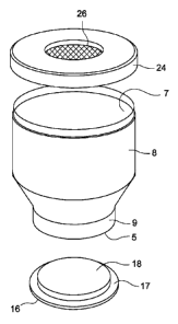
100221 Turning to Figs. 1-3, there is shown a disposable container 2 for
bioburden
sample selection and detection. The container is composed of a clear plastic
material such
as polycarbonate, polyethyleneterephlate, polypropylene, or other plastic
material. As
shown, the container comprises cylindrically cross sectioned body section 8
and a tapered
or decreasing diameter section 10 in the general form of a truncated conical
shape
superposed over the body 8. As shown in Fig. 1, the top most section of the
container is
defined by a narrowed neck portion 9 over which the cap sections are adapted
for
engagement by threaded engagement, snap fit, or other like attachment means.
With
specific reference to Fig. 1, a protective film 14 is provided on the top of
the container and
is, for example, shrink fit over the top of the container for easy removal.
The top cap 12
also includes an annular sealing lid portion 17. The top cap assembly fits
over and seals top
opening 5 of the container.
100231 At the bottom end of container 2, a removable cap 20 is provided over
the
bottom or base of the bottle. A fluid impervious rubber diaphragm 26 or
similar surface is
provided along the inside lower extremity of the bottle, and this may be
secured to the bottom
of the bottle via sealing cap 24 that can be threaded or snap fit over the
bottle base. The cap 20
seals the bottom opening 7 of the container.
[0024] In Fig. 2, the protective film 14 has been removed from the top cap 12
revealing annular sealing lid 17 that, in turn, is preferably friction fit
within the
circumference of the neck 9. Annular sealing lid 17 comprises a raised annular
land area
CA 2884671 2018-05-14
CA 02884671 2016-12-21
255255
6
surrounding a recessed, planar section having a multiplicity of apertures 16
therein to
facilitate fluid flow through sealing lid 17. On the underneath or bottom side
of lid 1 7 is
a filtration membrane 18 that may be composed of polyester, polycarbonate,
polypropylene, polytetrafluorethylene, nylon, and poly(vinylidene difluoride).
This
membrane functions to retain the microbial species thereon as shall be
referred to in
detail later.
[0025] Fig. 3 shows the container in inverted disposition. This is the
orientation in
which, in one embodiment, the bottle will be positioned in order to facilitate
bioburden
sampling and detection. Here, removable outer cap 20 has been removed,
revealing the
annular securing cap 24 and underlying fluid impenetrable rubber layer 26. The
layer 26 is
adapted to act as a barrier to fluid flow therethrough, but this layer may be
penetrated by
a sharp object such as a hypodermic needle or the like. More particularly,
layer 26 is
formed of an clastomeric material such as EPUM rubber, styrene butadiene,
acrylonitrile/
butadiene, polychloroprene, or natural rubber. Also, in Fig. 3, the protective
film 14 has
been removed from the now inverted top of the bottle, revealing filtration
membrane 18
on the bottom side of annular snap lid 17 with the recessed aperture side 16
of lid now
disposed under the filtration membrane 18.
[0026] In order to perform bioburden testing, the bottle 2 is filled with
aqueous or
other fluid sample containing the analyte microbial species. The bottle 2 may
be placed in
a bottle holding assembly 100 such as the type shown in Fig. 4. Here, assembly
100
comprises two opposed platen members 102, 104, with the top platen 102 joined
to platen
104 via connectors 106, 108, 110, 112 which can all be adjusted through the
spring
tension supplied by coil spring 114 and 116. A retainer member 120 such as
composed of
a rubber diaphragm or similar resilient material depends downwardly from the
platen 102
and has a general shape that is congruent to the shape of bottle 2 to securely
grasp and
hold same in the assembly 100.
[0027] As shown in Fig. 4, platform 140 is positioned underneath the platen
104
and the annular sealing lid 17 is engaged between yoke members 142, 144
carried on the
platform, which together, encircle the lid 17. Set screws or the like may be
used to vary
the tension of springs 146, 148 to provide for secure grasping of lid 17 by
the yoke
CA 02884671 2015-03-12
WO 2014/047770
PCT/CN2012/081889
7
members. At this point, the protective film 14 has been removed from the tap
12, so that
the membrane 18 (Fig. 3) is in direct fluid communication with the contents of
the bottle
2. A conduit 150 is connected via a funneling type connector or other device
(not shown)
to the now inverted top section of the bottle. A vacuum source 154 or pump
communicates with the bottle 2 via the provision of valve member 152. A
hypodermic
rubber needle or the like is inserted through the penetratable rubber layer of
the bottle to
inject staining, luminescence, cleaning, or other reagent or purified water to
the fluid
sample within bottle 2.
[0028] By turning on the vacuum or pump, the fluid in the sample is drained
out
through the filtration membrane 18 and the draining aperture 16 on the lid 17
to waste.
Due to the pore size of the filter membrane, the microbial species of the
sample will be
captured on the filtration membrane 18, and then the pump or vacuum source is
turned
off. Thus, after draining the fluid from the bottle, the microbial species and
reagent are
disposed on the membrane 18 so that the microbial count may then be made via
conventional techniques.
[0029] Turning to Fig. 5, as shown, the platform 140 has been removed from the
lower platen 104 with the microbial species and reagent disposed on the
surface 18.
Immediately prior to this step, the lid 17 has been removed from the remainder
of the
container, leaving lid 17 disposed on the platform. As shown in Fig. 5, the
spring tension
in springs 146 and 148 has been relaxed with the lid 17 loosely encircled by
the yoke
members 142, 144. At this stage, a detector such as a microscope or the like
can be
moved close to the membrane to count or otherwise detect the analyte microbial
species.
After the inspection, the bottle 2 may be disposed of.
[0030] It is thus apparent that a disposable container for bioburden sample
selection and detection is provided. This container works as a normal bottle
to select the
fluid sample and is sealed by a cap. In one aspect of the invention, the
container is then
placed upside down on stages for sample preparation (filtration, staining) and
detection
(imaging).
[0031] The bioburden measurement process may involve any one of many
conventional techniques such as a colormetric measurement technique in which a
staining
CA 02884671 2015-03-12
WO 2014/047770
PCT/CN2012/081889
8
reagent is injected into the fluid sample in container 2 via a syringe or the
like that
penetrates the layer 26. A microscope or the like is used to count the
microbial colonies
on the membrane 18 to assess bioburden level.
[0032] Additionally, another exemplary bioburdcn test method may comprise
injection of a luminescent or fluorescent reagent into the sample in the
container 2
through the penetrable layer 24. The desired analyte microbial species
captured on the
surface of membrane 18 can then be detected via a camera or the like placed
proximate
the membrane 18.
[0033] Another bioburden testing procedures that may be utilized involves the
"in
situ cell division" method. This method is a direct growth rapid method for
living target
cell detection before the cells become visible to the naked eye. In this
method, the
sample is filtered through the MF to capture the microorganism thereon. The MF
is
transferred to a nutrient agar plate and the target cells are allowed to form
microcolonies
by in situ respiration. (This usually takes about a three-hour incubation
time.) The
microcolonics arc illuminated with a blue light LED (Ex 450-500 nm, Em 510-560
nm).
A CCD chip is used to detect cellular autofluorescence and analysis software
is used to
automatically count the photosensitive pixels overlying each microcolony.
[0034] Another exemplary bioburden test procedure is known as the "solid phase
cytometry (SPC) method". This method is a rapid method for detection of total
viable
cells (TVC) by the sequential steps of: (a) filtering a sample over a black
polyester or
polycarbonate MF to capture microorganisms; (b) transferring the MF to a pad
comprising fluorocarbon dye, incubating, to label metabolically active cells
only; (c)
detecting fluorescence emitted by labeled cells with argon laser scanning (Ex
480 nm,
Em 515nm); (d) processing signals by computer to differentiate valid signals
from
fluorescent particles; and (c) quantifying total viable cells.
[0035] The pore size of the filter membrane is chosen to capture the secured
microbial analyte thereon. This pore size can vary over a wide range such as
to
encompass pore sizes from about 0.05 i.tm to about 0.65 ium. Exemplary
membrane
filters (MF) include the following: 1) 25 mm diameter, 7-20 1.im thickness,
0.4 lam pore
size; 2) 25 mm diameter, 6-11 [tm thickness, 0.2 [tm pore size; and 3) 47 mm
diameter,
CA 02884671 2016-12-21
255255
9
7-22 i.tm thickness, 0.45 um pore size. The selection of the appropriate
filter membrane
18 is within the skill of the artisan.
100361 The artisan will appreciate that bioburden testing can be conducted to
determine presence and count levels of a variety of microbes. For example,
counts of a
variety of bacteria, fungi, mold, yeast, and spores can be conducted using the
container
and methods of the present invention.
10037] It is to be understood that the invention is not limited to the
illustrations
described and shown herein, which arc deemed to be merely illustrative of
various
embodiments of the invention and which arc suitable for a variety of
modifications. The
invention is intended to encompass all such modifications which are in the
scope as
defined in the attached claims.