Note: Descriptions are shown in the official language in which they were submitted.
CA 02885981 2015-03-23
DEGRADABLE IMPLANTABLE MEDICAL DEVICES
BACKGROUND OF THE INVENTION
[0001] 1. Field of the Invention. The present invention relates to
medical devices and methods.
More particularly, the present invention relates to implantable luminal
prostheses and other medical
devices which degrade in a body environment.
[0002] 2. Description of the Background Art. Coronary artery disease is
the leading cause of
death in the industrialized countries around the world. It begins as the
accumulation of
atherosclerotic deposits in the walls of the major arteries which supply blood
to the heart. As the
deposits accumulate, normal blood flow to the heart is restricted. The heart
has several
compensatory mechanisms, which, to a point, can offset such diminished blood
flow. Beyond these
compensatory mechanisms, a number of well established pharmaceutical
treatments have been
shown to improve both symptoms and mortality in patients with mild to moderate
coronary artery
disease. However, as the disease progresses, its symptoms become more
apparent, despite drug
therapy. When the heart does not get enough blood, particularly during
exercise or stress, advanced
coronary artery disease is manifested as debilitating chest pain or angina. At
this point, mechanical
intervention is required to increase the amount of blood flowing to the heart.
[0003] Angioplasty is one of the most common interventional treatments
for advanced
coronary artery disease. Andreas Gruntzig performed the first percutaneous
transluminal coronary
angioplasty (PTCA) procedure. He advanced a catheter with a small balloon
through the aorta and
into a coronary artery with a partial occlusion. He then inflated the balloon,
compressing the plaque
against the arterial wall, and restoring blood flow to the heart.
[0004] PTCA has grown rapidly, and angioplasty catheters have become
smaller and more
maneuverable, allowing interventional cardiologists to access more difficult
coronary blockages.
However, restenosis, or reocclusion of the treated lesion, has plagued PTCA.
Typically 30-40% of
all patients have restenosis following PTCA.
[0005] Coronary stents were introduced in the mid 1990s to prevent
restenosis. A stent is a
small metal coil, slotted tube, mesh or scaffold structure that is placed in a
coronary artery. It is a
permanent implant that remains in the coronary artery following PTCA. The
stent helps hold the
artery open, improves the flow of blood, and relieves symptoms of coronary
artery disease.
1
CA 02885981 2015-03-23
Coronary stents were the first devices proven to reduce restenosis, dropping
the rate of restenosis to
15-20%. Stents have since been used in the majority of PICA procedures.
[0006] Conventional stents have taken two forms, balloon expandable
stents and self-
expanding stents. Both are typically made of metallic materials and may
include a biocompatible
coating. Such stents are permanently implanted into the human body by
deploying them on or
through a catheter. Such permanent implantation may increase the amount of
intimal hyperplasia,
thrombosis or other adverse medical effects. Coronary stents accomplish a
lower restenosis rate of
15-20% post angioplasty compared to PICA alone as a result of maintaining a
higher acute gain
post procedure.
[0007] Drug eluting stents, which elute drugs such as rapamycin and
paclitaxel, were designed
to further reduce intimal hyperplasia rates with stents. Such drug eluting
stents incorporate metal or
metal alloys with degradable or non degradable polymers which control release
of the drug. The use
of such drugs has further reduced the rates of restenosis as compared to
stents alone.
[0008] The metals or metal alloys used for both conventional and drug
eluting stents are
intended to be biologically stable and remain in the body for the patient's
life unless surgically
removed at a later date along with surrounding tissue. Thus, these stents do
not permit temporary
placement within the body unless patient and surgeon are prepared to undertake
a second procedure
to remove the stent, which is difficult or impossible in most cases.
[0009] Although one of the primary functions of stenting is to provide
mechanical support to
the blood vessel wall and to preserve the lumen for blood flow, once the
vessel wall heals the stent
serves little or no continuing purpose. Further, the presence of a stent which
remains mechanically
rigid could potentially cause complications to the patient. It has therefore
been desired to provide a
stent which dissolves or degrades during or shortly after healing of the
vessel or thereafter
[0010] There have been several attempts to make stents from
biodegradable polymer materials
such as poly-lactic acids (PLA). Such polymer stents, however, tend to provide
less mechanical
support for the vessel wall and therefore have to be substantially thicker
than a comparable metallic
stent. The thickness can reduce the available blood flow lumen and can cause
undesirable biologic
responses.
[0011] Recent attempts have been made to make metal stents which
decompose in the body, as
described for example in U.S. Patent Nos. 6,287,332 B1 and 6,854,172 B2. See
also
2
CA 02885981 2015-03-23
US2004/009808 and WO 02/053202. Such degradable metal stents , however, often
compromise
strength, profile, and other desirable characteristics which are found in
conventional metal stents.
[0012] For these reasons, it would be desirable to provide degradable
devices that have
improved physical and mechanical characteristics. In particular, it would be
desirable to provide a
stent or other luminal prosthesis which is degradable during and/or upon
healing of the vessel or
thereafter and which has features to reduce the risk of injury to the vessel
or restenosis. It would
also be desirable to provide localized and controlled release of a
pharmacological agent from the
stent or other device for the treatment of blood vessels and other body
structures at the location
being treated with the stent. Such pharmacological agents could minimize both
restenosis and any
inflammatory response towards the stent or other device and degradation
products thereof. At least
some of these objectives will be met by the aspects of the present invention.
BRIEF SUMMARY
[0013] Various medical devices and methods disclosed herein utilize an
implantable structure
comprising a body which is degradable over a clinically relevant period of
time. The body may
have a variety of forms and may be used in a variety of medical treatments. In
preferred
embodiments, the body has the form of a stent, particularly a vascular stent
of the type used in the
treatment of coronary artery disease. The body comprises or is formed or
constructed from a
material which provides desired physical and mechanical attributes for the
device. In preferred
embodiments, the body comprises a metal (pure or with impurities), a metal
alloy or a combination
thereof. The term "metal" as used hereinafter will include such pure and
impure metals as well as
metal alloys and other combinations of two or more metals and metal alloys.
The implantable
bodies are at least partially degradable in a physiologic environment.
Preferably, the materials of
implantable structures are fully degradable so that no structure remains after
a clinically relevant
time period, as discussed below, and produce degradation byproducts which are
physiologically
benign, preferably being of a type which is naturally occurring in the body
environment. More
preferably, the bodies of the implantable structures produce degradation
byproducts in amounts
lower than what is typically present in the physiologic environment. The
degradation rate of the
implantable structure may be controlled in a variety of aspects individually
or in combination
thereof. Exemplary physiologic environments include vascular and other body
lumens including the
ureter and the urethra, solid tissues, cerebral tissue, and the like.
3
CA 02885981 2015-03-23
[0014] In a first aspect of the present disclosure, the degradation rate
of the body of the
implantable structure is controlled by selection of the composition of the
implant material. The
implant material is selected from a metal or metal alloys or combination
thereof which can degrade
in a clinically relevant time period ranging from approximately one month to 5
years, usually from
4 months to 2 years, and often from 6 months to one year. Thus, the weight or
volume of the
implantable structure will typically diminish each day by a percentage in the
range from about
0.05% to 3%, usually from 0.1% to 0.75% per day, and more usually from 0.25%
to 0.5% per day.
[0015] The metal, alloy, or combination material of the implantable
structure will usually have
a corrosion current (Icon) in the range from 0.0001 amps/cm2 to 0.1 amps/cm2,
usually from
typically 0.001 amps/cm2 to 0.01 amps/cm2, and usually from 0.0025 amps/cm2 to
0.008 amps/cm2.
The corrosion current is proportional to the corrosion rate, so materials with
higher Icorr values will
corrode more rapidly in the vascular or other physiologic environment. Icon
varies with the
material property, geometry, and surface characteristics of the implant, and
also physiologic
environment among other factors. The Icorr value will typically represent an
average value for the
body as a whole or for any portion of the body.
[0016] In a second aspect, the degradation rate of the implantable
structure is controlled at least
in part by modifying its geometry. Such geometry modifications may include
surface area to
volume ratio. For example, attributes such as holes, reservoirs, trenches or
others can be
incorporated into the body to increase the surface area without significantly
increasing the volume
which can be used to control the degradation rate of the structure. When the
implantable body
comprises a stent having a strut, the geometry to be modified may include the
strut width to strut
thickness ratios.
[0017] In a third aspect disclosed herein, the degradation rate of the
implantable structure is
controlled at least in part by the addition of corrosion inducing features.
For example, in some
embodiments, the implantable structure comprises an implantable body having at
least one surface
and at least one corrosion inducing feature on the at least one surface which
causes at least a portion
of the structure to degrade at a controlled degradation rate. In preferred
embodiments, the
implantable body comprises a metal, a metal alloy or a combination thereof. In
some embodiments,
the corrosion inducing feature comprises a pit, pore, partial hole, void or
combination of these. In
other embodiments, the corrosion inducing feature comprises a surface
irregularity, scratch, streak,
ridge, bump, texture, sintered porous metal or alloy, roughened surface or
combination of these. In
4
CA 02885981 2015-03-23
still other embodiments, the corrosion inducing feature comprises a hole,
either partial or complete
sintered pores, or combination of these. Further, in some embodiments, the
implantable body has a
first surface with a first associated portion of the body and a second surface
with a second
associated portion of the body, wherein the first surface has corrosion
inducing features present in a
density and/or configuration which causes the first associated portion to
degrade at a rate which
differs from the second associated portion.
[0018] Exemplary metals include iron, cobalt, tungsten, molybdenum,
silver, and the like.
These metals may be substantially pure, typically having purities about 90% by
weight, often above
95% by weight, and frequently above 99.5% by weight. Alternatively, these
metals may be
combined as alloys with other metals or materials. Exemplary alloys include
iron-containing alloys,
such as AISI series 1000 carbon steels, AISI series 1300 manganese steels,
AISI series 4000
molybdenum steels, AISI series 4100 chromium-molybdenum steels, AISI series
4300 and AISI
series 8600 nickel-chromium-molybdenum steels, AISI series 4600 nickel-
molybdenum steels,
AISI series 5100 chromium steels, AISI series 6100 chromium-vanadium steels,
AISI series 9200
silicon steels, and the like. Other iron-containing alloys will have at least
25% iron, preferably 50%
iron, more preferably 75% iron, and often 90% iron, 95% iron, or 99% iron, or
greater by weight.
Iron alloys may contain carbon ranging from 0.05% to 3% by weight, preferably
0.05% to 1.0% by
weight, more preferably 0.1% to 0.6% by weight. Alloys of silver, tin, cobalt,
tungsten,
molybdenum, and the like, will usually have at least 25% by weight of the pure
metal, usually at
least 50% by weight, often at least 75% by weight, and sometimes 90% by
weight, 95% by weight,
or 98% by weight, or greater.
[0019] In a fourth aspect disclosed herein, the degradation rate of the
implantable structure is
controlled at least in part by the manipulation of corrosion enhancing and/or
corrosion resisting
elements in the implant structure. Thus, atoms or compounds which lower the
resistance of a metal
or metal alloy to corrosion can be added to or increased if already present in
these materials.
Likewise, one or more corrosion resisting elements may be depleted. Such
manipulation of
elements may occur on a surface of the implant structure, throughout the
implant structure or
adjacent to a grain boundary of a metal or alloy to control corrosion of the
metal or alloy.
[0020] In a fifth aspect disclosed herein, the degradation rate of the
implantable structure is
controlled at least in part by the addition of corrosion controlling agents.
Such agents may be
synthetic or biologic, such as acidic compounds, sodium chloride, calcium
chloride, magnesium
5
CA 02885981 2015-03-23
chloride, hydrochloric acid, citric acid, amino acid, hydroxyapatite, hydrogen
peroxide, basic
compounds such as potassium hydroxide, acidic and basic pharmaceutical agents,
or polymers with
acidic or basic byproducts, others or a combination thereof.
[0021] In a sixth aspect disclosed herein, the degradation rate of the
implantable structure is
controlled at least in part by the creation of a galvanic cell. In some
embodiments, metal or alloy
particles are delivered adjacent to the implant structure, either in fluid or
tissue. These particles are
in fluid contact with the implant and create a corrosion-inducing galvanic
cell. Galvanic cells can
be created, for example, by alloying metals having different electrochemical
potentials so that a
current may be generated to oxidize the alloy in the electrolytic physiologic
environment.
[0022] In a seventh aspect disclosed herein, the degradation rate of the
implantable structure is
controlled at least in part by layering of materials. In some embodiments, the
implantable structure
comprises an implantable body having a first layer which degrades at a first
degradation rate and a
second layer which degrades at a second degradation rate which differs from
the first rate. The first
layer and second layer comprises a metal, metal alloy or combination thereof
and the layers cause at
least a portion of the structure to degrade at a controlled degradation rate.
The first and second
layers may have different passive states. The first and second layers may
differ in an
electrochemical series. Alternatively or in addition, the degradation period
and/or degradation rates
may differ due to differing thicknesses of the layers.
[0023] In an eighth aspect disclosed herein, the degradation rate of the
implantable structure is
controlled at least in part by incorporating or manipulating a protective
layer. In some
embodiments, the implantable structure comprises an implantable body
comprising a metal, metal
alloy or combination thereof having a degradation rate, and a layer which
covers at least a portion
of the implantable body, wherein aspects of the layer are controlled which
controls the degradation
rate of the implantable body. The protective layer may comprise a passivation
layer or a coating.
Such coatings may comprise a polymer, metal, metal alloy, therapeutic agent,
corrosive agent,
radiopaque agent or combination of these. Such aspects of the protective layer
may include
thickness, chemical composition, chemical permeability, durability, amount of
coverage of the
implantable structure, or a combination of thereof, such aspects of the
protective layer may include
amount of corrosion-resistant oxides. Optionally, the protective layer may
have openings which
reveal underlying portions of the implantable body, wherein the openings
assist in controlling the
degradation rate of the implantable body.
6
CA 02885981 2015-03-23
=
[0024] In further aspects disclosed herein, an implantable structure is
provided comprising an
implantable body comprising a metal, metallic alloy or combination thereof
which has at least a
portion which degrades at a controlled degradation rate, wherein the
controlled degradation rate has
at least two phases of differing degradation rates. In some embodiments, the
at least two phases
comprises an initial degradation rate which is slower than a later degradation
rate. In other
embodiments, the at least two phases comprises an initial degradation rate
which is faster than a
later degradation rate.
[0025] In another aspect disclosed herein, an implantable structure is
provided comprising an
implantable body comprising a metal, metallic alloy or combination thereof
which has at least a
portion which degrades at a controlled degradation rate which varies along its
structure. In preferred
embodiments, the implantable body comprises a stent.
[0026] In yet another aspect disclosed herein, an implantable structure
is provided comprising
an implantable body comprising a metal, metal alloy or combination thereof
having a controlled
degradation rate, and at least one therapeutic agent which elutes from the
implantable structure.. In
some embodiments, the therapeutic agent includes a pharmacological agent
including but not
limited to an anti-cancer agent, an anti-inflammatory agent, an
immunosuppressive agent,
antiproliferative, antiplatelet , or a combination of these. In another
aspect, the implantable structure
further comprises at least one coating which at least partially covers the
implantable body. The
therapeutic agent may be contained in or adjacent to the coating. The coating
maybe metallic,
polymeric, ceramic; synthetic or natural; and or combination thereof. The
coatings may be
degradable, partially degradable, non degradable, and or combination thereof.
In other
embodiments, the therapeutic agent comprises one therapeutic agent which
elutes in one phase of
degradation and another therapeutic agent which elutes in another phase of
degradation. Further, in
other embodiments, the therapeutic agent is at least partially contained in a
corrosion inducing
feature.
[0026A] Various embodiments of the claimed invention relate to a degradable,
implantable
structure comprising: an implantable body having a first layer which degrades
at a first degradation
rate, wherein the first layer comprises a metal, metal alloy or combination
thereof, and a second
layer which degrades at a second degradation rate which differs from the first
rate, wherein the
second layer comprises a metal, metal alloy or combination thereof, wherein
the layers cause at
least a portion of the structure to degrade in a physiologic environment.
7
CA 02885981 2015-03-23
[0026B] Various embodiments of the claimed invention relate to a degradable,
implantable
structure comprising: an implantable body comprising a metal, metallic alloy
or combination
thereof which has at least a portion which degrades at a degradation rate,
wherein the degradation
rate has at least two phases of differing degradation rates.
[0026C] Various embodiments of the claimed invention relate to a degradable
structure
comprising: an implantable body comprising a metal, metallic alloy or
combination thereof which
has at least a portion which degrades at a degradation rate which varies along
its length.
[0026D] Various embodiments of the claimed invention relate to a degradable
structure
comprising: an implantable body comprising a metal, metallic alloy or
combination thereof,
wherein its geometry affects the degradation rate.
[0026E] Various embodiments of the claimed invention relate to a degradable
structure
comprising: an implantable body comprising a metal, metal alloy or combination
thereof having a
degradation rate; and at least one therapeutic agent which elutes from the
implantable structure.
[0026F] Various embodiments of the claimed invention relate to a
degradable structure
comprising: an implantable body comprising a metal, metallic alloy or
combination thereof which
has at least a portion which degrades at a rate to approximate dissolution in
the physiologic
environment in a period between one month to 5 years.
[0026G] Various embodiments of the claimed invention relate to a degradable
implant
comprising: a body composed of a metal and having a structure, wherein said
metal and structure
are selected to allow the body to degrade in a physiologic environment in a
period of from 1 month
to 5 years.
[0027] Various aspects, objects and advantages are covered by the
detailed description to
follow, together with the accompanying drawings.
BRIEF DESCRIPTION OF THE DRAWINGS
[0028] Figure 1 illustrates an example of an embodiment of the implant
of the present
disclosure in the form of a stent.
[0029] Figure 2 A shows examples of metal and metal alloy degradation
data.
7a
CA 02885981 2015-03-23
[0030] Figure 2B illustrates data for weight loss over time of some
metal and metal alloy
implant materials as compared to stainless steel.
[0031] Figure 3 illustrates a portion of a stent body having an example
of corrosion inducing
features thereon.
[0032] Figure 4 provides a close-up illustration of examples of corrosion
inducing features on a
stent body.
[0033] Figure 5 illustrates a cross-section of a strut of a stent body
having examples of
corrosion inducing features extending therein.
[0034] Figure 6 illustrates a surface of an implant having examples of
corrosion inducing
features of a variety of shapes and sizes extending from the surface into the
implant.
[0035] Figure 7 illustrates cross section of an implant body with an
example of corrosion
inducing features which extend from the surface and cross or join within the
implant.
[0036] Figure 8 illustrates an example of a corrosion inducing feature
which extends from the
surface and includes side-branches within the implant.
[0037] Figure 9 illustrates an example of a corrosion inducing feature
which extends from the
surface into the implant and includes at least one protrusion which extends
outwardly from the
surface.
[0038] Figure 10 illustrates examples of corrosion inducing features in
the form of scratches.
[0039] Figure 11 illustrates a surface of an implant having examples of
corrosion inducing
features in the form of, textured surfaces.
[0040] Figure 12 illustrates a cross-section of a portion of an implant
having examples of
corrosion inducing features in the form of holes extending through the
implant.
[0041] Figure 13 illustrates an example of a hole including a plurality
of side-branches.
[0042] Figure 14 illustrates an example of corrosion inducing features
substantially uniformly
distributed across a surface of an implant.
[0043] Figure 15 illustrates an example of corrosion inducing features
of Figure 14 non-
uniformly distributed across a surface of an implant.
8
CA 02885981 2015-03-23
100441 Figure 16 illustrates an example of corrosion inducing features
in the form of streaks
substantially uniformly distributed across a surface of an implant.
[0045] Figure 17 illustrates an example of corrosion inducing features
of Figure 16 non-
uniformly distributed across a surface of an implant.
[0046] Figure 18 illustrates an example of particles in fluid or tissue
adjacent to an implant.
[0047] Figure 19 illustrates an example of a cross-sectional view of an
implant having three
layers.
[0048] Figures 20-21 provide plots showing degradation rates of some
metals and alloys with
examples of corrosion inducing features.
DETAILED DESCRIPTION
[0049] The devices of the present disclosure may have a variety of forms
and may be used in a
variety of medical treatments. In preferred embodiments, the device has the
form of a vascular stent
that is used in the treatment of vascular disease. It may be appreciated that
stents may be used in a
variety of body lumens, such as an artery, vein, biliary duct, or esophagus.
[0050] In other embodiments, the devices of the present disclosure have
the form of a variety
of implants, such as graft implants, vascular implants, non- vascular
implants, implantable luminal
prostheses, wound closure implants, drug delivery implants, sutures, biologic
delivery implants,
urinary tract implants, inter-uterine implants, organ implants, bone implants
including bone plates,
bone screws, dental implants, spinal disks, or the like. The devices typically
allow for one or more
of the following: support, contain, hold together, affix, plug, close, deliver
drug, deliver biologies to
an organ, vessel, conduit, or bone for the treatment of hyper-proliferative
diseases, restenosis,
cardiovascular disease, wound healing, cancer, aneurysm, diabetic disease,
abdominal aortic
aneurysm, hyper-calcemia, ischemia, fibrillation, arrhythmia, or others.
[0051] Thus, the following detailed description utilizes the stent by way
of example and is not
intended to limit the scope of the invention.
9
CA 02885981 2015-03-23
Implant as a Stent
[0052] In preferred embodiments, the implant has the form of a stent.
Stent designs include
coils, slotted tubes, corrugated rings, sheets, rolled sheets, locking
designs, and stent grafts, to name
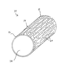
a few. Fig. 1 illustrates an embodiment of a stent 10 in an expanded state. As
shown, the stent 10
has a first end 12, a second end 14 and a central lumen 16. A stent body 18
extends from the first
end 12 to the second end 14. The body 18 has struts 20 which form a lattice-
type structure. The
struts 20 may have circular, rectangular or other shape cross-sections.
Typically, strut thicknesses
range from approximately 0.0005" to 0.010", preferably approximately 0.001" to
0.004", more
preferably approximately 0.0015" to 0.003". Strut widths typically range from
approximately
0.001" to 0.008", preferably approximately 0.002" to 0.004".
[0053] The stents 10 may be self-expanding or balloon expandable. Stent
pre-expansion
diameters typically range from approximately 0.3 mm to 10 mm, preferably
approximately 0.5 mm
to 4 mm, more preferably approximately 0.8 mm to 2 mm. Stent post expansion
diameters typically
range from approximately 1.5 mm to 35 mm, preferably approximately 2 mm to
lOmm, more
preferably approximately 2 mm to 5 mm. Depending on the material(s) from which
the stent 10 is
formed, the material(s) typically have a percent elongation range from
approximately 5% to 100%,
preferably approximately 15% to 70%, more preferably approximately 20% to 50%.
Degradation of Implant
[0054] As stated previously, devices disclosed herein are degradable in a
biological
environment. It may be appreciated that the terms degradation, biodegradation,
dissolution,
bioabsorption, absorption, resorption, corrosion, erosion, bioerosion,
erodible,
CA 02885981 2015-03-23
WO 2006/108065 PCT/US2006/012725
bioerodible and disintegration are used interchangeably along with other terms
which
describe any such deterioration in mass, volume or function by chemical,
biological,
electrical, mechanical, or any other means, unless stated otherwise.
[0055] It may also be appreciated that degradation as related to the
present invention
is considered to be degradation within a clinically relevant time period, such
as
approximately one month to 5 years. Although common metals or alloys may
corrode at
much longer rates ranging up to 1000 years or more depending on the inherent
properties of
the materials and the environmental conditions, such metals or metal alloys
are considered
nondegradable in a clinical setting. In preferred embodiments, the devices of
the present
invention substantially degrades in the body environment in the range of
approximately one
month to 5 years, preferably approximately 4 months to 2 years, more
preferably
approximately 6 months to I year. In some embodiments, the devices at least
partially
degrade in the body environment in less than one month, such as a few weeks,
one week, a
few days, one day, a few hours, one hour or less. For example, the device may
have at least a
portion which degrades at a controlled degradation rate to approximate
dissolution at or
within one month. Body environments effecting degradation typically have local
tissue pH
ranges from approximately 3 to 10, usually approximately 5 to 9, typically
approximately 6
to 8.
[0056] Degradation of devices of the present invention may occur in
multiple phases,
such as a slower degradation rate in one phase and a faster degradation rate
in another phase.
In some embodiments, the device degrades at a slower rate in an initial phase
and a faster rate
in a later phase. In other embodiments, the devices degrade at a faster rate
in an initial phase
and slower rate in a later phase. Likewise, degradation may be uniform along
the structure or
variable along the structure. The average mass or volume percentage loss may
range from
approximately 3% per day to 0.05% per day, preferably approximately 0.75% per
day to
0.1% per day, more preferably approximately 0.5% per day to 0.25% per day.
[00571
When the implant is comprised of a metal or metal alloy, degradation of the
implant
produces byproducts such as metal ions, salts or complexes. Preferably, these
byproducts are
naturally occurring elements in the body environment or cause no significant
harmful effects.
More preferably, the implantable structures produce degradation byproducts in
amounts
lower than what is typically present in the body environment. Further, the
rate of degradation
of the implant may be controlled to minimize the possibility of any negative
biologic
response from the degradation byproducts. Currently, long-term anti-platelet
therapy is
11
CA 02885981 2015-03-23
WO 2006/108065 PCT/US2006/012725
recommended for patients undergoing permanent implantation of conventional non-
degradable devices, such as stents, to prevent acute thrombosis or late
thrombosis. Systemic
anti-platelet therapy has side effects such as internal bleedings. Long- term
anti-platelet
therapy may not be necessary when degradable implants of the present invention
are used
since the risk of thrombosis is reduced once the implant has dissolved. This
may lower
procedure-associated cost and also minimize risks associated with patient
compliance of daily
doses of drugs. As an example, when the implant comprises or consists
essentially of iron or
an iron alloy, the degradation products may include biocompatible iron species
such as
oxidized iron species of a type which naturally occur in the human body. By
controlling the
degradation rate, the concentration of these species can be kept below 10 fold
the normal
amounts, preferably below five fold, more preferably below two fold, and most
preferably at
a level no greater than naturally present in the body. A particular preferred
metal is a carbon
steel where the degraded species will include primarily or exclusively iron
and carbon
compounds.
Metals and Metal Alloys
[0058] In preferred embodiments, the devices of the present invention
comprise at
least partially degradable metals, metal alloys, or a combination thereof.
[0059] Examples of metals include Cobalt, Tungsten, Bismuth, Silver,
Copper, Iron,
Zinc, Magnesium, Zirconium, Molybdenum, Indium, Tin or other metals. In some
embodiments, implant metal purity ranges from approximately 90% to 100%,
preferably
approximately 95% to 99.99%, more preferably from approximately 99.5 to 99.9%
by
weight.
[0060] Examples of metal alloys include: 1) silver containing alloys
(such as silver-
tin alloys , 2) cobalt containing alloys (such as cobalt-iron alloys), 3) iron
containing alloys
(such as 80-55-06 grade cast ductile iron, other cast ductile irons, AISI 1010
steel, AISI 1015
steel, AISI 1430 steel, AISI 8620 steel, AISI 5140 steel, or other steels, or
others), 4) tungsten
containing alloys, 5) melt fusible alloys (such as 40%bismuth-60%tin,
58%bismuth-42%fin,
bismuth-tin-indium alloys or others), 6) magnesium alloys, 7) zinc alloys, 8)
shape memory
or superelastic alloys, and 9) other alloys, to name a few.
[0061] In some embodiments, the devices of the present invention are
comprised of
more than one metal or metal alloy. Examples of metal+metal implants include
cobalt+tungsten, tungsten+iron, magnesium+iron, silver+zinc or others.
Examples of
12
CA 02885981 2015-03-23
WO 2006/108065 PCT/US2006/012725
metal+alloy implants include tungsten+8620 steel, titanium+low carbon steel,
magnesium
+1015 steel alloy, silver+bismuth-tin alloy or others. Examples of alloy+alloy
implants
include 8620 steel+silver-tin, 1015 steel+bismuth-tin or others.
[0062] Examples of metal and metal alloy degradation data are
illustrated in Fig. 2A.
Fig. 2B illustrates weight loss data over time for some metal and metal alloy
implant
materials as compared to stainless steel.
[0063] Degradation of metals is commonly termed corrosion, and the
terms
"degradation" and "corrosion" are used interchangeably herein for all
materials. Most metal
corrosion occurs via electrochemical reactions at the interface between the
metal and an
electrolyte solution, such as found in the body environment. Corrosion
typically occurs as a
result of anodic and cathodic reactions.
[0064] The potential of the metal is the means by which the anodic and
cathodic
reactions are kept in balance. The equilibrium potential assumed by the metal
in the absence
of electrical connections to the metal is called the Open Circuit Potential,
Eoc. The value of
either the anodic or cathodic current at Eoc is called the Corrosion Current,
Icorr. Icorr and
Corrosion Rate are a function of many system variables including type of
metal, solution
composition, temperature, solution movement, metal history, and many others.
[0065] For implant in saline solution at 37 C temperature, the
corrosion current flux
(Icon) is proportional to corrosion rate (CR) according to the following
formula:
CR = (Icorr x K x EW) / d
CR The corrosion rate (ram/yr)
Icon The corrosion current (amps/cm2)
Faraday's constant = 3272 (mm/(amp-cm-year))
EW The equivalent weight (grams/equivalent)
d Density (grams/cm3)
[0066] Typical implants of the present invention degrading in
physiological
conditionswill have Icon (corrosion current) ranges from approximately 0.0001
amps/cm2 to
13
CA 02885981 2015-03-23
WO 2006/108065 PCT/US2006/012725
0.1 amps/cm2; preferably approximately 0.001 amps/cm2, to 0.01 amps/cm2, more
preferably
approximately 0.0025 amps/cm2 to 0.008 amps/cm2.
[0067] In some embodiments, after one month of degradation, the metal
or metal
alloy maintains greater than approximately 25% of strength, preferably greater
than
approximately 50%, more preferably greater than approximately 60% of strength
as
compared to strength prior to implantation. In these or other embodiments,
after two months
of degradation, the metal or metal alloy implant maintains greater than
approximately 25% of
strength, preferably greater than approximately 50%, more preferably greater
than
approximately 60% of strength as compared to strength prior to implantation.
In these or
other embodiments, after four months of degradation, the metal or metal alloy
implant
maintains greater than approximately 25% of strength, preferably greater than
approximately
50%, more preferably greater than approximately 60% of strength as compared to
strength
prior to implantation.
[0068] In some embodiments, the implant will be corroded prior to implantation
with an
amount of corrosion greater than 0.01% by weight, preferably with greater than
0.1% by
weight, and more preferably with greater than 1% by weight (based on the
weight of the body
prior to corrosion). In some embodiments, the corrosion prior to implantation
may cover
greater than 1% of the surface area, preferably greater than 5% of the surface
area, and more
preferably greater than 10% of the surface area of the body which will be
exposed to the
physiologic environment. In such cases, the corrosion may result from
pretreatment or may
be the result of the device being packaged in a sterile environment with an
oxidizing
atmosphere, e.g., oxygen and some moisture.
[0069] In some embodiments, the implant may corrode prior to implantation by
less than
5% by mass, preferably less than 1% by mass, and more preferably less than
0.01% by mass.
In some embodiments, the implant may corrode prior to implanation by less than
10% of the
surface area, preferably less than 1% of the surface area, and more preferably
less than 0.1%
of the surface area.
Controlling degradation rates
I) Mod6ing Geometries
[0070] In some embodiments, the degradation rate of the implant is
increased by
maximizing the surface area to volume ratio. For example, when the implant is
in the form of
14
CA 02885981 2015-03-23
WO 2006/108065 PCT/US2006/012725
a stent, the stent strut thickness to width or width to thickness ratios may
be greater than
1.4:1, preferably greater than 2:1, more preferably greater than 3:1. In
preferred
embodiments, the stent strut thickness is less than approximately 100 microns,
preferably less
than approximately 70 microns, more preferably less than approximately 50
microns. This
minimizes absolute depth of degradation needed and minimizes localization of
corrosion
byproducts. Typically, the implant surface area/length ranges from
approximately 0.001
cm2/mm to 0.75cm2/mm, preferably approximately 0.005cm2/mm to 0.25 cm2/mm,
most
preferably approximately 0.01 to 0.1 cm2/mm.
[0071] In other embodiments, attributes such as holes, reservoirs,
trenches or other
can be incorporated in the implant to increase the surface area without
significantly
increasing the volume.
2) Addition of Corrosion Inducing Features
[0072] In some embodiments, corrosion inducing features are included
in the body of
the implant of the present invention to induce or assist degradation. Examples
of corrosion
inducing features present on at least one exposed surface include pits, pores,
partial holes,
voids, surface irregularities, scratches, streaks, ridges, bumps, texture,
sintered porous metal
or alloy, scoring, roughened surface, holes, thru holes, thru sintered pores,
or other geometric
or random features or combination thereof. Corrosion inducing features may be
present on
any surface of an implant, including surfaces of various shapes or design
configurations,
including examples where the implant has large holes, reservoirs, trenches or
others. Such
surface features will typically increase the degradation rate by 10% or more
based on weight,
often 20% or more based on weight, and frequently by 40% or more based on
weight. Some
surface features will be selected to provide a mean surface roughness (RA)
greater than
100 nm, often greater than 400 nm, and frequently 1000 run (1 p.m) or greater.
The surface
features may be provided on the entire exposed surface area of the implant, or
in other cases
may be provided only on a portion of the exposed surface where it is desired
to increase the
degradation rate. It would be appreciated that non-uniform distribution of the
surface
feature(s) will often result in a non-uniform degradation profile on the
implant.
[0073] In some embodiments, the rate of weight loss of the implant
with corrosion
inducing features is at least 10% greater, preferably, 20%, more preferably
40% greater than
the same implant without features. Further, in some embodiments, the rate of
dimension
CA 02885981 2015-03-23
WO 2006/108065 PCT/US2006/012725
reduction of the implant with features is at least 10%, preferably 20%, more
preferably 40%
greater than the same implant without features. =
[0074] Fig. 3 illustrates a portion of a stent body 18 (such as of the
stent body 18 of
Fig. 1) having examples of corrosion inducing features 30 thereon. Fig. 4
provides a close-up
illustration of a variety of corrosion inducing features 30 on a stent body
18. As shown, the
features 30 may have a variety of shapes, including circular, oval, square,
rectangular,
pentagonal, and polygonal, to name a few.
[0075] Fig. 5 illustrates a cross-section of a strut 20 of the stent
body 18 of Fig. 3. In
this embodiment, the strut 20 has a square cross-section. The strut 20
includes a lumen edge
32 which faces the lumen 16 of the stent 10, and an external edge 34 which
faces the wall of
the body lumen, such as an arterial wall. The strut 20 further includes side
edges 36 which
face other portions of the stent body 18 (i.e. other struts). Here, the strut
20 has corrosion
inducing features 30 on each of the lumen edge 32, external edge 34 and side
edges 36.
These corrosion inducing features 30 include pits, pores, partial holes,
voids, surface
irregularities or others. Fig. 6 illustrates a surface 17 of a stent body 18
having corrosion
inducing features 30 of a variety of shapes and sizes which extend from the
surface 17 and
into the stent body 18. Fig. 7 illustrates corrosion inducing features 30
which extend from the
surface 17 and cross or join in the stent body 18. Fig. 8 illustrates a
corrosion inducing
feature 30 which extends from the surface 17 and includes side-branches 30a
within the stent
body 18. Fig. 9 illustrates a corrosion inducing feature 30 which extends from
the surface 17
into the stent body 18 and includes at least one protrusion 30b which extends
outwardly from
the surface 17 of the stent body 18.
[0076] Fig. 10 provides a top view illustration of a surface of a
stent body 18 having
corrosion inducing features 30 in the form of scratches. The scratches or
streaks may have
any length, depth, width, orientation or shape. Example shapes include
straight lines, curved
lines, diagonal lines, overlapping lines, crossed lines, zig-zag lines, to
name a few.
[0077] Fig. 11 illustrates a surface of a stent body 18 having
corrosion inducing
features 30 in the form of textured surfaces. The resulting surface finish can
be described by
average roughness (Ra). The average roughness is the area between the
roughness profile
and its mean line, or the integral of the absolute value of the roughness
profile height over the
evaluation length. Surfaces of the implants of the present invention having
such corrosion
inducing features may have an Ra above approximately 100 nanometer, preferably
an Ra
16
CA 02885981 2015-03-23
WO 2006/108065 PCT/US2006/012725
above approximately 400 nanometer, and more preferably an Ra above
approximately 1000
nanometer.
[0078] Fig. 12 illustrates a cross-section of a portion of a stent
body 18 having an
external surface 34 and a lumen surface 32. Corrosion inducing features 30 in
the form of
holes are shown extending through the body 18 from the external surface 34 to
the lumen
surface 32. The holes may be smooth, jagged, straight, diagonal, curved,
connected, or
intersecting, to name a few. Further, in some embodiments, at least one
protrusion 30b may
extend outwardly from the surfaces 32, 34 of the stent body 18. Fig. 13
illustrates a corrosion
inducing feature 30 in the form of a hole extending from an external surface
34 to a lumen
surface 32 of a stent body 18, wherein the hole includes a plurality of side-
branches 30a.
2a) Creating Corrosion Inducing Features
[0079] Some of the above described corrosion inducing features can be
formed by
exposing the implant metal or alloy or combination thereof to chemicals, such
as but not
limited to hydrochloric acid, hydrofluoric acid, nitric acid, phosphoric acid,
acetic acid, citric
acid, formic acid, lactic acid, oxalic acid, aqua regia, fuming sulfuric acid
others at various
conditions or combination thereof.
[0080] Corrosion inducing features can also be formed by exposing the
implant metal
or alloy or combination thereof to salt spray, strong alkaline solutions such
as sodium
hydroxide, potassium hydroxide, solutions containing salts such as sodium,
potassium
carbonates, and phosphates, or other bases at various conditions. Or, the
features can be
formed by exposure of the implant to saline, sodium chloride, ferric chloride
or other salt
solution, Ferrolyte (Starlight Chemicals, Inc., Chicago, IL), or others at
various conditions.
Other chemicals which create such features include AlC13, CaC13 with MgC12,
CuSO4, HgC12,
H2SiF6, K2CO3, Na2CO3, Na2HS03, Na0C1, Na3PO4, NH4C1, NH2S03H, NI(NO3)2, Znat,
bromine, H202, gas oxidizer like oxygen, nitrogen, chlorine or other various
conditions or
combination thereof.
[0081] In another embodiment, corrosion inducing features are formed
by exposure of
the implant material to liquid metals at various conditions. Such liquid
metals include
bismuth, gallium, lithium, sodium, potassium, and sodium-potassium alloys,
thorium-
magnesium, zinc or others or combination thereof.
17
CA 02885981 2015-03-23
=
WO 2006/108065 PCT/US2006/012725
[0082] In another embodiment, corrosion inducing features are
formed by methods
such as but not limited to electron-induced etching, glow discharge plasma
etching, laser-
assisted etching, chemically assisted ion beam etching, laser-induced
photochemical dry
etching, pulsed plasma discharges, sputter beam, exposure to radioactive rays,
positive ion
beams, repetitive potentiodynamic polarization, ion bombardment, or other
methods or
combination thereof.
[0083] In another embodiment, corrosion inducing features are
formed by placing the
implant material in an electrolyte with a more noble metal for a sufficient
period of time to
form the desired corrosion inducing features.
[0084] Some corrosion inducing features, such as scratches or streaks, can
be made
with the use of a tool, such as a razor blade, needle, tweezers, sharp point
indenter, engraver,
knife, scalpel, bristle brush, steel wool, knurling tool, file, carbide burr,
pointed pick, grind
stone, tube cutter, chisel, scraper, laser, electro discharge machining (EDM),
or other tools or
combination thereof.
[0085] To obtain corrosion inducing features such as ridges, bumps,
texture, or
roughened surface a variety of methods can be used. Example methods include
sandblasting,
bead blasting, chemical etching, lasing, plasma etching, ion beam bombardment,
electro
discharge machining (EDM) imprinting, molding, sintering, chemical vapor
deposition
(CVD), sputtering, electroplating or other methods or combination thereof.
[0086] Some corrosion inducing features such as sintered pores, holes, and
thru holes
can be made by lasing, electro discharge machining, chemical etching with
chemicals used
for preparation of pits, partial holes, and voids, exposure to radioactive
rays or ion beam,
metal injection molding, sintering metal or alloy beads or other geometries,
or other methods
or combination thereof.
[0087] The corrosion inducing features can be formed during the
manufacturing
process or at any time prior to implantation. The features could also be
formed in situ using a
tool or a device such as rotablader, cutting balloon or other techniques or
other mechanical,
electrical, chemical means or a combination thereof.
2b) Dimensions and Distribution of Corrosion Inducing Features
[0088] The corrosion inducing features may have any suitable size,
diameter, width,
length, depth, circumference or dimension, etc. In some embodiments, the mean
diameter,
18
CA 02885981 2015-03-23
WO 2006/108065 PCT/US2006/012725
width or length of the features on the implant surface range from
approximately 1 rim to 1
mm, preferably approximately 10 nm to 100 micrometer, more preferably
approximately
100nm to 1 micrometer. The length of linear features such as streaks and
scratches may be
longer. In some embodiments, the mean depth of the features on the implant
surface range
from approximately 1 nm to 10 mm, preferably approximately 10 nm to 1 mm, more
preferably approximately 100 run to 1 micrometer. The features can be of
similar dimensions
or can vary in size or shape. Features can be contained or partially contained
in other
features.
[0089] The corrosion inducing features can be uniformly or non-
uniformly distributed
on the implant surface. Fig. 14 illustrates corrosion inducing features 30
substantially
uniformly distributed across a surface of an implant, such as a stent strut
20. Here, the
features 30 include pits, pores, holes, voids or surface irregularities. Fig.
15 illustrates these
corrosion inducing features 30 non-uniformly distributed across a surface of
an implant, such
as a stent strut 20. Similarly, Fig. 16 illustrates corrosion inducing
features 30 substantially
uniformly distributed across a surface of an implant, such as a stent strut
20. Here, the
features 30 include streaks. Fig. 17 illustrates these corrosion inducing
features 30 non-
uniformly distributed across a surface of an implant, such as a stent strut
20.
[0090] The corrosion inducing features can be partially or fully
covering the implant
surface. The features can be on one or more surfaces of the implant, such as
the external
surface, the lumen surface, edges or other surfaces. The features can be
limited to one or
more areas where it is desirable to for the implant to degrade while other
areas remain intact
or not degrade. The features can be present in variable densities at different
locations on the
surface of the implant. One or more areas can degrade faster than other areas.
Thus, the
degradation rate of the implant can be controlled in longitudinal,
circumferential or other
directions. As an example, the proximal and distal ends of an intraluminal
stent can degrade
before a section therebetween.
[0091] The surface density of the corrosion inducing features, such as
pits, pores,
partial holes, thru holes, voids, or surface irregularities, on the implant
surface can range from
approximately 1/cm2 to lx1014/cm2, preferably approximately 100/cm2 to
lx108/cm2, more
preferably approximately 1000/cm2 to lx106/cm2. The percentage of the implant
surface
without features can range from approximately 0% to 99.9%, preferably
approximately 5%
to 75%, more preferably approximately 10% to 50%.
19
CA 02885981 2015-03-23
WO 2006/108065 PCT/US2006/012725
3) Manipulation of Corrosion Enhancing/Resisting Elements
[00921 Alternatively or in addition to corrosion inducing features,
implants of the
present invention may be comprised of a material, such as metal or metal
alloy, which has
enrichment of one or more corrosion enhancing elements. Thus, atoms or
compounds which
lower the resistance of the metal or metal alloy or combination thereof, to
corrosion can be
added to or increased if already present in these materials. For example, an
alloy may be
processed to enrich elements like carbon, iron, copper, silicon, calcium,
sulphur, magnesium
sulphide, silicates or other elements within the alloy, or deplete certain
elements like
chromium, nickel, molybdenum or other corrosion resistant elements. In some
embodiments,
corrosion enhancing elements can be added to have a composition of greater
than
approximately 0.1%, preferably greater than approximately 1% more preferably
greater than
approximately 5%. Likewise, one or more corrosion resisting elements may be
depleted.
[00931 Such manipulation of elements may occur on a surface of the
implant,
throughout the implant or adjacent to a grain boundary of the alloy to control
corrosion of the
alloy. In some embodiments, metals or metal alloys have corrosion inducing
elements
greater than approximately 0.01% composition by weight, preferably greater
than
approximately 1%, more preferably greater than approximately 10%. For example,
steel may
contain percentage carbon by weight greater than approximately 0.03%,
preferably greater
than approximately 0.3%, more preferably greater than approximately 3%. In
some
embodiments, metal or metal alloys have preferential distribution of corrosion
inducing
elements on the surface of the implant with surface composition greater than
approximately
0.01% by weight of corrosion inducing elements, preferably greater than
approximately 5%,
more preferably greater than approximately 10% by weight of corrosion inducing
elements.
[00941 Further, in some embodiments, metals or metal alloys have
surface corrosion
protective elements less than approximately 15% composition by weight,
preferably less than
approximately 5%, more preferably less than approximately 1%. For example, an
implant
alloy such as steel may have a surface composition percentage of chromium
being less than
approximately 12%, preferably less than approximately 5%, more preferably less
than
approximately 1%.
4) Addition of Corrosion Controlling Agents
[0095] Alternatively or in addition to the features and elements
described above,
implants of the present invention may include corrosion controlling agents
that control the
CA 02885981 2015-03-23
WO 2006/108065 PCT/US2006/012725
implant's degradation. The agents may be synthetic or biologic, such as acidic
compounds,
sodium chloride, calcium chloride, magnesium chloride, hydrochloric acid,
citric acid, amino
acid, hydroxyapatite, hydrogen peroxide, basic compounds such as potassium
hydroxide,
acidic and basic pharmaceutical agents, or polymers with acidic or basic
byproducts, others
or a combination thereof. The amount of agent on the implant can range from
approximately
1 nanogram/cm2 to 1000 microgram/cm2, preferably approximately 1 to 500
microgram/cm2,
more preferably approximately 10 to 400 microgram/cm2.
[0096] In one embodiment, the agent does not significantly induce
corrosion of the
implant prior to implantation.
[0097] In another embodiment, agents are delivered to the tissue adjacent
the implant
in-situ by several means such as catheter, an infusion balloon, syringe,
syringe and needle, or
other methods.
5) Creation of a Corrosion Inducing Galvanic Cell
[0098] In some embodiments, metal or alloy particles are delivered
adjacent to the
implant, either in fluid or tissue. These particles are in fluid contact with
the implant and
create a corrosion-inducing galvanic cell. Such a galvanic cell controls
implant corrosion.
Fig. 18 illustrates particles 40 in fluid or tissue 42 adjacent to an implant,
such as a stent body
18. The particles are made from metals or alloys that are usually more passive
than the
implant. In other embodiments non-metallic particles are delivered adjacent to
the implant in
order to induce corrosion. The particles can range in size from approximately
1 nanometer
size to 1 millimeter, preferably ranging from approximately 0.1 micrometer to
10 micrometer
so as to minimize tissue response towards them. They can be delivered adjacent
to the
implant by means such as catheter, an infusion balloon, syringe, syringe and
needle, or other
methods.
[0099] Fig. 19 illustrates a cross-sectional view of an implant 46 adjacent
to fluid or
tissue 42. The implant 46 has three layers wherein the middle layer 48 is
comprised of a
non-conductive material and the outer layers 50 are comprised of a conductive
material, such
as metal or alloy, forming a galvanic cell. Any number of layers may be
present. Layering
will be further described below.
21
CA 02885981 2015-03-23
WO 2006/108065 PCT/US2006/012725
6) Layering of the Materials
[0100] In some embodiments, the implant of the present invention is
formed from two
or more layers of metal or alloy. In one embodiment, these metals or alloys
can be made
from different materials which differ in the electrochemical series or/and in
different passive
state. For example, one layer is made from tungsten while the other is made
from chromium.
In another example, one layer is made from iron containing alloy while the
other is made
from silver. In one embodiment, the layers can be in physical contact and in
fluid contact
upon implantation or after implantation. In one embodiment, the layers are
separated by a
layer or coating such as polymer, semi-conductor, or a dielectric coating but
they are in fluid
contact upon implantation or eventually as degradation of the coating occurs.
The thickness,
surface area, coverage of the layers may vary depending on the desired
corrosion rate.
7) Manipulating Protective Layer
[0101] Many metals form an oxide layer on their surface as they
corrode. If the oxide
layer inhibits further corrosion, the metal is said to passivate. Metals and
metal alloys in this
state are considered corrosion resistant. Examples of corrosion resistant
metal alloys include
316, 316L, 430, 304, 17-7, or other stainless steels, cobalt-chrome alloys
(such as L-605,
MP35N, Havar, cobalt-20chromium-15tungsten-1Onickel alloy, NiTi alloys, or
others.
[0102] Degradation of these metal alloys can be accelerated or
controlled by
eliminating or partially eliminating the protective passivation layer in a
controlled manner or
otherwise preventing the formation of the surface oxide layer. Likewise, the
initiation,
uniformity, or rate of implant degradation can be controlled by controlling
the presence,
coverage, thickness, chemical composition, chemical permeability, durability
or other aspects
of a protective layer such as an oxide layer. For example, the implant can be
protected from
forming a protective layer such as an oxide layer on its surface by packaging
the implant in a
low oxygen level environment or depleted oxygen environment. This minimizes
oxygen
from entering the inside of the package and causing premature corrosion of the
implant. In
one embodiment, the implant containing product is sealed in a pouch under
vacuum. In
another embodiment, the pouch is purged with nitrogen, argon or other inert
gas. In another
embodiment, oxygen scavengers are used to minimize available oxygen content in
package.
In addition, aspects of the protective layer can be controlled via chemical,
mechanical,
electrical, thermal means such as via chemical etching, bead blasting,
electropolishing, lasing
or other means. The protective layer such as oxide layer can be formed,
removed, or partially
22
CA 02885981 2015-03-23
WO 2006/108065 PCT/1JS2006/012725
removed in a controlled manner during the manufacturing process or prior to
implantation.
The layer can also be controlled in situ using a tool or device or other
technique such as
rotablader, cutting balloon or other technique or other mechanical,
electrical, chemical means
or combination thereof. Other means to controllably affect the surface
composition,
characteristics and the degree, thickness, location, or durability of a
protective/passive layer
can also be used.
[0103] Various techniques can be used to alter the passivation layer.
In one
embodiment, the implant is descaled and electropolished or partially
electropolished but not
passivated. In another embodiment, the implant is descaled but not
electropolished or
passivated. In another embodiment, the implant is not descaled or
electropolished or
passivated.
[0104] In one embodiment, the passivation layer thickness is limited
to less than mm
to provide for controlled degradation, preferably less than 0.5 nm and more
preferably less
than 0.1 mu. In another embodiment, the layer is only partially covering the
surface of the
implant to control degradation. This partial coverage can be on one or more
surface of the
implant or uniformly distributed or non uniformly distributed along the entire
length of the
implant such as the struts of a stent. For example the amount of corrosion-
resistant oxides
such as chromium oxides and/or the amount of less corrosion-resistant oxides
such as iron
oxides in the protective layer can be controlled in order to control
degradation. In one
embodiment, the protective layer composition is such that the implant degrades
in
approximately one month to 5 years, preferably 4 months to 2 years, and more
preferably
6 months to one year.
[0105] In some embodiments, the implant of the present invention can
be partially or
fully coated with a degradable or non-degradable coating. The coating material
can be
polymeric, metallic, metallic alloy, ceramic, therapeutic agents, corrosive
agents, or
radiopaque agents or a combination thereof. The coating can be hydrophobic
coating,
hydrophilic coating, porous, non-porous, water swellable coating, oxygen
barrier, gas
permeable barrier, semi-permeable barrier, or other or a combination thereof.
The implant
can have one or more coatings. In some embodiments, polymer coating can have
enhanced
porosity by incorporating agents (such as salts, small molecules, blowing
agents and the like)
into the polymer and leaching the agents out after coating or after
implantation. The coating
can provide for protection to the tissue wall from the degrading implant,
control degradation
23
CA 02885981 2015-03-23
WO 2006/108065 PCT/US2006/012725
of the implant, preferentially directing the degradation products, containing
degradation
products, neutralizing degradation products, releasing agents for therapeutic,
corrosion or
other purposes, provide radiopacity, or others or combination thereof.
[0106] In one embodiment, the coating can be covering at least a
portion of the
implant surface sections to initiate corrosion of the implant adjacent to or
at the uncovered
section. For example, a degradable coating preferentially covers the abluminal
surface of a
vascular stent to preferentially direct the implant degradation products away
from the vessel
tissue. In another example, the coating preferentially covers the luminal
surface of a vascular
stent, to control degradation rate of the implant.
[0107] In another example the coating has openings on its surface
connecting the
implant metal or alloy or combination thereof to the electrolyte or fluid to
control degradation
rate of the implant. In one embodiment, the mean diameter of the opening,
width or length
can range from approximately 1 nm to 10 mm, preferably approximately 100 nm to
1 mm,
more preferably approximately 1 micrometer to 100 micrometer. The length of
the opening
can further vary based on the length and size of the implant. The size and
shape of the
openings can be any shape such as circle, square, oval, polygonal, or other
uniform or
random shapes or combinations thereof. The surface density of openings on the
coating of
the implant can range from approximately 1/cm2 to lx109/cm2, preferably
approximately
10/cm2 to lx106/cm2, more preferably approximately 100/cm2 to lx103/cm2.
[0108] In one embodiment, the coating degrades at a slower rate than the
implant
metal or alloy or combination thereof. This controls the rate of degradation
of the implant
metal or alloy or combination thereof to achieve longer implant life prior to
degradation. In
another embodiment, the coating degrades faster than the implant metal or
metal alloy or
combination thereof. This delays degradation of implant for an initial period.
In another
embodiment, the coating delays the degradation of the implant for greater than
3 days,
preferably greater than one month, more preferably greater than 4 months. In
another
embodiment, the coating degrades in the body environment ranges from 3 days to
3 years,
preferably one month to 2 years, more preferably 4 months to one year. In yet
another
embodiment, the coating may degrade faster or slower than implant without
substantially
affecting the degradation rate of the implant. Two or more coating materials
may be
provided on any one implant device. The coating materials will typically be
selected to have
different degradation rates and/or different properties in the physiologic
environment. Often,
24
CA 02885981 2015-03-23
=
WO 2006/108065 PCT/US2006/012725
two or more coating materials will be layered one over the other so that one
layer degrades
faster than the other layer(s). Alternatively, the two or more coating
materials may be coated
over different regions of the exposed surface so that they will often degrade
simultaneously,
albeit usually at different rates. The different coating materials may also be
used to carry
different therapeutic and other active agents where it is desired to have
different release rates,
as described in more detail below.
[0109] The thickness of the coating can range from approximately
0.1 nm to 100
micrometer, preferably 1 micrometer to 25 micrometer, more preferably 5
micrometer to 10
micrometer. For some polymeric coatings, the thickness can range from
approximately 0.1
micrometer to 100 micrometer, preferably approximately 1 micrometer to 50
micrometer,
more preferably approximately 5 micrometer to 25 micrometer. For some metallic
or
metallic alloy coatings, the thickness can range from approximately 0.1 tun to
100
micrometer, preferably approximately 1 nm to 50 micrometer, more preferably
approximately
1 micrometer to 25 micrometer.
[0110] Suitable nondegradable or slow degrading coatings include, but are
not
limited to, polyurethane, polyethylenes imine, cellulose acetate butyrate,
ethylene vinyl
alcohol copolymer, silicone, C-flex, nylons, polyamide, polyimide,
polytetrafluoroethylene
(FIFE), parylene, parylast, poly (methyl methacrylate butyrate), poly-N-butyl
methacrylate,
poly butyl methacrylate copolymer with poly(ethylene vinyl acetate), poly
(methyl
methacrylate), poly 2-hydroxy ethyl methacrylate, poly ethylene glycol
methacrylates, poly
vinyl chloride, poly(dimethyl siloxane), poly ethylene vinyl acetate, poly
carbonate, poly
acrylamide gels, poly maleic anhydride, quartemary ammonium compounds
including
stearyl ammonium chloride and benzyl ammonium chloride, cellulose acetate
butyrate (CAB)
and the like, including other synthetic or natural polymeric substances;
mixtures, copolymers,
or combinations thereof.
[0111] Suitable biodegradable coatings include, but are not
limited to, poly(lactic
acid), poly lactates, poly(glycolic acid), poly glycolates and copolymers and
isomers, poly
dioxanone, poly (ethyl glutamate), poly (hydroxybutyrate), polyhydroxyvalerate
and
copolymers, polycaprolactone, polyanhydride, poly(ortho esters); poly(ether
esters), poly
(iminocarbonates), poly alkylene carbonates such as polyethylene carbonate,
poly
trimethylene carbonate, starch based polymers, polyester amides, polyester
amines,
polycyanoacrylates, polyphosphazenes, poly ethylene glycols, poly ethylene
oxide, N-vinyl-
CA 02885981 2015-03-23
WO 2006/108065 PCT/US2006/012725
2-pyrrolidione, copolymers and other aliphatic polyesters, or suitable
copolymers thereof
including copolymers of poly lactic acids (Poly-D-Lactic acids, Poly-L-Lactic
acids, Poly-
DL-Lactic acids and the like) and poly-e-caprolactone; mixtures, copolymers,
or
combinations thereof.
[0112] Suitable natural coatings include: fibrin, albumin, collagen,
gelatin,
glycosoaminoglycans, oligosaccharides & poly saccharides, chondroitin,
chondroitin
sulphates, phosholipids, phosphorylcholine, glycolipids, proteins, cellulose,
and mixtures,
copolymers, or combinations thereof.
[0113] Suitable metallic coatings include tungsten, magnesium, cobalt,
zinc, iron,
bismuth, tantalum, gold, platinum, stainless steel such as 316L, 304, titanium
alloys, semi-
metals such as carbon, nanoporous coatings or combination thereof.
[0114] The coatings can be applied by following methods which include
but are not
limited to spraying, dipping, inkjet dispension, plasma deposition, ion
implantation,
sputtering, evaporation, vapor deposition, pyrolysis, electroplating, glow
discharge coating,
or others or combination thereof.
[0115] The coating can be comprised of or contain or be adjacent to
agents that are
synthetic or biologic agents such as salts such as sodium chloride, calcium
chloride,
magnesium chloride, acidic compounds such as hydrochloric acid, citric acid,
amino acid,
hydrogen peroxide, basic compounds such as potassium hydroxide,
hydroxyapatite,
pharmaceutical agents, polymers of acidic or basic byproducts, others or a
combination
thereof which can control degradation of the implant or coating. The agents
contained
adjacent to the coatings can range from approximately 1 nanogram/cm2 to 1000
microgram/cm', preferably approximately 1 to 500 microgram/cm2, more
preferably
approximately 10 to 400 microgramicm2.
[0116] In one example, the agent covers the surface of the implant with a
coating on
top. In another example, the agent is mixed with the coating and sprayed on
the implant. In
another example the coating is the agent.
[0117] In one embodiment, the agent does not induce corrosion of the
implant prior to
implantation. In another embodiment, the agent does not significantly induce
corrosion of
the implant prior to implantation.
26
CA 02885981 2015-03-23
[0118] Implants of the present invention may contain degradable or non
degradable radio-opaque
material or markers or radio-opaque coatings.
Elution of Therapeutic Agents
[0119] Implants of the present invention may include pharmacological
agents, such as
immunomodulators , anti-cancer, anti-proliferative, anti-inflammatory,
antithrombotic, antiplatelet,
antifungal, antidiabetic, antihyperlipidimia, antiangiogenic, angiogenic,
antihypertensive,
contraceptives, anti depressants, anti siezures, pain control, anti-addictive,
healing promoting drugs,
fertility, metabolism control, or other therapeutic classes of drugs or
combination thereof. Illustrative
immunomodulators agents include but are not limited to rapamycin, everolimus,
ABT 578, AP20840,
AP23841, AP23573, CCI-779, deuterated rapamycin, TAFA93, tacrolimus,
cyclosporine, TKB662,
myriocin, their analogues, pro-drugs, salts, or others or combination thereof.
[0120] Illustrative anticancer agents include acivicin, aclarubicin,
acodazole, acronycine,
adozelesin, alanosine, aldesleukin, allopurinol sodium, altretamine,
aminoglutethimide, amonafide,
ampligen, amsacrine, androgens, anguidine, aphidicolin glycinate, asaley,
asparaginase, 5-azacitidine,
azathioprine, Bacillus calmette-guerin (BCG), Baker's Antifol (soluble), beta-
2'-deoxythioguanosine,
bisantrene hcl, bleomycin sulfate, busulfan, buthionine sulfoximine, BWA
773U82, BW 502U83.HC1,
BW 7U85 mesylate, ceracemide, carbetimer, carboplatin, carmustine,
chlorambucil, chloroquinoxaline-
sulfonamide, chlorozotocin, chromomycin A3, cisplatin, cladribine,
corticosteroids,
Corynebacterium parvum, CPT-11, crisnatol, cyclocytidine, cyclophosphamide,
cytarabine, cytembena,
dabis maleate, dacarbazine, dactinomycin, daunorubicin HC1, deazauridine,
dexrazoxane,
dianhydrogalactitol, diaziquone, dibromodulcitol, didemnin B,
diethyldithiocarbamate, diglycoaldehyde,
dihydro-5-azacytidine, doxorubicin, echinomycin, edatrexate, edelfosine,
eflomithine, Elliott's solution,
elsamitrucin, epirubicin, esorubicin, estramustine phosphate, estrogens,
etanidazole, ethiofos, etoposide,
fadrazole, fazarabine, fenretinide, filgrastim, finasteride, flavone acetic
acid, floxuridine, fludarabine
phosphate, 5-fluorouracil, FluosolTM, flutamide, gallium nitrate, gemcitabine,
goserelin acetate,
hepsulfam, hexamethylene bisacetamide, homoharringtonine, hydrazine sulfate, 4-
hydroxyandrostenedione, hydrozyurea, idarubicin HC1, ifosfamide, interferon
alfa, interferon beta,
interferon gamma, interleukin-1 alpha and beta, interleukin-3, interleukin-4,
interleukin-6, 4-ipomeanol,
iproplatin, isotretinoin, leucovorin calcium, leuprolide acetate, levamisole,
liposomal daunorubicin,
liposome encapsulated doxorubicin, lomustine, lonidamine, maytansine,
mechlorethamine
hydrochloride, melphalan, menogaril, merbarone, 6-mercaptopurine, mesna,
methanol extraction residue
of Bacillus calmette-guerin, methotrexate, N-methylformamide, mifepristone,
mitoguazone, mitomycin-
27
CA 02885981 2015-03-23
C, mitotane, mitoxantrone hydrochloride, monocyte/macrophage colony-
stimulating factor, nabilone,
nafoxidine, neocarzinostatin, octreotide acetate, ormaplatin, oxaliplatin,
paclitaxel, pala, pentostatin,
piperazinedione, pipobroman, pirarubicin, piritrexim, piroxantrone
hydrochloride, PIXY-321,
plicamycin, porfimer sodium, prednimustine, procarbazine, progestins,
pyrazofurin, razoxane,
sargramostim, semustine, spirogermanium, spiromustine, streptonigrin,
streptozocin, sulofenur, suramin
sodium, tamoxifen, taxotere, tegafur, teniposide, terephthalamidine,
teroxirone, thioguanine, thiotepa,
thymidine injection, tiazofurin, topotecan, toremifene, tretinoin,
trifluoperazine hydrochloride,
trifluridine, trimetrexate, tumor necrosis factor, uracil mustard, vinblastine
sulfate, vincristine sulfate,
vindesine, vinorelbine, vinzolidine, Yoshi 864, zorubicin, QP-2, epothilone D,
epothilone C Taxol, such
as, paclitaxel, docetaxel, ABJ879, patupilone, MN-029, BMS247550,
ecteinascidins such as ET-743,
tetrahydroisoquinoline alkaloid, sirolimus, actinomycin, methotrexate,
antiopeptin, vincristine,
mitomycin, 2-chlorodeoxyadenosine or others, antifungal agents such as
caspofungin, farnesylated
dibenzodiazepinone, ECO-4601, fluconazole, or others, angiogenesis drugs such
as follistatin, leptin,
midkine, angiogenin, angiopoietin-1, becaplermin, Regranex, anti-angiogenesis
drugs such as canstatin,
angiostatin, endostatin, retinoids, tumistatin, vasculostatin, angioarrestin,
vasostatin, bevacizumab,
prinomastat, or others, antidiabetic drugs such as metformin, hypertension
drugs such as candesartan,
diovan, diltiazem, atenolol, adalat or others, anti-ischemia drugs such as
ranolazine, isosorbide dinitrate,
or others.
[0121] Illustrative antiinflammatory agents include classic non-
steroidal anti-inflammatory drugs
(NSAIDS), such as aspirin, diclofenac, indomethacin, sulindac, ketoprofen,
flurbiprofen, ibuprofen,
naproxen, piroxicam, tenoxicam, tolmetin, ketorolac, oxaprosin, mefenamic
acid, fenoprofen,
nambumetone (relafen), acetaminophen (TylenolTm), and mixtures thereof; COX-2
inhibitors, such as
nimesulide, NS-398, flosulid, L-745337, celecoxib, rofecoxib, SC-57666, DuP-
697, parecoxib sodium,
JTE-522, valdecoxib, SC-58125, etoricoxib, RS-57067, L-748780, L-761066, APHS,
etodolac,
meloxicam, S-2474, and mixtures thereof; glucocorticoids, such as
hydrocortisone, cortisone,
prednisone, prednisolone, methylprednisolone, meprednisone, triamcinolone,
paramethasone
28
CA 02885981 2015-03-23
WO 2006/108065 PCT/US2006/012725
fluprednisolone, betamethasone, dexamethasone, fludrocortisone,
desoxycorticosterone,
fluticasone propionate, piroxicarn, celeoxib, mefenamic acid, tramadol,
meloxicam, methyl
prednisone, pseudopterosin, or others, hypercalcemia drugs such as zoledronic
acid, ,
alendronate or others, antithrombosis drugs like plavix, heparin, Arixtra and
Fraxiparine or
others or mixtures thereof.
[01221 Use of analogues, prodrugs, derivatives, precursors, fragments,
salts, or other
modifications or variations of pharmaceutical agents are all included.
[01231 Analogs, derivatives, prodrugs, salts, synthetic or biologic
equivalents of these
pharmaceutical agents can be released from the degradable implants depending
on the type of
treatment needed, such as hyperproliferative diseases, stenosis, wound
healing, cancer,
aneurysm, diabetic disease, abdominal aortic aneurysm, angiogenesis,
hypercalcemia,
ischemia, fibrillation, arrhythmia or others.
[0124] The agents can be released from the implant using non-
degradable, partially
degradable, fully degradable coatings or a combination. Illustrative examples
of these kinds
of coatings have been discussed above. The agents can be incorporated as a
matrix with the
coating or applied on the implant and covered with the coating as a rate
limiting barrier, or
the drug directly coated onto the implant surface. In one embodiment, the rate
of agent
release can be configured to be release at certain times and for certain
durations
corresponding to the degradation rate of the implant or biological response
events within the
implant environment. For example, an anti-inflammatory, antiproliferative, or
immunomodulator drug or a combination of these can be made to release during
the entire
degradation period. Multiple drugs can be released to match the degradation
rate of the
coating and/or degradation rate of the implant. Antiplatelet or anti-
thrombotic agents can be
released in the initial phase and anti-inflammatory or antiproliferative or
immunosuppressants
can be released concurrently or at the later phase.
[0125] In one embodiment, the agent is, contained or partially
contained in the
features such as pits, holes, th_ru holes, streaks, scratches, pores,
textures, or others or
combination thereof. In another embodiment, the agent is contained in the
implant attributes
such as reservoirs, trenches, holes, thru holes, channels, between ridges, or
other or
combination thereof, or combination with one or more of the above. In another
embodiment,
the agent is applied onto the surface of the implant with a top coat.
29
CA 02885981 2015-03-23
WO 2006/108065 PCT/US2006/012725
[0126] The amount of therapeutic or other agent on the implant can
range from
approximately 1 nanogram/cm2 to 1000 microgyam/cm2, preferably approximately 1
to 500
microgram/cm2, more preferably approximately 10 to 400 microgyarn/cm2. The
agent may be
released from the implant at rates ranging from approximately 1
nanogram/cm2/day to 1000
microgram /cm2/day, preferably approximately 1 microgram/cm2/day to 200
microgram/cm2/day, more preferably from approximately 5 mcg/cm2/day to 100
mcg/cm2/day. The agent uptake in the tissue adjacent to the implant can range
from
approximately 0.001 ngm/gm tissue to 1000 microgram/gm tissue, preferably
approximately
1 ngm/gm tissue to100 microgram/gm tissue, more preferably approximately 100
ng/gm
tissue to 10 microgram/gm tissue.
[0127] In some embodiments, the agent is released from the implant
over a period
ranging from 1 day to 3 years, preferably 2 weeks to 1 year, more preferably
one month to
6 months. In other embodiments, the agent is released from the implant over a
period greater
than 1 day, preferably greater than 2 weeks, more preferably greater than one
month, more
preferably greater than 4 months.
[0128] It may be appreciated that the agent can be contained within an
erodible or
non-erodible filament or filaments that are interlaced with the stent or
implant.
[0129] Degradation of the implant or coating may induce inflammatory
response in
the body environment. Inflammation can be controlled by pharmacological
therapy, provided
via systemic or local therapy. Illustrative anti-inflammatory agents include
classic non-
steroidal anti-inflammatory agents (NSAIDS), such as aspirin, diclofenac,
indomethacin,
sulindac, ketoprofen, fhubiprofen, ibuprofen, naproxen, piroxicam, tenmdcam,
tolmetin,
ketorolac, oxaprosin, mefenamic acid, fenoprofen, nambumetone (relafen),
acetaminophen
(Tylenol®), and mixtures thereof; COX-2 inhibitors, such as nimesulide, NS-
398,
flosulid, L-745337, celecoxib, rofecoxib, SC-57666, DuP-697, parecoxib sodium,
JTE-522,
valdecoxib, SC-58125, etoricoxib, RS-57067, L-748780, L-761066, APHS,
etodolac,
meloxicam, S-2474, and mixtures thereof; glucocorticoids, such as
hydrocortisone, cortisone,
prednisone, prednisolone, methylprednisolone, meprednisone, triamcinolone,
paramethasone,
fluprednisolone, betamethasone, dexamethasone, fludrocortisone,
desoxycorticosterone,
raparnycin or others or analogues of these agents or combination thereof. The
anti-
inflammatory agents can be coated or incorporated on the implant or preferably
given
systemically as the implant or coating dissolves in the body environment.
CA 02885981 2015-03-23
WO 2006/108065 PCT/US2006/012725
[0130] The metal ions from the dissolvable implant may be distributed
within the
tissue and/or systemically and are excreted from the system eventually which
may take a very
long time period. Additional therapy like chelation can be used to increase
the rate of
removal or redistribution of the metallic ions from or within the body.
Chelating agents such
as ethylenediarninetetraacetate (EDTA) can be given orally or intravenously or
local means
such as contained by the implant or implant coating or by other means.
Sequesting agents
can also be used to increase rate of removal or redistribution of metals or
metal alloys or
combination thereof from or within the body. Illustrative examples of
sequestering agents
include but are not limited to orthophosphates, orthosilicates, phosphates,
others or
combination thereof. In one embodiment, the chelating or sequestering agent is
delivered to
the site with the implant or incorporated within the implant or coating or is
applied on the
surface of the implant. In another embodiment, the chelating or sequestering
agent is control
released by a degradable or non degradable coating on the implant. These
agents can be
selected based on the metallic implant to ensure that the metallic ion is
redistributed or
eliminated from the system. The agent can also be introduced with an element
which can be
deposited at the site of the implant as the agent exchanges the element for
the metallic ion.
EXAMPLE 1
[0131] A 1.52 mm diameter hole is drilled in the center of a 5mm diameter
cobalt rod by
EDM drill or Swiss screw. The rod is then centerless grind to 1.63mm outer
diameter. The
resulting cobalt tube is annealed at 850 degrees C for 1 hr in a vacuum oven.
The tube is
laser cut into an 18mm long coronary stent. After the slag and scale are
removed by chemical
treatment, the stent is placed on a 0.020" diameter metal mandrel and rotated.
The air nozzle
of a sandblaster (Econoline of Grand Haven, Michigan) with 360 mesh aluminum
oxide
abrasive blasting media is directed at the stent and air activated for 10
seconds to create a
textured surface on all surfaces of the cobalt stent. The stent is cleaned by
dipping in hot
water beakers several times followed by dipping in 100% isopropanol beaker.
The stent is
then placed inside a beaker filled with 100% isopropanol. This beaker is
placed in an
ultrasonic bath (Branson Ultrasonic Corporation of Danbury Connecticut) and
the stent is
cleaned for 5 minutes in the bath. After drying, the stent is crimped onto the
balloon of a
3.0x2Omm stent delivery catheter. The stent delivery system is sealed inside a
pouch and
sterilized by Et0.
31
CA 02885981 2015-03-23
WO 2006/108065 PCT/US2006/012725
[0132] The stent delivery system is inserted over a guidewire until the stent
is within a
lesion in the right coronary artery. The stent is deployed and the cobalt
metal starts to
dissolve its mass over a period of time.
EXAMPLE 2
[0133] Before crimping onto a delivery catheter, the coronary stent made in
Example 1 is
coated with a matrix comprising of 33% Rapamycin and 66% Polyethylene-vinyl
alcohol
copolymer. After drying, the stent is crimped onto the balloon of a stent
delivery catheter.
The stent delivery system is sealed inside a pouch and sterilized by Et0.
[0134] The stent delivery system is inserted over a guidewire until the stent
is within a
lesion in the left circumflex artery. The stent is deployed onto the lesion
and the cobalt metal
starts to dissolve its mass over a period of time while the drug is released.
EXAMPLE 3
[0135] A coronary stent is made from 1.6mm OD and 0.6mm ID tungsten rod. The
hole is
enlarged to lmrn by an EDM drill. The rod is then centerless grind to 1.11mm
outer
diameter. The resulting tungsten tube is annealed at 1400 degrees C in a
vacuum oven for 1
hour. Slots are cut into the tube with a stamp EDM such that a 25mm long stent
pattern is
formed. The non EDM ends are removed. After the slag and scale are removed by
chemical
treatment, the stent is etched for 6 hours in 45 degree Celsius heated 2N
Hydrochloric Acid to
induce corrosive pit features on its surface.
[0136] The stent is cleaned by dipping in hot water beakers several times
followed by
dipping in 100% ethanol beaker. The stent is then placed inside a beaker
filled with 100%
ethanol. This beaker is placed in. an ultrasonic bath (Branson Ultrasonic
Corporation of
Danbury Connecticut) and the stent is cleaned for 5 minutes in the bath. After
drying, the
stent is crimped onto the balloon of a 3.5x27mm stent delivery catheter. The
stent delivery
system is sealed inside a pouch and sterilized by Et0. After sterilization,
the pouch is placed
inside a metalized foil pouch. The foil pouch is purged with inert nitrogen
gas, evacuated of
air, and then sealed.
[0137] The stent delivery system is inserted over a guidewire until the stent
is within a
lesion in the left circumflex artery. The stent is deployed onto the lesion
and the tungsten
metal starts to dissolve its mass over a period of time.
32
CA 02885981 2015-03-23
WO 2006/108065 PCT/US2006/012725
EXAMPLE 4
[0138] A silver hypotube is cut into a 8 mm coronary stent. After the slag and
scale is
removed by chemical treatment, the stent is placed in 20 N hydrochloric acid
and heated to
200 degrees C for 1 minute resulting in a corrosive pitted surface features.
The stent is
cleaned by dipping in boiling water beakers several times followed by dipping
in 100%
isopropanol beaker. The stent is then placed inside a beaker filled with 100%
isopropanol.
This beaker is placed in an ultrasonic bath (Branson Ultrasonic Corporation of
Danbury
Connecticut) and the stent is cleaned for 5 minutes in the bath. After drying,
the stent is
crimped onto the balloon of a 2.5x1Omm stent delivery catheter. A sheath made
with oxygen
scavenger embedded in the plastic is placed over the crimped stent. The stent
delivery
system is sealed inside a metalized pouch and sterilized by gamma
sterilization.
[0139] The stent delivery system is inserted over a guidewire until the stent
is within a
lesion in the LAD coronary artery. The stent is deployed onto the lesion and
the silver metal
starts to dissolve its mass over a period of time.
EXAMPLE 5
[0140] An 18mm coronary stent made in the same way as Example 4 is coated with
200ug
of epothilone D. After drying, the stent is crimped onto the balloon of a
3.25X20mm stent
delivery catheter. The stent delivery system is sealed inside a metalized
pouch and sterilized
by Et0.
[0141] The stent delivery system is inserted over a guidewire until the stent
is within a
lesion in the right coronary artery. The stent is deployed onto the lesion and
the silver metal
starts to dissolve its mass over a period of time as the drug is released.
EXAMPLE 6
[0142] A 13mm urinary tract stent made in the same way as Example 4 is coated
with taxol
followed by a coating of polydioxanone. After drying, the stent is crimped
onto the balloon
of a 3.0x15mm stent delivery catheter. The stent delivery system is sealed
inside a pouch and
sterilized by Et0.
[0143] The stent delivery system is inserted into and deployed in a blocked
section of the
urethra. After deployment, the stent metal starts to dissolve its mass into
the adjacent tissue
and urine over a period of time as the drug is released.
33
CA 02885981 2015-03-23
WO 2006/108065 PCMJS2006/012725
EXAMPLE 7
[0144] A magnesium hypotube is laser cut into a 18 mm coronary stent. After
the slag and
scale are removed by chemical treatment, the stent is then placed inside a
beaker filled with
100% isopropanol. This beaker is placed in an ultrasonic bath (Branson
Ultrasonic
Corporation of Danbury Connecticut) and the stent is cleaned for 5 minutes in
the bath. The
stent is coated with 1 Angstrom coating of chromium by sputtering. After
cleaning and
drying, the stent is crimped onto the balloon of a 2.5x2Omm stent delivery
catheter. The stent
delivery system is sealed inside a pouch and sterilized by Et0.
[0145] The stent delivery system is inserted over a guidewire until the stent
is at the lesion
in the left circumflex coronary artery. The stent is deployed onto the lesion
and the
magnesium starts to dissolve its mass into the adjacent tissue and blood over
a period of time
EXAMPLE 8
[0146] A tungsten hypotube is laser cut into a 28mm long bronchial stent.
After the slag
and scale is removed by chemical treatment, many small hillocks are deposited
on its surface
by chemical vapor deposition of tungsten vapors. The hillocks were closely
spaced with a Ra
of 400 nm. The stent is cleaned by dipping in boiling water beakers several
times followed
by dipping in 100% isopropanol beaker. After drying, the stunt is crimped onto
the balloon
of a 35x3Omm stent delivery catheter. The stent delivery system is sealed
inside a pouch and
sterilized by Et0.
[0147] The stent delivery system is inserted into a Left mainstem bronchus
with cancer.
The stent is deployed to keep open the airway block by the tumor while the
drug is treating
the cancer and the alloy starts to dissolve its mass into the adjacent tissue
over a period of
time.
EXAMPLE 9
[0148] A 40%bismuth-60%lin tube is secured in a rotating fixture. With the
tube turning,
the surface of the tube is scratched with a razor blade until the entire outer
surface of the tube
is scratched. The corrosive scratch features are non-uniformly distributed.
The tubing is then
laser cut using a femto laser into a 14mm long aneurysm stent pattern. After
the slag and
scale is removed by chemical treatment, the stent is cleaned by dipping in
boiling water
beakers several times followed by dipping in 100% isopropanol beaker. After
drying, the
34
= CA 02885981 2015-03-23
WO 2006/108065 PCT/US2006/012725
stent is crimped onto the balloon of an 2.5x15mm stent delivery catheter. The
stent delivery
system is sealed inside a pouch and sterilized by Et0.
[0149] The stent delivery system is inserted over a guidewire until the stent
is within a
brain aneurysm. The stent is deployed onto the aneurysm and the alloy starts
to dissolve its
mass into the adjacent tissue and blood over a period of time.
EXAMPLE 10
[0150] A rectangular bone plate is made from pure tungsten metal. The plate is
immersed
in boiling 20N Hydrochloric Acid for 10 minutes to create corrosive pit
features and then
cleaned with boiling water followed by ultrasonic cleaning in 100%
isopropanol. After
cleaning, the bone plate is packaged and sterilized with 25 kilogray of gamma
radiation. The
bone plate is fixed to a fractured femur on either side of the fracture to
create a temporary
scaffold. The bone plate starts to dissolve its mass into adjacent tissue over
a period of time.
EXAMPLE 11
[0151] Flat coupons made from cobalt, 40%bismuth-60%indium alloy tungsten, and
316L
stainless steel after different treatments were individually weighed. Each
coupon was placed
inside a vial filled with physiological saline. The vials were heated to 37
degrees C and
rotated at 12 rpm. After time, the coupons were removed from the vials, washed
in hot water,
and then immersed in a dish filled with 100% isopropanol. The dish was placed
in an
ultrasonic bath for 5 minutes. Plots showing the amount of rate loss from the
metals and
alloy are shown in Figs. 20-21.
EXAMPLE 12
[0152] A 1010 carbon steel tube is laser cut into a 14mm long coronary stent.
The stent is
annealed for 90 minutes at 750C in a furnace. The stent is then cleaned and
secured in a
rotating fixture. The stent is sandblasted with 20 micron aluminum oxide media
until the slag
and scale is removed and surface of the stent is textured with 400 nm Ra
roughness. After
cleaning, the stent is coated with 10 micron polyethylene carbonate containing
30%
rapamycin analogue and crimped onto the balloon of an 3.5x15mm stent delivery
catheter.
The stent delivery system is sealed inside a pouch and sterilized by Ebeam.
CA 02885981 2015-03-23
WO 2006/108065 PCT/US2006/012725
[0153] The stent delivery system is inserted over a guidewire until the stent
is within a
coronary artery. The stent is deployed onto the coronary artery and the alloy
starts to
dissolve its mass into the adjacent tissue and blood over a period of time.
EXAMPLE 13
[0154] A cobalt foil is laser cut into a 25mm flat patterned coronary stent.
The foil is
' annealed for 90 minutes at 700C in a vacuum furnace. The stent is then
descaled with HC1
and cleaned. After cleaning, one side (luminal side) of the patterned foil is
coated with 1
micron thick PLLA. The stent is then welded into a tubular stent. After
cleaning, the stent is
fully coated with 5 micron thick polyethylene carbonate containing 40%
rapamycin analogue
and crimped onto the balloon of an 3.0x26mm stent delivery catheter. The stent
delivery
system is sealed inside a pouch. The pouch is placed inside a cooled container
and the
container is exposed to Ebeam to sterilize the stent system.
[0155] The stent delivery system is inserted over a guidewire until the stent
is within a
coronary artery. The stent is deployed onto the coronary artery and the alloy
starts to
dissolve its mass into the adjacent tissue and blood over a period of time.
EXAMPLE 14
[0156] A 1010 carbon steel tube is laser cut into a 14mm long coronary stent.
The stent is
annealed for 90 minutes at 750C in a furnace. The stent is then cleaned and
secured in a
rotating fixture. The stent is sandblasted with 20 micron aluminum oxide media
until the slag
and scale is removed and surface of the stent is textured to a roughness (Ra)
of 400 nm. After
cleaning, the stent is coated with 150 microgram of rapamycin analogue and
crimped onto the
balloon of an 3.5x15mm stent delivery catheter. The stent delivery system is
sealed inside a
pouch and sterilized by Ebeam.
[0157] The stent delivery system is inserted over a guidewire until the stent
is within a
coronary artery. The stent is deployed onto the coronary artery and the alloy
starts to
dissolve its mass into the adjacent tissue and blood over a period of time.
[0158] The various embodiments, examples, and components within this
application
can apply separately or in combination with one another throughout this
application.
36
= CA 02885981 2015-03-23
[0159]
Although the claimed invention has been described in some detail by way of
illustration
and example, for purposes of clarity of understanding, it will be obvious that
various alternatives,
modifications and equivalents may be used and the above description should not
be taken as
limiting in scope of the invention which is defined by the appended claims.
37