Note: Descriptions are shown in the official language in which they were submitted.
CA 02905989 2015-09-28
,
REAL-TIME, ON-LINE AND OFFLINE TREATMENT DOSE
TRACKING
AND FEEDBACK PROCESS FOR VOLUMETRIC IMAGE GUIDED
ADAPTIVE RADIOTHERAPY
[0001] This paragraph intentionally left blank.
BACKGROUND OF THE INVENTION
Field of the Invention
[0002] The present invention relates generally to image guided radiotherapy,
and in
particular, the invention relates to volumetric image guided adaptive
radiotherapy.
Discussion of the Related Art
[0003] Presently, online treatment dose construction and estimation include
portal ex-
dose reconstruction to reconstruct treatment dose on a conventional linear
accelerator.
Specifically, the exit dose is measured using an MV portal imager to estimate
treatment dose in the patient. However, this method has not been employed for
patient treatment dose construction, since the dose reconstruction method
lacks
patient anatomic information during the treatment, and the scattered exit dose
is
difficult to calibrate properly.
[0004] In the past, a single pre-treatment computed tomography scan has been
used to
design a patient treatment plan for radiotherapy. Use of such a single
1
CA 02905989 2015-09-28
pre-treatment scan can lead to a large planning target margin and uncertainty
in
normal tissue dose due to patient variations, such as organ movement,
shrinkage and
deformation, that can occur from the start of a treatment session to the end
of the
treatment session.
BRIEF SUMMARY OF THE INVENTION
[0005] One aspect of the present invention regards a system for
radiotherapy
that includes an imaging system that generates volumetric image data of an
area of
interest of an object and a radiation source that emits a therapeutic
radiation beam
towards the area of interest of the object in accordance with a reference
plan. The
system for radiotherapy further includes a processing system that receives and
evaluates the volumetric image data and at least one parameter of the
therapeutic
radiation beam to provide a real-time, on-line or off-line evaluation and on-
line or
off-line modification of the reference plan.
[0006] A second aspect of the present invention regards a method of
treating
an object with radiation that includes generating volumetric image data of an
area of
interest of an object and emitting a therapeutic radiation beam towards the
area of
interest of the object in accordance with a reference plan. The method further
includes evaluating the volumetric image data and at least one parameter of
the
therapeutic radiation beam to provide a real-time, on-line or off-line
evaluation and
on-line or off-line modification of the reference plan.
[0007] A third aspect of the present invention regards a planning and
control
system for radiotherapy that includes a system that captures and evaluates
parameters
of a volumetric image of an area of interest of an object, a therapeutic
radiation
2
CA 02905989 2015-09-28
beam directed towards the area of interest of the object in accordance with a
reference
plan, and an image of the area of interest formed from the therapeutic
radiation beam
so as to provide a real-time, evaluation and a real-time modification of the
reference
plan. Real-time is defined to be a time period when the therapeutic radiation
beam is
emitted towards the area of interest of the object. The system constructs a
treatment
dose based on the captured parameters of the volumetric image and the
therapeutic
radiation beam and estimates a final treatment dose in the area of interest by
performing parameter estimation for random processes of patient anatomical
variation. The system further includes a monitor that displays information
based on
one or more of the captured parameters of the volumetric image and the
therapeutic
radiation beam.
[0008] A fourth aspect of the present invention regards a method of
planning
and controlling a radiation therapy session, the method including capturing
and
evaluating parameters of a volumetric image of an area of interest of an
object and a
therapeutic radiation beam directed towards the area of interest of the object
in
accordance with a reference plan so as to provide a real-time, on-line or off-
line
evaluation and on-line or off-line modification of the reference plan. The
method
further including displaying information based on one or more of the captured
parameters of the volumetric image and the therapeutic radiation beam.
[0009] A fifth aspect of the present invention regards a system for
radiotherapy that includes a radiation source that is programmed to emit a
therapeutic
radiation beam towards an area of interest of an object in accordance with a
reference
plan during a real-time time period when the object is on-line. The system
further
includes an imaging system that generates on-line volumetric image data of the
area
of interest of the object during the real-time time period when the object is
on-line,
3
CA 02905989 2015-09-28
,
. ,
and off-line volumetric image data of the area of interest of the object
during a non-
real time off-line time period. The system further includes a processing
system that
receives and processes one or more of the on-line and off-line volumetric
image data
to alter the reference plan.
[0010] A sixth aspect of the present invention regards
a method of treating
an object with radiation that includes planning on emitting a therapeutic
radiation
beam towards an area of interest of an object in accordance with a reference
plan
during a real-time time period when the object is on-line. The method includes
generating online volumetric image data of the area of interest of the object
during
the real-time time period when the object is on-line, and off-line volumetric
image
data of the area of interest of the object during a non-real time off-line
time period.
The method further includes altering the reference plan based on one or more
of the
on-line and off-line volumetric image data.
[0011] A seventh aspect of the present invention
regards a method of
forming a portal image, the method including forming a two-dimensional image
of an
object of interest and superimposing an image of a collimator element on the
two-
dimensional image. The image represents the position of the collimator element
when
a radiation therapy beam is to be directed towards the object of interest.
[0012] One or more aspects of the present invention
provide the advantage
of providing online and offline treatment dose reconstruction, and a treatment
decision tool that provides real-time, on-line and off-line treatment
evaluation and on-
line or off-line modification of a reference plan.
[0013] Additional objects, advantages and features of
the present invention
will become apparent from the following description and the appended claims
when
taken in conjunction with the accompanying drawings.
4
CA 02905989 2015-09-28
WO 2008/013598 PCT/US2007/012607
BRIEF DESCRIPTION OF THE DRAWINGS
100141 FIG. 1
schematically shows an embodiment of a radiation therapy
system that employs a dose tracking and feedback process and a possible
workflow
for auto-construction, estimation and evaluation of cumulative treatment dose,
and
patient anatomy and dose feedback for adaptive planning optimization in
accordance
with the present invention;
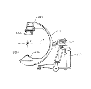
100151 FIGS, 2a-c show
various embodiments of onboard imaging systems
and/or radiation therapy systems to be used with the radiation therapy system
of FIG.
1 for performing dose tracking and feedback;
[00161 FIGS. 3a-b
provides a visual representation of a possible process to
form a kV portal image;
[0017] FIGS. 4a-b show a
reference image and a kV portal image with a
beam eye view of organs of interest:
[00181 FIG. 5 shows a
possible image on a quality assurance workstation that
shows kV portal images with a position/volume tracking chart for a daily kV
portal
image;
[00191 FIG. 6 is a flow
diagram of a sequence of steps for forming either of
the kV portal images of FIGS. 3-5; and
[00201 FIG. 7 shows an
embodiment of a radiotherapy process to be used
with the systems of FIGS. 1-2.
PREFERRED EMBODIMENTS OF THE INVENTION
[00211 In accordance
with the present invention, a volumetric image guided
adaptive radiotherapy system, such as cone-beam computerized tomography (CBCT)
CA 02905989 2015-09-28
image guided adaptive radiotherapy (IGART) system 100, and a corresponding
workflow
sequence for auto-construction and evaluation of daily cumulative treatment
dose are
shown in FIGS. 1-7, wherein like elements are denoted by like numerals. As
shown in
FIG. 1, the CBCT IGART system 100 includes a number of major systems: 1) a
three-
dimensional volumetric imaging system, such as an x-ray cone-beam computed
tomography system 200, 2) a megavoltage imaging system 300 that includes a
radiation
therapy source, such as a linear accelerator 302, and an imager 304, 3) a kV
portal imager
processor/software system 400 and 4) a treatment dose tracking and feedback
system
600, each of which are discussed below.
Three-Dimensional Volumetric Imaging System
[0022] Mechanical operation of a cone-beam computed tomography system 200 is
similar to that of a conventional computed tomography system, with the
exception that an
entire volumetric image is acquired through less than two rotations
(preferably one
rotation) of the source and detector. This is made possible by the use of a
two-
dimensional (2-D) detector, as opposed to the one-dimensional (1-D) detectors
used in
conventional computed tomography.
[0023] An example of a known cone-beam computed tomography imaging system is
described in U.S. Patent No. 6,842,502. The patent describes an embodiment of
a cone-
beam computed tomography imaging system that includes a kilovoltage x-ray tube
and a
flat panel imager having an array of amorphous silicon detectors. As a patient
lies upon a
treatment table, the x-ray tube and flat panel image rotate about the patient
in unison so
as to take a plurality of images as described previously.
6
CA 02905989 2015-09-28
[0024] As shown in FIGS. 2a-c, various volumetric imaging systems to be used
with the
present invention are illustrated. While the discussion to follow will
describe the cone-
beam computed tomography system 200 and megavoltage portal imaging system 300
of
FIG. 2a, the discussion will be equally applicable to the scanning slot cone-
beam
computed tomography and megavoltage portal imaging systems of FIGS. 2b-c. FIG.
2a
shows an embodiment of a wall-mounted cone-beam computed tomography system 200
and megavoltage portal imaging system 300 that can be adapted to be used with
the cone-
beam computed tomography and megavoltage portal imaging system sold under the
trade
name Synergy by Elekta of Crawley, the United Kingdom, Such systems 200 and
300 are
described in pending U.S. Patent Application Publication No. 20070280408,
entitled
"Scanning Slot Cone-Beam Computed Tomography and Scanning Focus Spot Cone-
BeamComputed Tomography" and flied on April 12, 2007.
10025] The cone-beam computed tomography system 200 includes an x-ray source,
such
as x-ray tube 202, a rotary collimator 204 and a flat-panel imager/detector
206 mounted
on a gantry 208. As shown in FIG. 2a, the flat-panel imager 206 can be mounted
to the
face of a flat, circular, rotatable drum 210 of the gantry 208 of a medical
linear
accelerator 302, where the x-ray beam produced by the x-ray tube 202 is
approximately
orthogonal to the treatment beam produced by the radiation therapy source 302.
Note that
an example of mounting an x-ray tube and an imager to a rotatable drum is
described in
U.S. Patent No. 6,842,502.
7
CA 02905989 2015-09-28
[00261 Note that the detector 206 can be composed of a two-dimensional array
of
semiconductor sensors that may be each made of amorphous silicon (a-Si:H) and
thin-
film transistors. The analog signal from each sensor is integrated and
digitized. The
digital values are transferred to the data storage server 102.
(00271 After the fan beams from collimator 204 traverse the width of a patient
and
impinge on the entire detector 206 in the manner described above, computer 234
of Fig. 1
instructs the drum 210 to rotate causing the x-ray source 202, the collimator
204 and the
detector 206 rotate about the patient to another position so that the scanning
process
described above can be repeated and another two-dimensional projection is
generated.
The above rotation of the x-ray source 202, collimator 204 and detector 206 is
continued
until a sufficient number of two-dimensional images are acquired for forming a
cone-
beam computed tomography image. Less than two rotations should be needed for
this
purpose (it is envisioned that images formed from a rotation of less than 360
can be
formed as well). The two-dimensional projections from each position are
combined in the
computer 234 to generate a three-dimensional image to be shown on display 236
of Fig. 1
in a manner similar to that of the cone-beam computed tomography systems
described
previously.
10028) While the above described embodiment for the collimator 208 is rotary,
a linear
moving collimator can be used instead as described in pending U.S. Patent
Application
Publication No. 20070280408, entitled "Scanning Slot Cone-Beam Computed
Tomography and Scanning Focus Spot Cone-Beam Computed Tomography" and filed on
April 12, 2007.
8
CA 02905989 2015-09-28
Radiation Therapy Source and Imager
[0029] As shown in FIG. 2a, the system 300 includes a separate radiation
therapy x-ray
source, such as a linear source 302, and a detector/imager 304 that are
separately
mounted to the rotating drum 210. The source 302 operates at a power level
higher than
that of x-ray tube 202 so as to allow for treatment of a target volume in a
patient lying on
movable table 306 (movable in x, y and z-direction via computer 234 of Fig.
1). The
linear source 302 generates a beam of x-rays or particles, such as photons,
protons or
electrons, which have an energy ranging from 4 MeV to 25 MeV.
[0030] As mentioned above, the particles are used to treat a specific area of
interest of a
patient, such as a tumor. Prior to arriving at the area of interest, the beam
of particles is
shaped to have a particular cross-sectional area via a multi-leaf collimator
308. The cross-
sectional area is chosen so that the beam of particles interacts with the area
of interest to
be treated and not areas of the patient that are healthy. The radiation
penetrating through
the area of interest can be imaged via imager 304 in a well known manner.
Alternative Embodiments for Volumetric Imaging System and Radiation Source
and Imager
[0031] Another embodiment of a cone-beam computed tomography system 200a and
megavoltage portal imaging system 300a is shown in FIG. 2b. In this
embodiment, the
systems 200a and 300a can be adapted to be used with the cone-beam computed
tomography and megavoltage portal imaging system sold under the trade name
TrilogyTm
by Varian Medical Systems of Palo Alto, California. The system 200a includes
an x-ray
tube 202, a rotary collimator 204 and a flat-panel imager/detector 206 similar
to those
9
CA 02905989 2015-09-28
used in the embodiment of FIG. 2a. Unlike the system 200 of FIG. 2a mounted on
a
drum, the x-ray tube 202 and collimator 204 are mounted on an arm 214
pivotably
mounted to a support 309 of the system 300a. Similarly, the flat panel imager
206 is
mounted on an arm 216 mounted to the support 309.
[0032] As with the embodiment of FIG. 2a, the x-ray beam 212 produced by the x-
ray
tube 202 of FIG. 2b is approximately orthogonal to the treatment beam produced
by the
radiation therapy source 302. As shown in FIG. 2b, the system 300a includes a
linear
source 302 and detector 304 similar to those described previously with respect
to FIG. 2a.
Accordingly, the linear source 302 generates a beam of x-rays or particles,
such as
photons or electrons, which have an energy ranging from 4 MeV to 25 MeV so as
to
allow for treatment of a target volume in a patient lying on movable table 306
(movable
in x, y and z-direction via computer 234 of Fig. 1). Unlike the system 300 of
FIG. 2a
mounted on a drum, the linear source 302 and the detector 304 are connected
with
support 309.
[0033] Another embodiment of a scanning slot cone-beam computed tomography
system
200b is shown in FIG. 2c. In this embodiment, the system 200b includes a kilo-
voltage x-
ray tube 202, a rotary collimator 204 and a flat-panel imager/detector 206
similar to those
used in the embodiment of FIG. 2a. Unlike the system 200 of FIG. 2a mounted on
a
drum, the x-ray tube 202 and collimator 204 are mounted at one end of a C-arm
218
while the flat panel imager 206 is mounted at the other end of the C-arm 218.
The C-arm
218 is mounted to a movable base 220 so that it can pivot about axes A and B
shown in
FIG. 2c.
CA 02905989 2015-09-28
Treatment Dose Tracking and Feedback System
[00341 As shown in FIG. 1, the treatment dose tracking and feedback system 600
includes a workstation or data server 110 that includes processors dedicated
to perform a
segmentation/registration process on a three-dimensional, volumetric image of
a patient
received from server 102 that was generated by cone-beam computed tomography
system
200. The workstation 110 is able to identify and register each volume of image
data
within each volumetric image. Such identification and registration allows for
the same
volume of image data to be tracked in position from one therapy session to
another
therapy session.
100351 The treatment dose tracking and feedback system 600 further includes a
workstation or data server 112 that includes processors dedicated to perform a
treatment
dose construction process based on 1) the segmentation/registration process
performed by
workstation 110 and 2) parameters of the beam of radiation emitted from the
source 302
as it impinges on the patient that are measured and stored in server 102, such
as angular
position, beam energy and cross-sectional shape of the beam, in accordance
with the
reference plan 502. Such parameters can be in the form of the angular position
of the
gantry 208, the angular orientation of the collimator 308, the positions of
the leaves of the
multi-leaf collimator 308, position of the table 306 and energy of the
radiation beam.
Once the position and shape of a subvolurne of image data is known, the
treatment
dosage received by that very same subvolume can be determined/constructed
based on
the above mentioned parameters of the beam of radiation emitted from the
source 302 as
it impinges on the patient. Such a determination is made for each of the
subvolumes of
image data for each of the volumetric images generated by system 200.
11
CA 02905989 2015-09-28
WO 2008/013598 PCT/US2007/012607
10036) The treatment
dose tracking and feedback system 600 further includes
a workstation or data server 114 that includes processors dedicated to perform
a an
adaptive planning process that can either 1) adjust the radiation therapy
treatment for
the particular day in a real-time manner based on off-line and on-line
information or
2) adjust a radiation therapy treatment plan in a non-real-time manner based
on off-
line information. The adjustment is based on how the dose calculated by the
workstation 112 differs from dose preferred by the treatment plan. Note that
the term
"real-time" refers to the time period when the radiation therapy source is
activated
and treating the patient. The term "on-line" regards when a patient is on the
treatment table and "off-line" refers to when the patient is off the treatment
table.
100371 In summary, the
treatment dose tracking and feedback system 600 can
perform real time treatment dose construction and 4D adaptive planning based
on
volumetric image information and therapy beam parameters that are measured in
a
real time manner during a therapy session. Thc system 600 can also perform
adaptive planning in a non-real-time manner as well. Such real time and non-
real
time processes will be discussed in more detail with respect to the process
schematically shown in FIG. 7. Note that in an alternative embodiment, the
workstations 110, 112 and 114 can be combined into a single workstation
wherein
the processes associated with workstations 110, 112 and 114 are performed by
one or
more processors. Note that the real time treatment dose construction
determined by
workstation 112 and the 4D adaptive planning determined by workstation 114 can
be
displayed on a monitor 117 of Quality Assurance (QA) evaluation station 116.
Based on the information displayed on monitor 117, medical personnel can
alter, if
required, the calculated 4D adaptive plan so as to be within acceptable
parameters.
12
CA 02905989 2015-09-28
WO 2008/013598 PCT/US2007/012607
Thus, the QA evaluation station 116 acts as a way to ensure confidence in
future real
time changes made to the therapy session. In this scenario, the QA evaluation
station
116 and the treatment dose tracking and feedback system 600 can be
collectively
thought of as a 4D planning and control system.
[00381 With the above
description of the onboard cone-beam computed
tomography system 200, megavoltage imaging and radiation therapy system 300,
QA
evaluation station 116 and the treatment dose tracking and feedback system 600
in
mind, the operation of the CBCT IGART system 100 of FIG, 1 can be understood.
In particular, the previously described online volumetric imaging information
and
real time therapy beam parameters are captured from systems 200, 300 and 400
and
stored in data storage server 102. The volumetric imaging information and
therapy
beam parameters are then sent to data monitor job controller 104 that
automatically
assigns tasks, based on pm-designed treatment schedule and protocol, to each
of the
work stations 110, 112 and 114 and controls the accomplishment of such tasks.
The
tasks are stored in temporal job queues 118 for dispatching, based on clinical
priorities, to each of the workstations 110, 112 and 114. The clinical
priority can be
reassigned from a clinical user's request 120 based on the treatment review
and
evaluation on the physician evaluation/decision making station 122. In
addition, the
station 122 also provides commands for treatment/plan modification decisions.
The
modification server 124 receives commands from the station 122 and modifies
the
ongoing treatment plan, beam or patient position on the system 300 based on
the
optimized adaptive plan created from the adaptive planning workstation 114.
[00391 As shown in FIG.
1, the raw data from server 102 is also sent to a
workstation 110. The workstation
110 is dedicated to perform an
13
CA 02905989 2015-09-28
autosegmentation/registration process on a three-dimensional, volumetric image
of a
patient generated by cone-beam computed tomography system 200. The raw data
from
server 102 is also sent to workstation 112 and workstation 114. Workstation
112
performs daily and cumulative treatment dose construction/evaluation from the
raw data.
Workstation 114 performs adaptive planning from the raw data. These three
workstations
110, 112 and 114 perform their tasks automatically with order of their job
queues 126,
128 and 130, respectively. The above described segmentation/registration,
treatment dose
construction/evaluation and adaptive planning will be described later with
respect to the
process schematically shown in FIG. 7.
[0040] As shown in FIG. 1, the segmentation/registration, treatment dose
construction
and adaptive planning information generated from workstations 110, 112 and 114
is sent
to the QA evaluation station 116 which interacts with a clinical user to
verify and modify,
if necessary, the results from the above workstations 110, 112 and 114. The
output from
QA evaluation station 116 is then stored in derived data server 103.
[0041] The QA station 116 provides an update execution status to job execution
log
server 132 that supplies information whether processing of information is
presently
occurring, whether processing is completed or whether an error has occurred.
Whenever a
task of treatment dose construction or adaptive planning modification is
completed by
workstations 112 and 1 14, respectively, the evaluation station 116 provides
treatment
evaluation information which includes both the current treatment status and
the
completed treatment dose and outcome parameters estimated based on the patient
and
treatment data from previous treatments. The user at QA evaluation station 116
can then
14
CA 02905989 2015-09-28
provide commands or a new clinical schedule to the high priority job request
server 120
to either request new information or modify clinical treatment schedule. In
addition, the
user can also make decisions to execute a new adaptive plan or perform a
treatment/patient position correction through the server 124.
[0042] The CBCT IGART system 100 performs a number of processes, including a
kV
portal imaging process via kV portal imaging processor/software 400 and a an
image
guided adapted radiation therapy process 500, both of which will be described
below with
respect to FIGS. 3-7.
Pre-Treatment Process
[0043] As an example of how the radiation therapy process proceeds, assume a
patient
who has undergone previous radiation therapy sessions at a clinic has another
session
scheduled for a particular day. The patient arrives at the clinic on the
scheduled day and
proceeds to the therapy room similar to that shown in FIG- 3a. The therapy
room includes
the cone-beam computed tomography system 200 and megavoltage portal imaging
system 300 previously described with respect to FIG. 2a. The patient lies on
the table 306
and is prepared for the on-line therapy session by the medical staff ("on-
line" being
defined as events and processes performed as the patient is positioned on the
radiation
therapy treatment table 306).
[0044] At this point of time, a reference treatment plan for applying
therapeutic radiation
to the patient has previously been determined for the patient based on the
previous
radiation therapy sessions. A reference treatment plan is designed before the
treatment
delivery based on the most likely planning volumetric image of the area of
interest to be
treated. The reference treatment plan contains patient setup position, therapy
machine
CA 02905989 2015-09-28
parameters and expected daily and cumulative doses to be applied to various
areas of the
patient. Such a reference plan specifies the area(s) of the patient to be
exposed to
radiation and the dosage the area(s) are to receive from the radiation source
during a
single session. Thus, the reference plan will include information regarding
the beam
angle/gantry position, beam energy and cross-sectional area of the beam formed
by the
multi-leaf collimator 308. Based on the reference plan, the patient is
instructed, per step
402 of a pre-treatment kV portal imaging process, to move to a particular
position, such
as on his or her side, that is optimal for applying radiation to the area of
interest within
the patient per the reference plan. While at the particular position, the
previously
mentioned pre-treatment kV portal imaging process employing kV
processor/software is
performed prior to the radiation therapy session. The pre-treatment kV portal
imaging
process is schematically shown in FIGS. 3-6. In particular, the process
includes forming a
two-dimensional projection/radiographic image from the cone-beam computed
tomographic image 404 of the patient prior to treatment, wherein the image 404
contains
the area of interest while the patient is at the particular position on the
table 306 per step
406 of the process. According to the reference plan, the radiation source 302
is to be
moved to one or more positions to apply Tadiation at each position while the
patient is at
the particular position. At each position of the radiation source 302, the
leaves of the
multi-leaf collimator 308 are to be moved to form a desired outline for
forming the
radiation beam to a particular cross-sectional shape. The positions of the
leaves at each
position of the radiation source are determined, per step 408, as
schematically represented
by the multi-leaf outlines 410 of FIGS. 3a-b.
16
CA 02905989 2015-09-28
WO 2008/013598 PCPUS2007/012607
[0045] The cone-beam computed tomographic image 404 of the area of
interest while the patient is at the particular position and the positions of
the
leaves/outlines 410 are then stored and processed in a processor of
workstation 110
as shown in FIGS. 3b and 4-6. Such processing involves, per step 412,
superimposing each outline 410 on a two-dimensional projection/radiographic
image
based on the cone-beam image 404 to form a treatment beam eye (REV) view kV
portal image such as shown in FIGS. 3b and 4b. Note that the kV portal image
can
be formed as a kV digital reconstructed radiographic (DRR) image for static
patient
anatomy verification or as a digital reconstructed fluoroscopic (DRF) image
for
verification of dynamic patient anatomy motion, such as respiratory motion,.
In
either case, each kV portal image with corresponding outline 410 (FIG. 4b, for
example) is compared with a treatment reference radiographic image (FIG. 4a,
for
example) that is generated according to the real-time radiation therapy plan
to be
executed. Should one or more areas of interest, such as a tumor or organ, of
the kV
portal image be displaced by at least a predetermined amount relative to the
position
of the corresponding area of interest of the reference image, then steps are
taken to
adjust the real-time radiation therapy plan for the day's treatment session.
If the
displacement is below the predetermined amount, then the real-time radiation
plan is
not adjusted.
10046] In addition to
the treatment dose, kV portal image can also be
constructed for treatment recordation and verification as shown in FIGS. 3a-b,
Further, organs of interest manifested on the CBCT image are auto-segmented
and
registered to the pre-treatment CT image. Therefore, daily and cumulative dose-
volume relationships of each organ of interest can be created. In some
17
CA 02905989 2015-09-28
implementations, a numerical filter is employed to estimate the final
treatment dose in
each organ of interest by performing parameter estimation for both stationary
and non-
stationary random processes of patient anatomical variation. Methods for
sample
estimation, such as the least square estimation, the principal component
analysis (PCA)
based estimation and singular value decomposition (SVD) estimation, may be
implemented.
[00471 The estimation is then used to provide information for the treatment
evaluation
and plan modification decision to determine when to switch on the adaptive
planning
modification engine.
On-Line, Off-Line Image Guided Adaptive Radiation Therapy Planning
[0048] After the kV imaging process is completed, resulting in the initial
radiation
therapy plan being modified or retained, the patient is repositioned to
receive radiation
therapy per the modified/original reference plan and image guided adapted
radiation
therapy process 500 is performed as schematically shown in FIG. 7. In
particular, the
reference plan 502 is applied to the linear source 302 per process 504 so as
to move the
source 302 to a position designated in the reference plan 502 and to format
parameters of
the beam of radiation emitted from the source 302 as it impinges on the
patient, such as
angular position, beam energy and cross-sectional shape of the beam, in
accordance with
the reference plan 502. Such on-line and realtime parameters can be in the
form of the
angular position of the gantry 208, the angular orientation of the collimator
308, the
positions of the leaves of the multi-leaf collimator 308, position of the
table 306 and
energy of the radiation beam. Process 504 can also involve moving individual
leaves of a
multi-leaf collimator 308 to desired positions per reference plan 502 so that
the radiation
therapy
18
CA 02905989 2015-09-28
therapy beam generated by the linear source 302 is collimated so as to radiate
a particular
shaped area of the patient per the reference plan 502,
[0049] Once the reference plan 502 is implemented per process 504, the
reference plan
502 can be altered to account for various factors that occur during the
radiation therapy
session. For example, the process 500 can entail having the system 100 monitor
real-time,
on-line machine treatment parameters of the linear source 302 and its
radiation output
online per process 506. The process 506 entails monitoring treatment
parameters, such as
beam angle, beam energy and cross-sectional shape of the beam. Such parameters
can
entail the position of the gantry, the angular position of the collimator 308,
position of the
leaves of the multi-leaf collimator 308, position of the table 306, the energy
of the beam.
[00501 The real-time, on-line information obtained by the above mentioned
monitoring
process 506 is fed to workstation 112 of FIG. 1 so that it can be used during
either the
online and offline daily and cumulative dose construction process 508.
[0051] While a radiation therapy beam is applied to the patient per process
504, the area
of interest to be treated is imaged via the cone-beam computed tomography
system 200.
The three-dimensional volumetric image is used to register and track various
individual
volumes of interest in a real-time and on-line manner. Prior to registration
and tracking, a
correction parameter must be determined by server 102 per process 510 so as to
be
applied to the volumetric image. The correction parameter is associated with
the fact that
rigid body components of the volumetric image are often not oriented in a
preferred
manner due to a number of factors, such as the position of the patient on the
table 306
19
CA 02905989 2015-09-28
and the angular position of the collimator. Based on the measurement of those
factors, a
correction parameter is determined per process 510 that when applied to the
three-
dimensional image the image is re-oriented to a preferred position. The re-
oriented three-
dimensional image is stored at workstation 102 of FIG. 1. The workstation 102
contains a
library of stored three-dimensional images of one or more areas of interest of
the patient.
[00521 Once the correction parameter is determined, the segmentation-
deformable organ
registration workstation 110 receives the volumetric image generated by system
200 and
correction parameter from server 102 via process 512. The workstation 110
executes
process 512 so as to match the patient anatomical elements manifested on the
volumetric
image to those on the reference planning volumetric image associated with the
reference
plan. The image registration results are used to map the pre-treatment organ
contours on
the planning volumetric image commonly delineated by clinicians, to the
corresponding
points on the treatment volumetric image automatically. The registration
methods applied
for this process are quite standard such as the finite element method and the
method of
image similarity maximization. However, there have been number of
modifications
performed to optimize these methods for the specific applications of the CBCT
image
and organs of interest in radiotherapy, such as described in the publications:
1) Liang J.,
et al., "Reducing Uncertainties in Volumetric Image Based Deformable Organ
Registration," Med Phys, 30(8), 2003, pp. 2116-2122, 2) Chi Y., et al.,
"Sensitivity Study
on the Accuracy of Deformable Organ Registration Using Linear Biomechical
Models,"
Med Phys, 33: (2006), pp. 421-33, 3) Zhang T., et al., "Automatic Delineation
of Online
Head and Neck CT Images: Towards Online Adaptive Radiotherapy," International
CA 02905989 2015-09-28
Journal of Radiation Oncology Biology Physics, 68(2), (2007) pp. 522-30 and 4)
Yan D.,
et al., "A Model to Accumulate Fractionated Dose in a Deforming Organ,"
International
Journal of radiation Oncology, Biology Physics, 44(3): (1999), pp. 665-675.
100531 Once each point in the volumetric image is tracked, that information is
sent to
workstation 112, which also receives the parameters per process 506. At
workstation 112,
an online daily and cumulative dose construction process 508 is performed. The
daily
dose construction process entails calculating/constructing for a real-time
treatment the
dose received for each volume of image data within the volumetric image
tracked per
process 512. After the treatment session for the day is completed, the daily
dose for each
volume of image data is stored in server 102. The daily dose for each volume
of image
data can be combined with daily doses for the same volumes of image data
calculated/constructed from previous therapy sessions so that an accumulated
dosage
over time for each volume of image data is determined per process 508 and
stored in
server 102. Further details of the construction of the daily and cumulative
treatment doses
are discussed in the publications: 1) Yan D., et at., "A Model to Accumulate
Fractionated
Dose in a Deforming Organ," International Journal of radiation Oncology,
Biology
Physics, 44(3): (1999), pp. 665-675, 2) Yan D. et at. "Organ/Patient Geometric
Variation
in External Beam Radiotherapy and Its Effect," Medical Physics, 28(4), (2001),
pp, 593-
602 and 3) Lockman D., et al., "Estimating the Dose Variation in a Volume of
Interest
with Explicit Consideration of Patient Geometric Variation," Medical Physics,
27: (2000)
21
CA 02905989 2015-09-28
pp. 2100-2108.
100541 As shown in FIG. 1, treatment evaluation 514 is performed by
workstation 114
following the patient organ registration and treatment dose construction
processes 512
and 508, respectively. There are two purposes for treatment evaluation, (a) to
determine if
the current treatment delivery is the same as the one previously planned for
the treatment
quality assurance; and (b) to modify the ongoing treatment plan by including
the patient
anatomy/dose variations observed and quantified so far to optimize the
treatment
outcome. Such treatment evaluation 514 can be performed real-time. on-line and
off-line.
10055] Final treatment dose and outcome estimation are used to provide
information for
the treatment evaluation and plan modification decision to determine when to
switch on
the adaptive planning modification engine per process 514 of FIG. 7. A
numerical filter is
employed to estimate the final treatment dose in each organ of interest by
performing
parameter estimation for both stationary and non-stationary random processes
of patient
anatomical variation. Methods for sample estimation, such as the least-square
estimation
(LSE), the principal component analysis (PCA) based estimation and singular
value
decomposition (SVD) estimation, are implemented. The detail discussions of
using these
filters for organ geometry and dose estimation of different treatment sites
have been
discussed in the following documents: 1) Yan D. et al. "Organ/Patient
Geometric
Variation in External Beam Radiotherapy and Its Effect," Medical Physics,
28(4), (2001),
pp. 593-602, 2) Lockman D., et al., "Estimating the Dose Variation in a Volume
of
Interest with Explicit Consideration of Patient Geometric Variation," Medical
Physics.
22
CA 02905989 2015-09-28
27: (2000) pp. 2100-2108, 3) Sohn M. et al.. "Modeling Individual Geometric
Variation
Based on Dominant Eigenmodes of Organ Deformation: Implementation and
Evaluation," Phys Med Biol, 50: (2005) pp. 5893-908 and 4) Yan D., "Image-
Guided/
Adaptive Radiotherapy," Medical Radiology-Radiation Oncology, Volume: New
Technologies in Radiation Oncology, Edited by W. Schlegel, T. Bortfeld and AL
Grosu,
Springer- Verlag Berlin Heidelberg New York Hong Kong, (2005) ISBN 3-540-00321-
5.
10056] The first task of treatment evaluation is related to treatment delivery
and plan
comparison performed by workstation 112 per process 514. If the comparison
shows that
the daily or cumulative treatment dosage for a particular subvolume of the
image and the
corresponding daily or cumulative planned dosages for the corresponding
subvolume are
outside a certain tolerance (see, Yan D., et al., "A New Model for 'Accept Or
Reject'
Strategies in On-Line and Off-Line Treatment Evaluation," International
Journal of
Radiation Oncology, Biology Physics, 31(4): (1995) pp. 943-952, then this
means that the
reference plan currently being implemented needs to be revised during the
present
therapy session. Note that the above described daily and cumulative dosages of
a
subvolume of interest can be tracked/displayed in time, such as on monitor 117
of FIG. 7
in a manner similar to the chart shown at the bottom of FIG. 5.
[0057] Besides comparing the dosages, the positioning of areas to be treated
with respect
to the therapeutic beam is tested by forming a kV portal image per the
previously
described process of FIG. 6. If the real-time kV portal image is compared
23
CA 02905989 2015-09-28
WO 2008/013598 PCT/US2007/012607
with a reference portal image and a subvolume of interest of the real-time kV
portal
image is found to be displaced in position or deformed in shape outside a
certain
tolerance with respect to a corresponding subvolume position in the reference
portal
image, then the reference plan, such as adjusting the leaves of the multi-leaf
collimator, needs to be changed in this instance as well Note that the above
described position of a subvolume of interest can be tracked/displayed in time
as
shown by the bottom chart of FIG. 5, wherein x, y and z positions of a
particular
subvolume is tracked from one daily treatment session to another daily
treatment
session.
100581 If either of the comparisons described above are outside the
corresponding tolerance, then a revision of the reference therapy treatment
plan is
performed in the on-line or off-line adaptive planning optimization process
516.
Adaptive planning optimization is different than conventional radiotherapy
planning
where only pre-treatment computed tomogaphic image data is used. Instead,
adaptive planning intends to utilize individual treatment history from patient
anatomy/dose tracking as feedback to optimize treatment control parameters.
Examples of techniques of adaptive planning optimization are described in the
following publications: 1) Yan D., et al., "An Off-Line Strategy for
Constructing a
Patient-Specific Planning Target Volume for Image Guided Adaptive Radiotherapy
of Prostate Cancer," International Journal of radiation Oncology, Biology
Physics,
48(1), (2000) pp. 289-302, 2) Birkner M., et al., "Adapting Inverse Planning
to
Patient and Organ Geometrical Variation: Algorithm and Implementation," Med
Phys, 30(10): (2003), pp. 2822-2831,3) Yan D., "On-Line Adaptive Strategy for
Dose Per Fraction Design," Proceeding, XIIIth International Conference on The
Use
24
CA 02905989 2015-09-28
WO 2008/013598 PCMIS2007/0 12607
of Computers in Radiotherapy, Heidelberg, Germany (2000), pp. 518-520 and 4)
Yan
D., et al., Strategies for Off-Line and On-Line Image Feedback Adaptive
Radiotherapy," Editors: BK Paliwal, DE Herbert, JF Fowler, MP Mehta,
Biological
& Physical Basis of IMRT & Tomotherapy, AAPM Symposium Proceeding No. 12,
2002, pp.139-50.
[00591 Note that the above-described process regarding FIG. 7 can
include
real-time data/information by capturing data volumetric image data from system
200
and therapy beam parameter information during the time the therapy beam is
generated. Such real-time information can be processed per processes 506, 508,
510,
512 and used in process 514 to determine if the therapy plan should be revised
in
"real-time." If it is so determined that revision is recommended, then the
real-time
data/information can be used in conjunction with prior dose information and
position/shape information of the volume of interest determined from previous
therapy sessions (off-line information) to reformulate the therapy plan.
[00601. While the above description demonstrates how "real-time"
data/information can be used to revise a therapy plan via the process of FIG.
7, the
description is equally applicable to non-real-time adaptive therapy. In this
case,
processes 506, 508, 510 and 512 use off-line information from previous
treatment
sessions and process 514 determines if a therapy plan to be used in the future
should
be revised. in "real-time."
[00611 In summary, the system 100 and process 500 provide volumetric
image guided adaptive radiotherapy, which can be performed in real time,
online and
offline for treatment dose construction and feedback. Therefore, they provide
all
possible feedback information for image guided real time, online and offline
CA 02905989 2015-09-28
WO 2008/013598
PCT/US2007/012607
radiotherapy. Thus, the system 100 and process 500 are able to fully utilize
individual treatment information, which primarily includes the patient dose
delivered
in the previous treatment, patient anatomy in the present treatment and
patient
anatomy estimated for remaining treatment deliveries.
[00621 The foregoing discussion discloses and describes merely
exemplary
embodiments of the present invention. One skilled in the art will readily
recognize
from such discussion, and from the accompanying drawings and claims, that
various
changes, modifications and variations can be made therein without departing
from
the scope of the invention as defined in the following claims.
26