Note: Descriptions are shown in the official language in which they were submitted.
CA 02906349 2015-09-14
WO 2014/149642 PCT/US2014/020071
SINCLE :PLANE TISSUE 'REPAIR :PATCH HAVING A I,OCATING
STRUCTURE
Reference to Related Applications
This is a continuation-inpart of co-pending commonly assigned US, Patent
Application Serial No. 13/443,3.-V filed on April 1.0, 2012, Which is -
incorporated hy
1.0 reference.
Technical Field
Thefield of art to which this invention pertainsis :implantable surgical
tissue
repair pat:du* Moreparticularly implantable surgical mesh. hernia patches for
iise in
hernia repair procedures.
BaClitlround of the invention
:Hernia -repair is a relatively straightfOrward.surgical. procedure, the
ultimate goal
of WWI is to reStom the mechanical. integrity of the:ztbdaminal :wall by
repairinga
musclo defect-thiough which the peritoneum and possibly a -section of
the-
underlying -viscera has protruded. There are various typesolhernias, each with
its own
-20 specific .surgicai repitifprocedure, 'including ventral hernias,
ntI1caI herniaS, incisional
hernias, sports hernias, &moral 'hennas, and inguinal hernias. It is
'believed. that most
herniaS are attributable to a weaknem in sections athe times of the abdominal
wall.
Precipitating -events, such as unusualmovements or lifting extremely heavy
weights,..rnay cause the 44,va1c. spot. in the abdominal wail:tissue to. be
eKcessively
.25 stressed, resulting ìn tissueseparation or rupture and-protmion of-a
section of
peritoneum and underlying visetitas eg.. inteAine,, through th6 separated or
ruptured
time. section. This weak-new, may be attributableto several factors. Weaknem
ín ttìc
-
abdomitad wall may be cong,enital or may beaSsociated with a prior incision
from a
surgical procedure or a trocar. wound.. Other factors rnay include trauma
genetic
30 predisposition, and aging.
CA 02906349 2015-09-14
WO 2014/149642
PCT/US2014/020071
5- Even though the eommonly used, conventional surgical procedures for
correcting
ortepaiting the various types of hernias.are Somewhat specific, there- is a
commonality
with respect to the niechanical repair. Typically, the protrusion of the
peritoneum
throngh a MIMIC or abdominal wall defect results in abernia sack containing
the
underlyingand protruding viscera, be hernia sack is dissected -and the
viscera. are
1.0 pushed back into the abdominal -cavity. Then, a tissue minforcirtg:or
repair implant such
amesh path device is typically lanted
arid -secured at the. site-of the abdominal wall
defect. Autologous tissue quidkly is into the mesh implant, .providing the
patient
wit-ha secure and strong repair. In certain patient presentations, itotay be
desirable to
suture or otherwise close the defect without .an implantõ although. dna is
typically tnuch
15 less desirable for the optimal outcome.
One,- common type of hernia is a ventral berni* This- type of hernia typically
occurs in the abdominal -small and may be caused by a prior -incision or
puncture-, orby an
arOg of tissue- weakness-that is,- gressed. Them are several repair procedures
that can be
employed by the surgeon to treat such hernias, depen.ding upon the individual
20 characteristics of the patientand the name of the hernia, hi one
technique, an Way
mesh is.implanted onthe dorsal surface. of the anterior fascia of the
abdornin.al wall.
Another tethnique provides -for an inlay .mesh, where the prosthetic material
is sutured to
the abdominal wall and..actS is a "bride to. close:the abdominal defect.
.Placement ola.
prosthetiomesh posteriorto the metus -muscle of the -abdominal wall is known
as the
Reeves Stoppa Or retrotriuseular technique. In this technique, a meshimplant
iS located
beneath the muscle of the abdominal wall but above the:mone:um. Implantation
of the
mesh in the intratperitoneal. location can. be-done via an -open or
laparoscopic approach.
The mesh is insert&I into the patient'sabdominal -cavity thrOughati open
anterior incision
or via a trocar and positioned to cover the defect. Theaurgeon then fixates
the mesh
30 implant-to the abdominal wall -with eonventimal mechanical fixation or
with statues
placed through the. full.thickness of the abdominal wall There :are a variety
of such
rrtceharticalfiXation devices that Call.be used in laparoseopic. or open
surgery, e,gõ
tacking instruments, lntraperitoneal placem.ent of mesh via an open approach
may -be the
desired technique of repair where the layers ofthe abdominal wall-are
attenuated and a.
2
CA 02906349 2015-09-14
WO 2014/149642 PCT/US2014/020071
5- laparoscopic approach. is not desired. Placement of MOSII via -this
technique presents
several unique challenges including-pootviMbility duringonesh handling and.
fixation,
poor handling, and -deficient ergonomics of the currently -available products.
Mesh repair
patch implants desi,gned for intraperitoneal placement typically requires
an.additional
treatment. or layer to function as a. tissue separating compotient.tosetvrate -
the viscera
from the .prosthetie abdominal wkIll repair layer, and thereby prevent or
substantially
inhibit the formation of post-operative adhesions. -Ile addition. of this
layer may add to
the complexity of 'wound healing due to the presence -and .mass of an -
additional layer,
Although hernia -.repair patch implants exist for open -ventral -hernia
repairs, there
are -deficiencies known to be associated veith their use. 11e- deficiencies
include- difficulty
inhandling the mesh, poor visibility during mesh handling, implantation and
fixation,
poor usability and. ergoriornics when. using alaparoscopie instrument, and
the. use of dual
or multiple layers of-mesh.. The commercially available meshes repair patch -
implants tbr
this- application typically h.ave at least dual. layers -of rnesh. or fabric
with poCkets or skirts.
to-provide for affixation to the parietal -wall via the top layer or skirt..
It can also be
:240 appreciated .that multiple layer meshes introduce more foreign body
rriaSs and te.nd to be
more expensive and complicated. to manufacture than a single layer.mesh.
implant.
Another deficiency associated with. hernia. repair patch-implants is-the ease
of locating the
periphery of the patches so that surgeon may affix the patch to ti.ssue by
emplacirtg
tacks or other fitsteners to properly secure the implants to. tissue in the
appropriate
-25 Manner.
Accordingly, there is a need in this art for novel 11,8,110 repair. implarns,
S11C11 ask.elitrat- hernia repair patch implants; that .can be- used in an
open surgical
procedure, and -which do not .require a mesh. anchoring ar affixation layer;
and 'which may
be secured to tissue using a 'single o.r multiple crown technique. There is
alsou need for
30 ti%1.1e repair implants which- facilitate the location ofthe
.peripheries of such implants by
the surgeon.
3
CA 02906349 2015-09-14
WO 2014/149642 PCT/US2014/020071
Summary a the Invention
Accordingly, nov:el..tissue repair-patches. a:re disclosed. The tissue. repair
patches
have asubstantially flat or .t)lanarbase member. The kik, member ikrirefbrably
a 1.11eth.
There is an opening located in thebase member, and, thcre, is a closure member
associated with the opening. The base member has a top side and a bottom side,
and an
outer periphery aid a peripheral edge.. Mounted to the bottom of the base
member
adjacent.to. or on the periphery of the-b.ase member is: a. locating
structure. The. locating.
structure -is preferably .an engagement ring member. Optionally, the locating -
structure has
a downwardly-extending flange-member. The patch -maY-have-a polymeric layer on
at
least part of at. least -one side of the base member. It is preferred that the
side of the mesh
that faces the viscera have a polymeric layer covering substa.ntially all of
that side. The
tissue repair patches of the :present invention are. especially tiseful in an
open hernia:repair
procedure, such as aventral hernia repair, and. are. also useftil in-other
types of body wall
tissue repairs:
Another aspect. of the present invention is.- a method.Of repairin a body wall
.20 defect,- such as a:hernia defect,:in an open surgical procedure
using.the a.bove-described
tissue repair patch ithplants-,
These and other aspects and-adVantages ofthe present inVention will betorne
mom apparent frorn the follovving:description and accompanyin.g drawing
prief Description of the Dramiitm
FIG. I is a plan. vicw of ait embodiment of a single plane ti&suerepair mesh
patch
o:f the present: invention; the patch has -a 'base meMber having an o.petting,
and a. closure
patch -camber mounted:to the top side attic. base -member over the Nang.
Fl....is an exploded perspective view ofthe :repair trieSh patch of FIG, 1.
30 .FIG. 3. is an illusMation showing a surgical tacldrig instrument having
an.
elongated .sbaft partially inserted undemeath the flap member and through the
opening of
4
CA 02906349 2015-09-14
WO 2014/149642 PCT/US2014/020071
5- the 'base, member of -the repair pat& of Hal; the instrument shall is
seen as-having
access to the bottom Side of the base nietnbcr..
NG, 4- is a plan view of a time repair patch of thepresent inVention that is
similar to the repair patch shown in Fki. I, but which i1t13 a rectangular
closure patch
member connected along its opposed Minor sides;. the clown: patch member is
seen to
contain a dimtionguide for use. by the surgeon in:orienting the patch dating
implantation.
'RC 5 is. an exploded perspective view illustrating two halves. of another
embodiment of a tissue repair patch atilt?, pment invention; the two halves
ate
connected to -fonn a repair mesh patch having closure flaps,
FIG, 6 isa plan view of a tsierepair- patch of the present invention made by
ning the two halves seen in Fla 5; the flaps- are in the at rest position.
FIG, 7 is a perspective view -of the tissue repair patch of FIG, 6; the as are
in
the at rest pt3sition.
Ha 8 is a perspective view of the tissue repair patch of FIG.? showing both of
the flaps .in the. up pcKsition, 411cQVcring the.opening ix the:base. member
thereby
ptvviding access through the base-member,
.FIG.. 9 illustrates the tissue repair patch of FIG. 8 with :a curved shaft of
a surgical
%eking instrumentinscrted partially through the opening .of the base member.
FIG, 10 is a plan view of another embodiment of.a ti stierepair patch of the
.25 present invention;The mesh patch is seen to have an opening with a
surgical suture and
surgical needle mounted about the opening: in a 'continuous mattress suture
cotfiguration..
PLtI 1.1. illustrates the. tissue repair patch of FIG, wherein the opening
has
been closed by applying tension. to :the sutureafter theputch .has been
affixed to the
parietal wall of -the patient over did- hbrnia defect,
CA 02906349 2015-09-14
WO 2014/149642
PCT/US2014/020071
5- FIG. 12 is
an exploded perspective view eat:I-other preferred embodiment of a
tissue repair patch of the present inVetition.; the, patch is seen to .have an
-upper olosure flap-
and a lower closure flap nmunted about an. opening in the base mentber:
Ha 1-3 is a .plai . view of th.e tissue repair mesh. of FIG. 13, showing the
elosure
flaps mounted bout the opening in the base member -with one closure flap
acijacent to the
la bottom site of the base Member and oneclosure fly adjacent to the top
side of the base
member; the flaps: atv in an at rest position.
FIG. 14 is a plan view of a preferred embodiment of a tissue repair patch of
the
present :invention; the patch is seen to have a -pair-of closure flap members.
FIG: 14a .is across-sectional view of the repair patch of FIG. :12 along View
Line
15 14a-14a,
FIG, 141 is a magnified partial viel,v Utile cross-section of MG.
12a.i1luStrating
the flaps posi timed-about the opening -in the base member of the patch.
.FIG 1-5 is an -exploded perspctctive view of two base member halves ofthe
tissue
repair patch of FIG. 121; both halves have a closure flap member extending
from the base
20 member sections.
FIG. 1.6 is a perspective _view of the tissue repair patch Made by joining
Wgether
the two halves seen in _FIG..15; one closure flap is positioned below the base
member and.
met-Imre:nap is positioned above. the base member.
FIG, 17 is aperspective view of the tissue repair Inoh patch of FIG. 16. both.
.25 closureflaps are in thenp position such-that the openingin the base-
member:is accessible
between the flaps.
FIG. .18 is a perspective- view of.the mesh repair patch ofFIG. 17,
illustrating the
distai end of a curved elongated shaft of a surgic.al tacking instrum.ent -
partially inserted
through the opening of the base member-in a positionblw the pat& to secure the
Mesh
30 repair patch to tissue.
6
CA 02906349 2015-09-14
WO 2014/149642 PCT/US2014/020071
5- FIG. 1.9 isa /1mila-five view of the tissue tepair patch of FICI, 1-8,
with both flaps
optionallysutured tog-ether hi an upward extending position to close the
opening in the
base member after the -patch has been atiixed-to tissue.
.PG. 20 is a cross-sectional side view of the tissue repair patch of MG. I
t":i. inserted
into the abdominal cavity of a patient and. .positioned adjacent to. the
patient's peritoneum;
1.0 a curved shaft of a surgical tacking instrument is.seen inserted
thorough- an access
opening such as a hernia defect in the .patienes.bo.dy wall and through the
openingin the
base member of the repair patch, such that the -distal end of the:shaft is in
position below--
the patch to- secure-a section of the base menther of the patch with .a tack
to the body walt.
.2.1 is kperspective view of the mesh -repair patch of Fla 17, illustrating
the
1.5 distal end of a straight elongated shaft of a sumical tae.king
instrument partially inserted
through the opening ofthe base member in a position-to Secure thetissue -
repair patch to
tis,sue,
HQ, 22 is a side view of the tissue-repair patch of FIG. 21 inserted into the
abdominai cavity of a patient and positioned adjacent to the patient's
peritoneum; a distal
20 section ofa straight
shaft of a surgical tacking instrument is. seen ed-thorough an
accem opening in the patient's body .wall- and through the opening: in
the.base merriber of
the repair patch, such that the distal end of the shaft- isitt -position WOW
the patch to
secure a section Orthe base member of the patch with a tack to the body wall,
FIG. 23 is an -illustration of a. hernia repair procedure wherein a-surgeon is
.25 securing the tissue repair patch of FIG. 17 in position over a hernia
defect using a
surgical tacking instillment having a curved elongated Shaft; the distal
section of the thaft
is insetted thmugh .an access opening in thepatient's body wall and through an
-opening
in the tissue repair patch ìn order to -secure the tisime patch to the
peritoneum; the
surgeon's hand is seen palpating the abdomen above the distal -end of the.
shaft of the
3 instrument to place a tack in adesired -position on the patch,
is a. cross-sectional side.view illustrating a-preferred embodiment of-a
tissue repair patch of the present invention in 01-al:cover a hernia defect
adjacent to a
7
CA 02906349 2015-09-14
WO 2014/149642 PCT/US2014/020071
5- patient's peritoneum; a curved elongated shaft ola surgical tacking
:instrument h.as been
positioned tinvagh an access opening it the patient's body wafl and through an
opening
in the patch to attach a section of the base member of:the patch to the
peritoneum; the
patient's visceral organs are seen .positioned adjacent. to the bottom side of
the patch and
the peritoneum, ad the closure- flaps an seen to exten.d. Upwardly through the-
opening in
the body wall.
FIG. 25 is an exploded :perspective view of an alternatt. embodiment of a mesh
tis.sue repair path of thepresent invention; the base member is seen to have
an opening-
in the base member surrounded by a closure ring,. and a closure patch haying a
mating.
closure ring is also: &bon.
MO, 26 is a peispective view of the tissue repair pateh of FIG. 25 -showing
the:
patch secumd to the base member.
Fla 27 illuStrates A Oritoneal view of the hnttorn side of
apreferredeinbodiment
ofa tissue repair path of the present invention secured to the peritoneum with
a double
tow of surgical :tacks referred to as a double crown technique; the opening in
the base
mernber is seen to- be closed, and both flaps have been positioned upwardly
away 'from
the top of the. base member; the flarg4 are smured to closefthe- opening in
the base
mother:
FIG. 28 is a perspective view of an alternate enibodiment oft mesh :tissue
repair
patch of the present invention; the-patch is seen -to have a slit in the base
member
.25 providing a central opening.;
.FIG, 29 is a perspectiv.e view Ofth.e patch of FIG, 28 :haying a surgical
suture
mounted about- the slit in a shoe lace :type configuration to-close the
tIpcning in the slit.
FIG. 'MI is a.petspective view of th.e tissue repair patch of FIG. 29 after
the suture
ends have:heen tensionekthereby closing the: opening and slit after the patch
is secured
30 to the .patticnt's body wall.
8
CA 02906349 2015-09-14
WO 2014/149642 PCT/US2014/020071
5- FIG. 31 is a. cro.wsectional view of a tissw repair patch of the present
invention
having a locating structure- positioned on the bottom of the base metriberon
the
periphery. The tissue repair patch is shown :located adjacent to a body wall
below a
hernia d.a&ct, A surgical tacking instrument is -shown with the-distal end. of
its. shaft:
positioned pioxlinal to .viscenti side of the body wall with the shaft: tip
adjacent to the
locating structutv and in -a position to firetacks through the base -member
into the body
wall. The !peeing structure is ìrt the- form of a ring.
Mi.. 32 is a -perspective view of the tissue repair patch of FIG. 31 looking
up from
a direction below the patch.
FIG. 33 is aperspective view olthe tissue repair patch of FIG..31 showing tbe
bottom of the mair patch and tissue repair -structure.
FIG. 3-4 is a partial magnified. side view of the -repair patch of Fla 31
showing the.
tip of thettteking insimment Shaft adjacent to the locating striicture.
FIG. .35 is cross-sectional view ofall Crlikv4inicut of a -tissue repair patch
of the.
present illVention having; a locating structure; the structure is seen. to
have a. textured
surface, The patch is it position to be affixed to-repair -a body wall defect
using a
surgical tacking instrument.
FIG. 36 is a partial. magnified -view. ofthe -pawh MG.
36.A-towing, the tip of the
sìtafL of the surgical tacking instrument -engaging the textured surfitce.
oldie locating
structure.
-.25 Ha 37 is a moss=-axtional. viewi-of art erribodiment of a tissue
repair patch of the
present invention having a locating structure; the structure is seen to-have a
downwattly
extending flange -member formed from the periphery of the base member. The
patch is in
twition to be affixed to repair a. body wall &Act .using a surgical tacking
instrument.
FIG. 38 is a partial magnified view of the-patch of FIG. 36 showing thc tip.
of the
shaft of the-surgical:tacking instrument engaging the downwardly extending
flange
Member of the locating structure..
9
CA 02906349 2015-09-14
WO 2014/149642 PCT/US2014/020071
5- FIG. 39 a partial magnified cross-sectional .view-a tissue -repair patch
wherein the
locating struCture is a downwardly extending-firm& Member mounted to the
periphery of
the base member.
FIG. 40 a partial magnified cross-sectional view a tittsue repair -patch.
wherein the
locating -structure is- a ring member having a dmeawardly extending flange
member; the
ring member is mounted .to the periphery of the to.p of the base member.
MG. 41 a 'partial. magnified croSs-sectional view a tissue repair patch
wherein. the
locating structure is a ring member having a downwardly extending flange
member; the.
ring member is -mounted to theperiphery of the 1)ottorn-of the base member.
Detailed lleseription of-the Invention
Mellow! tissue repair patches ordevices- of the. present invention are-
particularly
useful in opera- ventral or incisional hernia repair surgical procedums, The.
tissue repair
patch devices conSistof a base -merriber havingan opening. The base member
has; a
10 closure member or -d.evice associated with the opening fl-nt seeming the
opening after
implantation. The -repair patch -devices -of the present invention have
utility- in other
conventional tissue repair procedures. ibcluditg inguinal hernia repair
procedums, trOcar
puncture wounds, trocar incisional hernias, ete.,
Tissue, repair implants and .surgical in.struments for applying tacks to
fixalc tssue
.25 repair iinplatits aredisclosed in the. following commonly assigned,
pending patent
applicationS, whieh .are incorporated by reference: US Serial Nos. 12/464,151;
1.2/464,165;12/464,177; 12/464,143; 1.2J94051; and 121815,275.
The tissue repair patches of the present invention .may be madefrom. any
conventional biocOmpatible -materials. Thepatches and their components are
geeferably
30- made from conventional.biocompatible polymers ling may be non
absorbable or
bioabsorbable; The tertn bioabsorbable. ìs .iefined -to have. its
conve.ntional .meaning and
2.0
CA 02906349 2015-09-14
WO 2014/149642 PCT/US2014/020071
5- includes both- biodegradable arid bioresorbahle. Examples of such
nonabsorbable
poiymerS include polypropylene, polyester, nylonoiltra high molctular weight
polyethylene, and the like. 1-.1td.- combinations thereof, :Examples
otsuitable bioabsorbable
polymers include polylactides (PIA), polyglycolides (PGA)õ polydioxanones
Pl?S), copolymers of PGAStrimethylene carbonate WW1 copolymers of :PIAIRIC,
and
the like. If desired, conthinations of biocompatible nonabsorbable polymers
and
bioabsorbable polymerS May be utilized to construct the, tisaue repair implant
-patch
devices of the present invention.
Although it is preferred to use surgical meshes to construct the hernia repair
-
patches of the-present invention, other conventional woven or nonwoven
surgical repair
fabrics.or thermally formed implants may also be used. In addition, the-tissue
repair
patches.-may be -made frornother -conventional implantable materials such
as.PTFE
(polytetrafluoroethylene), &. ePITE films and. larninates. The patches.may
consist cif
composites of polymeric filnis and rueShes, andlor fabrics.
The -meshes useful. in the hernia repair patch devices of the .present
Mvention will
.20 be manufactured in a conventional manner using convet)tioital
manufacturing equipment
and methods ineluding-kriitting, veetiving, non-woven Ictliniques, and the
like. The,
meshes will typically have. a pore sizesufficient to effectively provide for
tissue
ingrowth; for example, they may have pore sizes :in the :range of
aboutØ.3mit. to about
5min, and. other conventional sizeranges, EXamples of commercially available
nonabsorbable arid bioabsorbable polymeric-meSlaes that ratty be used to -
construct -the
hernia -repair patches of:the present invention-include
PHYSIOIMESITEm and
ETHICON PROCEEDIm Surgical Mesh,- available from Ethicon, :Inc, Route. 22
\Vest,
Sotnerville, NJ 0.8876.
When constructing be novel tissue -repair patches-of-the-present inverition
from.
surgical fabrics other than trieShes, the fabrics will have open pores with a
pore, size
sufficient -to effectively provide for tissuo ingrowth; for example, with a
typical size of
about 03 -mtn. to about 3rrim. By. "open pores" is meant openings that extend
from-one
side of the -fabric to the opposed. side, providing a pathway through
the.fabric. 'The.fahric
CA 02906349 2015-09-14
WO 2014/149642 PCT/US2014/020071
repair members may be: constructed from monofilaments, multifilaments, or
combinations thereof, Examples of cornmercially available no
fTabrics that can be
used to manufacturethe hernia repair patches of the present invention. Melude
woven.
fabrics, textiles and. tapes fir surgical applications. Other fabrics or -
materials include
perforated condensed ePTFE films -and nonwoven falorica having f)ore Sizes of
at. leaSt
one millimeter. The non-mesh-fabrics may be constricted of conventional
biocompatible
Materials.
'The fabric or mesh rnay :contain, îr additiou.to a long-term stable polymer, -
a
resorbable polymer (i.e., bioabsorbable or biodegradable). The resorbable Ond
the long-
term stable polymer preferably. contain monofilaments and/or multifilaments.
The terms
re,sorbable polymers and bioabsorbable polymers are used interchangeably-
herein. The
tem. bioatks.orhable is defined to have its conventional meaning. Mthough..not
pmfe.rredõ
the fabric or -mesh tissue repair mentber may be- immufactumd from a
bioabsorbable
polymer or bioabsorbable polymersvidthout anylorig-tem stable -polymers.
The tissue repair patches of thepresent ittvention may also ineludepollner
.20 The films m.ay be attached. to the top surface, the bottom surface or
both surfaces- and
May also cover theperipheral edges of the repair patch. devices or extend
beyond the
periphery of the repair patch devices. Tlhc films that are used to
manu:fiteture. the tissue
repair patch implant devices of the present invention- will have a thickness
that is
sufficient to effectively prevent a.dhesio:ns Awn. forming, or qt.horwise
ftinction:as a tissue
barrier or tissue:separating:structure or membrane. For example, the thickness-
ma:y
typically range frOin about Ipm to about 500pm, and--prefertibly from about
5Arn to about
50pm, however this will d.epend upon the individu.al Characteristics of the
selected
polymeric films. The films suitable for .use with. the repair patches of the
present.
invention. inchide -both bioabso.rbable andnonabsorbable films. The filins.are
preferably
polyinerlatsed and may be -made from ..various conventional blocompatible
polyme.rs,
including bioabsorbable and noriabsorbable polythets.. Non,reso:tbable or very
slowly
resorbable substances includ:e polyaikenes (eõgõ, :polypropylene or
polyethylene),
fluorinated polyolefins polytetrafluoroethyleneor polyvin);./lidene
fluoride),
12
CA 02906349 2015-09-14
WO 2014/149642
PCT/US2014/020071
5- polyamides, polyurethanes, pol yisoprenes, polystyrenes, .poly
silicones, .polyearbonates,
polyarylether ketones (PEEKs), polyinethiterAicaeid esters polyacrylic acid
esters,
aromatic .polyesters, polyimides as well as mixtures andior co-pol.'111(TS of
these,
substanc.es. Also. usellil are synthetic bioabsoiltable polymer materials. for
example,
polyhydroxy acids (e.g., pcilylactides; polyglycolides, polyhydroxybutyrates,
polyhydroxyvaleriates), polycaprolactoms, polydioxanones, syntheticand natural
oligo-
and poly-amino acids, polyphosphazertesõ poll/anhydrides, polyorthoesters,
polyphosphates,polyphosphonates, polyalcohols, polysaccharides, and
polyethers,
I4owever,naturally occurring mate:rials such as collagen, gelantin or natural-
detivea
materials such ashioabsorbable Omega 3 .fatty acid cross4in.ked gel films or
oxygenated
I5 mgenerated cellulose (011.C) can also be used.
The_ films_ used in the tissue -repair patch. devices -of the present
invention n.tay
cover the- entire outer surfaces- of the hem ia patch nterriber or a part
thereof, ln some
cases, it is tlenofk.fal to have films overlapping the bordersandlor
peiipheries of the
repair patches', The repair patches of the present invention may also haw
adhesion
20 barrier layers attached to one or both sides. The adhesion barriers will
typically-consist
of conventional biocompatible polytneric.materials including but not limited:
to
absorbable and nonabsorbable polymers. Examples of conventional nonabsorbable
polymeric materials usefal for adhcsiortbaniers include expand.ed
polytetraftuoreethylenes polytetrafino.roethylene, silicones And the like.
Examples of
.25 conventional absOrbable .polytneric materials -useful for adhesion
baiTiers inelude
oxidized regenerated cellulose, poliglecaprone 25 (copolymer of glycolide and
epsilon-
caprolactone),. and the like.
It is particularly preferred that the tissue repair patches of the :present
invention
have a mesh constrUctions and the -embodirnents illustrated in the Figures
have such a
30 mesh -construction.- Th.e tissue repair it-pp./ants oldie present
invention have pa:rticular
ttiì.ity-for herniarepair ptocedureS, but .maybe used itt -othr tissue repair -
surgical
procOures as well,
13
CA 02906349 2015-09-14
WO 2014/149642
PCT/US2014/020071
5-
Referringnow to FIGS. 1:-3, a tissue repair patch IO-of the present invention
is:
seen.. The .patch 10 has a :meth conatruction. ilte repair .r)ateh 10 is seen
to have
substantially fi4t or planar base member 20 and closure patch mentber 30. 'The
base
meniber 20 is illustrated having-a substantially oval.shape or configuration,
but may havo
other configurations including square, tectangular, circular, polygonal,
etc,:combinations
thereof arid the like. The has member 20 is.seert to :have top.side 22, bottom
side 24, and
periphery 26. Extending through the base member 20:is:the Slot: 40 having
opening 42
bounded :by opposed sides 44 and opposed ends 43. The &star patch merriber 30
is seen.
to be a .substantially .flat or planar member having a substantially oval
configuration. The
closure patch member .30 is seen to have tOp side 32, b.ottom.side.34, and.
periphery 35.
1.5 Closure. patch member 30 is Seal to have. opposed. curved ends 37 and
opposed sides 38.
Patch member 30 is:mounted to the-top:of base member 20 via. connections 39
along the
ends 37 -stivh that the bottOm side. 34 of closure patch 30 is adjacent to the
top side 22 of
base member 20. The closure patch is mounted. using any conventional
affixation method
to.cma.te the connections 39, including but not limited to sewing, welding,
tacking,
.20 rivetine, stapling, gluing,. etc., and the like. The closure patch
30.Is mounted to the base
inerither 20 to:cover the slot 40 and opening 42. Openings 48 ad jacent to -
Sitie.8: 38
provide aCCeSS passages for surgical instruments-to and through opening 42 of -
Slot 40. A
partial schematic. ofa surgical tacking instrument 60 which can be used to
tack the bast.
member 20 of patch 10 to tissue is seen in FIG. 3, The instrument 60 has
proximal
25 handle 62 anddistally extending elongated:shaft 70 having. distal end.
78.. .A distal setAion
76 -of the shaft 70 is seen. to extend thnnigh opening 48, underneath. the
bottom side 34 of
closure flap 30 and through opening 42 of slot 40 sucb that it is positioned
below the
bottom side 24 of base .member 20. The distal end 78 is seen to be positioned
in
proximity to the periphery 26 of -the base .member 20 adjacent to bottom side
24 so that
30 surgical tacks may b.f.: fired to secure the patch tP ti$Stle adjacent
to the top. side. 22 of base
member 20 and the top side 32 of closure path member 30. The repair-patch. 10
is
-fixated around its petititeter-26 to tissue with fixation poitas placed, for
ex.araple, about
every l to 2 cm, c.. the fixation devices or nteks are -separated by about l -
cm. to 2 cm
distances, AltIvugh in many .embodiments of the. tissue or- hernia repair
patches of the
14.
CA 02906349 2015-09-14
WO 2014/149642 PCT/US2014/020071
5- present invention it -is preferred to have a slot in the base meniber to
.provide .art opening
through. the base member, the opening May b:e a. slit ot other types of
o.penirig,s having
different: geometric configurations may be -utilized including circular. OVal,
rectangular,
polygonal, ete,, on thereof and the like, >Although not preferred, it.
is pOssible
thform the tissue repair patches of the present invention such thatthe base
member
JM andior closure menther are-curved or otherwise in:more-than one plane.
Once -the tissue repair-patch 10 ofthe present invention. has been implanted
and
secured. to tissue by tanking:or other conventional methods. (e:g., stapling,
sunning. etc.),
the shall section 76 of-surgical affixation instnmient-60 is -removed. from
the body
through the slot O. The closure. patch member 30 prevents underlying -tissue
or viscera
15 from moving through the slot 40 and o.pening 42.
.An alternative embodiment of the :tissue repair patch 10 is seen in FIG. 4.
The
patch 10 is. seen to have similarly shaped base member 20, however the closure
meniber
50 is seen to havea. substantially rectangular shape with opposed minor end
sides 56 and
op-posed major sides: 5.7. Closure.rnember50 has top side 52 .and bottom
side...54 adjacent
.20 to top side 22 orbase tnernber 20.. 'The patch member-50 is mounted to
base member 20
Over.' Slot 40 by connecting-is 59 along rn orsìdos 56, Tbe.connections may be
-made as
described previously. Qpenings 48 beneath sides 57 provide access to slot 40
and
opening 42. _M seen in FIG. 4, the tissue repair .patch 10 is seen to have a
directional.
indicator -80 contained on pr -in the closure menftr 50. Indicator 50 may be.
25 conventionally sewn., 'molded orformed, printed, dyed oriamin.ated into
or onto the
member-50. The indicator 80 is seen to have -central section 81, having
oppeised
transverse se:ea:0ns 82 extending therefrom. Extending longitudinally in an
opposed
manner are the longitudinal.sections 85 and 87. Section. 87 is seen to be
thicker than
section 85. The indicator 80 allows the surgeon to deter.mine tbe location of
.the pate.h.
30 with rk.-sp.ect. to the patient after insertion by aligningthe
respective axes of the tissue
repair .pateh 10 with tespeet to the patient and the intiMon, -allowing for
mo.re- precise-
:fixation, either :using a tacking instrument or using surgical sutures. for
affixation, -Such
CA 02906349 2015-09-14
WO 2014/149642
PCT/US2014/020071
5- directional indicators may bettsed with other embodiments ofthe tissue
repair patches of
die present inVention.
Referring how-to MS. 5,-9, an -altemativeemboditnem of.
erepair patch
100 oftbe present invention is seen. Thcpatch 100 is seen to have
substantially at or
planar base member 110 formed from substantially fiat or planar base sections
120 and
140. The base member 110 has. bottom side 112,, to side 114 rind periphery
116. Base
section 120 is seento have straight side 122 having ends 124. Base section
120. is. also
seen to have curved side 126 having-ends 128 that conneetto ends 124..
Extending out.
from straight side 122 is the closure flap meniber 130 having hinged-Side 132
and flu
end :134 separated from- side 122 by slot 136. SW 136 .has closed end 137 and
open end
15 138: The closure flap member 130 i ken to have a generally rectangular
configuration,
but may hEWC other geometric configurations including eirc.ular, oval,
.polygonal, etc.,
combinations -thereof and the like. Base section 140-is seen to havestraight
side 142
having ends 144. Base section 1,40 is also seen to have curved -side .146
having ends -1.48
that:connect to ends 144. Extending out 'from straiaht side- 142 is the
closure flap
20 m.ember 150 having hi.nged side 152 and free end 154 separated from.
side .142 by slot
156.. $lot 156-has. closed end. 157 and. open end 158. The closure flap member
150 is
seen to have a. generally rectangular configuration, but may have
othergeometric
configurationS including circulars OVai., polygonal, etc, combinations thereof
and. the like:
The base -member 110:and the tissue repair patch 100 art formed. from the has
sections
25 120 and 140 by 'connecting the base sectiOns aim* straight sides 122 and
142 filet*.
seams. 118. This Cart be done in any conventional manner including sewing,
welding,
tacking, stapling, .gluing, etc., and combinations and equivalents thereof It
can. be seen
thatonly the straightSides 122 and 142 are connected on either side Of the
closure. flap
members. 130 and 150. The closure flaps members 130 and 150 are .mounted
together
-
30 such that hing(4. side 132 of:closure flap 130 is contained in slot 56
of flap member 150
and hinged side 1.52 of closure flap 140-is contained in skit 136 of closure
member 130,
This. creates the slit 168. basemetriber 110 having through opening.165
bounded by
interior -portions of straightsides 122 and 142 of the base Sections .120 and
142,
16
CA 02906349 2015-09-14
WO 2014/149642
PCT/US2014/020071
5- respectively, and also bounded b ythe hinged sidm 132 and 152 of the
flap members 130
an:d 150, respectively, In the at rest position as seen in FIG. 6, the flap
member.130 rests
upon the top side 145 of the base section 140 ofbase member 110, while the
flap member
150-rest upon the top side-125 of base section 120. In:this at rest
configuration the slit
160 and opening 165 are covered. The tissue repairpatch 100 is. seen: in the
ready
1.0 position in FIG. 8, with the closure flap members-130 and 150 in the
uprightposition
exposing the slit 160 and o.paning so that a fixation instruinent can. be
inserted thniugh
the -opening1.65. A tacking instrument .170 is illustrated in FIG. 9 with
tissue repair
patch 100 .of the present ilMution, The tacking instmme:nt .170 :is seen to
have proximal.
handle.172 and actuation trigger 174. Extending front the distal end 176 of
handle 170 i6
i5 the eutved shaft 180- having distal section 182:and-distal end 184. The
distal section 182
is Seen to .be -inserted through slit -160 and opening 165 betWeen upwardly
extending flaps
130 and 150 such that the distal end .184 May he about
the bottom side- 112 of the
base member 110 in order to secure the-base .member to tissue with surgical
tacks. Once
tacks am. placed through the base -member 1 .10 of pateh 100 to secure the
:patch 100 to
.20 tissue, the- tacking instrument 170 may be removed. from -theslit 160
and -the -two flap
Meta-en 130 and I.50.can beinterlock.ed by /biding or-rotating the flap
inetribers
down wardlyonto the top 114 of the base member 110. One or .both of the flap
members
may be optionally 'Waded or affixed. to the base member 110 using 'various
COMTeiltiOnai.
dOSUre Methods including adhesives, sutures, surgical fasteners, etc,
25 .Ati
alternate ernbodiment 400 of a Single plane tissue repai.r patch of the
present
invention is seen in FIGS. .10 and 11. The repair patch 400 has a base member
4/0
having .a top side 412 and a bottom side 414. The patch. has a periphery-416:
Located in
the base member 410 is a slit 420 having an. opening 424 bounded by sides 422.
Theslit
420 has -ends 428. Mowed about the slit 420 is a surgical suture 430
havingends 432-
30 and. 434 and surgical needle: 436 mounted to end 432, and Optionally,
although not
shown, to end 434. The suture 430 is -mounted alvut the opening 424 in a
conventional
mattress suture (continuous) configumtion. As-seen in..F1G.1.1, the opening
424 is
closed by tensioning the -suture ends 432 and 434. causing the Side s-422 to
approximate.
desired.-the suture-needles 436 can be used to engage tissue with the suture
430.
CA 02906349 2015-09-14
WO 2014/149642 PCT/US2014/020071
5- Referring to FIGS. 28 and 29, a variation-of suture.mounting is
ilInstrated: The rep-air
pateh-450 is similar to repair patch 400, but has a: rectangulady Shaped
basemember451
having mosed major sides 454 andepposed minor sides 456 connected by rotmded
Comers 457.- 1.7he base member 451 has bottom 4de 45-8. and top side 459, and
outer
periphery 452: 'The base MCMber 451 has centrally located slit 460 having an
opening
464 bounded by sides 462. The slit 460 has ends:468. Mounted about the slit
460 is a
Surgical sututt 470 having ends 472 and 474. The suture 470 -6 mounted it a
"shoe late"
type configuration.. The. suture 470 is seen to be .monnted to Ait 460 by
engaging
opposed -sides 462 of slit 460. about the opening 464õ Suture 470 is seen to
have ends 472
and 474 located.adjacent to OM mother along. one.. end 468 of slit 460. The
sin 460 is
:secured after placement-of the.patch 450 by pulling-on ends-472 and 474
thereby- closing
opening 464. The suture 460 may optionally haVe surgical needles- mounted tO
one or
both of the ends 472 and 474. The members 410 and 451 may -have any
suitable,
geometric: configuration.
A preferred embodiment ofa tissue repair patch 200 of the present ì ventiott
is
seen in FIGS. 12 and 13. The patch 200 ísstwn to have a substantially flat .or
planar base
member 210 having a. top 21.2, bottom 214 and periphery .216.. The base member
210 is
seen to havan oval shape, but may" have. other geometric shapes including
rectangular,
circular, square; polygonal, combinations therenf and tbelike. Located in the
base
member 210 is theslot 220 having opening 222 therethrough. Slot 220 is bounded
by
Oppsed sides 224 and 225 and curved ends 226. The patch 200 is seen -to have
upper.
closure flap. 230 and lower-closure flap 240. (ipper closure flap 230 is seen
to have a
sul-stantially rectangular shape., although it may have.other geometric
configurations
including circular, oval, rectangular, polygonal,-etc:and the likeõ Flap 2:30
is ken to
have top side 231 and bottom sido232. The flap 230 aiso .has opposed-sides 235
and :23.6
connected by opposed end sides 237. The flap 230 iS Mounted to the top shit
2.12 ofbase
member 21.0 adjacent to side 224 of slot.220 by connecting the flap 230 along
its side 235
in a conventional manner StiCh-Wl sewing, gluing, stapling, .welding, riveting
and the like
to-create a seam. 239. In this manner-, the flap :23.0 lui,s its bottom side
232 facing the top
side 212-of ba.se -member 210, and is -positioned to coverslot 220 and (vening-
222 in the
CA 02906349 2015-09-14
WO 2014/149642 PCT/US2014/020071
5- at rest -position. The closure flap inay be rotated upwardly about seam
239 to uncover
slot 220 and opening 222. Mounted toihe bottom side 214 or but menther2-10 i6
the
other closure .flap 240: Flap 240 is seen to have top side. 241 and bottom
side 242. The
flap 240. also has opposed sides 245 and 246..connected by opposed. end sides
247. The
flap 210 .ìs mottnted to the bottorn.Side 214 of base member 210 adjacent to
side-225: of
slot 220 by connecting the flap 240 along its -side 245 in aeon ye-IWO:nal
manner such as
Se:wing, gluing, stapling, welding, riveting and the like to create a Seam
249. In this
mannerõ the flap 240 has its -top- side 241 facing the bottom side 214 of base
meniber 210,
and is positioned. to cover slot 220 and. ope,:ning 222 :in the: at :rest
position, The -closure
flap may be rotated dowirwardly about .seam 249 to uncover slot 220 and
opening 222..
1.5 The. flap 240 may also be. rotated Upwardly aboutseam 249 through -Slot
220 and opening
Refbrring now :to FIGS. 14, 14a, 14b, and 15-17, a preferred tissue repair
patch
250 of the present invention is seen. The patch 250 is similar to patch-200,
but is-
constructed:in a different manner from. two separate base- section members.
The patch
250 is seen to have substantially flat or planar base member 260 formed from
substantially flat or planar b4.5,ze seotims 270 and. 280. 'The base member
260 has bottom
side 264, top side 262 and periphery 266, Base section 2:70 is seen to
havesmight side
272 having ends 274. Base-section 270 ia also Setil to:havoside 276 haing
curved. ends
-
278 that connect to ends 274, Extending -out from straight side 2-72 is the
closure flap
-25 inernber 290 having hinged side 292 and free side- 294,, The closure,
flap member. 290 is
seen to have a generally rectangular configuration., but may have other
geometric
configurations including,. circular, oval, rectangular, polygonal, ete. and
the-like. Base
section 280 is seen. ter sestxaight side-28.2 haVing ends..284. Base.
set...lion :280 isalso
seen -to have side 286 having curved ends 288 that connect to ends 284.
Extending oust
from straight side 282 is the elOsure flap member 300 .having hinged side 302
and free
side 304. The closure flap member 300 is seen to have a gencolly rectangular
configuration., butmay have other geometric configurations -including
circular, oval,
rectangular, polygonal, etc., and the lik.e. The .base member 260 and the
hernia closure
patch 250 are fbrmed torn the base sections 270 and 280 by connecting.; the.
base seetions
19
CA 02906349 2015-09-14
WO 2014/149642 PCT/US2014/020071
5- along straight sides 272 and 282-along seams 268. This can be done in
any conventional
m.annerincluding Sewing, weldikI, tacking, stapling, -gluing, etc., and
Combinations and
equivalents thereof. It can be seen that tbe. straight sides 2n and 282
areconneeted on
either side of the closure flap members, 290 and 300, thereby creating a slit
310 between
the nembers 290 and 300 having an opening 315. The slit.310 iS bounded by the
hinged.
sides-292 -and 302 of the closure. flap members 290 and 300 and has-opposed
ends 312.
When. assmbling the patch 250 and as member 260, closure -flap 290 is
h'iserted
through opening 315 in slit 310, :In the at rest position as seen in FIGS. 12
and 16, the
flap member 300 rests upon the top side of the haw- section 270 of base
:member 260,
Nvhile the flap member 290 rests upon the 'bottom side of base section-280: In
the at rest
state,. closure thin 290. and 300 each cover-the slit 3:10 and opening 315. It
will be
appreciated that either closure flap. thity: be rotated through the slit 310
And opening 315,
although patch:250 as illustrate,d shows closutv flap member 290 rotated
though the slit
and resting adjacent to the bottom side..264 of base member 260.1n addition
slit 310 may
have other geometric configurations and. shapes including a slot, etc;
Referring now to FIGS. :17-22, the repair patch 250 is seen in a ready
position for
securement to .tissue in a tissue repair procedim such, as a hernia repair
procedure, As
seen in Fla 17, the patch- has been placed. in &ready position by rotating-
flap 30.0
upwardly awayli7oin the top 262 of base member.260. Flap 290 is also seen to
be rotated
upwardly -through. slit 3.10 arid opening 315, By rotating closure 'flaps 290
and 300 in this
-25 manner, the slit 310 -and opening 315 ate uncOvered providing access to
a surgical
instrument, such as it tacking instrument, or the surgeon's fingers. A.
surgical tacking
in-sit-mem 320 is seen in..Fla 18 along with tiSSIle repair patch 250 of the
present
invention. The tacking instrument 320 is seen to haveproximal handle 322 :and
actnation
trigger 324. Extending fmm the distal end 326 of handle 332 is the-curved
shaft 330
having-distal section 332 and distal end 334. The distal -end section 332 is
seen to be
inserted through slit 310 and opening.3.15 between. upwardly Ntending closure
flaps 290
and 300 such that the distal end 334 may he moved about thebottom side 264of
the. basso
Member-260 in order to.Secure. Me base member 260 to tissue with surgical
tacks. The
-
hernia patch 250 is wen implanted in a patient in FIG. 20. A crossr-sectiort-
of a body wall
CA 02906349 2015-09-14
WO 2014/149642 PCT/US2014/020071
370 having a surgically created opening 37 is seen. The body wan 370 is seen
to have
an inner peritoneal layer 374, àitet upper .fascia layer 375, a next rankle
layer 376, it fat
layer 377, and finally a top demial layer 378. The top. side 262 of base
member 2.60is
seen-to be mounted adjacent to the peritoneal. -layer 334, with the closure -
flap member
0 and 300 extendingotaand thiough the:opening-332. Shaft 330 cif tacking
instrument.
320 is seen inserted through sumicalopening 332, through slit 310 and opening
315 and.
into the patient's underlying-body cavity.. The diStal end section 332 and
distal. end 334
are seen to be positioned adjacent to bottom side 264 of-base mentber .260 in
order to
attach a section of thebase member 260 to the peritoneal layer 374.. Referrinu
to FIG. 19,
the patch 250 is seen. with the flap members 290 and 300 optionally secured
along their
i5 bottom sidel.3302:and 292 respectfully by surgical- suture..380 having
ends 381 and 382.
SurOcal needle -388 is attached t SUture'end 28-1. The -Sutured- flap Members
'close- the
opening 315 in slit 310, Alternatively, the flap Members May be joined or
secured
together to close the slit 310 by conventional adhesives, surgical. fasteners,
etc. 'rhe flap.
tnernbm 290 and .3(10 may alternatively be unli.7*.xl in their at rest
position. during.
.20 implantation. The shaft ofa tacking instrument would. be inserted
beneath :flap. 300
through slit 110 and opening 315 without rotating he flaps upwardly. After
securement,
the flaps may be left in the-at rest position without additions/ securement Of
the flaps.
The flap 290 would prevent tissue or visceral from -moving ink) Slot 310 and
opening
315; any pressure against flap .290 would Cell$e it to seal against the
bouomside 264 of
25 basemember 260, closing off 4it 31Ø
A. -surgical tacking instilment :340 having a straight.shaft :350 that can be
used to
secure a tissue repair patch-of the. present- .invention is seen in FIGS. 2/
arid 22. The
instrument 340 WS apmximal handle:342 with an actnation trigger 344,
.Extetiding from
the distal. end 346 of handle 340 is the- straight shaft 350 having distal
section 352 and
30 distal end. 354. The distal end section 352 is WO to be inSerted-through
slit-310 and
opening 315 between. upwardly extending closure flaps 290 .and 300 such that -
the-distal
end 354 may be -moved -about the. bottom side 264 of the base member 260 ia .
order to
secure the base niernber 2.60 to tissue- with surgical tacks. The tissue
repair patch 250 is
seen implanted in a. patient in FIG. 22. A cross-section of a body wall 370
having-a
21
CA 02906349 2015-09-14
WO 2014/149642 PCT/US2014/020071
surgically created opening 372 is- seen. 'the body wall 370 is-seen to have
aninner
peritoneal-layer 374, a next upper fastia Ii13k1 375, aritylniuscle- layer
376, a fat layer
377, and finally a top dermal layer 378.. Thc top side 262 abase member 260 is
seen to
be mounted adjacent to the peritoneal layer 374, with the closure flap members
290 and
300 eNtending out and through.the opening 332. Shaft3.50 of tacking
instrument:350 is
seen -inserte /ough surgical -opening 372 ..through slit 310 and-opening
115 tuid into. the.
patient'S underlying body Cavity. The distal end section 352 and distal end
354 are seen
to be positioned adjacent to bottom si.de 264 of base member 260 in order to
attach a
section of the base member 260 to theperitoneal layer 374.
FIGS. 23 and 24 illustrate the implantation of a tissue repair patch 250 of
the
present invention in a patient during a .stirgieal procedureto repair a hernia
deka Tb.e
surgeon is. seen to be holding the handle 322_ of a surgical tacking
instillment 320. with
one band.while engaging the trigger 324. Theinstniment has a curved shaft.
330,. and the
proximal section 332 of shaft- 130 has been placed thrum/It opening 372 ofbody
wall 370,
and thivugh slit 3.15 and openi.ng 350 of hernia repair patch 250. Repair
patch 25 has
been implanted in. the patient's body cavity such that the upper Side_ 262 of
base mentber
260 -is adjacent to the peritoneal layer 374. The closure flaps 290 and 300.
have been
rotatedupwardly to :expose slit-310 and opening 315 and extend. Mt through -
opening 372
of body 370 so that they extend partially above dermal layer .378. The
patient's
viscera 379 are seen to be adjacent to the bottom side 264 of base rnembc,r
260. Shaft
-25 330 of tacking instrurnent 320 iS :wen inserted through surgical
cipening 372 thrmgh slit
3.10 anclopcning 315 and into the patient's underlying body cavity. The.
distal end
section 332 and distal end. 334 are: seen to be positiontkdadjacent to lvtiorn
side: 264 .of
base member 260 inorder to attach a section oldie base member 269 to the
peritoneal.
layer 374. The surgeon's other hand: is seen to be palpating the patient's
body wall 370
above the distal end 334 in order to locate the position of atack prior to
delivering it: by
actuating trigger 324. R.eferring to FIG 26, after implantation of -the patch
250 and
scourement.with tacks 3.80, the bottom side 264 .of base member 260 may have.
two
concentric CTOWYIS of tacks 382 and .384 to SeCitte the patch 250 to the
peritoneal layer
374.
22
CA 02906349 2015-09-14
WO 2014/149642
PCT/US2014/020071
5- Another
embodiment (-.f a- tissue-repair patch a the- present invention is seen in
F1QS. 25 and 26. The repair patch 500 is seen to have substantially at base
Member 510
having top side 512 and bottom side:-5.-I4. Base member 510 is seen to have
circular
opening; 520 bounded by peripher,y 522. Clostmering 530 is seen to be. mounted
about
periphery 522 of circular opening 520. The. patch 500 also has: closure patch
540 having
top side 542 and bottom side 544. Mounted to the bottom:side 544 of patch 540
is
Mating closure ring .548. Mating cloSure ring 54 ís removeably engageable-With
closure
ring 530. Vhen used in a surgical procedum the surgeon removes the closure
path 540
from base member 5:10 thereby exposing opening 520.. The base member 510 is
then.
implante.d in a body cavity-of a .patient such that the topside 512 of base
member 510 is
is adjacent.to. the inner layer of the body cavity such as-the peritoneum.
The surgeon .then
inserts a distal section of the shall. Ofan attachment. instrument such as a
surgical tac.ker
through opening 520 into the body cavity below bottOm side 514 of the base
merriber
5.10. After the base -member 510 has been secured. to. the inner layer of
tissue and the
-
shaft of the securement instrument has been remove.d, the surgeon mounts the-
closure
.20 patcb 540 to. the top side 51.2 of the base member 510 such that the-
mating closure ring
548 and the closure-ring:530 are engaged.
Referring.to. FIGS. .31 -41*. additional embrxlimentsof a tissue repair patch
600 of
the present invention. illustrated. As seen in FIGS. 31-34, the tissue-repair
patch. 600 is
seen to have a base member 610 having a top side 612 and a bottom. side 614.
The- patch
-25 has a periphery 616 and a peripheral edge 618. Located in the base
.member 610 iS
centrally located slot or slit 620 having an opening 624 bounded by sides 622.
The slft
620 hasends 628. If desired, the-slit or slot 620 .rnay be located such that
it is ollSet
ftom center, The base number 610 -is illustrated having a substantially oval
shape or
configuration, but may have other configurations including square,
rectangular, circular,
30 polygonal, etc, combinations thereof and the like. Although it is
.preferred -that the base
member 6.1.0 be substantially flat, it -may be shaped, for exam's.), curved,
etc... Mounted to
the bottom -side 6.14 of base member-610 is tholocating structure 650. The
structure.650
is &MI to bez ringrlike. :structure with a top surface .652 and lx)ttorn-
surface 654. As
illustrated, the top -surface-is. substantially flat and the-top:surface has a
rounded
23
CA 02906349 2015-09-14
WO 2014/149642 PCT/US2014/020071
5- configuration, such that -the crom-seetion D4;haped. However, it will be
appmeiated
that the-cross-Section of the locating StruCture May 'have a Vatiety of croSS-
sectional
shapes,. inchtding *hut not limited to, circular oval, square, rectangular,
polygonal, straight
sections and curved seetions, combinations thereof and the lik:e. The
structure 650 will
have a shape that .generally conforms to the periphery of the base member 610,
.for
example,. circular, oval, rectangular, square, polygortal, curved sides.,
straight sides., and
coithinations thereof. The structure 650- has miter edge 655, inner edge 656
and central.
opening 657, although if desired, although, not preferred, central opening 657
may he-
eliminated.. The locating strictures 650. may be made from.biocotible polymers
and.
hioabsorbahle polymers as. described herein above, but it is .particularly -
preferred to make
the structures 650 from bioabsorable pol3nnem. The stmetures (i50 may he -
manufacture4
using conventional manufacturing processes, -including Mjection Molding,
machining,.
three-dimensional ink. Jet printing, solutiOn casting, extrusion, composite
lamination., and.
the like. The locatina structures 650 may be: attached to the base members 610
in a
variety of conventional mannerS, including gluing, welding, sewing, 'fastening
with
.20 mechanical. fasteners, co -molding, the use of hot platens or presses.
Merino:I:brining, etc,
Inone embodiment as described below, -the-structuits 650 may be .molded-
orformed into
the base member .610.
R.eferning now- to FIG. 33., the tissue-repair patch 600 is :seen to be
implanted in.
patient below a hernia defect 700 in a body wall 710. Surgically created
opening 7.15 is
-25 contained .in body wall 710 above- the 'hernia defect 700. The bottom
side 6.14 of the base
member 61.0 is seen to be facing the patient's viscera., while the.top side
6.12. of theba.se
:member 6.10 is adjacent to the .interior side 7.12 ofbo4 wall 7.1Ø The
device 600 is
secured. to -the body wall 700 in. a conventional. manner by surgical.
fasteners such as
surgical. tacks, etc. The tacks orfasteners art applied.byinserting a distal
section .81-5 of
30 a- shaft 810 of a surgical tacking instillment 800 into opcning 640 and
lOcathigõ the
periplun 616 -of the base member 610 mi.ith the distal tip. 818 of thedistal
section 815 of
the shaft -810. The-periphery 616. is -conveniently and -accurately located by
the. surgeon
-
riming the distal. end section.81.5 of the shall 810 such that the tip 8.18
contacts or is
proximate-to -the locating structure 650µ Then, tue:ks or other securement or
fastening
24
CA 02906349 2015-09-14
WO 2014/149642 PCT/US2014/020071
5- d.evices are fired through the base member 610 into the -body wall 710
about the entire
periphery. 616 'abase Member 610 by ntoving and Manipulatthe tip 818 about the
locating strncture 650. 'The locating structure 6.50 assists the surgeon in
finding and
locating the periphery 616 -of the base member 610 ibrproper placement of the
tacks or
other securement or fasteningdevices. -The opening 624 M slit or slot -62.0 is
secured and
1.0 closed With an appitpriate closure member as -described herein above,
such as sutures.
Refining now to FIGS. 35 and 36, an. embodiment of a. time repair patch
.dovice-
600 of the present invention having locating structure 650 -with a textured
top surface()
is seen. The device 600 is seen to be-mounted adjacent to the bottom side-of-a
body
wall 710 beneath-a hernia. defect 700. The structure 650 is seen to.have a
bottom sulfate
15 660 that is textured. As shown,. the surface 6(4 has a.plurality of
peaked ridges 662
emending up from surface 660, and having bases 664 and peaks 667. The -ridges
6.62.-are
seen to have a rectangular ctOS;s-section. The rid.gesmay also be rounded and
have other
geometric cross-sections includingstware, rectangular, Mai, semicircular, etc.
Although
not shown, the texturedsurface may be textured by grooves or other
indentations, or by a
20 combination of grooves or indentations and projections. The distal tip
818 of thedistal
section 813 of shaft 810 oftacking or securement instrument 800 is seen tc. be
located in.
contact with textural surface-660 in position to firc tacks through base
member 610 into
body .wall. 710.
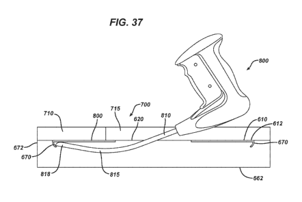
An embodiment of an erribodiment of a tissue repairpatch device 600 of the
25 piesent invention having locating structure. 650 with a downwardly
emending flango.
configukution is seen in FIGS. 37-41. Referiing first to FIGS. 37, 38 and 39,
the-tissue
repair patch device 600 is seen to have a: locating structure (60 ìn th.e fOnn
of a
downwardly extending flange member-670 that is made by- molding or otherwise
forming
prt of the periphery. 616 of base member 610. The flangemember 670.-is..secri
to have
30 bottorn edge-672, inner side 674, outer side 676-and top 678. Thellange
member has a
eurvederess-Section,:but may have other configurations and-cross-sections
including-
straight and angled, 'the device 600 when emplaced adjacent. to a patient's
body wall 710
on. the interior surface 712 as shown, is secured by m.anipulating the distal
tip -81.8 of the.
CA 02906349 2015-09-14
WO 2014/149642 PCT/US2014/020071
5- distal- seetion-8-15 ofshaft 810 of the ins/nu-nem 800 such that the tip
818 is .proximal to
or touching the inner side 674 of-flange member 6M. AS seen in FIG, 39, the
locating
structure 650 may consist of-a separate flange member 680. h.aving top 682,
bottom. 684,
itiner,side 686 and. outer side 688. The top 612 of -flange member 680 may be
mounted to
the periphery 616 or peripheral edge 618 of:base. member 610 in -#
conventional manner.
such as by gluing, welding, sewing, -fastening, co-ntolding, etc. The device
600 having
flange metriber seento be utilized and Unplowed in apatient to repair a
tissue
defect as previouSly described above.
Yet nother etrihodiment of the tissue repair member 600 having a locating
structure 650 with a downwardly extending flange structure is- seen in FIGS.
40. and-41.
The smicturc 690 is seen to consist of a ringor petiph.eral element 691 and a
downwardly
extending flange section 695.. The peripheral element 691 is seen to have top
side- 692,
bottom side 693 and outer sick 694. The dovenwardly extending:flange-section
695 hos-
top (i96, bottom: 697, inner Side 698, and. outer side 699. The structure
690may be
mounted such that. the bottom- side 693 of peripheral -element is on the top
side 612 of
base. member 614 adjacent to or on the periphery 616 and the peripheral edge
618 is
coveredoor the structure. 690 may be mounted :such that the top side 692 of
peripheral
clement 691 is on the bottom side 6-14 of base member-614 adjacent to oron -
the
periphery 616., The device 600 having structure 690 iS seen to he utilizexì
and implanted
in a patient to repair a tissue defectus previously described above.
The repair patches of th.e present invention may optionally contain or be
coated
with sufficiently effective amounts of an. active -agent Such- as -a
therapeutic -4gettt.
Substances *hid are suitable US active. agents include conventional agents.
that ntay be
naturally occurring or symhetic and ma:s,,, include but are not limited to,
.for example,
antibiotics, antimicrobials, antibacterialS, antiseptics, chemotherapeuticsõ
cytostatiesõ
metastasis inhibitors, anticleaboties, antitnycoties, lAynaecological. agents,
urologieal
agents, anti-allergic agents, sexual. hormones, sexual hormone inhibitors, -
haemostypties,
hormones,iieptidt.s4woriones, -antidepressants, vitamins. such as 'Vitamin C,
-antihistamines, naked DNA., plasmid DNA, cationic. DNA -complexes, RNA, cell
26
CA 02906349 2015-09-14
WO 2014/149642 PCT/US2014/020071
5- constituents, vaccines, and cells-.oceurring naturally in the body or
:genetically modified
cells.
la. one -embodiment, he active agents ntay be antibiotics: including suCh
agents as
gentatnicin or ZEVTERATN (ceflobiprole- medoctuil) brand antibiotic (available
from
Basilea Phannacentica Ltd., Basel Switzerland). In one embodiment, an implant -
may
include 'broadband antimicrobials. used against different bacteria and yeast
(even in the
presence of bodily liquids) such as octenidine, octenidine dihydrochloide
(availa.ble as
active- ingredient OeteniseptIP disinfectant from Schulke- & Mayr,
Norderstedt, any
as), polybexamethylene biguanide (PHMB) (available as active ingredient
Lavasept:k
from Braun. Switzerland), triclosan, copper (Cu), silver (Ag), nanosilver,õ
gold OW,.
selenium (Se), .gallium- (Ga),.tautolidinc,. N-thlorotaurine,:aleohol based-
antiseptics such
as ListerintA mouthwash, N a-lauryl-learginine ethyl ester (LAE),õ
myristamidopropyl
dimethylamine (MAPD, available as an active ingredient -in SCHERCODINPm I\4),
oleamidupropyl dirt ethylamine (GAD, available_ _as an active -ingredient = in
ScHEROODINErm 0),. and stearamidopropyl dimethylamine (SAPP* available as an
active ingmdient ín SCHER.CODINETh S). In one ernbodiment, the agent may be.
octenidine &hydrochloride (hereinafter-referred to as octernidine) andior
Although it is preferred. to have 4 single, centrally located opening in the
hernia
repair .patch devices of-the present invention, the opening and associated
closure tneniber
maybe offset from the. center. Additionally, more than oneopcning and closure
member
may be utilized in thetemia impair devices of the present invention.
The following examples are illustrative-of theprinciplesand practiee.of the
pmsent invention, -although not limited thereto,
Intattole 1
27
CA 02906349 2015-09-14
WO 2014/149642 PCT/US2014/020071
5. A. -patient with a ventral or ineisional hernia is prepared-for an open
hernia rep-air
procedure in the -following manner. The skin area Suntunding the hernia is -
scru'bbed
with a conventional antimicrOhial solution such. as betadine. The patent is -
administered.
conventional general anesthesia in. a conventional manner by -induction and
inhalation.
The surgeon then initiates die surgical procedure by making an ineision in -
the Ain and.
stibcutaneous tissueoverlying:-the hernia. In the. case of pianned ìitra-
peritoneal mesh
placement, the hernia Sae ísopened> l'heed.ges-of the healthy fakia arotmd the
defixt are
examined. and any attachments of the: viscera to the abdominal wall are
divided to create a
free space for fixation -of the mesh.
At this point in the procedure-, the surgeon then- prepares a mesh tissue
repair
hernia patch of the .pressent invention having a locating SITLICtUret and
lurving closure flaps
anda base member for insertion through the abdominal wall. defect and into the
abdominal cavity such that the top side of the mesh is adjacent to the p er
itoneurn.
surrounding the defect, and. the bottom side of the mesh device is -facing
down toward. the
patient's viscera. Stay sutures :may be placed through the meg) into the
abdominal. tissue
as desired, i.e. at thc four compass points of the mesh (North, -South, East,.
West). The
flaps are :rotated upwardly after placement to expose the opening :in the base
member of
the mesh. The- mesh is-fixated with a. conventionalsurgical tacker instrument
or other
means of fixation. A taek.er is inserted through the 0:pening- such that the
distal end of the
tacker-is between thernesh. and -the viscera, and the surgeon locates
theperiphery of the
-25 repair patch by engaging the locating struCture with the tip of th.e
shaft of th.e tacker'
instrument. The. perimeterof the mesh is then. fixated using a plurality of
tacks in a
crown configuration. The tacker 14:1 removed and the openingin the mes:h is
closed by
folding the flaps as appnoptiate -fix the present invention. The flapionay be
optionally
seettml. using adhesive-, -suture,- rivets., or other closure means, or may be
returned -to their
at rest position -without. Setatement to each other. The hernia defect may be
primarily
closed if desired.. The skin ineision is closed using. appropriate saluting.
or closure
techniques, ancl. the incision. is appropriately bandaged -and the patient is
moved to a
recovery -room.
28
CA 02906349 2015-09-14
WO 2014/149642 PCT/US2014/020071
5- The novel hernia repair devices of the present invention have numerous
advantageS. The novel repair patch -devices prOvi& a single layer mesh repair
device that
can bc affixed vi.a tacking in an open intraperitoneal. henna repair
procedure. The-repair
patch devices have .additional advantages including less foreigamaterial.
lowermaSs
of foreign materialyand the:ahility to implant a single layer tissue repair
mesh. -in -open
procedures-. The -tissue repair devices -of thepresent invention, preferably
made -from
niegh, May potentially aCeelerate the rate of tissue integration, provide less
area for
biofilm fonnation, have a lower cost ofmanufac.ture, and are easier to
package, sterilize,
and use with improved.ereonornics..
Although this invention has been shown anti described with respect -to
detailed
embodiments thereof, it will be understood by those...skilled inthe art that
various changes
in form and detail thereof -may be made, without. departing from the spirit
and scope of the.
claimed. invention,
29