Note: Descriptions are shown in the official language in which they were submitted.
METHODS AND DEVICES FOR ENDOMETRIAL
CELL AND TISSUE SAMPLING
RELATED APPLICATIONS
[0001] The present application claims the priority of and the benefit
of the filing date of
U.S. Provisional Patent Application Serial No. 611819,471, filed May 3, 2013,
TECHNICAL FIELD
[0002] The present invention relates to methods and devices for
obtaining samples,
simultaneously or sequentially, of cells, for example, endometrial cells.
BACKGROUND
[0003] Current methods of sampling the enclometrium for cells are
inadequate and may
be harmful to the outcome for the patient. What is needed are methods and
devices that can
obtain a more complete sampling of the area without degradation of the
anatomical area.
SUMMARY
[0004] Disclosed herein are methods and devices for sampling tissues,
such as
endometrial tissues, for example, sampling the uterine cavity for analysis as
an endometrial
biopsy procedure. The methods and devices may comprise only a scraping of the
endometrial
tissue to obtain a cellular sample, or in combination with negative pressure,
scraping the
uterine cavity to obtain an adequate sample. The present invention comprises
sampling
devices and methods that are improved over currently available devices. The
present
invention comprises improved sampling in the quantity of tissue and cells
obtained and in the
ability to broadly sample the target area, such as the endometrial lining, as
the target area is
assessed circumferentially and longitudinally. Additionally, the present
invention comprises
methods comprising devices disclosed herein for minimal contamination of the
sample once it
is acquired and removed from the patient Methods and devices disclosed herein
also offer
improved patient outcome in that an adequate sample minimizes the need for
further
evaluation and additional procedures to make an assessment of the patient's
condition. The
invention disclosed herein provides methods and devices that decrease patient
discomfort, for
example, during an endometrial biopsy (EMB) procedure, and devices that sample
the target
area to provide accurate and sensitive detection of endometrial abnormalities.
Aspects of the
CA 2911243 2019-07-09
CA 02911243 2015-11-02
WO 2014/179804 PCT/US2014/036828
present invention aid in providing a more patient-oriented device that when
used is more
comfortable with less pain for the patient. Devices may be provided in various
sizes to allow
for single or minimal entry into the uterine cavity to obtain an adequate and
broad sample
than is found when currently available devices are used. For example, a method
of the
present invention comprises collecting a sample of tissue and cells from the
endometrium by
insertion of the device only one time into the patient's uterine cavity.
Currently used devices
sometimes require more than one insertion of the device into the uterine
cavity to obtain
adequate samples. A device of the present invention comprises a sample
collection area and
wand that are cylindrical in shape that captures the sample by scraping along
the inner
surfaces of the uterine cavity which allows for deeper and more extensive
contact with the
lining which is more effective in obtaining an adequate sample than utilizing
a suction
mechanism to pull in a sample into a device. A device of the present invention
may be
offered with sample collection areas of different lengths to match the anatomy
of the patient.
Additionally, a device of the present invention may comprise an atraumatic tip
or a tapered
distal tip of the wand that allows for ease of entry of the device into the
endocervical canal
and subsequently into the uterine cavity. The tip may provide a dilation
function to aid in
insertion of the device through the endocervical canal.
[0005] A device of the present invention comprises handle, a wand
surrounded by a
sheath, tissue sampling elements, and a sample collection cavity. The device
is used for tissue
and cell sampling. The device handle remains outside of the patient while the
sample
collection cavity and tissue sampling elements are placed in proximity to the
target area. A
target area may be an endometrial surface. The wand is an elongated body
(tubular or solid)
that is designed to traverse along the entire length or partial length of the
inner circumference
of the sheath. In embodiments presented herein, the wand is connected to a
handle for
manipulation of the entire device and the handle may incorporate components to
expand the
sampling area, comprising tissue sampling elements and a sample collection
cavity to allow
for scraping of the target area, such as the endometrial lining, both
circumferentially and
longitudinally, against the inner surface of the uterine cavity to obtain
tissue or cells. The
sheath of a device of the present invention may be an elongated tubular body
designed to
allow for an inner shaft to traverse the sheath's inner circumference. The
sheath may function
to protect the tissue sample from contamination, and may provide the tissue
sampling
elements by which the sampling is achieved (i.e. serves to scrape the uterine
cavity), protect
the sample from being lost during retraction of the device through the
cervical os, and/or aid
2
in device placement. The sampling area comprises the portion of the device
where the tissue
sampling elements, such as opposing edges of a slit are used as scraping
edges(s), are located.
The sampling area provides for capturing the tissue and endometrial cells
located in the uterine
cavity.
[0005A] In a broad aspect, the present invention pertains to an endometrial
sampling apparatus,
comprising a handle having a distal end and a longitudinal axis, an elongate
wand extending
outwardly from the distal end of the handle substantially along the
longitudinal axis. The wand
has an exterior surface, a front end and a back end, the back end being
fixedly mounted to the
distal end of a portion of the handle. There is a selectively moveable
actuator coupled to a
portion of the handle and to a sheath. A sheath member selectively
encapsulates a portion of the
wand and is fixedly mounted to a portion of the wand proximate the front end
of the wand, and to
a portion of the first end of the actuator member. The sheath member defines a
slit(s) on a distal
end portion of the sheath member, the slit being bordered by opposing edges.
Thc distal end
portion of the sheath member and a portion of the exterior surface of the wand
underlying the
distal end portion of the sheath member define a sample collection cavity.
[0005B] In a further aspect, the present invention provides an endometrial
sampling apparatus,
comprising a handle have a distal end and a longitudinal axis, an elongate
wand extending
outwardly from the distal end of the handle substantially along the
longitudinal axis. The wand
has an exterior surface, a front end and a back end, and the back end is
fixedly mounted to the
distal end of the handle. An actuator member is rotatively coupled to the
distal end of the tube,
the actuator member defining an opening at a first end that is sized to
rotatively receive a portion
of the wand. A sheath member selectively encapsulates a portion of the wand,
and is fixedly
mounted to a portion of the wand proximate the front end of the wand and to a
portion of the first
end of the actuator member. The sheath member defines a slit on a distal end
portion of the
sheath member, the slit being bordered by opposing edges. The slit is
selectively movable
between a closed position, in which the opposing edges of the slit
substantially adjoin to
substantially seal a sample collection cavity, and an open position, the
opposing edges of the slit
being spaced from each other for selectively obtaining an endometrial sample
when positioned
within a uterine cavity.
2a
CA 2911243 2019-07-09
DESCRIPTION OF FIGURES
[0006] The accompanying figures, which are incorporated in and constitute
a part of this
specification, illustrate several aspects and together with the description
serve to explain the
principles of the invention.
[0007] Figure I shows an exemplary example of a device of the present
invention.
[0008] Figure 2A and B shows an enlarged view of a sampling area of the
device of
Figure 1.
[0009] Figures 3A and B shows an enlargement of the handle and actuator
portions of
the device of Figure 1, wherein 3A is an exterior view and 3B is a cross-
sectional interior
view.
[0010] Figure 4 shows an exemplary device of the present invention.
[0011] Figure 5 A-C shows an exemplary device of the present invention,
where 5B and
5C are an enlargement of the distal portion of the exemplary device shown in
5A.
[0012] Figure 6A-D shows the distal end of the handle of a device of Fig.
1 and 3,
wherein A is a front view, C is an enlargement of A, B is a back view and D is
an
enlargement of B.
[0013] Figure? is an exemplary example of a device of the present
invention.
[0014] Figure 8 A-C is an exemplary example of a device of the present
invention.
[0015] Figure 9 A-C is an exemplary example of a device of the present
invention.
[0016] Additional advantages of the invention will be set forth in part
in the description
which follows, and in part will be obvious from the description, or can be
learned by practice
of the invention. The advantages of the invention will be realized and
attained by means of
the elements and combinations particularly pointed out in the appended claims.
It is to be
3
CA 2911243 2019-07-09
CA 02911243 2015-11-02
WO 2014/179804 PCT/US2014/036828
understood that both the foregoing general description and the following
detailed description
are exemplary and explanatory only and are not restrictive of the invention,
as claimed.
DETAILED DESCRIPTION
[0017] Disclosed herein are methods and devices for cell sampling. The
present
invention provides for collection of cell and tissue samples from an area of
interest within an
organ or tissues, such as the uterine cavity, by having an expandable aspect
which allows for
broad contact, such as circumferentially to the inner surface of the walls of
the uterine cavity,
with capture of a sufficient sample volume for analysis and minimal
contamination of the
sample collected. The diagnosis of pre-malignancy and malignancy is dependent
on the
quantity and broad representation (a sufficient number of cells to be of
diagnostic value) of
the targeted area with the purest sample obtained. Obtaining an adequate
sample is usually
difficult as the customary available devices utilize suction to withdraw a
sample from a
limited area that often is rendered not adequate for diagnosis. The present
invention allows
the user to obtain a broad, representative and more substantive sample with
minimal
contamination from a patient with one or a few entries and removals from the
patient.
[0018] An aspect of the present invention comprises an expandable sampling
area
comprising tissue sampling elements and a sample collection cavity. A tissue
sampling
element may comprise opposing edges of a slit. Providing a device having an
expanding
sampling area provides for the insertion of the device and the collection of
the sample to
occur substantially along one plane or a single line of entry into the
patient. Once inserted, a
device of the present invention may be rotated around the single line of entry
to collect a
sample circumferentially and/or may be moved longitudinally in a distal to
proximal direction
or proximal to distal direction to collect a sample. A method of using a
device of the present
invention comprises insertion of the device and collection of the sample along
a single plane
or a single line of entry into the patient, wherein the rotation occurs in a
fixed location with a
very small to no rotational diameter of the wand and handle. In collecting a
sample using
currently available devices, not one of the present invention, a device end
having a sampling
area is inserted into a patient and then the sampling area is moved within the
uterine cavity by
the device, such as the insertion member and handle, being rotated through a
large arc or
circle to apply pressure to the sampling area. The rotation of the insertion
member and the
handle is not around a single line of entry, but comprises a large rotational
area that comprises
a large rotational diameter, and resembles a geometric cone with the sampling
area at the
4
CA 02911243 2015-11-02
WO 2014/179804 PCT/US2014/036828
apex of the cone and the large rotational diameter due to movement of the
handle at the base
of the cone.
[0019] In an aspect, the small to no rotational diameter of a device of the
present
invention is due to the expandable nature of the tissue sampling elements of a
device of the
present invention. Before insertion of a device of the present invention into
a patient, a tissue
sampling element comprising opposing edges of a slit are aligned with the
outer surface of the
sheath, which is referred to herein as the closed position. In the closed
position, the entire
sheath has a substantially uniform diameter from the proximal end to the
distal end of the
sheath. Once the sampling area of the device is in place in the cervical canal
or uterine
cavity, the slit (or slits) is opened, exposing the opposed edges of a slit
and creating the tissue
sampling element of the device. There is no need to rotate the entire device
in geometric
cone shaped rotation because the expanded tissue sampling element contacts the
inner surface
of the uterine cavity, a potential space. The opposed edges of the slit or
slits are then rotated
with a small to no rotational diameter along the single line of entry and
because the expanded
opposed edges of the slit or slits are in contact with the inner surface of
the uterine cavity, the
sample is scraped or cut from the inner surface by the edges and collected
within the sample
collection cavity. Once the sample is within the sample collection cavity, the
slit or slits are
moved to the closed position, so that the opposing edges are substantially
adjacent to each
other and the sample collection cavity is substantially covered so that no
sample within the
sample collection cavity can exit and no cellular material from outside the
sample collection
cavity can enter the cavity, which prevents contamination of the sample.
[0020] Having an expandable tissue sampling element also provides more
contacting
surface within the uterine cavity than is possible with currently available
devices. For a
currently available device, which does not have an expandable scraping
element, the thin
tube, which is designed to be small enough to go through the cervical canal,
utilizes an
opening at the end of the tube and provides suction at that opening to
withdraw tissue for a
sample of a limited area. When in place in the uterine cavity, a vacuum is
established within
the device by manually retracting its internal piston and the device is
rotated between the
fingers and it is passed several times between the fundus and the internal
cervical os. This
type of suction forces limited tissue and cells from the uterine cavity, which
is at least
uncomfortable and generally painful for a patient, and the sample obtained is
often
determined to be not adequate for diagnosis.
CA 02911243 2015-11-02
WO 2014/179804 PCT/US2014/036828
[0021] A method of the present invention comprises obtaining a sample of
the
endometrium of the uterine cavity by using a device comprising a sampling area
comprising
an expandable tissue sampling element. A device of the present invention
comprises an
expandable tissue sampling element. A device of the present invention
comprises a sampling
area comprising an expandable tissue sampling element comprising a sheath
having at least
one slit comprising two opposing edges wherein the two opposing edges may be
moved apart
from each other to expand the diameter of sheath so that the diameter of the
sheath is greater
where the two opposing edges are moved apart from each other than the diameter
of the
sheath where there is no slit. An expandable tissue sampling element is
expanded by the
movement of the opposing edges of a slit away from each other and the tissue
sampling
element is reduced to its original state by the opposing edges of a slit
moving together and
being adjacent again. When the slit is open, so that the opposing edges of the
slit are apart
from one another, the sheath and/or sampling area is said to be expanded. When
the slit is
closed, so that the opposing edges of the slit are substantially adjacent to
one another, the
sheath and/or sampling area is not expanded.
[0022] The standard management of patients with abnormal uterine bleeding,
postmenopausal bleeding, suspected uterine cancer, AGUS (abnormal glandular
cells of
unknown significance) as determined by pap smear, chronic anovulation, those
undergoing an
endometrial ablation or are infertile where the lining can determine ovulation
(i.e.
endometrial dating) is an endometrial biopsy (EMB). An endometrial biopsy is
the most
commonly performed test to assess the presence of endometrial hyperplasia or
endometrial
cancer.
[0023] It is common for endometrial tissue sampling to result in findings
that are
insufficient for diagnosis. In studies of patients with postmenopausal
bleeding, the range of
sampling failure (i.e. inadequate sample or inability to perform the biopsy)
with Pipelle
biopsy was 0-54% as noted by the Committee on Gynecologic Practice (ACOG
Committee
Opinion, August 2009). When endometrial biopsy is performed and tissue is
reported as
insufficient for diagnosis, some further examination is necessary, which may
include
transvaginal ultrasonography. If the endometrial biopsy sample doesn't provide
enough
tissue, or if the biopsy suggests cancer but the results are uncertain, a D&C
(dilation and
curettage) is performed. In this outpatient procedure, the opening of the
cervix is enlarged
(dilated) and a special instrument is used to scrape tissue from inside the
uterus. This may be
done with or without hysteroscopy. The procedure takes about an hour and may
require
6
CA 02911243 2015-11-02
WO 2014/179804 PCT/US2014/036828
general anesthesia or conscious sedation either with local anesthesia injected
into the cervix
or with spinal or epidural blocking anesthesia. A D&C is usually done in an
outpatient
surgery area of a clinic or hospital. Most women have little discomfort after
this procedure.
A D&C procedure is costly and invasive and would not be necessary if the
endometrial
biopsy captured an adequate sample for analysis.
[0024] A number of devices have been developed to collect samples from the
uterine
cavity, including Novak's curette, Vabra aspirator, Masterson endometrial
biopsy system and
Pipelle device (most commonly used). These devices incorporate a means of
suction to
obtain the tissue sample, whether by syringe, a device that generates a
vacuum, a reusable
hand operated pump, or an internal piston that is manually retracted to create
a vacuum for
sample collection.
[0025] Although, cytologic screening reduces mortality from uterine cancer
by earlier
diagnosis of invasive disease, there is still an unacceptable sample
inadequacy rate due to the
suction devices used and the method by which a sample is obtained. The current
EMB
devices obtain a questionable sample that is inadequate for pathological
evaluation, at the
expense of subjecting the patient to one or more subsequent, more invasive
procedures. This
inadequate finding often leads to unnecessary dilation & curettage or other
transvaginal
procedures. Furthermore due to the current device designs it is unlikely that
a broad sample
of the uterine cavity will be obtained so detection of pre-cancerous or
cancerous cells may be
missed.
[0026] The currently used or available device designs have inherent
deficiencies that
affect their ability to be a reliable and useful tool in diagnosing
endometrial hypemlasia or
endometrial cancer. The ideal EMB device is one that is simple, broad
contacting, and most
importantly provides an adequate volume sample of the endometrial lining of
the uterine
cavity that is minimally contaminated. The devices of the present invention
comprise easy to
use devices that are applicable to a variety of anatomical uterine cavity
variants, which
decrease the frequency of inadequate sample findings, which allow for improved
detection
rates of early high grade lesions, and that decrease the number of unnecessary
additional
treatments.
[0027] The present invention comprises methods and devices useful for
obtaining the
intended tissue sample of a body conduit under controlled conditions, for
example, where the
tissue are located in the uterine cavity. For example, the present invention
comprises
7
CA 02911243 2015-11-02
WO 2014/179804 PCT/US2014/036828
methods and devices for use in capturing a broad, adequate, and minimally
contaminated
sample with single or minimal number of entry insertions into the uterine
cavity. The
devices can be designed to be reusable or disposable for single use. As used
herein, broad
means that the sample comprises cells and tissues from a representative area
of the organ that
provide a more accurate diagnosis of the condition of the organ, as compared
to currently
available devices having one opening or a port oriented in one direction,
which then contacts
a small, limited area of the endometrium. For example, a broad sample would
comprise a
representative sample of the cells or tissue in that general area of the
organ. The sample may
be broad and encompass a representative sample by sampling from the area of
the organ
contacted by insertion of the sampling area into the uterus and expanding the
tissue sampling
element in that area contacted, or the tissue sampling clement may be moved in
one or more
directions, in contact with the endometrium, to obtain a sample of cells or
tissue in the entire
area contacted.
[0028] A device of the present invention may be a sterile, disposable
endometrial
sampling device with indications for single patient use in obtaining tissue
samples from the
uterine cavity for histological analysis. Clinical indications for performing
a method of the
present invention using a device disclosed herein include, but are not limited
to, further
evaluation of abnormal glandular cells of unknown significance (AGUS) as
determined by
pap smear; further evaluation of abnormal uterine bleeding, including
postmenopausal
bleeding; as a diagnostic device in patients suspected of uterine cancer; for
chronic
anovulation; prior to those undergoing an endometrial ablation procedure; for
patients who
are infertile where the lining can determine ovulation (i.e. endometrial
dating); or with other
clinical sequelae. A device of the present invention may collect a targeted
broad tissue
sample with minimal contamination that is adequate in volume for histological
evaluation. A
device of the present invention may comprise a wand that is somewhat rigid and
one
directional (not bent) or a wand may be bendable, or bent, so that an area of
interest is
contacted by the expandable sampling area.
[0029] A device of the present invention comprises a sampling area
comprising a sample
collection cavity comprising a portion of a wand having a reduced diameter and
an overlaying
sheath member having at least one slit that is capable of providing an opening
within the
sheath member for access to the sample collection cavity beneath. The slit may
have two
opposing edges. There may be one or more slits in the sheath member forming
one or more
access sites to the sample collection cavity. The slit may be in an open or
closed position. In
8
CA 02911243 2015-11-02
WO 2014/179804 PCT/US2014/036828
a closed position, the opposing edges of the slit are adjacent and adjoin each
other so as to
substantially close the slit and to prevent access to the sample collection
cavity underlying the
slit or slits. In an open position, the opposing edges of the slit are apart
from each other to
form an opening so that the sample collection cavity may be accessed. In an
aspect, when the
opposing edges are moved apart from each other, the diameter of the area of
the sheath where
the slit occurs is greater than the diameter of the area of the sheath where
the slit occurs when
the opposing edges are substantially adjoined or a closed position. The open
position is also
referred to herein as being expanded. When the opposing edges of a slit are
moved apart from
each other, each edge is exposed and forms a scraping edge, and the opposing
edges form a
tissue sampling element that is used to remove tissue from a soft tissue
location. The
removed tissue enters the sample collection cavity through the opening formed
by the moved
apart opposing edges. The removed tissue (a sample) is contained within the
sample
collection cavity and the opposing edges are moved together to substantially
close the sample
collection cavity so that no further tissue can enter the sample collection
cavity to
contaminate the sample and so that the sample does not leave the sample
collection cavity and
be lost.
DESCRIPTION OF DEVICE
[0030] The present invention comprises a device for sampling a tissue, for
example the
uterine cavity comprising endometrial lining. A device of the present
invention is not limited
to sampling a tissue and collecting cells as described herein for uterine
sampling, but may be
used for sampling tissues and obtaining cells from any tissue as may be used
by those of skill
in the art. The present invention is not limited to only the examples
disclosed herein but may
be used in methods of cellular sampling contemplated by those of skill in the
art.
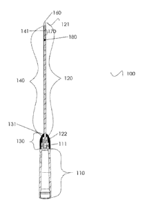
[0031] An exemplary device is shown in Figures 1, 2, 3 A and B, and 6, 8A-C
and 9A-C,
and exemplary devices are shown in Fig. 4, Fig. 5 and Fig. 7, and described
herein. Looking
at Fig. 1, a device of the present invention comprises a endometrial sampling
apparatus 100,
comprising, a handle 110 having a distal end 111 and a longitudinal axis, an
elongated wand
120 extending outwardly from the distal end 111 of the handle 110
substantially along the
longitudinal axis, wherein the wand 120 has an exterior surface, a front end
121 and a back
end 122, and wherein the back end 122 is fixedly mounted to the distal end 111
of the handle
110; an actuator member 130 rotatively coupled to the distal end 111 of the
handle 110, the
actuator member 130 defining an opening at a first end 131 that is sized to
rotatively receive a
portion of the wand; a sheath member 140 selectively encapsulating a portion
of the wand
9
CA 02911243 2015-11-02
WO 2014/179804 PCT/US2014/036828
120 and fixedly mounted to a portion of the wand proximate to the front end
121 of the wand
and to a portion of the first end 131 of the actuator member 130, wherein the
sheath member
140 defines a slit 141 on a distal end portion of the sheath member 140, the
slit 141 being
bordered by opposing edges 142 and 143, (see Fig. 2) wherein the distal end
portion of the
sheath member 140 and a portion of the exterior surface of the wand underlying
the distal end
portion of the sheath member define a sample collection cavity 150. As shown
in Fig. 2, the
slit 141 is selectively movable (expandable) between a closed position 200,
(Fig. 2A) in
which the opposing edges 142 and 143 of the slit 141 substantially adjoin to
substantially seal
the sample collection cavity 150, and an open position 201, (expanded) (Fig.
2B) in which the
opposing edges 142 and 143 of the slit 141 are spaced from each other for
obtaining a tissue
sample when positioned within a uterine cavity by contacting the surface of
the endometrial
lining. When spaced apart from each other, the opposing edges 142 and 143 form
a tissue
sampling element such that when contacting a soft tissue surface is capable of
removing
tissue from a soft tissue surface, such as the inner surface of the uterine
cavity. As shown in
2B, the sampling area 151 comprises an open sample collection cavity 150 with
opposing
edges 142 and 143 forming the tissue sampling element 146. Optionally, spaced
from the
proximal end of the sample collection cavity 150 is an indicator 180, which
may be used as a
depth indicator to a user to indicate the length of the apparatus inserted
into the patient and
approximate location of the sample collection cavity 150. A depth stop, not
shown, may be
placed on or proximate to the indicator 180. The device may comprise a closed
tip 160,
which may be an atraumatic tip.
[0032] As shown in Fig. 3A and B, actuator member 130 is selectively
rotatable about
the distal end 111 of the handle 110 between a first position 360, in which
the slit 141 is
positioned in the closed position 200 (shown in Fig. 2A), and a second
position 361, in which
the slit 141 is positioned in the open position 201 (Fig. 2B). The slit 141
may be a helical slit
141 that extends about the longitudinal axis. The helical slit may comprise a
plurality of
helical slits. A slit may have at least one round 170 around the longitudinal
axis. (See Fig. 2)
The opposing edges 142 and 143 of the slit 141 may be oriented substantially
parallel to each
other and substantially normal to an exterior surface of the sheath member 140
when the slit
141 is in the closed position 200. At least a portion of one of the opposing
edges 142 and 143
of the slit 141 is oriented at an acute angle to the longitudinal axis when
the slit 141 is in the
open position 201. The opposing edges 142 and 143 of the slit may be oriented
substantially
parallel to each other and are positioned at a face angle relative to an
exterior surface of the
CA 02911243 2015-11-02
WO 2014/179804 PCT/US2014/036828
sheath member 140 when the slit 141 is in the closed position 200. At least a
portion of one
of the opposing edges 142 and 143 of the slit 141 may be oriented at an acute
angle to the
longitudinal axis when the slit 141 is in the open position 201. In an aspect,
the face angle is
not normal to the exterior surface of the sheath member 140.
[0033] In an aspect, the front end of the wand 120 defines an atraumatic
tip 160. (See
Fig. 1 and 2). The atraumatic tip 160 may be tapered for ease of entry of a
device into the
cervical os. The atraumatic tip 160 may be made from the same or a different
material as the
wand, and may be more flexible than the wand for patient comfort. The distal
end portion of
the sheath member 140 is positioned proximate to the atraumatic tip 160 of the
wand 120.
[0034] In an aspect, a portion of the wand 120 has a reduced diameter. In
an aspect, the
portion of the wand 120 having a reduced diameter underlies the distal end
portion of the
sheath member. In an aspect, the portion of the wand 120 having a reduced
diameter
underlies the distal end portion of the sheath member where a slit 141 is
located. In an aspect,
the portion of the wand 120 having a reduced diameter and the distal end
portion of the sheath
member 140 where a slit 141 is located define a sample collection cavity 150.
[0035] See Fig. 3A which shows the exterior surface of handle 110 and
actuator 130.
Fig. 3B shows the interior view of actuator 130 as seen by looking from the
handle towards
the actuator in a distal direction, as sectioned along the line shown in Fig.
3A. In an aspect,
actuator 130 defines an interior cavity 301 having an interior peripheral edge
302 having a
plurality of spaced indentations 320 (320a and b) defined thereon and one or
more actuator
protrusions 340 (e.g., 340a and 340b). An actuator protrusion 340, when
interacting with the
distal end 111 of handle 110, may prevent the actuator 130 from continuing to
rotate in a
particular direction, depending on the location of the actuator protrusion.
For example, when
actuator 130 is in Position 1 (360), protrusion 340a stops actuator 130 from
rotating further in
a counterclockwise direction. Similarly, when actuator 130 is in Position 2
(361), actuator
protrusion 340b stops actuator 130 from rotating further in a clockwise
direction.
[0036] Looking at Fig. 3B and 6, the distal end of the handle 110 defines a
handle
protrusion 350 extending distally along the longitudinal axis, the handle
protrusion 350
defining a plurality of radially biasable keys 330 and a stationary portion
331. The radially
biasable key 330a is configured to be selectively received therein the
plurality of indentations
320 (e.g., 320a and 320b) in the respective first 360 and second 361
positions. A shown in
Figs. 3A and 6, key 330a is rounded so as to fit within a rounded indentation
320a and 320b.
11
CA 02911243 2015-11-02
WO 2014/179804 PCT/US2014/036828
Key 330b acts as a radially biasable key during assembly of the device and
then it acts as a
non-biasable key during device operation since it does not interact with the
spaced
indentations 320 during actuation/rotation. Stationary portion 331 provides a
stop 610 to
impede the movement of actuator 130. In an aspect, the plurality of spaced
indentations 320
comprise a pair of spaced indentations 320 positioned between about 1100 to
140 apart. In
an aspect, the plurality of radially biasable keys 330 (e.g., 330a and 330b)
comprise a pair of
spaced radially biasable keys 330 positioned between about 170 to 190 apart.
In an aspect,
the pair of spaced radially biasable keys 330 are positioned about 180 apart.
In an aspect,
moving the actuator position from closed 360 to open 361 requires a rotation
between about
220 to 250 .
[0037] Optionally, the elongate wand 120 is flexible. See for example, Fig.
1.
Optionally the distal end of the device comprising the sampling area 151 is
detachable. For
example, (not shown) the distal ends of the sheath and shaft, comprising the
sampling area
151, both could be scored so that with pressure or cutting, they break away
from the rest of
the device. A scored line around the diameter of the shaft could be used to
create a weak
section in the shaft so that the shaft would break when flexed. Depending on
the sheath
material, the sheath may or may not need to be scored also. In an aspect, the
sampling area
could be removed intact by cutting it off with a cutting tool or knife.
Alternatively, the tip of
the device could be sheared off using, a cutting tool.
[0038] As shown in Fig. 1 and 2, a device of the present invention
comprises a
endometrial sampling apparatus 100 comprising, a handle 110 having a distal
end 111 and a
longitudinal axis; an elongate wand 120 extending outwardly from the distal
end 111 of the
handle 110 substantially along the longitudinal axis, wherein the wand 120 has
an exterior
surface, a front end 121 and a back end 122, and wherein the back end 122 is
fixedly mounted
to the distal end 111 of the handle 110; an actuator member 130 rotatively
coupled to the
distal end 111 of the handle 110, the actuator member 130 defining an opening
at a first end
131 that is sized to rotatively receive a portion of the wand 120; a sheath
member 140
selectively encapsulating a portion of the wand 120 and fixedly mounted to a
portion of the
wand proximate the front end 121 of the wand 120 and to a portion of the first
end 131 of the
actuator member 130 , wherein the sheath member 140 defines a slit 141 on a
distal end
portion of the sheath member 120, the slit 141 being bordered by opposing
edges 142 and
143, wherein the slit 141 is selectively movable between a closed position
200, in which the
opposing edges 142 and 143 of the slit 141 substantially adjoin to
substantially seal a sample
12
CA 02911243 2015-11-02
WO 2014/179804 PCT/US2014/036828
collection cavity 150, and an open position 201, in which the opposing edges
142 and 143 of
the slit 141 are spaced from each other for selectively obtaining a tissue
sample when
positioned within a uterine cavity. In an aspect, the actuator member 130 is
selectively
rotatable about the distal end 111 of the handle 110 between a first position
360, in which the
slit 141 is positioned in the closed position 200, and a second position 361,
in which the slit
141 is positioned in the open position 201. In an aspect, the distal end
portion of the sheath
member 140 and a portion of the exterior surface of the wand 120 underlying
the distal end
portion of the sheath member define a sample collection cavity 150.
Optionally, an indicator
180 is located proximally to sample collection cavity 150. In an aspect, the
slit 141 is a
helical slit that extends about the longitudinal axis. A slit 141 may have at
least one round
170 around the longitudinal axis.
[0039] As shown in Fig. 3B and 6, in an aspect, actuator 130 defines an
interior cavity
301 having an interior peripheral edge 302 having a plurality of spaced
indentations 320
defined thereon, wherein the distal end 111 of the handle 110 defines a handle
protrusion 350
extending distally along the longitudinal axis, the handle protrusion 350
defining a plurality
of radially biasable keys 330, of which 330a is configured to be selectively
received therein
the plurality of spaced indentations 320 in the respective first 360 and
second 361 positions.
In an aspect, the plurality of spaced indentations comprise a pair of spaced
indentations 320
(e.g., 320a and 320b) positioned between about 1100 to 140 apart. In an
aspect, the plurality
of radially biasable keys comprises a pair of spaced radially biasable keys
330 (e.g., 330a and
330b) positioned between about 170 to 190 apart. In an aspect, the pair of
spaced radially
biasable keys 330 are positioned about 180 apart. In an aspect, moving the
actuator position
from closed 360 to open 361 requires a rotation between about 220 to 250 .
[0040] As shown in Fig.3B, actuator 130 is moved from position 1 (360) to
position 2
(361) by rotating actuator 130 around the handle 110 in a clockwise direction.
Biasable key
330a is present in indentation 320a in position 1, and when the actuator 130
is rotated in a
clockwise direction to position 2 (361), biasable key 330a then resides in
indentation 320b.
In position 1, actuator protrusion 340a abutting key 330b prevents actuator
130 from rotating
in a counterclockwise direction. In position 2, actuator protrusion 340b
abutting stop 610
formed in stationary portion 331 prevents actuator 130 from rotating in the
clockwise
direction. Handle protrusion 350 does not rotate, though keys formed therein
may be biased
radially, but the handle protrusion 350 remains in one location (other than
biasable keys
13
CA 02911243 2015-11-02
WO 2014/179804 PCT/US2014/036828
moving inward and returning to the starting position radially) and interacts
with the actuator
protrusions and indentations.
[0041] Fig. 4 shows an embodiment of the present invention comprising a
sample
collection cavity 450 formed by a distal section of the wand 420 having a
reduced diameter
470 and the overlying sheath 440, and having a sample area 415 comprising an
expandable
tissue sampling element formed by a slit 441 comprising two opposing edges,
which when
moved apart from each other form a tissue sampling element. The device
comprises a wand
420 and an overlying sheath member 440, a slit 441 in the distal portion of
the sheath
member, a handle 410 and a tip 460.
[0042] In a device of the present invention, a sheath may be moved, and a
slit may be
opened, in a variety of methods, and the present invention is not limited to
only those
exemplified herein. Moving a sheath member to affect the opposing edges of one
or more
slits, so that the opposing edges move apart from each other, may be
accomplished using an
actuator, and comprises holding one portion of the sheath immobile while
activating an
actuator which moves another portion of the sheath so that the force(s) in the
sheath from the
immobile portion and the moved portion force the opposing edges of a slit
apart. Relieving
the force(s) by returning the actuator and the moved portion of the sheath to
their original
locations brings the opposing edges of the slit together again to form the
closed position.
[0043] As shown in Figure 4, the sheath member is affixed on its distal end
445 to the
distal end of the wand 420 and/or to the tip 460. The proximal end of the
sheath is affixed to
an actuator, for example, a sheath nut 430. The sheath nut 430 is rotatable,
and when it
rotates, it also moves the sheath 440. In the first position, with no movement
by the sheath,
the slit 441, is closed with its opposing edges substantially adjacent and
adjoining each other.
When the sheath nut 430 is rotated to a second position, the sheath 440 moves
and the
opposing edges of the slit 441 move apart from each other, exposing the edges
and expanding
the diameter of the sheath in the area of the slit, as described above. The
sheath nut 430 may
be held in the second position by a locking member 480, which may be a screw
element that
is turned to engage the proximal end of the sheath nut 430 (not shown).
[0044] In an aspect, the locking member 480 may be a sliding element that
may be
moved in a longitudinal direction along the longitudinal axis of handle 410 to
engage the
proximal end of the actuator so as to hold the actuator in the second
position. In an aspect,
the actuator (sheath nut 430) may be held in position 1 by locking member 480
that is a
14
CA 02911243 2015-11-02
WO 2014/179804 PCT/US2014/036828
spring-loaded element such that when the locking mechanism is activated by
pushing on the
surface, an engaging element is released and the actuator is moved by the
force of the release
of the spring in a longitudinal direction along the axis of the wand or is
rotated
circumferentially around the wand to position 2. The actuator may be returned
to position 1
by manual manipulation and reengaging the engaging element. In an aspect, the
actuator may
be held in position 2, after manual movement of actuator from position 1 to
position 2, by
activating a spring-loaded locking mechanism 480 that engages with the
actuator in position
2. The actuator may be returned to position 1 by any method, for example, by
manual
manipulation. Position 2 may be one or more locations that are distally
removed from
position 1. Position 2 may be a defined distal location or may be any distally
removed
location chosen by the operator of the device. When position 2 is a defined
distal location,
the extent of the movement of the opposing edges away from each other is the
same extent,
and the opposing edges are moved apart to the same distance. When position 2
is an
optionally chosen distally removed distance from position 1, undefined by any
particular
structural stopping element, the extent of the distance between the opposing
edges of slit 441
is also an optional distance. Having an optionally distally removed distance
location for
position 2 allows for the opposing edges to be moved apart in a continuous
range, from the
maximum distance apart to a position of almost closed, allowing for control of
the amount of
expansion of the sheath member in the area of the slit 441. Once the actuator
is in position 2,
the position 2 location of the actuator is maintained by engaging a locking
member to hold
the actuator stationary, and, for example, the tissue sampling element is then
used to obtain a
sample that is contained within the sample collection cavity.
[0045] In an aspect, such as shown in Fig. 7, an actuator may rotate a gear
or set of
gears. One example of such a device, which may or may not place the sheath
member under
strain, is to have the actuator rotate a gear or set of gears which would
transfer the
longitudinal motion of an actuator into a rotational motion via a toothed ring
attached to the
proximal end of the sheath. Fig. 7 shows handle 710 comprising a gear system
712 which
comprises a gear that is moved by action of the actuator to move the gears and
affect the
sheath member 740 which is affixed to the gear system 712. Actuator movement
moves the
gears from position 1 where the slit 741 is closed and the sample collection
cavity 750 is
closed, to position 2 (not shown) which opens the slit exposing the opposing
edges to form
the tissue sampling element and opens the sample collection cavity. An
indicator 780 may be
in place on the sheath member 740 or the wand 720. An atraumatic tip 760 may
be on the
CA 02911243 2015-11-02
WO 2014/179804 PCT/US2014/036828
distal end of the wand. The sheath member 740 is affixed proximate to the tip
760. The
proximal end of the wand 720 is affixed to the handle 710. After a tissue
sample is acquired
and resident in the sample collection cavity, the actuator is moved so that
the gears return to
position 1, the slit closes so that the opposing edges are substantially
adjacent to one another,
the sample collection cavity is closed.
[0046] In an aspect, (not shown) an actuator may be moved in a longitudinal
direction, in
a proximal to distal direction, to move the sheath 440 so that the opposing
edges of slit 441
are moved apart from each other. An actuator is moved from its most proximal
site, position
1, where the slit is closed with its opposing edges substantially adjacent to
each other, and the
sheath member 440 is not under strain, to a second position, position 2 which
is distally
removed from position 1. When in position 2, the movement of sheath member 440
moves
the opposing edges of the slit 441 apart so that the tissue sampling element
is formed, as
described herein. The actuator may be held in position 2 by a locking member
480, which
may be a screw element, a sliding element or other such elements known to
those skilled in
the art that may interact with the actuator, the wand, the sheath member,
and/or the handle
410 to maintain the actuator in position 2 and maintain the slit in an open
configuration with
its opposing edges apart from each other. Position 2 may be one or more
locations that are
distally removed from position 1. Position 2 may a defined distal location or
may be any
distally removed location chosen by the operator of the device. When position
2 is a defined
distal location, the extent of the movement of the opposing edges away from
each other is the
same extent, and the opposing edges are moved apart to the same distance. When
position 2
is an optionally chosen distally removed distance from position 1, undefined
by any particular
structural stopping element, the extent of the distance between the opposing
edges of slit 441
is also an optional distance. Having an optionally distally removed distance
location for
position 2 allows for the opposing edges to be moved apart in a continuous
range, from the
maximum distance apart to a position of almost closed, allowing for control of
the amount of
expansion of the sheath member in the area of the slit 441. Once the actuator
is in position 2,
the position 2 location of the actuator is maintained by engaging a locking
member to hold
the actuator stationary, and, for example, the tissue sampling clement is then
used to obtain a
sample that is contained within the sample collection cavity.
[0047] Looking at Figs. 8 and 9, wherein like numbers indicate similarity
with those of
other figures, a device of the present invention comprises a tissue sampling
apparatus 800,
comprising, a handle 810 having a distal end 811 and a longitudinal axis, an
elongated wand
16
CA 02911243 2015-11-02
WO 2014/179804 PCT/US2014/036828
820 extending outwardly from the distal end 811 of the handle 810
substantially along the
longitudinal axis, wherein the wand 820 has an exterior surface, a front end
821 and a back
end 822, and wherein the back end 822 is fixedly mounted to the distal end 811
of the handle
810; an actuator member 830 rotatively coupled to the distal end 811 of the
handle 810, the
actuator member 830 defining an opening at a first end 831 (similar to 131 of
Fig. 1) that is
sized to rotatively receive a portion of the wand (not shown); a sheath member
840
selectively encapsulating a portion of the wand 820 and fixedly mounted to a
portion of the
wand proximate to the front end 821 of the wand and to a portion of the first
end 831 (similar
to 131 of Fig. 1) of the actuator member 830, wherein the sheath member 840
defines a slit
841 on a distal end portion of the sheath member 840, the slit 841 being
bordered by
opposing edges 842 and 843, (see Fig. 8C and 9C) wherein the distal end
portion of the
sheath member 840 and a portion of the exterior surface of the wand underlying
the distal end
portion of the sheath member define a sample collection cavity 850. As shown
in Fig. 8B and
9B, the slit 841 is selectively movable (expandable) between a closed
position, (Fig. 8B and
9B) in which the opposing edges 842 and 843 of the slit 841 substantially
adjoin to
substantially seal the sample collection cavity 850, and an open position,
(expanded) (Fig. 8C
and 9C) in which the opposing edges 842 and 843 of the slit 841 are spaced
from each other
for obtaining a tissue sample when positioned within a uterine cavity by
contacting the
surface of the endometrial lining. When spaced apart from each other, the
opposing edges
842 and 843 form a tissue sampling element such that when contacting a soft
tissue surface is
capable of removing tissue from a soft tissue surface, such as the inner
surface of the uterine
cavity. As shown in 8C and 9C, the sampling area 851 comprises an open sample
collection
cavity 850 with opposing edges 842 and 843 forming the tissue sampling element
846. As
shown in Figs. 8 and 9, spaced from the proximal end of the sample collection
cavity 850 is
an indicator 980, which may be used as a depth indicator to a user to indicate
the length of the
apparatus inserted into the patient and approximate location of the sample
collection cavity
850. A depth stop may be placed anywhere along the shaft to provide an
indication of the
depth of insertion of the device.
[0048] In an aspect, (not shown), suction can be incorporated into the
device to enhance
collection down the proximal end of the wand 820 and/ or for removal of the
sample collected
from the device. Reversing the suction, for example for dispensing the sample
from the
sample collection cavity, is also contemplated by the present invention.
17
CA 02911243 2015-11-02
WO 2014/179804 PCT/US2014/036828
[0049] The sheath may be made from a material that forms a tube covering, a
sheath,
having a thin wall that retains its shape. Suitable materials include but are
not limited to,
general classes of plastics, PTFE, PEEK, polycarbonate, nylon, polypropylene,
FEP, LDPE,
Topas, and other such plastics. The sheath material may also be constructed
from surgical
grade metals or alloys, such as stainless steel and Nitinol. The sheath
material may also be
fashioned from thermoset plastics such as epoxies. For example, qualities such
as rigidity and
transparency provide aspects desired in a sheath. Additionally, a sheath
having a thin wall
that is rigid allows for the formation of opposing edges of the slit that aid
in scraping tissue
during use. The wall of a sheath may be from about 0.100 inches to about 0.001
inches, from
about 0.001 inches to about 0.050 inches, from about 0.001 inches to about
0.030 inches,
from about 0.010 inches to about 0.100 inches, from about 0.010 inches to
about 0.020
inches, from about 0.001 inches to about 0.010 inches, from about 0.001 inches
to about
0.005 inches, from about 0.050 inches to about 0.100 inches, and widths there
inbetween.
[0050] The sample collection cavity is a contained space that cannot be
accessed except
when the slit is in an open position. Containing a sample within the closed
sample collection
cavity or having the cavity itself protected by being closed protects from
contamination by
the presence other types of cells and prevents sample disruption or tissue
loss, such as during
insertion or removal of the endometrial sampling apparatus into or from the
patient.
[0051] A slit may comprise one or more revolutions or rounds around the
longitudinal
axis of the sheath. For example, one revolution to ten revolutions may be made
in a sheath,
with consideration of the rigidity of the material and ability of the edges of
the slit to provide
an adequate scraping to obtain a sample. The number of revolutions of the slit
around the
longitudinal axis may affect the number of rotations of the sample collection
cavity and the
choice of direction, whether in one direction or both clockwise and
counterclockwise, used
and may be determined by the sample to be collected. One skilled in the art
can determine,
without undue experimentation, if an adequate sample is collected by a device
of the present
invention having a slit with a particular number of revolutions, and rotation
number and
direction.
[0052] A slit may comprise one or more slits, each having opposing edges.
In an aspect,
such as shown in Figure 5, four parallel slits are present in the distal end
of a sheath of a
device of the present invention, with two parallel slits shown, slit 541 and
slit 542. The
present invention contemplates devices having one or more slits. Such slits
may be shaped as
18
CA 02911243 2015-11-02
WO 2014/179804 PCT/US2014/036828
helical slits, longitudinal slits or perpendicular slits, or combinations of
any of these. A
longitudinal slit parallels the longitudinal axis of the wand. A perpendicular
slit is a slit cut
perpendicular to the longitudinal axis of the wand, and may comprise a slit
that extends in a
radial direction around a portion of the circumference of the sheath member.
Additional slit
design comprises a plurality of slits, for example, with four slits parallel
to the longitudinal
axis of the wand, and a small slit connected to and perpendicular to each long
slit. The small
slit can be anywhere along the long slit, and the small slit may provide a
different shape to the
tissue sampling element, i.e. the small slit location can change the bend
location of the
exposed opposing edges when the sheath is moved to the open position.
[0053] An exemplary device is shown in Fig. 5 comprising a handle 510, a
sheath
member 540, an actuator 530, a wand 520, and a locking member 515. When sheath
member
540 is moved in a longitudinal direction from a proximal position 1 to a
distally removed
position 2, the opposing edges of the parallel slits 541 and 542 are moved
apart from each
other, such movement also occurs with the other slits as shown in Fig. 5, slit
542 is bounded
by area 545 and 546. When the opposing edges of slit 542 are moved apart,
opposing edge
542a forms one border of area 545 and opposing edge 542b forms one border of
area 546.
The opposing edges 542a and 542b are capable of removing tissue when
contacting a soft
tissue surface. Slit 541 is bounded by area 545 and area 547. When opposing
edges of slit
541 are moved apart, opposing edge 541a forms one border of area 545 and
opposing edge
541b forms one border of area 547 (not shown). The opposing edges 541a and
541b are
capable of removing tissue when contacting a soft tissue surface. The tissue
sampling element
formed by the movement of the opposing edges of the slits apart from each
other may be used
to collect a sample, and the sample is contained within the sample collection
cavity formed by
the reduced diameter of the wand and sheath member overlying the area where
the slits are
located. Simultaneously, the slits 543 and 544 (not shown) are acted on to
move apart so that
each slit's opposing edges to form additional opposing edges for collecting a
sample.
[0054] In Fig. 5, a closed position 500 of the slits is shown and the
actuator is in position
1, with no movement forces on the sheath member 540. The actuator 530 is moved
to
position 2 which moves the sheath member so that the opposing edges of each
slit are moved
apart from each other, shown as the open position 501. The actuator is moved
from position
2 to position 1 to return the slits 541, 542, 543 and 544 to a closed position
500.
19
CA 02911243 2015-11-02
WO 2014/179804 PCT/US2014/036828
[0055] In use of a device of the present invention, the distal end of the
device is inserted
into the cervical canal and then into the uterine cavity, optionally using the
tip, such as an
atraumatic tip, to dilate the cervical os to some extent, so that the sampling
area comprising a
sample collection cavity is able to pass through the cervical canal and
position in the desired
location in the uterine cavity. The distance of insertion of the device may be
confirmed by
tactile feel by the healthcare provider or by utilizing a uterine sound. An
indicator located on
the sheath or wand, or a depth stop, which is a physical stop for the operator
can be
incorporated to assist in depth placement into the uterine cavity. For
example, a depth stop
may be positioned, such as slidably moved into position, at the indicator
location.
[0056] Once the sampling area of the a device is in the desired location,
the actuator is
moved from position 1 to position 2 so as to move the sheath which causes the
opposing
edges of the slits present in the sheath to move apart from each other. The
actuator may be
maintained in position 2 by mechanisms and components described herein. The
expansion of
the sheath diameter at the slit(s) location allows for the opposing edges to
broadly contact the
inner lining of the uterine cavity without the need to move the entire device.
Exposing each
of the opposing edges by their movement apart from each other forms the tissue
sampling
element of the device. The exposed opposing edges are moved in a direction,
either
longitudinally along the longitudinal axis of the device, or distally and
proximally from the
original location, or circumferentially around the interior of the uterine
cavity, or both, or
multiple movements in both forward and reverse directions, and tissue from the
soft tissue
surface of the uterine cavity is removed and collected in the sample
collection cavity. When
sample collecting is complete, the actuator is moved from position 2 to
position 1, and the
opposing edges of the slits present in the sheath are moved together to adjoin
so as to
substantially close the sample collection cavity. The device is withdrawn from
the patient.
The sample is then removed from the sample collection cavity by moving the
actuator from
position 1 to position 2, thus opening the sample collection cavity by moving
the opposing
edges of the slit apart from each other, and the tissue contained within the
sample collection
cavity is removed by known methods. For example, the distal end of the device,
comprising
the sample collection cavity, may be placed in a container containing a
histological fluid. The
actuator is moved from position 1 to position 2 to move apart the opposing
edges of the slit to
expose the sample collection cavity and the tissue therein to be exposed to
the histological
fluid. The distal end of the device may be moved so as to wash the tissue from
the sample
collection cavity. The device may then be sterilized or discarded.
CA 02911243 2015-11-02
WO 2014/179804 PCT/US2014/036828
[0057] A slit may be cut into a sheath using any known cutting means, for
example, by
laser cutting. The cut made into the sheath for a slit may be perpendicular to
the surface of
the sheath and the longitudinal axis of the sheath, so that the opposing edges
of the slit are
oriented substantially parallel to each other and substantially normal to the
exterior surface of
the sheath when the slit if closed. The cut made into the sheath for a slit
may be at a face
angle to the surface of the sheath, so that the opposing edges of the slit are
oriented
substantially parallel to each other and positioned at a face angle relative
to the exterior
surface of the sheath when the slit if closed. When the slit is in an open
position, at least a
portion of one of the opposing edges of the slit are oriented at an acute
angle to the
longitudinal axis. Cutting a slit with edges at a face angle to the surface of
the sheath may
provide a slit having sharper cutting edges. Cutting a slit with edges that
are saw toothed is
also contemplated by the present invention. A slit having toothed opposing
edges would
provide a closed sample collection cavity by interleaving the teeth of each
edge. The kerf or
width of the cut to make the slit in the sheath should be minimized so that
the sample
collection cavity is adequately closed to prevent contamination or sample
disruption. Cutting
a slit removes material and the more material lost, as in making the kerf or
width of the cut
wider or larger, the less tightly the sample collection cavity will close.
[0058] The wand of the endometrial sampling apparatus may be made from any
material
that provides the desired characteristics, for example, rigidity and/or
flexibility. Suitable
materials include, but are not limited to, plastics, nylon, PEEK, stainless
steel, surgical steels,
Ultem, Torlon, PPS, Grivory, carbon fiber, graphite, and glass-filled
Delrin,metals, any
thermoplastic or thermoset material, including compositions that incorporate
fillers or fibers
to enhance sufficient rigidity. Considerations in choosing a material for a
wand of a device of
the present invention include high flexural modulus and sufficiently high
rigidity, especially
for the reduced diameter section of the wand. The reduced diameter section of
the wand may
have a diameter from about 0.100 inches to about 0.001 inches, from about
0.001 inches to
about 0.050 inches, from about 0.020 inches to about 0.070 inches, from about
0.010 inches
to about 0.100 inches, from about 0.010 inches to about 0.060 inches, from
about 0.001
inches to about 0.080 inches, from about 0.050 inches to about 0.80 inches,
from about 0.050
inches to about 0.100 inches, and widths inbetween. For example, the flexural
modulus of
Grivory is 2,680,000 psi, for unfilled polycarbonate is 375,000 psi, and
600,000 psi for
unfilled PEEK.
21
CA 02911243 2015-11-02
WO 2014/179804 PCT/US2014/036828
[0059] A device of the present invention may have a wand of a particular
diameter of the
portion of the wand that does not form the sample collection cavity, which has
a reduced
diameter. The diameter may range from 0.050 inches to 1.0 inches, from about
0.100 inches
to about 0.200, from about 0.120 inches to about 0.300 inches, from about
0.130 inches to
about 0.200 inches, from about 0.140 inches to about 0.200 inches, from about
0.160 inches
to about 0.200 inches, from about 0.100 inches to about 0.500 inches, from
about 0.100
inches to about 0.700 inches, from about 0.050 inches to about 0.200 inches,
and all
diameters therein between. The length of a device of the present invention may
be any
desired length from the tip of the atraumatic tip to the proximal end of the
handle. For
example, the device may be from about 5 inches to about 25 inches, or from
about 7 inches to
about 15 inches, or from about 12 inches to about 15 inches, or from about 12
inches to about
20 inches, from about 5 inches to about 15 inches, and all lengths therein
between.
[0060] The length of the insertion depth into the uterine cavity, as
measured from the
fundus to the cervical os, may be any desired and functional length, for
example from about 1
to 4 cm, from 2 to 6cm, or from about 5 to 12cm, and all lengths therein
between. The length
of the area formed by the slit for total scraping length may be any desired
length that provides
an adequate and complete sample of the target area, and may be, for example,
from about
0.25 inches to about 2.5 inches, from about 0.75 inches to about 2 inch, or
from about 1 inch
to 4 inches, and all lengths therein in between.
[0061] The indicator can be a marker band present on the distal end of the
endometrial
sampling apparatus and may be on the wand, the sheath or both or a separate
depth stop set to
the uterine length. Such an indicator could be a marker band added to the wand
or sheath by
any means known, such as by pad printing on the wand or laser etching directly
on the sheath.
Alternatively, a material in a contrasting color to the wand or sheath may be
applied to the
wand or sheath, such as by heating the contrasting colored material to the
surface or to an
indentation in the surface of the wand, the sheath or both. The indicator may
be of any width,
such as from 0.05 inches to about 1.0 inches, that is of sufficient length to
be viewed during
use. The indicator is placed at a predetermined distance from the proximal end
of the slit, and
such distance may be from about 0.05 to about 6 inches from that end or set by
the user once
uterine length is determined. In use, the endometrial sampling apparatus is
placed into the
patient to a depth where the distal end of the device touches the fundus or to
a set length as
pre-determined by uterine sound. The indicator is set just within the patient
at the external
22
CA 02911243 2015-11-02
WO 2014/179804 PCT/US2014/036828
cervical os or to a slideable depth stop set by the user, or an affixed depth
stop is contacting
the subject.
[0062] The sample collection cavity may have any volume desired that can be
achieved
by the volume of the space created by a reduced diameter wand portion and the
overlaying
sheath. As the diameter of a endometrial sampling apparatus may be variable,
for example to
accommodate differing diameters of the uterine cavity, the diameter of a
opened sheath
member, as measured at the extent of the opposed edges in an open position,
may range from
0.05 inches to 1.0 inches, or from about 0.01 inches to about 0.75 inches, or
from about 0.2
inches to about 0.5 inches, or from about 0.1 inches to about 0.3 inches, from
about 0.05
inches to about 0.25 inches, and all diameters therein between. The sample
collection cavity
volume may differ also, and may range from 0.02 mL to about 1.2 mL. For
example, the
approximate volume of a sample collection cavity in a 9 FR device is 0.06 mL,
the
approximate volume of a sample collection cavity in a 11Fr device is 0.12 mL
and the
approximate volume of a sample collection cavity in a 13 Fr Device is 0.19 mL.
A diameter
of an opened sheath member, as measured at the extent of the opposed edges in
an open
position, may be 0.223 inches. A diameter of an opened sheath member, as
measured at the
extent of the opposed edges in an open position, may be 0.249 inches. A
diameter of an
opened sheath member, as measured at the extent of the opposed edges in an
open position,
may be 0.288 inches. The sample collection cavity may be extended beyond the
sample
collection cavity to allow for capture of additional tissue. Incorporation of
suction or other
negative pressure means will allow the tissue sample to travel down the wand.
[0063] In an aspect, the slit may be two separate slits, each of which is
substantially
parallel to the longitudinal axis of the device, and each is comprised of two
opposing edges.
When the actuator is moved from a first position to a second position, the
opposing edges are
separated from each other to provide an edge to be used for scraping and to
open the sample
collection cavity. The device functions in the manner and for the uses
described herein.
23
CA 02911243 2015-11-02
WO 2014/179804 PCT/US2014/036828
METHODS OF THE PRESENT INVENTION
[0064] A method of the present invention comprises using a device disclosed
herein,
such as one exemplified in Figs. 1, 2, 3 A and 3B, 8A-C and 9A-C, and in Figs.
4, 5, and 7 to
obtain a sample comprising tissue and cells. As used herein, a sample may
comprise tissue
and cells, including intracellular matrix, and cellular and extracellular
matter found when
scraping or cutting an area of a human or animal, and may be referred to as
tissue, cells or
both.
[0065] A method of the present invention comprises obtaining a tissue
sample,
comprising providing a sampling device comprising a selectably movable sheath
having at
least one slit comprising opposing edges, wherein moving the sheath moves
apart the
opposing edges of the slit; placing the slit adjacent to a soft tissue site,
moving the sheath so
as to move the opposing edges of the slit apart from one another, collecting a
sample by
contacting the soft tissue with the opposing edges; moving the sheath so as to
move the
opposing edges of the slit adjacent to each other and substantially adjoining
the edges; and
removing the slit from the soft tissue site. The movable sheath overlays a
portion of a wand.
The selectably movable sheath comprises one or more slits, may comprise two
slits, may
comprise three slits, may comprise four slits, may comprise five slits, may
comprise six slits,
may comprise seven slits, may comprise ten or more slits. Moving an actuator
affixed to the
sheath moves the sheath. A device may comprise a wand, a moveable sheath, an
actuator and
a sample collection cavity. The sheath may be affixed to a distal portion of a
wand
(proximate to a front end) or a tip positioned on a distal end of the wand,
and the sheath may
be affixed to an actuator, or a component that is moved by an actuator.
24
CA 02911243 2015-11-02
WO 2014/179804 PCT/US2014/036828
[0066] A method of the present invention comprises a method of obtaining
endometrial
samples, comprising, providing a endometrial sampling apparatus 100, as shown
in Figs. 1,
2A and B, 3 A and B, and 6 . The method comprises providing a endometrial
sample
apparatus comprising, a handle 110 having a distal end 111 and a longitudinal
axis; an
elongate wand 120 extending outwardly from the distal end 111 of the handle
110
substantially along the longitudinal axis, wherein the wand 120 has an
exterior surface, a
front end 121 and a back end 122, and wherein the back end 122 is fixedly
mounted to the
distal end 111 of the handle 110; an actuator member 130 rotatively coupled to
the distal end
111 of the handle 110, the actuator member 130 defining an opening at a first
end 131 that is
sized to rotatively receive a portion of the wand 120; a sheath member 140
selectively
encapsulating a portion of the wand 120 and fixedly mounted to a portion of
the wand
proximate the front end of the wand and to a portion of the first end of the
actuator member,
wherein the distal end portion of the sheath member and a portion of the
exterior surface of
the wand underlying the distal end portion of the sheath member define a
sample collection
cavity 150, wherein the sheath member 140 defines a helical slit 141 on a
distal end portion
of the sheath member, the helical slit 141 being bordered by opposing edges
142 and 143,
wherein the helical slit 141 is selectively movable between a closed position
200, in which
the opposing edges 142 and 143 of the helical slit substantially adjoin to
substantially seal the
sample collection cavity 150, and an open position 201, in which the opposing
edges of the
helical slit are spaced from each other; and wherein the actuator member 130
is selectively
rotatable about the distal end 111 of the handle 110 between a first position
360, in which the
helical slit 141 is positioned in the closed position 200, and a second
position 361, in which
the helical slit 141 is positioned in the open position 201; introducing and
advancing the distal
end portion of the sheath member to a desired location through the vaginal
cavity, through the
cervical canal and into the uterine cavity; rotating the actuator member to
the second position
361 to selectively extend the helical slit 141 to the open position 200;
selectively urging the
distal end portion of the sheath member having opposing edges, wherein the
opposing edges
are urged against tissue at the desired location in the uterine cavity while
simultaneously
rotating the handle 110 of the endometrial sampling apparatus 100 to affect
corresponding
rotation of the open positioned helical slit to collect tissue into the sample
collection cavity
150.
CA 02911243 2015-11-02
WO 2014/179804 PCT/US2014/036828
[0067] In an aspect, once the slit is positioned at a soft tissue site, to
collect a sample, a
endometrial sampling apparatus with the slit in the open position may be
rotated 360 degrees
one or more times in one direction, such as clockwise. In an aspect, to
collect a sample, a
endometrial sampling apparatus with the slit in the open position may be
rotated 360 degrees
one or more times in one direction, such as clockwise, followed by rotating
the endometrial
sampling apparatus with the slit in the open position 360 degrees one or more
times in the
other direction, such as counterclockwise. In an aspect, to collect a sample,
a endometrial
sampling apparatus with the slit in the open position may be rotated 360
degrees one time in
one direction, such as clockwise, followed by rotating the endometrial
sampling apparatus
with the slit in the open position 360 degrees one time in the other
direction, such as
counterclockwise. The number of rotations made in any one direction, and the
direction of
rotation, in one or both directions, may be variable, depending on the user
and the sample
desired. A method may comprise a step of collecting a sample, comprising
rotating the
handle of a endometrial sampling apparatus which includes rotating the handle
at least one,
two, three, four, five, six, seven, eight, nine, ten or more complete
revolutions, while the slit
is in the open position, in one or both clockwise and counterclockwise. A
benefit of the
present invention is the ability to take a sample broadly from the surface
contacted by the
device in an open position in a 360 degree rotation of the sample collection
cavity. In an
aspect, a portion of the handle, and not the entire handle, may be rotated so
as to rotate and
urge the tissue sampling element against the tissue surface. For example, a
knob portion of
the handle, in a distal or proximal portion of the handle, could be rotated so
as to move the
wand, sheath and sampling area comprising the tissue sampling element of the
device along
and/or against the tissue surface to collect cells and tissue.
26
CA 02911243 2015-11-02
WO 2014/179804 PCT/US2014/036828
[0068] In an aspect, in a method of collecting a sample, once the slit is
positioned at a
soft tissue site, to collect a sample, a endometrial sampling apparatus with
the slit in the open
position may be moved in a longitudinal direction along the longitudinal axis
of a endometrial
sampling apparatus, from a proximal location to a distal location along one or
more
longitudinal lines, one or more times. A method of collecting a sample may
comprise moving
the slit, which is in the open position wherein the opposing edges of the slit
are moved apart
from each other, by moving the endometrial sampling apparatus in a
longitudinal direction
and in a circumferential direction, or one or both directions one or more
times. Further the
movement of the apparatus may be made so that a movement in a longitudinal
direction
comprises moving from site A in a distal direction to site B and returning
from site B to site A
by moving in a proximal direction. Further, the movement of the apparatus may
be made so
that a movement in a circumferential direction comprises moving from site A in
a clockwise
direction to site B or in a complete 360 degree rotation to site A again, and
moving from site
A in a counterclockwise direction to site B or in a complete 360 degree
rotation to site A
again. In an aspect, a method further comprises rotating the actuator member
to the first
position 360 to selectively move the helical slit 141 to the closed position
200 to selectively
close the sample collection cavity 150; and withdrawing the endometrial
sampling apparatus
100 from the vaginal cavity. In an aspect, the method may comprise selecting
an endometrial
sampling apparatus having a wand plus sheath diameter that is appropriately
sized for the
patient on whom the apparatus is to be used. For example, for cervical anatomy
having a
small diameter, an endometrial sampling apparatus having a diameter of from
about 0Ø03
inches to about 3 inches may be used, and for cervical anatomy having a larger
diameter, a
endometrial sampling apparatus having a diameter of from about from about 0.05
inches to
about 5 inches or from about 0.1 inches to about 6 inches may be used.
Apparatuses of the
present invention may be provided in a range of wand plus sheath diameters,
wherein the
diameter is measured at an area where a slit is not located, of from about
0.03 inches to about
6 inches, and all diameters thereinbetween. For example, a wand plus sheath
diameter,
wherein the diameter is measured at an area where a slit is not located, is
approximately 0.118
inches. For example, a wand plus sheath diameter, wherein the diameter is
measured at an
area where a slit is not located, is approximately 0.145 inches. For example,
a wand plus
sheath diameter, wherein the diameter is measured at an area where a slit is
not located, is
approximately 0.170 inches. For example, a wand plus sheath diameter, wherein
the diameter
is measured at an area where a slit is not located, is approximately 0.200
inches.
27
CA 02911243 2015-11-02
WO 2014/179804 PCT/US2014/036828
[0069] In an aspect, the method may comprise selecting an endometrial
sampling
apparatus having a sample collection area length that is appropriately sized
for the patient on
whom the apparatus is to be used. For example, for uterine anatomy having a
small fundus to
cervical os length, an endometrial sampling apparatus having a length of from
about 0.025
inches to about 1.25 inches may be used, and for uterine anatomy having a
longer fundus to
cervical os length, an endometrial sampling apparatus having a length of from
about from
about 0.05 inches to about 3 inches or from about 0.1 inches to about 5 inches
may be used.
Apparatuses of the present invention may be provided in a range of sample
collection area
lengths, wherein the length is measured at an area where a slit is located, of
from about 0.03
inches to about 6 inches, and all lengths thereinbetween.
[0070] A method of the present invention comprises use of a device as shown
in Fig. 4.
A method comprises inserting into the uterine cavity of a female subject a
device comprising
a handle 410 having a longitudinal axis, an actuator 430 in a first position,
a flexible wand
420, a sheath member 440 overlying the flexible wand 420, wherein the sheath
member
comprises at least one slit 441 and is affixed proximate to the distal end of
the wand and is
affixed to the actuator, and a sampling area comprising the at least one slit
and a sample
collection cavity wherein the device is inserted to the extent that the
sampling area 415 is
within the uterine cavity; moving the actuator from a first position to a
second position so as
to move the opposing edges of the slit away from one another to form a tissue
sampling
element; optionally maintaining the actuator in a second position by moving a
locking
member; contacting the inner surface of the uterine cavity with the tissue
sampling element
by moving the entire device in a circumferential direction or in a
longitudinal direction along
the longitudinal axis of the device, or both to obtain a sample; containing
the sample in the
sample collection cavity; moving the actuator to the first position so as to
move the opposing
edges of the slit substantially adjacent to each other, and removing the
device from the
subject. The device may further comprise a tip 460, which may be an atraumatic
tip or a
closed tip.
[0071] An actuator may comprise a sheath nut 430. Moving the actuator
comprises
rotating the sheath nut. Moving the actuator also moves the sheath member 440.
In a first
position, with no movement by the sheath, the slit 441, is closed with its
opposing edges
substantially adjacent and adjoining each other. When the sheath nut 430 is
rotated to a
second position, the sheath 440 moves and the opposing edges of the slit 441
move apart from
each other, exposing the edges, the tissue sampling element. The sheath nut
430 may be held
28
CA 02911243 2015-11-02
WO 2014/179804 PCT/US2014/036828
in the second position by a locking member 480, which may be a screw element
that is turned
to engage the proximal end of the sheath nut 430 (not shown).
[0072] In an aspect, the locking member 480 may be a sliding element that
may be
moved in a longitudinal direction along the longitudinal axis of handle 410 to
engage the
proximal end of the actuator so as to hold the actuator in the second
position. In an aspect,
the actuator (sheath nut 430) may be held in a first position by locking
member 480 that is a
spring-loaded element such that when the locking mechanism is activated by
pushing on the
surface, an engaging element is released and the actuator is moved by the
force of the release
of the spring in a longitudinal direction along the axis of the wand or is
rotated
circumferentially around the wand to position 2. The actuator may be returned
to position 1
by manual manipulation and reengaging the engaging element. In an aspect, the
actuator may
be held in position 2, after manual movement of actuator from position 1 to
position 2, by
activating a spring-loaded locking mechanism 480 that engages with the
actuator in position
2. The actuator may be returned to position 1 by any method, for example, by
manual
manipulation. Position 2 may be one or more locations that are distally
removed from
position 1. Position 2 may a defined distal location or may be any distally
removed location
chosen by the operator of the device. When position 2 is a defined distal
location, the extent
of the movement of the opposing edges away from each other is the same extent,
and the
opposing edges are moved apart to the same distance. When position 2 is an
optionally
chosen distally removed distance from position 1, undefined by any particular
structural
stopping element, the extent of the distance between the opposing edges of
slit 441 is also an
optional distance. Having an optionally distally removed distance location for
position 2
allows for the opposing edges to be moved apart in a continuous range, from
the maximum
distance apart to a position of almost closed, allowing for control of the
amount of expansion
of the sheath member in the area of the slit 441. Once the actuator is in
position 2, the
position 2 location of the actuator is maintained by engaging a locking member
to hold the
actuator stationary, and, for example, the tissue sampling element is then
used to obtain a
sample that is contained within the sample collection cavity.
[0073] A method of the present invention comprises use of a device
comprising an
actuator that moves a gear or set of gears, such as shown in Fig. 7. A method
comprises
providing to a female subject a device comprising an actuator in mechanical
connection with
a gear or set of gears that when activated, the gear or set of gears move a
sheath member such
that opposing edges in one or more slits cut within the sheath member are
moved apart or
29
CA 02911243 2015-11-02
WO 2014/179804 PCT/US2014/036828
away from each other. The device may further comprise a handle 710 comprising
a gear
system 712 which comprises a gear that is moved by mechanical action of the
actuator to
move the gears and affect the sheath member 740 which is affixed to the gear
system 712.
Actuator movement moves the gears from a first position where the slit 741 is
closed and the
sample collection cavity 750 is closed, to position 2 (not shown) which opens
the slit
exposing the opposing edges to form the tissue sampling element and opens the
sample
collection cavity. An indicator 780 may be in place on the sheath member 740
or the wand
720. An atraumatic tip 760 may be on the distal end of the wand. The sheath
member 740 is
affixed proximate to the tip 760. The proximal end of the wand 720 is affixed
to the handle
710. After a tissue sample is acquired and resident in the sample collection
cavity, the
actuator is moved so that the gears return to position 1, the slit closes so
that the opposing
edges are substantially adjacent to one another, the sample collection cavity
is closed. As
used herein, the terms "a first position" and "position 1" may be used
interchangeably, and
similarly, the terms "a second position" and "position 2" may be used
interchangeably and
refer to the position of an actuator and/or the position of the opposing edges
of a slit, as can
be determined from a careful reading of the disclosure.
[0074] In a method, a device may be provided to a female subject comprising
an
actuator that moves in a longitudinal direction, in a proximal to distal
direction, to move the
sheath 440 so that the opposing edges of slit 441 are moved apart from each
other. An
actuator is moved from its most proximal site, position 1, where the slit is
closed with its
opposing edges substantially adjacent to each other, and the sheath member 440
is not under
strain, to a second position, position 2 which is distally removed from
position 1. When in
position 2, the movement of sheath member 440 moves the opposing edges of the
slit 441
apart so that the tissue sampling element is formed, as described herein. The
actuator may be
held in position 2 by a locking member 480, which may be a screw element, a
sliding element
or other such elements known to those skilled in the art that may interact
with the actuator,
the wand, the sheath member, and/or the handle 410 to maintain the actuator in
position 2 and
maintain the slit in an open configuration with its opposing edges apart from
each other.
Position 2 may be one or more locations that arc distally removed from
position 1. Position 2
may a defined distal location or may be any distally removed location chosen
by the operator
of the device. When position 2 is a defined distal location, the extent of the
movement of the
opposing edges away from each other is the same extent, and the opposing edges
are moved
apart to the same distance. When position 2 is an optionally chosen distally
removed distance
CA 02911243 2015-11-02
WO 2014/179804 PCT/US2014/036828
from position 1, undefined by any particular structural stopping element, the
extent of the
distance between the opposing edges of slit 441is also an optional distance.
Having an
optionally distally removed distance location for position 2 allows for the
opposing edges to
be moved apart in a continuous range, from the maximum distance apart to a
position of
almost closed, allowing for control of the amount of expansion of the sheath
member in the
area of the slit 441. Once the actuator is in position 2, the position 2
location of the actuator
is maintained by engaging a locking member to hold the actuator stationary,
and, for example,
the tissue sampling element is then used to obtain a sample that is contained
within the
sample collection cavity.
[0075] Disclosed herein is a method of collecting a sample from the uterine
cavity of a
female, comprising inserting the distal end of a device disclosed herein
through the vagina
and cervical canal of a female to locate a sampling area of the device within
the uterine
cavity; moving an actuator from a first position to a second position which
opens the sample
collection cavity and provides opposing edges of at least one slit; contacting
the uterine cavity
with the opposing edges to acquire a tissue sample; moving the actuator from a
second
position to a first position; and removing the device from the subject.
Alternatively, the
method further comprises utilizing suction or negative pressure to enhance the
collection of
the tissue into the device. The method further comprises removing the sample
from the
device by moving the actuator from a first position to a second position which
opens the
sample collection cavity, and removing the tissue sample within the sample
collection cavity.
Alternatively, the method further comprises removing the sampling area of the
device by
cutting off the distal end of the device or breaking the wand to release a
portion of the device
comprising the sampling area. Alternatively, the method further comprises
removing the
sample by reverse suction and blowing the sample from the sample collection
area when the
device is open.
[0076] A method of the present invention comprises inserting the distal end
of a device
disclosed herein, optionally comprising an atraumatic tip, through the vagina
and through the
cervical canal into the uterine cavity. The device is inserted until the
indicator passes from
sight as it enters the patient and insertion is stopped by the user or the
device is physically
stopped by a pre-set depth stop located on the outside of the sheath. The
actuator is moved
from a first position to a second position to move one or more slits to the
open position. The
entire device or portion of the device is rotated one or more times in a 360
degree motion in
one or both directions, clockwise and counterclockwise, or the device is moved
longitudinally
31
CA 02911243 2015-11-02
WO 2014/179804 PCT/US2014/036828
in a proximal to distal and/or distal to proximal direction, while contacting
the inner surfaces
of the uterine cavity with the opposing edges of the open slit to obtain
tissue samples from the
contacted area. Once an adequate sample is collected, the actuator is moved
from the second
position to the first position to close the slit and to cover and
substantially close the sample
collection cavity now containing the collected sample. The device is then
removed from the
patient. The collected sample is treated for histological examination. The
sample collection
cavity is protected from contamination during insertion and removal of the
device into and
from the patient, and is open only during sampling within the uterine cavity,
and afterwards
for release of the collected sample from the device. The closed position of
the slit shields the
sample collection cavity from contamination upon entry through the
endocervical canal and
into the uterine cavity, and after collection of the sample, protects the
collected sample
against loss of sample materials, and contamination during withdrawal of the
sample
collection cavity of the device from the uterine cavity and vagina.
[0077] An aspect of the present invention comprises a method of collecting
a sample
wherein the device is inserted one time into the patient. Once the distal end
of the device is
inserted into the uterine cavity of the patient, the sample is collected by
rotational or
longitudinal movements of the distal end of the device within the uterine
cavity, and then the
device is withdrawn from the patient. In contrast, methods comprising use of
currently
available devices to obtain a sample sometimes require multiple insertions of
the device into
the uterine cavity. Multiple insertions increase the opportunities for
contamination of the
sample, or loss of the sample, and increase the discomfort and/or pain felt by
the patient.
Multiple insertions also increase the variability in the sample collected as
it is difficult to
sample from the same site on the second and further insertions.
[0078] An example of a method of the present invention comprises inserting
the distal
end of the endometrial sample device, optionally comprising an atraumatic tip,
through the
vagina and through the cervical canal into the uterine cavity. The device is
inserted until the
indicator passes from sight as it enters the patient and insertion is stopped
by the user or
physically by a pre-set depth stop located on the outside of the sheath. The
actuator is rotated
from the first position to the second position to move the slit to the open
position. The entire
device is rotated one or more times in a 360 degree motion in one or both
directions,
clockwise and counterclockwise, while contacting the inner surfaces of the
uterine cavity
with the opposing edges of the open slit to obtain tissue samples from the
contacted area.
Once an adequate sample is collected, the actuator is rotated from the second
position to the
32
CA 02911243 2015-11-02
WO 2014/179804 PCT/US2014/036828
first position to close the slit and to cover and seal shut the sample
collection cavity
comprising the collected sample. The device is then removed from the patient.
The collected
sample is treated for histological examination. The sample collection cavity
is protected from
contamination during insertion and removal of the device into and from the
patient, and is
open only during sampling within the uterine cavity, and later for release of
the collected
sample from the device. The closed position of the slit shields the sample
collection cavity
from contamination upon entry through the endocervical canal and into the
uterine cavity, and
after collection of the sample, protects the collected sample against loss of
sample materials,
and contamination during withdrawal of the sampling area of the device from
the uterine
cavity and vagina.
[0079] A method of the present invention may comprise dilation of the
cervical os to
allow insertion of a device of the present invention. Only a small amount of
force should be
used to insert a device through the cervical canal, and resistance may be
found in nulliparous
or stenotic os patients. The device is inserted to a depth where the user
stops or to a pre-set
depth stop set by the user on the outside of the sheath.
[0080] A method may further comprise coating at least a portion of the wand
and sheath
member with a surgical lubricant or an anesthetic composition or pain
medication, prior to
insertion of the device into a subject.
[0081] A method of the present invention comprises insertion of a
endometrial sampling
apparatus so that the slit is positioned in the uterine cavity of a female,
which optionally the
depth of the slit (which may be one or more slits) within the patient may be
indicated by the
indicator located on the wand or by a depth stop contacting the outside of the
external
cervical os of the subject, moving (e.g., rotating) the actuator member so
that the slit is in an
open position; rotating the handle or part of the handle connected to the
sheath, and thus the
entire apparatus or the sampling part of the apparatus, 360 degrees while the
moved apart
opposing edges of the open slit, located in the distal end of the sheath
member, are adjacent to
and urged against the interior surfaces of the uterine cavity, and obtaining a
sample of the
endometrial tissue by scraping and/ or cutting action of the opposing edges of
the slit against
the uterine cavity interior surfaces and the endometrial tissue is removed to
the sample
collection cavity. The apparatus may also be moved in a longitudinal
direction, proximally to
distally and back. Once the one or more 360 degree rotations of the
endometrial sampling
apparatus is accomplished, and the sample is collected, the rotation or
movement of the
33
CA 02911243 2015-11-02
WO 2014/179804 PCT/US2014/036828
endometrial sampling apparatus is stopped, and the actuator member is rotated
or moved from
the first position to a second position so that the slit is moved from an open
position to a
closed position, and the sample collection cavity is closed. Once the sample
collection cavity
is closed, the endometrial sampling apparatus is withdrawn from the patient.
To aid in
prevention of contamination of the sample, the exterior of the sheath may be
rinsed to remove
any adhered tissue or cells. The rinsing solution should not enter the sample
collection
cavity, but only rinse the exterior surface of the sheath member so that cells
or tissue acquired
during movement of the endometrial sampling apparatus to and from the interior
of the
patient and the sample collected will not be contaminated by, for example,
ectocervical cells
or tissue.
[0082] The tissue may be removed from the sample collection cavity by
moving the
actuator member from the first position to a second position so that the slit
is moved from the
closed position 200 to an open position 201, and the sample collection cavity
is open and the
tissue is accessible to be removed. For example, while in a closed position,
the distal end of
the endometrial sampling apparatus, where the sample collection cavity is
located, may be
immersed in a liquid. The actuator member is then rotated from the first
position 360 to a
second position 361 so that the slit is moved from the closed position 200 to
an open position
201, and the sample collection cavity is open and the tissue may be washed
from the sample
collection cavity by flowing liquid into and out of the sample collection
cavity or by moving
the open sample collection cavity within the liquid. The distal tip of the
device where the
sample collection cavity is located may be submerged into a specimen container
with liquid,
and with the slit in an open position, the device is swirled or agitated to
dislodge the sample.
Alternatively, the sample may be retrieved from the sample collection cavity
by pipettes,
tweezers, graspers, or other instruments, suction, or other methods known to
those skilled in
the art.
[0083] It is also contemplated that the device may be designed with re-
usable
components and components that may be removed and discarded. It is
contemplated that the
removable components would contain the sampling portion of the device and can
be
assembled or attached on-site. The device may also be designed as a complete
single-use but
with a detachable segment containing the sampling portion of the device that
can be snapped
off or otherwise separated from the rest of the device for placement into
storage fluids or
containers for histological processing. For example, the distal end of the
wand, which
comprises the reduced diameter section forming the sample collection cavity,
may be
34
CA 02911243 2015-11-02
WO 2014/179804 PCT/US2014/036828
detachable from the rest of the wand. Additionally, the distal end of the
sheath, comprising at
least the slit, may also be detachable from the rest of the sheath. For
example, at a location
proximate to the sample collection cavity and slit, such as a site
corresponding to the site of
the indicator, the wand may be crimped or have a breakable section. Once the
sample is
collected and the device is removed from the patient, the distal end of the
sheath is cut free
from the rest of the sheath, for example by scissors or a scalpel,
approximately at the site of
the crimped or breakable section of the wand, and force is then used to break
the wand at the
crimped or breakable section. The released distal end of the wand and sheath,
containing the
tissue sample, may then be treated in a manner to collect and preserve the
tissue sample. In
an aspect, the distal end of a wand comprising the reduced diameter area may
be snap-fit or
screwed onto a longer segment of wand so as to form a complete assembled wand
structure.
The longer section of the wand may or may not be attached to a handle of a
device. In use,
the assembled wand structure is affixed to the handle. A sheath member may be
of one piece
of material or may be perforated in a location to allow the removal of a
portion of the sheath.
In an aspect, the sheath may be detachable from the actuator. For example,
detaching the
entire sheath from the actuator, and detaching the distal end of the wand but
leaving the wand
and the sheath attached to the tip may form a portion of the device that can
be used to retrieve
or store the collected tissue sample. The tip may also be removable from the
wand and or the
sheath. In a detached distal section of the device, comprising a tip, a
portion of the sheath (or
the entire sheath) and a portion of the wand, removal of the tip would allow
removal of the
sheath portion from the reduced diameter area of the wand so that the tissue
sample is easily
accessed.
[0084] Methods using a device disclosed herein may be used in obtaining
tissue samples
for diagnosing and prognosing disease, particularly uterine cancer. A method
using a device
as disclosed herein may be performed to obtain cells to diagnose endometrial
lesions, which
may be of particular medical significance in diagnosing high grade endometrial
lesions. A
method using a device as disclosed herein may be performed to assess abnormal
uterine
bleeding or postmenopausal bleeding. A method using a device as disclosed
herein may be
used prior to an endometrial ablation or any uterine procedure, for chronic
anovulation, or for
women that are infertile. A method using a device disclosed herein may be
performed when
an abnormal pap result is achieved, where there are abnormal glandular cells
of unknown
significance. Removing tissue samples using a device of the present invention
is not
CA 02911243 2015-11-02
WO 2014/179804 PCT/US2014/036828
particularly destructive to the uterine cavity and thus sampling with such a
device may be
performed routinely to monitor the uterine cavity.
[0085] It must be noted that, as used in this specification and the
appended claims, the
singular forms "a", "an", and "the" include plural referents unless the
context clearly dictates
otherwise.
[0086] All patents, patent applications and references included herein are
specifically
incorporated by reference in their entireties.
[0087] It should be understood, of course, that the foregoing relates only
to exemplary
embodiments of the present invention and that numerous modifications or
alterations may be
made therein without departing from the spirit and the scope of the invention
as set forth in
this disclosure.
[0088] Although the exemplary embodiments of the present invention describe
in detail
methods, delivery systems, and compositions to occlude the fallopian tubes of
human, the
present invention is not limited to these embodiments. There are numerous
modifications or
alterations that may suggest themselves to those skilled in the art for use of
the methods,
delivery systems, and compositions herein for the occlusion of a variety of
conduits in both
human and non-human mammals.
[0089] The present invention is further illustrated by way of the examples
contained
herein, which are provided for clarity of understanding. The exemplary
embodiments should
not to be construed in any way as imposing limitations upon the scope thereof
On the
contrary, it is to be clearly understood that resort may be had to various
other embodiments,
modifications, and equivalents thereof which, after reading the description
herein, may
suggest themselves to those skilled in the art without departing from the
spirit of the present
invention and/or the scope of the appended claims.
36
CA 02911243 2015-11-02
WO 2014/179804
PCT/US2014/036828
REFERENCES
1. Committee on Gynecologic Practice. The Role of Transvaginal
Ultrasonography in the
Evaluation of Postmenopausal Bleeding. ACOG Committee Opinion. August 2009.
37
CA 02911243 2015-11-02
WO 2014/179804 PCT/US2014/036828
EXAMPLES
[0090] The following examples are put forth so as to provide those of
ordinary skill in
the art with a complete disclosure and description of how the compounds,
compositions,
articles, devices and/or methods claimed herein are made and evaluated, and
are intended to
be purely exemplary and are not intended to limit the disclosure. Efforts have
been made to
ensure accuracy with respect to numbers (e.g., amounts, dimensions, etc.), but
some errors
and deviations should be accounted for. Unless indicated otherwise, parts are
parts by weight,
temperature is in C or is at ambient temperature, and pressure is at or near
atmospheric.
[0091] Unless otherwise expressly stated, it is in no way intended that any
method set
forth herein be construed as requiring that its steps be performed in a
specific order.
Accordingly, where a method claim does not actually recite an order to be
followed by its
steps or it is not otherwise specifically stated in the claims or descriptions
that the steps are to
be limited to a specific order, it is no way intended that an order be
inferred, in any respect.
This holds for any possible non-express basis for interpretation, including:
matters of logic
with respect to arrangement of steps or operational flow; plain meaning
derived from
grammatical organization or punctuation; and the number or type of embodiments
described
in the specification.
EXAMPLE 1
Sheath Collecting Endometrial Sampling System
[0092] Two endometrial sampling systems were designed having two sample
collection
area configurations in either 2cm or 5cm lengths. Each collection area version
contained a
spiral (or helical) cut slits with varying numbers of spiral cut revolutions
in the sample
collection area. The spiral cut slit is shown in Fig. 2. The devices were used
to collect a
tissue sample in a simulated uterine cavity. This study used a gelatin tissue
phantom (Vyse
Ballistic gelatin (prepared at 10% gelatin in water) to mimic the uterine
cavity, as the gelatin
is a type and composition used to mimic human body tissue. The 10% gelatin
models were
prepared using the following: 500g water, 55.6g gelatin, 11 drops foam eater,
and 7 drops
cinnamon oil. The uterine cavity was created using a mold with liquid gelatin
poured into a
square container approximately 5" x 5". After the gelatin had cooled and
solidified, the
gelatin block was ready for testing. To test the devices, the device tip
including the collection
38
CA 02911243 2015-11-02
WO 2014/179804 PCT/US2014/036828
area was inserted directly into the gelatin block as this model represents the
potential space
created by the uterine cavity.
[0093] Each device was constructed of an outer sheath with a spiral cut
slit. The sample
collecting cavity was overlaid by the sheath and closed by one of more slits.
The spiral slit
141 had either 1.5, 2 or 3 complete revolutions in the 2 or 5cm long section
of the sample
collection area. The distal end of the sheath was affixed to the tip 160 of
the wand 120 using
cyanoacrylate adhesive. The proximal end of the sheath was affixed to an
actuator 130. The
section beneath the sampling area (below where the slit(s) is located) has a
reduced diameter
to allow for the sample to be collected. The wand 120 is affixed on its
proximal end to a
handle 110.
[0094] Each device was inserted into a gelatin tissue phantom and evaluated
for being
able to remove gelatin from the simulated uterine cavity. For the spiral cut
design, the device
was operated by holding the wand 120 in place while moving the sheath by
rotating the
handle 130, which caused the opposing edges of slit 141 to move apart from
each other and
open the sample collecting cavity. Rotating the actuator caused the sample
collecting cavity
to be open by causing the opposing edges of the slit to be displaced and moved
apart from
contacting each other. Once in the open position, a locking mechanism 201 held
the sheath
open so that the sample collecting cavity remained open. Each device was then
rotated in the
gelatin phantom clockwise one complete revolution and then counter-clockwise
one complete
revolution. The actuator was then closed 200 moving the opposing edges of the
slit so as to
be adjacent once again and closing the sample collecting cavity. Each device
was removed
from the test model and the amount of gelatin collected was evaluated. The
number of cut
revolutions was tested, as was the length used for the sample collection area.
[0095] The following device prototypes were prepared for testing:
Sheath Spiral or Spiral Cut #Revolutions or
Device ID Wand Material
Material Straight Cut # of Slits for Straight cut
A PEEK Spiral 2 Stainless Steel
PEEK Spiral 1 Stainless Steel
Polycarbonate Spiral 2 Plastic Rod
LDPE Spiral 2 Plastic Rod
39
CA 02911243 2015-11-02
WO 2014/179804 PCT/US2014/036828
Sheath Spiral or Spiral Cut #Revolutions or
Device ID Wand Material
Material Straight Cut # of Slits for Straight cut
LDPE Spiral 1 Plastic Rod
F PEEK Straight 4 Stainless Steel
[0096] Each device was tested in a new uterine cavity gelatin tissue
phantom and the
following results were collected:
Device Closed Sheath Open Sheath Amount Gelatin
ID OD (mm) OD (mm) Collected (g) (n=1) Comments
1 4.3 7.1 0.231 Visible Material present
2 4.3 7.1 0.237 Visible Material present
3 4.3 7.5 0.194 Visible Material present
4 4.3 7.5 0.288 Visible Material present
4.3 5.3 0.165 Visible Material present
[0097] All devices tested in this study removed simulated tissue material
(i.e.
endometrium) from the simulated uterine cavity.
[0098] It will be apparent to those skilled in the art that various
modifications and
variations can be made in the present invention without departing from the
scope or spirit of
the invention. Other embodiments of the invention will be apparent to those
skilled in the art
from consideration of the specification and practice of the invention
disclosed herein. It is
intended that the specification and examples be considered as exemplary only,
with a true
scope and spirit of the invention being indicated by the following claims.